Trace mixed V3 anatomy, distinguish triggered neuralgia from nerve injury, and use sensory, motor, and imaging findings to guide localization and care.
Electric pain in the lower jaw identifies a sensory territory, not the place where the nerve is injured. The central question is: does this patient have triggerable attacks, loss of nerve function, or both? Follow the mandibular division from its roots to its branches, then use the examination and imaging to decide what the pain means.
By the end, you should be able to locate a V3 lesion, distinguish the pain phenotype from its cause, and explain why foramen ovale is an important landmark without calling it the usual source of trigeminal neuralgia.
Trace the shared trunk before choosing a branch
Does every trigeminal fiber pass through the ganglion? No. Sensory fibers connect with cell bodies in the trigeminal ganglion. The smaller motor root bypasses that ganglion, traverses foramen ovale, and joins the mandibular sensory component immediately below the skull. V3 has both general sensory and branchial motor components connecting the skull base with the infratemporal region. Its terminal territories are parallel destinations, not successive stops along one nerve. [4][5][12]
The sensory map includes the lower lip and chin, mandibular teeth, cheek mucosa, floor of the mouth, and general sensation from the anterior two-thirds of the tongue. The inferior alveolar nerve supplies the mandibular teeth; its mental branch supplies the chin and lower lip. The lingual nerve carries touch, pain, and temperature from the anterior tongue. The buccal branch of V3 is sensory; the facial nerve supplies the buccinator muscle. [4][5]
The motor map includes masseter, temporalis, medial and lateral pterygoids, mylohyoid, anterior digastric, tensor tympani, and tensor veli palatini. A proximal V3 lesion can therefore connect a numb lower face with weak chewing. An isolated mental nerve injury cannot explain temporal muscle atrophy. [4]
The nerve to mylohyoid leaves the inferior alveolar nerve before entry into the mandibular canal. An injury confined to the canal can therefore spare mylohyoid and anterior digastric function. [5]
A branch-cut prediction
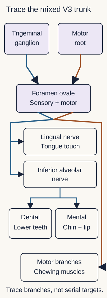
Use the accompanying branching diagram as a tracing exercise. Place a finger at the shared V3 trunk, then trace the pathways to the lower teeth, anterior tongue, and chewing muscles. Predict which functions disappear if the interruption is before those paths separate. Then repeat at the mental branch.
The sensory and motor components unite immediately below foramen ovale. Trace the shared trunk, then compare an interruption there with an interruption of the mental branch. Intermediate branches are simplified. Arrows show pathway connections, not the direction of every action potential. [4][5][12]
Worked result: interruption of the shared trunk can affect several sensory branches plus mastication. Interruption of the mental branch affects the lower lip and chin but spares dental sensation, tongue sensation, and chewing strength. Real lesions can be incomplete, so absence of one deficit does not establish an entirely normal trunk. [4][5]
Predict: numb chin plus weak masseter, one lesion
The lesion must account for both a sensory branch and a motor branch. A proximal V3 lesion fits more closely than an isolated mental nerve lesion. [4]
Predict: tongue touch and taste after dental trauma
A distal lingual nerve injury can affect V3 general sensory fibers and the facial nerve taste fibers that have joined it through chorda tympani. Taste loss does not mean that taste originated in V3. [5]
For transfer, compare injury near foramen ovale with injury beside a mandibular third molar. The proximal lesion can impair mastication and tongue touch while sparing taste because chorda tympani joins farther distally. A distal lingual injury can impair tongue touch and taste while sparing the masseter. Localization follows the combination, not the symptom count. [4][5]
Separate the attack pattern from its cause
Does a shock-like attack prove vascular compression? Trigeminal neuralgia is a clinical pain syndrome: recurrent severe, usually unilateral, electric or stabbing attacks in a trigeminal distribution, triggered by an ordinarily painless stimulus. Individual attacks typically last a fraction of a second to two minutes. V2 and V3 are common territories. Shaving, washing the face, speaking, or light contact during eating can trigger an attack. A short refractory interval often follows it. [1][2]
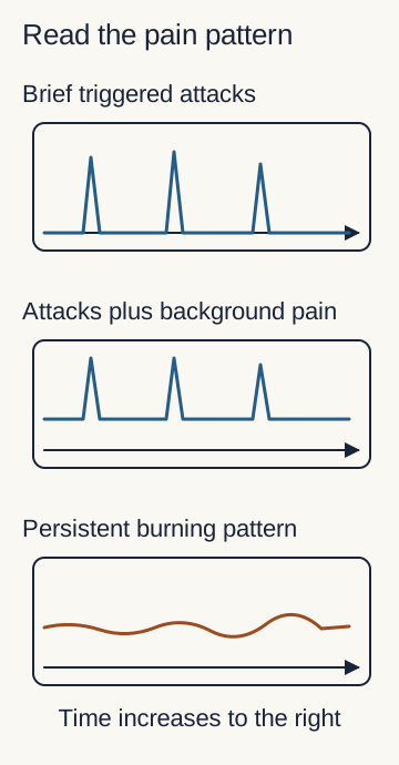
The pain timeline distinguishes isolated paroxysms from the same paroxysms superimposed on a continuous background ache. Continuous background pain does not by itself exclude trigeminal neuralgia. Ask whether the defining triggered attacks remain present. Predominantly persistent burning pain with objective sensory abnormalities raises a different neuropathic differential, especially after zoster or trauma. [1][9][10]
Higher traces represent more pain schematically; axes have no diagnostic scale. Neuralgia can include background pain. A time pattern alone does not establish vascular, traumatic, or postherpetic etiology. [1][2][9][10]
Two questions, kept separate
Question
Evidence needed
QuestionWhat pain phenotype is present?
Evidence neededAttack duration, triggers, distribution, and pain between attacks.
QuestionWhat causes the syndrome?
Evidence neededClinical context and adequate investigation, including MRI.
In the etiologic classification, classical trigeminal neuralgia requires neurovascular compression with a morphological nerve change, such as displacement or atrophy, demonstrated by MRI or surgery. The relevant contact is commonly near the root entry zone. A vessel simply touching a nerve is insufficient: contact also occurs without symptomatic compression. An appropriate negative investigation supports an idiopathic category; an explanatory lesion such as a tumor or multiple sclerosis supports a secondary category. [2][3]
Compare two reports: one describes a vessel touching the root with preserved nerve contour; the other describes indentation and displacement on the symptomatic side. Predict which report supports classical disease, then connect that answer to the visible distinction between contact and distortion.
Worked result: distortion supplies evidence of compression beyond mere proximity. Neither report replaces the clinical history. A normal examination supports the usual attack phenotype but does not prove its cause or exclude secondary disease. [2][3]
Apply the distinction to pain between attacks
A patient with qualifying triggered attacks, background pain, and demonstrated neurovascular compression with nerve distortion can have classical trigeminal neuralgia with concomitant continuous pain. The background ache does not cancel the other findings. [1][2]
For a new patient, do not jump directly from lower-jaw pain to an ovale lesion. The perceived territory identifies V3 involvement; the abnormal process may be at a distal branch, skull base, ganglion, root, or central pathway. The examination narrows that geography. [3][4][7]
Ask what the nerve can no longer do
Can a familiar pain trigger explain new weakness? A trigger identifies how pain is evoked. It does not account for an objectively weak muscle or a shrinking temporalis. Examine light touch and pin sensation by division, compare jaw closure, inspect the masseter and temporalis, and observe jaw opening. Persistent numbness, weakness, or atrophy requires an explanation beyond the attack description. [3][4][6]
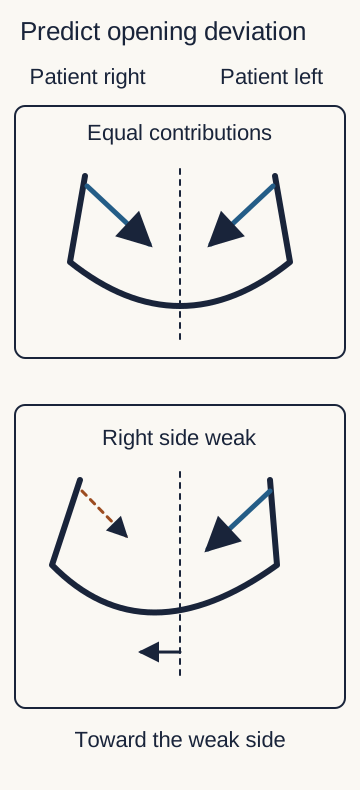
The two lateral pterygoids help translate the mandible forward. When one is weak, the intact side acts without an equal opposing contribution, so the jaw deviates toward the weak side during opening. For example, right temporal wasting, weak right jaw closure, and rightward deviation fit right motor V3 dysfunction. Pain-limited opening alone is not equivalent to denervation. [4][6]
Trace the two force arrows in the jaw diagram. First imagine both muscles acting; the centerline stays near the midline. Then identify the intact arrow when the right pterygoid is weak. The resulting rightward deflection links a visible examination finding to a side of nerve dysfunction. The drawing is a simplified force model, not a measurement of bite strength.
Solid arrows show effective contributions; the dashed arrow marks the weak side. Compare the midline with the displaced chin. This is a qualitative force model, not measured muscle vectors or an operative anatomical view. [4][6]
Worked result: rightward deviation together with right-sided wasting supports right motor dysfunction. If the same person also has numb mandibular teeth and tongue, a lesion proximal to several V3 branches is more economical than separate injuries to each target. [4][6]
Corneal reflex testing asks a different question. Corneal sensation reaches the brainstem through V1; closure of both eyelids uses facial nerve motor output. With an isolated left V1 afferent defect, left corneal stimulation fails to produce either blink, while right stimulation can produce both. With an isolated left facial motor defect, the left eyelid fails to close regardless of which cornea is stimulated. A normal corneal reflex does not establish normal V3 function. [6][14]
Predict the effect of adding diplopia to mandibular numbness
Diplopia suggests an additional ocular motor pathway abnormality. A diagnosis confined to a distal mandibular sensory branch no longer accounts for the full examination. Broader skull-base or intracranial assessment is needed. [3][7]
Young onset, bilateral symptoms, progressive deficits, visual symptoms, hearing changes, or other cranial nerve abnormalities increase concern for secondary disease. They do not specify one cause, and their absence is not a safe exclusion test. A younger person with bilateral attacks and prior episodes affecting vision or limb function needs assessment for a central disorder, including demyelination, rather than a diagnosis based only on age. [3]
For transfer, compare lower-lip numbness with isolated preserved strength against lower-lip numbness with muscle wasting. The first can be distal; the second requires a site or process that also reaches motor fibers. Add hearing loss, and the anatomical search must extend beyond foramen ovale. [3][4][7]
Investigate the cause while addressing the pain
Should imaging wait until the examination becomes abnormal? No. The EAN guideline recommends MRI as part of the work-up for suspected trigeminal neuralgia because clinical features cannot reliably exclude a secondary cause. A normal examination changes the level of concern; it does not eliminate the investigation. Progressive sensory or motor loss adds urgency and directs attention to the nerve's wider course. [3]
Begin by documenting attack length, quality, side, distribution, triggers, and interictal symptoms. Ask about prior zoster, dental procedures, trauma, cancer, and earlier neurological episodes. Follow with the trigeminal and other cranial nerve examinations, then inspect the oral cavity, teeth, and temporomandibular region. The order organizes evidence; it is not a requirement to postpone pain relief until every test is complete. [1][3][8][9][10]
Compare a patient whose lower-lip contact triggers a ten-second shock with one whose familiar preauricular ache is reproduced by joint loading and accompanied by stiffness. Both may say that chewing hurts, but the provocation and time course differ. Painful joint or muscle findings support a temporomandibular disorder; painless clicking alone does not. Localized dental findings warrant dental assessment rather than assuming that every mandibular symptom is neuralgia. Tooth-centered pain with swelling and pus needs urgent dental source treatment; spreading swelling or difficulty swallowing or breathing requires emergency assessment. [1][8][11]
For postherpetic pain, connect a prior rash in the same trigeminal territory with persistent burning, altered sensation, and allodynia. Pain persisting four months after that eruption meets the time component for trigeminal postherpetic neuralgia. After a procedure or injury, pain in the affected nerve territory with sensory dysfunction and an appropriate temporal relationship supports painful post-traumatic trigeminal neuropathy. Either condition can contain paroxysms; a single electric descriptor does not settle the diagnosis. [9][10]
For MRI, communicate the suspected territory and objective deficits. High-resolution trigeminal imaging evaluates the root and nearby vessels; the broader examination determines whether the brainstem, skull base, and extracranial course require focused assessment. A mass enlarging foramen ovale can injure V3, and tumor can extend along a nerve toward the skull base. Enlargement locates disease but does not, by itself, establish a specific tumor type. [3][7]
Compare two plans: symptom treatment with cause-directed investigation, versus escalating medication while ignoring newly documented weakness. The first addresses two different problems. Less pain does not demonstrate recovery of motor function or exclude an explanatory lesion.
Apply the plan after an initial normal scan
New progressive deficits warrant reassessment of the patient and the adequacy, timing, and anatomical coverage of prior imaging. An earlier normal study is not a permanent explanation for a changed examination. [3][7]
For a new case, specify what imaging must answer: root compression, demyelination, skull-base disease, or another process suggested by the combined history and examination. Do not replace that question with a nonspecific confidence score. [3][7]
Let evidence guide treatment, not a homemade score
Can several reassuring features cancel one objective deficit? No validated numerical score is supplied here. Brief triggered paroxysms support the neuralgia phenotype, while progressive numbness or weakness asks a separate anatomical question. Counting those observations as equal positive and negative points would conceal their different meanings. Drug response is likewise evidence about symptom control, not proof that the nerve has no structural lesion. [1][3]
Consider a patient whose electric attacks become infrequent during treatment, but whose chewing strength declines. Predict whether the next priority is simply a larger dose. The visible result is less pain alongside worse nerve function: the investigation must address that divergence rather than treating one outcome as a substitute for the other.
Carbamazepine or oxcarbazepine is generally the first pharmacological choice for long-term trigeminal neuralgia treatment. Selection and monitoring depend on the patient's comorbidities, interactions, tolerability, and clinical assessment. This lesson does not prescribe an individual dose. Cause-directed care proceeds alongside symptom treatment when a secondary lesion is identified. [3]
When medication provides inadequate control or is poorly tolerated, specialist discussion includes surgical options. Microvascular decompression addresses the vessel-nerve relationship in suitable patients with classical disease. Neuroablative treatments instead injure selected trigeminal fibers to reduce pain transmission; some percutaneous approaches reach the trigeminal region through foramen ovale. That access route does not mean the foramen caused the original pain. [3][7][10]
Compare relieving external compression with interrupting nerve fibers. They have different targets and tradeoffs. Recurrence and adverse effects remain possible, and sensory injury can itself become painful. Suitability, imaging, general health, patient priorities, and specialist assessment matter; no single procedure guarantees permanent relief. [3][10]
Apply the distinction after a pain procedure
New persistent burning in an objectively numb territory after neuroablative treatment can represent painful post-traumatic trigeminal neuropathy. It is not automatically the same process as recurrent brief triggerable attacks, although both patterns can coexist. [10]
Worked result: track pain and neurological function separately. Explain the purpose and limitations of treatment without converting an educational case into a personal prescription or a cure promise.
Use skull-base geography to test the localization
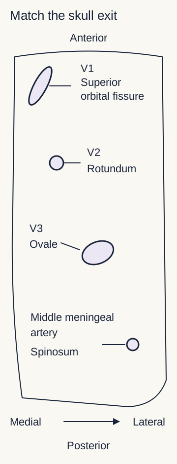
Which opening fits both the sensory and motor findings? V1 reaches the orbit through the superior orbital fissure. V2 passes through foramen rotundum toward the pterygopalatine fossa. V3 passes through foramen ovale toward the infratemporal and masticator region. In the greater wing of the sphenoid, ovale lies posterolateral to rotundum, and spinosum lies posterolateral to ovale. The accompanying schematic shows relationships, not a surgical trajectory. [4][7]
Foramen spinosum chiefly transmits the middle meningeal artery, with accompanying structures including a recurrent meningeal branch of V3. That small returning branch is not the main mandibular trunk. Confusing it with V3's exit would mislocalize a lesion that combines lower-face sensory loss and masticatory weakness. [7]
Trace three patterns on the map. Upper-cheek and upper-dental sensory loss with intact mastication suggests V2 and makes rotundum relevant. Lower-dental and tongue sensory loss with masticatory weakness suggests V3 and makes ovale relevant. Forehead and midface sensory abnormalities accompanied by ocular motor deficits require consideration of the cavernous sinus region, where V1 and V2 occupy the lateral wall alongside the third and fourth nerves, with the sixth nerve situated within the sinus. V3 does not traverse the cavernous sinus. [4][7][13]
Use position and contents together. This schematic shows relative geography, not dimensions or a needle trajectory. Spinosum also transmits accompanying structures, including a recurrent meningeal V3 branch; the main V3 trunk uses ovale. [4][7]
Worked result: a right ovale mass with right lower-face numbness, temporal wasting, and rightward jaw deviation supports structural right V3 dysfunction. Its sensory map and motor findings agree. A rotundum-only lesion does not explain the same motor deficit, and a named foramen does not establish a tumor's histology. [4][6][7]
Transfer: what remains intact in an isolated V2 lesion?
Mastication and mandibular dental sensation can remain intact because their fibers travel with V3. The preserved functions help test the proposed V2 localization rather than merely adding normal findings to a list. [4][7]
Finish with three questions: what is the pain pattern, what function is lost, and where can one process explain the combination? Use imaging to investigate the cause. The territory, phenotype, and etiology are related, but none is a substitute for the others. [1][3][4]
Apply the evidence to new patients
Case 1
Show answer and explanations for case 1
A. Panoramic radiography of the mandible (Why this does not fit)
It evaluates dental and mandibular bone abnormalities. The local examination is unrevealing, and radiography does not assess intracranial causes of the triggered attacks.
Reasoning steps for option A
What would mandibular radiography principally investigate?
It evaluates dental and mandibular bone abnormalities.
Why is it not the best investigation here?
The local examination is unrevealing, and radiography does not assess intracranial causes of the triggered attacks.
B. Noncontrast CT of the facial bones (Why this does not fit)
It depicts bony facial abnormalities, including fractures. There is no traumatic history, and the unresolved causes include trigeminal root and central disease.
Reasoning steps for option B
What pathology is facial-bone CT suited to assess?
It depicts bony facial abnormalities, including fractures.
Why is its coverage insufficient for this decision?
There is no traumatic history, and the unresolved causes include trigeminal root and central disease.
C. Brain MRI with dedicated trigeminal sequences (Best answer)
A structural or demyelinating cause can still be present. MRI of the brain and trigeminal pathway can assess these causes despite the typical attack history.
Reasoning steps for option C
What does the normal examination leave unresolved?
A structural or demyelinating cause can still be present.
Which study evaluates that remaining question?
MRI of the brain and trigeminal pathway can assess these causes despite the typical attack history.
D. Electromyography of the masticatory muscles (Why this does not fit)
It can assess dysfunction of motor supply to chewing muscles. Jaw strength is normal, and testing motor units does not exclude a structural cause along the sensory pathway.
Reasoning steps for option D
What deficit can masticatory electromyography help characterize?
It can assess dysfunction of motor supply to chewing muscles.
Why does it not resolve this patient's main uncertainty?
Jaw strength is normal, and testing motor units does not exclude a structural cause along the sensory pathway.
E. MRI focused on the temporomandibular joint (Why this does not fit)
Mechanical joint symptoms and reproduced joint pain would direct attention to the temporomandibular region. Brief shocks from facial washing with a normal joint examination favor a trigeminal pathway question.
Reasoning steps for option E
Which pattern would support joint-focused imaging?
Mechanical joint symptoms and reproduced joint pain would direct attention to the temporomandibular region.
What points away from that target here?
Brief shocks from facial washing with a normal joint examination favor a trigeminal pathway question.
Takeaway: A typical history supports trigeminal neuralgia, but normal examination findings do not replace cause-directed MRI.
A. Classical neuralgia with concomitant continuous pain (Best answer)
Severe brief shocks triggered by light contact remain present despite the background ache. An artery indents and displaces the symptomatic trigeminal root.
Reasoning steps for option A
Do the attacks still satisfy the neuralgia phenotype?
Severe brief shocks triggered by light contact remain present despite the background ache.
What supplies the classical etiologic classification?
An artery indents and displaces the symptomatic trigeminal root.
B. Secondary neuralgia from a space-occupying lesion (Why this does not fit)
An explanatory space-occupying lesion affecting the trigeminal pathway would support that category. It shows vascular indentation and displacement without a mass.
Reasoning steps for option B
What would support tumor-related secondary neuralgia?
An explanatory space-occupying lesion affecting the trigeminal pathway would support that category.
What does this MRI instead demonstrate?
It shows vascular indentation and displacement without a mass.
C. Trigeminal postherpetic neuralgia (Why this does not fit)
A prior zoster eruption in the same trigeminal territory and the subsequent pain course establish that relationship. The defining triggered attacks accompany symptomatic root compression, with no zoster history supplied.
Reasoning steps for option C
What evidence connects persistent pain to postherpetic disease?
A prior zoster eruption in the same trigeminal territory and the subsequent pain course establish that relationship.
What explains this presentation more directly?
The defining triggered attacks accompany symptomatic root compression, with no zoster history supplied.
D. Idiopathic neuralgia with concomitant continuous pain (Why this does not fit)
Adequate investigation has not established classical compression or another explanatory disease. The symptomatic root has demonstrated neurovascular compression with morphological change.
Reasoning steps for option D
When is the idiopathic category appropriate?
Adequate investigation has not established classical compression or another explanatory disease.
Which finding prevents that classification here?
The symptomatic root has demonstrated neurovascular compression with morphological change.
E. Painful post-traumatic trigeminal neuropathy (Why this does not fit)
A relevant nerve injury must be linked to pain and trigeminal dysfunction. There is preserved function and documented vascular compression, rather than a stated injury-related deficit.
Reasoning steps for option E
What relationship is necessary for traumatic neuropathy?
A relevant nerve injury must be linked to pain and trigeminal dysfunction.
What competing explanation is supplied here?
There is preserved function and documented vascular compression, rather than a stated injury-related deficit.
Takeaway: Background pain can coexist with classical trigeminal neuralgia when the attack phenotype and compression criteria are met.
A. Classical trigeminal neuralgia (Why this does not fit)
Neurovascular compression must be accompanied by a morphological nerve change. The vessel touches the root without altering its contour or position.
Reasoning steps for option A
What additional MRI feature is required for classical disease?
Neurovascular compression must be accompanied by a morphological nerve change.
Does this scan provide that feature?
The vessel touches the root without altering its contour or position.
B. Tumor-related trigeminal neuralgia (Why this does not fit)
An explanatory tumor affecting the trigeminal pathway would be needed. It excludes an identified mass and describes only nondeforming vascular contact.
Reasoning steps for option B
What finding would establish the proposed etiology?
An explanatory tumor affecting the trigeminal pathway would be needed.
What does the supplied investigation show?
It excludes an identified mass and describes only nondeforming vascular contact.
C. Post-traumatic trigeminal neuropathy (Why this does not fit)
A relevant injury and positive or negative sensory dysfunction must fit the pain territory. Normal function and an adequate negative etiologic assessment accompany an established neuralgia phenotype.
Reasoning steps for option C
What evidence links pain to traumatic neuropathy?
A relevant injury and positive or negative sensory dysfunction must fit the pain territory.
Which supplied findings favor another classification?
Normal function and an adequate negative etiologic assessment accompany an established neuralgia phenotype.
D. Postherpetic trigeminal neuralgia (Why this does not fit)
Persistent pain follows zoster in the same trigeminal branch. The established syndrome is brief triggered neuralgia, and no preceding zoster or corresponding persistent deficit is supplied.
Reasoning steps for option D
What clinical history supports a postherpetic category?
Persistent pain follows zoster in the same trigeminal branch.
Why is that not the best explanation here?
The established syndrome is brief triggered neuralgia, and no preceding zoster or corresponding persistent deficit is supplied.
E. Idiopathic trigeminal neuralgia (Best answer)
No indentation, displacement, or atrophy is demonstrated. Idiopathic trigeminal neuralgia is supported because the clinical syndrome is established but its cause is not.
Reasoning steps for option E
Does vessel contact alone meet classical criteria?
No indentation, displacement, or atrophy is demonstrated.
What classification remains after the stated negative investigation?
Idiopathic trigeminal neuralgia is supported because the clinical syndrome is established but its cause is not.
Takeaway: Contact without nerve distortion does not establish classical trigeminal neuralgia.
A. Strength of right mandibular protrusion (Why this does not fit)
The V3-supplied pterygoid muscles contribute to mandibular protrusion. The lesion affects the mixed trunk and already produces objective masticatory motor loss.
Reasoning steps for option A
Which muscles contribute to protruding the mandible?
The V3-supplied pterygoid muscles contribute to mandibular protrusion.
What supplied finding makes preservation less likely?
The lesion affects the mixed trunk and already produces objective masticatory motor loss.
B. Taste from the right anterior tongue (Best answer)
Facial nerve fibers reach the tongue through chorda tympani after joining the lingual nerve distally. The lesion is confined to V3 at skull exit, proximal to the entry of those taste fibers.
Reasoning steps for option B
Which cranial nerve supplies anterior tongue taste?
Facial nerve fibers reach the tongue through chorda tympani after joining the lingual nerve distally.
Why can that modality survive this lesion?
The lesion is confined to V3 at skull exit, proximal to the entry of those taste fibers.
C. Sensation from the right lower gingival tissues (Why this does not fit)
V3 branches, including lingual and buccal branches, supply lower gingival tissues. The lesion is proximal to the involved mandibular sensory branches, rather than isolated to one terminal twig.
Reasoning steps for option C
Which division supplies lower gingival sensory territories?
V3 branches, including lingual and buccal branches, supply lower gingival tissues.
Why are those fibers not reliably spared here?
The lesion is proximal to the involved mandibular sensory branches, rather than isolated to one terminal twig.
D. Touch sensation from the right lower lip (Why this does not fit)
The mental branch of the inferior alveolar nerve carries this V3 territory. Yes; a proximal mixed V3 lesion can affect it along with the already numb chin.
Reasoning steps for option D
Which branch carries lower-lip touch?
The mental branch of the inferior alveolar nerve carries this V3 territory.
Does the lesion lie before that branch separates?
Yes; a proximal mixed V3 lesion can affect it along with the already numb chin.
E. General sensation from the right floor of mouth (Why this does not fit)
The lingual nerve supplies the floor of the mouth. Lingual general sensory fibers are involved, so floor-of-mouth sensation is also at risk.
Reasoning steps for option E
Which V3 branch supplies this general sensory territory?
The lingual nerve supplies the floor of the mouth.
What does anterior tongue sensory loss suggest here?
Lingual general sensory fibers are involved, so floor-of-mouth sensation is also at risk.
Takeaway: A proximal V3 lesion can impair tongue touch while sparing taste carried by chorda tympani.
A. Mandibular nerve at the foramen ovale (Why this does not fit)
It can affect mandibular dental sensation and masticatory motor fibers as well as tongue touch. Those functions are preserved, while taste fibers entering distally are affected.
Reasoning steps for option A
What broader pattern can a proximal V3 lesion produce?
It can affect mandibular dental sensation and masticatory motor fibers as well as tongue touch.
Why does it poorly explain this selective pattern?
Those functions are preserved, while taste fibers entering distally are affected.
B. Inferior alveolar nerve within the mandibular canal (Why this does not fit)
It supplies mandibular teeth and continues toward the mental nerve territory. Dental and lower-lip sensation are intact while tongue touch and taste are impaired.
Reasoning steps for option B
Which sensory territories does this segment serve?
It supplies mandibular teeth and continues toward the mental nerve territory.
What contradicts that localization?
Dental and lower-lip sensation are intact while tongue touch and taste are impaired.
C. Facial nerve proximal to the chorda tympani origin (Why this does not fit)
Facial expression may be weak along with loss of taste fibers. Facial expression is intact, and tongue touch loss localizes a V3 contribution not carried by the facial trunk.
Reasoning steps for option C
What accompanying motor deficit could follow a proximal facial lesion?
Facial expression may be weak along with loss of taste fibers.
Why is the observed combination more distal and mixed?
Facial expression is intact, and tongue touch loss localizes a V3 contribution not carried by the facial trunk.
D. Lingual nerve distal to the chorda tympani junction (Best answer)
The lingual nerve carries both after chorda tympani joins it. Intact dental sensation, lower-lip sensation, and chewing favor a lingual branch injury rather than proximal V3 injury.
Reasoning steps for option D
Which nerve segment carries tongue touch and the joined taste fibers?
The lingual nerve carries both after chorda tympani joins it.
How do the spared functions refine the location?
Intact dental sensation, lower-lip sensation, and chewing favor a lingual branch injury rather than proximal V3 injury.
E. Chorda tympani before joining the lingual nerve (Why this does not fit)
It carries anterior tongue taste but not V3 touch sensation. Loss of tongue touch requires injury to general sensory fibers in addition to chorda tympani.
Reasoning steps for option E
Which tongue modality travels in chorda tympani?
It carries anterior tongue taste but not V3 touch sensation.
Which deficit would remain unexplained?
Loss of tongue touch requires injury to general sensory fibers in addition to chorda tympani.
Takeaway: Distal lingual nerve injury can interrupt both V3 general sensation and facial nerve taste fibers.
A. Left inferior alveolar nerve before entering the mandible (Why this does not fit)
Mandibular dental fibers travel within the inferior alveolar nerve. Normal sensation in the mandibular teeth favors a distal mental branch injury.
Reasoning steps for option A
Which additional territory lies downstream from that location?
Mandibular dental fibers travel within the inferior alveolar nerve.
Which preserved finding makes the proposed proximal injury less fitting?
Normal sensation in the mandibular teeth favors a distal mental branch injury.
B. Left infraorbital nerve (Why this does not fit)
It supplies a V2 territory including the upper lip and adjacent midface. This patient's deficit is in the lower lip and chin, which belong to the mental branch of V3.
Reasoning steps for option B
Which facial region does the infraorbital nerve serve?
It supplies a V2 territory including the upper lip and adjacent midface.
Why does the side of the lip matter?
This patient's deficit is in the lower lip and chin, which belong to the mental branch of V3.
C. Left mandibular nerve at skull exit (Why this does not fit)
Multiple V3 sensory branches and masticatory motor fibers share the proximal trunk. The deficit is confined to chin and lip near a local wound, with dental, tongue, and motor function preserved.
Reasoning steps for option C
How much territory converges at this location?
Multiple V3 sensory branches and masticatory motor fibers share the proximal trunk.
Why is a smaller branch the better explanation?
The deficit is confined to chin and lip near a local wound, with dental, tongue, and motor function preserved.
D. Left mental nerve (Best answer)
The mental nerve supplies the chin and lower lip. It places the injury beyond the inferior alveolar branches serving the mandibular teeth.
Reasoning steps for option D
Which terminal branch supplies the numb skin?
The mental nerve supplies the chin and lower lip.
What does preserved dental sensation add?
It places the injury beyond the inferior alveolar branches serving the mandibular teeth.
E. Left lingual nerve (Why this does not fit)
It affects general sensation of the anterior tongue and floor of the mouth. Tongue sensation is normal while chin and lower-lip sensation are lost.
Reasoning steps for option E
Which territory would a lingual injury affect?
It affects general sensation of the anterior tongue and floor of the mouth.
What is the sensory contrast here?
Tongue sensation is normal while chin and lower-lip sensation are lost.
Takeaway: Chin and lower-lip numbness with intact dental sensation favors the mental branch over the more proximal inferior alveolar nerve.
A. Mylohyoid contraction preserved; lower-incisor sensation preserved (Why this does not fit)
Its motor branch has already separated before the nerve enters the canal. They remain within the inferior alveolar pathway through the injured canal before continuing toward the anterior teeth.
Reasoning steps for option A
Why is preserved mylohyoid contraction plausible?
Its motor branch has already separated before the nerve enters the canal.
Why should the incisor fibers not be treated as equally spared?
They remain within the inferior alveolar pathway through the injured canal before continuing toward the anterior teeth.
B. Mylohyoid contraction preserved; lower-incisor sensation reduced (Best answer)
The nerve to mylohyoid separates before the inferior alveolar nerve enters the mandibular canal. Incisive sensory fibers continue through the canal, so lower-incisor sensation is expected to be reduced while mylohyoid contraction is preserved.
Reasoning steps for option B
Which motor branch leaves before the injured segment?
The nerve to mylohyoid separates before the inferior alveolar nerve enters the mandibular canal.
Which sensory fibers still pass through the injured segment?
Incisive sensory fibers continue through the canal, so lower-incisor sensation is expected to be reduced while mylohyoid contraction is preserved.
C. Mylohyoid contraction reduced; lower-incisor sensation preserved (Why this does not fit)
It would overlook their branch point before entry into the mandibular canal. It would place their separation before the injured proximal canal segment, although those sensory fibers continue through it.
Reasoning steps for option C
What error would place the mylohyoid fibers inside this injury?
It would overlook their branch point before entry into the mandibular canal.
What error would spare the incisor fibers?
It would place their separation before the injured proximal canal segment, although those sensory fibers continue through it.
D. Mylohyoid contraction reduced; lower-incisor sensation reduced (Why this does not fit)
The incisive sensory fibers remain downstream of the injured proximal canal segment. Mylohyoid fibers leave before canal entry, so this confined injury does not predict reduced mylohyoid contraction.
Reasoning steps for option D
Why is reduced lower-incisor sensation plausible?
The incisive sensory fibers remain downstream of the injured proximal canal segment.
Which motor prediction is misplaced?
Mylohyoid fibers leave before canal entry, so this confined injury does not predict reduced mylohyoid contraction.
Takeaway: An inferior alveolar injury inside the mandibular canal can spare the earlier mylohyoid motor branch.
A. Midline opening from isolated left facial motor weakness (Why this does not fit)
The mandibular division of the trigeminal nerve supplies them, rather than the facial nerve. Facial expression is symmetric while left masticatory muscles are weak and wasted.
Reasoning steps for option A
Which nerve drives the principal chewing muscles?
The mandibular division of the trigeminal nerve supplies them, rather than the facial nerve.
What separates the two motor systems here?
Facial expression is symmetric while left masticatory muscles are weak and wasted.
B. Rightward deviation from reduced left pterygoid force (Why this does not fit)
Other left V3 motor muscles are wasted or weak. Opening deviation is toward the weak side, not away from it.
Reasoning steps for option B
Why is left pterygoid weakness a plausible premise?
Other left V3 motor muscles are wasted or weak.
Which part of the prediction is reversed?
Opening deviation is toward the weak side, not away from it.
C. Rightward deviation from reduced right pterygoid force (Why this does not fit)
Right pterygoid weakness can cause rightward deviation. It localizes the weakness opposite the left-sided wasting and weak jaw closure.
Reasoning steps for option C
Is the proposed direction internally consistent?
Right pterygoid weakness can cause rightward deviation.
Why does it not fit this patient?
It localizes the weakness opposite the left-sided wasting and weak jaw closure.
D. Leftward deviation from reduced right pterygoid force (Why this does not fit)
It favors rightward opening deviation. The demonstrated weakness and atrophy are on the left.
Reasoning steps for option D
What direction would right pterygoid weakness favor?
It favors rightward opening deviation.
Which observation contradicts a right-sided motor lesion?
The demonstrated weakness and atrophy are on the left.
E. Leftward deviation from reduced left pterygoid force (Best answer)
Left-sided temporalis wasting and masseter weakness indicate left motor V3 dysfunction. The intact opposite side acts relatively unopposed, producing deviation toward the weak left side.
Reasoning steps for option E
Which side has demonstrated masticatory denervation?
Left-sided temporalis wasting and masseter weakness indicate left motor V3 dysfunction.
How does unilateral pterygoid weakness affect opening?
The intact opposite side acts relatively unopposed, producing deviation toward the weak left side.
Takeaway: Unilateral motor V3 dysfunction can cause the opening jaw to deviate toward the affected side.
A. Left ophthalmic division of the trigeminal nerve (Best answer)
Both facial motor outputs can close their eyelids. The left corneal afferent pathway travels through V1.
Reasoning steps for option A
What does bilateral blinking after right stimulation establish?
Both facial motor outputs can close their eyelids.
Where must the failure specific to left stimulation lie?
The left corneal afferent pathway travels through V1.
B. Left mandibular division of the trigeminal nerve (Why this does not fit)
V3 supplies the chewing muscles and mandibular sensory territories. Corneal afferent input uses V1, and jaw closure is normal.
Reasoning steps for option B
What would make a V3 lesion relevant to jaw function?
V3 supplies the chewing muscles and mandibular sensory territories.
Which pathway does the observed reflex test instead?
Corneal afferent input uses V1, and jaw closure is normal.
C. Right ophthalmic division of the trigeminal nerve (Why this does not fit)
Stimulation of the right cornea would fail to provide effective afferent input. It produces bilateral blinking, demonstrating a functioning right afferent route.
Reasoning steps for option C
What would right V1 dysfunction predict?
Stimulation of the right cornea would fail to provide effective afferent input.
What happens when that cornea is tested?
It produces bilateral blinking, demonstrating a functioning right afferent route.
D. Left facial nerve supplying orbicularis oculi (Why this does not fit)
The left eyelid would fail to close regardless of which cornea supplied the stimulus. The left eyelid closes after right corneal stimulation and with voluntary effort.
Reasoning steps for option D
What would a left facial efferent defect predict?
The left eyelid would fail to close regardless of which cornea supplied the stimulus.
Which observed response excludes that pattern?
The left eyelid closes after right corneal stimulation and with voluntary effort.
E. Right facial nerve supplying orbicularis oculi (Why this does not fit)
The right eyelid would fail to close from either corneal input. Right eyelid closure occurs with right corneal stimulation and voluntary effort.
Reasoning steps for option E
What would a right facial efferent defect predict?
The right eyelid would fail to close from either corneal input.
What directly contradicts that prediction?
Right eyelid closure occurs with right corneal stimulation and voluntary effort.
Takeaway: Failure tied to the stimulated cornea identifies an afferent problem; failure tied to one eyelid identifies an efferent problem.
A. Right superior orbital fissure (Why this does not fit)
V1 and ocular motor nerves pass through the superior orbital fissure. Upper-cheek V2 sensory loss favors a more proximal region that includes V2 as well.
Reasoning steps for option A
Which involved pathways traverse the fissure?
V1 and ocular motor nerves pass through the superior orbital fissure.
Which supplied deficit lies outside that isolated group?
Upper-cheek V2 sensory loss favors a more proximal region that includes V2 as well.
B. Right foramen rotundum (Why this does not fit)
V2 sensory loss over the upper cheek could follow a rotundum lesion. Forehead sensory loss and multiple ocular motor abnormalities are not explained by an isolated V2 exit lesion.
Reasoning steps for option B
Which observed deficit could rotundum explain?
V2 sensory loss over the upper cheek could follow a rotundum lesion.
Which findings require a broader region?
Forehead sensory loss and multiple ocular motor abnormalities are not explained by an isolated V2 exit lesion.
C. Right cavernous sinus (Best answer)
Ocular motor nerves and V1/V2 have closely related courses through the cavernous sinus region. V3 does not pass through the cavernous sinus, so its preservation fits that localization.
Reasoning steps for option C
Which affected nerves share this region?
Ocular motor nerves and V1/V2 have closely related courses through the cavernous sinus region.
Why does preserved mandibular function help?
V3 does not pass through the cavernous sinus, so its preservation fits that localization.
D. Right foramen ovale (Why this does not fit)
The mixed mandibular division, V3, exits there. V3 function is preserved while ocular motor and V1/V2 functions are abnormal.
Reasoning steps for option D
Which division passes through foramen ovale?
The mixed mandibular division, V3, exits there.
Which mismatch makes this location unlikely?
V3 function is preserved while ocular motor and V1/V2 functions are abnormal.
E. Right internal acoustic meatus (Why this does not fit)
The facial and vestibulocochlear nerves pass through it. Hearing is preserved, and the deficits involve ocular motor nerves and V1/V2 rather than that canal's nerves.
Reasoning steps for option E
Which nerves are principally associated with this canal?
The facial and vestibulocochlear nerves pass through it.
How does the observed cluster differ?
Hearing is preserved, and the deficits involve ocular motor nerves and V1/V2 rather than that canal's nerves.
Takeaway: Combined ocular motor and V1/V2 deficits with preserved V3 function fit the cavernous sinus region better than foramen ovale.
V3 dysfunction can affect mandibular sensation and mastication. Lower-lip and tongue sensation and chewing strength are preserved.
Reasoning steps for option A
What deficits would implicate the main nerve at ovale?
V3 dysfunction can affect mandibular sensation and mastication.
Which supplied findings make that less fitting?
Lower-lip and tongue sensation and chewing strength are preserved.
B. Superior orbital fissure (Why this does not fit)
V1 passes through the superior orbital fissure. Forehead sensation and eye motility are normal while V2 territories are affected.
Reasoning steps for option B
Which trigeminal division accompanies ocular motor nerves here?
V1 passes through the superior orbital fissure.
What is the distribution mismatch?
Forehead sensation and eye motility are normal while V2 territories are affected.
C. Internal acoustic meatus (Why this does not fit)
Facial and vestibulocochlear structures pass through the internal acoustic meatus. The upper lip, upper teeth, and palate identify V2 rather than those nerves.
Reasoning steps for option C
Which cranial nerves use this passage?
Facial and vestibulocochlear structures pass through the internal acoustic meatus.
Which sensory map instead identifies the lesion here?
The upper lip, upper teeth, and palate identify V2 rather than those nerves.
D. Foramen rotundum (Best answer)
The maxillary division supplies the upper lip, upper teeth, palate, and much of the midface. V2 passes through foramen rotundum.
Reasoning steps for option D
Which division supplies the affected sensory territories?
The maxillary division supplies the upper lip, upper teeth, palate, and much of the midface.
Where does that division leave the middle cranial fossa?
V2 passes through foramen rotundum.
E. Foramen spinosum (Why this does not fit)
The middle meningeal artery passes through foramen spinosum. The deficit identifies the main maxillary sensory division, not a meningeal vascular pathway.
Reasoning steps for option E
Which major structure characterizes this opening?
The middle meningeal artery passes through foramen spinosum.
Why is this not the exit being sought?
The deficit identifies the main maxillary sensory division, not a meningeal vascular pathway.
Takeaway: Upper-dental and midfacial sensory loss without masticatory weakness points toward V2 and foramen rotundum.
Foramen spinosum occupies that neighboring position. The middle meningeal artery normally enters through foramen spinosum.
Reasoning steps for option A
Which opening lies posterolateral to ovale?
Foramen spinosum occupies that neighboring position.
Which principal artery passes through it?
The middle meningeal artery normally enters through foramen spinosum.
B. Ophthalmic artery (Why this does not fit)
It accompanies the optic pathway toward the orbit. The described opening is posterolateral to ovale, not at the optic canal.
Reasoning steps for option B
Which region is the ophthalmic artery directed toward?
It accompanies the optic pathway toward the orbit.
Why does it not fit this fracture location?
The described opening is posterolateral to ovale, not at the optic canal.
C. Accessory meningeal artery (Why this does not fit)
It can enter through foramen ovale. The fractured opening is behind and lateral to ovale, identifying spinosum.
Reasoning steps for option C
Why is this artery a plausible neighboring structure?
It can enter through foramen ovale.
Which positional detail favors another artery?
The fractured opening is behind and lateral to ovale, identifying spinosum.
D. Internal carotid artery (Why this does not fit)
It enters the skull through the carotid canal and follows a distinct intracranial course. No; the relative position identifies spinosum rather than the carotid canal.
Reasoning steps for option D
Why is the internal carotid relevant to skull-base vascular anatomy?
It enters the skull through the carotid canal and follows a distinct intracranial course.
Does that identify the small opening described here?
No; the relative position identifies spinosum rather than the carotid canal.
E. Vertebral artery (Why this does not fit)
It enters through the foramen magnum. A small middle-fossa opening beside ovale is not the foramen magnum.
Reasoning steps for option E
Where does the vertebral artery enter the cranial cavity?
It enters through the foramen magnum.
How does the supplied location differ?
A small middle-fossa opening beside ovale is not the foramen magnum.
Takeaway: Foramen spinosum is the major middle meningeal arterial route, not the main V3 exit.
A. Mandibular neuropathy from bilateral ovale tumors (Why this does not fit)
It could affect peripheral V3 sensory and motor pathways. No skull-base mass is present, whereas the pontine trigeminal pathway contains new plaques.
Reasoning steps for option A
What could bilateral ovale disease affect?
It could affect peripheral V3 sensory and motor pathways.
Which supplied imaging result favors another site?
No skull-base mass is present, whereas the pontine trigeminal pathway contains new plaques.
B. Classical neuralgia from vascular root compression (Why this does not fit)
Compression with a morphological change of the symptomatic root is required. There is an anatomically relevant demyelinating plaque and no neurovascular compression.
Reasoning steps for option B
What imaging evidence would support this cause?
Compression with a morphological change of the symptomatic root is required.
What cause is demonstrated instead?
There is an anatomically relevant demyelinating plaque and no neurovascular compression.
C. Secondary neuralgia related to central demyelination (Best answer)
Yes; the trigger and duration support neuralgia even though the examination is abnormal. The new plaques involve both central trigeminal pathways in a patient with multiple sclerosis.
Reasoning steps for option C
Do the brief triggered attacks fit a neuralgia phenotype?
Yes; the trigger and duration support neuralgia even though the examination is abnormal.
What supplies an explanatory secondary cause?
The new plaques involve both central trigeminal pathways in a patient with multiple sclerosis.
D. Idiopathic neuralgia without an identified cause (Why this does not fit)
Adequate evaluation has not found an explanatory classical or secondary cause. The neurological disease and new anatomically relevant plaque provide a secondary explanation.
Reasoning steps for option D
When is an idiopathic category defensible?
Adequate evaluation has not found an explanatory classical or secondary cause.
Why does that condition fail here?
The neurological disease and new anatomically relevant plaque provide a secondary explanation.
E. Lingual neuropathy from a peripheral dental injury (Why this does not fit)
It affects tongue and floor-of-mouth general sensation. The attacks involve the chin on both sides and accompany a central trigeminal lesion rather than a supplied dental injury.
Reasoning steps for option E
Which distribution would lingual injury primarily affect?
It affects tongue and floor-of-mouth general sensation.
What is discordant in this patient?
The attacks involve the chin on both sides and accompany a central trigeminal lesion rather than a supplied dental injury.
Takeaway: A demyelinating lesion can produce secondary trigeminal neuralgia, including a phenotype with bilateral attacks or sensory deficits.
A. Venous spread through the cavernous sinus (Why this does not fit)
It is an intracranial venous space near several cranial nerves. The contiguous extracranial abnormality follows V3 through ovale, not a demonstrated venous pathway.
Reasoning steps for option A
Why might the cavernous sinus enter a skull-base differential?
It is an intracranial venous space near several cranial nerves.
What favors a different route here?
The contiguous extracranial abnormality follows V3 through ovale, not a demonstrated venous pathway.
B. Direct extension through a mandibular fracture (Why this does not fit)
A demonstrated bony breach could provide a direct path for local disease. There is no fracture, and the abnormality follows the named nerve rather than a fracture plane.
Reasoning steps for option B
What would make a fracture route plausible?
A demonstrated bony breach could provide a direct path for local disease.
Which supplied findings argue against it?
There is no fracture, and the abnormality follows the named nerve rather than a fracture plane.
C. Transdural spread through foramen spinosum (Why this does not fit)
Meningeal vessels and a recurrent meningeal branch pass through it. MRI identifies the main mandibular pathway through ovale, matching lower-lip and chewing deficits.
Reasoning steps for option C
Which nearby structures traverse spinosum?
Meningeal vessels and a recurrent meningeal branch pass through it.
Which opening and nerve are actually abnormal?
MRI identifies the main mandibular pathway through ovale, matching lower-lip and chewing deficits.
D. Hematogenous spread to separate sphenoid deposits (Why this does not fit)
They could produce separate metastatic foci in skull-base bone. Enhancement follows the inferior alveolar nerve into V3, without a separate destructive marrow lesion.
Reasoning steps for option D
What distribution could blood-borne bone deposits produce?
They could produce separate metastatic foci in skull-base bone.
Which feature instead identifies a continuous route?
Enhancement follows the inferior alveolar nerve into V3, without a separate destructive marrow lesion.
E. Perineural extension along the mandibular division (Best answer)
The inferior alveolar nerve joins the V3 pathway that passes through foramen ovale. It supports tumor extension along the nerve rather than unrelated lesions at separate sites.
Reasoning steps for option E
Which path connects the abnormal extracranial and skull-base findings?
The inferior alveolar nerve joins the V3 pathway that passes through foramen ovale.
What does continuous enlargement along that path support?
It supports tumor extension along the nerve rather than unrelated lesions at separate sites.
Takeaway: Contiguous abnormality along a mandibular nerve toward foramen ovale supports perineural extension; the opening alone does not identify tumor histology.
It could explain mandibular sensory loss. V1/V2 sensory loss and sensorineural hearing loss require a broader region.
Reasoning steps for option A
Which part of the examination could ovale disease explain?
It could explain mandibular sensory loss.
Which findings remain outside that isolated site?
V1/V2 sensory loss and sensorineural hearing loss require a broader region.
B. Right mandibular canal (Why this does not fit)
Inferior alveolar and mental sensory loss can affect lower teeth, lip, and chin. Forehead and cheek sensation, corneal input, and hearing are also impaired.
Reasoning steps for option B
Which sensory deficits could canal injury cause?
Inferior alveolar and mental sensory loss can affect lower teeth, lip, and chin.
Which combined deficits contradict a canal-only explanation?
Forehead and cheek sensation, corneal input, and hearing are also impaired.
C. Right superior orbital fissure (Why this does not fit)
V1 corneal and forehead sensory fibers pass through this region. The V2/V3 and auditory findings extend beyond an isolated superior orbital fissure lesion.
Reasoning steps for option C
Which affected sensory input could a fissure lesion involve?
V1 corneal and forehead sensory fibers pass through this region.
What additional abnormalities make it incomplete?
The V2/V3 and auditory findings extend beyond an isolated superior orbital fissure lesion.
D. Right foramen rotundum (Why this does not fit)
V2 passes through it and supplies the midface. It does not explain V1/V3 involvement or the auditory deficit.
Reasoning steps for option D
Which division passes through rotundum?
V2 passes through it and supplies the midface.
Why does it not unite the examination?
It does not explain V1/V3 involvement or the auditory deficit.
E. Right cerebellopontine angle (Best answer)
Forehead and cheek sensation and the corneal afferent response are also abnormal. Ipsilateral sensorineural hearing loss brings nearby vestibulocochlear structures into the localization.
Reasoning steps for option E
Why is this not an isolated mandibular sensory lesion?
Forehead and cheek sensation and the corneal afferent response are also abnormal.
What additional finding directs the proximal search?
Ipsilateral sensorineural hearing loss brings nearby vestibulocochlear structures into the localization.
Takeaway: Trigeminal sensory loss plus ipsilateral sensorineural hearing loss warrants a broader intracranial search, including the cerebellopontine angle.
A. Idiopathic neuralgia with a prolonged pain background (Why this does not fit)
It can, provided the defining neuralgia attacks are present. There are no brief lip-triggered shocks; the familiar pain is reproduced mechanically at the joint.
Reasoning steps for option A
Can neuralgia include pain between attacks?
It can, provided the defining neuralgia attacks are present.
What prevents background pain from establishing that diagnosis here?
There are no brief lip-triggered shocks; the familiar pain is reproduced mechanically at the joint.
B. Postherpetic neuropathy with touch-evoked facial pain (Why this does not fit)
Pain and sensory change would follow zoster in the corresponding territory. The pain is reproduced by joint loading rather than cutaneous contact, without objective sensory loss.
Reasoning steps for option B
What kind of history would support postherpetic disease?
Pain and sensory change would follow zoster in the corresponding territory.
Why is the proposed mechanism a poorer fit to these observations?
The pain is reproduced by joint loading rather than cutaneous contact, without objective sensory loss.
C. Temporomandibular pain with incidental vessel contact (Best answer)
Joint palpation and loading reproduce the familiar prolonged preauricular ache. Simple contact lacks morphological compression, and the characteristic brief triggered attacks are absent.
Reasoning steps for option C
Which observations identify the painful structure most directly?
Joint palpation and loading reproduce the familiar prolonged preauricular ache.
Why does the MRI not establish neuralgia instead?
Simple contact lacks morphological compression, and the characteristic brief triggered attacks are absent.
D. Classical neuralgia with an incidental joint click (Why this does not fit)
Vascular compression can cause classical trigeminal neuralgia. The clinical attack phenotype is absent, and imaging shows no nerve distortion.
Reasoning steps for option D
Why might the MRI report attract attention?
Vascular compression can cause classical trigeminal neuralgia.
Which two requirements are missing here?
The clinical attack phenotype is absent, and imaging shows no nerve distortion.
E. Structural V3 neuropathy with masticatory denervation (Why this does not fit)
Objective sensory loss, weakness, or masticatory atrophy would support nerve dysfunction. Sensation and muscle strength are normal while joint testing reproduces the pain.
Reasoning steps for option E
What examination pattern would support denervation?
Objective sensory loss, weakness, or masticatory atrophy would support nerve dysfunction.
Which observations instead favor a joint pain source?
Sensation and muscle strength are normal while joint testing reproduces the pain.
Takeaway: MRI vessel contact does not outweigh a clinical pattern of reproducible temporomandibular pain.
A. Carbamazepine treatment with outpatient dose titration (Why this does not fit)
It is a first-choice drug for trigeminal neuralgia. It would not treat the purulent dental source producing this continuous tooth-centered pain.
Reasoning steps for option A
What pain syndrome is carbamazepine used to treat?
It is a first-choice drug for trigeminal neuralgia.
Why would symptom suppression miss the main problem here?
It would not treat the purulent dental source producing this continuous tooth-centered pain.
B. Urgent dental assessment and source treatment (Best answer)
Tooth-centered throbbing, caries, gingival swelling, and pus support an odontogenic infection. Urgent dental assessment can provide definitive treatment rather than only suppressing the pain.
Reasoning steps for option B
Which findings point to a local infectious source?
Tooth-centered throbbing, caries, gingival swelling, and pus support an odontogenic infection.
Which action addresses that source?
Urgent dental assessment can provide definitive treatment rather than only suppressing the pain.
C. Dedicated trigeminal MRI and neurology referral (Why this does not fit)
A neuralgia phenotype or unexplained nerve deficits would support assessment of the trigeminal pathway. There is a visible focal dental infection with no brief triggered attack pattern or neurological loss.
Reasoning steps for option C
When would a trigeminal investigation be important?
A neuralgia phenotype or unexplained nerve deficits would support assessment of the trigeminal pathway.
What currently requires source treatment instead?
There is a visible focal dental infection with no brief triggered attack pattern or neurological loss.
D. Temporomandibular therapy with a fitted oral appliance (Why this does not fit)
Reproducible joint or masticatory muscle pain and mechanical symptoms would direct that care. The pain centers on a carious molar with adjacent pus rather than an identified joint disorder.
Reasoning steps for option D
What findings could support a temporomandibular approach?
Reproducible joint or masticatory muscle pain and mechanical symptoms would direct that care.
Which supplied observations point elsewhere?
The pain centers on a carious molar with adjacent pus rather than an identified joint disorder.
E. Mental nerve injection for prolonged regional analgesia (Why this does not fit)
It can reduce sensation in the lower lip and chin. The infection is at a mandibular tooth and needs dental treatment, not analgesia of a terminal skin territory.
Reasoning steps for option E
Which territory could mental nerve anesthesia affect?
It can reduce sensation in the lower lip and chin.
Why does this not address the identified source?
The infection is at a mandibular tooth and needs dental treatment, not analgesia of a terminal skin territory.
Takeaway: Pain in a V3 territory can arise from an infected tooth; focal dental findings require dental source-directed care.
A. Purely paroxysmal classical trigeminal neuralgia (Why this does not fit)
Pain would consist of characteristic brief neuralgia attacks without concomitant continuous pain. Persistent burning dominates, and the history provides a zoster-related cause with sensory dysfunction.
Reasoning steps for option A
Which temporal pattern would that label require?
Pain would consist of characteristic brief neuralgia attacks without concomitant continuous pain.
Which findings contradict that pattern?
Persistent burning dominates, and the history provides a zoster-related cause with sensory dysfunction.
B. Painful post-traumatic trigeminal neuropathy (Why this does not fit)
A temporally relevant injury to the affected nerve must explain the pain and dysfunction. The documented antecedent is a vesicular zoster eruption, not a mechanical or treatment-related nerve injury.
Reasoning steps for option B
What cause distinguishes a traumatic neuropathy?
A temporally relevant injury to the affected nerve must explain the pain and dysfunction.
Which preceding event is actually supplied?
The documented antecedent is a vesicular zoster eruption, not a mechanical or treatment-related nerve injury.
C. Acute painful trigeminal herpes zoster (Why this does not fit)
The eruption and painful sensory territory are consistent with trigeminal zoster. Pain persists four months after the eruption, supporting postherpetic rather than acute zoster pain.
Reasoning steps for option C
Why is an acute zoster diagnosis initially plausible?
The eruption and painful sensory territory are consistent with trigeminal zoster.
Which time-course detail requires a chronic category?
Pain persists four months after the eruption, supporting postherpetic rather than acute zoster pain.
D. Trigeminal postherpetic neuralgia (Best answer)
The pain occupies the same territory as the prior eruption and remains four months later. Sensory loss and broad allodynia accompany a predominantly persistent burning pain pattern.
Reasoning steps for option D
What establishes the anatomical and temporal relationship?
The pain occupies the same territory as the prior eruption and remains four months later.
How does the current examination reinforce that diagnosis?
Sensory loss and broad allodynia accompany a predominantly persistent burning pain pattern.
E. Idiopathic neuralgia with concomitant continuous pain (Why this does not fit)
The defining brief, stereotyped, innocuously triggered attacks must still be present. That attack pattern is absent, while prior same-territory zoster supplies an explanatory cause.
Reasoning steps for option E
What must still be present when neuralgia has background pain?
The defining brief, stereotyped, innocuously triggered attacks must still be present.
Why is idiopathic neuralgia a poorer fit here?
That attack pattern is absent, while prior same-territory zoster supplies an explanatory cause.
Takeaway: Persistent burning with sensory abnormalities after same-territory zoster supports postherpetic neuralgia, even when occasional shooting pains occur.
A. Painful post-traumatic trigeminal neuropathy (Best answer)
A documented lingual injury is followed within two days by pain in its sensory territory. Reduced sensation and touch-evoked pain remain after the local wound has healed.
Reasoning steps for option A
What connects the procedure to the current pain?
A documented lingual injury is followed within two days by pain in its sensory territory.
What establishes persistent nerve dysfunction?
Reduced sensation and touch-evoked pain remain after the local wound has healed.
B. Temporomandibular joint-related facial pain (Why this does not fit)
The joint and associated masticatory muscles generate the relevant mechanical pain. The abnormality is confined to an injured lingual nerve territory with numbness and allodynia, while jaw strength is normal.
Reasoning steps for option B
Which structures principally generate temporomandibular pain?
The joint and associated masticatory muscles generate the relevant mechanical pain.
Why does that not explain this sensory pattern?
The abnormality is confined to an injured lingual nerve territory with numbness and allodynia, while jaw strength is normal.
C. Trigeminal postherpetic neuralgia (Why this does not fit)
A zoster eruption in the affected trigeminal territory must precede the chronic pain. There was no zoster, and the pain began after a documented surgical nerve injury.
Reasoning steps for option C
What antecedent event is needed for postherpetic pain?
A zoster eruption in the affected trigeminal territory must precede the chronic pain.
How does the stated history discriminate?
There was no zoster, and the pain began after a documented surgical nerve injury.
D. Idiopathic trigeminal neuralgia without sensory injury (Why this does not fit)
No explanatory cause has been established for an otherwise qualifying neuralgia phenotype. The injury is documented, the affected territory matches, and sensory dysfunction is present.
Reasoning steps for option D
What does an idiopathic classification imply about the investigation?
No explanatory cause has been established for an otherwise qualifying neuralgia phenotype.
Which facts directly conflict with that premise?
The injury is documented, the affected territory matches, and sensory dysfunction is present.
E. Classical trigeminal neuralgia with continuous pain (Why this does not fit)
Characteristic neuralgia attacks and demonstrated morphological neurovascular compression would be required. A documented distal nerve injury matches both the onset and the persistent sensory deficit.
Reasoning steps for option E
What additional evidence would support that classification?
Characteristic neuralgia attacks and demonstrated morphological neurovascular compression would be required.
What supplied cause better accounts for the findings?
A documented distal nerve injury matches both the onset and the persistent sensory deficit.
Takeaway: Pain beginning after a matching nerve injury with sensory dysfunction supports painful post-traumatic trigeminal neuropathy.
The brief triggered attacks and morphological neurovascular compression support classical trigeminal neuralgia. Carbamazepine is a recommended initial long-term option, alongside oxcarbazepine.
Reasoning steps for option A
Which pain syndrome is established by the history and imaging?
The brief triggered attacks and morphological neurovascular compression support classical trigeminal neuralgia.
Which proposed drug has first-choice guideline support?
Carbamazepine is a recommended initial long-term option, alongside oxcarbazepine.
B. Lamotrigine (Why this does not fit)
It may be used as an alternative or additional medication in selected cases. This is initial long-term therapy, for which carbamazepine or oxcarbazepine is preferred.
Reasoning steps for option B
Where can lamotrigine fit in management?
It may be used as an alternative or additional medication in selected cases.
What distinguishes the requested treatment stage?
This is initial long-term therapy, for which carbamazepine or oxcarbazepine is preferred.
C. Baclofen (Why this does not fit)
It is among the agents that may be used alone or as additional therapy. No intolerance or contraindication displaces the guideline's preferred initial carbamazepine or oxcarbazepine approach.
Reasoning steps for option C
Can baclofen be considered for difficult trigeminal neuralgia?
It is among the agents that may be used alone or as additional therapy.
Why is it not the best first-choice answer here?
No intolerance or contraindication displaces the guideline's preferred initial carbamazepine or oxcarbazepine approach.
D. Pregabalin (Why this does not fit)
No; it may have a role in selected patients. The stem supplies no reason to depart from the guideline-supported first-choice drugs.
Reasoning steps for option D
Is pregabalin outside all neuralgia treatment options?
No; it may have a role in selected patients.
Why is it not the strongest answer under these conditions?
The stem supplies no reason to depart from the guideline-supported first-choice drugs.
E. Gabapentin (Why this does not fit)
It can be used in selected patients as an alternative or additional agent. Carbamazepine has first-choice guideline status in a patient without a supplied reason to avoid it.
Reasoning steps for option E
Can gabapentin have a role in trigeminal neuralgia?
It can be used in selected patients as an alternative or additional agent.
Why is another answer preferred for this initial-treatment question?
Carbamazepine has first-choice guideline status in a patient without a supplied reason to avoid it.
Takeaway: Carbamazepine or oxcarbazepine is generally first-choice long-term pharmacotherapy; individual selection and monitoring require clinical assessment.
A. Arrange temporomandibular therapy and defer neurological testing (Why this does not fit)
Jaw discomfort or difficulty chewing can occur in joint and muscle disorders. Chin sensory loss and focal temporal atrophy accompany objective unilateral weakness.
Reasoning steps for option A
Which complaint can overlap with temporomandibular disease?
Jaw discomfort or difficulty chewing can occur in joint and muscle disorders.
Which supplied findings demand a nerve-focused explanation?
Chin sensory loss and focal temporal atrophy accompany objective unilateral weakness.
B. Refer for percutaneous ablation using the previous imaging (Why this does not fit)
It may be considered when suitable patients have uncontrolled or poorly tolerated neuralgia. The changed sensory and motor examination may indicate a structural process not addressed by an old routine scan.
Reasoning steps for option B
When can an ablative procedure enter the treatment discussion?
It may be considered when suitable patients have uncontrolled or poorly tolerated neuralgia.
What question must now be clarified for appropriate planning?
The changed sensory and motor examination may indicate a structural process not addressed by an old routine scan.
C. Substitute gabapentin and attribute weakness to medication intolerance (Why this does not fit)
Neuralgia medications can have adverse effects requiring reassessment. Progressive unilateral V3 sensory loss and focal muscle atrophy form an anatomical deficit pattern needing investigation.
Reasoning steps for option C
Why might medication intolerance be considered during treatment?
Neuralgia medications can have adverse effects requiring reassessment.
What makes that assumption inadequate here?
Progressive unilateral V3 sensory loss and focal muscle atrophy form an anatomical deficit pattern needing investigation.
D. Increase carbamazepine and reassess symptoms in three months (Why this does not fit)
Carbamazepine has reduced the painful attacks. New sensory loss and muscle atrophy require assessment of the cause, not only more suppression of paroxysms.
Reasoning steps for option D
Which observation could make dose adjustment seem attractive?
Carbamazepine has reduced the painful attacks.
What would a dose-only plan fail to address?
New sensory loss and muscle atrophy require assessment of the cause, not only more suppression of paroxysms.
E. Repeat targeted imaging and neurological assessment (Best answer)
Progressive numbness and masticatory atrophy demonstrate worsening nerve function. It predates the new deficits and may not adequately assess the now-implicated V3 course.
Reasoning steps for option E
What new problem is not measured by attack frequency?
Progressive numbness and masticatory atrophy demonstrate worsening nerve function.
Why should the old imaging not settle the current question?
It predates the new deficits and may not adequately assess the now-implicated V3 course.
Takeaway: Reduced attack frequency does not explain new sensory loss or masticatory atrophy; changed function requires renewed evaluation.
A. Compress the trigeminal ganglion with a temporary balloon (Why this does not fit)
It is a percutaneous neuroablative approach rather than separation of a vessel from a nerve. The symptomatic root is indented by an artery in a patient suitable for surgical discussion.
Reasoning steps for option A
What treatment category does balloon compression represent?
It is a percutaneous neuroablative approach rather than separation of a vessel from a nerve.
Which demonstrated abnormality favors decompression in this case?
The symptomatic root is indented by an artery in a patient suitable for surgical discussion.
B. Produce a thermal lesion in selected trigeminal fibers (Why this does not fit)
Thermal neuroablation injures selected fibers involved in transmitting pain. The patient is medically fit with a demonstrated compressive vessel suitable for decompression.
Reasoning steps for option B
How can this approach reduce pain?
Thermal neuroablation injures selected fibers involved in transmitting pain.
Why is it not the guideline-preferred first surgical principle for this profile?
The patient is medically fit with a demonstrated compressive vessel suitable for decompression.
C. Deliver focused radiation to a segment of the trigeminal nerve (Why this does not fit)
It targets a nerve segment to alter pain transmission. Classical compression in a medically fit patient favors directly addressing the vessel-nerve relationship.
Reasoning steps for option C
What is the intended target of radiosurgical treatment?
It targets a nerve segment to alter pain transmission.
Why does the requested first surgical preference differ?
Classical compression in a medically fit patient favors directly addressing the vessel-nerve relationship.
D. Separate the compressing artery from the trigeminal root (Best answer)
An artery indents the symptomatic trigeminal root. Microvascular decompression separates the offending vessel from the nerve and is the preferred first surgical approach for suitable classical cases.
Reasoning steps for option D
What anatomical abnormality is established?
An artery indents the symptomatic trigeminal root.
Which surgical principle addresses that abnormality?
Microvascular decompression separates the offending vessel from the nerve and is the preferred first surgical approach for suitable classical cases.
E. Interrupt a peripheral mandibular sensory branch surgically (Why this does not fit)
It could reduce input from a selected mandibular sensory territory. Imaging identifies a proximal root compression rather than a lesion confined to one peripheral branch.
Reasoning steps for option E
What could peripheral interruption change?
It could reduce input from a selected mandibular sensory territory.
Why is that not the best anatomical match here?
Imaging identifies a proximal root compression rather than a lesion confined to one peripheral branch.
Takeaway: In suitable patients with classical disease, microvascular decompression targets the vessel-nerve relationship rather than intentionally ablating sensory fibers.
A. Temporomandibular pain caused by persistent joint inflammation (Why this does not fit)
Reproduced mechanical joint pain, stiffness, or painful joint findings would be relevant. Pain began in the treated, objectively numb sensory territory immediately after neuroablation.
Reasoning steps for option A
Which features would support a joint-centered cause?
Reproduced mechanical joint pain, stiffness, or painful joint findings would be relevant.
Which supplied relationship points to a nerve injury instead?
Pain began in the treated, objectively numb sensory territory immediately after neuroablation.
B. Recurrence of the same purely paroxysmal neuralgia (Why this does not fit)
Return of the prior stereotyped brief triggerable shocks would support recurrence. The old attacks are absent, while continuous burning and sensory loss began after the procedure.
Reasoning steps for option B
What would support recurrence of the original phenotype?
Return of the prior stereotyped brief triggerable shocks would support recurrence.
How does the present pattern differ?
The old attacks are absent, while continuous burning and sensory loss began after the procedure.
C. Painful post-traumatic trigeminal neuropathy after ablation (Best answer)
The old paroxysms ceased and new continuous burning appeared in an objectively numb territory. Painful post-traumatic trigeminal neuropathy can follow neuroablative injury.
Reasoning steps for option C
What changed after the nerve-directed procedure?
The old paroxysms ceased and new continuous burning appeared in an objectively numb territory.
What causal category fits that change?
Painful post-traumatic trigeminal neuropathy can follow neuroablative injury.
D. Idiopathic facial pain without demonstrable sensory dysfunction (Why this does not fit)
It assumes there is no explanatory injury or objective sensory dysfunction. A recent neuroablative injury and persistent sensory loss are both documented.
Reasoning steps for option D
What assumption is built into this proposed explanation?
It assumes there is no explanatory injury or objective sensory dysfunction.
Which observations directly contradict it?
A recent neuroablative injury and persistent sensory loss are both documented.
E. Postherpetic pain caused by an unrecognized eruption (Why this does not fit)
Evidence of zoster in the same territory preceding the persistent pain would be needed. The new sensory loss and pain follow a known nerve-ablating procedure.
Reasoning steps for option E
What would establish a postherpetic relationship?
Evidence of zoster in the same territory preceding the persistent pain would be needed.
Which documented event supplies a closer explanation?
The new sensory loss and pain follow a known nerve-ablating procedure.
Takeaway: Ablation can be followed by painful sensory dysfunction; treatment does not guarantee permanent relief without new neuropathic pain.
A. Deep temporal branches of the mandibular nerve (Why this does not fit)
They supply motor fibers to the temporalis muscle. Jaw closure is normal, and the demonstrated deficit is mucosal sensation rather than temporalis function.
Reasoning steps for option A
What do the deep temporal branches principally supply?
They supply motor fibers to the temporalis muscle.
Which sensory-motor contrast excludes them as the best answer?
Jaw closure is normal, and the demonstrated deficit is mucosal sensation rather than temporalis function.
B. Buccal branch of the mandibular nerve (Best answer)
The buccal sensory branch of V3 supplies cheek mucosa and adjacent buccal gingiva. The buccinator receives facial nerve motor supply, separate from the sensory buccal branch.
Reasoning steps for option B
Which branch supplies the affected mucosal territory?
The buccal sensory branch of V3 supplies cheek mucosa and adjacent buccal gingiva.
Why can cheek inflation remain strong?
The buccinator receives facial nerve motor supply, separate from the sensory buccal branch.
C. Lingual branch of the mandibular nerve (Why this does not fit)
Anterior tongue and floor-of-mouth general sensation are key lingual territories. Tongue sensation is preserved while buccal mucosal sensation is reduced.
Reasoning steps for option C
Which sensory region identifies the lingual branch?
Anterior tongue and floor-of-mouth general sensation are key lingual territories.
Which observed finding directs attention elsewhere?
Tongue sensation is preserved while buccal mucosal sensation is reduced.
D. Buccal motor branches of the facial nerve (Why this does not fit)
It could impair buccinator-mediated control of the cheek. Cheek motor function is preserved, while the deficit is sensory in a V3 mucosal territory.
Reasoning steps for option D
What function would facial buccal motor injury threaten?
It could impair buccinator-mediated control of the cheek.
How does the current pattern differ?
Cheek motor function is preserved, while the deficit is sensory in a V3 mucosal territory.
E. Mental branch of the inferior alveolar nerve (Why this does not fit)
It supplies the lower lip and chin. Lower-lip sensation is normal, with loss confined to a different buccal territory.
Reasoning steps for option E
Which external territories does the mental branch supply?
It supplies the lower lip and chin.
Which preservation argues against it here?
Lower-lip sensation is normal, with loss confined to a different buccal territory.
Takeaway: The buccal branch of V3 is sensory; preserved buccinator function does not exclude its injury.