Maxillary facial pain: from trigeminal neuralgia to cavernous sinus localization
Distinguish brief V2 pain from regional cranial neuropathy, trace the maxillary nerve, interpret eye findings, and recognize cavernous sinus emergencies.
A brief shock in the upper lip and a painful swollen eye can share a trigeminal territory without sharing a cause. The central question is whether the findings fit an isolated pain syndrome or a lesion affecting neighboring nerves and vessels. By the end, you should be able to trace V2, localize a combined cranial nerve pattern, and recognize when facial pain needs emergency assessment.
First identify the pain pattern, not its presumed cause
Trigeminal neuralgia produces recurrent, severe, unilateral facial pain in one or more trigeminal divisions. A typical attack feels electric, shooting or stabbing, lasts from a fraction of a second to two minutes, and can follow an ordinarily harmless stimulus. In a V2 distribution, washing the cheek, touching the upper lip or chewing may trigger pain felt in the cheek or upper teeth. A brief interval during which the trigger no longer provokes another attack can occur. These are features of a clinical pattern, not proof of one anatomical cause. [1]
Compare two patients. One has ten-second shocks when a washcloth touches the cheek, then returns to comfortable conversation with normal eye function. Another has worsening facial pain all day, fever, a swollen eye and new double vision. Both report facial pain, but the second patient has evidence of a regional process affecting more than a sensory pain pathway. Do not wait for every textbook sign before acting. [1][6]
Dental inflammation more often provides tooth-specific evidence, such as percussion tenderness, lingering thermal pain, local gingival swelling or a corresponding abnormal dental examination. Sinus-related pain needs a compatible nasal or sinus illness; the location of pain by itself does not establish sinusitis. Chewing is not exclusive to neuralgia because it also loads painful teeth. Interpret the trigger together with duration, examination and associated findings. [1][3][6]
Many patients with trigeminal neuralgia have no sensory abnormality on routine examination. Nevertheless, a normal examination cannot exclude an underlying lesion. Some also experience continuous background pain between the characteristic attacks. Mild tearing or redness during a painful attack can occur; persistent chemosis, meaning conjunctival swelling, or proptosis, meaning forward displacement of the eye, calls for another explanation. [1][3]
Predict before checking: a patient has cheek shocks with brief tearing, but normal vision and ocular alignment between attacks. Does tearing alone establish a cavernous sinus disorder?
Check the distinction
No. Brief autonomic symptoms may accompany trigeminal neuralgia. Persistent congestion or an objective cranial nerve deficit would change the assessment. The time course and examination matter more than redness alone.
Apply the distinction to a new finding: when the same patient develops continuous numbness or impaired eye abduction, the earlier pain label is no longer an adequate explanation for the entire presentation. Reassess rather than simply increasing pain medication. [3][4]
Trace V2 before deciding where the problem is
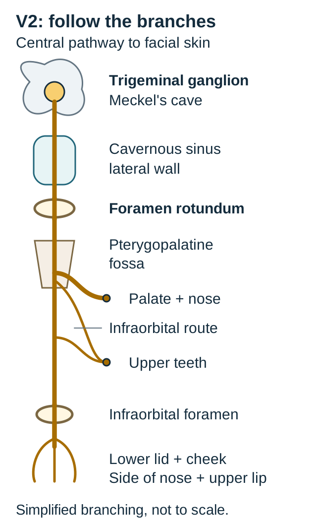
Why can disease at several different sites cause similar cheek pain? The maxillary division, V2, is a purely sensory division with a long course. Its sensory cell bodies lie in the trigeminal ganglion in Meckel's cave. V2 courses in the lower part of the cavernous sinus lateral wall, exits through the foramen rotundum, and enters the pterygopalatine fossa. Its branches distribute sensation to the midface, upper teeth, palate, nasal cavity and maxillary sinus. [4][9]
Use the route diagram to follow the pathway from the skull base toward its branches. The infraorbital continuation passes through the inferior orbital fissure into the orbital floor region, then reaches the face through the infraorbital foramen. Its terminal cutaneous territory includes the lower eyelid, cheek, side of the nose and upper lip. Dental branches arise before the terminal facial branches. Palatal and nasal routes diverge in the pterygopalatine region. Therefore, numbness of the upper lip alone and numbness of the upper lip plus palate need not have the same localization. [9]
Trace the common route, then compare a proximal interruption with injury at the terminal facial exit. The diagram simplifies the dental branch pattern and does not depict every branch or true distances. [4][9][4][9]
Three exits prevent a common anatomical substitution: V1 passes through the superior orbital fissure; V2 through foramen rotundum; V3 through foramen ovale. V3 does not travel in the cavernous sinus lateral wall. V2 does not supply the muscles of mastication. Jaw weakness or lower-lip and chin sensory loss therefore broadens the localization beyond an isolated V2 lesion. [4][8]
Trace-and-predict activity: place a finger on the terminal infraorbital branches in the diagram. Mentally interrupt that route after the dental branches have separated. Which remains available: upper-lip sensation or palatal sensation?
Check the first interruption
Palatal sensation remains available because its route separated earlier. Upper-lip sensation is affected downstream of the interruption. A distal branch lesion cannot explain every V2 territory.
Now trace back to V2 at foramen rotundum. An interruption there can affect several downstream territories together, including the palate and upper teeth. This is the complete worked comparison even with the optional answer closed: distal branch injury has a narrower distribution; a proximal divisional lesion can have a broader one. [9]
Transfer the map to a mass in the pterygopalatine fossa: proximal extension along V2 reaches the skull through foramen rotundum. The route explains possible spread, but numbness alone cannot distinguish a fossa lesion from a ganglion, root or cavernous sinus lesion. Additional nerves and imaging supply the missing localization. [4][9]
Separate the lateral wall from the venous compartment
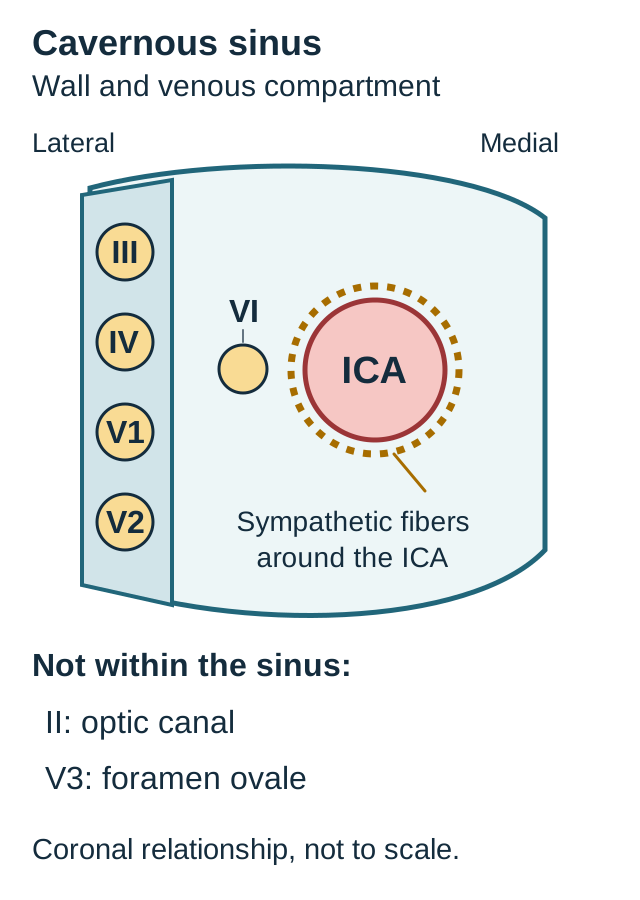
Which neighboring structures could produce facial sensory loss and double vision together? In a simplified coronal view, the lateral wall contains III, IV, V1 and V2, arranged from superior to inferior. The internal carotid artery, or ICA, traverses the venous compartment with its sympathetic plexus. Cranial nerve VI lies within that compartment, close to the ICA, rather than within the same wall layer as the other listed nerves. These relationships are a localization model, not a fixed order of nerve failure. [4]
CN VI activates the lateral rectus, so weakness limits abduction and causes horizontal diplopia. The pericarotid sympathetic pathway helps maintain pupillary dilation and a component of upper-eyelid tone. Injury can produce ipsilateral miosis and partial ptosis, the ocular features of Horner syndrome. Anisocoria is more apparent in darkness when the smaller pupil does not dilate adequately. Facial sweating may be preserved in a postganglionic lesion. [4][8]
Use the compartment diagram for a three-comparison exercise. The reference facts remain visible: V2 is in the lower lateral wall, VI is near the ICA, and the sympathetic fibers accompany the artery. For each example, point to the smallest region that could contain the implicated structures before reading the optional check.
Point to the smallest region that contains the affected nerves. The geometry is schematic and is not a fixed sequence of clinical deficits. ICA means internal carotid artery. [4][8][4][8]
Comparison A: cheek sensory loss with intact eye alignment. Locate V2, then ask whether this finding requires the cavernous sinus.
Check comparison A
It does not. The same sensory territory can be affected at several locations along V2. A distribution identifies a pathway before it identifies a site.
Comparison B: impaired abduction and a small pupil with mild ptosis on the same side. Point to the structures shared by this combination.
Check comparison B
VI and the pericarotid sympathetic fibers are close in the cavernous compartment. Their combination makes this region important to investigate, although examination alone does not establish the lesion's cause. Combined deficits can be more informative than either alone.
Comparison C: add forehead and upper-lip sensory loss while lower-lip sensation remains normal. Extend the proposed region to include the relevant wall structures.
Check comparison C
The pattern now includes V1 and V2 with VI, while sparing V3. That combination supports a cavernous sinus localization more than an isolated peripheral branch injury. Account for the affected and the spared territories.
The visible synthesis is that a central ICA-related process can affect VI and sympathetic fibers, while extension to the wall can add ocular motor or trigeminal sensory deficits. Real lesions vary in size and direction, so this is not a staged progression rule. An isolated VI palsy has many possible causes and does not by itself prove cavernous sinus disease. [4][8]
Connect venous congestion to an urgent differential
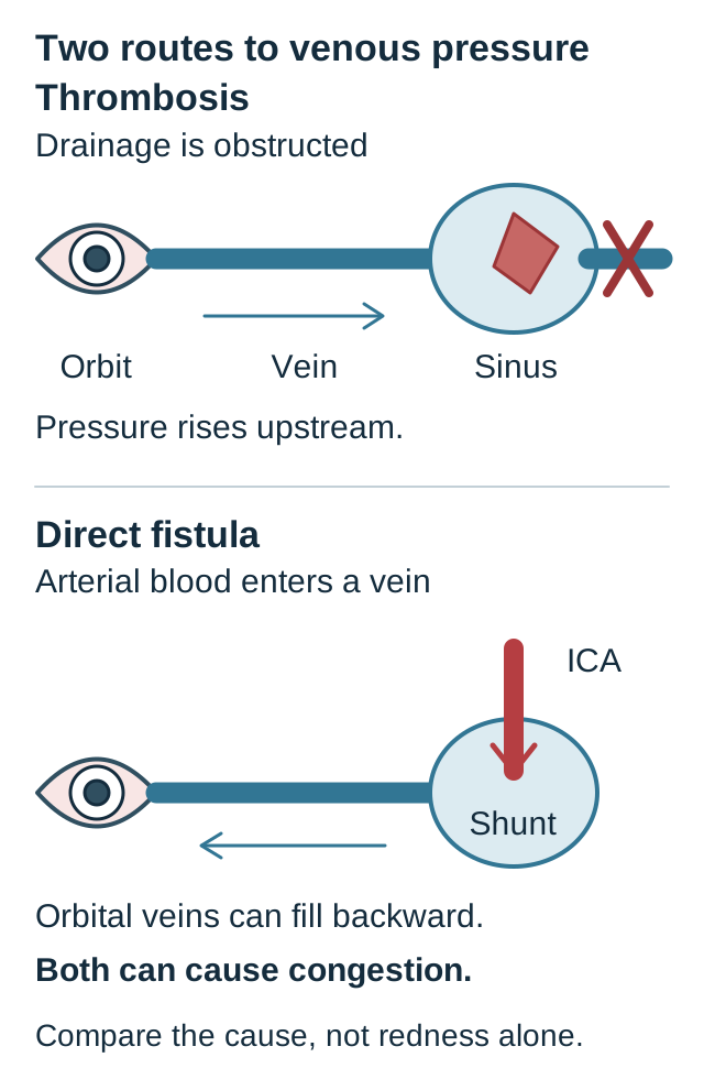
The cavernous sinus receives venous drainage from the orbit and communicates with other cranial venous channels. Intercavernous venous channels connect the two sides around the sella, providing a route that helps explain bilateral involvement. Ophthalmic veins link the orbit to this region, while petrosal routes provide drainage rather than the direct cross-sellar connection. [12] If outflow is impaired, orbital venous pressure can rise. If arterial blood enters the sinus through a fistula, venous pressure can also rise. The pressure diagram contrasts these two mechanisms. Similar eye congestion can therefore have different causes. [5][7]
Upper: obstructed venous drainage. Lower: one direct fistula configuration with arterial inflow. Indirect fistulas use arterial branches rather than the illustrated direct ICA connection. Arrow directions are simplified; real drainage depends on the lesion. [5][7][5][7]
Septic cavernous sinus thrombosis should be considered when facial, nasal, dental or sinus infection accompanies progressive headache or facial pain, fever, ocular motor dysfunction and orbital swelling. V1 or V2 sensory loss may occur, and findings may become bilateral. The sixth nerve is often affected early, but its absence does not provide reassurance against the syndrome. Confusion, seizures or additional focal deficits raise concern for intracranial complications. [5][6]
This combination warrants emergency hospital assessment. Obtain urgent contrast-enhanced neuroimaging, with venous imaging as appropriate, and blood cultures without delaying treatment. Begin clinician-directed intravenous antibiotics for suspected septic disease, then tailor therapy and arrange source control. The team assesses anticoagulation and contraindications, including bleeding risk; this is not a decision made from a facial-pain label alone. [6]
On contrast imaging, an expanded cavernous sinus with abnormal internal filling defects supports thrombosis in the appropriate clinical setting. A dilated superior ophthalmic vein supports disturbed orbital drainage but is not specific for a clot; it can also accompany a fistula. Do not substitute a single diameter cutoff for the full study. The cited MRI investigation was a small retrospective series, not validation of a universal screening rule. [5][7]
A carotid-cavernous fistula is an abnormal arterial connection to the venous sinus. A direct connection from the ICA can follow trauma and produce conspicuous pulsatile proptosis or an orbital bruit. Indirect connections involve arterial branches and can be less dramatic. Lack of trauma or an audible bruit does not exclude an indirect fistula. Angiographic assessment establishes the vascular connection and guides specialist treatment. [7]
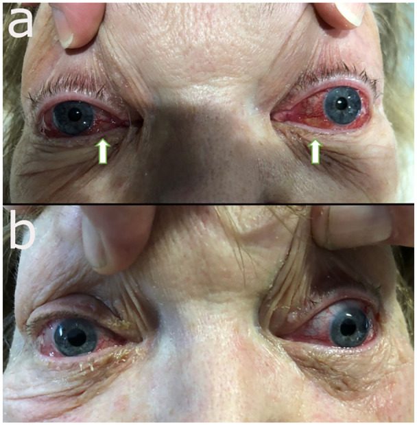
Inspect the paired clinical photograph from a published bilateral fistula case: the upper panel shows congestion before treatment; the lower panel shows the reported appearance one day after embolization. The original arrows and full panels are preserved. This is a real fistula example, not a thrombosis photograph, and the appearance alone does not establish the diagnosis or predict another patient's outcome. [7]
Real clinical photograph, not a schematic: bilateral carotid-cavernous fistula before treatment (upper panel) and the reported appearance one day after embolization (lower panel). Reproduced unmodified from Figure 5 of Sharma et al., 2022, Journal of Investigative Medicine High Impact Case Reports, DOI 10.1177/23247096221094181. Copyright 2022 American Federation for Medical Research. CC BY 4.0. Appearance alone does not diagnose a fistula or predict outcomes. Image: [7] Rohan Sharma, Christian Ponder, Mudassar Kamran, Joseph Chacko, Nidhi Kapoor, Krishna Mylavarapu, Sanjeeva Onteddu and Krishna Nalleballe; original source; CC BY 4.0. [7].
Predict the distinction: after head trauma, arterial-phase imaging shows early filling of the cavernous sinus and ophthalmic veins. Is the primary demonstrated abnormality blocked venous outflow or arterial inflow into a vein?
Check the pressure mechanism
Early venous filling demonstrates an arteriovenous connection. A clot can impede outflow, but it does not by itself explain this arterial-phase shunting. Congestion describes a consequence; imaging must identify its cause.
Other possibilities include aneurysm, tumor and inflammatory disease. A gradually progressive deficit without fever still needs localization and imaging. Painful ophthalmoplegia that improves with corticosteroids is not automatically Tolosa-Hunt syndrome: that diagnosis requires appropriate evidence and exclusion of competing infectious, neoplastic and other causes. [4][10]
Use the examination to test the proposed location
What finding would make the cavernous sinus explanation less complete? Examine visual acuity, color perception, pupils and ocular alignment, then compare sensation in V1, V2 and V3 territories. Assess facial strength, the corneal reflex when clinically appropriate, jaw strength and the rest of the neurologic examination. A symptom map should be tested against functions that travel elsewhere, not merely against the first suspected nerve. [8]
For the touch-evoked corneal reflex, V1 provides sensory input to the brainstem and VII supplies the eyelid-closing muscles. Either cornea normally elicits bilateral closure. The input side and output side are different questions. A trained examiner interprets the test in context; this lesson is not an instruction to touch one's own cornea. [11]
Worked comparison: touching the left cornea produces no blink in either eye, while touching the right produces both blinks. The right-sided test demonstrates that both motor outputs can function. Failure only when stimulating the left therefore points to the left sensory input. In contrast, a left facial motor deficit can prevent the left eyelid from closing after stimulation of either cornea while the right eyelid still responds. An isolated V2 deficit does not remove V1 corneal input. [8][11]
Predict before checking: a patient has upper-lip numbness and cannot close the left eye after either cornea is stimulated. Does V2 injury alone account for both findings?
Check the additional nerve
No. The eyelid pattern requires assessment of the left facial motor output, not just maxillary sensation. Do not assign every facial finding to the trigeminal nerve.
Now compare neighboring skull-base regions. The superior orbital fissure carries III, IV, V1 and VI, but not V2. The optic nerve travels through the optic canal and is not a cavernous sinus content. Ophthalmoplegia accompanied by impaired acuity, color vision and a relative afferent pupillary defect therefore raises concern for optic nerve involvement, including an orbital apex process. A strictly confined superior orbital fissure lesion would not directly explain that optic deficit. [4][8]
These borders are useful, not absolute disease barriers: a cavernous sinus process can extend into the orbital apex or impair vision through vascular complications. Conversely, V3 sensory loss with chewing weakness makes a broader trigeminal or adjacent motor-root process important. Ipsilateral ocular motor dysfunction with contralateral limb weakness suggests a brainstem localization rather than a lesion confined to the cavernous sinus. Always explain the whole examination. [4][5][8]
Keep diagnostic confidence separate from treatment response
A typical pain history can support trigeminal neuralgia without establishing its subtype. Classical trigeminal neuralgia requires neurovascular compression with morphological changes of the trigeminal root demonstrated by imaging or surgery. Mere contact between a vessel and nerve is insufficient. Secondary neuralgia has a demonstrated underlying cause, such as an appropriate demyelinating lesion or mass; idiopathic neuralgia lacks a demonstrated causative abnormality after adequate evaluation. [2][3]
MRI is part of the diagnostic workup even when the history is typical and the routine examination is normal. The EAN guideline describes high-resolution sequences to assess the trigeminal pathway, neurovascular relationships and secondary causes. Testing is individualized when MRI cannot be performed. A report of vascular contact should be read for root distortion or atrophy and correlated with the symptomatic side, rather than treated as an automatic operative indication. [2][3]
Carbamazepine or oxcarbazepine are recommended first-choice medicines for long-term trigeminal neuralgia treatment. Selection and titration require clinician assessment of tolerability, interactions, contraindications and monitoring. Symptom improvement supports continued therapeutic assessment, but it does not prove a compression mechanism or exclude another lesion. Dental treatment similarly requires a supported dental diagnosis rather than pain location alone. [3]
When pain remains inadequately controlled or adverse effects limit treatment, arrange specialist review. Procedural choices depend on the cause, imaging, health status and patient priorities. Microvascular decompression is an important option for an appropriate patient with classical neuralgia. Neuroablative procedures, including percutaneous techniques or radiosurgery, act on trigeminal signaling rather than separating the compressing vessel from the root. Other procedures have different benefits and sensory risks; no intervention guarantees permanent relief for every patient. [3]
Consider a patient whose attacks fall markedly on medication, but who develops diplopia and persistent facial numbness. Should the improvement make the new deficits less important?
Check the reassessment decision
No. Pain response and new neurologic deficits answer different questions. The changed examination requires reassessment of the cause and location. Treat symptoms while continuing to account for the anatomy.
Finish with a qualitative check, not an invented risk score: does the timing fit neuralgia, which sensory territory is involved, what additional nerves are affected, and is there infection, congestion or threatened vision? Preserve uncertainty where only a pathway is identified, and escalate promptly when the new findings indicate a potentially dangerous regional process. [1][3][6][8]
Apply the anatomy to new patients
Case 1
Show answer and explanations for case 1
A. Begin neuralgia medication and reserve MRI for an abnormal examination (Why this does not fit)
Brief triggered attacks make neuralgia medication reasonable. Deferring all imaging until a deficit appears would leave secondary causes insufficiently assessed. A normal examination does not end the neuralgia workup. [1] [2] [3]
Reasoning steps for option A
Why is medication plausible for this woman's 5 to 20 second cheek shocks?
Light touch triggers brief unilateral V2 attacks, a clinical trigeminal neuralgia pattern for which first-choice medication can be assessed.
Why is waiting for an abnormal examination before MRI insufficient here?
Her normal interictal sensory and eye examination does not exclude a secondary trigeminal cause, and she has never had neuroimaging.
What should accompany symptom treatment under this plan?
Trigeminal-pathway MRI should investigate the cause now rather than being conditional on a future deficit.
B. Combine trigeminal-pathway MRI with first-choice medication assessment (Best answer)
The history fits trigeminal neuralgia, for which carbamazepine or oxcarbazepine can be considered. A normal examination does not exclude secondary disease, and she has not had imaging. Treat the clinical syndrome while investigating its cause. [1] [2] [3]
Reasoning steps for option B
Which features support treating a trigeminal neuralgia syndrome rather than persistent regional pain?
The unilateral upper-lip and cheek shocks last seconds, follow innocuous facial contact and resolve between attacks.
What diagnostic uncertainty survives the normal examination and unrevealing dental evaluation?
Neither finding establishes whether a lesion underlies the neuralgia; she has not yet had trigeminal-pathway imaging.
How does the combined plan address both problems?
Arrange MRI to assess the trigeminal pathway and secondary causes while assessing carbamazepine or oxcarbazepine for attack control.
C. Request dental extraction and reassess after the socket heals (Why this does not fit)
Upper-lip and cheek pain can be perceived near maxillary teeth. The dental assessment is unrevealing and the attacks follow light cutaneous contact rather than a demonstrated tooth lesion. An irreversible dental procedure requires a supported dental diagnosis. [1] [2] [3]
Reasoning steps for option C
Why might upper-lip and cheek pain initially suggest a maxillary tooth source?
Maxillary teeth and adjacent facial skin share V2 territory, so perceived location alone can mislead.
Which two case findings argue against extraction?
Dental evaluation found no tooth lesion, and light contact while washing her face triggers seconds-long attacks rather than a demonstrated tooth stimulus.
What evidence would be needed before an irreversible dental procedure?
A supported dental diagnosis, such as a concordant abnormal tooth, is needed; V2 pain location alone is not enough.
D. Arrange immediate decompression based on the sensory distribution (Why this does not fit)
A procedure can help selected patients with a demonstrated cause of neuralgia. No compression or morphological root abnormality has been established, and medical treatment has not been assessed. A sensory territory is not an operative indication. [1] [2] [3]
Reasoning steps for option D
What would decompression aim to change in a selected neuralgia patient?
It separates a compressing vessel from the trigeminal root when clinically appropriate neurovascular compression is demonstrated.
What is missing before immediate decompression can be justified here?
She has no imaging evidence of root compression or morphological change, and medication has not been assessed.
Why does the right V2 distribution not itself select surgery?
The sensory map identifies an affected pathway, not a compressing vessel or a procedural indication.
Takeaway: Treat the clinical syndrome while investigating its cause.
A. Trigeminal neuralgia requiring primary neurologic treatment (Why this does not fit)
The upper teeth lie in a trigeminal territory. Persistent thermal pain and demonstrable tooth pathology explain the presentation better than brief innocuous-touch-triggered attacks. Pain location alone does not establish neuralgia. [1] [3] [9]
Reasoning steps for option A
Why can pain in an upper molar be mistaken for trigeminal neuralgia?
Upper molar afferents travel in V2, but territory alone does not specify the pain mechanism.
How does this patient's cold response differ from a neuralgic trigger?
Cold produces lingering pain at a particular diseased tooth, whereas typical neuralgia causes brief attacks with innocuous touch.
What examination finding most strongly redirects the initial referral from neurology?
Percussion reproduces pain at the carious molar, supporting focused dental assessment rather than primary neuralgia treatment.
B. Infraorbital neuropathy requiring peripheral nerve imaging (Why this does not fit)
Infraorbital branches supply parts of the symptomatic region. Sensation is normal, and the pain is reproducible at a diseased tooth rather than a damaged cutaneous nerve. A nerve territory and a nerve lesion are not equivalent. [1] [3] [9]
Reasoning steps for option B
What territory could make an infraorbital lesion seem plausible?
The infraorbital nerve carries sensation from nearby midface structures, including skin near the upper lip and cheek.
What findings fail to support a damaged infraorbital cutaneous nerve?
Facial sensation is normal and touching cheek or upper lip triggers no attack; pain instead localizes to a carious, percussion-sensitive tooth.
Why would peripheral nerve imaging not be the first referral?
The demonstrated focal dental pathology and lingering thermal response explain the symptoms without evidence of infraorbital neuropathy.
C. Sinus inflammation requiring primary sinonasal assessment (Why this does not fit)
Maxillary sinus disease can refer pain toward upper teeth. The supplied findings identify one abnormal tooth rather than a nasal or sinus illness. Prefer the demonstrated focal source over an unsupported neighboring source. [1] [3] [9]
Reasoning steps for option C
Why can a maxillary sinus disorder enter the upper-tooth pain differential?
Sinus disease may refer pain toward upper teeth because of the neighboring maxillary anatomy.
What positive evidence favors the molar over the sinus in this case?
One molar has deep caries, lingering cold sensitivity and reproducible percussion tenderness; no nasal or sinus illness is supplied.
What should guide the initial referral when these sources compete?
Refer for assessment of the demonstrated abnormal tooth rather than prioritizing an unsupported sinonasal source.
D. Odontogenic pain requiring focused dental assessment (Best answer)
Tooth pathology can produce pain in the maxillary region. Lingering thermal pain, percussion tenderness and caries all converge on the same tooth. Use local findings to identify a supported dental source. [1] [3] [9]
Reasoning steps for option D
Which examination localizes this pain to a particular tooth?
Percussion of the upper molar reproduces the pain, and that tooth has deep caries.
What does pain persisting after cold is removed add to the localization?
Lingering tooth-specific thermal pain supports an odontogenic process rather than seconds-long touch-triggered facial neuralgia.
Which initial referral follows from these convergent findings?
Focused dental assessment of the symptomatic carious molar is appropriate.
Takeaway: Use local findings to identify a supported dental source.
A. The pain syndrome is supported; its classical subtype is not established (Best answer)
A typical clinical phenotype supports trigeminal neuralgia. The MRI demonstrates contact but not the morphological root changes required to establish classical neuralgia. Separate syndrome recognition from etiological classification. [1] [2] [3]
Reasoning steps for option A
What can be inferred from shaving-triggered electric cheek attacks without relying on MRI?
Recurrent unilateral shocks lasting under a minute support the clinical trigeminal neuralgia syndrome.
Why does the vessel touching the symptomatic root not establish the classical subtype?
The scan shows contact but no root displacement, indentation or atrophy, so the required morphological evidence of compression is absent.
What distinction should the MRI conclusion preserve?
The clinical syndrome remains supported, but vascular contact alone does not establish classical trigeminal neuralgia.
B. The pain syndrome is supported; its classical subtype is established (Why this does not fit)
Neurovascular compression can cause classical trigeminal neuralgia. The reported finding is contact without displacement, indentation or atrophy. Contact is not interchangeable with causative compression. [1] [2] [3]
Reasoning steps for option B
What imaging finding would strengthen a claim of classical neurovascular compression?
A compressing vessel with morphological root change, such as displacement, indentation or atrophy, would meet the relevant distinction.
What does this man's scan show instead?
It reports vessel contact on the symptomatic side without any of those root changes.
Why is the classical subtype therefore premature?
An adjacent vessel is not equivalent to demonstrated compression with morphological effects on the trigeminal root.
C. The pain syndrome is supported; a demyelinating subtype is established (Why this does not fit)
Demyelination can cause secondary trigeminal neuralgia. The imaging explicitly shows no demyelinating lesion and identifies only uncomplicated contact. A possible disease mechanism still requires evidence. [1] [2] [3]
Reasoning steps for option C
What lesion could support demyelination as a secondary cause of these shocks?
A demyelinating lesion concordant with the symptomatic trigeminal pathway could account for secondary neuralgia.
Does the reported MRI identify that lesion?
No. It explicitly finds no demyelinating lesion anywhere along the assessed trigeminal pathway.
Why cannot simple vascular contact be relabeled a demyelinating subtype?
Contact supplies no evidence of myelin injury; the proposed secondary cause requires its own demonstrated lesion.
D. The pain syndrome is excluded; normal root shape contradicts neuralgia (Why this does not fit)
Imaging contributes to finding the cause of facial pain. Neuralgia is diagnosed clinically and can occur without the morphological changes required for the classical subtype. A negative etiological study does not erase a compatible phenotype. [1] [2] [3]
Reasoning steps for option D
What clinical evidence remains if the root has a normal shape?
Recurrent brief unilateral electric pain provoked by shaving still supports trigeminal neuralgia.
What question does absent root deformation answer instead?
It limits evidence for the classical compression subtype, not the existence of a clinically recognizable pain syndrome.
Why is excluding neuralgia from this MRI a category error?
Imaging helps classify cause; normal root morphology cannot negate a compatible attack phenotype.
Takeaway: Separate syndrome recognition from etiological classification.
A. Neurosurgical discussion of percutaneous trigeminal glycerol treatment (Why this does not fit)
Glycerol treatment is another neuroablative option for selected patients. It affects trigeminal signaling but does not directly correct the demonstrated neurovascular compression. Discuss alternative procedures with their differing targets rather than treating them as equivalent. [2] [3]
Reasoning steps for option A
Why might percutaneous glycerol treatment be discussed after these medication trials?
Persistent disabling shocks and intolerance of carbamazepine and oxcarbazepine justify discussion of procedural alternatives.
What does glycerol treatment target instead of the displaced trigeminal root's cause?
It alters trigeminal signaling through a neuroablative approach rather than separating the compressing artery from the root.
Why is it not the best match for this patient's stated procedural goal?
She is an operative candidate who wants the demonstrated arterial compression addressed directly, so decompression better matches that goal.
B. Neurosurgical discussion of trigeminal radiofrequency treatment (Why this does not fit)
Radiofrequency treatment can reduce pain by targeting trigeminal fibers. It does not directly relieve the vascular compression that this patient seeks to address. Match a procedure's target and sensory risks to the patient's stated goal. [2] [3]
Reasoning steps for option B
What therapeutic role could trigeminal radiofrequency treatment have here?
It can reduce neuralgic pain by lesioning trigeminal fibers when a neuroablative procedure is selected.
Does radiofrequency treatment directly relieve the MRI-demonstrated arterial compression?
No. It changes trigeminal signaling rather than separating the compressing artery from the root.
Which patient preference makes radiofrequency a less fitting consultation?
She specifically seeks a procedure aimed at the demonstrated cause and is otherwise a reasonable operative candidate.
C. Neurosurgical discussion of trigeminal balloon compression (Why this does not fit)
Balloon compression is a neuroablative option for selected trigeminal neuralgia. It alters trigeminal nerve function rather than separating the demonstrated root-compressing vessel in this otherwise suitable candidate. Procedures differ in whether they address vascular compression or interrupt trigeminal signaling. [2] [3]
Reasoning steps for option C
Why is balloon compression an actual procedural alternative for this neuralgia?
Disabling pain despite adequate medication trials can prompt consideration of percutaneous neuroablative options.
What anatomical problem remains after balloon compression?
The root-compressing artery is not separated from the displaced and atrophic trigeminal root; the intervention alters nerve function.
How should the patient's operative fitness and goal affect the choice?
Because she can reasonably undergo surgery and wants the vascular cause addressed, discussion of microvascular decompression fits better.
D. Neurosurgical discussion of stereotactic trigeminal radiosurgery (Why this does not fit)
Radiosurgery can be considered for selected patients with trigeminal neuralgia. It targets the nerve rather than directly separating an offending vessel from the root. A less invasive option is not mechanically identical to decompression. [2] [3]
Reasoning steps for option D
Why could stereotactic radiosurgery be considered for uncontrolled trigeminal pain?
It is a procedural means of targeting trigeminal signaling when medication is ineffective or poorly tolerated.
How does its target differ from the MRI finding?
Radiation targets the nerve; it does not physically separate the compressing artery from the morphologically changed root.
Why is a potentially less invasive procedure not automatically the answer here?
The question specifies a reasonable operative candidate seeking treatment of the demonstrated compression itself.
E. Neurosurgical discussion of microvascular decompression (Best answer)
Microvascular decompression addresses an established neurovascular compression. The patient has morphological root changes and medication-limited disabling disease, with reasonable operative fitness. Match the procedure discussion to the demonstrated cause and patient priorities. [2] [3]
Reasoning steps for option E
Which MRI details support classical neurovascular compression rather than incidental contact?
An artery compresses the symptomatic root with displacement and focal atrophy, demonstrating morphological change.
Why is procedural consultation warranted now?
Disabling attacks persist after supervised first-choice medication trials, and adverse effects limit further use.
Which procedure most directly addresses her goal and anatomical cause?
Discuss microvascular decompression with neurosurgery: it aims to separate the offending vessel from the root in a suitable operative candidate.
Takeaway: Match the procedure discussion to the demonstrated cause and patient priorities.
A. Classical trigeminal neuralgia from vascular compression (Why this does not fit)
Classical neuralgia can produce the same attack phenotype. The study shows no compressive vascular root change and instead identifies relevant demyelination. A familiar phenotype does not establish the classical subtype. [1] [3] [8]
Reasoning steps for option A
Why can this patient's touch-triggered shocks resemble classical neuralgia?
Brief unilateral triggered cheek shocks fit the trigeminal neuralgia pain phenotype regardless of its eventual cause.
What imaging finding prevents attributing this episode to vascular compression?
There is no compressive vascular root change; MRI instead identifies a new lesion in the ipsilateral pontine trigeminal pathway.
Why does the phenotype alone not establish the classical subtype?
Classical neuralgia requires demonstrated neurovascular compression with morphological root change, not merely characteristic attacks.
B. Distal infraorbital neuropathy from local trauma (Why this does not fit)
A distal infraorbital lesion can affect cheek sensation. No local trauma is supplied, and a matching central lesion is demonstrated. Integrate structural evidence with the sensory distribution. [1] [3] [8]
Reasoning steps for option B
Which symptom could a distal infraorbital nerve lesion potentially produce?
It could affect cutaneous cheek sensation within part of the maxillary distribution.
What evidence weighs against a traumatic distal nerve explanation here?
No local injury is reported, while MRI shows a new left pontine trigeminal lesion concordant with the left-sided symptoms.
Why should cheek location not override the MRI localization?
The same sensory territory can be affected centrally or peripherally; the demonstrated pathway lesion favors a central secondary cause.
C. Secondary trigeminal neuralgia from pontine demyelination (Best answer)
A demyelinating lesion along the trigeminal pathway can cause secondary neuralgia. The newly demonstrated lesion is ipsilateral and anatomically relevant to the new symptoms. Link a pain phenotype to a demonstrated, concordant cause. [1] [3] [8]
Reasoning steps for option C
Which symptom pattern identifies the pain syndrome?
Brief recurrent shocks in the left cheek triggered by touch fit trigeminal neuralgia.
How do the new sensory change and MRI identify its cause?
A new ipsilateral demyelinating lesion involves the pontine trigeminal pathway, anatomically matching the new symptoms in a patient with multiple sclerosis.
Why is the classification secondary rather than classical?
A concordant demyelinating lesion is demonstrated, while compressive vascular root change is absent.
D. Cavernous sinus neuropathy from a regional mass (Why this does not fit)
A cavernous lesion can affect V2 and other cranial nerves. Imaging shows no cavernous lesion and places the new abnormality in the pons. Localize using the demonstrated lesion rather than the word cheek. [1] [3] [8]
Reasoning steps for option D
Why might a cavernous sinus lesion be considered for cheek sensory loss?
V2 passes through the cavernous sinus lateral wall, so a regional lesion could affect its sensory fibers.
Where does MRI actually place the new abnormality?
It places a demyelinating lesion in the left pontine trigeminal pathway and reports no cavernous sinus abnormality.
What makes a cavernous regional mass less explanatory than pontine demyelination?
No cavernous mass is demonstrated, whereas the pontine lesion is new, ipsilateral and directly relevant to the neuralgic symptoms.
Takeaway: Link a pain phenotype to a demonstrated, concordant cause.
A. Terminal infraorbital branches at the facial exit (Best answer)
These branches supply the lower eyelid, side of the nose and upper lip. The trauma is at their exit, with dental and palatal routes spared. Preserved upstream branches support a distal localization. [4] [9]
Reasoning steps for option A
Which skin fields identify the terminal infraorbital branches?
The lower eyelid, side of the nose and upper lip are the affected terminal cutaneous fields.
Why does preserved upper-tooth and palatal sensation favor the facial exit?
Those V2 pathways have branched off before the infraorbital nerve reaches the face, so a facial-exit injury can spare them.
How does the laceration site reinforce this localization?
It is centered just below the infraorbital foramen, matching a distal branch injury rather than a proximal division lesion.
B. Trigeminal ganglion with broad sensory involvement (Why this does not fit)
The ganglion contains sensory cell bodies for the face. A broad ganglionic injury is less consistent with this narrow post-traumatic branch territory and spared neighboring fields. A highly focal deficit favors the matching peripheral branch. [4] [9]
Reasoning steps for option B
Could a trigeminal ganglion injury affect the lower eyelid and upper lip?
Yes, their sensory cell bodies belong to the trigeminal system, but a ganglion lesion usually threatens a wider distribution.
Which preserved field argues against broad ganglionic involvement?
Forehead sensation remains normal despite V1 cell bodies also lying in the ganglion.
What does the sharply circumscribed post-traumatic deficit favor instead?
The injured terminal infraorbital branches at the nearby facial exit, rather than broad ganglionic damage.
C. Mandibular nerve at foramen ovale (Why this does not fit)
V3 supplies lower-face sensation and mastication pathways. The deficit is in the midface, not the lower lip or chin. Map the skin before assigning the division. [4] [9]
Reasoning steps for option C
What territory would a foramen ovale V3 injury principally disturb?
Mandibular territory, including the lower lip and chin, and potentially motor fibers to mastication.
Does the post-traumatic deficit occupy that mandibular field?
No. It involves the lower eyelid, nasal side and upper lip, a V2 cutaneous distribution.
How does the laceration site distinguish an infraorbital injury from a foramen ovale lesion?
The wound lies just below the infraorbital foramen, matching the affected midface skin rather than the proximal V3 route through foramen ovale.
D. Maxillary nerve at foramen rotundum (Why this does not fit)
Proximal V2 includes fibers serving the affected skin. An injury at this level would be less compatible with selective preservation of dental and palatal sensation. Use the width of the sensory deficit to locate the level. [4] [9]
Reasoning steps for option D
Why can proximal V2 at rotundum initially seem plausible?
V2 carries the sensory fibers serving the affected midface skin.
What should a lesion before V2 branches to the teeth and palate also threaten?
Upper-tooth and palatal sensation, both explicitly preserved here.
Where along V2 does the narrow skin-only loss therefore point?
Distal terminal infraorbital branches near the laceration, not the common V2 trunk at rotundum.
E. Ophthalmic nerve at the superior orbital fissure (Why this does not fit)
V1 serves forehead and corneal sensation near the orbit. The affected lower eyelid and upper lip are V2 territory, while forehead sensation is normal. Orbital proximity does not make a sensory deficit V1. [4] [9]
Reasoning steps for option E
What does V1 near the superior orbital fissure supply that can be tested here?
Forehead sensation is a V1 field and is preserved.
Do the numb lower eyelid and upper lip indicate V1 despite their orbital proximity?
No. Those skin fields belong to the infraorbital continuation of V2.
Why does the facial wound matter more than the word orbital in this option?
Its position below the infraorbital foramen directly matches the involved V2 terminal branches, not the V1 route through the fissure.
Takeaway: Preserved upstream branches support a distal localization.
Terminal infraorbital branches emerge onto the face here. That distal facial exit does not connect the pterygopalatine fossa to the cranial cavity. Distinguish the nerve division exit from a terminal branch exit. [4] [9]
Reasoning steps for option A
What part of V2 passes through the infraorbital foramen?
Terminal infraorbital cutaneous branches emerge onto the face there.
Does that facial exit join the pterygopalatine fossa to the cranial cavity?
No. It is downstream of the fossa along the infraorbital course.
Which opening must imaging follow for the broad dental, palatal and facial V2 pattern?
Foramen rotundum is the proximal V2 passage from the cranial cavity into the fossa.
B. Superior orbital fissure (Why this does not fit)
V1 and ocular motor nerves reach the orbit here. V1 and ocular motor function are preserved, and the route asked for follows V2 from the pterygopalatine fossa. Separate the orbital fissure from V2 intracranial passage. [4] [9]
Reasoning steps for option B
Which nerves make the superior orbital fissure a tempting skull-base opening?
It transmits V1 and ocular motor nerves toward the orbit.
What does normal forehead sensation and ocular alignment suggest about that route?
The characteristic V1 and ocular motor functions associated with the fissure are spared.
Why does the requested fossa-to-cranium trajectory exclude this fissure?
The symptomatic V2 division traverses foramen rotundum, not the superior orbital fissure.
C. Foramen ovale (Why this does not fit)
This opening transmits V3. Chin sensation and mastication are intact, while the impaired territories belong to V2. Do not substitute the mandibular exit for the maxillary exit. [4] [9]
Reasoning steps for option C
Which trigeminal division uses foramen ovale?
V3, the mandibular division, passes through it.
Which findings oppose a V3 route in this woman?
Chin sensation and jaw strength remain normal while upper teeth and palate are numb.
What alternative opening follows the impaired division proximally?
The V2 route from the pterygopalatine fossa is foramen rotundum.
D. Foramen rotundum (Best answer)
V2 enters the pterygopalatine fossa through this opening. The broad maxillary sensory pattern identifies the division being followed proximally. V2 reaches the pterygopalatine fossa through rotundum. [4] [9]
Reasoning steps for option D
Which division unites the numb lower eyelid, upper lip, upper teeth and palate?
V2 carries these maxillary cutaneous, dental and palatal sensory pathways.
Where does that division pass between the pterygopalatine fossa and cranial cavity?
V2 runs through foramen rotundum.
Why does the broad pattern make a terminal facial opening insufficient?
Dental and palatal involvement extends beyond terminal infraorbital skin branches, supporting proximal V2 imaging through rotundum.
E. Optic canal (Why this does not fit)
The optic nerve and ophthalmic artery traverse this canal. The supplied abnormality is maxillary somatic sensation, not an optic pathway deficit. The optic canal does not transmit V2. [4] [9]
Reasoning steps for option E
What normally traverses the optic canal?
The optic nerve and ophthalmic artery, not the maxillary nerve.
Is an optic-pathway complaint part of the supplied deficit?
No. The progressive loss concerns V2 skin, upper teeth and palate.
Which skull-base opening, rather than the optic canal, is on the stated V2 route?
Foramen rotundum connects V2 with the pterygopalatine fossa.
Takeaway: V2 reaches the pterygopalatine fossa through rotundum.
A. Superior orbital fissure alone (Why this does not fit)
The fissure carries V1 and VI and can explain two new deficits. It does not provide the proximal V2 route through foramen rotundum or explain the complete palatal and upper-lip sensory pattern. Use the original territory as well as the newly impaired nerves. [4] [8] [9]
Reasoning steps for option A
Which newly abnormal functions could the superior orbital fissure explain?
Its V1 and VI contents can account for forehead numbness and impaired abduction.
What pre-existing deficit and imaging route make the fissure alone insufficient?
Palatal and upper-lip V2 loss began in the fossa, and enhancement tracks posteriorly through V2's skull-base opening.
Which proximal compartment can link that V2 route to V1 and VI?
The cavernous sinus fits extension from rotundum together with the added nerve deficits.
B. Cavernous sinus (Best answer)
V1 and V2 are related to the lateral wall, while VI lies near the ICA. Proximal V2 extension plus the added V1 and VI findings fits this compartment. Combine the route of extension with the newly affected neighbors. [4] [8] [9]
Reasoning steps for option B
Through which opening does the tumor track posteriorly along V2?
It follows the maxillary nerve through foramen rotundum from the pterygopalatine fossa.
Which cavernous neighbors account for new forehead loss and failed abduction?
V1 relates to the lateral wall and VI courses near the cavernous ICA.
Why does unchanged chin sensation fit the cavernous sinus localization?
V3 follows a different route through foramen ovale and need not be involved in this V2, V1 and VI pattern.
C. Foramen ovale region (Why this does not fit)
A lesion here can affect V3 and associated motor function. V3 sensation is preserved, while the newly impaired nerves are V1 and VI. Spared lower-face function helps distinguish adjacent skull-base routes. [4] [8] [9]
Reasoning steps for option C
What nerve does the foramen ovale region principally put at risk?
V3, including lower-face sensory fibers and mandibular motor pathways.
Does unchanged chin sensation support new V3 injury?
No. Chin sensation is unchanged, whereas the new deficits are forehead sensation and eye abduction.
Which compartment better joins the tracked V2 extension to those two new nerves?
The cavernous sinus contains the relevant V2, V1 and VI relationships.
D. Trigeminal sensory ganglion alone (Why this does not fit)
The ganglion can account for maxillary and ophthalmic sensory deficits. A lesion confined to the sensory ganglion does not account for VI dysfunction without additional regional involvement. A sensory-only compartment is insufficient when ocular motor function is also lost. [4] [8] [9]
Reasoning steps for option D
Which sensory changes might a trigeminal ganglion lesion explain?
It could affect V2 sensation in the palate and upper lip and V1 sensation in the forehead.
Which new deficit is not explained by the sensory ganglion alone?
Limited right-eye abduction indicates CN VI motor dysfunction.
Where can the sensory pattern and VI weakness coexist along the proximal V2 route?
In the cavernous sinus region, rather than a lesion confined to the sensory ganglion.
E. Terminal infraorbital canal (Why this does not fit)
The infraorbital continuation carries part of V2 distally. It does not contain V1 or VI and cannot explain the new forehead deficit and abduction weakness. A distal V2 lesion cannot explain additional cranial nerves. [4] [8] [9]
Reasoning steps for option E
What original complaint can the infraorbital continuation partly explain?
Distal V2 fibers can carry upper-lip sensation.
Why does palatal numbness already exceed a terminal infraorbital lesion?
Palatal V2 fibers take another route rather than traversing the terminal infraorbital canal.
Can this distal canal account for new forehead loss and abduction weakness?
No. It contains neither V1 nor VI; posterior spread toward the cavernous sinus better fits all findings.
Takeaway: Combine the route of extension with the newly affected neighbors.
A. Trochlear nerve and ophthalmic sensory fibers (Why this does not fit)
IV and V1 are both associated with the cavernous lateral wall. Their dysfunction does not explain lateral rectus weakness or sympathetic pupillary dysfunction. A neighboring pair must account for the specific lost functions. [4] [8]
Reasoning steps for option A
Where are IV and V1 located relative to the cavernous region?
Both course in the cavernous lateral wall, so they are anatomically nearby.
Would IV dysfunction produce the observed failure of abduction?
No. The lateral rectus depends on VI, whereas IV supplies the superior oblique.
Can V1 sensory fibers explain anisocoria greatest in darkness?
No. That small right pupil reflects impaired sympathetic dilation along the cavernous ICA.
B. Optic nerve and oculomotor parasympathetic fibers (Why this does not fit)
This pairing can affect visual afferent function and pupillary constriction. The stem instead describes abduction weakness and reduced dilation in darkness, with an ICA-centered lesion. Separate visual afferent function from sympathetic dilation. [4] [8]
Reasoning steps for option B
What deficit would optic nerve involvement primarily predict?
A visual afferent disturbance rather than isolated lateral rectus weakness.
What pupil direction follows loss of oculomotor parasympathetic constriction?
The affected pupil tends to be larger, not the smaller pupil described here.
Which ICA-adjacent pair matches both right gaze failure and dark-enhanced anisocoria?
CN VI accounts for abduction weakness and pericarotid sympathetic fibers for impaired dilation.
C. Oculomotor nerve and its parasympathetic fibers (Why this does not fit)
III dysfunction can produce ptosis and ocular misalignment. Parasympathetic failure favors a larger pupil, and it does not directly paralyze lateral rectus. A small pupil with abduction weakness is not a complete third-nerve pattern. [4] [8]
Reasoning steps for option C
Why might III initially be considered for ptosis and diplopia?
Oculomotor dysfunction can lower the lid and disrupt ocular alignment.
What eye movement localizes the observed deficit more specifically than diplopia alone?
The right eye fails to abduct, implicating lateral rectus and CN VI rather than III.
Why does the pupil argue against III parasympathetic failure?
The right pupil is small with anisocoria greater in darkness, consistent with sympathetic loss, not failed parasympathetic constriction.
D. Maxillary nerve and facial motor fibers (Why this does not fit)
V2 and VII can affect facial sensation and eyelid closure. Neither explains the demonstrated abduction deficit and impaired pupillary dilation. Ptosis is not the same finding as weak eyelid closure. [4] [8]
Reasoning steps for option D
What would maxillary nerve V2 injury alter?
It would change midface or upper-lip sensation, not lateral rectus action.
What eyelid finding could facial nerve VII weakness cause instead of this ptosis?
Weak eyelid closure; the stem describes mild drooping associated with a small pupil.
Why does this pair miss the ICA-centered examination?
Neither V2 nor VII directly explains right abduction weakness plus impaired sympathetic dilation.
E. Abducens nerve and pericarotid sympathetic fibers (Best answer)
VI and sympathetic fibers are close to the cavernous ICA. This pairing explains abduction weakness plus a small pupil with partial ptosis. Use ocular alignment and pupil behavior as complementary localization findings. [4] [8]
Reasoning steps for option E
Which nerve accounts for inability to abduct the right eye?
Right CN VI supplies lateral rectus, explaining gaze-dependent horizontal diplopia.
Why does anisocoria increase in darkness when the right pupil is smaller?
Right sympathetic dilation is impaired, producing a Horner-pattern small pupil and mild ptosis.
What makes these two structures a coherent pair at the imaged lesion?
CN VI and pericarotid sympathetic fibers lie close to the cavernous ICA, matching both deficits.
Takeaway: Use ocular alignment and pupil behavior as complementary localization findings.
A. Right orbital apex alone (Why this does not fit)
An apex lesion can affect ocular motor nerves and V1. It does not directly account for the broad V2 pattern including the hard palate, while optic function is preserved. Palatal sensory loss argues against a purely orbital explanation. [4] [8] [9]
Reasoning steps for option A
Which parts of the examination could a right orbital apex lesion explain?
It may impair ocular motor function and V1 forehead sensation.
Which sensory site extends beyond a purely orbital V1 or infraorbital explanation?
The hard palate is a V2 territory reached through the pterygopalatine route.
What does preserved visual acuity and color add to the apex comparison?
There is no supplied optic nerve deficit, while the V2 palatal loss points toward a cavernous rather than purely apical location.
B. Right superior orbital fissure (Why this does not fit)
This fissure transmits VI and V1. It does not carry V2 and cannot directly account for the palatal and upper-lip sensory deficits. V2 involvement helps distinguish the cavernous sinus from the superior orbital fissure. [4] [8] [9]
Reasoning steps for option B
Which two affected nerves pass through the superior orbital fissure?
VI can account for abduction weakness and V1 for forehead sensory loss.
Why are the upper lip and hard palate decisive against the fissure alone?
They indicate V2 involvement, and V2 does not traverse the superior orbital fissure.
Which neighboring compartment accommodates VI, V1 and V2 together?
The right cavernous sinus, with VI near the ICA and V1 and V2 along its lateral wall.
C. Right cavernous sinus (Best answer)
The compartment places VI near the ICA and V1 and V2 in the lateral wall. The combination explains ocular motor and sensory deficits while allowing V3 and optic function to remain intact. Localize the complete nerve pattern, including important spared functions. [4] [8] [9]
Reasoning steps for option C
What does right-eye abduction weakness identify?
Right CN VI dysfunction, because VI innervates lateral rectus.
Which two divisions explain forehead versus upper-lip and palatal pinprick loss?
V1 serves the forehead; V2 supplies the upper lip and hard palate.
Why does the right cavernous sinus fit the affected and spared structures?
It neighbors VI, V1 and V2, while V3 jaw and chin function and optic testing may remain normal.
D. Right terminal infraorbital nerve (Why this does not fit)
A distal V2 lesion can affect the upper lip. It cannot account for forehead sensory loss, palatal sensory loss and VI palsy together. A peripheral branch does not explain multiple cranial nerve functions. [4] [8] [9]
Reasoning steps for option D
Which reported symptom could a terminal infraorbital lesion explain?
Upper-lip sensory loss can arise from distal infraorbital V2 fibers.
Why does hard-palate numbness exceed that distal branch territory?
Palatal V2 fibers branch through a separate route before the terminal infraorbital nerve.
What additional findings definitively require a wider localization?
Forehead V1 loss and VI-related abduction weakness cannot arise from a terminal infraorbital lesion.
E. Right trigeminal sensory ganglion alone (Why this does not fit)
A ganglionic lesion can affect V1 and V2 sensation. A lesion restricted to that sensory structure does not explain VI palsy. Sensory and ocular motor findings must be explained together. [4] [8] [9]
Reasoning steps for option E
Why could a trigeminal ganglion lesion account for some sensory findings?
The ganglion contains V1 and V2 sensory cell bodies relevant to forehead, upper lip and palate.
What part of the examination remains unexplained by a sensory ganglion lesion alone?
Right-eye abduction weakness requires CN VI motor involvement.
Which nearby site incorporates the sensory divisions and VI while sparing V3 and optic function?
The right cavernous sinus best unifies that complete pattern.
Takeaway: Localize the complete nerve pattern, including important spared functions.
A. Reduced color vision with an afferent pupillary defect (Why this does not fit)
An optic nerve lesion can produce this visual pattern. The optic nerve is not a cavernous lateral-wall content. An optic deficit requires a broader or different localization. [4] [8]
Reasoning steps for option A
Which visual signs would an optic nerve lesion produce?
Reduced color vision and a relative afferent pupillary defect indicate optic afferent dysfunction.
Why does inferior extension confined to the cavernous lateral wall not affect the optic nerve?
The optic nerve travels in the optic canal, not in the cavernous lateral wall traversed by this lesion.
What localization would new optic afferent dysfunction require?
New optic signs would require extension beyond the specified wall or a separate visual pathway lesion.
B. Weakness of eyelid closure and the lower face (Why this does not fit)
CN VII supplies facial expression and eyelid closure. It does not run through the cavernous lateral wall. Do not confuse third-nerve ptosis with facial motor weakness. [4] [8]
Reasoning steps for option B
Which nerve supplies eyelid closure and lower-face movements?
CN VII innervates orbicularis oculi for eyelid closure and supplies facial expression muscles.
Why is existing third-nerve ptosis not evidence of facial motor weakness?
Third-nerve impairment weakens eyelid elevation and causes ptosis, whereas seventh-nerve impairment weakens closure and facial movement.
Does CN VII travel through the cavernous lateral wall?
CN VII does not travel in the cavernous lateral wall, so the specified inferior wall extension cannot directly injure it.
C. Reduced sensation over the chin and lower lip (Why this does not fit)
V3 supplies these lower-face territories. V3 is not in the cavernous lateral wall described in the stem. Inferior wall extension does not automatically become V3 involvement. [4] [8]
Reasoning steps for option C
Which division supplies the chin and lower lip?
V3 supplies sensation to the chin and lower lip.
Why does inferior wall extension encounter V2 rather than V3?
V2 lies below V1 in the cavernous lateral wall, whereas V3 exits through foramen ovale and is outside that wall.
What broader localization would chin numbness imply?
Chin numbness would suggest V3 involvement at another or broader trigeminal site, beyond the described wall-confined extension.
D. Reduced sensation over the upper lip and palate (Best answer)
V2 occupies the lower part of the cavernous lateral wall. The lesion extends inferiorly within that wall from the region containing the more superior nerves. Use the specified direction of extension rather than an assumed universal sequence. [4] [8]
Reasoning steps for option D
Which sensory division lies immediately inferior to V1 in this lateral wall?
V2 lies inferior to V1 in the cavernous lateral wall.
Which new sensory territories would involvement of that division impair?
V2 involvement reduces upper-lip and palatal sensation rather than the V1-mediated forehead sensation already lost.
Why does wall-confined inferior extension predict upper-lip and palatal numbness?
The lesion extends inferiorly within the right wall toward V2, predicting upper-lip and palatal sensory loss without assuming spread toward VI beside the ICA.
E. Weakness of eye abduction with preserved facial sensation (Why this does not fit)
VI dysfunction would impair lateral rectus. The specified extension remains in the wall, whereas the VI region near the ICA is not involved. Distinguish the wall from the intraluminal nerve relationship. [4] [8]
Reasoning steps for option E
Which cranial nerve would cause eye abduction weakness?
CN VI innervates lateral rectus, and its injury impairs abduction.
Where does VI lie relative to the spared ICA and the involved lateral wall?
VI lies in the cavernous venous compartment near the ICA rather than in the lateral wall to which extension is restricted.
Why is isolated VI weakness with preserved facial sensation not the predicted wall-extension pattern?
Wall-confined inferior growth approaches sensory V2 and should not selectively cause VI palsy with preserved facial sensation.
Takeaway: Use the specified direction of extension rather than an assumed universal sequence.
A. Trigeminal sensory ganglion and adjacent motor-root involvement (Best answer)
Meckel's cave contains the sensory ganglion, with the motor root nearby. The broad sensory deficit and chewing weakness require both components described on MRI. Do not assign motor loss to the sensory ganglion alone. [4] [8]
Reasoning steps for option A
Which divisions explain simultaneous forehead, cheek and chin sensory loss?
Forehead loss implicates V1, cheek loss V2 and chin loss V3, so all three sensory divisions are affected.
Why does left jaw deviation require more than the sensory ganglion?
Left jaw deviation and reduced chewing strength indicate left trigeminal motor root involvement, which the sensory ganglion alone cannot produce.
How does the Meckel cave mass contacting the motor root unify these deficits?
The Meckel cave mass affects the sensory ganglion across V1, V2 and V3 and contacts the nearby motor root, accounting for both sensory and chewing deficits.
B. Isolated maxillary nerve involvement at foramen rotundum (Why this does not fit)
A V2 lesion explains part of the cheek sensory deficit. It cannot explain V1 and V3 sensory loss or mastication weakness. One sensory division cannot account for a pan-trigeminal deficit. [4] [8]
Reasoning steps for option B
Which portion of the sensory deficit could isolated V2 disease at rotundum explain?
An isolated left V2 lesion at foramen rotundum could explain the cheek sensory loss.
Which sensory territories and motor finding fall outside isolated V2 disease?
V1 forehead and V3 chin loss plus weak chewing lie outside an isolated sensory V2 lesion.
Why can a single maxillary division lesion not explain this examination?
A single maxillary sensory division cannot produce pan-trigeminal sensory loss or trigeminal motor weakness.
C. A lesion restricted to the cavernous sinus lateral wall (Why this does not fit)
The wall contains V1 and V2. V3 and the motor root are not part of that restricted wall pattern, and MRI identifies another center. A confined cavernous wall lesion does not explain trigeminal motor loss. [4] [8]
Reasoning steps for option C
Which affected divisions pass through the cavernous lateral wall?
The cavernous lateral wall carries V1 and V2, which could account for forehead and cheek loss.
Why do chin loss and weak chewing exceed a lesion restricted to that wall?
V3 sensation and the trigeminal motor root are outside a lesion strictly restricted to the cavernous lateral wall.
How does MRI identify a more appropriate center than the lateral wall?
MRI centers the mass in Meckel cave and shows motor-root contact, matching the pan-trigeminal and motor findings.
D. Isolated mandibular nerve involvement at foramen ovale (Why this does not fit)
V3 involvement can explain chin sensory loss and mastication weakness. It cannot also explain the supplied forehead and cheek sensory deficits. V3 findings do not erase evidence that other trigeminal divisions are affected. [4] [8]
Reasoning steps for option D
Which chin and chewing findings might a V3 lesion at foramen ovale explain?
V3 sensory fibers may explain chin numbness, and associated V3 motor fibers may explain weak chewing and ipsilateral jaw deviation.
Why do the forehead and cheek findings defeat isolated V3 localization?
An isolated V3 lesion cannot impair V1 forehead or V2 cheek sensation.
Which broader structure accounts for all three sensory divisions?
The trigeminal sensory ganglion in Meckel cave can involve all three sensory divisions, unlike isolated V3 at foramen ovale.
E. Isolated ophthalmic nerve involvement at the superior orbital fissure (Why this does not fit)
V1 involvement explains forehead sensory loss. The cheek, chin and motor abnormalities require additional trigeminal components outside this route. Include every affected division in the localization. [4] [8]
Reasoning steps for option E
Which forehead finding could isolated V1 injury at the fissure produce?
Left V1 injury at the superior orbital fissure could explain forehead sensory loss.
Which cheek, chin and chewing findings remain unexplained by V1?
Cheek V2 and chin V3 loss plus chewing weakness require structures beyond isolated V1.
Why does an ophthalmic fissure lesion not match the mass contacting the motor root?
The trigeminal motor root is not carried through the superior orbital fissure; the MRI mass contacts that root in Meckel cave.
Takeaway: Do not assign motor loss to the sensory ganglion alone.
A. Left facial motor output (Why this does not fit)
VII dysfunction can prevent ipsilateral eyelid closure. The left eyelid closes after right corneal stimulation, demonstrating a functioning left output. An eyelid that closes under another input is not explained by a fixed motor-output loss. [8] [11]
Reasoning steps for option A
What blink failure would left facial motor output loss predict after either corneal stimulus?
A left VII output lesion would prevent the left eyelid from closing after stimulation of either cornea.
What does left eyelid closure after right corneal stimulation prove?
The left eyelid closes after right corneal stimulation, showing that the left facial efferent pathway functions.
Why does failure of both lids only after left stimulation favor an afferent lesion?
Both lids fail only after left stimulation, implicating left corneal sensory input rather than persistent left VII output failure.
B. Left maxillary trigeminal sensory input (Why this does not fit)
V2 carries midface sensation near the eye. Corneal input is V1, and forehead sensory loss also points to V1 rather than V2. Anatomical proximity does not make the maxillary division the corneal afferent. [8] [11]
Reasoning steps for option B
Why might nearby V2 midface sensation be confused with corneal sensation?
V2 supplies nearby cheek and upper-lip sensation, which can be mistaken for the corneal territory.
Which trigeminal division actually carries corneal touch?
V1, the ophthalmic trigeminal division, carries corneal touch to the brainstem reflex circuit.
How does concurrent left forehead loss reinforce the V1 localization?
Reduced left forehead sensation is also a V1 deficit, consistent with loss of left corneal afferent signaling.
C. Right ophthalmic trigeminal sensory input (Why this does not fit)
Right V1 initiates responses to right corneal touch. The right-sided stimulus produces both expected responses. The successful input provides a useful internal comparison. [8] [11]
Reasoning steps for option C
What would impaired right V1 input predict after right corneal touch?
A right V1 lesion would impair bilateral blink responses when the right cornea is stimulated.
What does bilateral closure after right stimulation show about that input?
Right stimulation closes both eyelids, confirming functioning right V1 input and both facial motor outputs.
Which stimulus side actually determines the failure in this patient?
Only stimulation of the left cornea causes bilateral blink failure, pointing to left rather than right V1 input.
D. Right facial motor output (Why this does not fit)
The right facial nerve closes the right eyelid. The right eyelid responds when the right cornea is stimulated. Motor output failure would persist regardless of which cornea supplies input. [8] [11]
Reasoning steps for option D
What would right VII weakness do to the right eyelid after either corneal stimulus?
Right VII weakness would prevent right eyelid closure after either left or right corneal stimulation.
What does the right eyelid response after right stimulation demonstrate?
The right eyelid closes after right stimulation, demonstrating an intact right facial motor output.
Why is the bilateral failure after left stimulus not a right motor-output deficit?
Failure of both lids specifically after left stimulation identifies a left afferent problem, not a fixed right output problem.
E. Left ophthalmic trigeminal sensory input (Best answer)
V1 carries corneal touch input to the brainstem. Both motor outputs work after right stimulation, while left stimulation and forehead sensation are impaired. Compare responses to each input before assigning an output deficit. [8] [11]
Reasoning steps for option E
What do bilateral blinks after right corneal touch establish about motor output?
Bilateral closure after right corneal stimulation establishes that both left and right VII motor outputs can function.
Why does bilateral failure specifically after left corneal touch identify input failure?
Only left corneal stimulation fails to activate either lid, identifying a defect before bilateral output, in the left sensory afferent limb.
How do left forehead loss and the corneal pathway identify left V1?
Left forehead sensory loss and deficient left corneal input both map to the left ophthalmic trigeminal division, V1.
Takeaway: Compare responses to each input before assigning an output deficit.
A. Right ophthalmic sensory input (Why this does not fit)
Right V1 supplies input from the right cornea. Right corneal stimulation still causes a right blink, so that input is not absent. Separate the stimulated side from the side with motor failure. [8] [11]
Reasoning steps for option A
Which stimulus depends on right V1 sensory input?
Right V1 conveys sensory input when the right cornea is stimulated.
What does the preserved right blink after right stimulation prove?
Right corneal touch still triggers a right blink, so right V1 input reaches the reflex circuit.
Why cannot right V1 failure explain absent left closure after both stimuli?
The left lid fails after both right and left stimuli while the right lid responds, so right V1 loss cannot explain the consistent left output failure.
B. Left facial motor output (Best answer)
The facial nerve activates eyelid closure. The left lid fails under both inputs, while the right responds to both and left facial weakness is present. A response that fails for both inputs points toward the affected output. [8] [11]
Reasoning steps for option B
What does the right blink after stimulation of either cornea prove about afferent inputs?
The right eyelid closes after either cornea is touched, demonstrating that sensory input from both sides reaches the reflex circuit.
Which eyelid output fails regardless of stimulus side?
The left eyelid alone fails to close after both right and left corneal stimulation.
How does ipsilateral upper- and lower-face weakness support left VII dysfunction?
Left CN VII supplies orbicularis oculi and other facial muscles; concomitant left upper- and lower-face weakness supports left facial motor output failure.
C. Left ophthalmic sensory input (Why this does not fit)
Left V1 carries the left corneal stimulus. The right eyelid responds to left stimulation, indicating that this input is functioning. A successful opposite eyelid response can demonstrate preserved afferent input. [8] [11]
Reasoning steps for option C
What would left V1 sensory failure do to blinks after left stimulation?
Absent left V1 input would abolish bilateral blinking after left corneal touch.
What does a right blink after left stimulation establish?
The right lid blinks after left corneal touch, proving that left V1 afferent signaling reaches the circuit.
Why does failure of the left eyelid even after right stimulation indicate an efferent deficit?
The left lid also fails after right stimulation, which bypasses left V1 input and instead identifies deficient left facial motor output.
D. Left oculomotor motor output (Why this does not fit)
III supplies the main upper-eyelid opener. The deficit is closure with upper and lower facial weakness, not isolated ptosis from impaired opening. Opening and closing the eyelid depend on different motor pathways. [8] [11]
Reasoning steps for option D
Which eyelid action is controlled by the left oculomotor nerve?
CN III raises the upper eyelid through levator palpebrae; injury can cause ptosis.
Is this patient unable to open or to close the left eyelid?
The left eyelid cannot close after either corneal stimulus, rather than being unable to open.
How does accompanying lower-face weakness distinguish VII from III?
Left upper- and lower-face weakness implicates CN VII, which supplies eyelid closure, rather than isolated CN III injury.
E. Left maxillary sensory input (Why this does not fit)
V2 supplies cutaneous sensation in the midface. The corneal reflex uses V1 input, and the abnormality is consistently one eyelid output. V2 does not supply corneal afferent input or eyelid closure. [8] [11]
Reasoning steps for option E
Which territory is supplied by left V2 rather than the cornea?
V2 supplies midface and upper-lip sensation; corneal touch is conveyed by V1.
Why does a right blink after left corneal touch show intact left afferent input?
A right blink follows left corneal touch, showing that left V1 input is intact.
Why can V2 injury not explain failure of left eyelid closure after both stimuli?
V2 supplies neither the corneal afferent limb nor the left orbicularis oculi efferent limb; persistent left closure failure implicates VII.
Takeaway: A response that fails for both inputs points toward the affected output.
A. Left trigeminal sensory ganglion alone (Why this does not fit)
Ganglionic disease can affect trigeminal sensation. It does not directly explain the optic afferent deficit and ophthalmoplegia as a confined lesion. A sensory ganglion is not the common passage for visual and ocular motor pathways. [4] [5] [8]
Reasoning steps for option A
Which forehead sensory deficit could ganglionic disease explain?
The trigeminal sensory ganglion could explain reduced left V1 forehead sensation.
Which acuity, color and pupil findings require optic afferent involvement?
Reduced left acuity, desaturated colors and a relative afferent pupillary defect indicate left optic afferent dysfunction, not trigeminal sensory loss.
Why does a sensory ganglion alone fail to explain ophthalmoplegia as well?
A sensory ganglion lesion alone does not affect ocular motor nerves producing ophthalmoplegia or the optic nerve producing visual loss.
B. Left cavernous lateral wall alone (Why this does not fit)
The lateral wall contains ocular motor nerves and V1. The additional optic afferent deficit requires extension, vascular complications or a different center rather than a lesion restricted to that wall. Optic neuropathy is not directly explained by cavernous wall contents. [4] [5] [8]
Reasoning steps for option B
Which ocular motor and V1 findings could a cavernous lateral-wall lesion explain?
The cavernous lateral wall carries III, IV and V1 and could account for ocular motor impairment and forehead sensory loss.
Why does a wall-only lesion not directly account for the optic afferent signs?
The optic nerve does not travel in the cavernous lateral wall, so wall confinement cannot directly cause the acuity, color and afferent pupil deficits.
What extension or complication would be needed to link cavernous disease to optic loss?
Cavernous disease would need orbital-apex extension, vascular complications or another lesion to explain optic involvement; wall-only localization is insufficient.
C. Left orbital apex (Best answer)
The apex places the optic pathway near structures reaching the orbit through the superior orbital fissure. Optic neuropathy plus ophthalmoplegia is broader than an isolated fissure or V2 lesion. Visual afferent dysfunction adds an important localization dimension. [4] [5] [8]
Reasoning steps for option C
Which three visual findings implicate the left optic nerve?
Reduced left visual acuity, color desaturation and a relative afferent pupillary defect together indicate left optic afferent dysfunction.
Which adjacent pathway accounts for ophthalmoplegia and V1 loss?
Ocular motor nerves and V1 travel through the superior orbital fissure toward the orbit, explaining ophthalmoplegia and forehead loss.
Why can the orbital apex affect both optic and fissure pathways while sparing V2?
At the left orbital apex the optic canal pathway neighbors fissure pathways; V2 travels separately through rotundum and may remain normal.
D. Left foramen rotundum alone (Why this does not fit)
A rotundum lesion can affect maxillary sensation. V2 is preserved and this opening does not contain the optic and ocular motor combination. A preserved sensory territory can defeat an otherwise nearby localization. [4] [5] [8]
Reasoning steps for option D
What V2 deficit would left rotundum disease usually cause?
A rotundum lesion affects V2 and would commonly reduce maxillary or upper-lip sensation.
How does intact upper-lip sensation argue against this site?
Upper-lip sensation is intact, while V1 forehead sensation is diminished, arguing against isolated V2 injury at rotundum.
Why can rotundum alone not account for optic dysfunction and ophthalmoplegia?
Foramen rotundum transmits V2, not the optic nerve or ocular motor nerves required to explain visual afferent loss and ophthalmoplegia.
E. Left superior orbital fissure alone (Why this does not fit)
A fissure lesion can affect ocular motor nerves and V1. The optic nerve is in the optic canal, so a strictly confined fissure lesion does not explain the additional optic deficit. Account for optic function when distinguishing adjacent orbital passages. [4] [5] [8]
Reasoning steps for option E
Which ocular motor and V1 findings fit a superior orbital fissure lesion?
A superior orbital fissure lesion can affect ocular motor nerves and V1, producing ophthalmoplegia and forehead sensory loss.
Why do the visual afferent signs exceed a fissure-only lesion?
The optic nerve traverses the optic canal, not the fissure, and the acuity, color and afferent pupil findings indicate optic dysfunction.
How does proximity of the optic canal at the orbital apex resolve this discrepancy?
The orbital apex brings the optic canal and superior orbital fissure pathways into proximity, allowing both visual and ocular motor abnormalities.
Takeaway: Visual afferent dysfunction adds an important localization dimension.
A. Vertical diplopia from superior oblique muscle weakness (Why this does not fit)
IV supplies the superior oblique. IV also traverses the superior orbital fissure, so this is anatomically compatible with the original region. Compare each new function with the region's actual contents. [4] [8] [11]
Reasoning steps for option A
Why could superior oblique weakness follow this fracture?
The trochlear nerve supplies superior oblique and traverses the right superior orbital fissure.
Does vertical diplopia imply optic canal involvement?
No. Trochlear dysfunction can arise within the documented fissure injury without implicating the optic nerve.
How does this compare with the requested sign of extension?
It adds a deficit of another nerve already inside the fissure, so it is weaker evidence of extension than an optic deficit.
B. Color desaturation with a relative afferent pupillary defect (Best answer)
Optic nerve involvement can produce this paired visual finding. The injury is specified as confined to the fissure and excludes the optic canal and apex. A new optic deficit requires reassessment beyond the original compartment. [4] [8] [11]
Reasoning steps for option B
What do color desaturation and a relative afferent pupillary defect implicate?
They implicate visual afferent dysfunction, particularly the optic nerve, rather than just the ocular motor nerves.
Why does the stated fracture site fail to contain that pathway?
The optic nerve passes through the optic canal, whereas III, IV, V1 and VI pass through the superior orbital fissure.
What should the new paired visual findings prompt?
Reassessment for optic nerve or orbital apex extension despite the initial CT description of a fissure-confined injury.
C. More severe eye abduction weakness on lateral gaze (Why this does not fit)
VI supplies the lateral rectus. VI passes through the injured fissure and already shows dysfunction. Severity change alone does not establish spread outside a compartment. [4] [8] [11]
Reasoning steps for option C
Which injured pathway controls right eye abduction?
The right abducens nerve activates lateral rectus and passes through the superior orbital fissure.
What does worsening abduction add to the initial examination?
It increases the severity of an existing right abduction deficit, not the number of anatomical compartments implicated.
Would this finding alone overturn the fissure localization?
No. More severe VI dysfunction remains directly compatible with the documented right fissure lesion.
D. Greater upper-eyelid ptosis on the affected side (Why this does not fit)
III dysfunction can impair the upper-eyelid opener. III already traverses the injured fissure, so this does not require a new anatomical compartment. A worsening included nerve differs from a newly implicated pathway. [4] [8] [11]
Reasoning steps for option D
Which fissure nerve could account for greater right upper-eyelid ptosis?
The oculomotor nerve innervates levator palpebrae superioris and passes through the fissure.
How does the existing adduction deficit affect interpretation?
Impaired adduction already suggests III involvement, so increasing ptosis can reflect worsening of an already implicated nerve.
Does ptosis by itself require optic canal extension?
No. An ocular motor deficit within the original fissure compartment can explain it without a new optic pathway lesion.
E. Reduced corneal sensation on the affected side (Why this does not fit)
V1 supplies corneal touch sensation. V1 is already among the pathways affected at the fissure. Corneal input belongs to the ophthalmic division. [4] [8] [11]
Reasoning steps for option E
Which sensory division carries corneal touch from the right eye?
The ophthalmic division V1 supplies corneal sensory input, not V2 or the optic nerve.
Is that division outside the documented fissure lesion?
No. V1 traverses the superior orbital fissure, and forehead sensation is already reduced.
What does new reduced corneal sensation indicate here?
Further V1 sensory impairment can fit the original fissure injury rather than prove extension to the optic canal.
Takeaway: A new optic deficit requires reassessment beyond the original compartment.
A. Left pons involving the abducens fascicle and corticospinal fibers (Why this does not fit)
A pontine lesion can combine ocular motor and pyramidal deficits. The proposed side reverses the observed right ocular and left limb pattern. Use the cranial deficit to identify the ipsilateral brainstem side. [4] [8]
Reasoning steps for option A
What makes a pontine explanation attractive for right abduction loss plus left weakness?
A pontine lesion can involve an abducens fascicle and descending corticospinal fibers together.
Which side does the right lateral rectus deficit select?
It selects the right abducens pathway; a left pontine fascicular lesion would ordinarily impair left, not right, abduction.
Why is the proposed left pons the wrong crossed localization?
Left pontine injury would predict left ocular dysfunction with right limb weakness, the reverse of this examination.
B. Right superior orbital fissure involving the abducens nerve (Why this does not fit)
VI traverses the fissure and can be injured there. That orbital passage does not explain contralateral limb weakness. A peripheral ocular motor lesion does not produce a crossed pyramidal syndrome. [4] [8]
Reasoning steps for option B
Can a right superior orbital fissure lesion cause the observed ocular deficit?
Yes. Right VI traverses the fissure, so local injury could prevent right eye abduction.
What part of the examination remains unexplained at that site?
Left arm and leg pyramidal weakness requires a descending central motor pathway, not just an orbital nerve passage.
Why does intact left adduction not rescue this localization?
It supports a right abducens deficit rather than a conjugate gaze palsy, but does not place corticospinal fibers in the fissure.
C. Left cavernous sinus involving the abducens nerve (Why this does not fit)
A cavernous lesion can produce an abducens palsy. It predicts a left rather than right ocular deficit and does not directly contain corticospinal fibers. Check both laterality and anatomical neighbors. [4] [8]
Reasoning steps for option C
What ocular finding might a left cavernous sinus lesion produce?
It could injure left VI and weaken left eye abduction.
How does the side compare with the patient's deficit?
The patient cannot abduct the right eye, so a left-sided cavernous lesion has the wrong ocular laterality.
Can the left cavernous sinus account directly for left pyramidal weakness?
No. It does not contain descending corticospinal fibers; this proposal misses both the crossed pattern and ocular side.
D. Right cavernous sinus involving the abducens nerve (Why this does not fit)
A cavernous lesion can impair right abduction. A lesion confined there does not directly explain left pyramidal limb weakness. Do not stop after explaining only the cranial nerve finding. [4] [8]
Reasoning steps for option D
What finding does a right cavernous sinus lesion explain?
It can affect right VI and impair right eye abduction.
Which accompanying finding is not explained by a confined right cavernous lesion?
The left-sided pyramidal arm and leg weakness implicates descending motor fibers outside that sinus.
How do the absent proptosis and chemosis affect the comparison?
Their absence offers no positive orbital congestion evidence for a cavernous process; the crossed motor pattern instead favors the pons.
E. Right pons involving the abducens fascicle and corticospinal fibers (Best answer)
The pons contains the abducens fascicle and descending motor pathways. The crossed right ocular deficit and left limb weakness fit a brainstem lesion. A cranial nerve deficit plus contralateral long-tract signs redirects localization centrally. [4] [8]
Reasoning steps for option E
Which ocular pathway is affected if the right eye cannot abduct while left adduction remains intact?
The right abducens output to lateral rectus is impaired, rather than an obligatory conjugate right gaze mechanism.
Where can that right pathway coexist with motor fibers destined for the left limbs?
In the right pons, the abducens fascicle can lie near descending corticospinal fibers before their caudal crossing.
What site accounts for both sides of the examination?
The right pons: ipsilateral right VI dysfunction and contralateral left pyramidal weakness form a crossed brainstem pattern.
Takeaway: A cranial nerve deficit plus contralateral long-tract signs redirects localization centrally.
A. Begin corticosteroid monotherapy and use the response to classify the illness (Why this does not fit)
Inflammatory causes can produce painful ophthalmoplegia. The febrile infectious presentation requires infection assessment and treatment rather than a diagnostic steroid trial alone. Steroid responsiveness does not exclude infection. [5] [6] [10]
Reasoning steps for option A
Why might steroids be considered in painful ophthalmoplegia?
An inflammatory cavernous-region process can cause pain with ocular motor dysfunction.
What makes steroid monotherapy hazardous in this patient?
Purulent sinus illness, fever, progressive orbital swelling and confusion raise concern for septic disease requiring prompt antimicrobial treatment.
Could improvement after steroids classify the cause safely?
No. A symptomatic response cannot exclude infection or replace emergency investigation of the cranial neuropathies.
B. Begin oral antibiotics and arrange outpatient imaging after reassessment (Why this does not fit)
Oral therapy can be appropriate for selected uncomplicated infections. This patient already has neurologic dysfunction, orbital involvement and confusion. An intracranial complication requires escalation beyond an uncomplicated infection pathway. [5] [6] [10]
Reasoning steps for option B
When might oral antibiotics with outpatient follow-up fit a sinus illness?
That approach may fit selected uncomplicated infections without orbital, neurologic or intracranial warning signs.
Which findings make this illness more than uncomplicated sinusitis?
Right VI, V1 and V2 deficits, emerging left orbital swelling and confusion suggest spreading regional or intracranial disease.
Why is delayed outpatient imaging inappropriate?
Potential septic cavernous sinus involvement needs immediate hospital assessment, imaging and intravenous treatment rather than reassessment after discharge.
C. Arrange emergency contrast neuroimaging and begin intravenous antibiotics (Best answer)
Septic cavernous sinus disease needs urgent imaging and antimicrobial therapy. The progressive cranial nerve findings, fever and confusion make delay unsafe. Investigate the compartment while promptly treating suspected infection. [5] [6] [10]
Reasoning steps for option C
Which combination points toward septic cavernous sinus involvement?
Febrile purulent sinus illness with swollen eye, abduction weakness and V1/V2 sensory loss links infection to neighboring cavernous pathways.
What do new left swelling and confusion add to urgency?
They suggest progression across regions and possible intracranial complication, making delayed evaluation unsafe.
What immediate paired actions address both diagnosis and treatment?
Arrange emergency contrast neuroimaging, with venous assessment as appropriate, and begin clinician-directed intravenous antibiotics without waiting for imaging to justify treatment.
D. Begin carbamazepine and defer imaging until attack control is assessed (Why this does not fit)
Carbamazepine can treat trigeminal neuralgia. Continuous progression, fever and multiple objective deficits are not accounted for by an isolated neuralgia syndrome. Do not use pain response to postpone evaluation of new neurologic signs. [5] [6] [10]
Reasoning steps for option D
What symptom might carbamazepine address?
It treats characteristic trigeminal neuralgia attacks, but treating facial pain alone does not identify the cause of this syndrome.
Which features contradict isolated trigeminal neuralgia here?
Fever, continuous worsening headache, orbital swelling, objective VI/V1/V2 deficits and confusion require a broader infectious explanation.
Why not wait to see whether pain improves before imaging?
Analgesic response would not resolve the risk of septic cavernous involvement or intracranial spread; emergency evaluation is needed now.
Takeaway: Investigate the compartment while promptly treating suspected infection.
A. Cavernous sinus thrombosis with impaired orbital drainage (Best answer)
Thrombus can appear as abnormal filling defects in an expanded venous sinus. The defects are separate from the normal ICA course and occur with congestion and febrile orbital symptoms. Interpret filling defects by structure and clinical setting. [5] [6] [7]
Reasoning steps for option A
Where exactly are the irregular nonenhancing defects?
They lie within the expanded enhancing cavernous venous space, not along the separately seen ICA course.
What does an enlarged superior ophthalmic vein signify alongside those defects?
It supports disturbed orbital venous drainage, while fever and orbital pain strengthen suspicion for septic thrombosis.
Which interpretation integrates the anatomy and presentation?
Cavernous sinus thrombosis with impaired orbital drainage explains the venous filling defects, sinus expansion and congestion.
B. Normal ICA flow accounting for all reported abnormalities (Why this does not fit)
Flowing blood in the ICA can create a normal MRI flow void. The report separately identifies irregular venous-space defects and sinus expansion, which that arterial finding does not explain. A normal arterial flow void is not the same as a venous filling defect. [5] [6] [7]
Reasoning steps for option B
Why can a round ICA flow void be normal on MRI?
Rapid arterial flow can appear dark along the expected internal carotid artery course.
Does that ICA observation explain irregular defects elsewhere?
No. The report separately locates nonenhancing defects inside an expanded cavernous venous sinus.
Which distinction rules out normal ICA flow as a complete answer?
A defined arterial flow void is not a set of venous filling defects, nor does it alone explain the enlarged ophthalmic vein.
C. Carotid-cavernous fistula established by ophthalmic vein enlargement (Why this does not fit)
A fistula can enlarge ophthalmic veins through venous hypertension. Vein enlargement alone does not establish an arterial connection, while the venous defects and fever support thrombosis. Congestion is shared by more than one vascular process. [5] [6] [7]
Reasoning steps for option C
Why might the enlarged superior ophthalmic vein suggest a fistula?
Arterial inflow into the cavernous sinus can increase venous pressure and dilate orbital veins.
What evidence for a fistula is missing from this report?
There is no demonstrated arterial-to-venous shunt or early venous filling; vein enlargement alone is nonspecific.
What finding instead favors thrombosis in this febrile patient?
Irregular defects within the expanded cavernous venous space, together with fever and orbital pain, support impaired drainage from thrombus.
D. Isolated trigeminal root compression causing the orbital changes (Why this does not fit)
Root compression can cause a neuralgia phenotype. It does not explain the abnormal venous sinus or enlarged ophthalmic vein in this febrile presentation. A pain mechanism must also account for positive vascular findings. [5] [6] [7]
Reasoning steps for option D
Why is trigeminal root compression a tempting facial-pain explanation?
Compression can produce a neuralgia-like trigeminal pain phenotype.
Which reported abnormalities lie beyond an isolated trigeminal root mechanism?
Cavernous venous filling defects, sinus expansion and a dilated superior ophthalmic vein are vascular and orbital findings.
Why does this option fail the combined-findings question?
It explains possible pain but not febrile orbital congestion or the positive venous imaging evidence for thrombosis.
Takeaway: Interpret filling defects by structure and clinical setting.
A. The ipsilateral ophthalmic artery (Why this does not fit)
The ophthalmic artery supplies the orbit and is a relevant nearby vessel. It is arterial, not the direct venous communication between the cavernous sinuses. Identify the vessel type as well as the region it serves. [6] [12]
Reasoning steps for option A
Why does the ophthalmic artery seem relevant to the new left eye findings?
It supplies the orbit, so its name and location can be confused with structures involved in orbital congestion.
What type of pathway must connect imaging-confirmed right and left cavernous sinus disease?
A venous channel between the two cavernous spaces, not an artery supplying one orbit.
Why is the ipsilateral ophthalmic artery not the requested bridge?
It does not directly join the right and left cavernous venous compartments around the sella.
B. The ipsilateral inferior petrosal sinus (Why this does not fit)
The inferior petrosal route provides venous drainage from the cavernous region. It is not the direct cross-sellar connection between the right and left cavernous spaces. A drainage route is not necessarily an interconnecting route. [6] [12]
Reasoning steps for option B
What is the inferior petrosal sinus's relationship to the cavernous sinus?
It provides venous outflow from the cavernous region toward downstream drainage.
Does that route lead directly to the opposite cavernous sinus?
No. Ipsilateral petrosal drainage is not the cross-sellar communication needed for right-to-left extension.
Which anatomical role distinguishes this option from the correct one?
It is an exit route for venous blood, whereas intercavernous channels link the paired sinus spaces.
C. The ipsilateral superior ophthalmic vein (Why this does not fit)
This vein connects orbital drainage with the cavernous sinus. It does not directly cross between the two cavernous sinuses around the sella. Distinguish orbital drainage from a direct connection between sides. [6] [12]
Reasoning steps for option C
How does the superior ophthalmic vein connect to cavernous disease?
It drains the orbit toward the cavernous sinus and can reflect orbital venous congestion.
Does its ipsilateral course explain extension into the left cavernous sinus?
No. It connects an orbit to its cavernous region rather than directly joining the two cavernous sinuses.
What does the repeat scan require beyond an orbital drainage explanation?
A route between right and left cavernous venous spaces because the opposite sinus itself is now involved.
D. Intercavernous venous channels (Best answer)
The cavernous sinuses communicate through intervening venous channels. That route directly connects the initial and subsequently involved compartments. Venous communication can help explain bilateral evolution. [6] [12]
Reasoning steps for option D
What changed beyond the woman's original right-sided thrombosis?
New left chemosis and ocular motor dysfunction accompany imaging-confirmed involvement of the left cavernous sinus.
What venous route directly links the original and new sites?
Intercavernous venous channels communicate between the paired cavernous sinuses around the sella.
How does that route explain the observed progression?
It provides a direct anatomical path for bilateral cavernous involvement, unlike ipsilateral orbital or petrosal drainage routes.
Takeaway: Venous communication can help explain bilateral evolution.
A. Arterial inflow raises pressure in the orbital venous drainage (Best answer)
A direct fistula delivers arterial blood into the cavernous venous space. The angiogram demonstrates this connection and early filling of the enlarged ophthalmic vein. An arteriovenous shunt can cause venous hypertension without primary venous occlusion. [7]
Reasoning steps for option A
What does contrast entering the cavernous sinus directly from the injured ICA establish?
A direct carotid-cavernous arteriovenous fistula, rather than an isolated orbital lesion.
Why does the superior ophthalmic vein fill early and enlarge?
The shunt arterializes cavernous venous outflow and transmits elevated pressure into ophthalmic drainage.
How do bruit, pulsation and chemosis follow from that flow?
High-pressure arterial inflow into orbital venous drainage produces turbulent flow and venous congestion.
B. Septic venous occlusion blocks drainage from the affected orbit (Why this does not fit)
Thrombosis can impair orbital drainage and cause chemosis. The defining finding is direct arterial-to-venous contrast passage after trauma, not a demonstrated septic clot. Congestion alone does not distinguish a fistula from thrombosis. [7]
Reasoning steps for option B
Can cavernous sinus thrombosis produce chemosis and diplopia?
Yes, impaired venous drainage can congest the orbit and affect ocular motor nerves.
What angiographic observation argues against septic occlusion here?
Contrast passes from a cavernous ICA defect directly into the sinus and ophthalmic vein; no septic clot is demonstrated.
Why is the postcollision bruit more consistent with a fistula?
Turbulent arterial-to-venous flow accounts for the pulsatile bruit, unlike a diagnosis based only on venous obstruction.
C. Shunting lowers venous pressure by diverting blood out of the orbit (Why this does not fit)
A shunt changes the distribution of vascular flow. Here arterial blood enters the cavernous venous space and the enlarged ophthalmic vein fills early, supporting raised rather than reduced venous pressure. Predict the pressure consequence from the actual direction of the connection. [7]
Reasoning steps for option C
Which side of the ICA-to-sinus defect supplies the abnormal flow?
The high-pressure ICA supplies the lower-pressure cavernous venous compartment.
Would that inflow decrease pressure in the enlarged ophthalmic vein?
No. Early filling and enlargement reflect arterialized, pressurized venous drainage.
What predicted ocular effect contradicts lowered venous pressure?
Reduced venous pressure would not account for pulsatile chemosis and orbital congestion.
D. Arterial narrowing reduces retinal inflow without venous hypertension (Why this does not fit)
Reduced arterial inflow can impair ocular perfusion. It does not explain the direct shunt, early venous filling, bruit and pulsating congested eye. Use the direction of abnormal contrast flow to identify the mechanism. [7]
Reasoning steps for option D
What feature might suggest a primary arterial perfusion problem?
An ICA injury can alter arterial flow to the eye.
What does early superior ophthalmic venous filling specifically show instead?
Arterial blood is entering the cavernous sinus and draining into ophthalmic veins.
Why does isolated reduced ocular arterial inflow fail to explain the examination?
It cannot explain the bruit, pulsating congested eye and directly imaged arteriovenous shunt.
Takeaway: An arteriovenous shunt can cause venous hypertension without primary venous occlusion.
A. Bilateral septic cavernous sinus thrombosis (Why this does not fit)
Septic thrombosis can cause bilateral orbital congestion and cranial nerve deficits. There is no febrile illness, and the positive vascular finding is arterial shunting rather than venous filling defects. Explain positive angiographic findings, not just shared eye signs. [7]
Reasoning steps for option A
Which signs could cavernous sinus thrombosis share with this presentation?
Bilateral chemosis and diplopia can occur with impaired cavernous venous drainage.
What vascular finding is not explained by septic thrombosis?
Small dural arteries opacify both cavernous sinuses early, demonstrating arterial-to-venous shunting.
How does absence of fever weigh alongside that finding?
It further weakens a septic process, although the positive shunt finding is the decisive discriminator.
B. Bilateral nonfistulous cavernous ICA aneurysms (Why this does not fit)
Aneurysms can cause regional compression and ocular motor deficits. A nonfistulous aneurysm does not explain early filling of venous sinuses through dural arterial feeders. Compression and arterial-to-venous communication are distinct abnormalities. [7]
Reasoning steps for option B
Why can cavernous ICA aneurysms initially seem plausible?
They can compress adjacent ocular motor nerves and cause diplopia.
Would nonfistulous aneurysms make both sinuses fill early from dural feeders?
No. That pattern requires arterial-to-venous communication, not compression alone.
What vascular abnormality is missing for the aneurysm explanation?
No aneurysmal sac is described; the demonstrated finding is bilateral dural arterial shunting.
C. Bilateral indirect carotid-cavernous fistulas (Best answer)
Indirect fistulas connect dural arterial branches with the cavernous venous space. The described arterial supply, early venous opacification and congestion fit this mechanism despite absent bruit. Classify the demonstrated vascular connection rather than requiring a dramatic presentation. [7]
Reasoning steps for option C
What does early filling of both cavernous sinuses through dural branches identify?
Bilateral indirect carotid-cavernous fistulas supplied by meningeal arterial branches.
Does the absence of trauma or an audible bruit rule out indirect fistulas?
No. Indirect dural shunts may develop without trauma and need not produce a detectable bruit.
How do enlarged ophthalmic veins and high intraocular pressures fit the shunts?
Arterialized cavernous venous drainage raises orbital venous pressure on both sides.
D. Bilateral direct carotid-cavernous fistulas (Why this does not fit)
Direct fistulas also cause arterialized venous drainage and congestion. The study shows small dural feeders without a direct ICA tear. Distinguish direct ICA communication from indirect branch connections. [7]
Reasoning steps for option D
What structural finding would establish a direct carotid-cavernous fistula?
A defect connecting the cavernous ICA lumen directly to the cavernous sinus.
What route does angiography show in this woman instead?
Small dural arterial branches feed both sinuses, with no direct ICA tear.
Why can congestion alone not distinguish direct from indirect fistulas?
Both arterialize orbital venous drainage; the feeder anatomy determines their classification.
Takeaway: Classify the demonstrated vascular connection rather than requiring a dramatic presentation.
A. Assign Tolosa-Hunt syndrome and defer imaging during prolonged steroid therapy (Why this does not fit)
Tolosa-Hunt syndrome can produce steroid-responsive orbital pain and ocular motor palsies. Response alone is insufficient; evidence of the relevant process and exclusion of mimics are still required. Do not convert a nonspecific response into a confirmed diagnosis. [3] [10]
Reasoning steps for option A
Why might pain improvement after corticosteroids suggest Tolosa-Hunt syndrome?
Steroid-responsive painful ophthalmoplegia is compatible with cavernous sinus or orbital-apex inflammation.
Why does that response not confirm Tolosa-Hunt syndrome here?
Pain relief is nonspecific, the ocular deficit persists and no relevant inflammatory lesion or exclusion of mimics has been established.
What accompanying history makes deferring imaging unsafe?
Unintended weight loss and prolonged unilateral nasal obstruction raise concern for competing structural or infectious disease.
B. Obtain targeted contrast imaging and investigate competing causes (Best answer)
Several causes of painful ophthalmoplegia can improve symptomatically with steroids. Persistent deficits and concerning associated symptoms remain unexplained, without evidence establishing Tolosa-Hunt syndrome. A treatment response does not replace a cause-directed evaluation. [3] [10]
Reasoning steps for option B
What has the short steroid course actually established?
Orbital pain improved, but its cause remains undetermined and ophthalmoplegia persists.
Which details require a cause-directed investigation now?
Persistent ocular motor deficit with weight loss and unilateral nasal obstruction warrants targeted contrast imaging and evaluation of neoplastic, infectious and inflammatory causes.
Why is imaging preferable to assuming a steroid-responsive diagnosis?
It assesses the cavernous sinus, orbital apex and adjacent structures for lesions that symptomatic pain relief cannot exclude.
C. Classify the illness as neuralgia because pain improved with medication (Why this does not fit)
Medication response can demonstrate symptomatic benefit. The persistent ocular deficit and the systemic and nasal findings do not fit an isolated neuralgia explanation. Classify the full syndrome rather than its response to one treatment. [3] [10]
Reasoning steps for option C
Could an analgesic response alone classify this as isolated neuralgia?
No. Relief of pain does not determine the anatomical cause.
Which finding lies outside an isolated pain-only syndrome?
Persistent partial ophthalmoplegia is an objective ocular motor deficit.
What additional clues further challenge isolated neuralgia?
Weight loss and months of unilateral nasal obstruction suggest a broader regional or systemic process requiring evaluation.
D. Exclude Tolosa-Hunt because ocular weakness has not immediately resolved (Why this does not fit)
The temporal response of pain and motor deficits can differ. Persistent weakness after pain relief does not by itself exclude an inflammatory cause or identify another cause. Neither improvement nor nonresolution is a substitute for an etiological assessment. [3] [10]
Reasoning steps for option D
Does persistent ophthalmoplegia after pain improves prove inflammation is absent?
No. Pain and ocular motor function may recover at different rates.
Does the persistence instead establish a specific alternative cause?
No. It signals unresolved neurological dysfunction but cannot identify neoplasm, infection or inflammation on its own.
What should resolve the uncertainty about the persistent deficit?
Targeted contrast imaging and investigation of the nasal and systemic red flags, not inference from response timing alone.
Takeaway: A treatment response does not replace a cause-directed evaluation.
A. Increase neuralgia medication because the new signs indicate undertreated attacks (Why this does not fit)
Escalation can be considered for recurrent neuralgic pain. Attacks remain suppressed, while the new symptoms are persistent deficits involving additional nerves. Do not treat a new neuropathy pattern as merely more pain. [3] [4] [8]
Reasoning steps for option A
When would increasing trigeminal neuralgia medication address the main problem?
When brief neuralgic attacks remain inadequately controlled.
Are the cheek attacks worsening in this case?
No. They remain much less frequent on medication while new constant numbness and diplopia have appeared.
Why would a higher pain dose miss the new concern?
Objective V1 and V2 sensory loss with abduction weakness suggests a new anatomical neuropathy, not merely undertreated attacks.
B. Attribute the entire presentation to an isolated terminal infraorbital injury (Why this does not fit)
A distal infraorbital lesion could affect upper-lip sensation. It does not explain new forehead sensory loss and abduction weakness. An explanation must include the full distribution of deficits. [3] [4] [8]
Reasoning steps for option B
Which symptom can a distal infraorbital nerve injury explain?
Upper-lip sensory loss within part of the V2 distribution.
Can that terminal branch explain forehead numbness?
No. Forehead sensation is carried by V1, outside the infraorbital branch.
What further finding rules out an isolated infraorbital injury?
Right-eye abduction weakness implicates cranial nerve VI and demands a more proximal or regional explanation.
C. Continue unchanged follow-up because pain suppression excludes structural disease (Why this does not fit)
Improved pain can show symptomatic medication benefit. It does not exclude structural disease or negate a newly abnormal examination. Therapeutic benefit and diagnostic certainty are separate judgments. [3] [4] [8]
Reasoning steps for option C
What does reduced cheek-attack frequency demonstrate?
The medication controls much of her paroxysmal neuralgic pain.
What changed despite that symptomatic success?
Constant forehead and upper-lip sensory loss and horizontal diplopia developed, with corresponding objective deficits.
Why is unchanged follow-up inadequate?
Pain suppression cannot exclude a regional lesion causing new V1, V2 and VI abnormalities.
D. Reassess for a regional lesion with targeted imaging and specialist evaluation (Best answer)
A combined V1, V2 and VI deficit can localize to the cavernous region. These are new objective deficits that the prior pain label and treatment response do not explain. A changed examination requires a fresh anatomical assessment. [3] [4] [8]
Reasoning steps for option D
Which findings distinguish the new syndrome from her prior brief cheek attacks?
Persistent right V1 and V2 numbness plus right-eye abduction weakness are objective deficits beyond episodic V2 pain.
Where can trigeminal divisions and the abducens nerve be affected together?
A regional skull-base process involving the cavernous sinus is one important localization to assess.
What action follows from this changed localization?
Obtain targeted imaging and specialist assessment rather than attributing the examination to the old neuralgia diagnosis.
E. Attribute the entire presentation to an isolated abducens palsy (Why this does not fit)
A sixth-nerve palsy explains the abduction weakness and horizontal diplopia. An isolated VI lesion does not explain simultaneous objective V1 and V2 sensory loss. Do not ignore sensory findings when assessing ocular motor palsy. [3] [4] [8]
A. Require several more intolerable drug trials before considering specialist options (Why this does not fit)
Medical therapy is usually assessed before selecting a procedure. The guideline does not require a fixed number of additional intolerable trials before a patient can discuss specialist options. Medication intolerance warrants individualized referral rather than a rigid trial count. [2] [3]
Reasoning steps for option A
What does the reduction from 40 to 3 daily attacks show?
Carbamazepine substantially reduces this man's neuralgic attacks at the current dose.
Why is insisting on more intolerable trials not justified?
At the effective dose, somnolence prevents usual activities; lowering the dose restores disabling attacks, so treatment is not acceptably tolerated.
What decision should intolerance prompt without a fixed trial quota?
Individualized discussion of alternative medication strategies and specialist options.
B. Recommend decompression because medication response proves vascular compression (Why this does not fit)
Decompression can help appropriately selected classical neuralgia. Drug response does not establish neurovascular compression or the required morphological imaging findings. A medication response is not a surgical selection test. [2] [3]
Reasoning steps for option B
Does a strong carbamazepine response prove neurovascular compression?
No. Symptomatic response supports treatment efficacy but does not establish a compressive vascular lesion.
What evidence is needed before selecting decompression?
Appropriate clinical assessment and imaging of neurovascular contact with relevant morphological changes, rather than drug response alone.
What other problem would immediate decompression overlook?
The current decision concerns balancing benefit against disabling somnolence and discussing suitable alternatives, not assuming a surgical target.
C. Review alternatives and specialist options because tolerability limits benefit (Best answer)
Treatment assessment includes function and adverse effects as well as attack counts. The effective dose causes unacceptable somnolence and the lower dose fails to control disabling pain. Medication intolerance is a legitimate reason to reconsider the treatment plan. [2] [3]
Reasoning steps for option C
How large is the observed treatment benefit?
Recorded attacks decline from 40 to 3 per day on clinician-guided carbamazepine titration.
Why is maintaining that effective dose problematic?
Marked daytime somnolence prevents usual activities, while dose reduction relieves somnolence but brings back disabling attacks.
What plan incorporates both observations?
Review alternative drug regimens and specialist treatment options because substantial efficacy does not erase dose-limiting intolerance.
D. Maintain the effective dose because attack counts outweigh functional effects (Why this does not fit)
A large fall in attack frequency demonstrates benefit. The stated dose-limiting adverse effect substantially impairs function and cannot be ignored. An effective treatment still needs an acceptable benefit-harm balance. [2] [3]
Reasoning steps for option D
What result might favor leaving the dose unchanged?
At the current dose the daily attack count falls dramatically from 40 to 3.
What functional cost makes that plan unacceptable without reassessment?
Marked daytime somnolence prevents his usual activities.
Why is simply lowering the dose not an adequate solution either?
Although somnolence improves, disabling attacks return, necessitating a different benefit-harm strategy.
Takeaway: Medication intolerance is a legitimate reason to reconsider the treatment plan.