Neurodegeneration Decoder: Anatomy, Circuits and Tempo
Use anatomy, time course and circuit physiology to distinguish neurodegenerative syndromes from treatable causes of cognitive, motor and gait dysfunction.
A person becomes forgetful and unsteady. Is the problem a motor circuit, a memory network, or widespread brain injury? The useful first question is not which disease owns a nucleus. It is which function has failed, how quickly it failed, and whether an urgent treatment could prevent further injury.
Can one brain structure identify one disease? No: localization narrows the problem; it does not establish its cause. The caudate and putamen form the dorsal striatum, an input region for basal ganglia circuits. The caudate head borders the frontal horn of the lateral ventricle. The putamen lies farther laterally, with the globus pallidus medial to it. The internal pallidal segment, or GPi, is an output nucleus; the external segment, or GPe, is part of the indirect pathway. [3]
Map a predominant cognitive or motor deficit to a distributed network, then use tempo as an independent finding. Arrows link clinical findings to affected networks. Open full-size image.
The subthalamic nucleus lies beneath the thalamus. The substantia nigra lies in the midbrain, not beside the caudate head on one convenient coronal slice. Its pars compacta supplies striatal dopamine; its pars reticulata is an inhibitory output region. Mammillary bodies and thalamic memory relays belong to a different network. Cortex, cerebellum and spinal cord must remain on the map too. A schematic can connect these regions without pretending they occupy a single imaging plane. [3][6]
First sort the time course, then test the explanation
Course
Examples to consider
Why it matters
CourseSeconds to days
Examples to considerStroke, seizures, hypoglycemia, delirium
Why it mattersStabilization and emergency evaluation may be needed now.
CourseWeeks to months
Examples to considerPrion disease, autoimmune or infectious encephalitis, nutritional or toxic injury
Why it mattersInvestigate several causes in parallel.
CourseYears
Examples to considerTypical Alzheimer, Huntington and Parkinson courses; many Lewy body dementias
Why it mattersDefine the dominant cognitive or motor syndrome.
These are typical patterns, not exclusion rules. Vascular cognitive impairment can begin abruptly, recur in steps, or progress gradually. Alzheimer disease and dementia with Lewy bodies can occasionally progress rapidly, and either can be complicated by delirium. Establish baseline function with an informant before labeling a recent deterioration as the natural history of dementia. [2]
Try a two-coordinate description. One patient develops left arm flinging within minutes; another develops bilateral irregular movements and impaired planning over three years. Locate the motor network in both, then use tempo to separate an acute focal insult from a progressive disorder. Sudden onset warrants a vascular and metabolic assessment rather than an automatic Huntington label. Transfer the same method to new forgetfulness: an abrupt change deserves a search for an acute cause even if chronic dementia already exists.
Why can neuronal loss cause either too little or too much movement?
The answer depends on which signal is lost. GPi normally inhibits motor thalamus using GABA. Less GPi output means less inhibition of thalamic activity. More GPi output means more inhibition. The thalamus provides excitatory input to motor cortex. This simplified rate model explains useful directional predictions, not every feature of tremor, dystonia or treatment response. [3]
Follow each signed connection rather than memorizing a single arrow: loss of indirect-pathway striatal neurons weakens the movement-suppressing brake. Open full-size image.
In the indirect pathway, striatal GABA neurons inhibit GPe; GPe inhibits the subthalamic nucleus; the subthalamic nucleus excites GPi using glutamate. Activating the first inhibitory projection reduces GPe activity, permits more subthalamic activity and increases GPi inhibition of thalamus. In the direct pathway, striatal neurons inhibit GPi directly, reducing its restraint of thalamus. Dopamine favors motor activity by facilitating D1 direct-pathway neurons and inhibiting D2 indirect-pathway neurons. [3]
Trace a changed signal
Use the diagram to predict a downstream change before opening either optional comparison. Read inhibitory connections as "reduces the next region's activity." The complete reference outcomes are also stated below, so the lesson does not depend on an interaction.
Predict the result of reduced subthalamic output Open the circuit comparison, then close it to retry.With STN output lost, GPi receives less excitation and releases thalamus from some inhibition; compare with intact circuit. A unilateral lesion can cause contralateral hemiballismus. [3]Open full-size image.
First, GPi receives less excitation. Next, thalamus receives less inhibition. The predicted motor effect is excessive activity. If you predicted slowing, check the final connection: GPi inhibits rather than excites thalamus. A loss of excitation can reduce an inhibitory output.
Predict the result of reduced nigral dopamine Open the circuit comparison, then close it to retry.Dopamine loss removes a D2-pathway brake on striatal indirect neurons; GPi output rises, suppressing thalamic motor drive. Compare with intact circuit. [3]Open full-size image.
Direct-pathway facilitation falls while indirect-pathway activity rises. Both favor greater inhibitory GPi output and reduced thalamic support of motor cortex. If you predicted excessive activity merely because neurons died, identify which neurons were lost. Dopamine loss and subthalamic loss have opposite model predictions.
Huntington disease: loss within striatum
Progressive chorea means irregular, nonrhythmic movements that appear to flow between body parts. Huntington disease also affects behavior, mood and executive function, with later dementia. It usually begins in adulthood, often in the thirties or forties, but juvenile disease can feature rigidity, slowness and seizures rather than prominent chorea. Caudate and putaminal degeneration includes loss of GABAergic medium spiny projection neurons. Caudate head atrophy enlarges the adjacent frontal horns, producing the familiar squared or "boxcar" appearance. This is tissue loss, not proof of obstructive hydrocephalus. [4][12]
Early preferential dysfunction of indirect-pathway neurons reduces inhibition of GPe. More GPe inhibition of the subthalamic nucleus reduces GPi output, favoring chorea. Later disease involves broader circuits and may become hypokinetic. The cause is a CAG repeat expansion in HTT on chromosome 4, inherited in an autosomal dominant pattern. A heterozygous affected parent has a 50% transmission probability in each pregnancy. Longer expansions are generally associated with earlier onset; anticipation is not guaranteed in every generation. Predictive testing requires informed genetic counseling, not an unrequested test in a healthy relative. [4][12]
Parkinson disease: loss of a modulating input
Degeneration and depigmentation of substantia nigra pars compacta reduce striatal dopamine. Typical pathology includes alpha-synuclein-containing Lewy bodies in surviving neurons. Bradykinesia is slowness with declining amplitude or speed during repetitive activity. Together with rest tremor or rigidity, it defines motor parkinsonism. Reduced facial expression, small handwriting, diminished arm swing and a shuffling gait support the pattern. Postural instability may develop, but early recurrent falls should raise concern for another parkinsonian disorder. Rest tremor is not mandatory. [3][5]
Levodopa replenishes the precursor for dopamine synthesis and often improves bradykinesia and rigidity. A convincing response supports Parkinson disease in context; it does not prove every feature is due to dopamine loss. Parkinson disease and dementia with Lewy bodies share alpha-synuclein pathology, so a Lewy body alone cannot determine the clinical syndrome. [3][11]
Hemiballismus: loss of excitatory subthalamic output
Large-amplitude, irregular proximal flinging on one side is hemiballismus. A contralateral subthalamic infarct is the classic localization: reduced excitation of GPi lowers thalamic inhibition. Left-sided flinging therefore fits a right-sided lesion. Other basal ganglia or network lesions and severe hyperglycemia can also cause this syndrome. A report that flinging diminishes during sleep does not by itself establish a location or rule out seizures. [3][15][16]
Apply the circuit, not just the label. A right subthalamic lesion and early striatal indirect-pathway loss can both reduce GPi output, yet their tempo and distribution differ. Explain why sudden left-sided flinging and gradually evolving generalized chorea should not receive the same etiologic diagnosis. Then reverse the prediction: increased GPi inhibition would reduce thalamic activity, not produce the same excessive-motion pattern.
Try it here · Checkpoint 1 of 3
Make your prediction before reading the choices. A first attempt is just a starting point.
Case 6
Show answer and explanations for case 6
A. Loss of dopaminergic input from substantia nigra pars compacta to the striatum. (Why this does not fit)
This predicts a Parkinson-like hypokinetic circuit, not the stated selective D2-neuron loss.
Reasoning steps for option A
Which neuron population is lost in the stated Huntington model, rather than in the proposed nigral lesion?
The stated lesion is loss of D2-expressing striatopallidal neurons, not loss of dopamine-producing substantia nigra pars compacta neurons.
How would primary nigrostriatal dopamine loss alter movement compared with this patient's chorea?
Dopamine depletion weakens direct-pathway facilitation and strengthens indirect braking, favoring hypokinesia rather than the excess movement described.
Why does this alternative point toward hypokinesia?
Primary dopamine loss favors reduced movement, unlike this selective indirect-pathway chorea.
B. GPi inhibition of motor thalamus falls after D2 striatopallidal neuron loss. (Best answer)
Loss of indirect-pathway striatal inhibition disinhibits GPe, which suppresses STN and reduces GPi inhibitory output to thalamus.
Reasoning steps for option B
What inhibition of GPe is lost with D2 striatal neurons?
D2 striatopallidal degeneration removes inhibitory input to GPe.
How do GPe, STN and GPi respond in sequence?
Disinhibited GPe suppresses STN, reducing excitation of GPi.
What happens to thalamic inhibition and chorea?
Reduced GPi inhibition releases motor thalamus and favors excess movement.
C. STN excitation of GPi rises after D2 striatopallidal neuron loss. (Why this does not fit)
That strengthens the indirect brake rather than releasing chorea.
Reasoning steps for option C
What does STN normally excite?
STN normally provides excitatory input to GPi.
Why does GPe disinhibition suppress STN here?
Increased GPe output after indirect striatal loss inhibits STN.
Which direction of GPi output follows?
STN drive and consequently GPi output decrease rather than rise.
D. GPi inhibition of motor thalamus rises after D2 striatopallidal neuron loss. (Why this does not fit)
That would suppress thalamocortical drive and favor bradykinesia.
Reasoning steps for option D
What is GPi’s effect on motor thalamus?
GPi output inhibits the motor thalamus.
Does GPi inhibition rise or fall after D2 neuron loss?
This lesion decreases GPi output by suppressing STN excitation.
Why does increased inhibition oppose chorea?
Greater GPi inhibition would brake thalamocortical movement rather than yield chorea.
Takeaway: Early indirect-pathway failure reduces pallidal braking of thalamocortical movement.
Which memory problem requires treatment before confirmation?
Confusion and gait difficulty after poor intake should prompt consideration of Wernicke encephalopathy, not just chronic alcohol-related brain injury. Thiamine, vitamin B1, supports carbohydrate metabolism. Deficiency can cause energy failure in vulnerable periventricular regions, including medial thalami, mammillary bodies and periaqueductal gray. The injury is a metabolic emergency, not a primary degenerative disease. Prolonged vomiting, bariatric surgery, severe malnutrition and increased metabolic demand can create risk without alcohol use. [6]
A memory complaint plus confusion, gait difficulty or eye findings can signal urgent thiamine deficiency rather than isolated degenerative memory loss. This schematic is not a diagnostic scan. Open full-size image.
The classic triad is altered mental state, ocular abnormalities and gait ataxia. Many patients do not have all three. In the appropriate setting, two of nutritional deficiency, altered cognition or memory, ocular signs, and cerebellar dysfunction should raise concern. Nystagmus or gaze palsy may be easier to detect than complete ophthalmoplegia. MRI can support the diagnosis but a normal scan does not exclude it; do not wait for imaging or a thiamine result before treatment. [6][2]
Give parenteral thiamine promptly. Oral absorption is unreliable for acute suspected Wernicke encephalopathy. Administer thiamine before or with planned carbohydrate delivery when feasible, but do not postpone emergency dextrose for hypoglycemia while waiting for thiamine. Prolonged carbohydrate administration without supplementation is the concern, not a reason to leave a severely hypoglycemic brain untreated. Treatment dose and duration should follow the local acute-care protocol; one universal regimen is not established by strong trial evidence. [6][7]
Korsakoff syndrome is a persistent amnestic syndrome that can follow thiamine-related injury. Severe difficulty forming new episodic memories, variable retrograde amnesia and sometimes confabulation reflect damage across diencephalic memory networks, not one isolated mammillary lesion. Confabulation is not intentional deception and is not required in every case. Deficits can be lasting even after the acute ocular or confusional features improve. [6][13]
Separate two treatment decisions. Imagine a malnourished patient with confusion, nystagmus and symptomatic hypoglycemia while thiamine is being obtained. Predict what waiting would do: it prolongs glucose deprivation. Treat hypoglycemia immediately and provide thiamine promptly. Now change the glucose to normal and make a scan available in one hour: suspected Wernicke still deserves immediate parenteral thiamine. Neither laboratory confirmation nor a complete triad is a prerequisite.
Try it here · Checkpoint 2 of 3
Make your prediction before reading the choices. A first attempt is just a starting point.
Case 3
Show answer and explanations for case 3
A. Favor inflammatory brainstem and cord disease; wait for MRI before nutritional treatment. (Why this does not fit)
Inflammation is a possible differential, but postoperative nutritional risk and acute compatible symptoms warrant immediate empiric thiamine while workup proceeds.
Reasoning steps for option A
How could inflammatory disease unite ocular and cord findings?
Brainstem inflammation could cause ocular abnormalities, and cord inflammation could cause pyramidal findings.
What recent bypass-related symptoms require urgent empiric treatment?
Recent bypass and vomiting with confusion and nystagmus make thiamine depletion urgent.
Why must MRI not hold up thiamine tonight?
Unavailable MRI should not delay parenteral thiamine.
B. Give parenteral thiamine now for acute symptoms; assess and treat possible B12-related cord disease in parallel. (Best answer)
Ocular-cognitive deterioration after poor intake demands urgent thiamine; chronic posterior-column and corticospinal signs merit B12 testing and timely replacement without assuming one lesion explains both.
Reasoning steps for option B
Which sensory and pyramidal signs began before vomiting?
Position-sense loss, brisk knees and extensor plantars preceded the vomiting by months.
How do those chronic signs localize apart from acute confusion and nystagmus?
The older posterior-column and corticospinal pattern differs from new ocular-confusional illness.
Which two nutritional risks need parallel attention?
Give thiamine immediately while evaluating and promptly replacing possible B12 deficiency.
C. Treat both phases as B12 myelopathy; defer thiamine until MRI clarifies new symptoms. (Why this does not fit)
B12 can explain the longstanding cord signs, not safely the acute ocular-confusional syndrome; thiamine should not await imaging.
Reasoning steps for option C
Which old findings fit B12 myelopathy?
Proprioceptive loss with extensor plantars supports a chronic B12-related cord syndrome.
Why does that diagnosis not safely explain new gaze and cognitive symptoms?
B12 myelopathy alone does not safely account for acute confusion and gaze-evoked nystagmus after vomiting.
Should thiamine await MRI when B12 treatment has begun for the older cord signs?
Start thiamine now even as B12 deficiency is investigated and treated.
D. Treat both phases as thiamine depletion; omit workup of older proprioceptive and pyramidal signs. (Why this does not fit)
Thiamine is urgent, but proprioceptive loss plus pyramidal signs predating vomiting requires a separate myelopathy evaluation.
Reasoning steps for option D
Which new findings favor thiamine deficiency?
Confusion, nystagmus and gait decline after vomiting fit acute thiamine depletion.
Why do older toe-position and plantar findings require separate workup?
Months of position-sense loss and pyramidal signs imply an older cord process.
What must accompany urgent thiamine replacement?
Investigate possible B12 myelopathy rather than attribute all signs to thiamine.
Takeaway: Acute nutritional encephalopathy and a chronic cord syndrome can coexist after bariatric surgery; treat suspected thiamine deficiency immediately.
A person who loses independent function over six weeks, develops stimulus-sensitive myoclonus and becomes ataxic needs urgent evaluation for rapidly progressive dementia. Creutzfeldt-Jakob disease, or CJD, is a major possibility, not an automatic conclusion. Misfolded prion protein promotes abnormal folding of normal protein. Neuronal loss, spongiform vacuolation and gliosis characterize the tissue injury; a prominent inflammatory-cell infiltrate is not typical. Sporadic CJD is the commonest form. Its course is usually measured in months, with a median survival around four to five months after symptom onset. [1][2]
Variant CJD: find the pale cortical vacuoles characteristic of spongiform change. Compare the pattern with clinical and pathology findings before assigning a prion-disease subtype. Image: CDC PHIL 10131; Teresa Hammett, Sherif Zaki and Wun-Ju Shieh; source and public-domain license. [19]
On MRI, assess diffusion-weighted images together with the apparent diffusion coefficient map and FLAIR. Cortical ribbon-like restricted diffusion or caudate/putaminal involvement can support CJD. Seizures, hypoglycemia, hypoxic injury and other disorders can produce overlapping appearances. Wernicke encephalopathy can also overlap clinically and radiographically. A bright diffusion signal is a pattern requiring interpretation, not a disease name. [2]
What each CJD investigation can and cannot establish
Finding
Meaning in context
Limit
FindingCSF RT-QuIC positive
Meaning in contextDetects prion seeding activity; supports probable CJD with a compatible neuropsychiatric disorder
LimitNot the same as tissue-confirmed definite disease; a negative result does not exclude CJD in every clinical and specimen context
FindingCSF 14-3-3 positive
Meaning in contextEvidence of rapid neuronal injury
LimitAlso occurs with nonprion injury; not a prion-specific assay
FindingPeriodic sharp-wave complexes on EEG
Meaning in contextCan support CJD with the appropriate syndrome
LimitMay be absent and require distinction from other encephalopathic or ictal patterns
FindingCompatible brain tissue examination
Meaning in contextEstablishes definite disease using neuropathologic or prion-specific methods
LimitBrain biopsy is not a routine prerequisite for clinical management
The alternative probable sporadic-CJD criteria combine rapidly progressive dementia, at least two specified neurologic features, a qualifying EEG, CSF or MRI result, and no better diagnosis on routine investigation. Myoclonus, visual or cerebellar signs, pyramidal or extrapyramidal signs, and akinetic mutism are the relevant clinical categories. For this alternative criteria branch, CSF 14-3-3 qualifies when illness duration is under two years. Qualifying MRI shows high signal in caudate or putamen, or in at least two temporal, parietal or occipital cortical regions on DWI or FLAIR. Do not treat one supportive biomarker as the whole definition. [1]
Check immediate threats and common causes in parallel: glucose, oxygenation, medication or toxin exposure, electrolytes, renal and liver function, thyroid function and B12. Obtain brain MRI, EEG when seizures are possible, and appropriately targeted CSF studies for infection, inflammation and prion testing. Subacute behavioral change with seizures or inflammatory CSF should increase concern for autoimmune or infectious encephalitis.
Depending on the phenotype, investigate neuronal-antibody disorders such as anti-NMDA receptor encephalitis and paraneoplastic disease alongside infection. Normal routine CSF does not exclude autoimmune disease. Treat suspected urgent reversible causes while testing proceeds; do not wait to finish a prion workup first. [2]
There is no established disease-stopping treatment for CJD. Care includes symptom relief, supportive and palliative planning, specialist consultation and appropriate prion precautions for relevant tissue or instrument exposure. A likely diagnosis does not justify abandoning treatable alternatives prematurely. [1][17]
Change one result. Start with rapid decline, myoclonus and a positive 14-3-3 assay. Add recent prolonged seizures and resolving diffusion abnormalities: the injury marker no longer separates seizure-related injury from CJD. Now consider a compatible progressive syndrome with positive RT-QuIC and no better explanation: confidence in probable prion disease rises. The useful distinction is a nonspecific injury marker versus evidence of prion seeding, not "positive test equals diagnosis."
Typical Alzheimer disease begins with progressively impaired episodic memory over years and later affects other cognitive functions. Amyloid-beta plaques and neurofibrillary tangles containing hyperphosphorylated tau are characteristic pathology, distinct from alpha-synuclein Lewy bodies and prion-associated spongiform injury. Tempo helps rank these possibilities but does not demonstrate a protein pathology in an individual. [2][18]
For a slower cognitive disorder, recurrent well-formed visual hallucinations, fluctuations in attention, REM sleep behavior disorder and spontaneous parkinsonism support dementia with Lewy bodies. Dementia preceding or appearing within one year of parkinsonism favors that clinical label; dementia after established Parkinson disease favors Parkinson disease dementia. The one-year boundary is a clinical convention, not a different protein. Antipsychotic sensitivity is an important safety concern. [11]
Try it here · Checkpoint 3 of 3
Make your prediction before reading the choices. A first attempt is just a starting point.
Case 1
Show answer and explanations for case 1
A. A has probable CJD; B lacks sufficient evidence for CJD; neither is definite without tissue confirmation. (Best answer)
A has compatible clinical and MRI findings plus prion seeding; B has a plausible nonprion source of neuronal injury. Definite CJD requires neuropathologic or immunodiagnostic tissue confirmation.
Reasoning steps for option A
What separates A’s progression from B’s postictal confusion?
A patient says "I cannot walk steadily." That describes disability, not localization. Ask which function is missing: speed and scaling, coordination, or sensory feedback. Preserve this distinction even when several findings share the word ataxia.
Use the examination to choose a different system
Pattern
Localization
Next interpretation
PatternPosition and vibration loss, worse balance in darkness, extensor plantar responses
LocalizationPosterior columns plus lateral corticospinal tracts
Next interpretationConsider B12-related subacute combined degeneration
PatternBroad-based gait, truncal instability, dysarthria or dysmetria despite visual guidance
LocalizationCerebellar networks
Next interpretationEvaluate nutritional, toxic, structural and other causes
PatternProgressive focal deficits with multifocal white matter lesions in immunosuppression
LocalizationMyelinated cerebral pathways
Next interpretationConsider PML among infectious, inflammatory and neoplastic alternatives
Subacute combined degeneration can combine sensory ataxia with upper motor neuron signs. B12 deficiency also causes peripheral neuropathy and cognitive symptoms; it is not exclusively a spinal disorder. Neurological disease can occur without anemia or macrocytosis. Low or indeterminate B12 may require methylmalonic acid interpretation using the laboratory range and clinical context. Renal impairment can raise methylmalonic acid independently, so an abnormal result is not automatically proof of B12 deficiency. Treatment should not be delayed when serious neurological deficiency is strongly suspected. Recovery may be incomplete after prolonged injury. [8][14]
Alcohol-related cerebellar degeneration preferentially affects the anterior superior vermis, with Purkinje and granular-layer involvement. Gait and stance can be more affected than hand coordination. It is not equivalent to acute Wernicke encephalopathy, though nutritional injury and alcohol-related toxicity can coexist. A patient who remains ataxic with eyes open and has preserved joint position sense fits cerebellar dysfunction better than isolated sensory ataxia. A positive Romberg by itself is not specific for B12 deficiency. [9]
Progressive multifocal leukoencephalopathy, or PML, is JC virus-associated demyelination, usually in immunosuppression. Oligodendrocyte injury disconnects white matter pathways; deficits depend on the areas involved. MRI often shows asymmetric T2/FLAIR-bright white matter lesions without substantial mass effect. Enhancement can occur, especially with immune reconstitution. Compatible imaging plus CSF JC viral DNA strongly supports the diagnosis; a negative assay may require repeat investigation if suspicion remains high. In HIV-associated PML, prompt effective antiretroviral therapy supports immune restoration. Inherited leukodystrophies are another category of white matter disease, not another name for JC virus infection. [10]
Transfer the examination. Compare two patients with unsteady gait: one cannot sense toe position and becomes much less stable when visual input is removed; the other has normal position sense but overshoots a target with eyes open. Predict which patient relies on vision to compensate. The first pattern is sensory; the second is cerebellar. Neither alone establishes a nigral, striatal or prion disorder. Return to time course and context before naming the cause.
Pause here and summarize any new patient in one sentence: the failing function, the likely network, the tempo, and the reversible danger to address first. The independent cases ask you to use those relationships without a diagnostic heading.
Independent clinical practice
Case 2
Show answer and explanations for case 2
A. Obtain brain biopsy for suspected prion disease before giving empiric parenteral thiamine. (Why this does not fit)
Invasive confirmation is neither necessary nor appropriate before empiric low-risk thiamine therapy.
Reasoning steps for option A
Why might 14-3-3 prompt a prion biopsy?
Rapid cognitive decline with elevated injury marker raises concern for CJD.
Which history and MRI pattern point to treatable Wernicke?
Prolonged vomiting and symmetric medial thalamic and periaqueductal lesions favor Wernicke.
Why is biopsy not a prerequisite to thiamine?
A potentially reversible deficiency warrants immediate parenteral replacement, not biopsy-first delay.
B. Give parenteral thiamine now and continue the rapid-decline evaluation. (Best answer)
The nutritional history and symmetric periventricular distribution favor treatable Wernicke despite nonspecific 14-3-3 elevation.
Reasoning steps for option B
How does prolonged vomiting threaten thiamine stores?
Gastric obstruction with prolonged vomiting can deplete thiamine.
What do medial thalamic and periaqueductal abnormalities imply?
The symmetric vulnerable-region MRI pattern supports Wernicke despite nonspecific 14-3-3.
How should urgent treatment coexist with rapid-decline evaluation?
Give parenteral thiamine immediately while investigating other causes of rapid decline.
C. Repeat CSF 14-3-3 to corroborate the injury marker before beginning any intervention. (Why this does not fit)
Repeating a nonspecific neuronal-injury marker delays treatment of a plausible reversible deficiency.
Reasoning steps for option C
What would another positive 14-3-3 measure?
Another positive value would still indicate neuronal injury rather than its cause.
Why can repetition not identify the injury’s cause?
Repeated 14-3-3 cannot separate nutritional injury from prion disease.
What intervention should not await repeat CSF?
Treat suspected Wernicke now instead of waiting for another injury-marker result.
D. Wait for negative CSF RT-QuIC before treating the possible thiamine deficiency. (Why this does not fit)
RT-QuIC may refine prion probability, but suspected Wernicke should be treated immediately and in parallel.
Reasoning steps for option D
What advantage does RT-QuIC have over 14-3-3?
RT-QuIC is a more specific prion seeding assay than 14-3-3.
Why does a pending prion assay not remove Wernicke risk?
A pending result cannot make the vomiting-associated thiamine risk safe to ignore.
When should thiamine begin relative to assay results?
Give parenteral thiamine before the assay returns and continue the workup.
Takeaway: Treat plausible Wernicke immediately even if a neuronal injury marker is positive; continue the rapid-dementia workup.
A. External pallidal activity falls and subthalamic excitation of the internal pallidum rises. (Why this does not fit)
That is the opposite of losing inhibitory input to GPe and implies greater, not lesser, STN drive.
Reasoning steps for option A
Why might GPe activity mistakenly seem to fall with caudate atrophy?
It is tempting to infer that neuronal loss lowers all downstream activity.
What effect does removing striatal inhibition have on GPe?
Indirect striatal neurons inhibit GPe, so their loss disinhibits GPe.
Why would STN excitation not rise in this pair?
Higher GPe inhibits STN, reducing rather than increasing its excitation of GPi.
B. External pallidal activity rises and subthalamic excitation of the internal pallidum rises. (Why this does not fit)
The first step is right, but increased inhibitory output from GPe suppresses STN rather than increasing its excitation of GPi.
Reasoning steps for option B
Which GPe prediction follows indirect-neuron loss?
Striatal inhibitory loss correctly predicts elevated GPe activity.
How does stronger GPe output affect STN?
The elevated GPe sends more inhibitory drive to STN.
What happens to STN excitation of GPi?
Suppressed STN supplies less excitation to GPi, not more.
C. External pallidal activity rises and subthalamic excitation of the internal pallidum falls. (Best answer)
Reduced inhibition of GPe disinhibits it; increased GPe inhibition lowers STN activity and thus excitatory STN drive to GPi.
Reasoning steps for option C
What early striatal lesion fits caudate atrophy and chorea?
Early caudate indirect-pathway degeneration accounts for the choreiform syndrome.
How does loss of inhibitory striatal input change GPe activity?
Loss of striatal inhibition raises GPe activity.
What follows for STN excitation of GPi?
Increased GPe inhibition lowers STN activity and its excitatory input to GPi.
D. External pallidal activity falls and subthalamic excitation of the internal pallidum falls. (Why this does not fit)
STN drive may fall, but loss of striatal inhibition raises rather than lowers GPe activity.
Reasoning steps for option D
Which STN projection change is correctly predicted?
Reduced STN excitation of GPi is the correct downstream half.
Why must GPe rise rather than fall?
Removing inhibitory caudate input makes GPe more active, not less.
How do the two linked predictions diverge?
The option pairs a correct STN fall with an incorrect GPe fall.
Takeaway: Early loss of indirect-pathway striatal inhibition disinhibits GPe, suppressing STN and reducing excitatory drive to GPi; this is a simplified circuit model.
A. 25%; anticipation is possible, but this pedigree cannot fix a child’s onset age. (Best answer)
Each child has a 1/2 probability of not inheriting the heterozygous dominant allele, so neither inheriting is 1/2 multiplied by 1/2, or 1/4. Anticipation can occur, but the pedigree cannot determine an individual onset age.
Reasoning steps for option A
What is the chance a single child does not inherit the HTT expansion?
Heterozygosity gives each child a one-half chance of not inheriting HTT expansion.
What is the probability of two independent nontransmissions?
Independence makes the joint noninheritance chance one-half times one-half, or one-quarter.
Can paternal and filial onset ages fix a future child’s onset?
The 25% risk is calculable; longer repeats may contribute to anticipation but do not fix onset age.
B. 50%; anticipation is possible, but this pedigree cannot fix a child’s onset age. (Why this does not fit)
50% is the noninheritance probability for one pregnancy, not for both independent pregnancies. The qualification about onset is appropriate.
Reasoning steps for option B
Why is 50% correct for one pregnancy?
Fifty percent describes noninheritance in a single pregnancy.
What multiplication is required for neither of two children?
Two independent nontransmissions occur with probability one-quarter.
What probability answers the actual question?
Neither of two inherits with 25%, not the single-pregnancy 50%.
C. 25%; an affected child should begin symptoms at the same age as this parent with the longer repeat. (Why this does not fit)
The joint probability is correct, but repeat instability and other modifiers prevent an exact onset-age prediction.
Reasoning steps for option C
Is 25% the correct probability that neither of this heterozygous parent's two children inherits the expansion?
The chance neither child inherits is correctly given as 25%.
Why does the longer repeat not impose an exact onset age?
Repeat length and family onset differences cannot specify a future affected child’s age.
How should inheritance risk be distinguished from onset prediction?
A valid inheritance calculation does not validate an exact age prediction.
D. 75%; the longer repeat guarantees earlier symptoms in every affected descendant of this parent. (Why this does not fit)
75% is the probability at least one child inherits the expansion, not that neither does. Expansion and earlier onset are not guaranteed in every generation.
Reasoning steps for option D
Which inheritance outcome has probability 75%?
At least one inheritance has probability 75%, complementing neither inheritance.
What probability applies to neither inheriting?
Neither inherits with 25%; expansion-related anticipation varies among descendants.
Is earlier onset guaranteed for every affected descendant?
No. Repeat expansion can contribute to anticipation, but earlier onset is not guaranteed in every affected descendant; the proposed 75% also describes the wrong inheritance outcome.
Takeaway: Multiply independent inheritance probabilities; do not translate a repeat-length trend into an exact onset forecast.
A. Parkinsonism is present; levodopa increases GPi/SNr inhibition of motor thalamus. (Why this does not fit)
The syndrome is correctly recognized, but the predicted thalamic inhibition changes in the wrong direction in the simplified model.
Reasoning steps for option A
Which signs qualify as parkinsonism without rest tremor?
Small decremental finger taps and rigidity establish parkinsonism without tremor.
In which direction does levodopa shift GPi/SNr output?
Levodopa reduces excessive GPi/SNr inhibition of thalamus in the simplified model.
Why does the proposed increase in GPi/SNr output conflict with the observed levodopa response?
Its proposed increase in inhibitory output reverses the actual treatment prediction.
B. Without rest tremor, parkinsonism is absent; levodopa increases GPi/SNr thalamic inhibition. (Why this does not fit)
Rest tremor is not mandatory when rigidity accompanies bradykinesia, and the proposed output direction would worsen movement initiation.
Reasoning steps for option B
Is tremor required alongside decremental finger taps and rigidity?
Bradykinesia with rigidity suffices even without rest tremor.
Would increased GPi/SNr output explain levodopa improvement?
Increasing already excessive GPi/SNr output would not account for levodopa improvement.
How do both the tremor requirement and the predicted rise in pallidal inhibition conflict with this presentation?
Bradykinesia with rigidity already establishes parkinsonism without tremor, and levodopa should reduce excessive GPi/SNr inhibition rather than raise it.
C. Parkinsonism is present; levodopa reduces GPi/SNr inhibition of motor thalamus. (Best answer)
Bradykinesia plus rigidity fulfills the motor definition of parkinsonism; restoring dopaminergic drive tends to reduce excessive basal-ganglia output inhibition.
Reasoning steps for option C
What clinical findings establish parkinsonism here?
Progressively smaller finger taps show bradykinesia alongside cogwheel rigidity.
How does dopamine depletion alter direct and indirect pathway drive?
Dopamine loss weakens direct facilitation and strengthens indirect braking, raising GPi/SNr output.
Why does replacing dopamine improve movement?
Restoration of dopamine lowers thalamic inhibition and improves initiation of movement.
D. Without rest tremor, parkinsonism is absent; levodopa reduces GPi/SNr thalamic inhibition. (Why this does not fit)
The circuit prediction is reasonable, but the diagnostic premise incorrectly requires tremor.
Reasoning steps for option D
Which output effect of levodopa is plausible?
Reduced GPi/SNr thalamic inhibition is a plausible levodopa effect.
Why does no resting tremor not negate the diagnosis?
Tremor is optional because bradykinesia and rigidity are already present.
What unnecessary tremor requirement would exclude parkinsonism despite decremental finger taps and rigidity?
It wrongly requires rest tremor even though the combination of decremental bradykinesia and rigidity satisfies the motor definition of parkinsonism.
Takeaway: Bradykinesia with rigidity establishes parkinsonism without tremor; dopaminergic therapy can lessen pathologically high inhibitory basal-ganglia output.
What happens to indirect-pathway output after dopamine withdrawal?
Its loss releases those neurons from suppression and strengthens indirect braking.
Why should GPi inhibition rise rather than fall?
Dopamine loss releases D2 indirect neurons from inhibition; increased indirect braking raises STN drive to GPi and therefore increases GPi inhibition of the thalamus.
D. STN output rises while GPi inhibition of the motor thalamus falls. (Why this does not fit)
STN excitation drives GPi output, so stronger STN activity does not reduce pallidal inhibition.
Reasoning steps for option D
Why can STN output increase with dopamine depletion?
A stronger indirect brake can increase STN excitation.
What does excitatory STN input do to GPi?
STN excites GPi, whose output inhibits thalamus.
Why is decreased thalamic inhibition incompatible with increased STN?
Increased STN therefore predicts increased GPi inhibition, contrary to the pair.
Takeaway: Nigrostriatal dopamine loss weakens D1 movement facilitation and releases the D2-associated indirect brake.
A. Both have Parkinson disease dementia because brainstem inclusions prove motor symptoms came first. (Why this does not fit)
Brainstem inclusions do not reconstruct symptom chronology, and A's dementia preceded parkinsonism.
Reasoning steps for option A
What can brainstem synuclein inclusions show?
Brainstem synuclein inclusions are compatible with Lewy pathology.
Can autopsy pathology reconstruct A’s symptom order?
Autopsy location does not reveal whether A’s cognition or motor signs came first.
Which onset history rules against PDD in A?
A’s disabling dementia preceded parkinsonism, favoring DLB rather than PDD.
B. A has Parkinson disease dementia and B has DLB because hallucinations predict late motor onset. (Why this does not fit)
The labels are reversed; hallucinations support a Lewy phenotype but do not overturn the one-year clinical convention.
Reasoning steps for option B
Why do hallucinations fit Lewy disease without fixing onset timing?
Hallucinations support a Lewy phenotype but do not date its motor onset.
Whose dementia came before parkinsonism and whose followed years later?
A developed dementia first; B had Parkinson disease six years before dementia.
How does the one-year convention label each?
A meets DLB timing and B meets PDD timing, not the reverse.
C. Both have DLB because cortical inclusions supersede the recorded motor-cognitive sequence. (Why this does not fit)
Cortical Lewy pathology may occur in both; it cannot replace the longitudinal clinical timing distinction.
Reasoning steps for option C
Can cortical inclusions occur across Lewy syndromes?
Cortical inclusions can occur in both Lewy clinical syndromes.
What does B’s six-year Parkinson history establish clinically?
B’s motor disease was established for six years before dementia.
Why can pathology not supersede that chronology?
That chronology supports PDD despite shared cortical pathology.
D. A has DLB and B has Parkinson disease dementia based on clinical timing. (Best answer)
Dementia before or within one year of parkinsonism favors the DLB clinical label; dementia years after established Parkinson disease favors PDD. Shared pathology cannot supply onset order.
Reasoning steps for option D
What was A’s sequence of dementia and parkinsonism?
A’s dementia preceded spontaneous parkinsonism by eight months.
How long had B’s motor disease preceded dementia?
B’s dementia followed six years of established Parkinson disease.
Which labels follow the one-year clinical convention?
The clinical one-year convention favors DLB for A and PDD for B.
A. Give dextrose immediately but rely on oral thiamine instead of parenteral replacement. (Why this does not fit)
Emergency glucose is appropriate, but oral thiamine absorption is unreliable for acute suspected Wernicke encephalopathy; prompt parenteral replacement is preferred.
Reasoning steps for option A
Why must glucose 34 mg/dL be corrected immediately?
Glucose of 34 mg/dL requires immediate dextrose to avert brain injury.
Why is oral thiamine unreliable in suspected Wernicke?
Oral absorption is unreliable for acute suspected Wernicke encephalopathy.
What thiamine route is required alongside rescue?
Give parenteral thiamine promptly alongside emergency glucose rescue.
B. Give dextrose immediately and parenteral thiamine as soon as available. (Best answer)
Severe symptomatic hypoglycemia must be corrected without delaying dextrose; suspected Wernicke also needs prompt thiamine.
Reasoning steps for option B
Which measured glucose value requires immediate dextrose?
Symptomatic glucose of 34 mg/dL is an immediate brain threat.
Which nutritional and ocular signs require thiamine?
Malnutrition, confusion and bilateral abduction failure raise concurrent Wernicke concern.
How should dextrose and parenteral thiamine be timed when glucose is 34 mg/dL and thiamine is not at bedside?
Do not delay dextrose; administer parenteral thiamine as soon as available.
C. Finish parenteral thiamine before correcting glucose of 34 mg/dL. (Why this does not fit)
This reflects the traditional thiamine-first rule, but severe hypoglycemia must not remain untreated while thiamine is obtained or infused.
Reasoning steps for option C
Why might clinicians prefer thiamine near carbohydrate delivery?
Thiamine should accompany carbohydrate when available without delaying rescue.
What happens if dextrose waits while thiamine is fetched?
Waiting for thiamine prolongs severe hypoglycemia.
Should severe hypoglycemia remain untreated until parenteral thiamine finishes?
Correct glucose immediately, even while parenteral thiamine is being obtained.
D. Give dextrose immediately but wait for MRI before starting parenteral thiamine. (Why this does not fit)
Correcting glucose is appropriate, but the nutritional risk and ocular-confusional syndrome already warrant empiric parenteral thiamine without waiting for MRI.
Reasoning steps for option D
Which immediate danger does dextrose address?
Dextrose promptly treats the measured hypoglycemia.
Why is MRI unnecessary before treating Wernicke risk?
Malnutrition and ocular-confusional findings justify thiamine before MRI.
When should parenteral thiamine begin if dextrose is available but MRI has not confirmed deficiency?
Provide parenteral thiamine promptly rather than await imaging.
Takeaway: Give thiamine before or alongside carbohydrates when feasible, but never delay lifesaving correction of hypoglycemia.
A. The thiamine episode is fully reversible; defer memory assessment until all nutritional labs normalize. (Why this does not fit)
Persistent amnesia can remain despite resolution of ocular and attentional signs; normalizing labs is not a reason to delay assessment and support.
Reasoning steps for option A
Which symptoms resolved after thiamine?
Attention and gaze palsy improved following parenteral thiamine.
What persists despite recovered attention?
Inability to retain a conversation persists despite alertness.
Why is laboratory normalization no reason to defer memory care?
Persistent amnesia warrants assessment and support now, not after laboratory normalization.
B. Ongoing delirium explains retention failure; defer rehabilitation until the acute confusional phase resolves. (Why this does not fit)
Sustained alertness and recovered attention distinguish an amnestic deficit from active delirium, so support should not be deferred.
Reasoning steps for option B
How can delirium impair memory during acute illness?
Delirium can disrupt recall during an inattentive acute illness.
Which current findings separate amnesia from ongoing delirium?
He is now alert and attentive but cannot retain newly presented information.
Why should rehabilitation begin now?
The residual amnestic deficit needs support rather than delayed delirium treatment.
C. Diagnose primary hippocampal degeneration from recall failure; stop nutritional follow-up. (Why this does not fit)
Recall failure alone cannot establish degenerative etiology, and the temporal relationship to acute nutritional illness supports diencephalic injury while follow-up remains important.
Reasoning steps for option C
Can new-learning failure also occur with hippocampal disease?
Hippocampal disorders can impair acquisition of new memories.
What recent nutritional episode prevents proving degeneration?
Recent thiamine-deficient illness offers another plausible cause involving memory networks.
Why must nutritional follow-up continue?
One recall deficit proves no degeneration; continue nutritional follow-up.
D. Persistent thiamine-associated amnesia is possible; provide memory support and nutritional follow-up while reassessing other contributors. (Best answer)
Recovered attention makes ongoing delirium less likely. Persistent anterograde memory impairment after the nutritional episode supports a lasting amnestic syndrome involving diencephalic networks, while other contributors still deserve reassessment.
Reasoning steps for option D
Which deficits recovered after thiamine, and which conversational memory problem persists?
Confusion and gaze palsy resolved, but anterograde retention remains poor.
What network could explain continuing failure to retain conversations?
Thiamine-associated diencephalic network injury may cause lasting memory impairment.
What care is warranted for persistent amnesia despite recovered attention and eye movements?
Begin memory supports and nutritional care while reassessing other contributors.
Takeaway: Resolution of delirium does not exclude persistent thiamine-associated diencephalic amnesia; support and follow-up should begin now.
A. Right STN lesion with reduced right GPi output and increased right motor thalamic activity. (Why this does not fit)
The downstream signs fit loss of STN excitation, but a right-sided circuit lesion classically causes left-sided rather than right-sided hemiballismus.
Reasoning steps for option A
What circuit effects correctly follow reduced GPi output?
Less GPi inhibition would release motor thalamus and increase movement.
Which body side would a right STN lesion affect?
Right STN injury classically affects the contralateral left body.
Where should right-sided hemiballismus localize?
Right-sided flinging instead points to the left STN circuit.
B. Left STN lesion with increased left GPi output and decreased left motor thalamic activity. (Why this does not fit)
The left side fits the right body findings, but loss of STN excitation reduces rather than increases GPi output.
Reasoning steps for option B
Which STN side matches right proximal flinging?
A left STN lesion fits right-sided hemiballismus.
What does loss of STN excitation do to ipsilateral GPi?
Loss of excitatory STN input lowers ipsilateral GPi output.
How does that alter motor thalamic activity?
Left motor thalamus is disinhibited, not suppressed.
C. Left STN lesion with reduced left GPi output and increased left motor thalamic activity. (Best answer)
Right hemiballismus classically localizes to the left motor circuit. A left STN lesion would reduce excitation of left GPi, decreasing inhibition of left motor thalamus and increasing drive affecting the right body.
Reasoning steps for option C
Which side of the circuit controls the flinging right limbs?
Left motor circuitry controls the right-sided proximal movements.
What happens to GPi after left STN infarction?
Left STN infarction reduces excitation of left GPi.
How does released left motor thalamus produce hyperkinesia?
Less GPi inhibition raises left motor thalamic activity and produces right hemiballismus.
D. Right STN lesion with increased right GPi output and decreased right motor thalamic activity. (Why this does not fit)
Both side and projection-sign predictions are mismatched: right-sided body flinging favors the left circuit, and STN loss reduces GPi excitation.
Reasoning steps for option D
Why might STN injury be suspected at all?
STN lesions can cause ballistic movements by releasing thalamic motor drive.
How do both lesion side and GPi direction conflict with findings?
A right lesion predicts left-body effects; greater GPi inhibition would suppress thalamus.
Which STN side and GPi output direction fit sudden right arm and leg flinging instead?
The matching lesion is left STN with reduced left GPi output.
Takeaway: A contralateral STN lesion is a classic cause of hemiballismus through reduced pallidal inhibition of motor thalamus.
A. Lower right GPi output; abrupt left-sided flinging is an acute contralateral hyperkinetic syndrome. (Best answer)
Right STN injury reduces excitation of right GPi, disinhibiting motor thalamus in the circuit controlling the left body. Abrupt onset favors stroke.
Reasoning steps for option A
Which finding links the abrupt left shoulder and hip flinging to the right subthalamic infarct?
The movements began within minutes, and the infarct is contralateral to the affected limbs. This pattern is consistent with acute left hemiballismus.
How does a right subthalamic infarct change right GPi output and motor thalamic activity?
The injured subthalamic nucleus provides less excitation to the right GPi, so GPi inhibitory output falls and the right motor thalamus is released from inhibition.
What clinical interpretation follows from the predicted fall in right GPi output?
The circuit change supports an acute contralateral hyperkinetic syndrome caused by the infarct, rather than a progressive striatal disorder.
B. Left GPi output falls, releasing the left motor thalamus; the abrupt left-sided flinging fits an ipsilateral hyperkinetic syndrome. (Why this does not fit)
The lesion is right-sided and the motor findings are contralateral; left GPi is not the implicated output.
Reasoning steps for option B
Why might left GPi output initially seem relevant to left-sided flinging?
Matching the side of the movements to the side of the GPi is tempting, but it overlooks that the identified subthalamic lesion is on the right.
Which case detail rules out the proposed left GPi and left motor thalamus circuit?
MRI localizes the infarct to the right subthalamic nucleus. Its downstream GPi and motor thalamic effects are right-sided, while the abnormal limb movements are contralateral.
What diagnostic error would result from calling these movements an ipsilateral syndrome?
It would assign the lesion's effect to the wrong basal ganglia circuit and mislocalize the cause of the left-sided flinging.
C. Both GPi outputs fall as inherited striatal disease progresses; the left-sided flinging reflects a bilateral chronic syndrome. (Why this does not fit)
Early Huntington disease can reduce output through indirect-pathway dysfunction, but bilateral progression and caudate loss are not supplied.
Reasoning steps for option C
Why could reduced GPi output from inherited striatal disease appear plausible in a hyperkinetic presentation?
Early Huntington disease can impair the indirect pathway and reduce inhibitory basal ganglia output, producing excess movement.
Which findings argue against the proposed bilateral, progressive inherited syndrome?
The flinging began within minutes, MRI shows a focal right subthalamic infarct with preserved caudate volume, and there is no family history. These findings do not support the proposed chronic striatal progression.
How should the acute onset and MRI findings affect the diagnostic decision?
They favor infarct-related left hemiballismus over attributing the movements to progressive inherited striatal disease.
D. Right GPi output rises, suppressing motor thalamus; the abrupt left-sided flinging fits an acute contralateral hyperkinetic syndrome. (Why this does not fit)
Higher GPi output increases thalamic inhibition, opposite to the prediction after STN loss.
Reasoning steps for option D
Why does this option's right-sided circuit and contralateral clinical pattern sound partly plausible?
A right basal ganglia lesion can produce left-sided abnormal movements, so its lesion side and clinical laterality match the stem.
Why is increased right GPi output the wrong prediction for this infarct?
The right subthalamic infarct removes excitatory drive to the right GPi. GPi output therefore falls rather than rises.
What movement effect would increased right GPi output predict instead?
It would increase inhibition of the motor thalamus and suppress movement, contrary to the large-amplitude left-sided flinging.
Takeaway: Use lesion side, projection sign and tempo together.
A. Striatal injury explains the original sensory and reflex findings; normal MMA after B12 replacement excludes a nutritional cause of persistent gait loss. (Why this does not fit)
Striatal dysfunction is a poorer fit for the combined proprioceptive and pyramidal findings. A normalized biochemical marker after treatment does not prove the prior neurological syndrome was unrelated.
Reasoning steps for option A
Why might striatal injury initially seem relevant to this patient's worsening gait?
Striatal dysfunction can impair movement and gait, so it is a plausible first thought when gait is abnormal.
Which examination findings argue against striatal injury as the explanation for the original syndrome?
Impaired vibration and toe position sense point to posterior-column dysfunction, while brisk knee reflexes and extensor plantar responses indicate corticospinal injury. Striatal injury does not account for that combination.
Does normal methylmalonic acid after B12 replacement exclude a nutritional cause of the persistent gait loss?
No. It supports correction of the metabolic deficiency, but neurological injury from the preceding B12 deficiency can persist, so the gait deficit cannot be dismissed as unrelated.
B. Posterior-column and corticospinal injury explains the original findings; normal MMA after B12 replacement does not ensure full gait recovery. (Best answer)
Proprioceptive loss and pyramidal signs implicate posterior columns and corticospinal tracts. Correcting B12 metabolism can normalize MMA while longstanding neurological deficits recover incompletely.
Reasoning steps for option B
Which original findings support posterior-column and corticospinal injury?
Loss of vibration and toe position sense supports posterior-column injury; brisk knee reflexes and extensor plantar responses support corticospinal injury.
How can methylmalonic acid normalize while gait remains abnormal after adequate B12 replacement?
Methylmalonic acid reflects B12-dependent metabolism, whereas recovery of established spinal tract injury may be slow or incomplete. Biochemical correction and neurological recovery are not equivalent.
What is the practical interpretation of the persistent gait deficit at follow-up?
The original examination and low B12 with elevated methylmalonic acid support B12-related posterior-column and corticospinal injury. Persistent gait impairment despite normalized methylmalonic acid is compatible with incomplete neurological recovery.
C. Posterior-column and corticospinal injury explains the original findings; persistent gait loss proves B12 replacement failed biochemically. (Why this does not fit)
The localization is appropriate, but normalized MMA supports a biochemical response. Persistent neural dysfunction and failure of biochemical correction are not equivalent.
Reasoning steps for option C
What makes the tract localization in this option appropriate?
Impaired proprioception and vibration localize to posterior columns, and hyperreflexia with extensor plantar responses localizes to corticospinal pathways.
Which follow-up result refutes the claim that persistent gait loss proves biochemical treatment failure?
Methylmalonic acid normalized after adequate B12 replacement, providing evidence of a biochemical response despite the remaining gait abnormality.
What clinical error would follow from equating persistent gait loss with failed biochemical replacement?
It would misclassify a potentially lasting neurological deficit as ongoing B12 metabolic deficiency, despite the normalized methylmalonic acid.
D. Isolated cerebellar injury explains the original findings; persistent gait loss proves ongoing cerebellar degeneration despite normal MMA. (Why this does not fit)
An isolated cerebellar process does not explain impaired position sense with extensor plantar responses as well. Persistence alone cannot establish continued cerebellar degeneration.
Reasoning steps for option D
Why could isolated cerebellar injury seem plausible from the presenting complaint?
Cerebellar disease can cause an abnormal gait, making it a possible initial consideration when gait is worsening.
Which stem findings disconfirm an isolated cerebellar explanation?
Impaired toe position and vibration sense indicate a proprioceptive pathway lesion, while extensor plantar responses indicate corticospinal involvement. Neither is explained by isolated cerebellar injury.
Can persistent gait loss despite normal methylmalonic acid establish ongoing cerebellar degeneration?
No. Persistence alone does not identify a degenerative process; incomplete recovery from the documented B12-related neurological injury remains a fitting explanation.
Takeaway: Separate localization from treatment response: biochemical correction does not guarantee reversal of longstanding neurological injury.
A. Indeterminate B12 excludes deficiency; obtain cord imaging before offering replacement. (Why this does not fit)
An indeterminate level does not exclude tissue deficiency, and delay risks neurological injury; imaging can proceed in parallel.
Reasoning steps for option A
Why might an indeterminate serum B12 level seem to support option 0?
It does not establish deficiency, so cord imaging may seem like the next diagnostic step for progressive gait difficulty.
Which case detail refutes the claim in option 0 that indeterminate B12 excludes deficiency?
Prior gastric surgery raises the risk of B12 deficiency, and an indeterminate serum level cannot rule out tissue deficiency.
What is the clinical risk of waiting for cord imaging before offering B12 replacement, as option 0 proposes?
Delay could allow potentially irreversible neurological injury; imaging can proceed while B12 is replaced.
B. Renal impairment limits MMA specificity; promptly replace B12 while investigating posterior-column and corticospinal dysfunction. (Best answer)
Reduced kidney function can raise MMA independently; combined proprioceptive and pyramidal findings are concerning enough to warrant timely B12 treatment alongside evaluation for other causes.
Reasoning steps for option B
Which examination findings support the action in option 1?
Impaired toe position sense and a positive Romberg test suggest posterior-column dysfunction, while brisk knees and extensor plantar responses suggest corticospinal involvement.
Why does an estimated GFR of 30 mL/min/1.73 m2 change how the mildly elevated MMA should be interpreted?
Reduced kidney function can raise MMA independently of B12 deficiency, so this result is less specific than it would be with normal renal function.
What practical approach follows from option 1 despite the diagnostic uncertainty?
Promptly replace B12 given the gastric-surgery risk and concerning cord signs, while investigating other causes of the posterior-column and corticospinal syndrome in parallel.
C. Attribute progressive gait dysfunction to renal-associated peripheral neuropathy; defer B12 replacement while monitoring the reduced kidney function. (Why this does not fit)
Peripheral neuropathy alone is a poorer explanation for brisk knees and extensor plantar responses. Gastric-surgery risk and a combined cord pattern still warrant timely B12 replacement and a broader evaluation.
An estimated GFR of 30 indicates substantial renal impairment, which can be associated with peripheral neuropathy and gait difficulty.
Which findings most strongly argue against peripheral neuropathy alone as the explanation in option 2?
Brisk knees and extensor plantar responses are pyramidal signs; peripheral neuropathy alone does not adequately explain them.
What would be the consequence of deferring B12 replacement to monitor kidney function, as option 2 recommends?
It could delay treatment of a potentially progressive B12-related myelopathy in a patient with prior gastric surgery.
D. Elevated MMA proves B12 deficiency even with reduced GFR; replace B12 and abandon the other myelopathy workup. (Why this does not fit)
The renal confounder prevents treating MMA as definitive; replacement may be appropriate but the differential remains open.
Reasoning steps for option D
Why might the elevated MMA make option 3 attractive?
MMA can rise with tissue B12 deficiency, and prior gastric surgery increases this patient's risk of deficiency.
Which case detail prevents MMA from proving B12 deficiency as option 3 claims?
The estimated GFR is 30 mL/min/1.73 m2; reduced renal clearance can elevate MMA independently of B12 status.
Why is abandoning the other myelopathy workup unsafe even if B12 is replaced?
B12 deficiency remains plausible but unconfirmed, and the combined posterior-column and corticospinal findings require evaluation for other causes.
Takeaway: Renal impairment weakens MMA specificity, while posterior-column and corticospinal signs warrant prompt treatment and continued diagnostic assessment.
A. Acute intoxication explains both the long-standing gait disorder and new confusion; observe until sober before considering thiamine. (Why this does not fit)
Intoxication is possible but does not account for years of vermian-pattern gait loss, and suspected Wernicke cannot wait for observation.
Reasoning steps for option A
Why might acute intoxication seem plausible as an explanation for the new confusion?
Heavy alcohol exposure makes intoxication possible, and intoxication can cause confusion and impaired coordination.
What finding argues against intoxication explaining the long-standing gait disorder?
Years of gradually worsening broad-based gait and superior vermian atrophy indicate chronic cerebellar injury, not a transient intoxication effect.
What is the danger of observing until sober before considering thiamine?
It delays treatment of possible Wernicke encephalopathy despite acute confusion and gaze-evoked nystagmus after several days of vomiting and minimal food intake.
B. Superior vermian atrophy explains both the old gait impairment and new ocular-confusional findings; continue rehabilitation alone. (Why this does not fit)
A chronic structural finding does not adequately explain acute confusion and ocular signs after vomiting.
Reasoning steps for option B
Which case finding makes superior vermian atrophy a plausible explanation for the old gait impairment?
The superior vermis contributes to axial coordination, so its atrophy fits the gradual broad-based gait and impaired tandem walking.
Why does superior vermian atrophy not adequately explain the new ocular-confusional findings?
It is a chronic structural finding, whereas confusion and gaze-evoked nystagmus appeared over one day after vomiting and minimal food intake.
What would continuing rehabilitation alone fail to address?
Rehabilitation for chronic gait dysfunction would not treat a possible acute thiamine deficiency causing Wernicke encephalopathy.
C. Acute thiamine deficiency may overlay chronic vermian injury; give parenteral thiamine and evaluate other acute causes concurrently. (Best answer)
The slow axial ataxia fits chronic alcohol-associated cerebellar damage; sudden ocular and mental-status changes after poor intake demand empiric treatment for Wernicke encephalopathy.
Reasoning steps for option C
Which findings support chronic vermian injury with a superimposed acute process?
Years of broad-based gait and superior vermian atrophy support chronic cerebellar damage, while new confusion and gaze-evoked nystagmus indicate an acute change.
How does poor intake make acute thiamine deficiency a better explanation for the change than vermian atrophy alone?
Several days of vomiting and minimal food intake can deplete available thiamine in a person with heavy alcohol exposure; chronic vermian atrophy does not explain the sudden ocular and mental-status changes.
What should be done when Wernicke encephalopathy is suspected here?
Give parenteral thiamine immediately while evaluating other acute causes of confusion and nystagmus concurrently.
D. A new posterior circulation event may overlay chronic vermian injury; complete vascular imaging before giving thiamine. (Why this does not fit)
The acute symptoms warrant assessment for stroke when indicated, but suspected Wernicke encephalopathy should receive parenteral thiamine immediately while the vascular assessment proceeds.
Reasoning steps for option D
Why is a new posterior circulation event worth considering?
An acute posterior circulation event can cause abrupt eye-movement abnormalities and altered mental status, so the one-day change warrants assessment when clinically indicated.
What case details make vascular imaging an insufficient reason to withhold thiamine?
Vomiting and minimal food intake precede the acute confusion and gaze-evoked nystagmus, making Wernicke encephalopathy a treatable concern even if stroke is also possible.
What is the clinical consequence of completing vascular imaging before giving thiamine?
It unnecessarily delays treatment for suspected Wernicke encephalopathy; parenteral thiamine should be given while vascular evaluation proceeds.
Takeaway: Do not anchor on chronic alcohol-related ataxia when acute ocular-confusional symptoms after poor intake require urgent thiamine.
A. Nonviral inflammatory demyelination of left posterior white matter disrupts the visual pathway and language connections. (Why this does not fit)
Inflammatory demyelination can injure the same connections, but untreated HIV and JCV detected in CSF strongly support viral PML in this compatible pattern.
Reasoning steps for option A
Why might nonviral inflammatory demyelination of left posterior white matter initially seem plausible?
Subcortical white matter injury on the left could interrupt both left retrochiasmal visual fibers and language connections, producing right homonymous loss and word-finding difficulty.
What case evidence argues against nonviral inflammatory demyelination as the cause?
Untreated HIV, detectable JC virus DNA in CSF, and progressive white matter lesions without mass effect favor JC virus-associated progressive multifocal leukoencephalopathy (PML).
Why should left posterior localization not lead to a nonviral interpretation of these lesions?
The localization fits the visual and language deficits, but positive CSF JCV DNA in untreated HIV with compatible white-matter lesions supports viral PML rather than unrelated inflammatory demyelination.
B. Primary left cortical neuronal vacuolation secondarily disrupts visual and language connections in the affected hemisphere. (Why this does not fit)
Left-sided networks fit the deficits, but positive JCV PCR with predominant subcortical white matter injury favors oligodendrocyte infection rather than primary prion-related cortical injury.
Reasoning steps for option B
Why is primary left cortical neuronal vacuolation superficially compatible with the symptoms?
Left-hemisphere cortical disease could impair word retrieval and, if it involved visual cortex, cause right homonymous visual loss.
Which findings disfavor primary cortical neuronal vacuolation?
The lesions are predominantly subcortical white matter, and CSF contains JC virus DNA; together these favor oligodendrocyte infection over primary prion-related cortical injury.
Which affected cell type better fits the subcortical lesions and detected JCV than cortical neuronal vacuolation?
JCV-related oligodendrocyte injury fits the white-matter compartment; left-sided symptoms alone do not establish primary cortical prion disease.
C. JCV infection of right posterior white-matter oligodendrocytes disrupts visual and language connections. (Why this does not fit)
The infected cell type fits PML, but right homonymous visual loss localizes to the left retrochiasmal pathway. A right posterior lesion would instead affect the left visual field.
Reasoning steps for option C
Which part of the right posterior white matter option fits the established diagnosis?
JC virus infection of oligodendrocytes causes the demyelinating white matter lesions characteristic of PML.
Why does the reported visual deficit rule out right posterior localization?
A right retrochiasmal lesion causes left homonymous visual loss. Right homonymous loss instead localizes to the left retrochiasmal pathway.
What localization error would result from accepting the right posterior white matter option?
It would direct attention to right-sided visual pathways despite a deficit that requires left-sided retrochiasmal involvement.
D. JCV infection of left posterior white-matter oligodendrocytes disrupts visual and language connections. (Best answer)
The virologic and imaging findings support PML. Left retrochiasmal visual-pathway injury causes right homonymous loss, and adjacent left language-network connections explain word-finding difficulty.
Reasoning steps for option D
Which findings support JC virus infection of left posterior white matter oligodendrocytes?
Untreated HIV, a positive CSF JC virus test, and progressive subcortical white matter lesions without mass effect support PML.
How can one left posterior white matter process explain both deficits?
Demyelination can interrupt left retrochiasmal fibers to cause right homonymous visual loss and adjacent left language-network connections to impair word finding.
Can the lesion side be inferred when the MRI description provides no side label?
Yes. Right homonymous visual loss localizes to left retrochiasmal pathways, and word-finding difficulty supports left language-network involvement, favoring left posterior white-matter injury.
Takeaway: JC infection targets oligodendrocytes; left posterior white matter disease can generate right visual field deficits and language dysfunction.
A. PML-related immune-reconstitution inflammation explains the edema; continue ART and consider steroids if inflammatory deterioration is significant. (Best answer)
New enhancement and edema with immune recovery support PML-IRIS. Maintain immune restoration; steroids may be considered when inflammatory edema causes significant deterioration.
Reasoning steps for option A
Which MRI change after ART initiation supports PML-related immune-reconstitution inflammation?
New enhancement and edema appeared three weeks after ART began, consistent with an inflammatory response in the PML lesions.
Why does immune reconstitution better explain the new edema than untreated PML alone?
The lesions were initially nonenhancing. As CD4 count rose and HIV RNA fell, new enhancement and edema developed, linking the change to recovering immune activity.
What is the practical treatment inference when PML-IRIS causes worsening weakness?
Continue ART to sustain immune control of PML. Consider corticosteroids if inflammatory edema is causing significant clinical deterioration.
B. Worsening weakness demonstrates ART failure; replace the regimen despite falling HIV RNA and rising CD4 count. (Why this does not fit)
Falling HIV RNA and rising CD4 indicate a virologic and immunologic response. Neurologic worsening with new inflammation does not itself establish ART failure.
Reasoning steps for option B
Why might worsening weakness initially raise concern for ART failure?
Progressive neurologic impairment after treatment begins can suggest uncontrolled disease, making treatment failure a plausible initial concern.
Which results argue decisively against replacing ART for presumed failure?
HIV RNA is falling and CD4 count is rising, demonstrating virologic and immunologic responses despite the neurologic worsening.
What would be the consequence of replacing the ART regimen solely because weakness worsened?
It would treat inflammatory deterioration as evidence of ART failure without support from the HIV response and could disrupt an effective regimen.
C. Continued JCV replication without inflammation explains enhancement; add cidofovir while continuing ART. (Why this does not fit)
Ongoing viral injury remains a consideration, but new enhancement and edema alongside immune recovery favor an inflammatory contribution. Cidofovir has not shown clinical benefit for PML.
Reasoning steps for option C
Why could continued JC virus injury seem plausible when weakness progresses?
CSF JC virus PCR is positive, and PML can continue to cause neurologic injury after ART is started.
What finding contradicts the claim that ongoing replication without inflammation explains the new MRI changes?
New enhancement and edema appeared during a rising CD4 count and falling HIV RNA, favoring an inflammatory contribution rather than injury without inflammation.
Why should cidofovir not be added on the basis of this deterioration?
Cidofovir has not shown clinical benefit for PML, and the new inflammatory imaging findings do not establish a need for it.
D. PML-related immune-reconstitution inflammation explains the edema; suspend ART until enhancement and edema resolve. (Why this does not fit)
The inflammatory interpretation is plausible, but stopping ART undermines immune control of PML. NIH guidance recommends continuing ART during PML-IRIS.
Reasoning steps for option D
What makes the proposed PML-IRIS diagnosis in the suspend-ART option plausible?
Enhancement and edema developed soon after ART initiation while CD4 count was rising and HIV RNA was falling, supporting immune-reconstitution inflammation.
Which feature of PML management argues against suspending ART until the edema resolves?
Immune recovery is needed to control JC virus in PML; stopping ART would undermine that control even when PML-IRIS is present.
What is the clinical consequence of suspending ART for PML-IRIS?
Suspension risks weakening immune control of PML. ART should be continued, with corticosteroids considered if inflammatory deterioration is significant.
Takeaway: PML can worsen from immune-reconstitution inflammation after ART; generally maintain ART and consider steroids for clinically important edema.
A. Investigate infection and autoimmunity together in A; investigate the cause of B’s seizures despite improvement. (Best answer)
A has an inflammatory limbic syndrome requiring concurrent infectious and autoimmune assessment. B has a demonstrated peri-ictal contribution, but seizure control does not establish why seizures or cognitive decline developed.
Reasoning steps for option A
Which findings in A support the concurrent encephalitis investigation proposed in option 0?
Eight weeks of amnesia, agitation, and focal seizures, together with medial temporal FLAIR abnormalities and CSF pleocytosis of 18 leukocytes per microliter, support an inflammatory limbic syndrome. Infectious and autoimmune causes both require assessment.
Why does B's improved attention after seizure treatment not resolve the uncertainty addressed by option 0?
EEG-confirmed nonconvulsive seizures can impair attention, so improvement identifies a peri-ictal contribution. It does not explain why B developed new seizures or rapid cognitive decline.
What investigation decision follows from option 0 for both patients?
Assess infection and autoimmunity concurrently in A, including time-sensitive infectious causes, while continuing an etiologic evaluation of B's new seizures despite symptomatic improvement.
B. Investigate infectious and autoimmune encephalitis concurrently in A; stop B's etiologic workup after attention improves. (Why this does not fit)
A's plan is appropriate. B's response establishes a treatable contribution, not the underlying cause of new seizures, so further investigation remains warranted.
Reasoning steps for option B
What makes option 1 initially attractive for A?
Its concurrent infectious and autoimmune assessment fits A's subacute limbic symptoms, medial temporal MRI abnormalities, and CSF pleocytosis.
Which finding shows why option 1 cannot end B's workup?
B's staring spells were confirmed as nonconvulsive seizures on EEG. Improved attention after treatment shows that seizures contributed to impairment, not what caused the seizures.
What could be missed if B's etiologic evaluation stops as option 1 proposes?
A treatable cause of the new seizures and rapid decline, such as encephalitis or a structural lesion, could remain undiagnosed despite improved attention.
C. Test A only for prion disease before infection studies; investigate why B developed seizures despite improvement. (Why this does not fit)
B's plan is appropriate, but a prion-first delay in A overlooks the inflammatory seizure syndrome and potentially treatable infection or autoimmunity.
Reasoning steps for option C
Which part of option 2 is supported by B's course?
Investigating why B developed seizures remains appropriate: seizure treatment improved attention, but the cause of the EEG-confirmed seizures is still unknown.
What in A's presentation argues against testing only for prion disease before infection studies?
A's focal seizures, medial temporal FLAIR abnormalities, and CSF pleocytosis of 18 leukocytes per microliter warrant prompt evaluation for inflammatory encephalitis, including infection.
What is the clinical risk of the prion-first sequence in option 2?
It could delay testing for and treatment of a time-sensitive infection such as herpes simplex encephalitis. Prion testing, if indicated, should not postpone that assessment.
D. Test A only for neuronal antibodies before infection studies; stop B's etiologic workup after improvement. (Why this does not fit)
A needs infection assessed alongside autoimmunity rather than serially. B's improvement does not supply an etiologic diagnosis or justify ending evaluation.
Reasoning steps for option D
Why might neuronal antibody testing seem relevant to A under option 3?
Subacute amnesia and focal seizures with medial temporal FLAIR abnormalities are compatible with autoimmune limbic encephalitis, making neuronal antibody testing reasonable.
Which case detail makes antibody testing alone an insufficient first plan for A?
A's CSF contains 18 leukocytes per microliter. This inflammatory finding does not distinguish autoimmunity from infection, so infectious studies must proceed alongside antibody testing.
Why is the proposed end to B's workup in option 3 clinically unsafe?
B's attention improved when nonconvulsive seizures were treated, but neither that response nor the EEG finding identifies their cause. Stopping evaluation could leave the driver of seizures and decline untreated.
Takeaway: Investigate treatable inflammatory syndromes in parallel; a seizure-treatment response identifies a contribution, not necessarily its cause.
A. Typical isolated Alzheimer disease explains the infarct-linked cognitive declines and persistent right-sided weakness. (Why this does not fit)
Alzheimer disease may coexist, but abrupt infarct-linked decline and persistent focal weakness require a vascular contribution.
Reasoning steps for option A
Why could five years of impaired memory and planning initially suggest an Alzheimer process?
A prolonged course of memory and executive decline can occur in Alzheimer disease, but it does not by itself explain the two abrupt infarct-linked losses.
What case detail most strongly argues against isolated Alzheimer disease as the explanation for the cognitive course?
Two lasting cognitive declines occurred abruptly after documented left hemispheric infarcts, with stable performance between them. That timing is not typical of isolated Alzheimer disease.
What would be missed by attributing both the declines and right-sided weakness solely to Alzheimer disease?
The vascular contribution would be missed: cortical infarcts account for the abrupt losses, and a left hemispheric lesion can produce persistent right pronator drift.
Infarcts linked in time to lasting cognitive losses and contralateral motor findings support vascular contribution. Mixed dementia remains possible.
Reasoning steps for option B
Which finding directly supports a substantial vascular contribution to cognitive impairment?
Records link two abrupt, lasting cognitive declines to documented left hemispheric infarcts.
How do the stable intervals and right pronator drift strengthen the vascular interpretation?
Stable performance between infarct-linked losses supports stepwise vascular injury, while right pronator drift is a focal motor finding consistent with left hemispheric damage.
What is the practical diagnostic inference from the infarcts without assuming they explain every symptom?
Vascular cognitive impairment contributes substantially; a coexisting neurodegenerative process remains possible.
C. A Lewy body disorder explains the infarct-linked cognitive declines and persistent right-sided weakness. (Why this does not fit)
Lewy body fluctuations differ from lasting infarct-linked deficits; hallucinations or parkinsonism are not supplied.
Reasoning steps for option C
What feature of a Lewy body disorder could superficially invite consideration in a patient with cognitive symptoms?
Lewy body disorders can cause cognitive impairment and changes in cognitive performance.
Why do the documented declines argue against a Lewy body disorder as their explanation?
They were lasting losses immediately following infarcts, not the recurrent cognitive fluctuations characteristic of Lewy body disease; hallucinations and parkinsonism are not supplied.
What is the consequence of assigning the persistent right pronator drift to a Lewy body disorder?
It would obscure a focal deficit consistent with the documented left hemispheric infarcts and understate their contribution to cognition.
D. Persistent nutritional encephalopathy explains the discrete declines accompanying documented cortical infarcts. (Why this does not fit)
Nutritional disease can affect cognition, but it does not explain the discrete imaging-confirmed vascular events as well.
Reasoning steps for option D
Why could persistent nutritional encephalopathy be considered when evaluating this patient's memory and planning problems?
Nutritional disorders can cause cognitive impairment, including problems with memory and executive function.
Which findings decisively weaken nutritional encephalopathy as the explanation for the discrete declines?
Each abrupt, lasting decline followed a documented left hemispheric infarct, and imaging confirms multiple cortical infarcts.
What diagnostic error would follow from treating nutritional encephalopathy as the sole cause?
It would fail to account for the imaging-confirmed vascular events and the persistent contralateral motor finding, thereby missing a substantial vascular contribution.
Takeaway: Infarct-linked deterioration and matching focal deficits support vascular contribution without excluding mixed pathology.
A. Typical Alzheimer disease explains the early hallucinations and fluctuations; the later parkinsonism is incidental. (Why this does not fit)
Alzheimer disease can coexist, but early hallucinations, fluctuations and parkinsonism together favor DLB over a typical isolated amnestic course.
Reasoning steps for option A
Why is Alzheimer disease a possible initial consideration for this three-year loss of cognitive independence?
Gradual cognitive dependence at age 72 can occur in Alzheimer disease, but the hallucinations, fluctuations and subsequent spontaneous bradykinesia must also be explained.
Which early symptoms argue against typical Alzheimer disease as the sole explanation?
Recurrent detailed visual hallucinations and marked fluctuations in attention are more characteristic of dementia with Lewy bodies than of a typical isolated Alzheimer course.
What is the consequence of treating the later bradykinesia as incidental?
It separates a spontaneous motor sign from the hallucinations and fluctuations, obscuring their shared support for dementia with Lewy bodies.
B. Strategic recurrent infarcts explain the fluctuating attention and hallucinations despite no reported vascular events. (Why this does not fit)
No abrupt deficits, stepwise course or infarcts are supplied; the phenotype supports Lewy body disease more strongly.
Reasoning steps for option B
Why could recurrent strategic infarcts be considered for the attention changes?
Vascular injury can produce variable cognitive performance, making fluctuating attention a superficially plausible reason to consider infarcts.
What case information fails to support recurrent strategic infarcts?
The stem reports no vascular events, abrupt focal deficits, stepwise decline, or infarcts. It instead describes a progressive syndrome with visual hallucinations and spontaneous bradykinesia.
What diagnostic error would follow from attributing this syndrome to strategic infarcts?
It would assign a vascular cause without evidence of infarction and underrecognize the more strongly supported Lewy body phenotype.
C. Dementia with Lewy bodies fits the hallucinations, fluctuations and motor signs beginning near cognitive decline. (Best answer)
Hallucinations, fluctuations and spontaneous parkinsonism fit DLB. Dementia preceding or near motor onset supports that clinical label.
Reasoning steps for option C
Which findings directly support dementia with Lewy bodies?
Recurrent detailed visual hallucinations, marked fluctuations in attention, and spontaneous bradykinesia are characteristic clinical features.
How does the timing of bradykinesia distinguish dementia with Lewy bodies from Parkinson disease dementia?
Cognitive symptoms began several months before bradykinesia. Dementia preceding or arising near the onset of parkinsonism supports dementia with Lewy bodies, whereas Parkinson disease dementia follows an established motor disorder.
What is the practical interpretation of the combined phenotype and chronology?
Dementia with Lewy bodies is the best clinical label because the characteristic cognitive and visual symptoms accompany parkinsonism that began only after cognitive decline.
D. Parkinson disease dementia fits because an established motor disorder long preceded the cognitive syndrome. (Why this does not fit)
The motor disorder was not established years before dementia; cognitive symptoms began first.
Reasoning steps for option D
What feature makes Parkinson disease dementia worth considering?
The patient has dementia and spontaneous bradykinesia, a parkinsonian motor sign that can also occur in Parkinson disease dementia.
Why does the stated chronology rule against Parkinson disease dementia?
The cognitive symptoms began first, and bradykinesia appeared several months later; an established motor disorder did not precede dementia.
What happens if the motor symptoms are incorrectly assumed to have begun years earlier?
The patient would be mislabeled with Parkinson disease dementia rather than dementia with Lewy bodies, reversing the chronology that distinguishes the two clinical labels.
Takeaway: Combine the cognitive phenotype with its timing relative to parkinsonism.
A. A Lewy body syndrome is likely; avoid haloperidol but routinely prescribe another antipsychotic to eliminate the familiar, nonthreatening hallucinations. (Why this does not fit)
Avoiding haloperidol addresses one hazard, but familiar hallucinations without distress or danger do not automatically justify suppression with another antipsychotic. Assess contributors, impact and risk before medication decisions.
Reasoning steps for option A
Why might replacing haloperidol with a different antipsychotic initially seem reasonable in this patient?
Fluctuating attention, well-formed visual hallucinations, and rigidity beginning near cognitive decline suggest dementia with Lewy bodies, in which haloperidol poses a substantial risk of severe sensitivity reactions.
What case detail argues against routinely substituting another antipsychotic to eliminate the hallucinations?
The images are familiar and nonthreatening, and the family reports no dangerous behavior. Their presence alone does not establish a need for antipsychotic treatment.
What should the clinician do instead of automatically prescribing a substitute antipsychotic?
Assess potential contributors to the hallucinations, their effect on the patient, and any safety risk before deciding whether medication is warranted.
B. A Lewy body syndrome is likely; routinely block dopamine because one transmitter deficit causes hallucinations and rigidity. (Why this does not fit)
DLB is plausible, but its symptoms do not justify routine dopamine blockade. Such drugs can markedly worsen parkinsonism and cause severe sensitivity reactions.
Reasoning steps for option B
Why could a dopamine-based explanation of both hallucinations and rigidity seem appealing here?
Rigidity suggests parkinsonism, while the hallucinations suggest a Lewy body syndrome; dopamine signaling is relevant to both motor symptoms and antipsychotic effects.
Which clinical feature makes routine dopamine blockade particularly unsafe rather than a solution to both symptoms?
The patient's rigidity indicates existing parkinsonism. Dopamine-blocking drugs can markedly worsen it and can trigger severe sensitivity reactions in dementia with Lewy bodies.
What could result from routinely blocking dopamine to suppress these nonthreatening hallucinations?
The patient could develop substantially worse parkinsonism or a severe antipsychotic sensitivity reaction without a demonstrated clinical need to suppress the images.
C. Parkinson disease dementia is likely; routinely use haloperidol because rigidity prevents sensitivity reactions. (Why this does not fit)
Dementia did not follow years of established Parkinson disease. Neither motor symptoms nor a PDD label protect against worsening from potent dopamine blockade.
Reasoning steps for option C
Why might the rigidity make Parkinson disease dementia seem plausible at first?
Parkinson disease dementia includes motor parkinsonism and cognitive decline, and this patient has both rigidity and loss of independence.
Which timing detail weighs against Parkinson disease dementia as the best interpretation?
Rigidity began within months of cognitive symptoms, rather than dementia developing after years of established Parkinson disease. The near-concurrent onset favors dementia with Lewy bodies.
Does labeling the illness Parkinson disease dementia make routine haloperidol safe?
No. A Parkinson disease dementia label would not protect against potent dopamine blockade; haloperidol could worsen motor symptoms and cause a severe sensitivity reaction.
D. Likely Lewy body disease; assess reversible contributors and distress before drugs, given the risk of severe dopamine-blocker sensitivity. (Best answer)
The phenotype and near-concurrent cognitive and motor onset support DLB. Nonthreatening hallucinations do not automatically require suppression; routine potent dopamine blockade carries important sensitivity risk.
Reasoning steps for option D
Which findings support a Lewy body syndrome in this patient?
Fluctuating attention, recurrent well-formed visual hallucinations, and rigidity beginning close to cognitive decline support dementia with Lewy bodies.
How does the near-concurrent onset of rigidity and cognitive symptoms help distinguish the likely diagnosis?
Dementia with Lewy bodies is favored when cognitive impairment and parkinsonism emerge around the same time; Parkinson disease dementia typically follows years of established motor disease.
What is the practical response to the proposed routine haloperidol prescription?
Do not prescribe it routinely. First assess contributors, distress, and safety: the hallucinations are nonthreatening, while potent dopamine blockade carries a risk of severe sensitivity reactions.
Takeaway: Combine a Lewy body phenotype with treatment-risk assessment; benign hallucinations do not automatically justify potent dopamine blockade.
Why might autoimmune encephalitis initially seem plausible in this patient with impaired concentration?
A subacute change in cognition over three weeks can prompt consideration of autoimmune encephalitis, but the stem provides no specific supporting features such as seizures or inflammatory findings.
Which case finding argues against making immunotherapy the priority over treatment of hypercalcemia?
Corrected calcium is 14.1 mg/dL, accompanied by constipation, polyuria, dehydration and cognitive symptoms. This is a severe, symptomatic metabolic abnormality requiring immediate attention.
What is the consequence of starting immunotherapy before addressing the calcium and volume abnormalities?
It would delay treatment of severe hypercalcemia and dehydration, leaving a potentially reversible cause of cognitive impairment and ongoing medical risk untreated.
B. Thiamine deficiency is the sole explanation; replace vitamins without addressing marked hypercalcemia and dehydration. (Why this does not fit)
Thiamine may be appropriate if nutritional risk exists, but it does not address marked hypercalcemia and dehydration.
Reasoning steps for option B
Why could thiamine replacement be considered in an evaluation of impaired concentration?
Thiamine deficiency can cause cognitive dysfunction, particularly when nutritional risk is present. The stem, however, gives no nutritional history establishing it as the sole cause.
What finding rules out treating vitamin deficiency alone as an adequate response?
The corrected calcium of 14.1 mg/dL, together with polyuria, constipation and dehydration, identifies severe symptomatic hypercalcemia that vitamin replacement does not treat.
What would be the clinical cost of giving vitamins without treating the hypercalcemia?
The patient would remain exposed to the acute renal, volume and neurologic effects of severe hypercalcemia, even if thiamine were also indicated.
C. Symptomatic severe hypercalcemia is an urgent reversible cause; treat it and investigate its origin while reassessing cognition. (Best answer)
The gastrointestinal, renal and cognitive findings fit severe hypercalcemia and dehydration. Address the reversible metabolic danger promptly while considering coexisting disease.
Reasoning steps for option C
Which findings most directly support symptomatic severe hypercalcemia as the working interpretation?
A corrected calcium of 14.1 mg/dL coincides with constipation, polyuria, dehydration and impaired concentration, a compatible gastrointestinal, renal and cognitive symptom cluster.
How can hypercalcemia account for both dehydration and impaired concentration in this case?
Hypercalcemia can impair renal urine concentration, causing polyuria and volume depletion; severe hypercalcemia and dehydration can both contribute to cognitive dysfunction.
What should clinicians do while determining the origin of this patient's hypercalcemia?
Treat the severe symptomatic hypercalcemia and dehydration promptly, investigate the cause of the elevated calcium, and reassess cognition after metabolic correction rather than assuming it explains every symptom.
D. Primary neurodegeneration is the priority; delay calcium treatment until repeat cognitive testing in several months. (Why this does not fit)
Three-week deterioration with severe symptomatic hypercalcemia requires urgent treatment, not delayed chronic-dementia assessment.
Reasoning steps for option D
Why might primary neurodegeneration enter the differential for a 64-year-old with impaired concentration?
Neurodegenerative disease can affect cognition at this age, but the presentation must also be assessed for acute, treatable causes.
Which details argue against prioritizing a chronic neurodegenerative diagnosis now?
The decline occurred over only three weeks in the presence of symptomatic severe hypercalcemia at 14.1 mg/dL. That combination requires urgent metabolic treatment before attributing the change to primary neurodegeneration.
What is the consequence of waiting several months for repeat cognitive testing before treating calcium?
It would prolong severe hypercalcemia and dehydration and postpone treatment of a potentially reversible contributor to the cognitive symptoms.
Takeaway: A reversible metabolic danger can explain rapid decline and change immediate priorities.
A. Nonconvulsive seizures are incidental; cortical diffusion restriction specifically establishes protein-folding disease. (Why this does not fit)
Seizures can cause cortical restricted diffusion; this pattern is not prion-specific.
Reasoning steps for option A
Why might the initial cortical diffusion hyperintensity raise concern for protein-folding disease?
Cortical diffusion restriction can occur in prion disease, particularly in a patient with rapidly progressive cognitive decline.
What case evidence contradicts the claim that the nonconvulsive seizures are incidental and the MRI finding is prion-specific?
EEG captured frequent nonconvulsive seizures, and both attention and diffusion signal improved after antiseizure treatment. Seizures themselves can cause cortical diffusion abnormalities.
What diagnostic error would follow from treating the initial diffusion pattern as proof of protein-folding disease?
It would discount a documented, treatable peri-ictal cause of the imaging abnormality and cognitive impairment.
B. Improved diffusion signal excludes any need for continued clinical follow-up after seizure treatment. (Why this does not fit)
Improvement supports a peri-ictal component but does not explain every cognitive symptom or eliminate follow-up.
Reasoning steps for option B
Why is the diminished follow-up diffusion signal reassuring after antiseizure treatment?
Its improvement alongside attention supports a reversible peri-ictal component to the MRI abnormality.
Which stem detail prevents the imaging improvement from establishing that follow-up is unnecessary?
The patient presented with eight weeks of cognitive decline; improvement in attention and diffusion signal does not establish that all cognitive symptoms have resolved or identify every cause.
What is the clinical consequence of ending assessment solely because the diffusion signal diminished?
Persistent or recurrent decline could go unrecognized and would not receive reassessment for seizure activity or another underlying disorder.
C. Initial cortical diffusion restriction establishes probable CJD despite EEG seizures and later improvement. (Why this does not fit)
CJD assessment requires a compatible syndrome and consideration of alternatives; EEG findings and treatment response change interpretation.
Reasoning steps for option C
Why could the initial MRI and clinical course prompt consideration of probable CJD?
Rapid cognitive decline with cortical diffusion hyperintensity is a combination that can raise concern for CJD.
Why does initial cortical diffusion restriction not establish probable CJD in this case?
EEG documented frequent nonconvulsive seizures, and the diffusion signal diminished after antiseizure treatment. A peri-ictal explanation must therefore be considered rather than treating the initial MRI as decisive.
What would be the consequence of assigning probable CJD despite the EEG and treatment response?
It could prematurely attribute potentially seizure-related cognitive and imaging findings to a progressive prion disorder.
D. Peri-ictal diffusion change plausibly mimics prion disease; reassess any persistent decline. (Best answer)
Documented seizures and parallel clinical and imaging improvement support peri-ictal injury. Persistent or recurrent decline still deserves reassessment.
Reasoning steps for option D
Which paired treatment responses support a peri-ictal explanation for the cortical diffusion abnormality?
Attention improved and follow-up diffusion signal diminished after antiseizure treatment, in a patient whose EEG had captured frequent nonconvulsive seizures.
How can nonconvulsive seizures mimic prion disease on MRI without establishing it?
Seizure-related cortical dysfunction can produce diffusion hyperintensity. Unlike an interpretation based solely on the initial scan, the parallel clinical and imaging improvement supports a peri-ictal component.
What should be inferred if cognitive decline persists or recurs despite seizure treatment?
Reassess the patient rather than assuming seizures explain the entire course; persistent decline warrants renewed evaluation for ongoing seizures and other causes, including prion disease when clinically appropriate.
Takeaway: Interpret diffusion imaging with EEG and longitudinal response.
A. A negative first CSF JCV PCR excludes PML; return to routine surveillance despite enlarging white-matter lesions. (Why this does not fit)
Progressive focal deficits and enlarging compatible lesions maintain concern; one negative PCR cannot reliably exclude PML.
Reasoning steps for option A
Why might the initial negative CSF JCV PCR make routine surveillance seem reasonable?
A negative pathogen-specific test lowers the probability of PML, but it does not establish that PML is absent.
Which stem findings most strongly argue against excluding PML after one negative PCR?
Seven weeks of progressive focal deficits and enlarging asymmetric white-matter lesions without substantial mass effect remain compatible with PML.
What is the clinical risk of returning to routine surveillance now?
It could delay diagnosis of PML while neurologic deficits and lesions continue to progress.
B. A negative first CSF JCV PCR proves inflammatory relapse; discontinue evaluation for infection. (Why this does not fit)
Inflammatory relapse may compete with PML, but a negative assay does not establish it; further evaluation is needed.
Reasoning steps for option B
Why is inflammatory relapse a plausible alternative in a patient with new focal deficits?
An inflammatory demyelinating lesion can cause focal neurologic deficits and white-matter MRI abnormalities, so relapse belongs in the differential.
Why does the negative CSF JCV PCR not prove inflammatory relapse in this case?
CSF JCV PCR has imperfect sensitivity; the progressive course and enlarging compatible lesions leave PML unresolved.
What could happen if evaluation for infection stops on the assumption of relapse?
An active JCV infection could remain undiagnosed, delaying PML-directed management and potentially prompting inappropriate treatment for relapse.
C. CSF 14-3-3 substitutes for JCV PCR because both identify the same oligodendrocyte injury mechanism. (Why this does not fit)
14-3-3 is a neuronal injury marker, not an alternative test for JCV oligodendrocyte infection.
Reasoning steps for option C
What makes CSF 14-3-3 appear superficially relevant to this progressive neurologic presentation?
It is a CSF marker that can rise with neurologic tissue injury, but that broad association does not make it a test for JCV.
Why can 14-3-3 not substitute for JCV PCR when the lesions raise concern for PML?
14-3-3 reflects neuronal injury; PML involves JCV infection of oligodendrocytes, which CSF JCV PCR tests for directly.
What diagnostic consequence would follow from using 14-3-3 instead of repeat JCV testing?
It would leave the suspected JCV infection untested after an initial result that cannot reliably exclude PML.
D. Repeat sensitive CSF JCV testing and reassess MRI and alternatives because a negative initial result does not exclude PML. (Best answer)
CSF sensitivity is imperfect. Clinical and imaging suspicion justify repeat sensitive testing while evaluating inflammatory, neoplastic and other infectious alternatives.
Reasoning steps for option D
Which findings justify repeating JCV testing despite the first negative result?
Natalizumab exposure, progressive left arm incoordination and right visual field loss, and enlarging asymmetric cerebral white-matter lesions sustain concern for PML.
How can PML remain possible when the initial CSF JCV PCR is negative?
CSF viral DNA may be below the assay's detection threshold, so imperfect PCR sensitivity must be weighed against the compatible clinical course and MRI pattern.
What diagnostic plan follows from that remaining uncertainty?
Repeat CSF JCV PCR with a sensitive assay, reassess interval MRI findings, and evaluate inflammatory, neoplastic, and other infectious alternatives.
Takeaway: A negative assay changes probability but does not erase a strong clinical and imaging pattern.

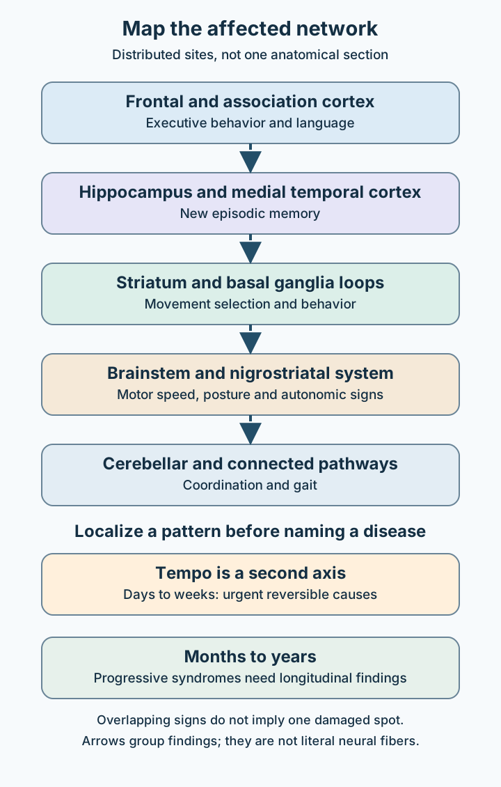
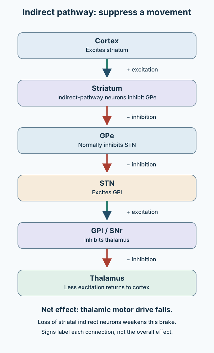
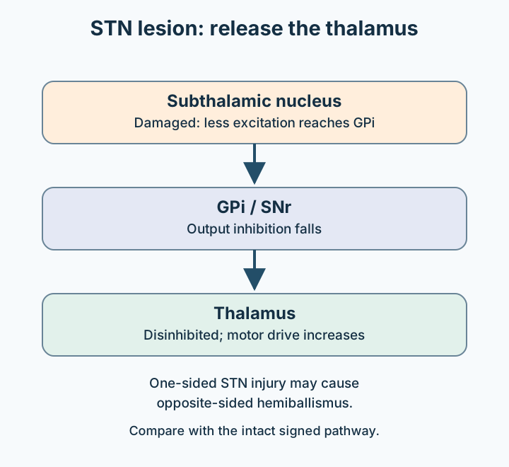
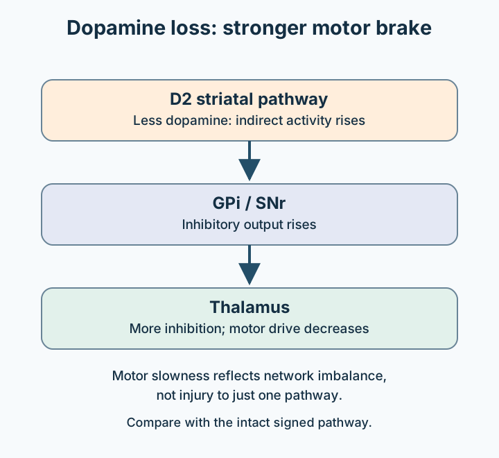
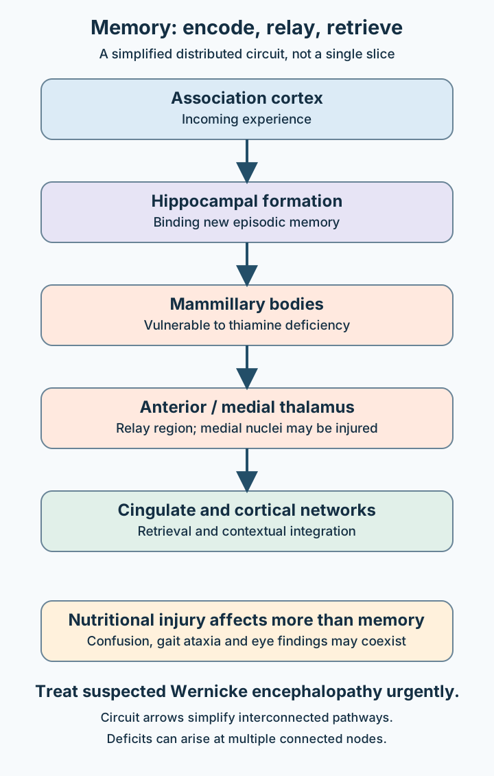
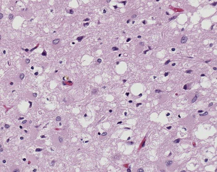
,_H%26E.jpg)