Separate diagnostic thresholds from severe features, treat pressure and seizure risks, and use maternal-fetal stability to guide delivery and postpartum care.
Two patients can have the same blood pressure and very different immediate risks. The central question is not simply, "Is there protein in the urine?" It is: Does this patient have a pregnancy-associated hypertensive disorder, which organs are affected, and what cannot safely wait?
Follow the pressure, then the organs, then the timing. By the end, you should be able to distinguish a diagnostic threshold from a severe feature, identify disease without proteinuria, pair the right treatment with the right threat, and explain when delivery takes priority over additional pregnancy time. These are educational examples using the ACOG diagnostic framework, not a patient-specific treatment calculator.
Why can one placental disorder affect several organs?
A patient at 31 weeks develops hypertension, visual disturbances, and reduced urine output. An isolated kidney explanation is too narrow. Preeclampsia involves maternal vascular dysfunction associated with placental disease. Impaired placental vascular development and stress can contribute to circulating signals that affect the maternal endothelium, the inner lining of blood vessels. The resulting problem is distributed, not confined to the urine. [1]
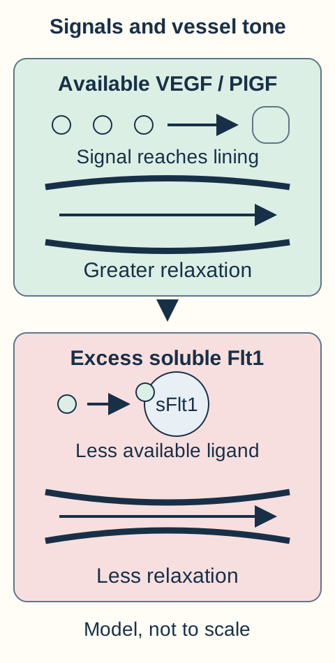
One studied contributor is soluble fms-like tyrosine kinase 1, abbreviated sFlt1. It binds vascular endothelial growth factor, VEGF, and placental growth factor, PlGF. Less available signaling can impair endothelial function. Experimental work linked excess sFlt1 to reduced microvascular relaxation, hypertension, proteinuria, and glomerular endothelial swelling. This is a useful explanatory model, not proof that one molecule accounts for every clinical presentation. [4]
Trace the accompanying vessel diagram from available growth-factor signal to the vessel lining. Increased vascular resistance can raise pressure. Glomerular endothelial injury can impair filtration and permit protein loss. Increased vascular permeability contributes to tissue fluid accumulation. Placental dysfunction can also limit fetal growth. These processes need not become abnormal at the same moment; a normal urine result does not report what is happening in the brain or platelet circulation. [1,4]
Trace the available ligand, not simply the total growth-factor amount. Binding by soluble Flt1 can reduce endothelial signaling and relaxation. This experimental relationship is one contributor to a complex clinical disorder, not a complete cause or a treatment recommendation. [1,4]
Predict before reading the answer: At a fixed high pressure, would a larger urine protein value tell you that the lungs are filling with fluid? Name the additional evidence you would need.
Compare the kidney measurement with the lung question
No. Urine protein describes a renal finding. New breathlessness, oxygenation, examination and appropriate imaging assess possible pulmonary edema. The supplied pressure and protein cannot substitute for respiratory assessment.
Transfer the model: when a patient has a new visual disturbance but little proteinuria, assess the neurologic risk directly rather than requiring the kidney to become abnormal first. Endothelial disease explains why the diagnostic evaluation surveys several organs. [1]
Which number establishes disease, and which changes urgency?
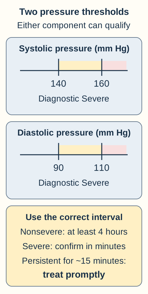
Begin with a reliable pressure measurement: an appropriately sized cuff, supported positioning and the arm at heart level. For previously normal blood pressure, new systolic pressure at least 140 mm Hg or diastolic pressure at least 90 mm Hg after 20 weeks establishes hypertension when confirmed on two occasions at least four hours apart. Both numbers do not have to cross their thresholds. A single nonsevere reading requires reassessment rather than an immediate definitive label. [1,2]
Use a different clock for severe pressure. Systolic pressure at least 160 or diastolic pressure at least 110 mm Hg can be confirmed within minutes. Acute severe pressure persistent for about 15 minutes requires urgent treatment as soon as possible, within 30 to 60 minutes of confirmation. Do not wait four hours, for a urine collection, or for both pressure components to become severe. A systolic pressure of 166 with a diastolic pressure of 96 still qualifies. These emergency rules are not a substitute for a separate chronic-hypertension maintenance plan. [1,2]
First establish reliable readings. A diagnostic threshold is not itself an emergency-drug threshold. Either severe component is sufficient; normal protein and a nonsevere second pressure component do not cancel it. Treat confirmed sustained severe pressure as soon as possible, within 30 to 60 minutes. [1,2]
With confirmed new hypertension, proteinuria establishes one route to preeclampsia: at least 300 mg in a 24-hour collection, or a spot protein-to-creatinine ratio at least 0.30 mg/mg. Dipstick 2+ is a fallback when quantitative testing is unavailable. A ratio is not a urine protein concentration. A normal ratio also does not exclude preeclampsia when qualifying organ dysfunction is present. [1]
Keep the labels distinct. Gestational hypertension is new hypertension after 20 weeks without proteinuria or qualifying organ findings. Preeclampsia without severe features adds proteinuria but no severe feature. New gestational hypertension that reaches severe range is classified as preeclampsia with severe features in the ACOG framework, even with normal protein and other laboratory results. [1]
Chronic hypertension predates pregnancy, appears before 20 weeks, or remains beyond the usual postpartum resolution period, commonly 12 weeks. New proteinuria or organ dysfunction later in pregnancy raises concern for superimposed preeclampsia. Worsening pressure warrants evaluation but is not, by itself, proof of superimposed disease. Compare baseline studies and perform serial assessment when the distinction is uncertain. Treat a severe-pressure emergency while that evaluation proceeds. [5,7]
Change one finding, then trace the diagnostic fork
Start with this openly visible reference patient: 32 weeks, previously normal pressure, 146/94 on two readings four hours apart, protein-to-creatinine ratio 0.36, platelets 180,000/microliter, creatinine 0.7 mg/dL, normal liver studies, no concerning symptoms. This is preeclampsia without severe features. In each independent comparison below, return to these starting values. Predict whether the category, the emergency pressure response, both, or neither changes.
Comparison A: only the protein-to-creatinine ratio becomes 3.6
The diagnosis remains preeclampsia without severe features. More protein does not itself create a severe feature or an indication for emergency intravenous pressure treatment. Continue the appropriate maternal and fetal assessment; do not use protein quantity as a severity scale. [1]
Comparison B: only platelets fall to 75,000/microliter, with no other cause found
The category changes to preeclampsia with severe features because of thrombocytopenia. Inpatient stabilization, seizure prophylaxis and delivery planning are needed. The pressure is still 146/94, so the low platelet count is not an instruction to give an emergency antihypertensive for a severe-pressure threshold that has not been reached. [1]
Comparison C: only systolic pressure becomes 166, persisting for 15 minutes
Both category and immediate pressure action change. Severe systolic hypertension requires prompt antihypertensive treatment, alongside the separate severe-preeclampsia pathway. The diastolic value of 94 and an unchanged protein result do not cancel this emergency. [1,2]
Now apply the same fork to a different patient: new hypertension plus platelets of 85,000/microliter but a ratio of 0.10. Qualifying thrombocytopenia, after considering other causes, provides the diagnostic route and the severe feature. Proteinuria was never a mandatory intermediate step. [1]
Which organ finding makes the disease severe?
Ask what the patient is losing: neurologic function, platelet reserve, filtration, hepatic integrity, or oxygenation. The pressure does not have to be severe when another qualifying severe feature is present. Conversely, dependent ankle edema, protein quantity and brisk reflexes alone do not establish severe preeclampsia. [1]
Platelet circulation
A platelet count below 100,000/microliter is a severe feature when attributable to the disorder. Interpret the trend and alternative explanations; mild isolated thrombocytopenia does not automatically establish HELLP.
Kidney filtration
Creatinine above 1.1 mg/dL or twice the patient’s baseline qualifies in the absence of another renal cause. From 0.5 to 1.0 mg/dL is a doubling even though the final value is below 1.1. Urine protein and filtration are different measurements.
Liver
Transaminases at least twice the laboratory upper limit support hepatic involvement when not explained by another disorder. Severe persistent right-upper-quadrant or epigastric pain unresponsive to medication and without another explanation is also a severe feature; do not require a dramatic enzyme rise before evaluating concerning pain.
Lungs
Pulmonary edema is a severe feature. Breathlessness, hypoxemia and crackles require prompt cardiopulmonary evaluation, not automatic attribution to normal pregnancy.
Brain and vision
A new headache unresponsive to medication and not explained by another diagnosis, or visual disturbance, is concerning for severe disease. Headache is nonspecific: assess its pattern, persistence and alternative causes. Focal deficits, thunderclap onset or altered consciousness demand urgent neurologic investigation as well as obstetric stabilization. [1]
HELLP describes hemolysis, high liver enzymes and low platelets. Look for a pattern rather than a single abnormal value: schistocytes, increased indirect bilirubin or low haptoglobin support hemolysis; LDH at least 600 U/L is commonly used with the other findings. Substantial transaminase abnormalities and platelets below 100,000/microliter complete the pattern. Hypertension or proteinuria may be absent, so right-upper-quadrant pain, nausea and malaise with this laboratory pattern need urgent assessment. Other thrombotic microangiopathies and liver diseases remain in the differential. [1]
Complications connect to these organs: eclampsia is a new seizure in the relevant pregnancy or postpartum context not explained by another cause; stroke and vasogenic cerebral edema associated with posterior reversible encephalopathy syndrome are neurologic threats. Hepatic hematoma or rupture can cause pain and circulatory deterioration. Platelet consumption and hemolysis may accompany disseminated intravascular coagulation. Placental insufficiency can produce fetal growth restriction; abruption and abnormal fetal testing can make continued pregnancy unsafe. None of these is excluded by a reassuring urine strip. [1]
Compare two patterns: Patient A has platelets 118,000/microliter, normal AST, normal LDH and no hemolysis. Patient B has platelets 68,000/microliter, AST 220 U/L with an upper limit of 35, LDH 850 U/L and schistocytes. Which pattern supports HELLP, and which part of A’s evaluation must continue?
Explain the difference between an isolated count and a syndrome
B combines hemolysis, hepatic injury and thrombocytopenia. A does not establish HELLP from the platelet count alone. Continue assessing blood pressure, symptoms, platelet trend and other causes rather than assuming that a modestly low count either proves HELLP or guarantees benign disease. [1]
Transfer: a patient with severe persistent epigastric pain needs evaluation even before the next laboratory panel. Trends and symptoms can change the assessment between scheduled blood draws. [1]
Which treatment addresses pressure, and which addresses seizures?
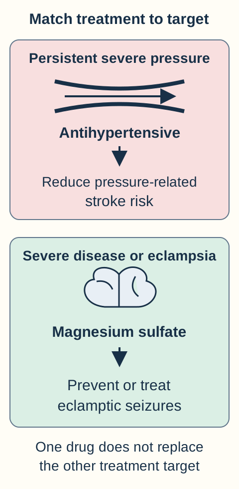
Consider a patient with persistent pressure of 172/112 mm Hg and visual scotomata. There are two immediate treatment targets, not one interchangeable drug choice. Severe pressure requires urgent antihypertensive therapy to reduce maternal cerebrovascular risk. Magnesium sulfate prevents or treats eclamptic seizures; it is not the drug used to resolve the severe-pressure emergency. Maternal stabilization includes airway and breathing assessment, monitoring and obstetric help while fetal assessment proceeds when appropriate. [1,2]
First-line severe-pressure choices include intravenous labetalol, intravenous hydralazine and immediate-release oral nifedipine, with selection and repeat dosing guided by contraindications and a monitored obstetric protocol. Avoid labetalol in patients with asthma, significant bradycardia or heart block. When intravenous access is delayed and the patient can swallow, immediate-release oral nifedipine is an option; do not give it sublingually. ACE inhibitors are not the acute pregnancy treatment in this pathway. Do not lower pressure precipitously toward a normal nonpregnant value. [2,7]
Magnesium is recommended for preeclampsia with severe features and for eclampsia. For disease without severe features, prophylaxis is individualized rather than triggered by a protein ratio of 0.30 alone. A commonly used intravenous regimen is a 4 to 6 g loading dose over 20 to 30 minutes followed by 1 to 2 g/hour; this requires a clinical protocol, appropriate dilution, monitoring and renal assessment, not self-administration. It is commonly continued through delivery and for 24 hours afterward, with the ongoing plan adjusted to the clinical situation. [1]
Magnesium leaves through the kidneys. Reduced urine output or impaired renal function can produce accumulation during an unchanged infusion. Monitor respiratory rate, oxygenation, tendon reflexes and urine output; obtain levels and adjust treatment when renal dysfunction or toxicity is a concern. Myasthenia gravis and significant renal failure require specialist consideration of contraindications and alternatives, not automatic use of a routine infusion. [1,3]
Loss of reflexes plus slow breathing is not reassurance from a quiet nervous system. Stop the magnesium infusion, support ventilation and give intravenous calcium gluconate for clinically important toxicity under emergency supervision. Do not delay respiratory support while waiting for the magnesium level. Advanced toxicity can also impair cardiac conduction. Flushing alone with normal respiration, reflexes and urine output is a different pattern from progressive neuromuscular and respiratory depression. [1,3]
Use the two treatment paths in the diagram. After magnesium is started, the pressure remains 170/114 and the patient is alert with normal reflexes. Decide which path still needs treatment.
Follow each path to the complication it addresses. Blood-pressure rescue and magnesium serve different purposes. Drug selection, contraindications, renal function and respiratory monitoring remain part of the clinical protocol; the diagram is not a dosing tool. [1,2,3]Check the target that remains untreated
The severe-pressure target remains. Give a first-line antihypertensive according to the emergency protocol while continuing the indicated seizure prophylaxis. Increasing magnesium solely to lower blood pressure confuses the two treatment purposes. [2]
Transfer: if the pressure instead improves but urine output falls and reflexes disappear, investigate magnesium accumulation. A successful pressure response does not prove that every medication remains safe at its current rate. [1,3]
When does more pregnancy time become less safe?
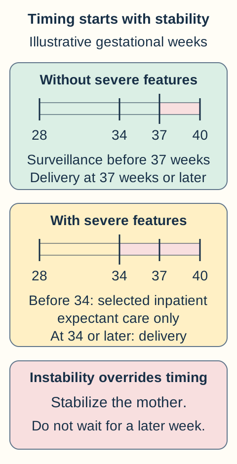
Use the accompanying timeline only after assessing maternal and fetal stability. Delivery ends the ongoing placental contribution, but it is not an instantaneous reversal of maternal organ injury. Stabilize the mother before delivery whenever possible, even when delivery is urgent. Route of birth follows obstetric circumstances, not the diagnosis alone. Preeclampsia or a resolved eclamptic seizure is not an automatic instruction for cesarean delivery. [1,2]
Read maternal and fetal stability before following either timeline. Severe-disease expectant care before 34 weeks requires appropriate inpatient resources and repeated reassessment. An urgently indicated birth is not delayed for a gestational milestone or completion of corticosteroids. [1]
Without severe features: at 37 weeks or later, delivery is recommended rather than continued expectant management. Before 37 weeks, surveillance can continue when maternal and fetal status are reassuring and reliable follow-up is feasible. Outpatient care is reserved for appropriate patients without severe features; uncertain access to monitoring can justify admission. [1]
With severe features: at 34 weeks or later, delivery after maternal stabilization is recommended. Do not postpone an indicated late-preterm delivery just to complete corticosteroids. Before 34 weeks, carefully selected patients with stable maternal and fetal status may be offered inpatient expectant management in a center with appropriate obstetric, neonatal and critical-care resources. This is a monitored risk-benefit decision, not a promise to reach a particular date. [1]
Expectant care requires repeated pressure and symptom assessment, serial platelet, renal and hepatic studies, and fetal evaluation. Uncontrolled severe pressure despite treatment, persistent neurologic symptoms, eclampsia, HELLP, pulmonary edema, worsening renal injury, abruption, DIC or abnormal fetal testing can end the expectant pathway regardless of gestational age. Give fetal-maturation corticosteroids when appropriate, but do not make their completion a prerequisite for urgently indicated birth. [1]
Fetal growth restriction signals placental risk, but a small estimated weight alone does not dictate the same action as persistent reversed umbilical artery end-diastolic flow or abnormal fetal testing. With otherwise reassuring fluid, Doppler and surveillance findings, growth restriction alone need not end a carefully selected expectant pathway. Reassuring fetal tests also do not excuse maternal deterioration. [1]
Compare two moments in one admission: At 29 weeks, pressure is controlled, symptoms have resolved, laboratory trends are stable and fetal testing is reassuring. Later, oxygen saturation falls with new pulmonary edema. Before opening the answer, identify what changed: the gestational age rule or eligibility to continue waiting.
Explain why the same gestational age now leads to a different plan
Maternal instability ends eligibility for expectant care. Stabilize breathing, circulation and the obstetric emergency, then proceed with delivery planning despite prematurity. The earlier stable assessment was conditional, not permission to ignore a new severe complication. [1]
Transfer: a 35-week patient with controlled pressure after medication and severe features still has reached the delivery threshold. Successful stabilization makes delivery safer; it does not erase the indication. [1]
What must continue after the placenta is delivered?
A patient returns six days after birth with a new persistent headache and severe pressure. A normal antepartum course does not exclude postpartum preeclampsia. It can first appear after delivery, including up to six weeks later. Severe headache, visual change, dyspnea, seizure, chest symptoms or severe pressure warrants urgent assessment; do not wait for the routine postpartum visit. Severe hypertension is treated promptly in the postpartum period too. [2,5]
Plan follow-up before discharge. ACOG recommends blood-pressure evaluation within 72 hours after birth for patients with severe hypertension, and no later than 7 to 10 days postpartum for hypertensive disorders generally. A scheduled review is not a reason to defer care for warning symptoms. An individualized monitoring and medication plan should specify whom to contact and when to seek emergency care. [6]
Continue organ and medication assessment as indicated. Clinical improvement after delivery does not justify overlooking falling platelets, renal dysfunction or respiratory changes. The pregnancy history also matters later: hypertensive pregnancy disorders are associated with greater future cardiovascular risk. Document the episode and arrange ongoing primary care, cardiovascular risk assessment and counseling before another pregnancy. [1,6]
Explain to a recovering patient why a normal pressure today does not cancel the early blood-pressure check. Give one reason related to the next few days and one related to future health.
Compare immediate recovery with longer-term follow-up
Pressure and symptoms can worsen again during early postpartum recovery, so early reassessment and clear warning instructions still matter. Beyond recovery, this history identifies a need for continued cardiovascular prevention and follow-up rather than being erased from the medical record. [5,6]
The complete decision is: establish the hypertensive context, identify the organ finding, treat the immediate threat, and reassess whether continued pregnancy or postpartum observation remains appropriate.
Apply the distinctions to new patients
Choose a single best answer from the supplied information. Three cases appear at related points in the lesson; the remaining cases provide mixed practice. Explanations address every option, and the clinical reasoning remains available without a timed sequence.
Case 1
Show answer and explanations for case 1
A. Intravenous hydralazine bolus (Why this does not fit)
Hydralazine treats an acute severe-pressure emergency. Both confirmed pressures here are below 160 systolic and 110 diastolic. A severe platelet feature supports magnesium and inpatient care, not automatic emergency pressure lowering.
Reasoning steps for option A
What acute complication would intravenous hydralazine treat?
Hydralazine treats sustained severe hypertension, not the seizure risk established by thrombocytopenia.
Do 152/96 and 150/94 mm Hg reach either severe-pressure threshold?
Neither reading reaches 160 mm Hg systolic or 110 mm Hg diastolic.
Why does a platelet count of 78,000 not automatically call for hydralazine?
Thrombocytopenia establishes a severe feature requiring the severe-preeclampsia pathway, but emergency pressure lowering requires severe blood pressure.
B. Oral prednisone course (Why this does not fit)
Prednisone can treat selected immune-mediated platelet disorders. The new hypertension and pregnancy-associated platelet decline have no alternative cause on evaluation. Do not replace the severe-preeclampsia pathway with treatment for an unsupported isolated immune disorder.
Reasoning steps for option B
Which platelet disorder could make prednisone an appropriate treatment?
Selected immune-mediated platelet disorders may respond to prednisone.
Does the evaluation support isolated immune thrombocytopenia in this newly hypertensive patient?
No alternative explanation was found for the new platelet fall accompanying hypertension at 32 weeks.
Why should prednisone not replace severe-preeclampsia care for this platelet decline?
New hypertension with unexplained platelets of 78,000 supports severe preeclampsia rather than an unsupported isolated immune disorder.
C. Magnesium sulfate infusion (Best answer)
New hypertension with platelets below 100,000/microliter establishes a severe feature in this context. The ratio of 0.11 does not exclude this nonproteinuric presentation. Severe preeclampsia warrants seizure prophylaxis even when pressure is below the emergency antihypertensive threshold.
Reasoning steps for option C
Which severe-feature threshold is crossed by platelets of 78,000/microliter?
The count is below 100,000/microliter, a severe feature in this newly hypertensive pregnancy without another cause.
Does a protein-to-creatinine ratio of 0.11 exclude this platelet-defined presentation?
No. A qualifying organ feature can establish preeclampsia without diagnostic proteinuria.
Why is magnesium indicated even with pressures below 160/110?
Severe preeclampsia warrants seizure prophylaxis; magnesium addresses that risk independently of the emergency antihypertensive threshold.
D. Intravenous calcium gluconate (Why this does not fit)
Calcium can antagonize dangerous magnesium effects. There is no magnesium exposure, absent reflexes or respiratory depression in this presentation. Calcium rescue treats toxicity; it is not seizure prophylaxis.
Reasoning steps for option D
What magnesium-related emergency is treated with calcium gluconate?
Calcium gluconate antagonizes clinically important magnesium toxicity.
Which missing exposure and clinical signs argue against calcium rescue here?
There is no magnesium exposure, loss of reflexes or respiratory depression to suggest toxicity.
Can calcium gluconate prevent seizures from the platelet-defined severe disease?
No. Calcium is toxicity rescue, not a substitute for magnesium-based seizure prophylaxis.
E. Oral nifedipine rescue dose (Why this does not fit)
Immediate-release nifedipine is a first-line option for sustained severe hypertension. The severe feature here is thrombocytopenia, not a pressure at or above 160/110. Identify which immediate threat the medication actually treats.
Reasoning steps for option E
What pressure pattern would justify an immediate-release nifedipine rescue dose?
Sustained severe hypertension can be treated with immediate-release oral nifedipine under an emergency protocol.
Is the qualifying severe feature here a pressure elevation or thrombocytopenia?
It is thrombocytopenia at 78,000/microliter; both confirmed pressures remain below severe range.
Why does oral nifedipine rescue miss the complication risk identified in the stem?
It lowers severe pressure rather than providing the seizure prophylaxis warranted by this severe platelet feature.
Takeaway: Severe preeclampsia warrants seizure prophylaxis even when pressure is below the emergency antihypertensive threshold.
Hypertension plus a ratio above 0.30 establishes preeclampsia without supplied severe features. At 38 weeks the pregnancy is beyond the recommended 37-week delivery threshold. Term preeclampsia without severe features favors delivery; the diagnosis alone does not require cesarean birth.
Reasoning steps for option A
What diagnosis follows from new hypertension and a protein ratio of 0.44 with normal organ studies?
The ratio exceeds 0.30 and accompanies confirmed new hypertension, establishing preeclampsia without supplied severe features.
How does 38 weeks compare with the delivery threshold for nonsevere preeclampsia?
She is beyond the recommended 37-week delivery threshold.
Why is induction appropriate rather than automatic cesarean delivery?
Delivery is indicated, but the cephalic fetus and absence of a labor contraindication permit induction; preeclampsia alone does not require cesarean birth.
B. Continue surveillance until 40 weeks (Why this does not fit)
Surveillance can extend pregnancy before the delivery threshold in stable nonsevere disease. This patient is already at 38 weeks. Reassuring testing does not negate the term delivery recommendation.
Reasoning steps for option B
When can surveillance extend a pregnancy with nonsevere preeclampsia?
Selected stable patients may be monitored before the recommended delivery threshold.
Does this 38-week pregnancy remain before that surveillance endpoint?
No. She has already passed 37 weeks.
Can reassuring fetal testing justify waiting until 40 weeks here?
No. Reassuring testing does not remove the term delivery indication from established preeclampsia.
C. Obtain serial 24-hour urine collections (Why this does not fit)
Quantitative urine testing helps establish proteinuria when it is uncertain. The ratio of 0.44 already supplies that evidence. Repeated protein quantification should not delay an established delivery indication.
Reasoning steps for option C
What diagnostic uncertainty could quantitative urine collection resolve?
Urine testing can determine whether diagnostic proteinuria is present when that is uncertain.
What does the existing protein-to-creatinine ratio of 0.44 already establish?
It already meets the proteinuria criterion, so another collection is not needed to establish that finding.
Why should serial 24-hour collections not postpone induction at 38 weeks?
The diagnosis and gestational age already support delivery; repeated protein measurement would not remove that indication.
D. Give emergency intravenous labetalol (Why this does not fit)
Labetalol can rapidly treat severe pressure. Pressures of 146/92 and 148/94 are diagnostic but not severe range. A diagnostic pressure threshold is not an emergency-drug threshold.
Reasoning steps for option D
What blood-pressure emergency is intravenous labetalol designed to treat?
Intravenous labetalol can rapidly treat sustained severe hypertension.
Do 146/92 and 148/94 mm Hg meet the emergency treatment threshold?
No. They establish hypertension but remain below 160 systolic and 110 diastolic.
Why does meeting a diagnostic pressure threshold not justify emergency labetalol?
The threshold establishing hypertension is lower than the severe-pressure threshold used for urgent rescue treatment.
E. Schedule prelabor cesarean birth today (Why this does not fit)
Cesarean delivery is appropriate for an obstetric indication. The fetus is cephalic and labor has no contraindication in the supplied circumstances. Determine the need for delivery separately from the route of birth.
Reasoning steps for option E
What would justify choosing prelabor cesarean rather than induction?
An obstetric indication or a contraindication to labor would support cesarean delivery.
Do fetal presentation and labor eligibility supply such an indication here?
No. The fetus is cephalic and there is no contraindication to labor.
Does the 38-week preeclampsia delivery indication also determine the route of birth?
No. It establishes the need for delivery, while route is decided separately from the obstetric circumstances.
Takeaway: Term preeclampsia without severe features favors delivery; the diagnosis alone does not require cesarean birth.
A. Repeat the pressure four hours after the initial value (Why this does not fit)
A four-hour interval helps confirm nonsevere pregnancy hypertension. Severe systolic values have already persisted for 15 minutes. Do not apply the nonsevere confirmation interval to an acute severe-pressure emergency.
Reasoning steps for option A
For which pressure category is four-hour diagnostic confirmation commonly used?
Nonsevere pregnancy hypertension is commonly confirmed with readings four hours apart.
What changes the confirmation interval when systolic values remain 166 to 170 for 15 minutes?
Persistent systolic pressure at or above 160 is severe and can be confirmed over minutes for urgent treatment.
Why would waiting another four hours be unsafe despite the absence of symptoms?
The sustained severe systolic pressure already establishes an emergency requiring prompt antihypertensive therapy.
B. Collect a 24-hour urine before selecting pressure therapy (Why this does not fit)
Protein measurement can help classify nonsevere pregnancy hypertension. Pressure urgency here is established independently of proteinuria. Diagnostic urine testing must not delay treatment of sustained severe pressure.
Reasoning steps for option B
What classification question can urine protein testing help answer in nonsevere hypertension?
It can distinguish proteinuric preeclampsia from hypertension without diagnostic proteinuria.
Does a ratio of 0.08 change the urgency of systolic readings near 170?
No. Sustained severe systolic pressure requires treatment independently of proteinuria.
Should a 24-hour urine result precede selection of an emergency pressure drug?
No. Collecting urine must not delay treatment of the established severe-pressure emergency.
C. Start magnesium and reassess pressure after its loading dose (Why this does not fit)
Magnesium is appropriate seizure prophylaxis in the severe-preeclampsia pathway. It does not replace an antihypertensive for the persistent systolic emergency. Begin the two indicated treatments for their distinct targets rather than using magnesium as a pressure trial.
Reasoning steps for option C
Which complication does magnesium prophylaxis address in this severe-pressure presentation?
Magnesium addresses the seizure risk associated with the severe-preeclampsia pathway.
Can a magnesium loading dose substitute for an antihypertensive at 166 to 170 systolic?
No. Magnesium is not a replacement for urgent treatment of the persistent severe systolic pressure.
How should pressure treatment be timed relative to magnesium prophylaxis?
Treat the pressure promptly while seizure prophylaxis proceeds rather than waiting to test a pressure response to magnesium.
D. Arrange outpatient pressure monitoring with an oral maintenance plan (Why this does not fit)
Outpatient follow-up can serve selected stable nonsevere patients. Repeated systolic values near 170 require monitored urgent treatment now. Absence of symptoms does not make sustained severe hypertension suitable for outpatient observation.
Reasoning steps for option D
What disease severity can sometimes be managed with outpatient pressure follow-up?
Selected stable patients with nonsevere hypertension may be suitable for outpatient monitoring.
Why do repeated systolic values near 170 exclude that plan now?
The values are persistently severe and require monitored urgent treatment.
Do normal organ studies and no symptoms make severe systolic hypertension safe for discharge?
No. Those reassuring findings do not cancel the acute risk from sustained severe pressure.
E. Administer intravenous labetalol under the emergency protocol (Best answer)
Sustained systolic pressure at or above 160 mm Hg is an obstetric hypertensive emergency. Normal urine and nonsevere diastolic readings do not cancel the persistent severe systolic values. Treat severe pressure promptly while the complete severe-preeclampsia assessment and seizure-prophylaxis plan proceed.
Reasoning steps for option E
Which emergency threshold is crossed despite diastolic values below 100?
Systolic values of 166 to 170 exceed the 160 mm Hg severe threshold and have persisted for 15 minutes.
Can normal urine protein and nonsevere diastolic pressure negate the systolic emergency?
No. Either severe pressure component is sufficient; diagnostic proteinuria is not needed before treatment.
Why is intravenous labetalol an appropriate immediate choice with the supplied access and history?
Intravenous access is available and no labetalol contraindication is supplied, so it can promptly treat severe pressure while assessment and seizure prophylaxis proceed.
Takeaway: Treat severe pressure promptly while the complete severe-preeclampsia assessment and seizure-prophylaxis plan proceed.
A. The original nonsevere value requires another four hours of observation (Why this does not fit)
The first reading alone would have required nonsevere confirmation. Later persistent severe diastolic readings supersede that observation schedule. Management responds to the current severe physiology, not only the first measurement.
Reasoning steps for option A
What confirmation schedule would apply if the only finding were 146/94 mm Hg?
That isolated nonsevere reading would ordinarily need further diagnostic confirmation over the nonsevere interval.
How do subsequent diastolic readings of 112 to 114 over 15 minutes alter that schedule?
They establish persistent severe diastolic hypertension, superseding the four-hour observation plan.
Should treatment follow the original 146/94 reading or the newer severe diastolic pattern?
Management must respond to the current severe physiology and begin urgent pressure treatment.
B. The sustained diastolic rise requires urgent antihypertensive treatment now (Best answer)
A diastolic value at or above 110 mm Hg independently reaches severe range. This severe component has persisted despite systolic values below 160. Either severe pressure component is sufficient for the emergency pathway.
Reasoning steps for option B
What diastolic cutoff independently defines severe pregnancy hypertension?
A diastolic pressure at or above 110 mm Hg reaches severe range.
Do systolic readings of 152 to 154 prevent the repeated 112 to 114 diastolic values from being severe?
No. The diastolic component is persistently severe even though systolic pressure is below 160.
What immediate treatment follows this sustained isolated diastolic rise?
Urgent antihypertensive treatment is required because either severe component can establish the emergency.
C. The normal protein ratio supports discharge with close reassessment (Why this does not fit)
A normal ratio can occur in gestational hypertension without organ findings. It does not make persistent diastolic pressure above 110 safe. Proteinuria is not required before treating severe hypertension.
Reasoning steps for option C
Can gestational hypertension without organ injury have a protein ratio of 0.12?
Yes. A normal protein ratio can occur without diagnostic proteinuria.
Does that normal ratio make persistent diastolic pressure above 110 safe for discharge?
No. It does not reduce the immediate risk from the sustained severe-pressure readings.
Is proteinuria required before treating this diastolic emergency?
No. Severe hypertension requires prompt treatment regardless of the protein ratio.
D. The systolic value supports oral maintenance adjustment without rescue (Why this does not fit)
Maintenance adjustment addresses ongoing pressure control. The diastolic values already meet an acute severe threshold. Do not let a nonsevere systolic value conceal a severe diastolic emergency.
Reasoning steps for option D
What goal does adjustment of oral maintenance pressure therapy serve?
Maintenance therapy supports ongoing blood-pressure control rather than replacing acute rescue.
Which component of 154/112 and 152/114 demands more than maintenance adjustment?
The diastolic values are at or above 110 mm Hg and have remained severe over 15 minutes.
Why is using only the systolic value to choose maintenance care misleading?
A systolic value below 160 does not cancel a severe diastolic emergency requiring urgent treatment.
E. The normal platelet count supports magnesium as the sole acute drug (Why this does not fit)
Normal platelets do not exclude severe-pressure disease. Magnesium cannot substitute for pressure treatment in these repeated readings. Seizure prevention and urgent antihypertensive therapy have separate purposes.
Reasoning steps for option E
Do platelets of 192,000 exclude severe disease driven by blood pressure?
No. Normal platelets do not negate the persistently severe diastolic pressure.
Would magnesium alone resolve the repeated diastolic readings above 110?
No. Magnesium addresses seizure risk and cannot substitute for antihypertensive treatment.
Why must the acute plan distinguish magnesium from a pressure rescue drug?
Seizure prophylaxis and urgent pressure lowering treat separate risks; the severe diastolic pressure needs its own therapy.
Takeaway: Either severe pressure component is sufficient for the emergency pathway.
A. Gestational hypertension without renal involvement (Why this does not fit)
New hypertension with normal urine protein can resemble uncomplicated gestational hypertension. The creatinine has more than doubled from 0.48 to 1.02. Normal protein excretion does not exclude loss of filtration.
Reasoning steps for option A
Why might a protein ratio of 0.09 initially suggest gestational hypertension?
New hypertension without proteinuria can fit gestational hypertension if no qualifying organ involvement is present.
How does creatinine of 1.02 compare with the baseline of 0.48 mg/dL?
It is more than twice baseline, demonstrating a qualifying severe renal change when another cause is excluded.
Why does absent proteinuria not justify the label 'without renal involvement'?
Urine protein and filtration are different findings; the marked creatinine rise shows renal involvement despite a normal ratio.
B. Preeclampsia without severe features (Why this does not fit)
Nonsevere pressure can accompany preeclampsia without severe features. The relative creatinine increase is itself a qualifying severe feature. Severity is not determined by the pressure value alone.
Reasoning steps for option B
Do pressures of 148/94 and 150/96 alone establish severe-pressure disease?
No. They are hypertensive but below severe range.
Which non-pressure finding makes the 'without severe features' classification incorrect?
Creatinine has more than doubled from 0.48 to 1.02 mg/dL without another identified renal cause.
Why must renal findings be assessed separately from blood-pressure magnitude?
A qualifying organ feature can establish severe preeclampsia even when pressure is nonsevere.
C. Chronic hypertension with established kidney disease (Why this does not fit)
Chronic pressure or renal abnormalities would change the baseline interpretation. The record instead shows new hypertension and a low early-pregnancy creatinine without known renal disease. Do not assume chronic disease solely because a renal abnormality is present now.
Reasoning steps for option C
What baseline history would support chronic hypertension with established kidney disease?
Pressure or renal abnormalities predating this presentation would support that interpretation.
What do the early creatinine of 0.48 and absence of known renal disease indicate here?
The record supports a low early-pregnancy creatinine and new hypertension rather than established chronic renal disease.
Can the current creatinine elevation alone establish that the kidney disorder is chronic?
No. The documented rise must be interpreted against the patient's earlier baseline, not assumed to be longstanding.
D. Preeclampsia with severe renal involvement (Best answer)
The new hypertension accompanies a creatinine value more than twice baseline. The relative rise qualifies despite a final value below 1.1 mg/dL and absent proteinuria. Assess severe renal involvement using either the absolute threshold or doubling, after excluding another renal cause.
Reasoning steps for option D
What relative renal threshold is met by the rise from 0.48 to 1.02 mg/dL?
Creatinine has more than doubled, meeting the relative severe renal criterion alongside new hypertension.
Does a final creatinine below 1.1 mg/dL or a ratio of 0.09 negate that doubling?
No. The relative rise qualifies independently of the absolute creatinine threshold and without proteinuria.
How does exclusion of dehydration, obstruction and known renal disease affect classification?
With no alternative renal cause identified, the new hypertension and qualifying creatinine rise support preeclampsia with severe renal involvement.
E. Physiologic gestational change with no hypertensive disorder (Why this does not fit)
A population laboratory interval can make a value near 1.0 appear unremarkable. Repeated hypertension plus more than doubling of creatinine is not explained by that interval. The patient’s baseline and pregnancy context are necessary to interpret a renal result.
Reasoning steps for option E
Why could a general laboratory range obscure the significance of creatinine near 1.0?
A population range may make that value appear unremarkable without considering the lower pregnancy baseline.
Can a normal population range explain confirmed hypertension plus a greater-than-twofold creatinine rise?
No. Those changes cannot be dismissed as normal gestational physiology.
Which comparison prevents mislabeling the 1.02 result as physiologic?
Compare it with her 0.48 mg/dL early-pregnancy value and the concurrent new hypertension, not just the general laboratory interval.
Takeaway: Assess severe renal involvement using either the absolute threshold or doubling, after excluding another renal cause.
A. It establishes severe disease requiring delivery after stabilization (Why this does not fit)
Delivery may be necessary when severe disease is unstable or reaches the relevant gestational threshold. The only new finding here is protein quantity, which is not a severe-feature criterion. Do not convert a protein increase into an unsupported preterm delivery indication.
Reasoning steps for option A
When would severe preeclampsia at 30 weeks require delivery?
Delivery may be required for maternal or fetal instability, rather than a urine value alone.
Does a protein-to-creatinine ratio rising to 3.2 define a severe feature?
No. Protein quantity is not a severe-feature criterion; pressure and organ assessment remain nonsevere.
Why should this urine change not trigger immediate preterm delivery?
No severe pressure, organ injury, symptoms or fetal deterioration accompanies the higher ratio.
B. It does not itself change the severe-feature classification (Best answer)
The original ratio already established proteinuria in a hypertensive pregnancy. The larger ratio is not accompanied by a pressure or organ criterion for severe disease. Protein quantity alone is not a severe feature; continue the indicated maternal and fetal surveillance.
Reasoning steps for option B
What did the original protein-to-creatinine ratio of 0.5 establish?
A ratio above 0.30 already established proteinuria with her new pregnancy hypertension.
Which severe-feature criteria appear after the ratio rises to 3.2?
None are supplied: pressure stays below 160/110, organ studies are normal and symptoms are absent.
How should a larger ratio affect classification and follow-up?
It does not independently confer severe features; continue appropriate maternal and fetal surveillance.
C. It establishes a pressure emergency requiring intravenous rescue (Why this does not fit)
Severe pressure requires prompt rescue therapy. The current pressures remain below both emergency thresholds. Urine protein magnitude cannot substitute for the measured pressure.
Reasoning steps for option C
What blood-pressure values call for urgent antihypertensive rescue?
Sustained pressure at or above 160 systolic or 110 diastolic requires prompt treatment.
Do readings of 146 to 150/92 to 96 meet those thresholds?
No. Both systolic and diastolic readings remain below the severe-pressure thresholds.
Can a urine ratio of 3.2 substitute for a severe blood-pressure reading?
No. Treat a pressure emergency according to measured pressure, not urine protein magnitude.
D. It excludes preeclampsia in favor of an isolated renal syndrome (Why this does not fit)
Substantial proteinuria can occur in renal disease outside preeclampsia. This patient already has new pregnancy hypertension and established preeclampsia. A larger amount of protein does not, by itself, overturn the established clinical diagnosis.
Reasoning steps for option D
When could substantial proteinuria suggest another renal disorder?
Protein loss can also occur with primary renal disease, depending on the broader clinical history.
What evidence still supports preeclampsia in this 30-week patient?
She already has new pregnancy hypertension and diagnostic proteinuria, without evidence that another syndrome replaces them.
Does increasing proteinuria alone overturn the established diagnosis?
No. A higher ratio does not by itself exclude preeclampsia or establish an isolated renal syndrome.
E. It requires magnesium because protein loss predicts an imminent seizure (Why this does not fit)
Magnesium reduces seizure risk in the appropriate severe-preeclampsia setting. No qualifying severe feature has appeared in the supplied assessment. Magnesium decisions in nonsevere disease are individualized rather than determined by a protein cutoff above the diagnostic threshold.
Reasoning steps for option E
What risk does magnesium sulfate prevent in severe preeclampsia?
It reduces eclamptic seizure risk when severe disease warrants prophylaxis.
Which supplied finding makes this 3.2 ratio a magnesium indication?
None. The ratio is not a severe feature, and pressure, symptoms and organ studies do not show one.
How are magnesium decisions made in disease without severe features?
They are individualized, not triggered automatically by protein exceeding a second numeric cutoff.
Takeaway: Protein quantity alone is not a severe feature; continue the indicated maternal and fetal surveillance.
A. Gestational hypertension without proteinuria (Why this does not fit)
Gestational hypertension would fit confirmed new pressure without proteinuria or organ findings. The calculated ratio is 0.30, which meets rather than falls below the threshold. A cutoff written as at least 0.30 includes exactly 0.30.
Reasoning steps for option A
When would gestational hypertension without proteinuria fit?
New pressure elevation after 20 weeks without qualifying proteinuria or organ injury could fit.
What ratio results from urine protein 45 mg/dL and creatinine 150 mg/dL?
The protein-to-creatinine ratio is 45/150, or exactly 0.30 mg/mg.
Why does exactly 0.30 exclude the no-proteinuria classification?
The diagnostic threshold is at least 0.30, so equality meets it.
B. Preeclampsia with severe features (Why this does not fit)
Severe disease requires severe pressure or a qualifying organ feature. A diagnostic ratio of 0.30 does not supply such a feature, and the organ assessment is normal. The proteinuria threshold establishes diagnosis, not severity.
Reasoning steps for option B
What distinguishes severe features from diagnostic proteinuria?
Severe pressure or qualifying organ dysfunction, not proteinuria alone, defines severe features.
Do a ratio of 0.30 and the supplied normal studies establish severity?
No. Pressures are below 160/110 and platelets, kidney function and AST are normal.
What diagnosis does the 0.30 ratio establish without severe findings?
It supports preeclampsia without severe features alongside confirmed new hypertension.
C. Chronic hypertension without superimposed disease (Why this does not fit)
Chronic hypertension depends on an earlier or persistent hypertension history. The patient was normotensive before these readings at 29 weeks. Use the pregnancy timeline before applying a chronic-hypertension label.
Reasoning steps for option C
When does hypertension qualify as chronic in pregnancy?
It is present before pregnancy or early in pregnancy, rather than first appearing at 29 weeks.
What does the previously normal pressure history imply here?
Her confirmed 29-week elevation is new, not evidence of a preexisting chronic disorder.
Why is a chronic-hypertension label unsupported?
No earlier hypertension history is supplied, and the timeline places onset after 20 weeks.
D. Isolated gestational proteinuria without hypertension (Why this does not fit)
Isolated proteinuria describes protein loss without established hypertension. Two readings four hours apart exceed the nonsevere pressure threshold. Interpret the urine result together with the confirmed pressure pattern.
Reasoning steps for option D
What is required for isolated gestational proteinuria?
Proteinuria occurs without established hypertension.
Do readings of 144/92 and 146/94 four hours apart establish hypertension?
Yes. Both exceed 140/90 and are separated by the diagnostic interval.
How should the urine ratio be interpreted with those pressure readings?
The ratio of 0.30 joins confirmed new hypertension to support preeclampsia, not isolated proteinuria.
E. Preeclampsia without severe features (Best answer)
Dividing 45 by 150 gives a protein-to-creatinine ratio of 0.30 mg/mg. That ratio meets the proteinuria threshold alongside confirmed new hypertension, without a supplied severe feature. Use the ratio in matching units, then assess severity separately.
Reasoning steps for option E
How is the urine protein-to-creatinine ratio calculated from 45 and 150 mg/dL?
Dividing 45 by 150 gives 0.30 mg/mg.
What do that ratio and two elevated pressures establish?
They establish new hypertension with proteinuria at the diagnostic cutoff, without supplied severe features.
Why must severity be assessed separately from this positive ratio?
A ratio of 0.30 establishes proteinuria, whereas severe pressure or organ findings determine severe classification.
Takeaway: Use the ratio in matching units, then assess severity separately.
A. Manage superimposed severe preeclampsia and prepare for delivery (Best answer)
Baseline proteinuria does not account for new marked thrombocytopenia and hepatic injury. These organ findings establish severe superimposed disease at 35 weeks. Beyond 34 weeks, superimposed severe features favor delivery after maternal stabilization.
Reasoning steps for option A
Why does baseline proteinuria of 0.8 not explain the new presentation?
A small shift to 0.9 does not explain platelets falling to 72,000 and AST rising to 190.
Which new findings identify severe superimposed disease?
Marked thrombocytopenia and hepatic injury in chronic hypertension, without another cause, establish severe features.
How does gestational age 35 weeks affect the plan?
After maternal stabilization, delivery is favored for superimposed preeclampsia with severe features beyond 34 weeks.
B. Adjust maintenance pressure therapy and continue routine surveillance (Why this does not fit)
Chronic hypertension often requires medication adjustment. A maintenance-only plan does not address the new platelet and liver injury. Separate chronic pressure control from a newly superimposed multisystem disorder.
Reasoning steps for option B
What could a maintenance-medication adjustment address?
It can address ongoing pressure control in chronic hypertension.
Why is that insufficient with platelets 72,000 and AST 190?
Those new abnormalities signal severe multisystem injury, not an isolated need to adjust pressure medication.
What separate condition must be managed alongside chronic hypertension?
Superimposed preeclampsia with severe features requires stabilization and delivery planning.
C. Wait for a substantial protein increase before planning delivery (Why this does not fit)
A changing urine result can help when baseline chronic renal findings complicate diagnosis. New severe platelet and hepatic findings already provide evidence here. Superimposed disease need not announce itself through a further protein rise.
Reasoning steps for option C
When can a changing urine ratio help assess superimposed disease?
It may clarify changes when baseline proteinuria complicates the diagnosis.
Why is a substantial increase beyond 0.8 unnecessary here?
New severe platelet and liver abnormalities already support superimposed disease.
Can a nearly unchanged ratio of 0.9 rule out severe superimposed disease?
No. Organ injury can establish it without a further protein rise.
D. Treat isolated gestational thrombocytopenia and repeat studies next week (Why this does not fit)
An isolated platelet decrease can occur during pregnancy. The marked count reduction occurs with substantial hepatic injury and hypertension. A multisystem pattern should not be relabeled as benign isolated thrombocytopenia.
Reasoning steps for option D
What pattern might support isolated gestational thrombocytopenia?
A platelet decline without accompanying systemic organ injury may prompt that differential.
What makes the count of 72,000 nonisolated here?
The platelet fall coincides with AST 190 and chronic hypertension.
Why is waiting until next week inappropriate?
A severe multisystem pattern requires prompt assessment, stabilization and delivery planning at 35 weeks.
E. Start magnesium and continue inpatient pregnancy to 37 weeks (Why this does not fit)
Magnesium addresses seizure risk in severe disease. It does not erase the 35-week severe-disease delivery indication. Seizure prophylaxis is stabilization, not permission to continue an otherwise indicated delivery delay.
Reasoning steps for option E
What does magnesium address in this severe presentation?
Magnesium provides seizure prophylaxis for severe preeclampsia when appropriate.
Does magnesium reverse the delivery indication at 35 weeks?
No. It does not eliminate the severe platelet and hepatic findings.
Why not continue pregnancy to 37 weeks after starting magnesium?
Seizure prophylaxis is part of stabilization, not a reason to postpone indicated delivery beyond 34 weeks.
Takeaway: Beyond 34 weeks, superimposed severe features favor delivery after maternal stabilization.
A. New gestational hypertension that has reached severe range (Why this does not fit)
New severe gestational hypertension is classified within the severe-preeclampsia framework. This patient already had hypertension years before pregnancy. The rule for newly developed gestational hypertension should not erase a documented chronic baseline.
Reasoning steps for option A
How is new severe gestational hypertension ordinarily classified?
When hypertension first develops during pregnancy, severe-range readings are managed in the severe-preeclampsia framework.
Why does that new-onset label not describe this patient?
Hypertension was documented two years before pregnancy.
What diagnostic history must not be discarded after a 170/112 episode?
The chronic hypertension baseline predates the severe episode and guides interpretation of subsequent findings.
B. Superimposed preeclampsia established by the initial pressure alone (Why this does not fit)
Severe pressure in chronic hypertension appropriately raises concern for superimposed disease. The missed doses and unchanged serial organ findings provide a competing explanation rather than definitive proof. Continue evaluation when necessary; do not equate suspicion with a confirmed superimposed diagnosis.
Reasoning steps for option B
Why did the initial 170/112 pressure merit investigation?
Severe pressure in a chronically hypertensive patient raises concern for superimposed preeclampsia and needs urgent treatment.
Which observations offer another explanation for the episode?
Three missed doses preceded it, and pressure improved on treatment without new serial organ or urine abnormalities.
Does one severe reading confirm superimposed preeclampsia here?
No. Continue indicated assessment; a pressure exacerbation alone does not prove new superimposed disease.
C. HELLP syndrome preceding its expected laboratory abnormalities (Why this does not fit)
HELLP can have an atypical pressure or urine presentation. Repeated platelet and liver results are normal without evidence of hemolysis. HELLP is an organ-injury pattern, not a label assigned from pressure alone.
Reasoning steps for option C
Can HELLP occur without striking hypertension or proteinuria?
Yes. Its diagnosis depends on a compatible hemolysis, liver injury and platelet pattern, not just pressure.
What do repeated platelet and transaminase results show here?
They remain normal, with no supplied hemolysis evidence.
Why is HELLP unsupported by the severe pressure alone?
No qualifying laboratory organ-injury pattern accompanies the treated pressure episode.
D. An exacerbation of chronic hypertension associated with missed medication (Best answer)
The pressure disorder clearly predates pregnancy, and the episode followed missed maintenance doses. After treatment, serial organ studies and urine findings remain at baseline without symptoms. A severe episode still needs treatment and surveillance, but pressure worsening alone does not prove superimposed preeclampsia.
Reasoning steps for option D
What preceded the sustained severe-pressure episode?
She had established chronic hypertension and missed three maintenance doses.
What changed during observation after resuming treatment?
Pressure settled to 132 to 142/82 to 90 while serial organ studies and the 0.10 urine ratio stayed at baseline.
What diagnosis best fits without dismissing the acute risk?
This is a treated chronic-hypertension exacerbation; the severe episode still required rescue and surveillance.
E. Transient white-coat hypertension without a chronic pressure disorder (Why this does not fit)
A transient office rise can produce misleading readings. There is a documented chronic history and a sustained severe episode after missed treatment. Do not discard established hypertension because pressure improves after medication.
Reasoning steps for option E
Can white-coat effect sometimes explain a single office reading?
It can explain isolated elevated office measurements without sustained hypertension.
What facts contradict a transient white-coat explanation?
She has documented chronic hypertension and sustained 170/112 pressure after missing therapy.
Does improvement on medication erase the chronic diagnosis?
No. A response to treatment does not undo the documented predating hypertension.
Takeaway: A severe episode still needs treatment and surveillance, but pressure worsening alone does not prove superimposed preeclampsia.
A. Begin magnesium for severe proteinuric preeclampsia (Why this does not fit)
Magnesium is indicated for the appropriate severe-preeclampsia syndrome. Neither hypertension nor a qualifying severe organ feature is supplied here. Protein quantity and ankle edema alone are not indications for severe-preeclampsia seizure prophylaxis.
Reasoning steps for option A
When is magnesium indicated for preeclampsia-related seizure prevention?
It is indicated for appropriate severe-preeclampsia presentations, not proteinuria alone.
What diagnostic elements are missing despite 520 mg of urine protein?
Repeated pressures are normal, and no qualifying severe organ feature is supplied.
Why do ankle edema and proteinuria not establish a magnesium indication?
Dependent swelling is nonspecific, and protein loss alone does not establish severe preeclampsia.
B. Give an urgent antihypertensive for concealed vascular injury (Why this does not fit)
Urgent pressure therapy treats a documented severe-pressure emergency. All supplied pressure measurements are normal. Do not infer an emergency pressure threshold from urine protein.
Reasoning steps for option B
What finding triggers emergency antihypertensive treatment?
Documented sustained severe blood pressure, not suspected vascular injury inferred from urine testing.
How do this patient’s repeated home and clinic pressures compare?
They remain normal at 116 to 126/70 to 80 mm Hg.
Does 520 mg/24 hours of protein imply concealed severe pressure?
No. Urine protein cannot substitute for actual blood-pressure measurements.
C. Assess proteinuria and continue blood-pressure surveillance (Best answer)
The collection establishes proteinuria but the repeated pressures do not establish hypertension. Dependent ankle edema does not supply the missing hypertensive criterion or a severe feature. Isolated proteinuria warrants evaluation and follow-up rather than being automatically labeled preeclampsia.
Reasoning steps for option C
What does the valid 24-hour collection establish?
It documents 520 mg proteinuria, while repeated readings do not establish hypertension.
Does bilateral dependent ankle swelling supply the missing criterion?
No. Edema improving overnight is nonspecific and is not a hypertensive or severe-feature criterion.
What follow-up is appropriate without diagnosing preeclampsia now?
Assess the proteinuria and continue blood-pressure surveillance for evolving disease.
D. Arrange delivery for preeclampsia diagnosed before 34 weeks (Why this does not fit)
Delivery is appropriate for an established obstetric indication. This isolated protein finding does not establish the hypertensive syndrome or instability. A renal investigation and surveillance are different decisions from an indicated preterm delivery.
Reasoning steps for option D
When is preterm delivery justified in hypertensive disease?
It requires an established maternal or fetal indication, not isolated urine protein alone.
Why is preeclampsia not established at 27 weeks here?
Repeated properly measured pressures are normal despite proteinuria.
What should replace immediate delivery planning?
Investigate proteinuria and monitor blood pressure, rather than assume an indicated preterm birth.
E. Stop surveillance because ankle edema is physiologic (Why this does not fit)
Dependent edema can be a common pregnancy finding. The measured proteinuria still needs assessment and continued follow-up. Recognizing a nonspecific symptom as insufficient for diagnosis does not make an objective abnormality irrelevant.
Reasoning steps for option E
How should the overnight improvement in ankle swelling be interpreted?
Dependent edema is common and nonspecific in pregnancy.
What objective finding remains even if edema is physiologic?
The valid 24-hour collection documents 520 mg proteinuria that warrants assessment.
Why should surveillance continue despite normal pressure now?
Proteinuria needs evaluation and repeated pressure checks can detect any later hypertensive change.
Takeaway: Isolated proteinuria warrants evaluation and follow-up rather than being automatically labeled preeclampsia.
A. Continue outpatient surveillance until the scheduled 37-week delivery (Why this does not fit)
Expectant outpatient care is an option for selected patients without severe features. The refractory severe pain establishes a concerning severe feature here. Do not use a near-normal enzyme value to dismiss a qualifying persistent symptom.
Reasoning steps for option A
When would outpatient surveillance until 37 weeks be reasonable in preeclampsia?
Expectant outpatient care is an option for selected patients without severe features.
Why does refractory right-upper-quadrant pain make discharge inappropriate at 35 weeks?
Her severe right-upper-quadrant pain persists despite medication, making outpatient waiting unsafe.
Does AST 42 U/L negate this patient’s persistent severe epigastric pain?
AST 42 U/L is below twice normal, but the refractory unexplained pain independently qualifies.
B. Observe until transaminases reach twice the reference limit (Why this does not fit)
Marked enzyme abnormalities are one route to identifying hepatic involvement. Qualifying persistent pain provides another route under the supplied conditions. A severe-feature evaluation is not restricted to numerical laboratory cutoffs.
Marked enzyme abnormalities are one route to identifying hepatic involvement.
Can this patient’s refractory epigastric pain qualify before AST reaches 70 U/L?
Yes. Persistent severe pain without another cause is a qualifying hepatic-region symptom even though AST is 42 U/L.
Should the proposed wait for AST to double govern recognition of her severe feature?
No. Waiting for AST to double misses the already qualifying pain.
C. Treat presumed biliary colic and reassess at the next visit (Why this does not fit)
Right-upper-quadrant pain can arise from gallbladder disease. Evaluation finds no such explanation, and the pain remains severe despite medication. A plausible alternative should be supported by the actual examination and investigation.
Reasoning steps for option C
What alternative diagnosis could explain her right-upper-quadrant pain?
Right-upper-quadrant pain can arise from gallbladder disease.
What in the gallbladder evaluation and response to medication argues against biliary colic?
The ultrasound and clinical assessment found no gallbladder explanation; medication has not relieved the severe pain.
Why should biliary colic not be assumed before the next visit?
Biliary colic has no supporting evaluation here, so reassessment only at the next visit is unsafe.
D. Give emergency intravenous pressure treatment as the only intervention (Why this does not fit)
Emergency antihypertensives address sustained severe pressure. The supplied values are nonsevere, while persistent pain drives the severe-disease assessment. Match the intervention to the severe feature rather than assuming all severe disease is a pressure emergency.
Reasoning steps for option D
For what pressure pattern would emergency intravenous antihypertensives be indicated?
Emergency antihypertensives address sustained severe pressure.
Do pressures of 150/96 and 152/98 explain the emergency intervention proposed?
No. Both 150/96 and 152/98 mm Hg are below 160/110 mm Hg; severe pain is the key finding.
Why does intravenous pressure treatment alone miss the significant finding?
Pressure rescue alone would neither address the symptom-defined severe feature nor plan delivery.
E. Stabilize severe disease and prepare for delivery (Best answer)
Severe persistent pain in this location, refractory to medication and without another cause, is a severe feature. The enzyme value below twice the upper limit does not negate this qualifying symptom. At 35 weeks, severe features favor delivery after stabilization even with reassuring fetal testing.
Reasoning steps for option E
Which attributes of this patient’s abdominal pain establish a severe feature?
The severe right-upper-quadrant and epigastric pain persists despite medication and lacks another identified cause.
Does AST 42 U/L, below twice the upper limit, exclude symptom-defined severity?
No. AST 42 U/L is less than twice the 35 U/L upper limit, but qualifying pain is an independent severe feature.
What delivery plan follows stabilization of severe disease at 35 weeks despite reassuring fetal testing?
After stabilization, delivery is favored at 35 weeks despite reassuring fetal testing.
Takeaway: At 35 weeks, severe features favor delivery after stabilization even with reassuring fetal testing.
A. Prepare for delivery today because any headache defines severe disease (Why this does not fit)
Persistent refractory unexplained headache can establish a severe feature. This headache instead matched a longstanding pattern and resolved completely. Evaluate headache carefully rather than ignoring the qualification in the severe-feature criterion.
Reasoning steps for option A
What sort of headache can support a severe-feature diagnosis?
Persistent refractory unexplained headache can establish a severe feature.
How did this patient’s familiar migraine respond to acetaminophen and rest?
It matched her established migraines and resolved completely after acetaminophen and rest.
Why is delivery today not justified merely by the reported headache?
A familiar, resolved headache does not meet the persistent unexplained refractory-headache criterion.
B. Begin urgent intravenous pressure therapy before any further assessment (Why this does not fit)
Urgent intravenous therapy is appropriate for sustained severe-range pressure. The repeated values here are below 160 systolic and 110 diastolic. An abnormal pressure is not necessarily an emergency pressure.
Reasoning steps for option B
At what blood pressure range is urgent intravenous treatment appropriate?
Urgent intravenous therapy is appropriate for sustained severe-range pressure.
How do her serial values of 146/94 and 148/92 compare with severe-range thresholds?
Both 146/94 and 148/92 mm Hg are below the 160 systolic or 110 diastolic severe threshold.
Why is her confirmed hypertension not itself an intravenous-pressure emergency?
Confirmed nonsevere hypertension requires care but not emergency intravenous pressure rescue.
C. Continue close maternal and fetal surveillance toward 37 weeks (Best answer)
The pressure and protein pattern establishes preeclampsia without another supplied severe feature. The familiar headache resolved with treatment and has an established alternative explanation. A transient explained headache does not automatically convert nonsevere preeclampsia into severe disease.
Reasoning steps for option C
What do four-hour-separated hypertension and protein ratio 0.41 establish?
The repeated hypertensive values and protein-to-creatinine ratio 0.41 establish preeclampsia.
Why does the fully resolved familiar migraine not add a severe feature?
The familiar migraine resolved completely with acetaminophen and rest, so it does not establish a severe feature.
Why is surveillance toward 37 weeks appropriate despite the earlier headache?
At 34 weeks without a supplied severe feature, continue close maternal and fetal surveillance toward 37 weeks.
D. Discontinue obstetric surveillance after the migraine has resolved (Why this does not fit)
The response supports the migraine explanation for this headache. Confirmed hypertension and proteinuria still establish preeclampsia. Explaining one symptom does not erase the independent pregnancy diagnosis.
Reasoning steps for option D
What does complete relief of her familiar migraine suggest about that symptom?
The response supports the migraine explanation for this headache.
What independent findings remain after the migraine resolves?
Readings of 146/94 and 148/92 plus protein ratio 0.41 still establish preeclampsia.
Why would discontinuing obstetric surveillance misclassify her pregnancy?
Relief of one explained migraine leaves the independently diagnosed preeclampsia needing surveillance.
E. Wait until 40 weeks provided a second headache does not occur (Why this does not fit)
Absence of new symptoms supports continued surveillance before the delivery threshold. Known nonsevere preeclampsia has a recommended delivery threshold of 37 weeks. Delivery timing is not determined only by whether headache returns.
Reasoning steps for option E
What supports observation at 34 weeks without new severe symptoms?
Absence of new symptoms supports continued surveillance before the delivery threshold.
At what gestational age should known nonsevere preeclampsia prompt delivery rather than waiting until 40 weeks?
For preeclampsia without severe features, delivery is recommended at 37 weeks, not 40.
Why should the absence of another headache not extend pregnancy to 40 weeks?
The 37-week threshold applies even if her migraine never recurs.
Takeaway: A transient explained headache does not automatically convert nonsevere preeclampsia into severe disease.
A. Gestational thrombocytopenia; repeat the count as an outpatient (Why this does not fit)
Gestational thrombocytopenia can produce an isolated platelet reduction. It does not explain the simultaneous hemolysis and substantial liver injury. A multisystem laboratory pattern requires more than a benign platelet label.
Reasoning steps for option A
What platelet pattern could gestational thrombocytopenia explain?
Gestational thrombocytopenia can produce an isolated platelet reduction.
Why do AST 240 and schistocytes contradict isolated gestational thrombocytopenia?
Schistocytes and LDH 940 U/L show hemolysis, while AST 240 U/L shows liver injury.
Why is outpatient platelet rechecking inadequate with hemolysis and liver injury?
The simultaneous hemolysis and hepatic injury rule out an isolated benign platelet explanation.
B. HELLP syndrome; stabilize and arrange expedited delivery (Best answer)
Schistocytes, indirect bilirubin and LDH support hemolysis alongside hepatic injury and low platelets. Normal-range pressure and limited proteinuria do not exclude this HELLP presentation. Treat the complete organ-injury syndrome urgently rather than waiting for hypertension or proteinuria to become dramatic.
Reasoning steps for option B
Which findings support hemolysis alongside platelets 58,000 and AST 240?
Schistocytes, LDH 940 U/L and indirect bilirubin 2.2 mg/dL support hemolysis alongside AST 240 U/L and platelets 58,000/microliter.
Do pressure 134/86 and protein ratio 0.13 rule out HELLP?
No. HELLP can present without marked hypertension or proteinuria; her values are 134/86 and 0.13.
Why should suspected HELLP prompt stabilization and expedited delivery now?
The complete hemolysis, liver-injury and low-platelet syndrome requires urgent stabilization and expedited delivery.
C. Immune thrombocytopenia; treat the count and continue pregnancy (Why this does not fit)
An immune platelet disorder can cause a low count. The schistocytes, indirect bilirubin and high AST show injury outside isolated platelet destruction. Use the full hematologic and hepatic pattern before selecting an isolated platelet treatment.
Reasoning steps for option C
What hematologic abnormality might immune thrombocytopenia account for?
An immune platelet disorder can cause a low count.
Which smear, bilirubin and hepatic findings argue against isolated immune platelet destruction?
Schistocytes, indirect bilirubin 2.2 mg/dL and AST 240 U/L indicate hemolysis and liver injury beyond isolated platelet destruction.
Why would treating the platelet count alone miss the 33-week syndrome?
Treating only the platelet count would miss her combined hemolytic and hepatic injury.
D. Acute viral hepatitis; investigate the liver and defer obstetric intervention (Why this does not fit)
Hepatitis can cause abdominal symptoms and transaminase abnormalities. The marked thrombocytopenia and microangiopathic hemolysis make the combined HELLP pattern more coherent. A hepatic diagnosis must also explain the concurrent blood-cell findings.
Reasoning steps for option D
Which of this patient’s symptoms or results could initially suggest hepatitis?
Hepatitis can cause abdominal symptoms and transaminase abnormalities.
Why does hepatitis alone fit platelets 58,000 and schistocytes poorly?
Platelets 58,000/microliter with schistocytes and indirect hyperbilirubinemia favor microangiopathic hemolysis and HELLP.
What concurrent hematologic abnormalities must a proposed liver diagnosis explain?
A hepatitis explanation must also account for the severe thrombocytopenia and schistocytes.
E. Gestational hypertension; quantify urine protein before choosing delivery (Why this does not fit)
Protein testing helps characterize a hypertensive pregnancy. The current pressure does not establish gestational hypertension, and the organ-injury pattern already demands action. Urine confirmation should not postpone care of suspected HELLP.
Reasoning steps for option E
When can urine protein quantification help characterize hypertension?
Protein testing helps characterize a hypertensive pregnancy.
Does 134/86 establish gestational hypertension despite the organ injury?
No. The pressure of 134/86 is not hypertensive, while HELLP-pattern organ injury is already present.
Why must urine testing not delay action on the HELLP pattern?
Waiting for urine confirmation would delay urgent treatment of hemolysis, hepatic injury and thrombocytopenia.
Takeaway: Treat the complete organ-injury syndrome urgently rather than waiting for hypertension or proteinuria to become dramatic.
A. HELLP syndrome requiring stabilization and delivery (Why this does not fit)
HELLP requires a combined hemolysis, liver and platelet injury pattern. Normal AST, LDH and smear findings oppose that pattern in this patient. A platelet count alone is not the complete syndrome.
Reasoning steps for option A
Which combined laboratory injuries would support HELLP rather than isolated mild thrombocytopenia?
HELLP requires a combined hemolysis, liver and platelet injury pattern.
How do normal AST, LDH and a smear without schistocytes challenge HELLP?
AST and LDH are normal, and the smear has no schistocytes.
Why does a stable platelet count of 118,000 not itself diagnose HELLP?
Platelets around 118,000/microliter without liver injury or hemolysis do not establish HELLP.
B. Preeclampsia with severe features from the platelet count (Why this does not fit)
A platelet count below 100,000/microliter is a qualifying severe feature. Her count remains above that cutoff and no other severe finding is supplied. Apply the actual threshold rather than treating every low platelet result as severe.
Reasoning steps for option B
What platelet threshold constitutes a severe feature in hypertensive pregnancy?
A platelet count below 100,000/microliter is a qualifying severe feature.
How does her stable 118,000 compare with the 100,000 cutoff?
Her stable range of 116,000 to 122,000/microliter stays above the less-than-100,000/microliter severe-feature cutoff.
Why should her mildly reduced platelet count not trigger a severe-feature label?
A mildly low platelet count above the actual threshold is not itself a severe feature.
C. Chronic hypertension with established hematologic disease (Why this does not fit)
Chronic hypertension would require earlier hypertension or persistence in the appropriate follow-up context. The pressure was normal before this new third-trimester pattern. Separate the timing of hypertension from the duration of a mildly abnormal platelet count.
Reasoning steps for option C
What timing would favor chronic rather than new gestational hypertension?
Chronic hypertension would require earlier hypertension or persistence in the appropriate follow-up context.
How does previously normal pressure weigh against chronic hypertension here?
She was previously normotensive; the hypertension is new at 34 weeks.
Why does four weeks of mild platelet reduction not establish chronic hypertension?
Four weeks of mildly reduced platelets do not make the new hypertension chronic.
D. Gestational hypertension with continued platelet and organ surveillance (Best answer)
The patient has new hypertension without diagnostic proteinuria or a supplied severe feature. The stable platelet count is above 100,000 and is not accompanied by hemolysis or hepatic injury. A modest platelet reduction still deserves follow-up but does not by itself establish severe preeclampsia or HELLP.
Reasoning steps for option D
What diagnosis fits new 34-week hypertension and protein ratio 0.14?
New hypertension without diagnostic proteinuria or a supplied severe feature fits gestational hypertension.
Why do platelets 116,000 to 122,000 with normal AST, LDH and smear not prove HELLP?
The 116,000-to-122,000 platelet range is above the severe threshold; AST, LDH and smear show no HELLP injury.
Why continue platelet and organ surveillance despite no current severe feature?
Continue following platelet and organ findings because mild thrombocytopenia can evolve even without current severe disease.
E. Preeclampsia without severe features based on urine protein (Why this does not fit)
New hypertension with quantitative proteinuria can establish nonsevere preeclampsia. A ratio of 0.14 does not reach 0.30, and no qualifying organ feature supplies an alternative route. Use the whole diagnostic framework instead of assuming proteinuria from the page topic.
Reasoning steps for option E
What urine protein finding could establish preeclampsia after new hypertension?
New hypertension with quantitative proteinuria can establish nonsevere preeclampsia.
How does protein ratio 0.14 compare with the diagnostic 0.30 threshold?
Her ratio of 0.14 is below 0.30, with no other qualifying organ feature supplied.
Why is protein-based preeclampsia unsupported without another qualifying organ finding?
Neither the urine protein value nor the stable platelets establish preeclampsia here.
Takeaway: A modest platelet reduction still deserves follow-up but does not by itself establish severe preeclampsia or HELLP.
A. Stabilize oxygenation and prepare for delivery for severe disease (Best answer)
Pulmonary edema is a qualifying severe feature in this newly hypertensive pregnancy. The nonsevere pressure and low protein ratio do not negate documented hypoxemic pulmonary edema. Maternal pulmonary edema ends eligibility for expectant management despite prematurity.
Reasoning steps for option A
What severe feature is indicated by hypoxemia, crackles and radiographic pulmonary edema?
Imaging-confirmed pulmonary edema with hypoxemia is a severe organ feature in this newly hypertensive pregnancy.
Do nonsevere pressure and low urine protein erase documented pulmonary edema?
Neither nonsevere pressure nor low protein ratio negates oxygen saturation 88% with pulmonary edema.
Why does pulmonary edema at 32 weeks rule out continued expectant management?
Stabilize oxygenation and plan delivery because maternal pulmonary edema precludes expectant management.
B. Continue outpatient surveillance after treating dependent pregnancy edema (Why this does not fit)
Dependent ankle edema can occur without severe disease. This patient instead has hypoxemia, crackles and pulmonary edema on imaging. Do not substitute a benign peripheral-edema explanation for respiratory organ dysfunction.
Reasoning steps for option B
What benign pregnancy finding could account for peripheral ankle swelling?
Dependent ankle edema can occur without severe disease.
Why do oxygen saturation 88% and pulmonary edema on imaging reject dependent edema as the explanation?
Saturation 88%, bilateral crackles and imaging-confirmed pulmonary edema indicate lung dysfunction, not ankle edema.
Why would outpatient surveillance leave respiratory organ dysfunction untreated?
Outpatient treatment for dependent edema would leave hypoxemic pulmonary edema untreated.
C. Delay obstetric decisions until proteinuria is confirmed by timed collection (Why this does not fit)
Proteinuria is one route to a preeclampsia diagnosis. A qualifying pulmonary severe feature already supplies the alternative organ route. Urine confirmation must not delay respiratory stabilization or indicated delivery planning.
Reasoning steps for option C
What role can proteinuria play in diagnosing preeclampsia?
Proteinuria is one route to a preeclampsia diagnosis.
What pulmonary finding establishes a severe organ feature despite a low protein ratio?
Pulmonary edema itself provides a severe organ route even without diagnostic proteinuria.
Why should a timed urine collection not delay respiratory stabilization and delivery planning?
Timed urine protein testing cannot take priority over hypoxemia and indicated delivery planning.
D. Use emergency pressure rescue as the sole acute intervention (Why this does not fit)
Severe pressure can contribute to acute cardiopulmonary complications. The supplied pressures are nonsevere and the immediate problem is impaired oxygenation. Treat the actual respiratory emergency and complete severe-disease care rather than reducing the problem to a pressure number.
Reasoning steps for option D
How might severe blood pressure contribute to cardiopulmonary illness?
Severe pressure can contribute to acute cardiopulmonary complications.
What immediate problem is shown by 88% oxygen saturation despite nonsevere pressures?
At saturation 88% with pulmonary edema, impaired oxygenation needs immediate attention despite nonsevere pressures.
Why is pressure rescue alone inadequate for imaging-confirmed pulmonary edema?
Pressure rescue alone misses respiratory support and severe-disease delivery planning.
E. Continue pregnancy because normal ejection fraction excludes severe disease (Why this does not fit)
Preserved systolic function helps assess a cardiomyopathic explanation. It does not exclude pulmonary edema from vascular permeability and other preeclampsia-associated factors. Normal systolic function is not a test that rules out severe preeclampsia.
Reasoning steps for option E
What alternative cardiac cause might preserved ejection fraction help assess?
Preserved systolic function helps assess a cardiomyopathic explanation.
Can preserved systolic function rule out preeclampsia-associated pulmonary edema?
No. Pulmonary edema can arise with preserved ejection fraction through preeclampsia-associated vascular and fluid mechanisms.
Why is normal ejection fraction not a reason to continue pregnancy with hypoxemia?
Normal systolic function does not erase imaging-confirmed pulmonary edema and hypoxemia.
A. Observe until magnesium has completed its maintenance infusion (Why this does not fit)
Magnesium addresses eclamptic seizure risk. It does not establish the cause of new unilateral weakness and aphasia. Seizure prophylaxis does not replace assessment for stroke.
Reasoning steps for option A
What complication risk does the ongoing magnesium infusion address?
Magnesium addresses eclamptic seizure risk.
Can waiting for magnesium infusion establish the cause of new right-arm weakness and difficulty producing speech?
No. Waiting for magnesium infusion does not establish why right-arm weakness and aphasia developed.
Why must stroke assessment proceed despite seizure prophylaxis?
Seizure prophylaxis cannot replace urgent assessment for stroke causing new focal deficits.
B. Assume posterior reversible encephalopathy and reassess after birth (Why this does not fit)
Reversible vasogenic edema can occur with preeclampsia. The supplied focal deficits do not distinguish that process from a cerebrovascular event without investigation. Do not infer reversibility before evaluating a focal neurologic emergency.
Reasoning steps for option B
What reversible neurologic process can accompany severe preeclampsia?
Reversible vasogenic edema can occur with preeclampsia.
Can unilateral weakness and aphasia distinguish reversible edema from stroke without imaging?
No. Right-arm weakness and aphasia need brain evaluation to distinguish reversible edema from stroke.
Why should delivery not be awaited before investigating these focal deficits?
Assuming reversibility and deferring assessment until birth risks missing a cerebrovascular emergency.
C. Activate urgent neurologic assessment and brain imaging (Best answer)
A new focal deficit can represent ischemic or hemorrhagic stroke. The unilateral weakness and aphasia cannot safely be assumed to be a nonspecific headache or uncomplicated preeclampsia symptom. Investigate the neurologic emergency in parallel with pressure and obstetric treatment.
Reasoning steps for option C
What vascular emergencies can produce sudden unilateral weakness and aphasia?
A new focal deficit can represent ischemic or hemorrhagic stroke.
Why are focal deficits unlike a nonspecific preeclampsia headache?
Unilateral right-arm weakness and aphasia are focal findings concerning for stroke, not a generic headache.
How should urgent brain imaging be timed relative to blood-pressure and obstetric care?
Obtain urgent neurologic assessment and imaging in parallel with pressure and obstetric care.
D. Wait for platelet and liver trends to establish neurologic severity (Why this does not fit)
Laboratory trends help monitor systemic disease. A focal neurologic deficit already requires urgent assessment regardless of the next laboratory panel. Brain symptoms cannot be graded solely through liver and platelet tests.
Reasoning steps for option D
What systemic disease information might serial platelets and liver enzymes provide?
Laboratory trends help monitor systemic disease.
Does a new focal neurologic deficit require waiting for those laboratory trends?
No. A new focal deficit requires immediate evaluation independent of the next laboratory results.
Why can liver and platelet tests not determine this patient’s brain injury?
Platelets and liver tests monitor systemic disease but cannot identify the cause of aphasia and weakness.
E. Administer another magnesium loading dose before neurologic evaluation (Why this does not fit)
Additional magnesium may be used for recurrent eclamptic seizures in an appropriate monitored setting. No seizure has occurred, and the new findings are focal weakness and aphasia. Do not use an extra antiseizure dose as a substitute for diagnostic evaluation of a different emergency.
Reasoning steps for option E
When could an additional magnesium dose be considered during eclampsia care?
Additional magnesium may be used for recurrent eclamptic seizures in an appropriate monitored setting.
What does the absence of seizure and presence of aphasia suggest about another loading dose?
There has been no seizure; the findings are focal weakness and difficulty producing speech.
Why is repeat magnesium not a substitute for urgent neurologic evaluation?
Another magnesium dose would not evaluate a possible stroke and could delay brain imaging.
Takeaway: Investigate the neurologic emergency in parallel with pressure and obstetric treatment.
A. Proceed directly to cesarean birth before treating pressure or seizures (Why this does not fit)
Cesarean delivery may become necessary for persistent fetal or obstetric indications. The tracing has recovered, while severe maternal pressure and seizure recurrence risk remain untreated. Stabilize the mother before delivery whenever feasible rather than bypassing treatment for a resolved fetal deceleration.
Reasoning steps for option A
Under what persistent fetal or obstetric circumstances might cesarean delivery become necessary?
Cesarean delivery may become necessary for persistent fetal or obstetric indications.
Why does recovered fetal heart rate not outrank untreated seizure risk and 172/112 pressure?
The fetal tracing recovered, while 172/112 mm Hg pressure and risk of another seizure remain untreated.
What maternal care should precede delivery after this transient deceleration?
Treat seizure risk and severe pressure before delivery when feasible; recovered bradycardia alone does not mandate immediate cesarean.
B. Continue inpatient pregnancy if the fetal tracing remains reassuring (Why this does not fit)
Reassuring fetal testing is necessary for selected expectant care. An eclamptic seizure is itself a reason to end that expectant pathway. A recovered fetal tracing does not erase serious maternal instability.
Reasoning steps for option B
What fetal condition is needed for selected expectant management?
Reassuring fetal testing is necessary for selected expectant care.
How does a witnessed generalized seizure change expectant-management eligibility?
A witnessed eclamptic seizure ends eligibility for expectant management even when the fetal tracing recovers.
Why does recovery from fetal bradycardia not justify continuing pregnancy indefinitely?
The fetal tracing does not erase the maternal seizure or need for stabilization and delivery.
C. Obtain a 24-hour urine result before deciding whether to give magnesium (Why this does not fit)
Urine protein can assist the diagnosis of nonsevere hypertensive disease. A seizure with severe hypertension demands emergency management without awaiting urine collection. Proteinuria is not a prerequisite for treatment of suspected eclampsia.
Reasoning steps for option C
For which diagnostic question could a 24-hour urine collection be useful?
Urine protein can assist the diagnosis of nonsevere hypertensive disease.
Why do seizure and 172/112 pressure demand action before urine results?
The witnessed seizure and pressure 172/112 mm Hg require emergency treatment now.
Is proteinuria required before magnesium for suspected eclampsia?
No. Suspected eclampsia is treated promptly without waiting for proteinuria confirmation.
D. Give magnesium and urgent pressure treatment, then plan delivery (Best answer)
The seizure in this hypertensive pregnancy is concerning for eclampsia after other causes are considered. Maternal breathing is recovering and the fetal tracing has recovered, permitting immediate stabilization before birth. Eclampsia requires stabilization and delivery planning; route depends on obstetric circumstances rather than the label alone.
Reasoning steps for option D
What does a new generalized seizure with severe hypertension at 30 weeks suggest?
The seizure in this hypertensive pregnancy is concerning for eclampsia after other causes are considered.
How do recovered breathing and fetal tracing affect the sequence before delivery?
Her breathing and fetal tracing have recovered, allowing immediate maternal stabilization before delivery.
What determines delivery planning and route after magnesium and urgent pressure control?
After magnesium and urgent pressure therapy, plan delivery; choose route by obstetric indications, not eclampsia alone.
E. Give a magnesium infusion and defer treatment of the blood pressure (Why this does not fit)
Magnesium is the preferred eclamptic antiseizure treatment when appropriate. The persistent severe pressure needs its own urgent therapy. Treat seizure risk and pressure risk together rather than expecting one drug to cover both.
Reasoning steps for option E
Which immediate risk does magnesium infusion address following a seizure?
Magnesium is the preferred eclamptic antiseizure treatment when appropriate.
What separate risk remains at a pressure of 172/112?
The 172/112 mm Hg pressure independently needs urgent antihypertensive treatment.
Why must antihypertensive treatment accompany magnesium rather than be deferred?
Magnesium prevents seizures but does not treat severe hypertension, so both therapies are needed.
Takeaway: Eclampsia requires stabilization and delivery planning; route depends on obstetric circumstances rather than the label alone.
A. Give another magnesium loading dose and reassess the respiratory rate (Why this does not fit)
Additional magnesium can be appropriate for recurrent eclamptic seizures under monitoring. There is no seizure; absent reflexes and slow breathing after oliguria instead indicate toxicity. An extra dose would worsen the suspected accumulation rather than treat this respiratory emergency.
Reasoning steps for option A
When could another magnesium loading dose be appropriate?
Additional magnesium can be appropriate for recurrent eclamptic seizures under monitoring.
Do absent reflexes and respiratory rate 7/min indicate seizure recurrence or magnesium accumulation?
Absent reflexes and breathing at 7/min after oliguria indicate magnesium accumulation, not another seizure.
What effect would an extra loading dose have after four hours of oliguria?
Another dose could intensify magnesium-associated respiratory depression.
B. Continue magnesium and titrate labetalol while awaiting the next assessment (Why this does not fit)
Antihypertensive therapy treats a concurrent severe-pressure emergency when present. It does not reverse magnesium-associated neuromuscular and respiratory depression. Address the demonstrated toxicity rather than continuing its source while treating a different target.
Reasoning steps for option B
What target would labetalol address if severe hypertension coexisted?
Antihypertensive therapy treats a concurrent severe-pressure emergency when present.
Would labetalol reverse absent reflexes and hypoventilation during magnesium infusion?
No. Labetalol lowers pressure but does not reverse absent reflexes or hypoventilation.
Why should magnesium not continue unchanged while treating a different target?
Stop the accumulating magnesium and treat respiratory compromise instead of continuing its infusion.
C. Pause magnesium and await a serum concentration before giving further support (Why this does not fit)
A serum magnesium concentration can guide subsequent dosing and assessment. A respiratory rate of 7/min with oxygen saturation of 86% already requires immediate intervention. Laboratory confirmation must not postpone ventilation or indicated calcium rescue.
Reasoning steps for option C
What information might a serum magnesium concentration provide after oliguria?
A serum magnesium concentration can guide subsequent dosing and assessment.
Why do respiratory rate 7/min and oxygen saturation 86% demand action before a level returns?
Breathing at 7/min with saturation 86% requires immediate respiratory rescue before a level returns.
What support and antidote should not await laboratory confirmation?
Ventilatory support and intravenous calcium gluconate should not await laboratory confirmation.
D. Stop magnesium, support ventilation and give intravenous sodium bicarbonate (Why this does not fit)
Respiratory support is essential during dangerous drug-related hypoventilation. Sodium bicarbonate does not specifically antagonize magnesium at its physiologic targets. Calcium gluconate is the appropriate magnesium antagonist alongside stopping the infusion and supporting breathing.
Reasoning steps for option D
What aspect of stopping magnesium and supporting ventilation is appropriate here?
Respiratory support is essential during dangerous drug-related hypoventilation.
Why is sodium bicarbonate not the antidote for magnesium toxicity?
Sodium bicarbonate does not antagonize magnesium-induced neuromuscular depression.
Which intravenous antagonist belongs with ventilation and stopping magnesium?
Give intravenous calcium gluconate after stopping magnesium while supporting ventilation.
E. Stop magnesium, support ventilation and give intravenous calcium gluconate without delay (Best answer)
Renal dysfunction reduces magnesium clearance and can cause accumulation during an unchanged infusion. Absent reflexes with profound respiratory depression strongly supports clinically important toxicity. Treat respiratory compromise and antagonize magnesium effects immediately rather than waiting for the serum level.
Reasoning steps for option E
How do urine output 15 mL/hour and creatinine rising to 1.6 affect magnesium clearance?
Urine output 15 mL/hour and creatinine rising from 0.7 to 1.6 mg/dL reduce magnesium clearance.
What do absent reflexes and profound respiratory depression suggest during the unchanged infusion?
Absent reflexes with respiratory rate 7/min and saturation 86% strongly indicate dangerous toxicity.
Why should calcium gluconate and ventilatory support be given without waiting for a level?
Stop the infusion, support ventilation and give intravenous calcium gluconate immediately.
Takeaway: Treat respiratory compromise and antagonize magnesium effects immediately rather than waiting for the serum level.
A. Continue protocol-based prophylaxis with clinical monitoring (Best answer)
Flushing can occur during magnesium administration without dangerous accumulation. Normal breathing, oxygenation, reflexes and urine output do not show the toxicity pattern. Interpret an infusion symptom alongside objective neuromuscular, respiratory and renal findings.
Reasoning steps for option A
Can flushing during a magnesium loading infusion occur without toxicity?
Flushing can occur during magnesium administration without dangerous accumulation.
What do respiratory rate 18/min, reflexes 2+ and urine output 60 mL/hour show?
Respiratory rate 18/min, saturation 98%, reflexes 2+ and urine output 60 mL/hour do not suggest toxicity.
Why should objective safety findings guide continued prophylaxis despite warmth?
Continue monitored prophylaxis because isolated flushing lacks objective respiratory, reflex or renal impairment.
B. Give intravenous calcium and permanently abandon seizure prophylaxis (Why this does not fit)
Calcium is used for clinically important magnesium toxicity. Flushing alone with intact reflexes and normal respiration does not establish that emergency. Do not expose the patient to an unnecessary antidote or abandon indicated prophylaxis without evidence of toxicity.
Reasoning steps for option B
For what magnesium-related complication is intravenous calcium used?
Calcium is used for clinically important magnesium toxicity.
Do flushing with 2+ reflexes and 98% oxygen saturation establish that complication?
No. Reflexes remain 2+ and saturation 98% with normal breathing despite flushing.
Why is permanent abandonment of seizure prophylaxis unwarranted here?
Calcium and permanent discontinuation would be unnecessary without clinically important toxicity.
C. Increase the magnesium rate to normalize the residual hypertension (Why this does not fit)
Magnesium provides seizure prophylaxis in severe preeclampsia. A pressure of 146/94 does not justify titrating magnesium as an antihypertensive. Magnesium dosing follows the seizure-prophylaxis protocol and safety assessment, not a normal-pressure target.
Reasoning steps for option C
What is magnesium’s intended role in this severe preeclampsia treatment?
Magnesium provides seizure prophylaxis in severe preeclampsia.
Does residual pressure 146/94 justify increasing magnesium as a pressure drug?
No. Pressure 146/94 mm Hg does not make magnesium an antihypertensive treatment.
What should govern magnesium dosing rather than pressure normalization?
Follow seizure-prophylaxis dosing and toxicity monitoring, not a normal-pressure target.
D. Withhold further magnesium until flushing has been absent for 24 hours (Why this does not fit)
Stopping an infusion is appropriate when a concerning adverse effect requires reassessment. The supplied findings show an expected transient effect without objective toxicity. A fixed symptom-free waiting period would unnecessarily interrupt indicated prophylaxis.
Reasoning steps for option D
When might an infusion symptom warrant pausing magnesium for reassessment?
Stopping an infusion is appropriate when a concerning adverse effect requires reassessment.
What evidence distinguishes this flushing episode from dangerous accumulation?
Flushing alone occurs without objective toxicity when breathing, reflexes and renal output remain normal.
Why is a mandatory 24-hour flushing-free pause not justified?
A fixed 24-hour interruption would needlessly withhold indicated seizure prophylaxis.
E. Replace magnesium with routine calcium infusions through delivery (Why this does not fit)
Calcium can antagonize excessive magnesium effects. It is not an alternative anticonvulsant prophylaxis regimen for this stable patient. An antidote and a seizure-prevention medicine are not interchangeable treatments.
Reasoning steps for option E
What can calcium do when excessive magnesium effects occur?
Calcium can antagonize excessive magnesium effects.
Can routine calcium infusions replace seizure prophylaxis in this stable patient?
No. Calcium is an antidote for toxicity, not a seizure-prevention replacement in a stable patient.
Why are an antidote and an anticonvulsant regimen not interchangeable?
Routine calcium would counter magnesium without providing the indicated prophylactic effect.
Takeaway: Interpret an infusion symptom alongside objective neuromuscular, respiratory and renal findings.
A. Labetalol by intravenous injection (Why this does not fit)
Intravenous labetalol is often effective in an obstetric severe-pressure emergency. Access is not yet available, and asthma with recent bronchospasm is a further reason to avoid it. A familiar first-line drug is not the best choice when route and contraindication constraints both oppose it.
Reasoning steps for option A
Why might intravenous labetalol usually be considered for sustained severe pregnancy pressure?
Intravenous labetalol is often effective in an obstetric severe-pressure emergency.
Which two barriers here oppose intravenous labetalol?
Intravenous access is not yet available, and recent bronchospasm makes beta blockade undesirable.
Why does the familiar first-line drug lose priority with delayed access and recent bronchospasm?
Delayed intravenous access and asthma favor an available oral first-line option instead.
B. Immediate-release nifedipine by mouth (Best answer)
Immediate-release oral nifedipine is a first-line treatment for sustained severe pregnancy hypertension when appropriate. It can be given while intravenous access is being obtained and avoids the supplied beta-blocker concern. Select an effective available route promptly; oral nifedipine is swallowed, not administered sublingually.
Reasoning steps for option B
What orally administered formulation treats sustained severe hypertension promptly?
Immediate-release oral nifedipine is a first-line treatment for sustained severe pregnancy hypertension when appropriate.
How do delayed intravenous access and asthma favor swallowed immediate-release nifedipine?
She can swallow oral immediate-release nifedipine while access is obtained; it avoids beta-blocker concerns.
Why should this patient swallow rather than place immediate-release nifedipine under the tongue?
Swallow the immediate-release formulation under monitoring; sublingual dosing is not recommended.
C. Extended-release nifedipine by mouth (Why this does not fit)
Extended-release nifedipine is useful in maintenance blood-pressure control. The current question is prompt rescue for sustained severe pressure. Choose the immediate-release formulation for this emergency protocol rather than substituting a maintenance preparation.
Reasoning steps for option C
For what setting is extended-release nifedipine usually used?
Extended-release nifedipine is useful in maintenance blood-pressure control.
Why do pressures 168/112 and 170/114 over 18 minutes require rescue rather than maintenance treatment?
Sustained readings of 168/112 and 170/114 mm Hg for 18 minutes require prompt rescue.
Which nifedipine formulation is appropriate for this acute emergency?
Use immediate-release, not extended-release, nifedipine for acute severe-pressure treatment.
D. Immediate-release nifedipine under the tongue (Why this does not fit)
Nifedipine can lower pressure rapidly. Sublingual administration can cause an unpredictable precipitous response and is not the recommended route. Swallow immediate-release nifedipine according to the monitored obstetric protocol.
Reasoning steps for option D
What therapeutic effect makes nifedipine relevant to this severe pressure?
Nifedipine can lower pressure rapidly.
What danger distinguishes sublingual from swallowed immediate-release nifedipine?
Sublingual administration risks an unpredictable precipitous pressure drop.
Which route should the emergency team use for immediate-release nifedipine?
Give immediate-release nifedipine by mouth, swallowed under the obstetric protocol.
E. Magnesium sulfate by intramuscular injection (Why this does not fit)
Magnesium may be administered for seizure treatment or prophylaxis using an appropriate regimen. It does not address the immediate antihypertensive target. An alternative magnesium route does not make magnesium a replacement for pressure treatment.
Reasoning steps for option E
For what obstetric complication might intramuscular magnesium be used?
Magnesium may be administered for seizure treatment or prophylaxis using an appropriate regimen.
Does intramuscular magnesium lower this sustained severe blood pressure?
No. Magnesium does not treat this sustained severe-pressure emergency.
Why must a pressure drug be selected separately from seizure prophylaxis?
Use an acute antihypertensive separately; intramuscular magnesium cannot replace pressure rescue.
Takeaway: Select an effective available route promptly; oral nifedipine is swallowed, not administered sublingually.
Labetalol is a standard severe-pressure option when its contraindications are absent. The heart rate of 46/min and atrioventricular block make further beta blockade unsuitable. First-line status does not override a patient-specific conduction hazard.
Reasoning steps for option A
When is intravenous labetalol ordinarily an option for severe pregnancy hypertension?
Intravenous labetalol can rapidly treat sustained severe pregnancy hypertension when beta blockade is safe.
Why do a pulse of 46/min and second-degree AV block argue against labetalol?
Her pulse is 46/min with second-degree atrioventricular block, so beta blockade could worsen bradycardia or conduction.
How does this conduction disorder change the usual first-line drug choice?
A standard first-line drug is unsuitable here; choose a parenteral alternative without further beta blockade.
B. Oral extended-release nifedipine (Why this does not fit)
Extended-release nifedipine can support ongoing pressure control. It is a maintenance preparation, and repeated vomiting also limits reliable oral delivery now. Acute rescue requires a suitable rapidly acting preparation and a dependable route.
Reasoning steps for option B
What role does extended-release nifedipine have in pressure management?
Extended-release nifedipine is used for ongoing rather than immediate pressure control.
Why do its formulation and repeated vomiting make it a poor rescue choice here?
Its extended-release formulation is not acute rescue, and repeated vomiting makes oral absorption unreliable.
What onset and administration route does a pressure of 174/112 require?
Sustained 174/112 pressure needs a promptly acting drug by a dependable route, such as intravenous hydralazine.
C. Intravenous magnesium sulfate (Why this does not fit)
Magnesium is used for the seizure risk associated with severe preeclampsia, subject to its own contraindication assessment. It is not an antihypertensive substitute, and the conduction disorder makes specialist medication assessment especially important. Treat the acute pressure target with an appropriate antihypertensive rather than assuming magnesium covers it.
Reasoning steps for option C
Which severe-preeclampsia complication does magnesium sulfate target?
Magnesium sulfate addresses seizure risk in severe preeclampsia, subject to assessment of its safety in this conduction disorder.
Why does magnesium not resolve the sustained pressure of 174/112?
Magnesium is not an antihypertensive and cannot be relied upon to lower her sustained 174/112 pressure.
What separate therapy is needed for the immediate hypertensive risk?
Urgent appropriate antihypertensive therapy is needed separately from the seizure-risk plan.
D. Intravenous hydralazine (Best answer)
Hydralazine is a first-line parenteral treatment for acute severe pregnancy hypertension. It avoids additional beta blockade in a patient with bradycardia and heart block, and it does not require oral retention. Choose the antihypertensive using both the clinical contraindications and the available administration route.
Reasoning steps for option D
What acute-pressure role can intravenous hydralazine serve at 174/112?
Intravenous hydralazine is a first-line parenteral treatment for sustained severe pregnancy hypertension.
Why is hydralazine preferable to beta blockade or oral dosing in this patient?
It avoids beta blockade with her heart rate of 46/min and heart block, and intravenous dosing bypasses vomiting.
Which two patient-specific constraints determine this antihypertensive selection?
Bradycardia with atrioventricular block rules against labetalol, while vomiting favors a reliable intravenous route.
E. Oral methyldopa (Why this does not fit)
Methyldopa can be used for maintenance pressure control in pregnancy. Its onset and the unreliable oral route do not fit this sustained severe-pressure emergency. A drug suitable for chronic use may be unsuitable for immediate rescue.
Reasoning steps for option E
When might oral methyldopa be used in pregnancy?
Methyldopa can serve as maintenance pressure therapy during pregnancy.
Why is methyldopa unsuitable for this vomiting patient with sustained severe pressure?
Its slower onset and oral route are unsuited to a vomiting patient with sustained pressure of 174/112.
How does maintenance treatment differ from acute pressure rescue?
Maintenance treatment does not replace prompt parenteral treatment of an acute severe-pressure emergency.
Takeaway: Choose the antihypertensive using both the clinical contraindications and the available administration route.
A. Repeat the magnesium loading dose and reassess for a pressure response (Why this does not fit)
Additional magnesium can be considered for a recurrent eclamptic seizure under appropriate monitoring. There is no seizure and the untreated target is severe pressure. Do not titrate magnesium as a substitute antihypertensive.
Reasoning steps for option A
When might another magnesium loading dose be considered?
An additional magnesium loading dose may be considered for recurrent eclamptic seizures under appropriate monitoring.
Does persistent pressure of 170/114 without a seizure justify repeating magnesium?
She has no seizure; the unresolved finding is severe pressure of 170/114 despite magnesium prophylaxis.
Why can magnesium dosing not replace antihypertensive rescue?
Magnesium prevents seizures but is not an antihypertensive; severe pressure needs its own urgent drug.
B. Give intravenous labetalol and reassess the pressure promptly (Best answer)
Sustained severe pressure requires a separate urgent antihypertensive. The magnesium load has addressed seizure prophylaxis, not the ongoing pressure of 170/114. Treat both targets and then proceed with indicated delivery planning after maternal stabilization.
Reasoning steps for option B
What immediate danger does sustained pressure of 170/114 pose despite magnesium?
Which risk has magnesium addressed, and which remains untreated?
Magnesium addresses seizure risk, whereas severe pressure remains untreated until an antihypertensive is given.
What sequence follows intravenous labetalol and prompt pressure reassessment?
Give intravenous labetalol, reassess pressure promptly, and plan indicated delivery after stabilization.
C. Give intravenous calcium gluconate and stop the magnesium infusion (Why this does not fit)
Calcium rescue is indicated for clinically important magnesium toxicity. Respiration, reflexes, alertness and urine output do not show that pattern. Persistent hypertension alone is not a sign that magnesium requires reversal.
Reasoning steps for option C
Which findings would support calcium rescue for magnesium toxicity?
Respiratory depression, absent reflexes or other clinically important toxicity would support stopping magnesium and giving calcium gluconate.
What do intact reflexes, normal respirations and urine output show here?
She is alert with respiratory rate 18/min, intact reflexes and normal urine output, arguing against toxicity.
Why is persistent hypertension not a reason to reverse magnesium?
Persistent hypertension signals an untreated pressure target, not a need to reverse magnesium.
D. Wait four hours to verify that the hypertension is sustained (Why this does not fit)
Nonsevere diagnostic pressure confirmation commonly uses a four-hour interval. Repeated values are already in severe range and treatment has not begun. The severe-pressure emergency uses confirmation over minutes, not a prolonged diagnostic delay.
Reasoning steps for option D
When is a four-hour pressure confirmation interval relevant?
A four-hour interval is commonly used to confirm nonsevere diagnostic hypertension.
Why do the repeated 172/116 and 170/114 readings call for treatment now?
Repeated severe pressures of 172/116 and 170/114 require urgent antihypertensive treatment instead of a four-hour delay.
Over what time frame should severe-range pressure be confirmed?
Severe-range hypertension is confirmed over minutes, not four hours.
E. Begin delivery before treating the persistent pressure (Why this does not fit)
Delivery is indicated after stabilization for severe preeclampsia at this gestational age. A pressure of 170/114 still exposes the mother to immediate cerebrovascular risk. Stabilization makes the indicated delivery safer rather than replacing it.
Reasoning steps for option E
Why is delivery indicated for severe preeclampsia at 35 weeks?
Severe preeclampsia at 35 weeks calls for delivery after maternal stabilization.
Why must a persistent 170/114 pressure be addressed before delivery?
Untreated 170/114 pressure carries immediate cerebrovascular risk during delivery preparation.
How does maternal stabilization relate to the delivery plan?
Control the severe pressure promptly, then proceed with the indicated delivery plan.
Takeaway: Treat both targets and then proceed with indicated delivery planning after maternal stabilization.
A. Proceed toward delivery after maternal stabilization (Best answer)
Severe preeclampsia at or beyond 34 weeks favors delivery after stabilization. Controlled pressure and reassuring fetal testing do not erase the severe presentation or persistent visual symptoms. Do not postpone an indicated delivery solely to finish a fetal-maturation corticosteroid course.
Reasoning steps for option A
What delivery timing applies to severe preeclampsia at 34 weeks 5 days?
At 34 weeks 5 days, severe preeclampsia favors delivery after maternal stabilization.
Why do improved pressure and reassuring fetal testing not reverse this indication?
A treated pressure of 144/92 and reassuring fetus do not erase her severe presentation or persistent visual symptoms.
Should delivery wait for completion of corticosteroids in this presentation?
No. Do not postpone indicated delivery solely to complete a corticosteroid course.
B. Wait 48 hours to complete the corticosteroid course (Why this does not fit)
Corticosteroids can benefit selected patients at risk of preterm birth. They do not justify delaying this established severe-disease delivery indication. The timing of an urgent maternal indication takes precedence over completing a medication schedule.
Reasoning steps for option B
What fetal benefit might a corticosteroid course offer?
Corticosteroids can aid fetal maturation when preterm birth is likely and safe time remains.
Why is a 48-hour steroid delay inappropriate with persistent visual symptoms?
A 48-hour wait is inappropriate because established severe disease and ongoing visual symptoms require delivery planning.
Which takes precedence: completing steroids or the maternal delivery indication?
The maternal indication for delivery takes precedence over finishing corticosteroids.
C. Continue inpatient pregnancy until 37 weeks if pressure stays controlled (Why this does not fit)
Continued surveillance to 37 weeks fits selected patients without severe features. This patient has severe disease and persistent visual disturbance after 34 weeks. A treatment response should not be mistaken for reclassification into a nonsevere course.
Reasoning steps for option C
For which nonsevere patients might surveillance to 37 weeks be appropriate?
Selected stable patients without severe features may be monitored until 37 weeks.
Why do severe pressure and ongoing visual disturbance preclude that plan here?
This patient had sustained 168/112 pressure and continues to have visual symptoms after 34 weeks.
Does lowering pressure reclassify the initial severe disease?
No. Successful pressure lowering does not reclassify established severe disease as nonsevere.
D. Discharge on oral pressure therapy with a next-day assessment (Why this does not fit)
Outpatient management can suit reliable patients without severe features. The current severe presentation and ongoing visual symptoms require inpatient management and delivery planning. A lower pressure after rescue is not enough to justify discharge.
Reasoning steps for option D
When is outpatient pressure surveillance a reasonable option?
Reliable patients without severe features may sometimes have outpatient surveillance.
Why do persistent visual symptoms after severe hypertension require inpatient care?
Her severe presentation and persistent visual disturbance require inpatient assessment and delivery planning.
Why is a pressure of 144/92 after rescue insufficient to support discharge?
A treated 144/92 reading cannot negate ongoing neurologic symptoms or justify discharge.
E. Await a 24-hour urine collection before choosing the delivery date (Why this does not fit)
Urine protein can support diagnosis in an appropriate uncertain presentation. Persistent severe pressure and neurologic features already determine the urgent disease pathway. Urine collection should not delay an established severe-disease delivery indication.
Reasoning steps for option E
What diagnostic question can urine protein testing answer?
Urine protein testing can clarify a diagnosis when proteinuria is uncertain and relevant.
Which existing pressure and neurologic findings already establish the urgent pathway?
Sustained severe pressure and persistent visual disturbance already establish the severe-disease pathway.
Why should a 24-hour urine collection not determine delivery timing here?
Waiting for 24-hour protein results would delay delivery indicated by existing severe features.
Takeaway: Do not postpone an indicated delivery solely to finish a fetal-maturation corticosteroid course.
A. Discharge with home pressure checks and weekly laboratory assessment (Why this does not fit)
Home monitoring can support selected nonsevere outpatient pathways. This patient has established severe features and requires the intensive inpatient resources described. A controlled pressure after treatment does not make severe preeclampsia an outpatient condition.
Reasoning steps for option A
For which disease severity can home pressure checks be appropriate?
Home monitoring may fit selected patients with nonsevere disease and reliable follow-up.
Why does a treated severe-pressure episode still require inpatient monitoring?
What resources make this selected expectant pathway different from outpatient care?
The tertiary center offers serial maternal and fetal assessments and the capacity for urgent delivery.
B. Plan cesarean birth immediately because any prior severe reading requires it (Why this does not fit)
Delivery becomes necessary with instability or when the severe-disease timing threshold is reached. This patient is stable before 34 weeks and no obstetric indication for cesarean birth is supplied. Neither a previous severe reading nor the disease label independently fixes the immediate route and timing of birth.
Reasoning steps for option B
When would immediate delivery become necessary during expectant care?
Maternal or fetal instability, or reaching the severe-disease delivery threshold, can require delivery.
Why does current stability at 29 weeks not mandate immediate cesarean?
At 29 weeks she is currently stable with reassuring maternal and fetal assessments, and no cesarean indication is supplied.
Does a past severe reading alone determine delivery route and timing?
No. A prior severe reading does not by itself fix immediate birth timing or require cesarean.
C. Continue pregnancy to 34 weeks even if new organ injury develops (Why this does not fit)
Reaching a later gestation can benefit the fetus when continued pregnancy remains safe. That benefit does not override new maternal or fetal deterioration. Eligibility must be reassessed throughout expectant care rather than guaranteed by its starting date.
Reasoning steps for option C
What fetal benefit motivates carefully selected care before 34 weeks?
Additional pregnancy time before 34 weeks can improve neonatal outcomes if mother and fetus remain stable.
What happens to that benefit if new maternal organ injury develops?
New maternal organ injury would make continued pregnancy unsafe despite potential fetal benefit.
Why must eligibility for expectant care be reassessed continuously?
Serial assessment must continue; expectant care stops if maternal or fetal deterioration develops.
D. Delay further maternal studies while fetal testing remains reassuring (Why this does not fit)
Fetal surveillance assesses important placental and fetal consequences. It cannot establish that maternal renal, hepatic or neurologic status remains stable. Maternal and fetal surveillance are complementary, not substitutes for one another.
Reasoning steps for option D
What information does reassuring fetal testing provide?
Reassuring fetal testing assesses current fetal status and aspects of placental function.
Why can fetal testing not substitute for serial maternal labs and symptoms?
It cannot demonstrate stable maternal creatinine, platelets, liver studies or neurologic status.
Which two risk domains need ongoing surveillance?
Continue both maternal and fetal surveillance during inpatient expectant care.
E. Offer monitored inpatient expectant care with predefined maternal and fetal delivery triggers (Best answer)
Before 34 weeks, selected stable patients may benefit from carefully monitored additional pregnancy time. Both maternal and fetal assessments are currently reassuring and appropriate inpatient resources are available. Expectant care is conditional on continued stability; it is not a commitment to reach 34 weeks.
Reasoning steps for option E
Why can expectant care be considered at 29 weeks after stabilization?
Selected stable severe-preeclampsia patients before 34 weeks may undergo closely monitored inpatient expectant care.
What maternal, fetal and facility findings make this patient eligible now?
Her pressure and symptoms are controlled, labs and fetal assessments are stable, and urgent-delivery resources are available.
What changes must trigger reconsideration of delivery before 34 weeks?
New maternal or fetal deterioration must trigger delivery planning; reaching 34 weeks is not guaranteed.
Takeaway: Expectant care is conditional on continued stability; it is not a commitment to reach 34 weeks.
A. Continue the original plan until fetal testing becomes abnormal (Why this does not fit)
Fetal deterioration is an important reason to expedite delivery. Maternal renal deterioration can independently end the expectant pathway. A reassuring fetus does not make worsening maternal organ injury safe to observe indefinitely.
Reasoning steps for option A
Which fetal finding could end expectant management?
Nonreassuring fetal surveillance can independently end expectant management.
Why does creatinine rising to 1.4 independently end this pathway?
Creatinine rose from 0.7 to 1.4 mg/dL with oliguria, a separate maternal reason to end expectant care.
Can reassuring fetal testing override worsening maternal kidney function?
No. Reassuring fetal testing does not override progressive maternal renal injury.
B. Give repeated fluid boluses until urine output normalizes (Why this does not fit)
Fluid replacement may help documented volume depletion. Assessment finds no volume depletion, and renal injury in preeclampsia is not automatically fluid responsive. Avoid treating every oliguria episode with repeated fluid loading, which can add pulmonary risk.
Reasoning steps for option B
When could a fluid bolus address low urine output?
A fluid bolus may help oliguria caused by documented volume depletion.
Why do the excluded volume deficit and preeclampsia argue against repeated boluses?
No volume depletion or obstruction is found; repeated fluid loading may not correct preeclamptic renal injury.
What pulmonary complication can unnecessary fluid loading worsen?
Unnecessary fluid boluses can worsen pulmonary edema risk in preeclampsia.
C. Stabilize the mother and proceed with delivery planning (Best answer)
Progressive renal dysfunction is a maternal reason to discontinue expectant management. The creatinine trajectory and oliguria show deterioration despite controlled pressure and reassuring fetal testing. Do not continue an unsafe pregnancy solely to complete corticosteroids or reach a later gestation.
Reasoning steps for option C
What does rising creatinine with urine output of 18 mL/hour signify?
Creatinine rising from 0.7 to 1.4 mg/dL with urine output 18 mL/hour indicates progressive renal dysfunction.
Why is maternal deterioration present despite controlled pressure and fetal reassurance?
The worsening renal trend shows maternal instability despite controlled pressure and reassuring fetal tests.
Why should stabilization and delivery planning not wait for more steroid doses?
Stabilize and plan delivery rather than delay for the remaining corticosteroids or later gestation.
D. Wait for both proteinuria and creatinine to increase further (Why this does not fit)
Protein measurements help establish one diagnostic route. The established syndrome and current worsening renal function already determine the concern. Protein change is not a prerequisite for recognizing dangerous filtration loss.
Reasoning steps for option D
What is the diagnostic role of proteinuria in preeclampsia?
Proteinuria can provide one route to establishing preeclampsia.
Why is further protein increase unnecessary to recognize this renal deterioration?
The syndrome is established and creatinine has doubled from 0.7 to 1.4 with oliguria; more protein is not required.
Which current kidney trend drives the safety decision?
The rise from 0.7 to 1.4 mg/dL and urine output of 18 mL/hour show worsening filtration.
E. Delay delivery for 48 hours while finishing corticosteroids (Why this does not fit)
Fetal-maturation therapy can be useful when time permits. The ongoing maternal renal decline makes a fixed delay inappropriate. Maternal or fetal instability takes precedence over completing a steroid course.
Reasoning steps for option E
When can fetal corticosteroids be completed before delivery?
A corticosteroid course may be completed if maternal and fetal conditions permit safe delay.
Why is a fixed 48-hour wait unsafe during progressive renal dysfunction?
Progressive creatinine elevation and oliguria make a predetermined 48-hour wait unsafe.
What priority overrides completion of the steroid course?
Maternal renal deterioration overrides the goal of completing fetal corticosteroids.
Takeaway: Do not continue an unsafe pregnancy solely to complete corticosteroids or reach a later gestation.
A. Growth restriction alone need not end carefully selected expectant care (Best answer)
A low growth percentile indicates placental risk and requires continued fetal surveillance. Normal fluid, forward Doppler flow and reassuring testing do not show the fetal deterioration that would independently compel delivery now. Interpret growth together with Doppler, fluid, testing and maternal stability rather than using fetal size alone.
Reasoning steps for option A
What risk does fetal weight at the third percentile signal?
Estimated fetal weight at the third percentile signals growth restriction and placental risk.
How do normal fluid, forward Doppler flow and reassuring testing affect immediate delivery need?
Normal fluid, forward end-diastolic flow and reassuring testing do not themselves show compromise requiring immediate delivery.
Why should size be weighed with maternal status and ongoing fetal assessment?
Continue inpatient maternal and fetal surveillance; do not use fetal size alone as an immediate delivery trigger.
B. The growth percentile requires immediate cesarean birth despite normal testing (Why this does not fit)
Fetal compromise may require expedited delivery and sometimes cesarean birth. A small estimated weight alone is not equivalent to abnormal surveillance or a stated route-of-birth indication. Separate fetal size, present physiologic compromise and obstetric route decisions.
Reasoning steps for option B
When might fetal compromise require expedited birth?
Current fetal compromise may require expedited delivery, with route chosen by obstetric circumstances.
Why does the third-percentile estimate alone not require cesarean today?
Third-percentile estimated weight alone does not establish abnormal surveillance or a cesarean indication.
How are growth restriction, surveillance compromise and route of birth distinct?
Growth percentile marks risk; Doppler and testing assess current compromise, and route needs a separate indication.
C. Normal Doppler permits reducing the frequency of maternal surveillance (Why this does not fit)
A reassuring Doppler result helps assess current placental circulation. It cannot establish stable maternal kidney, liver, platelet or neurologic function. Fetal and maternal risk must continue to be assessed independently.
Reasoning steps for option C
What does retained forward umbilical artery flow suggest now?
Forward end-diastolic flow is reassuring about current umbilical artery circulation.
Can a normal Doppler establish stable maternal renal, hepatic or platelet status?
No. Doppler cannot show whether maternal kidneys, liver, platelets or neurologic findings remain stable.
What surveillance remains necessary despite reassuring fetal circulation?
Serial maternal assessment remains necessary alongside fetal Doppler and testing.
D. The current testing guarantees that pregnancy can continue to 34 weeks (Why this does not fit)
Reassuring findings support current eligibility for selected expectant care. They are a snapshot rather than a guarantee against later maternal or fetal deterioration. Continue repeated assessment with predefined triggers for delivery.
Reasoning steps for option D
What do reassuring fetal studies establish at this moment?
Reassuring studies support current eligibility for selected monitored expectant care.
Why do they not ensure stability through 34 weeks?
They are a snapshot; maternal or fetal deterioration could arise before 34 weeks.
What ongoing condition governs continuation of expectant care?
Continue only while serial maternal and fetal assessments remain reassuring, with delivery triggers defined.
E. Severe preeclampsia is excluded because end-diastolic flow remains forward (Why this does not fit)
Doppler assesses fetal placental circulation and contributes to management. The maternal severe-preeclampsia diagnosis is already established independently of this reassuring fetal result. A normal test in one compartment does not exclude disease in another.
Reasoning steps for option E
Which compartment does umbilical artery Doppler evaluate?
A. Continue expectant care because the pressure is below severe range (Why this does not fit)
Controlled pressure is one element of maternal stability. It does not resolve painful bleeding, coagulopathy or persistent fetal compromise. A pressure response is not a global measure of obstetric safety.
Reasoning steps for option A
What does a pressure of 142/92 suggest about current pressure control?
The current 142/92 pressure is below severe range after treatment.
Which bleeding, coagulation and fetal findings still make observation unsafe?
Painful bleeding, fibrinogen 110, prolonged PT and persistent fetal heart rate 80 still indicate an emergency.
Why is controlled pressure not proof of overall obstetric stability?
Controlled pressure does not make placental abruption with coagulopathy and fetal bradycardia safe to observe.
B. Complete the steroid course before addressing the delivery indication (Why this does not fit)
Corticosteroids can benefit a preterm fetus when there is safe time for treatment. Ongoing bleeding, consumption and fetal bradycardia do not allow a fixed medication-related delay. An acute maternal-fetal emergency takes priority over completing fetal-maturation treatment.
Reasoning steps for option B
When might corticosteroid completion benefit a preterm fetus?
Steroids may aid fetal maturation when there is safe time before preterm delivery.
Why do fetal bradycardia, bleeding and consumptive labs preclude waiting?
Ongoing bleeding, consumptive coagulation and persistent fetal bradycardia make waiting unsafe.
Which immediate emergency takes priority over the steroid schedule?
Resuscitation and urgent delivery take priority over completing fetal-maturation steroids.
C. Treat thrombocytopenia alone and repeat the fetal assessment tomorrow (Why this does not fit)
Platelet support may be needed as part of hemorrhage management. Low fibrinogen, prolonged prothrombin time and persistent fetal bradycardia identify a broader emergency. One laboratory intervention cannot substitute for coordinated management of the cause and delivery indication.
Reasoning steps for option C
How can platelet support fit into hemorrhage management?
Platelet support may be needed as part of coordinated hemorrhage and coagulopathy treatment.
What do fibrinogen 110, PT 19 and fetal heart rate 80 add beyond thrombocytopenia?
Fibrinogen 110 mg/dL, PT 19 seconds and sustained fetal heart rate 80/min show consumption and fetal compromise beyond low platelets.
Correcting platelets alone does not treat suspected abruption or replace resuscitation and urgent delivery.
D. Wait for imaging confirmation before mobilizing the delivery team (Why this does not fit)
Imaging may contribute to evaluating abdominal pain and bleeding. The clinical presentation and persistent fetal compromise already require urgent action. Do not delay emergency treatment while seeking confirmation that would not make waiting safe.
Reasoning steps for option D
What can imaging contribute to evaluating painful bleeding?
Imaging can sometimes assist evaluation of abdominal pain and bleeding.
Why do a tender firm uterus and persistent fetal bradycardia justify action without imaging?
A tender firm uterus with bleeding and persistent fetal bradycardia already strongly suggests abruption requiring urgent action.
Should diagnostic confirmation delay mobilization for delivery?
No. Mobilize resuscitation and delivery support rather than wait for confirmatory imaging.
E. Coordinate resuscitation, coagulopathy treatment and urgent delivery (Best answer)
Painful bleeding with uterine tenderness suggests abruption, and the coagulation pattern supports consumption. Persistent fetal bradycardia and evolving maternal bleeding risk make continued expectant care unsafe. Treat the maternal hemorrhagic emergency and expedite delivery in parallel with obstetric and transfusion support.
Reasoning steps for option E
Which findings point to placental abruption with consumption?
Sudden painful bleeding, firm tender uterus, fibrinogen 110 and PT 19 suggest abruption with consumption.
How do fetal heart rate 80 and maternal bleeding risk end expectant care?
Persistent fetal heart rate 80/min and maternal hemorrhagic risk make expectant care unsafe.
What maternal and delivery interventions must proceed together?
Coordinate maternal resuscitation and coagulopathy treatment with urgent obstetric delivery.
Takeaway: Treat the maternal hemorrhagic emergency and expedite delivery in parallel with obstetric and transfusion support.
A. Treat a routine tension headache and keep the six-week postpartum visit (Why this does not fit)
Benign headache causes remain part of the differential. Persistent severe pressure and new visual symptoms make routine delayed follow-up unsafe. Evaluate the hypertensive neurologic presentation urgently rather than assume a benign postpartum headache.
Reasoning steps for option A
What benign cause might initially explain a postpartum headache?
Tension headache remains a possible benign cause of postpartum headache.
Why do visual spots and persistent 168/112 pressure defeat routine follow-up?
New visual spots and sustained severe pressure of 168/112 require urgent evaluation instead of routine follow-up.
What makes this neurologic presentation an urgent hypertensive evaluation?
Neurologic symptoms with severe hypertension six days postpartum may signal postpartum preeclampsia or another serious cause.
B. Collect 24-hour urine before considering any pregnancy-related diagnosis (Why this does not fit)
Urine protein can support preeclampsia diagnosis when the clinical context requires it. Severe pressure and concerning neurologic symptoms already demand treatment and assessment. A normal spot ratio does not justify delaying emergency postpartum care.
Reasoning steps for option B
When can urine protein testing aid a preeclampsia diagnosis?
Protein testing can support diagnosis when the clinical context leaves proteinuria relevant or uncertain.
Why does a ratio of 0.12 not permit delaying treatment here?
A spot ratio of 0.12 does not negate severe pressure and neurologic symptoms requiring urgent care.
Which severe postpartum findings demand action before 24-hour urine results?
Treat sustained 168/112 pressure and assess the visual symptoms without waiting for a 24-hour urine collection.
C. Treat severe pressure and assess promptly for postpartum preeclampsia (Best answer)
Preeclampsia can first present after delivery, even after a normotensive pregnancy. The severe pressure and neurologic symptoms are urgent despite the low protein ratio. Begin prompt antihypertensive therapy, obstetric evaluation and the appropriate magnesium-based seizure-risk plan while considering other headache causes.
Reasoning steps for option C
Can preeclampsia first appear six days after a normotensive pregnancy?
Yes. Preeclampsia can first present postpartum after a previously normotensive pregnancy.
Why are severe pressure and visual symptoms concerning despite little proteinuria?
At six days postpartum, 168/112 pressure with headache and visual spots is urgent despite a ratio of 0.12.
Which separate pressure and seizure-risk measures belong in the initial plan?
Treat severe pressure promptly, assess for postpartum preeclampsia and other headache causes, and plan magnesium for seizure risk as appropriate.
D. Observe for 12 weeks to determine whether hypertension becomes chronic (Why this does not fit)
Longer follow-up helps distinguish persistent chronic hypertension from a transient pregnancy-related course. That classification question does not postpone treatment of a current severe-pressure emergency. Immediate safety and later diagnostic persistence are different timelines.
Reasoning steps for option D
What question can 12-week postpartum follow-up answer?
Follow-up at 12 weeks can help classify whether hypertension persists as chronic disease.
Why is that classification timeline irrelevant to immediate 168/112 treatment?
That later classification cannot delay treatment of the current sustained 168/112 emergency.
How do emergency care and later chronic-hypertension assessment differ?
Treat the immediate severe pressure now; reassess chronicity during later follow-up.
E. Give magnesium alone because placental delivery has resolved the pressure risk (Why this does not fit)
Magnesium addresses seizure risk in the appropriate postpartum presentation. Delivery does not immediately eliminate maternal severe hypertension or its cerebrovascular risk. Postpartum severe pressure still requires a separate urgent antihypertensive.
Reasoning steps for option E
Which postpartum risk can magnesium address?
Magnesium addresses eclampsia risk in an appropriate postpartum presentation.
Why does prior delivery not eliminate the severe-pressure danger?
Delivery does not immediately eliminate severe maternal hypertension or cerebrovascular risk.
What urgent treatment is needed in addition to seizure prophylaxis?
Give an urgent antihypertensive for sustained severe pressure separately from magnesium-based seizure prophylaxis.
Takeaway: Begin prompt antihypertensive therapy, obstetric evaluation and the appropriate magnesium-based seizure-risk plan while considering other headache causes.
A. Defer pressure reassessment until the six-week visit if symptoms stay absent (Why this does not fit)
A routine postpartum visit addresses important recovery and preventive needs. It is too late to serve as the only pressure reassessment after severe hypertension. Early postpartum worsening can occur despite reassuring discharge measurements.
Reasoning steps for option A
What does the routine six-week postpartum visit cover?
The six-week visit addresses general postpartum recovery and longer-term needs.
Why is six weeks too late for the only pressure review after severe disease?
Six weeks is too late as the only pressure assessment after severe hypertension.
What risk persists despite an asymptomatic discharge pressure of 136/86?
Pressure may worsen early postpartum even if she is asymptomatic at discharge with a treated 136/86 reading.
B. Arrange blood-pressure evaluation within 72 hours after birth (Best answer)
Severe hypertension warrants early postpartum blood-pressure evaluation even after initial improvement. At 48 hours postpartum, relying only on a six-week visit leaves the recommended early interval uncovered. Pair early reassessment with urgent symptom instructions and an ongoing cardiovascular follow-up plan.
Reasoning steps for option B
Why does prior severe hypertension warrant early postpartum evaluation?
Severe hypertension warrants blood-pressure evaluation within 72 hours after birth despite early improvement.
What early-follow-up gap remains at 48 hours with only a six-week visit booked?
At 48 hours postpartum, the only booked visit is six weeks away, leaving the within-72-hours assessment unarranged.
What symptom advice and longer-term care should accompany the early check?
Arrange evaluation within 72 hours after birth, urgent warning-symptom instructions and longer-term cardiovascular follow-up.
C. Stop the oral medication because the discharge pressure is normal (Why this does not fit)
Medication needs should be reassessed as postpartum physiology changes. A controlled pressure while taking medication does not establish that treatment is no longer needed. Adjust therapy through the clinical plan and follow-up rather than from one treated value alone.
Reasoning steps for option C
Why might antihypertensive needs change after birth?
Postpartum physiology and blood-pressure trends may change medication requirements.
What does a pressure of 136/86 while taking medication fail to establish?
A reading of 136/86 on an oral antihypertensive does not show that untreated pressure is normal.
How should medication changes be guided after discharge?
Continue or adjust medication through clinician-directed reassessment and early follow-up, not one treated value.
D. Schedule the first pressure review at 12 weeks to classify chronic disease (Why this does not fit)
The later course helps evaluate persistent hypertension. The immediate gap is early surveillance after severe disease, not merely eventual classification. Later diagnostic follow-up does not replace prompt postpartum safety assessment.
Reasoning steps for option D
What can the blood-pressure course at 12 weeks help classify?
Later measurements can help determine whether hypertension persists as chronic disease.
Why does later classification not address this discharge-plan gap?
A first review at 12 weeks would miss the early postpartum risk after severe hypertension.
Which early safety assessment remains necessary?
Arrange a blood-pressure evaluation within 72 hours after birth as well as subsequent care.
E. Arrange routine protein measurements without an early pressure check (Why this does not fit)
Renal findings may require follow-up when clinically indicated. Urine protein does not monitor the severe-pressure risk that requires early reassessment here. Match the follow-up measurement to the complication risk rather than substituting a different organ marker.
Reasoning steps for option E
When might postpartum renal findings warrant follow-up?
Renal abnormalities may merit follow-up if they persist or are otherwise clinically indicated.
Why cannot routine protein testing substitute for early blood-pressure evaluation?
Protein testing does not assess the early recurrence or worsening of severe pressure.
Which measurement directly tracks the prior severe-hypertension risk?
Early blood-pressure measurement within 72 hours after birth directly addresses the severe-hypertension risk.
Takeaway: Pair early reassessment with urgent symptom instructions and an ongoing cardiovascular follow-up plan.
A. Less available growth factor and greater vascular relaxation (Why this does not fit)
Greater relaxation requires an explanation for improved endothelial signaling in this model. Preventing sequestration increases rather than decreases available VEGF and PlGF. Follow the change in free ligand before predicting the vascular response.
Reasoning steps for option A
What signaling change would explain greater vessel relaxation?
Improved endothelial growth-factor signaling would explain greater relaxation in these isolated vessels.
Does blocking soluble Flt1 binding lower or raise free VEGF and PlGF?
Preventing soluble Flt1 sequestration raises, rather than lowers, free VEGF and PlGF.
What ligand direction must precede the predicted relaxation response?
More free VEGF and PlGF should produce greater vascular relaxation in the supplied model.
B. More available growth factor and less vascular relaxation (Why this does not fit)
Preventing soluble Flt1 binding should increase ligand availability. The supplied vessels relax in response to these growth factors, so the predicted response is in the opposite direction. Connect the direction of ligand availability to the demonstrated functional response.
Reasoning steps for option B
How does blocking soluble Flt1 affect available VEGF and PlGF?
Blocking soluble Flt1 binding increases free VEGF and PlGF.
Given the observed ligand-induced relaxation, why is less relaxation unlikely?
These ligands cause relaxation in the experiment, so increased availability predicts greater, not less, relaxation.
How must the two predicted changes align in this experiment?
The paired prediction is more available VEGF and PlGF with greater vascular relaxation.
C. Less available growth factor and less vascular relaxation (Why this does not fit)
Reduced free growth factor and reduced relaxation fit adding an effective binding antagonist. The second preparation prevents that binding rather than increasing it. Predict the consequence of the actual experimental perturbation, not the initial exposure.
Reasoning steps for option C
When would lower free ligand and less relaxation be expected?
Lower free ligand and less relaxation would follow effective sequestration by soluble Flt1.
Does the second preparation strengthen or prevent soluble Flt1 sequestration?
The second preparation prevents soluble Flt1 binding, rather than strengthening it.
Which experimental condition does this paired prediction actually describe?
That pairing describes the initial soluble Flt1 exposure, not the binding-blocked preparation.
D. More available growth factor and greater vascular relaxation (Best answer)
Soluble Flt1 can bind VEGF and PlGF and reduce their availability for endothelial signaling. Preventing that binding should leave more available ligand in the experimental preparation. The predicted restoration of relaxation explains a vascular relationship; it does not establish a treatment recommendation for pregnant patients.
Reasoning steps for option D
What does soluble Flt1 do to VEGF and PlGF availability?
Soluble Flt1 sequesters VEGF and PlGF, limiting their availability for signaling.
How does prevention of binding alter free growth factor in the second preparation?
Blocking that binding leaves more VEGF and PlGF available to interact with unchanged vessel receptors.
What vascular response follows, and what clinical claim cannot be inferred?
Greater growth-factor availability predicts greater relaxation here, but does not establish a clinical treatment for pregnant patients.
E. Unchanged growth factor availability and unchanged vascular relaxation (Why this does not fit)
No response would fit an intervention unrelated to ligand availability in this model. The intervention directly prevents the demonstrated soluble binding interaction. A functional signaling effect can change without an accompanying structural vessel change.
Reasoning steps for option E
When would growth factor availability remain unchanged?
No availability change would require an intervention that leaves soluble Flt1 binding unaffected.
Why does preventing a demonstrated binding interaction change this system?
The second preparation directly prevents binding that previously reduced growth-factor-driven relaxation.
Can vascular signaling change without structural vessel changes?
Yes. Ligand availability and functional relaxation can change even while vessel structure stays unchanged.
Takeaway: The predicted restoration of relaxation explains a vascular relationship; it does not establish a treatment recommendation for pregnant patients.