Build the receptor map, then use it for standing, urinary symptoms, drug families, and catecholamine responses.
Opening question: Which receptor squeezes the vessel, and which receptor limits further norepinephrine release from the nerve?
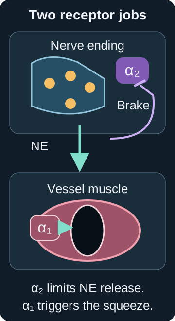
α₁ squeezes while α₂ limits release
α₁ is prominent on vascular smooth muscle. Norepinephrine activation tightens the muscle and narrows the lumen. The same family helps maintain tone at the bladder neck and prostate.
α₂ often sits on a sympathetic nerve terminal. It senses norepinephrine released from that nerve and limits further release. This is a local feedback brake, although α₂ also exists elsewhere.
Trace one receptor job
Endogenous norepinephrine reaches vascular α₁.
Nerve release→α₁ on vessel muscle→Muscle squeeze
Released norepinephrine feeds back at presynaptic α₂.
α₁ worked state: endogenous norepinephrine reaches vascular α₁, smooth muscle squeezes, and the lumen narrows.
α₂ worked state: norepinephrine released from the nerve activates presynaptic α₂ feedback, limiting further endogenous release. This does not control an administered catecholamine or tumor secretion.
Separate the presynaptic feedback job from the postsynaptic vascular squeeze.Enlarge diagramBone Wizardry original illustration. Physiological references: [17]
The feedback model concerns endogenous nerve norepinephrine. It does not mean injected epinephrine, infused norepinephrine, or tumor secretion is automatically controlled by that local brake.
Phenylephrine is a directly acting α₁ agonist: it activates that receptor rather than requiring a nerve to release norepinephrine. A reduced response to directly applied phenylephrine therefore points beyond the release step. Receptor compendium [17].
Release delivers transmitter from a nerve; uptake removes transmitter from the junction. More measured norepinephrine can reflect more release or less removal. Compare a pulse train with an uptake control before assigning a feedback mechanism. Receptor compendium [17].
Central α₂ activation can reduce sympathetic outflow from the brain. A peripheral α₂ autoreceptor senses the transmitter released by its own terminal and restrains further release. An isolated peripheral nerve contains no central circuit. Receptor compendium [17].
The short inside-cell relay map
A G protein is an inside-cell relay that passes a receptor signal onward. These are the predominant board-level pairings, not exclusive rules for every cell:
Alpha-1 chiefly uses Gq/11. Gq/11 activates phospholipase C, an enzyme commonly shortened to PLC. PLC makes IP3, a small messenger that releases stored calcium. More available calcium supports smooth-muscle squeeze.
Alpha-2 chiefly uses Gi/o. Gi/o reduces adenylyl cyclase and cyclic adenosine monophosphate, shortened to cyclic AMP or cAMP. At many sympathetic nerve terminals, alpha-2 also inhibits further endogenous norepinephrine release; that release control is not claimed to be one single exclusive cAMP mechanism.
Beta receptors chiefly use Gs. Gs activates adenylyl cyclase and raises cAMP. Beta-1 supports cardiac stimulation, while vascular beta-2 can support relaxation.
Bypass localization asks which downstream steps still work. If direct calcium restores contraction, the muscle can still generate force; the interruption lies earlier under the experiment's assumptions. That result alone cannot distinguish a receptor defect from a shared signaling defect.
Compare a second receptor using the same relay, such as an angiotensin II receptor using Gq, to test shared signaling. Also test a plausible opposing relaxant pathway. These are experimental controls built from the receptor map [17]; they do not by themselves measure a drug's molecular binding site.
The iris radial dilator pulls the pupil open when it contracts through α₁ signaling. The circular sphincter narrows the pupil through muscarinic signaling. Blocking the radial muscle reduces adrenergic dilation without directly activating the sphincter. Predict each muscle's effect from its geometry. Phentolamine ophthalmic label, section 12.1 [20].
Exam frame Blocking α₁ reduces smooth-muscle squeeze. Blocking presynaptic α₂ can remove a brake on endogenous norepinephrine release.
Predict one link, open it to check, then build on it.
1Why consider signaling upstream of calcium?
Answer: A signaling lesion could leave the final calcium-dependent machinery intact.
2Which results oppose this combined explanation?
Answer: Angiotensin II remains effective and the original phenylephrine curve returns after washout.
Read the complete worked explanation
A shared signaling lesion could weaken contraction upstream of calcium. This proposal fails both the intact angiotensin II response and recovery after washout. It therefore does not explain either the pathway specificity or the reversibility.
Why it is tempting A signaling lesion could leave the final calcium-dependent machinery intact.
Exact discriminator Angiotensin II remains effective and the original phenylephrine curve returns after washout.
Rule: Use pathway controls to localize the functional defect, then use surmountability and washout to characterize antagonism; neither alone proves a molecular binding site.
Review option B reasoning
Predict one link, open it to check, then build on it.
1Which part fits the intact parallel pathway?
Answer: Selective α₁ impairment could spare angiotensin II contraction.
2What contradicts persistent inactivation?
Answer: Full washout recovery contradicts a lasting loss of functional receptors under these conditions.
Read the complete worked explanation
A selective α₁ effect fits the parallel-pathway controls. Persistent receptor inactivation does not fit full recovery after washout. Spare receptors could preserve an initial maximum, but they do not explain restoration of the original curve after removal of free drug in this short experiment.
Why it is tempting Selective α₁ impairment could spare angiotensin II contraction.
Exact discriminator Full washout recovery contradicts a lasting loss of functional receptors under these conditions.
Rule: Use pathway controls to localize the functional defect, then use surmountability and washout to characterize antagonism; neither alone proves a molecular binding site.
Review option C reasoning
Predict one link, open it to check, then build on it.
1What does preserved angiotensin II contraction narrow down?
Answer: The defect favors the α₁ pathway over shared Gq signaling.
2What do surmountability and washout recovery add?
Answer: The inhibition behaves as reversible competitive antagonism.
3What remains unproven by these functional tests?
Answer: The exact molecular binding site remains unproven.
Read the complete worked explanation
The intact angiotensin II and calcium responses favor an α₁ pathway effect over shared signaling or force failure. Recovery of the maximum at higher phenylephrine concentrations and recovery after washout support reversible competitive antagonism. The β₂ control excludes the tested opposing relaxation route. These functional findings are consistent with α₁ receptor antagonism but do not directly demonstrate its binding site.
Rule: Use pathway controls to localize the functional defect, then use surmountability and washout to characterize antagonism; neither alone proves a molecular binding site.
Review option D reasoning
Predict one link, open it to check, then build on it.
1Why consider a reversible shared signaling defect?
Answer: A shared Gq defect could spare direct calcium contraction and reverse on washout.
2Which response distinguishes the proposed shared site?
Answer: Angiotensin II still contracts through the other tested Gq receptor.
Read the complete worked explanation
Reversibility fits the washout result, and shared Gq inhibition could blunt phenylephrine while sparing direct calcium contraction. However, the intact response of the other tested Gq receptor argues against that shared site.
Why it is tempting A shared Gq defect could spare direct calcium contraction and reverse on washout.
Exact discriminator Angiotensin II still contracts through the other tested Gq receptor.
Rule: Use pathway controls to localize the functional defect, then use surmountability and washout to characterize antagonism; neither alone proves a molecular binding site.
Compare with the correct option
The correct option is C. Open option C reasoning above, predict each link, and reveal one answer at a time.
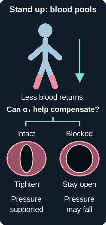
Standing shifts venous blood toward the legs. Baroreceptor signaling normally raises sympathetic output, and α₁-mediated vasoconstriction helps maintain venous return. α₁ blockade leaves less vascular tightening available at that moment.
Postural lightheadedness or syncope is most concerning after initiation, a dose increase, or a restart after interruption. Volume depletion and other blood-pressure-lowering drugs can increase risk. Reflex tachycardia is possible, not mandatory with every drug or patient.
Step through standing
Standing: gravity shifts venous blood toward the legs.
Normal compensation: sympathetic α₁ squeeze narrows capacitance vessels and supports venous return.
α₁ blocked: the sympathetic signal arrives, but the vessel cannot tighten as much.
Read every worked state
Stand: gravity pools venous blood in the legs and briefly reduces venous return.
Compensate: sympathetic signaling normally uses α₁ to tighten vessels.
Blocked: the signal may still arrive, but less vessel tightening is available, so orthostatic symptoms are more likely.
Connect first-dose and restart warnings to failed standing compensation.Enlarge diagramBone Wizardry original illustration. Physiological references: [4][15][16]
Venous capacity is the amount of blood veins hold at a given pressure. Less venous tightening permits more dependent pooling and less return to the heart during standing. Receptor effects [17]; postural physiology [25].
Preload describes ventricular filling and stretch before contraction; stroke volume is blood ejected per heartbeat. In the lesson's otherwise stable circulation, restoring lost volume improves filling and supports stroke volume. The alpha antagonist can remain active while filling improves. Volume and postural context [25]; doxazosin label [16].
Compensation can accelerate the sinus pulse through sympathetic activation and withdrawal of the vagal brake. A faster pulse supports compensation but does not directly measure sympathetic nerve activity. Better circulatory support can reduce the need for reflex tachycardia. Postural responses [25]; autonomic-blockade experiment [27].
A nerve recording measures the net effect of drug action and compensation. A recorded increase may coexist with an inhibitory drug component if the compensatory increase is larger; it does not exclude every additional central effect. This is a limit on interpreting the postural response [25].
After a substantial terazosin interruption, the prescriber reassesses reinitiation and low-dose retitration. A previously tolerated maintenance dose is not automatically an appropriate restart dose. Terazosin label [15]; NIH restart guidance [21].
Clinical boundary This lesson teaches recognition and mechanism. Individual initiation, titration, and interaction decisions belong to the treating clinician and current labeling.
Answer: Pulse alone cannot separate sympathetic activation from vagal withdrawal.
Read the complete worked explanation
Less α₁-mediated venous tightening permits dependent veins to hold more blood, reducing venous return on standing. The accelerating sinus pulse supports cardiac compensation despite the pressure fall. Sympathetic activation and vagal withdrawal can both contribute; pulse alone does not identify their individual magnitudes.
Rule: Postural vascular failure can coexist with cardiac compensation; sinus acceleration does not isolate sympathetic activity from vagal withdrawal.
Review option B reasoning
Predict one link, open it to check, then build on it.
1Which component fits the vascular mechanism?
Answer: Greater capacity permits more dependent pooling.
Answer: The sinus pulse rises from 72 to 108/min on standing.
Read the complete worked explanation
Increased venous capacity fits the vascular action and positional symptoms by permitting dependent pooling. A failed cardiac response would not explain the substantial sinus acceleration from 72 to 108/min observed during the pressure fall.
Why it is tempting Greater capacity permits more dependent pooling.
Exact discriminator The sinus pulse rises from 72 to 108/min on standing.
Rule: Postural vascular failure can coexist with cardiac compensation; sinus acceleration does not isolate sympathetic activity from vagal withdrawal.
Review option C reasoning
Predict one link, open it to check, then build on it.
1Why consider separate venous and cardiac functions?
Answer: Either inadequate filling or inadequate cardiac adjustment can impair standing tolerance.
2Which findings reverse both assignments?
Answer: α₁ blockade permits pooling while the sinus pulse accelerates.
Read the complete worked explanation
Effective venous tightening combined with cardiac failure could produce some postural problems, but it does not fit this combination. Prazosin limits venous tightening, and the sinus rate increases rather than failing to respond.
Why it is tempting Either inadequate filling or inadequate cardiac adjustment can impair standing tolerance.
Exact discriminator α₁ blockade permits pooling while the sinus pulse accelerates.
Rule: Postural vascular failure can coexist with cardiac compensation; sinus acceleration does not isolate sympathetic activity from vagal withdrawal.
Review option D reasoning
Predict one link, open it to check, then build on it.
1Which component fits the pulse response?
Answer: The sinus acceleration supports active cardiac compensation.
2Why is the venous prediction reversed?
Answer: Blocking α₁ venous tightening allows greater capacity and pooling.
Read the complete worked explanation
The faster sinus pulse supports active compensation. Lower venous capacity would favor venous return by reducing pooling. α₁ blockade instead limits the venous tightening needed during standing, so the proposed capacity change reverses the expected vascular effect.
Why it is tempting The sinus acceleration supports active cardiac compensation.
Benign prostate enlargement: relax the outlet, do not shrink it
Benign prostatic hyperplasia (BPH) means noncancerous enlargement of the prostate. Symptoms reflect both a static tissue-growth component and a dynamic smooth-muscle tone component at the prostate and bladder outlet.
Tamsulosin and silodosin favor alpha-1A receptors in the prostate and bladder neck. They can improve urinary symptoms with less average vascular effect than less selective α₁ blockers, but orthostatic hypotension and syncope risk are not zero.
Alfuzosin is clinically used for urinary symptoms and is often called functionally uroselective. That does not make it an alpha-1A-specific receptor blocker, and it still carries postural risk. None of these alpha blockers shrink prostate tissue.
Compare outlet tone with tissue growth
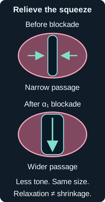
Alpha blockade: same gland size, wider outlet lumen.
Five-alpha-reductase inhibition: a later growth effect can reduce gland size; it is not the immediate alpha-blocker mechanism.
Earlier: enlarged glandLater: growth pathway reducedTime course is not immediate
Read every worked state
Alpha-blocker state: outlet smooth-muscle tone falls, the passage widens, symptoms may improve, and gland size is unchanged.
Growth-target state: five-alpha-reductase inhibition reduces dihydrotestosterone production and can reduce the growth component over time.
Make the dynamic tone-versus-static tissue-size distinction visible.Enlarge diagramBone Wizardry original illustration. Physiological references: [18][5][6][7]
Storage is the filling phase; urgency and frequency are storage symptoms. Voiding is emptying; hesitancy and a weak stream are voiding symptoms. The detrusor is bladder-wall smooth muscle that generates emptying pressure. NIDDK BPH overview [18]; urodynamic testing [23].
β₃ activation helps the detrusor relax during storage. Muscarinic M3 activation supports detrusor contraction; antimuscarinic treatment can reduce unwanted contractions. Those bladder actions differ from α₁-mediated prostate and bladder-neck tone. Receptor compendium [17].
During comparable voiding tests, higher detrusor pressure with lower flow favors increased outlet opposition. Low flow alone could instead reflect a weak detrusor. Detrusor pressure subtracts abdominal pressure from bladder pressure; sphincter electromyography records striated-muscle activity. NIDDK pressure-flow and muscle testing [23].
Use matched measurements to make a prediction: less pressure for the same urinary flow supports lower outlet resistance. At the old driving pressure, that reduced resistance should permit more flow if other conditions remain comparable. This is the mechanical interpretation used in the lesson's pressure-flow comparisons [23], not a clinical cutoff.
Five-alpha-reductase converts testosterone to dihydrotestosterone, or DHT. Inhibiting that enzyme reduces the androgen signal supporting prostate growth. Gland-volume change takes longer than relaxation of outlet tone. Finasteride mechanism [26]; NIDDK treatment distinctions [18].
Binding affinity measures how tightly a drug binds under specified assay conditions; a smaller Ki indicates tighter binding. Functional tissue selectivity describes the resulting effects in different tissues. Different unbound tissue exposures can produce different effects even without a receptor-subtype affinity difference. Receptor compendium [17].
Clearance describes drug removal; exposure reflects drug concentration over time. At the same dose and absorption, greater exposure with slower elimination favors reduced clearance. Silodosin exposure and half-life increase in renal impairment, requiring renal-function-specific prescribing review. Relative alpha-1A preference does not guarantee vascular sparing. Silodosin label, sections 5.2 and 12.3 [6].
Emission delivers seminal contents into the posterior urethra through coordinated smooth-muscle activity. Retrograde ejaculation diverts contents into the bladder. Low external ejaculate with preserved erection and orgasm does not alone distinguish these routes. Adrenergic receptor physiology [17]; primary ejaculatory-function comparison [24].
Sperm in post-ejaculatory urine supports diversion into the bladder. Repeated adequate samples without sperm, considered with prior semen findings, favor reduced emission but do not prove a diagnosis. Drug history alone cannot settle the distinction. Ejaculate and urine compartment measurements [24].
Outlet relaxation
Alpha blockers target the dynamic smooth-muscle component. Symptoms can improve without a smaller gland.
Growth pathway
Five-alpha-reductase inhibitors address the growth component over a longer interval and are a different class.
Drug families make sense when tradeoffs stay attached
Prazosin, terazosin, doxazosin
Reversible α₁ antagonists with vascular effects. Prazosin has an off-label niche for PTSD-related nightmares. Terazosin and doxazosin can affect urinary symptoms and blood pressure, but they are not universal first-line hypertension choices.
Tamsulosin, silodosin
Alpha-1A-preferring urinary symptom drugs. Ejaculatory adverse effects can occur. Orthostasis, syncope, interactions with vasodilating phosphodiesterase type 5 (PDE5) inhibitors, and intraoperative floppy iris syndrome (IFIS) disclosure still matter.
Alfuzosin
A clinically uroselective BPH drug without the same receptor-subtype preference label. It is not indicated to treat hypertension, and postural hypotension remains possible.
Phenoxybenzamine, phentolamine
Both block α₁ and α₂. Phenoxybenzamine binds irreversibly and lasts beyond its plasma presence. Phentolamine is reversible and shorter acting, with specific monitored uses.
Inspect receptor coverage and reversibility
Prazosin, terazosin, doxazosin
α₁ coveredα₂ sparedReversibleVascular and urinary effects
Tamsulosin and silodosin
Alpha-1A preferredOther α₁ not absentReversibleUrinary emphasis, residual BP risk
Prazosin group: reversible α₁ blockade with vascular and urinary effects.
Tamsulosin group: alpha-1A preference emphasizes the outlet but does not erase vascular or IFIS risk.
Alfuzosin: clinically uroselective without an alpha-1A-specific binding claim.
Nonselective pair: both cover α₁ and α₂; phenoxybenzamine is irreversible while phentolamine is reversible.
Reversible competitive antagonism can be overcome by enough agonist, preserving the maximum response; sufficient washout can restore the original response. Compare both the agonist curve and recovery rather than relying on duration alone. Phentolamine label [9]; reversible receptor binding [20].
Receptor reserve means spare receptors can preserve a maximum despite loss of some functional receptors. A known irreversible antagonist can reduce the maximum once that reserve is exceeded. Persistent inhibition after washout supports a lasting effect but alone does not prove covalent binding; slow dissociation can also persist. Phenoxybenzamine label [8]; primary irreversible-binding evidence [19].
After irreversible receptor inactivation, recovery requires replacement of functional receptors rather than only disappearance of free drug from plasma. Plasma clearance alone cannot specify when responsiveness returns. Phenoxybenzamine label [8]; binding evidence [19].
Extravasation means an infused medicine has escaped the intended vessel into surrounding tissue. Norepinephrine there can constrict nearby vessels and cause local blanching or coolness even when distal pulses remain. Clinician-directed local phentolamine opposes vascular alpha effects; this mechanism is not a complete extravasation management protocol. Phentolamine injection label [9].
Tamsulosin treats urinary symptoms and is not indicated as hypertension therapy. Judge those endpoints separately; combining alpha antagonists is not a default solution for uncontrolled pressure. Tamsulosin label [5]; silodosin interaction precautions [6].
Nitric oxide, or NO, activates soluble guanylyl cyclase, which makes cyclic GMP (cGMP). PDE5 breaks down cGMP; inhibiting PDE5 prolongs an existing relaxant signal. If the main NO input falls, there is less cGMP signal to preserve. VIAGRA label, section 12.1 [22].
cGMP is distinct from the cAMP produced through beta-receptor activation of adenylyl cyclase. PDE5 inhibition does not remove an alpha antagonist: extra relaxation and less alpha-mediated contraction can add to hypotension. Beta coupling [17]; VIAGRA label, section 5.5 [22].
Interaction pattern Phosphodiesterase type 5 inhibitors, usually shortened to PDE5 inhibitors, are vasodilating medicines such as sildenafil and tadalafil. Their vascular relaxation can add to hypotension from an alpha blocker. A class label never replaces patient-specific review.
Predict one link, open it to check, then build on it.
1Does the uptake test support slower transmitter removal?
Answer: Normal tracer uptake argues against slower removal.
2Why does the later-pulse effect suggest feedback?
Answer: Earlier release normally restrains subsequent release through terminal autoreceptors.
3Which direction of α₂ action fits increased later release?
Answer: Antagonism removes the inhibitory feedback.
Read the complete worked explanation
Normal tracer uptake argues against impaired removal as the source of the higher transmitter signal. The preferential increase during later pulses fits loss of feedback generated by previously released norepinephrine. α₂ autoreceptor antagonism removes that peripheral brake while leaving the separately tested α₁ vascular response intact.
Rule: Distinguish transmitter release from removal before assigning the direction of peripheral α₂ feedback.
Review option B reasoning
Predict one link, open it to check, then build on it.
1Why is this a plausible anatomical competitor?
Answer: Peripheral α₂ autoreceptors regulate release during repeated stimulation.
2Which directional prediction fails?
Answer: Agonism predicts less subsequent release, but later release increases.
Read the complete worked explanation
α₂ agonism acts at the relevant peripheral terminal and can alter release during a pulse train. Its direction is wrong: stronger inhibitory feedback would reduce later release, whereas X increases it.
Why it is tempting Peripheral α₂ autoreceptors regulate release during repeated stimulation.
Exact discriminator Agonism predicts less subsequent release, but later release increases.
Rule: Distinguish transmitter release from removal before assigning the direction of peripheral α₂ feedback.
Review option C reasoning
Predict one link, open it to check, then build on it.
1Why consider a vascular alpha receptor?
Answer: Postsynaptic α₁ receptors translate norepinephrine into contraction.
2What opposes that localization?
Answer: Direct phenylephrine contraction is preserved while the measured change is at repeated nerve release.
Read the complete worked explanation
α₁ blockade can reduce the vessel response to nerve-derived norepinephrine. It does not explain a selective increase in later nerve release, and the direct phenylephrine test shows that the vascular α₁ response remains intact at this exposure.
Why it is tempting Postsynaptic α₁ receptors translate norepinephrine into contraction.
Exact discriminator Direct phenylephrine contraction is preserved while the measured change is at repeated nerve release.
Rule: Distinguish transmitter release from removal before assigning the direction of peripheral α₂ feedback.
Review option D reasoning
Predict one link, open it to check, then build on it.
1Why could uptake inhibition mimic this result?
Answer: Slower removal can increase transmitter accumulation during a pulse train.
2Which measurement separates removal from release?
Answer: Labeled norepinephrine uptake is unchanged.
Read the complete worked explanation
An uptake inhibitor could increase the measured norepinephrine signal by slowing removal, particularly as pulses accumulate. The unchanged tracer uptake contradicts that mechanism and directs attention to increased release instead.
Why it is tempting Slower removal can increase transmitter accumulation during a pulse train.
Exact discriminator Labeled norepinephrine uptake is unchanged.
Rule: Distinguish transmitter release from removal before assigning the direction of peripheral α₂ feedback.
Compare with the correct option
The correct option is A. Open option A reasoning above, predict each link, and reveal one answer at a time.
Change the blockade and follow every surviving pathway
Use this qualitative model to compare α₁ smooth-muscle squeeze, α₂ feedback on endogenous nerve norepinephrine release, β₁ cardiac stimulation, and β₂ vascular relaxation. Geometry and pathway output change together.
Qualitative receptor and selectivity model
Defined setting: one alpha-dominant experimental catecholamine challenge in a conceptual vascular bed. This is not every dose or vascular bed.
Epi means epinephrine. Norepi means norepinephrine.
Reversible and irreversible choices block both alpha receptors.
Vessel effect
α₁ vessel squeezeAvailable
α₂ nerve feedbackAvailable
β₁ cardiacAvailable
β₂ vessel relaxationAvailable
Endogenous nerve norepinephrine
Cardiac pathway
All model states without JavaScript
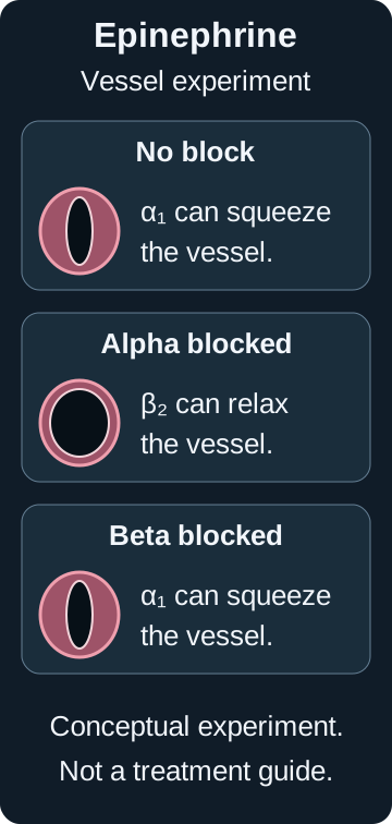
No block plus epinephrine: α₁ squeeze and β₂ relaxation are both available; α₁ dominates the vascular direction in this defined challenge. α₂ nerve feedback and β₁ cardiac stimulation remain.
No block plus norepinephrine: α₁ squeeze and β₁ cardiac stimulation are available, with little β₂ vascular relaxation. α₂ nerve feedback remains.
Selective α₁ block plus epinephrine: β₂ relaxation remains after α₁ blockade, so dilation can set the direction in this defined challenge. α₂ feedback on endogenous nerve release remains.
Selective α₁ block plus norepinephrine: the alpha pressor path is blunted without the same classic β₂ direction switch.
Alpha-1A preference: urinary effects are emphasized, but vascular α₁ is not absent.
Reversible or irreversible nonselective blockade plus epinephrine: the instantaneous receptor state and vessel geometry are the same. Alpha pathways are blocked and β₂ relaxation can set the vascular direction.
Reversible or irreversible nonselective blockade plus norepinephrine: the instantaneous alpha pressor response is blunted without the same classic β₂ reversal.
Irreversibility changes persistence after free phenoxybenzamine is gone; it does not create extra instantaneous dilation without ongoing agonist.
α₂ feedback here concerns endogenous nerve norepinephrine release, not the administered catecholamine or tumor secretion.
This qualitative model is not a dose or blood pressure prediction tool, and it is not a treatment recommendation.
Teach qualitative experimental epinephrine reversal without an anaphylaxis treatment scenario.Enlarge diagramBone Wizardry original illustration. Physiological references: [17][8]
For a vascular bed, pressure gradient = flow × resistance. The gradient is inflow pressure minus outflow pressure. With flow and outflow pressure fixed, lower inflow pressure means lower resistance. If the gradient is fixed instead, lower resistance permits higher flow. This is the pressure-flow relation used in the model.
An isolated vascular bed has no cardiac-output contribution, and a denervated preparation cannot generate a nerve reflex. Compare baseline resistance, maximal dilatory reserve, drug exposure, and effective blockade before attributing a difference to an agonist. These controls separate alternative explanations of the receptor effects [17].
A crossover tests each challenge in each preparation: a difference that follows the challenge is less likely to be a fixed tissue difference. A reference agonist tests whether the preparations respond comparably. Removing endothelium separates a direct smooth-muscle effect from a required endothelial contribution.
A downstream intervention can interrupt a surviving pathway even when its receptor remains available. Inhibiting adenylyl cyclase in isolated atrium can reduce β₁ signaling; it cannot remove an antagonist from a separately maintained artery. Predict each compartment from its own exposure. β₁ signaling [17].
In a defined alpha-dominant experimental challenge, substantial nonselective alpha blockade can remove epinephrine alpha-mediated vascular squeeze while β₂ relaxation remains. The net vascular direction can switch toward dilation. This does not describe every epinephrine dose or vascular bed. Norepinephrine has much less β₂ activity, so alpha blockade blunts its pressor pathway rather than producing the same classic reversal.
Safety boundary This is an experimental receptor demonstration and not a treatment recommendation. It does not say to withhold epinephrine in anaphylaxis, and it is not a dosing or blood pressure prediction tool.
Construction basis: documented receptor effects [17] and the phenoxybenzamine label warning [8]. This original qualitative model is not presented as a reconstruction of an unverified historical experiment.
Pheochromocytoma preparation begins with alpha blockade
A pheochromocytoma is an adrenal tumor that produces catecholamines such as norepinephrine and epinephrine. Those signals can drive marked vascular alpha effects and cardiac beta effects.
Preoperative management is clinician-led and individualized. The durable board principle is that alpha blockade must be established before a beta blocker is considered. A beta blocker may be added only when tachycardia persists after adequate alpha blockade.
Choose the first receptor target
Safe sequence under specialist care
1. Establish adequate alpha blockadeVessel squeeze is controlled first2. Add beta only if tachycardia still needs treatment
Beta first leaves vascular alpha squeeze unopposed
Beta effects blocked firstAlpha vasoconstriction remainsNarrow vessel and crisis risk
Read every worked state
Alpha-first state: establish adequate alpha blockade, then consider beta blockade only for a remaining indication such as persistent tachycardia.
Beta-first state: vascular alpha constriction remains without beta-mediated compensation, which can provoke crisis.
Starting beta blockade first can leave catecholamine-driven alpha vasoconstriction without beta-mediated compensation and provoke a hypertensive crisis. Alpha blockade lowers risk but does not guarantee tumor manipulation will be hemodynamically quiet.
Metanephrines are metabolites produced from catecholamines. Plasma free or urinary fractionated metanephrines help evaluate a suspected catecholamine-producing tumor; an adrenal mass alone does not establish secretion. Endocrine Society guideline [10].
Board rule Alpha first. Beta later only for a remaining indication such as persistent tachycardia, under specialist management.
Source: Endocrine Society pheochromocytoma and paraganglioma guideline [10].
Past α₁ blocker exposure belongs in the cataract history
Intraoperative floppy iris syndrome (IFIS) is a surgical iris behavior associated with α₁ blocker exposure, especially tamsulosin. The ophthalmic surgeon should know about current or past exposure before cataract or glaucoma surgery.
Stopping a drug shortly before surgery does not reliably erase risk. Patients should not alter prescribed therapy on their own. Early disclosure lets the surgical team plan.
Test the eye-surgery medication history
Current α₁ blocker
Medication historyCurrent exposureTell the ophthalmic surgeon before surgery
Past α₁ blocker
Medication historyPast exposure still mattersTell the ophthalmic surgeon before surgery
No known exposure
Review the complete medication historyNo α₁ blocker identifiedDo not invent a self-directed stop or restart
Read every worked state
Current exposure: disclose it before cataract or glaucoma surgery.
Past exposure: disclose it even if the drug was stopped; a short stop does not reliably erase IFIS risk.
No known exposure: continue a complete medication review and do not change prescriptions without the treating team.
α₁ signaling contracts the radial iris dilator; muscarinic signaling contracts the circular sphincter. Reduced adrenergic dilation is therefore different from directly stimulating pupil constriction. Office dilation does not replace the exposure history used for surgical preparation. Iris mechanism [20]; IFIS warning [5].
Sources: tamsulosin label [5] and silodosin label [6].
Prazosin guidance separates nightmares from global PTSD symptoms
The 2023 VA and DoD guidance suggests prazosin for PTSD-associated nightmares and suggests against using it for global PTSD symptoms. This is an off-label, symptom-specific decision, not a broad endorsement for PTSD.
Orthostatic risk, concurrent drugs, comorbidities, and shared decision-making still apply. Trauma-focused psychotherapy and other evidence-based care remain outside this receptor lesson.
Set the prazosin target
Target: PTSD-associated nightmares
Guidance suggests prazosin may be consideredOff label and individualizedOrthostatic safety still matters
Target: global PTSD symptoms
Guidance suggests against prazosin for this broad targetDo not generalize the nightmare recommendationUse the full PTSD treatment evidence base
Read every worked state
Nightmare target: current VA and DoD guidance suggests prazosin for PTSD-associated nightmares as an off-label individualized option.
Global-symptom target: the same guidance suggests against prazosin for global PTSD symptoms.
Track the intended symptom separately from daytime and global outcomes. Fewer nightmares in one person do not establish benefit for avoidance, hypervigilance, or the population as a whole. VA symptom-specific guidance [13].
Dose dechallenge means observing what happens when exposure is reduced under clinical supervision. If a lower dose preserves the target benefit while orthostasis resolves, the higher dose added observed harm without added target benefit. That supports an individualized dose discussion, not a population efficacy claim. Prazosin postural risk [4]; nightmare treatment scope [13].
Scope rule Nightmares are the narrow target in current guidance. Do not generalize the suggestion to every PTSD symptom.
Each case keeps every option's reasoning available. Predict one link, open it to check, then build on it. Previously opened steps stay open. A separate worked explanation remains available after the steps.
Patients and experimental measurements are fictional educational constructions, not reports of studies.
Use a case as an inference test
Predict one link, open it to check, then build on it. Each option keeps its own atomic steps and worked explanation.
How retrieval works without JavaScript
Selecting and checking are optional. Open one option, predict the answer to its first step, reveal that single answer, and continue. Every option also has a separate complete worked explanation.
Free case preview
These three cases preserve a representative free practice sample.
Predict one link, open it to check, then build on it.
1Why is Gi/o a relevant alternative?
Answer: α₂ receptors predominantly signal through Gi/o.
2What separates this relay from the failure?
Answer: α₂-dependent inhibition remains intact in the tested terminal preparation.
Read the complete worked explanation
Gi/o is a relevant adrenergic signaling relay, but the α₂ terminal response is preserved in the separate preparation. The lost arterial response and its rescue by PLC activation instead involve the α₁-to-PLC connection, rather than the intact inhibitory terminal relay.
Why it is tempting α₂ receptors predominantly signal through Gi/o.
Exact discriminator α₂-dependent inhibition remains intact in the tested terminal preparation.
Rule: Downstream rescue localizes an interruption upstream of the rescued step without identifying one molecular lesion.
Review option B reasoning
Predict one link, open it to check, then build on it.
1What does direct PLC rescue establish?
Answer: The pathway downstream of PLC can still contract the muscle.
2Where is the failed norepinephrine signal localized?
Answer: The interruption is upstream of PLC activation.
3Which listed connection normally occupies that interval?
Answer: α₁ receptor coupling to Gq/11 occupies that interval.
Read the complete worked explanation
Rescue by direct PLC activation shows that the downstream IP3-to-contraction pathway can work. The failed signal lies upstream of PLC, where α₁ receptors predominantly signal through Gq/11. The separate α₂ response remains intact. This localizes a connection, not a specific defective protein.
Rule: Downstream rescue localizes an interruption upstream of the rescued step without identifying one molecular lesion.
Review option C reasoning
Predict one link, open it to check, then build on it.
1Why could a final muscle defect mimic receptor failure?
Answer: Even an intact receptor needs functional contractile machinery.
2What tests that machinery directly?
Answer: Supplied intracellular calcium still produces contraction.
Read the complete worked explanation
A failed contractile apparatus could prevent contraction despite normal receptor signaling. Direct calcium still contracts the artery, so the final machinery is functional. Loss of the norepinephrine response must therefore be explained at an earlier signaling step.
Why it is tempting Even an intact receptor needs functional contractile machinery.
Exact discriminator Supplied intracellular calcium still produces contraction.
Rule: Downstream rescue localizes an interruption upstream of the rescued step without identifying one molecular lesion.
Review option D reasoning
Predict one link, open it to check, then build on it.
1Why could an IP3 defect weaken contraction?
Answer: A loss of stored calcium release would reduce the contractile signal.
2Which rescue contradicts that site?
Answer: PLC activation restores contraction through the downstream pathway.
Read the complete worked explanation
Impaired IP3-dependent calcium release could weaken contraction despite receptor activation. However, direct PLC activation restores both IP3 and contraction, showing that the pathway through IP3 and calcium release remains usable. The interruption must lie earlier in the tested sequence.
Why it is tempting A loss of stored calcium release would reduce the contractile signal.
Exact discriminator PLC activation restores contraction through the downstream pathway.
Rule: Downstream rescue localizes an interruption upstream of the rescued step without identifying one molecular lesion.
Compare with the correct option
The correct option is B. Open option B reasoning above, predict each link, and reveal one answer at a time.
Predict one link, open it to check, then build on it.
1Why might the pulse attract treatment attention?
Answer: A racing pulse can be distressing during near-syncope.
2What does the paired pressure result imply?
Answer: The pulse is responding to a pressure fall rather than explaining the restart problem.
Read the complete worked explanation
The faster pulse may feel like the cause of the episode. Here it accompanies a large positional pressure fall and represents compensation; suppressing it does not restore venous α₁ tightening or correct restart dosing.
Why it is tempting A racing pulse can be distressing during near-syncope.
Exact discriminator The pulse is responding to a pressure fall rather than explaining the restart problem.
Rule: After a substantial terazosin interruption, prior maintenance-dose tolerance does not replace reassessment and low-dose retitration.
Review option B reasoning
Predict one link, open it to check, then build on it.
1Why consider a related medicine?
Answer: Another α₁ blocker may also relieve outlet symptoms.
2What does the proposed switch fail to address?
Answer: Another maintenance-dose start can retain the same initiation-related postural risk.
Read the complete worked explanation
A related α₁ blocker may retain urinary efficacy, but switching agents does not remove initiation-related orthostatic risk. A maintenance-dose substitution bypasses the reassessment and titration required by the new intolerance.
Why it is tempting Another α₁ blocker may also relieve outlet symptoms.
Exact discriminator Another maintenance-dose start can retain the same initiation-related postural risk.
Rule: After a substantial terazosin interruption, prior maintenance-dose tolerance does not replace reassessment and low-dose retitration.
Review option C reasoning
Predict one link, open it to check, then build on it.
1Why might bedtime dosing seem useful?
Answer: Less activity after dosing may reduce exposure to early dizziness.
2What risk does timing leave unresolved?
Answer: The maintenance dose was resumed after a prolonged interruption and caused postural intolerance.
Read the complete worked explanation
Bedtime administration may limit activity during early dizziness, so it is a plausible precaution. Timing does not address the excessive restart dose after weeks without exposure or the demonstrated standing intolerance.
Why it is tempting Less activity after dosing may reduce exposure to early dizziness.
Exact discriminator The maintenance dose was resumed after a prolonged interruption and caused postural intolerance.
Rule: After a substantial terazosin interruption, prior maintenance-dose tolerance does not replace reassessment and low-dose retitration.
Review option D reasoning
Predict one link, open it to check, then build on it.
1Which event changes the relevance of old tolerance?
Answer: A prolonged interruption precedes the maintenance-dose restart.
2What do the standing measurements establish?
Answer: The resumed regimen is associated with symptomatic orthostatic intolerance.
3What prescribing principle follows?
Answer: Reassess low-dose reinitiation and retitration.
Read the complete worked explanation
The interruption followed by a maintenance-dose restart recreates initiation-related orthostatic risk. The standing measurements document intolerance despite compensatory tachycardia. Prescriber-directed reassessment and low-dose retitration address that risk while retaining the urinary indication.
Rule: After a substantial terazosin interruption, prior maintenance-dose tolerance does not replace reassessment and low-dose retitration.
Compare with the correct option
The correct option is D. Open option D reasoning above, predict each link, and reveal one answer at a time.
Predict one link, open it to check, then build on it.
1Which prediction fits restored filling?
Answer: A higher stroke volume fits improved preload.
2Why does the heart-rate prediction fail?
Answer: Correcting the volume deficit reduces rather than enlarges the compensatory stimulus.
Read the complete worked explanation
Improved filling supports the predicted higher stroke volume. However, restoring volume reduces the deficit that provoked reflex tachycardia. Unchanged doxazosin exposure does not require a larger reflex response after the additional volume problem is corrected.
Why it is tempting A higher stroke volume fits improved preload.
Exact discriminator Correcting the volume deficit reduces rather than enlarges the compensatory stimulus.
Rule: Restored filling can raise stroke volume and reduce compensatory tachycardia even while alpha antagonism remains active.
Review option B reasoning
Predict one link, open it to check, then build on it.
1When would this pair be plausible?
Answer: Worsening effective filling could reduce stroke volume and increase reflex tachycardia.
2What intervention reverses that expectation here?
Answer: The new volume deficit is corrected without a new cardiac problem.
Read the complete worked explanation
Lower stroke volume with greater reflex tachycardia could fit worsening effective filling. The intervention instead restores circulating volume while contractility and medications remain stable. Both predictions therefore describe the opposite of the expected response to the supplied perturbation.
Why it is tempting Worsening effective filling could reduce stroke volume and increase reflex tachycardia.
Exact discriminator The new volume deficit is corrected without a new cardiac problem.
Rule: Restored filling can raise stroke volume and reduce compensatory tachycardia even while alpha antagonism remains active.
Review option C reasoning
Predict one link, open it to check, then build on it.
1Which part fits less circulatory stress?
Answer: The heart-rate increase should become smaller.
2Which stipulated condition defeats a lower stroke volume?
Answer: Restored filling at stable contractility supports higher stroke volume.
Read the complete worked explanation
A smaller reflex pulse response fits improved circulatory support. Lower stroke volume does not follow from successful volume replacement with stable contractility and no heart failure; restored filling should support ejection.
Why it is tempting The heart-rate increase should become smaller.
Rule: Restored filling can raise stroke volume and reduce compensatory tachycardia even while alpha antagonism remains active.
Review option D reasoning
Predict one link, open it to check, then build on it.
1What does volume replacement improve before ejection?
Answer: Venous return and ventricular preload improve.
2What follows for stroke volume in this bounded model?
Answer: Stroke volume rises at stable contractility.
3How does better circulatory support affect the reflex pulse demand?
Answer: The compensatory heart-rate increase becomes smaller.
Read the complete worked explanation
Volume replacement increases venous return and ventricular filling, supporting a higher stroke volume at the stipulated stable contractility. Better circulatory support reduces the reflex demand for tachycardia. Doxazosin can still blunt vascular tightening, so improvement need not imply complete normalization or loss of antagonist activity.
Rule: Restored filling can raise stroke volume and reduce compensatory tachycardia even while alpha antagonism remains active.
Compare with the correct option
The correct option is D. Open option D reasoning above, predict each link, and reveal one answer at a time.
Predict one link, open it to check, then build on it.
1What changed when the same flow required less pressure?
Answer: Outlet resistance fell rather than requiring a stronger bladder pump.
2What is the new resistance in this stated model?
Answer: 30 cm H2O divided by 6 mL/s gives 5 cm H2O per mL/s.
3What flow follows at a gradient of 60?
Answer: 60 divided by 5 gives 12 mL/s.
Read the complete worked explanation
The same flow at half the driving pressure means the outlet offers less resistance; greater detrusor force is not required to explain it. The new resistance is 30/6 = 5 cm H2O per mL/s. At a gradient of 60, the stated local model predicts 60/5 = 12 mL/s. This is a bounded teaching calculation, not a universal urinary-flow law or clinical prediction.
Rule: Use matched pressure and flow to distinguish reduced outlet resistance from increased pumping force, then calculate the consequence of a new driving pressure.
Review option B reasoning
Predict one link, open it to check, then build on it.
1Which part fits the measured comparison?
Answer: Less pressure for the same flow supports lower outlet resistance.
2Which calculation separates 9 from the supported prediction?
Answer: The new resistance is 5, so a gradient of 60 produces 12 mL/s under the stated assumption.
Read the complete worked explanation
Reduced outlet resistance fits the matched pressure-flow observations. A predicted flow of 9 incorrectly transfers the absolute earlier flow or uses an unsupported proportional change. With the new resistance fixed at 30/6 = 5, the specified gradient of 60 gives 12, not 9 mL/s.
Why it is tempting Less pressure for the same flow supports lower outlet resistance.
Exact discriminator The new resistance is 5, so a gradient of 60 produces 12 mL/s under the stated assumption.
Rule: Use matched pressure and flow to distinguish reduced outlet resistance from increased pumping force, then calculate the consequence of a new driving pressure.
Review option C reasoning
Predict one link, open it to check, then build on it.
1Why could stronger detrusor contraction seem relevant?
Answer: A stronger bladder pump can improve emptying in other circumstances.
2What matched finding identifies a different mechanism here?
Answer: The same flow needs less pressure, which demonstrates less outlet opposition in this model.
Read the complete worked explanation
The numerical flow matches the calculation using the new observed resistance. Increased pumping strength alone does not explain why less driving pressure is required at the same flow. The matched comparison identifies a lower outlet resistance, not merely a stronger detrusor.
Why it is tempting A stronger bladder pump can improve emptying in other circumstances.
Exact discriminator The same flow needs less pressure, which demonstrates less outlet opposition in this model.
Rule: Use matched pressure and flow to distinguish reduced outlet resistance from increased pumping force, then calculate the consequence of a new driving pressure.
Review option D reasoning
Predict one link, open it to check, then build on it.
1Which competing interpretation could follow from improved emptying alone?
Answer: A stronger detrusor could be considered without pressure-flow measurements.
2Which two supplied results defeat this pair?
Answer: Less pressure at the same flow identifies lower resistance, and 60 divided by the new resistance of 5 gives 12 mL/s.
Read the complete worked explanation
Improved emptying may tempt a stronger-pump explanation, but the matched pressure-flow comparison demonstrates reduced resistance instead. The second prediction also fails the given relation: new resistance is 5 and a gradient of 60 gives 12 mL/s.
Why it is tempting A stronger detrusor could be considered without pressure-flow measurements.
Exact discriminator Less pressure at the same flow identifies lower resistance, and 60 divided by the new resistance of 5 gives 12 mL/s.
Rule: Use matched pressure and flow to distinguish reduced outlet resistance from increased pumping force, then calculate the consequence of a new driving pressure.
Compare with the correct option
The correct option is A. Open option A reasoning above, predict each link, and reveal one answer at a time.
Predict one link, open it to check, then build on it.
1What pharmacokinetic change fits both exposure and half-life?
Answer: Reduced clearance fits the same-dose exposure rise and slower elimination.
2What do the matched vascular controls localize?
Answer: The concentration-dependent defect favors the α₁ response over final force generation.
3What follows for earlier urinary selectivity?
Answer: Earlier tolerability does not guarantee vascular safety at the new exposure.
Read the complete worked explanation
At unchanged dose and absorption, greater exposure together with a longer terminal half-life supports reduced clearance. The high-concentration assay selectively weakens phenylephrine contraction while sparing angiotensin II and calcium responses. Relative urinary receptor preference therefore does not prevent vascular antagonism at this exposure; renal-function-specific prescribing reassessment is needed. Silodosin illustrates this exposure-sensitive urinary-versus-vascular tradeoff.
Rule: Infer an exposure mechanism from dose and elimination measurements, then localize the concentration-dependent vascular effect with matched pathway controls.
Review option B reasoning
Predict one link, open it to check, then build on it.
1Why consider both exposure and tissue response?
Answer: More circulating drug can change the magnitude of vascular effects.
2Which controls reject this proposed combination?
Answer: Unchanged absorption, longer elimination, and intact comparator contractions favor reduced clearance with a selective pathway effect.
Read the complete worked explanation
Higher exposure could in principle arise from absorption and cause broader tissue effects. Here absorption conditions are unchanged, elimination is slower, and comparator contractions remain intact. Both parts of the proposed explanation fail the supplied controls.
Why it is tempting More circulating drug can change the magnitude of vascular effects.
Exact discriminator Unchanged absorption, longer elimination, and intact comparator contractions favor reduced clearance with a selective pathway effect.
Rule: Infer an exposure mechanism from dose and elimination measurements, then localize the concentration-dependent vascular effect with matched pathway controls.
Review option C reasoning
Predict one link, open it to check, then build on it.
1Which part fits the vessel experiment?
Answer: Preserved comparator contractions support a selective α₁ response defect.
2Which findings favor clearance over absorption?
Answer: Absorption is unchanged and terminal elimination takes longer.
Read the complete worked explanation
The vascular localization fits the selective phenylephrine impairment. Increased absorption does not fit the stipulated unchanged absorption conditions together with a doubled elimination half-life; reduced clearance is the supported pharmacokinetic explanation.
Why it is tempting Preserved comparator contractions support a selective α₁ response defect.
Exact discriminator Absorption is unchanged and terminal elimination takes longer.
Rule: Infer an exposure mechanism from dose and elimination measurements, then localize the concentration-dependent vascular effect with matched pathway controls.
Review option D reasoning
Predict one link, open it to check, then build on it.
1Which part fits the pharmacokinetics?
Answer: Reduced clearance explains higher exposure and slower elimination at the same dose.
Answer: Direct calcium and angiotensin II contractions remain near control values.
Read the complete worked explanation
Reduced clearance fits the exposure and half-life changes. Generalized force failure does not fit preserved angiotensin II and direct calcium contractions at the same drug concentration. The assay instead supports a selective α₁ pathway effect.
Why it is tempting Reduced clearance explains higher exposure and slower elimination at the same dose.
Exact discriminator Direct calcium and angiotensin II contractions remain near control values.
Rule: Infer an exposure mechanism from dose and elimination measurements, then localize the concentration-dependent vascular effect with matched pathway controls.
Compare with the correct option
The correct option is A. Open option A reasoning above, predict each link, and reveal one answer at a time.
Predict one link, open it to check, then build on it.
1Which forecast follows the higher arterial concentration?
Answer: More vascular antagonism can increase standing hypotension risk.
2Why is the initial affinity explanation unsupported?
Answer: Ki is similar across all three tested receptor subtypes.
Read the complete worked explanation
Increased arterial exposure can raise postural risk, but the proposed binding explanation is contradicted by the similar Ki values across alpha-1A, alpha-1B, and alpha-1D. The measured exposure difference explains the initial emphasis without overriding the direct subtype-affinity measurements.
Why it is tempting More vascular antagonism can increase standing hypotension risk.
Exact discriminator Ki is similar across all three tested receptor subtypes.
Rule: Separate receptor affinity from tissue exposure, and use a controlled exposure change to predict vascular risk.
Review option B reasoning
Predict one link, open it to check, then build on it.
1Which explanation fits the original tissue difference?
Answer: Higher outlet exposure can create functional urinary emphasis without subtype affinity.
2What changes the standing prediction?
Answer: Arterial exposure rises to a concentration that inhibits arterial α₁ responses in the matched assay.
Read the complete worked explanation
The initial exposure explanation fits the binding and matched-response measurements. The risk prediction does not: the artery now receives a concentration shown to inhibit its α₁ response, so the original small vascular effect cannot be assumed to persist.
Why it is tempting Higher outlet exposure can create functional urinary emphasis without subtype affinity.
Exact discriminator Arterial exposure rises to a concentration that inhibits arterial α₁ responses in the matched assay.
Rule: Separate receptor affinity from tissue exposure, and use a controlled exposure change to predict vascular risk.
Review option C reasoning
Predict one link, open it to check, then build on it.
1Why might tissue emphasis be mistaken for binding exclusivity?
Answer: A larger urinary effect can resemble a subtype-preferring drug pattern.
2Which two controls defeat that interpretation?
Answer: Similar Ki values and comparable equal-concentration tissue responses oppose binding exclusivity and continued vascular sparing.
Read the complete worked explanation
A subtype-exclusive interpretation could suggest preserved vascular sparing, but neither part fits this dataset. Binding affinity is similar across subtypes, and equal-concentration assays show that the new arterial exposure inhibits the vascular response.
Why it is tempting A larger urinary effect can resemble a subtype-preferring drug pattern.
Exact discriminator Similar Ki values and comparable equal-concentration tissue responses oppose binding exclusivity and continued vascular sparing.
Rule: Separate receptor affinity from tissue exposure, and use a controlled exposure change to predict vascular risk.
Review option D reasoning
Predict one link, open it to check, then build on it.
1Do the Ki values establish alpha-1A-specific affinity?
Answer: The similar values show no marked alpha-1A preference.
2What measured difference explains the functional tissue emphasis?
Answer: Unbound drug exposure is higher at the outlet.
3What does raising only arterial exposure predict?
The similar Ki values provide no marked alpha-1A binding preference, and comparable equal-concentration responses favor exposure as the supplied explanation of the tissue difference. Raising arterial exposure should increase vascular antagonism and can impair standing support despite unchanged urinary exposure. This is a fictional experiment, not a reported alfuzosin clinical study.
Rule: Separate receptor affinity from tissue exposure, and use a controlled exposure change to predict vascular risk.
Compare with the correct option
The correct option is D. Open option D reasoning above, predict each link, and reveal one answer at a time.
Predict one link, open it to check, then build on it.
1What changes would this option require?
Answer: Alpha antagonism would have to decline while the cGMP signal increased.
2Which stipulated conditions contradict both changes?
Answer: Alpha-antagonist exposure is fixed and NO-dependent cGMP generation is reduced.
Read the complete worked explanation
Loss of the phenylephrine shift with greater sildenafil relaxation would require relief of alpha antagonism and amplification of the cGMP signal. The intervention does neither: antagonist exposure is unchanged and the main source of cGMP generation is reduced.
Why it is tempting Alpha antagonism would have to decline while the cGMP signal increased.
Exact discriminator Alpha-antagonist exposure is fixed and NO-dependent cGMP generation is reduced.
Rule: Removing the source of a messenger can reduce the effect of blocking its degradation without removing a separate receptor antagonist.
Review option B reasoning
Predict one link, open it to check, then build on it.
1What happens to cGMP generation when the main NO input falls?
Answer: Less cGMP is generated.
2How does that change the extra effect of PDE5 inhibition?
Answer: There is less cGMP signal for sildenafil to prolong.
3What happens to the unchanged alpha antagonist?
Answer: Its receptor-dependent phenylephrine shift persists.
Read the complete worked explanation
Inhibiting NO synthesis reduces activation of guanylyl cyclase and the supply of cGMP that PDE5 inhibition preserves. Sildenafil therefore adds less relaxation in this bounded model. The separate α₁ antagonist remains present, so its effect on phenylephrine responsiveness persists even if net vessel tone changes.
Rule: Removing the source of a messenger can reduce the effect of blocking its degradation without removing a separate receptor antagonist.
Review option C reasoning
Predict one link, open it to check, then build on it.
Answer: The phenylephrine response remains shifted.
2Why is greater extra relaxation unsupported?
Answer: Less NO input produces less cGMP for PDE5 inhibition to preserve.
Read the complete worked explanation
Persistence of the phenylephrine shift fits unchanged alpha antagonism. More sildenafil-dependent relaxation is the wrong direction because inhibiting the principal NO source reduces cGMP generation rather than increasing the substrate signal preserved by PDE5 inhibition.
Why it is tempting The phenylephrine response remains shifted.
Exact discriminator Less NO input produces less cGMP for PDE5 inhibition to preserve.
Rule: Removing the source of a messenger can reduce the effect of blocking its degradation without removing a separate receptor antagonist.
Review option D reasoning
Predict one link, open it to check, then build on it.
1Which component fits less NO-dependent cGMP input?
Answer: Sildenafil adds less relaxation.
2Why should the alpha-dependent shift persist?
Answer: The NO intervention does not remove the alpha antagonist.
Read the complete worked explanation
The smaller sildenafil contribution fits reduced NO-dependent cGMP generation. Disappearance of the alpha-antagonist shift does not follow: blocking NO synthesis changes a parallel relaxant pathway without removing the unchanged antagonist from the receptor-dependent pathway.
Why it is tempting Sildenafil adds less relaxation.
Exact discriminator The NO intervention does not remove the alpha antagonist.
Rule: Removing the source of a messenger can reduce the effect of blocking its degradation without removing a separate receptor antagonist.
Compare with the correct option
The correct option is B. Open option B reasoning above, predict each link, and reveal one answer at a time.
Predict one link, open it to check, then build on it.
1Which assignment fits surmountability and washout recovery?
Answer: Phentolamine fits X.
2What specifically defeats the Y assignment?
Answer: Y remains inhibited after extensive free-drug washout.
Read the complete worked explanation
X fits phentolamine. Y retains loss of responsiveness after free drug is removed and after spare-receptor reserve is exceeded, so a reversible competitive phentolamine assignment does not fit that preparation.
Why it is tempting Phentolamine fits X.
Exact discriminator Y remains inhibited after extensive free-drug washout.
Rule: Interpret each preparation independently using surmountability, washout, and receptor reserve; persistent functional inhibition alone does not prove covalent binding.
Review option B reasoning
Predict one link, open it to check, then build on it.
1Why are both drugs relevant candidates?
Answer: Both antagonize alpha receptors but differ in persistence.
2What reverses the proposed pairing?
Answer: X recovers after washout; Y retains inhibition despite free-drug removal.
Read the complete worked explanation
Assigning phenoxybenzamine to X and phentolamine to Y reverses the supported assignments. X shows the reversible and surmountable pattern expected from phentolamine, whereas Y shows persistent loss after washout consistent with phenoxybenzamine. Both preparations must be interpreted to select the correct pair.
Why it is tempting Both antagonize alpha receptors but differ in persistence.
Exact discriminator X recovers after washout; Y retains inhibition despite free-drug removal.
Rule: Interpret each preparation independently using surmountability, washout, and receptor reserve; persistent functional inhibition alone does not prove covalent binding.
Review option C reasoning
Predict one link, open it to check, then build on it.
1What behavior does X demonstrate?
Answer: X demonstrates surmountable, reversible antagonism.
2What does Y demonstrate after free drug is removed?
Answer: Y retains a loss of receptor-mediated response despite viable muscle.
3Which known drug pairing fits these distinct behaviors?
Answer: Phentolamine fits X and phenoxybenzamine fits Y.
Read the complete worked explanation
X fits reversible competitive phentolamine antagonism because higher agonist restores the maximum and washout restores the original response. Y fits the known irreversible action of phenoxybenzamine after receptor reserve is exceeded. Preserved calcium contraction limits a muscle-injury explanation. The constructed curves are consistent with these drugs but do not independently prove covalent binding.
Rule: Interpret each preparation independently using surmountability, washout, and receptor reserve; persistent functional inhibition alone does not prove covalent binding.
Review option D reasoning
Predict one link, open it to check, then build on it.
1Which assignment fits the persistent preparation?
Answer: Phenoxybenzamine fits Y.
2What specifically defeats the X assignment?
Answer: X fully recovers after washout, rather than retaining receptor inactivation.
Read the complete worked explanation
Y fits phenoxybenzamine under the stated reserve and washout conditions. X does not: full recovery of its original curve after washout is inconsistent with persistent receptor inactivation in this short comparison. Preserved maximum alone would be less decisive because spare receptors can mask irreversible loss.
Why it is tempting Phenoxybenzamine fits Y.
Exact discriminator X fully recovers after washout, rather than retaining receptor inactivation.
Rule: Interpret each preparation independently using surmountability, washout, and receptor reserve; persistent functional inhibition alone does not prove covalent binding.
Compare with the correct option
The correct option is C. Open option C reasoning above, predict each link, and reveal one answer at a time.
Predict one link, open it to check, then build on it.
1Why consider vasodilation and reduced nerve activity?
Answer: Both can reduce vascular tone when their relevant pathways supply the tone.
2Which controls reject this combination?
Answer: The rescue survives NO-pathway blockade and the tissue catecholamine does not require local nerve release.
Read the complete worked explanation
Both parts fail the supplied evidence. The rescue remains active when the tested NO-donor pathway is blocked, and the constrictor remains in tissue independently of nerve activity. A mechanism that reduces nerve release alone would not neutralize this exogenous source.
Why it is tempting Both can reduce vascular tone when their relevant pathways supply the tone.
Exact discriminator The rescue survives NO-pathway blockade and the tissue catecholamine does not require local nerve release.
Rule: Distinguish receptor antagonism from an opposing NO pathway, then ask whether the tissue catecholamine source depends on local nerve release.
Review option B reasoning
Predict one link, open it to check, then build on it.
1Which part fits the vessel controls?
Answer: Competitive alpha antagonism fits the reversible, surmountable shift.
2Why would nerve silencing not remove the local source?
Answer: The remaining norepinephrine was supplied by infiltration rather than local nerves.
Read the complete worked explanation
The antagonist assignment fits the controlled vascular experiment. The nerve prediction fails because infusion-derived norepinephrine already present in tissue can stimulate vascular receptors without sympathetic release. Intact distal pulses do not erase that local source.
Why it is tempting Competitive alpha antagonism fits the reversible, surmountable shift.
Exact discriminator The remaining norepinephrine was supplied by infiltration rather than local nerves.
Rule: Distinguish receptor antagonism from an opposing NO pathway, then ask whether the tissue catecholamine source depends on local nerve release.
Review option C reasoning
Predict one link, open it to check, then build on it.
1What does the effective guanylyl-cyclase control exclude?
Answer: The rescue does not require the tested NO-donor pathway.
2What mechanism fits the reversible agonist-curve shift?
Answer: Competitive vascular alpha antagonism fits the shift.
3Would silencing local nerves remove the supplied catecholamine?
Answer: Infiltrated norepinephrine remains independent of local nerve release.
Read the complete worked explanation
The reversible, surmountable inhibition of a direct alpha agonist supports competitive vascular antagonism. The guanylyl-cyclase control argues against NO donation as the rescue mechanism. Local nerve silencing would not remove norepinephrine already delivered into tissue from the infusion. Local phentolamine illustrates the antagonist mechanism; this case does not provide a complete extravasation protocol.
Rule: Distinguish receptor antagonism from an opposing NO pathway, then ask whether the tissue catecholamine source depends on local nerve release.
Review option D reasoning
Predict one link, open it to check, then build on it.
1Why could NO donation initially seem plausible?
Answer: An opposing vasodilator could improve flow through constricted local vessels.
2What directly distinguishes the rescue from the NO donor?
The nerve prediction is appropriate for exogenous tissue norepinephrine. NO donation is a plausible vasodilator competitor, but the effective guanylyl-cyclase inhibitor blocks the NO donor while sparing this agent, contradicting that rescue mechanism.
Why it is tempting An opposing vasodilator could improve flow through constricted local vessels.
Predict one link, open it to check, then build on it.
1Why might adding the old drug seem useful?
Answer: The earlier drug had a larger vascular effect.
2What limits making that the default plan?
Answer: Overlapping alpha blockade can aggravate the patient's prior postural intolerance.
Read the complete worked explanation
An additional alpha antagonist could lower pressure, but overlapping alpha blockade can increase hypotensive adverse effects. Prior postural intolerance makes dual blockade an especially poor default inference from the persistent hypertension.
Why it is tempting The earlier drug had a larger vascular effect.
Exact discriminator Overlapping alpha blockade can aggravate the patient's prior postural intolerance.
Rule: Urinary efficacy and blood-pressure control are separate treatment endpoints.
Review option B reasoning
Predict one link, open it to check, then build on it.
1Why could the regimen feel successful?
Answer: The urinary symptoms improve and dizziness lessens.
Answer: Repeated home pressure remains above the established hypertension goal.
Read the complete worked explanation
Satisfactory urinary symptoms can make the overall regimen seem successful. Urinary relief does not address the repeated above-goal home pressure, which remains a separate treatment concern despite improved flow and less dizziness after the switch.
Why it is tempting The urinary symptoms improve and dizziness lessens.
Exact discriminator Repeated home pressure remains above the established hypertension goal.
Rule: Urinary efficacy and blood-pressure control are separate treatment endpoints.
Review option C reasoning
Predict one link, open it to check, then build on it.
Answer: Home pressure remains above the hypertension goal.
3What principle follows for the regimen?
Answer: Evaluate urinary treatment and hypertension treatment separately.
Read the complete worked explanation
Continued flow improvement supports the urinary purpose of tamsulosin. Repeated above-goal pressure remains a separate unresolved indication and needs an appropriate hypertension assessment. Urinary benefit does not establish a pressure-targeted tamsulosin titration strategy.
Rule: Urinary efficacy and blood-pressure control are separate treatment endpoints.
Review option D reasoning
Predict one link, open it to check, then build on it.
1Why might one-drug titration seem efficient?
Answer: The earlier alpha blocker affected both urinary symptoms and pressure.
2What indication boundary defeats that strategy?
Answer: Tamsulosin urinary efficacy does not establish a hypertension indication.
Read the complete worked explanation
A single regimen for both problems may seem attractive because the previous alpha blocker affected both. Tamsulosin is selected for urinary symptoms and is not indicated as hypertension treatment; the flow response does not validate pressure-targeted escalation.
Why it is tempting The earlier alpha blocker affected both urinary symptoms and pressure.
Exact discriminator Tamsulosin urinary efficacy does not establish a hypertension indication.
Rule: Urinary efficacy and blood-pressure control are separate treatment endpoints.
Compare with the correct option
The correct option is C. Open option C reasoning above, predict each link, and reveal one answer at a time.
Predict one link, open it to check, then build on it.
1Which interpretation fits the observed response?
Answer: Rate control occurred while vascular constriction persisted.
2What is still missing from the proposed sequence?
Answer: Adequate vascular alpha protection must precede beta blockade.
Read the complete worked explanation
The interpretation of the discordant endpoints is sound. The sequence is not: choosing a cardiac-selective beta blocker does not remove the prerequisite to control vascular alpha effects in this preoperative tumor setting.
Why it is tempting Rate control occurred while vascular constriction persisted.
Exact discriminator Adequate vascular alpha protection must precede beta blockade.
Rule: Interpret the cardiac and vascular endpoints independently before applying the alpha-before-beta sequence.
Review option B reasoning
Predict one link, open it to check, then build on it.
1Which component follows the preoperative rule?
Answer: Alpha blockade should precede any needed beta blocker.
2What rejects inadequate rate suppression as the explanation?
Answer: Pressure and cold extremities worsen even as the pulse falls from 126 to 86/min.
Read the complete worked explanation
The proposed order follows the tumor-preparation principle. The claimed cause of the pressure rise fails the paired observations: the pulse fell while peripheral constriction worsened. A lower pulse does not ensure lower vascular resistance.
Why it is tempting Alpha blockade should precede any needed beta blocker.
Exact discriminator Pressure and cold extremities worsen even as the pulse falls from 126 to 86/min.
Rule: Interpret the cardiac and vascular endpoints independently before applying the alpha-before-beta sequence.
Review option C reasoning
Predict one link, open it to check, then build on it.
1Why could a high pressure prompt more rate treatment?
Answer: Cardiac stimulation can contribute to pressure in catecholamine excess.
2Which evidence and sequencing principle defeat this plan?
Answer: Rate slowed despite worse constriction, and tumor preparation requires alpha protection before beta blockade.
Read the complete worked explanation
Prioritizing β₁ blockade treats the higher pressure as a demand for more initial cardiac blockade. The measured pulse already fell, while the vascular findings worsened. Both the interpretation and the beta-first sequence leave the demonstrated vascular alpha problem unaddressed.
Why it is tempting Cardiac stimulation can contribute to pressure in catecholamine excess.
Exact discriminator Rate slowed despite worse constriction, and tumor preparation requires alpha protection before beta blockade.
Rule: Interpret the cardiac and vascular endpoints independently before applying the alpha-before-beta sequence.
Review option D reasoning
Predict one link, open it to check, then build on it.
1Did the cardiac rate endpoint respond to propranolol?
Answer: The pulse slowed substantially.
2What do higher pressure and colder hands imply despite that response?
Answer: The vascular constrictor problem remained uncontrolled.
3Which sequence addresses this risk before further rate control?
Answer: Establish alpha blockade before adding a beta blocker if needed.
Read the complete worked explanation
The pulse fall demonstrates a cardiac drug response while rising pressure and colder hands support uncontrolled vascular constriction. In the catecholamine-tumor context, vascular alpha effects need protection before beta blockade is added for a remaining indication. The blood-pressure response is not evidence that more initial rate suppression is needed.
Rule: Interpret the cardiac and vascular endpoints independently before applying the alpha-before-beta sequence.
Compare with the correct option
The correct option is D. Open option D reasoning above, predict each link, and reveal one answer at a time.
Predict one link, open it to check, then build on it.
1Why might persistent symptoms suggest more alpha treatment?
Answer: Catecholamine excess initially produced both vascular and cardiac symptoms.
2Which endpoint distinction limits that inference?
Answer: Vascular control is adequate while the separate rate endpoint remains abnormal.
Read the complete worked explanation
Both vascular and cardiac symptoms arise from catecholamine excess, making more alpha treatment tempting. The vascular endpoint is already controlled; cardiac beta-mediated stimulation can remain and does not by itself show inadequate alpha dosing.
Why it is tempting Catecholamine excess initially produced both vascular and cardiac symptoms.
Exact discriminator Vascular control is adequate while the separate rate endpoint remains abnormal.
Rule: Persistent clinically important tachycardia can justify beta blockade after adequate alpha protection is established and maintained.
Review option B reasoning
Predict one link, open it to check, then build on it.
1Which prerequisite has already been met?
Answer: Adequate alpha blockade is established.
2Which separate endpoint remains abnormal?
Answer: Symptomatic sinus tachycardia persists.
3What adjustment preserves protection while addressing that endpoint?
Answer: Add beta blockade while maintaining alpha blockade.
Read the complete worked explanation
Adequate alpha blockade has met the vascular prerequisite, but a distinct symptomatic sinus-rate problem persists after volume restoration. Specialist-directed beta blockade can address that remaining endpoint while continued alpha blockade preserves protection against catecholamine-driven vasoconstriction.
Rule: Persistent clinically important tachycardia can justify beta blockade after adequate alpha protection is established and maintained.
Review option C reasoning
Predict one link, open it to check, then build on it.
1Why consider volume before additional rate treatment?
Answer: Low filling can produce reflex tachycardia.
2What makes further volume alone an inadequate answer here?
Answer: Volume has already been restored and no new loss is supplied.
Read the complete worked explanation
Volume depletion can cause reflex tachycardia during alpha blockade and deserves assessment. The stipulated volume restoration and absence of new losses limit that explanation here, while the persistent symptomatic rate remains a separate indication.
Why it is tempting Low filling can produce reflex tachycardia.
Exact discriminator Volume has already been restored and no new loss is supplied.
Rule: Persistent clinically important tachycardia can justify beta blockade after adequate alpha protection is established and maintained.
Review option D reasoning
Predict one link, open it to check, then build on it.
1Why might substitution seem attractive?
Answer: The active complaint is now a fast pulse.
2What essential protection would substitution remove?
Answer: Vascular alpha blockade would be lost during beta blockade.
Read the complete worked explanation
Changing the regimen may seem appropriate because the remaining complaint is tachycardia. Replacing alpha blockade would remove vascular protection at the point beta effects are being blocked, recreating the sequencing risk.
Why it is tempting The active complaint is now a fast pulse.
Exact discriminator Vascular alpha blockade would be lost during beta blockade.
Rule: Persistent clinically important tachycardia can justify beta blockade after adequate alpha protection is established and maintained.
Compare with the correct option
The correct option is B. Open option B reasoning above, predict each link, and reveal one answer at a time.
Predict one link, open it to check, then build on it.
1Which component follows the pressure-flow calculation?
Answer: The challenged resistance of 4 predicts flow 20 at gradient 80.
2Which control separates the proposed pathway from the observed one?
Answer: NO-donor relaxation persists under beta blockade, while the challenge response disappears.
Read the complete worked explanation
The flow calculation is correct. Direct NO donation is contradicted by the differential blockade: the beta antagonist removes the unnamed challenge response but leaves the NO-donor response intact. A correct pressure-flow prediction does not identify the correct molecular pathway.
Why it is tempting The challenged resistance of 4 predicts flow 20 at gradient 80.
Exact discriminator NO-donor relaxation persists under beta blockade, while the challenge response disappears.
Rule: Subtract outflow pressure before calculating resistance, then distinguish beta-dependent relaxation from an independently preserved NO-donor pathway.
Review option B reasoning
Predict one link, open it to check, then build on it.
1Which part fits the pharmacologic control?
Answer: Selective loss under beta blockade supports a beta-dependent relaxant response.
2Which omitted pressure produces the numerical error?
Answer: Outflow pressure is 10, so the driving gradient is 40 rather than 50 mmHg.
Read the complete worked explanation
The beta-dependent pathway fits the antagonist experiment. Flow 16 results from using inflow pressure 50 as though it were the full gradient: 50/10 = 5 and 80/5 = 16. The nonzero outflow pressure must be subtracted first, giving resistance 4 and flow 20.
Why it is tempting Selective loss under beta blockade supports a beta-dependent relaxant response.
Exact discriminator Outflow pressure is 10, so the driving gradient is 40 rather than 50 mmHg.
Rule: Subtract outflow pressure before calculating resistance, then distinguish beta-dependent relaxation from an independently preserved NO-donor pathway.
Review option C reasoning
Predict one link, open it to check, then build on it.
1What two interpretations must be checked independently?
Answer: The pressure gradient determines the numerical prediction, while antagonist controls identify the pathway.
2Which findings defeat both components?
Answer: Subtracting outflow gives flow 20; beta blockade removes the challenge but preserves NO-donor relaxation.
Read the complete worked explanation
This pair combines two separate errors. Ignoring outflow pressure overestimates resistance and gives flow 16 instead of 20. Assigning direct NO donation also conflicts with preservation of the NO-donor response when beta blockade removes the challenge response.
Why it is tempting The pressure gradient determines the numerical prediction, while antagonist controls identify the pathway.
Exact discriminator Subtracting outflow gives flow 20; beta blockade removes the challenge but preserves NO-donor relaxation.
Rule: Subtract outflow pressure before calculating resistance, then distinguish beta-dependent relaxation from an independently preserved NO-donor pathway.
Review option D reasoning
Predict one link, open it to check, then build on it.
1Which pressure difference drives the challenged flow?
Answer: Inflow 50 minus outflow 10 gives a gradient of 40 mmHg.
2What flow follows from that resistance at the new gradient?
Answer: Resistance is 40/10 = 4; a gradient of 80 gives flow 20 mL/min.
3Which control identifies the remaining relaxant route?
Answer: Beta blockade abolishes the challenge response while NO-donor relaxation remains, supporting beta-dependent cAMP signaling.
Read the complete worked explanation
The challenged pressure gradient is 50 minus 10 = 40 mmHg. Dividing by flow 10 gives resistance 4 mmHg per mL/min. At a clamped gradient of 80, flow is 80/4 = 20 mL/min. The beta antagonist removes the challenge effect without removing NO-donor relaxation, identifying the beta-dependent pathway. Epinephrine after alpha blockade is the receptor example illustrated here.
Rule: Subtract outflow pressure before calculating resistance, then distinguish beta-dependent relaxation from an independently preserved NO-donor pathway.
Compare with the correct option
The correct option is D. Open option D reasoning above, predict each link, and reveal one answer at a time.
Predict one link, open it to check, then build on it.
1Which component fits loss of the C1 effect under beta blockade?
Answer: The C1 relaxant component is beta-dependent.
2Which result defeats a fixed preparation explanation?
Answer: The larger effect follows C1 into either bed rather than staying with one preparation.
Read the complete worked explanation
Beta dependence fits the antagonist result. A fixed preparation difference would remain with one bed when the challenges are crossed over. The greater effect instead follows C1, while reference β₂ responsiveness and dilatory reserve are comparable.
Why it is tempting The C1 relaxant component is beta-dependent.
Exact discriminator The larger effect follows C1 into either bed rather than staying with one preparation.
Rule: Use selective blockade to identify the relaxant pathway and crossover controls to decide whether the response difference follows the challenge or the preparation.
Review option B reasoning
Predict one link, open it to check, then build on it.
1What does the antagonist comparison identify?
Answer: C1 relaxation is beta-dependent because beta blockade removes it while another relaxant route remains usable.
2Does the larger effect stay with one vascular bed?
Answer: No; the greater fall follows C1 when the challenges are crossed over.
3How do the reference responses refine the explanation?
Answer: Comparable reference responses support a challenge-dependent activity difference rather than a fixed difference in tissue responsiveness.
Read the complete worked explanation
A lower pressure gradient at fixed flow indicates lower resistance. Loss of C1 relaxation under beta blockade while NO-donor relaxation persists identifies a beta-dependent component. C1 remains stronger in either bed, and the reference responses match, so a challenge-dependent difference best explains its size. Epinephrine and norepinephrine can illustrate such a relative β₂ difference under defined conditions, but the coded observations alone do not prove their chemical identities.
Rule: Use selective blockade to identify the relaxant pathway and crossover controls to decide whether the response difference follows the challenge or the preparation.
Review option C reasoning
Predict one link, open it to check, then build on it.
1What could otherwise create different falls in two vascular beds?
Answer: Different tissue responsiveness or a different relaxant pathway could alter the observed response.
2Which independent controls resolve both possibilities here?
Answer: Selective beta blockade identifies the pathway; crossover with matched reference responses attributes the difference to the challenge.
Read the complete worked explanation
Neither part fits the controls. The beta antagonist selectively removes the C1 response, supporting beta dependence. The larger response follows C1 through crossover, while reference responses and reserve match, opposing a fixed preparation difference.
Why it is tempting Different tissue responsiveness or a different relaxant pathway could alter the observed response.
Exact discriminator Selective beta blockade identifies the pathway; crossover with matched reference responses attributes the difference to the challenge.
Rule: Use selective blockade to identify the relaxant pathway and crossover controls to decide whether the response difference follows the challenge or the preparation.
Review option D reasoning
Predict one link, open it to check, then build on it.
1Which component fits the crossover?
Answer: The size difference follows the challenge rather than one fixed preparation.
Answer: Beta blockade removes C1 relaxation while the independently acting NO donor remains effective.
Read the complete worked explanation
Crossover supports a challenge-dependent difference. Beta-independent relaxation does not fit selective removal of C1 relaxation by the beta antagonist when the NO donor can still relax the tissue. The crossover and pathway findings answer different questions.
Why it is tempting The size difference follows the challenge rather than one fixed preparation.
Exact discriminator Beta blockade removes C1 relaxation while the independently acting NO donor remains effective.
Rule: Use selective blockade to identify the relaxant pathway and crossover controls to decide whether the response difference follows the challenge or the preparation.
Compare with the correct option
The correct option is B. Open option B reasoning above, predict each link, and reveal one answer at a time.
Predict one link, open it to check, then build on it.
1Why might a still-present receptor seem sufficient?
Answer: The β₁ receptor remains available to bind epinephrine.
2What two constraints determine the actual responses?
Answer: Atrial adenylyl cyclase is inhibited and arterial X remains present.
Read the complete worked explanation
Neither predicted response follows the specified intervention. The atrial downstream pathway is inhibited, while the separate arterial blockade remains. Receptor identity alone does not establish a preserved response when a necessary downstream step has been interrupted.
Why it is tempting The β₁ receptor remains available to bind epinephrine.
Exact discriminator Atrial adenylyl cyclase is inhibited and arterial X remains present.
Rule: A compartment-specific downstream intervention can interrupt surviving cardiac signaling without reversing blockade in another tissue.
Review option B reasoning
Predict one link, open it to check, then build on it.
1Which component fits the downstream intervention?
Answer: Atrial acceleration should be reduced.
2What prevents the proposed arterial recovery?
Answer: The arterial inhibitor remains present outside the intervention's compartment.
Read the complete worked explanation
Reduced atrial acceleration follows inhibition of the atrial adenylyl-cyclase pathway. Restored arterial contraction does not follow because the intervention neither reaches the artery nor removes its original inhibitor. The two preparations have deliberately separate exposures.
Why it is tempting Atrial acceleration should be reduced.
Exact discriminator The arterial inhibitor remains present outside the intervention's compartment.
Rule: A compartment-specific downstream intervention can interrupt surviving cardiac signaling without reversing blockade in another tissue.
Review option C reasoning
Predict one link, open it to check, then build on it.
1What does atrial adenylyl-cyclase inhibition interrupt?
Answer: The surviving β₁ cAMP signaling route is interrupted.
2What happens to epinephrine-induced atrial acceleration?
Answer: The atrial acceleration is reduced.
3Does this intervention remove X from the artery?
Answer: The arterial inhibition persists because X remains and the intervention is atrium-specific.
Read the complete worked explanation
The direct β₁ chronotropic pathway predominantly uses Gs, adenylyl cyclase, and cAMP, so inhibiting atrial adenylyl cyclase reduces that response. X remains in the artery, and the new intervention is restricted to the atrium. The arterial phenylephrine response therefore remains blunted.
Rule: A compartment-specific downstream intervention can interrupt surviving cardiac signaling without reversing blockade in another tissue.
Review option D reasoning
Predict one link, open it to check, then build on it.
1Which component fits persistence of X?
Answer: The arterial phenylephrine response remains blunted.
2Why is the atrial prediction wrong?
Answer: Atrial adenylyl-cyclase inhibition interrupts the predominant β₁ chronotropic pathway.
Read the complete worked explanation
Persistent arterial inhibition is appropriate because X remains present there. Preserved atrial acceleration ignores the required downstream cAMP-generating step: a β₁ receptor can still bind epinephrine while its adenylyl-cyclase-dependent response is reduced by the new intervention.
Why it is tempting The arterial phenylephrine response remains blunted.
Predict one link, open it to check, then build on it.
1Which component fits the semen and urine observations?
Answer: Reduced emission is favored over diversion into the bladder.
2What reverses the proposed urinary prediction?
Answer: Lower resistance permits more flow at the same driving pressure.
Read the complete worked explanation
The seminal-delivery interpretation fits the compartment evidence. The urinary prediction reverses the pressure-flow relation: needing less pressure for the same flow implies less resistance, so restoring the old pressure should increase rather than decrease flow under matched conditions.
Why it is tempting Reduced emission is favored over diversion into the bladder.
Exact discriminator Lower resistance permits more flow at the same driving pressure.
Rule: Use seminal compartments to favor reduced emission over retrograde diversion, and independently translate lower outlet resistance into a flow prediction.
Review option B reasoning
Predict one link, open it to check, then build on it.
1Why might both complaints suggest a common outlet effect?
Answer: Adrenergic smooth-muscle changes can affect both urinary flow and seminal delivery.
2Which findings reject this specific combination?
Answer: Urinary sperm is absent and pressure-flow measurements indicate lower urinary resistance.
Read the complete worked explanation
Retrograde diversion would more naturally produce sperm in the post-ejaculatory urine, which is not found here. The flow prediction also conflicts with the lower pressure required after treatment: lower resistance predicts higher flow at the old pressure. Neither component best fits the supplied data.
Why it is tempting Adrenergic smooth-muscle changes can affect both urinary flow and seminal delivery.
Exact discriminator Urinary sperm is absent and pressure-flow measurements indicate lower urinary resistance.
Rule: Use seminal compartments to favor reduced emission over retrograde diversion, and independently translate lower outlet resistance into a flow prediction.
Review option C reasoning
Predict one link, open it to check, then build on it.
1Why is retrograde diversion a close competitor?
Answer: It can reduce external ejaculate while erection and orgasm remain intact.
2Which finding favors reduced emission instead?
Answer: Repeated adequately collected post-ejaculatory urine samples contain no sperm.
Read the complete worked explanation
The urinary prediction follows the reduced outlet resistance. Retrograde diversion is a close sexual-function competitor because it can reduce visible ejaculate with preserved orgasm, but the expected recovery of sperm from the bladder is absent in repeated adequate samples. Reduced emission is therefore better supported.
Why it is tempting It can reduce external ejaculate while erection and orgasm remain intact.
Rule: Use seminal compartments to favor reduced emission over retrograde diversion, and independently translate lower outlet resistance into a flow prediction.
Review option D reasoning
Predict one link, open it to check, then build on it.
1Which seminal-delivery explanation is favored by absent urinary sperm?
Answer: Reduced emission is favored over retrograde diversion.
2What does less pressure for the same urinary flow imply?
Answer: Dynamic outlet resistance has fallen.
3What follows if the old driving pressure is restored?
Answer: Urinary flow should increase with the lower outlet resistance.
Read the complete worked explanation
Low external output with no sperm recovered from adequately collected post-ejaculatory urine favors reduced delivery into the urethra over diversion into the bladder. This is supportive localization rather than proof of a diagnosis. The lower pressure needed for the same urinary flow indicates lower outlet resistance, so the old driving pressure would produce more flow in the matched comparison. An α₁ antagonist can connect these distinct smooth-muscle effects.
Rule: Use seminal compartments to favor reduced emission over retrograde diversion, and independently translate lower outlet resistance into a flow prediction.
Compare with the correct option
The correct option is D. Open option D reasoning above, predict each link, and reveal one answer at a time.
Predict one link, open it to check, then build on it.
1What could a plasma measurement describe?
Answer: It could describe current circulating drug exposure.
2Why would that not settle the history question?
Answer: Loss of circulating drug does not establish loss of the past-exposure surgical association.
Read the complete worked explanation
A plasma level might describe current drug exposure, but clearance does not establish disappearance of the associated surgical risk. Even an undetectable result would not replace communicating the documented tamsulosin history to the ophthalmic team.
Why it is tempting It could describe current circulating drug exposure.
Exact discriminator Loss of circulating drug does not establish loss of the past-exposure surgical association.
Rule: Disclose past α₁ blocker exposure as well as the complete current medication history before cataract surgery.
Review option B reasoning
Predict one link, open it to check, then build on it.
1Why might the current list seem sufficient?
Answer: Many immediate drug effects decline after treatment stops.
2What makes this history different?
Answer: IFIS has been associated with past alpha-blocker exposure.
Read the complete worked explanation
Current exposure would be the relevant boundary if risk tracked only circulating drug. The associated iris complication can occur with past exposure, so restricting precautions to the active medication list would omit the documented earlier tamsulosin use.
Why it is tempting Many immediate drug effects decline after treatment stops.
Exact discriminator IFIS has been associated with past alpha-blocker exposure.
Rule: Disclose past α₁ blocker exposure as well as the complete current medication history before cataract surgery.
Review option C reasoning
Predict one link, open it to check, then build on it.
1Why could office dilation seem reassuring?
Answer: The pupil responds satisfactorily during the examination.
2What does that observation fail to exclude?
Answer: It does not reliably exclude associated intraoperative iris instability.
Read the complete worked explanation
Satisfactory office dilation may seem reassuring, but it describes one examination condition rather than iris behavior during surgery. That observation does not reliably exclude the intraoperative association with previous alpha-blocker exposure or justify omitting the documented history.
Why it is tempting The pupil responds satisfactorily during the examination.
Exact discriminator It does not reliably exclude associated intraoperative iris instability.
Rule: Disclose past α₁ blocker exposure as well as the complete current medication history before cataract surgery.
Review option D reasoning
Predict one link, open it to check, then build on it.
1What relevant history is missing from the current list?
Answer: The list omits previous tamsulosin exposure.
2Do discontinuation and office dilation rule out the surgical association?
Answer: Neither reliably excludes the associated intraoperative iris behavior.
3What action follows for preoperative communication?
Answer: Disclose past exposure together with the complete current list.
Read the complete worked explanation
The current list omits a relevant previous α₁ blocker exposure. Past exposure remains associated with intraoperative iris behavior, and satisfactory office dilation does not establish that this risk is absent. Sharing the complete history allows the ophthalmic team to plan.
Rule: Disclose past α₁ blocker exposure as well as the complete current medication history before cataract surgery.
Compare with the correct option
The correct option is D. Open option D reasoning above, predict each link, and reveal one answer at a time.
Predict one link, open it to check, then build on it.
1Which part fits the dechallenge result?
Answer: The lower dose retains benefit without the higher-dose orthostasis.
2Which outcome defeats the claim of adequate coverage across domains?
Answer: Daytime avoidance and hypervigilance remain unchanged throughout.
Read the complete worked explanation
The lower-dose choice fits the benefit and dechallenge observations. Calling it adequate across symptom domains does not: avoidance and hypervigilance remain unchanged, so the specific nightmare response cannot stand in for a global response.
Why it is tempting The lower dose retains benefit without the higher-dose orthostasis.
Exact discriminator Daytime avoidance and hypervigilance remain unchanged throughout.
Rule: Use dose dechallenge to assess the marginal benefit-harm tradeoff, and keep the observed symptom target separate from global PTSD treatment.
Review option B reasoning
Predict one link, open it to check, then build on it.
1What does the return to the lower dose show about marginal benefit and harm?
Answer: Nightmare benefit persists while the higher-dose orthostatic harm resolves.
2Which dose is favored by that observed tradeoff?
Answer: The lower tolerated dose is favored for discussion.
3What do unchanged daytime symptoms imply for the treatment target?
Answer: Nightmare benefit should remain separate from broader PTSD treatment.
Read the complete worked explanation
The dechallenge preserves the observed nightmare benefit and removes the higher-dose orthostatic symptoms. That supports discussing the lower tolerated dose for the patient's stated target. Unchanged daytime symptoms require a separate treatment assessment; this individual response does not establish global PTSD efficacy or population efficacy.
Rule: Use dose dechallenge to assess the marginal benefit-harm tradeoff, and keep the observed symptom target separate from global PTSD treatment.
Review option C reasoning
Predict one link, open it to check, then build on it.
1Why might a higher total dose seem attractive?
Answer: An initial response can encourage pursuit of additional symptom coverage.
2Which separated outcomes contradict that extension?
Answer: Nightmares do not improve further, daytime symptoms remain unchanged, and orthostasis appears only at the higher dose.
Read the complete worked explanation
Returning to higher-dose prazosin for coverage across PTSD domains assumes both an incremental dose benefit and broader symptom efficacy. The observed higher dose adds no nightmare improvement, causes reversible orthostasis, and does not improve the daytime domains. Neither assumption is supported by this individual course.
Why it is tempting An initial response can encourage pursuit of additional symptom coverage.
Exact discriminator Nightmares do not improve further, daytime symptoms remain unchanged, and orthostasis appears only at the higher dose.
Rule: Use dose dechallenge to assess the marginal benefit-harm tradeoff, and keep the observed symptom target separate from global PTSD treatment.
Review option D reasoning
Predict one link, open it to check, then build on it.
1Why might this plan seem clinically balanced?
Answer: It retains broader therapy and proposes mitigation of a known adverse effect.
2What supplied outcome defeats the reason to retain higher exposure?
Answer: The higher dose produced no additional nightmare benefit and its orthostasis resolved on dose reduction.
Read the complete worked explanation
Keeping daytime treatment separate respects the endpoint distinction. Returning to the higher dose would reintroduce a documented adverse effect without added nightmare benefit in this course. Precautions may mitigate harm but do not supply the missing incremental benefit.
Why it is tempting It retains broader therapy and proposes mitigation of a known adverse effect.
Exact discriminator The higher dose produced no additional nightmare benefit and its orthostasis resolved on dose reduction.
Rule: Use dose dechallenge to assess the marginal benefit-harm tradeoff, and keep the observed symptom target separate from global PTSD treatment.
Compare with the correct option
The correct option is B. Open option B reasoning above, predict each link, and reveal one answer at a time.
Answer: The α₁-sensitive outlet relaxes; detrusor weakness is not the tested effect.
Read the complete worked explanation
The iris prediction correctly follows reduced radial dilator activity. The outlet prediction is reversed because the affected tissue is outlet smooth muscle, which relaxes as its α₁ response is inhibited. This is not a test of impaired detrusor force.
Why it is tempting Adrenergic pupil dilation decreases.
Exact discriminator The α₁-sensitive outlet relaxes; detrusor weakness is not the tested effect.
Rule: Localize the inhibited response before translating smooth-muscle effects through outlet and radial-iris geometry; reduced dilation is not direct sphincter activation.
Review option B reasoning
Predict one link, open it to check, then build on it.
1Why must muscle geometry be considered separately?
Answer: Outlet and radial iris contraction produce different changes in an opening.
2What does the localized inhibition predict in each tissue?
Answer: Outlet resistance falls and radial-muscle dilation decreases.
Read the complete worked explanation
Both predictions reverse the direct consequences of the localized defect. Inhibited outlet contraction lowers resistance, and inhibited radial iris contraction reduces dilation. Preserved muscarinic sphincter responsiveness supplies a separate intact pathway rather than a reason to increase dilation.
Why it is tempting Outlet and radial iris contraction produce different changes in an opening.
Exact discriminator Outlet resistance falls and radial-muscle dilation decreases.
Rule: Localize the inhibited response before translating smooth-muscle effects through outlet and radial-iris geometry; reduced dilation is not direct sphincter activation.
Review option C reasoning
Predict one link, open it to check, then build on it.
2Why does the iris prediction reverse the geometry?
Answer: Radial contraction pulls the pupil open, so inhibiting it reduces dilation.
Read the complete worked explanation
The outlet prediction fits reduced α₁ contraction. The iris prediction applies the wrong geometry: radial fibers enlarge the pupil by contracting, unlike a constricting outlet ring. Reducing radial contraction therefore diminishes adrenergic dilation.
Why it is tempting Resistance to urine passage decreases.
Exact discriminator Radial contraction pulls the pupil open, so inhibiting it reduces dilation.
Rule: Localize the inhibited response before translating smooth-muscle effects through outlet and radial-iris geometry; reduced dilation is not direct sphincter activation.
Review option D reasoning
Predict one link, open it to check, then build on it.
1What functional pathway do the controls localize?
Answer: The defect favors α₁-dependent contraction.
2What does less outlet contraction do to urine passage?
Answer: Urinary outlet resistance decreases.
3What does less radial iris contraction do to dilation?
Answer: Adrenergic pupil dilation decreases without direct sphincter activation.
Read the complete worked explanation
The parallel-pathway and calcium controls favor impairment of the α₁ contractile response. Relaxing outlet smooth muscle lowers resistance to urine passage. Radial iris fibers pull the pupil open when they contract, so reducing their contraction reduces adrenergic dilation. The preserved circular sphincter response does not mean X activates that muscle.
Rule: Localize the inhibited response before translating smooth-muscle effects through outlet and radial-iris geometry; reduced dilation is not direct sphincter activation.
Compare with the correct option
The correct option is D. Open option D reasoning above, predict each link, and reveal one answer at a time.
Predict one link, open it to check, then build on it.
1Why is weak detrusor force a credible low-flow alternative?
Answer: A bladder that cannot generate pressure can empty poorly.
2Which paired observation separates this dataset?
Answer: True detrusor pressure doubles while urinary flow falls.
Read the complete worked explanation
A weak bladder can produce low flow, so flow alone would not localize the defect. Here true detrusor pressure rises substantially as flow falls, favoring greater outlet opposition rather than reduced driving force as the explanation of the new change.
Why it is tempting A bladder that cannot generate pressure can empty poorly.
Exact discriminator True detrusor pressure doubles while urinary flow falls.
Rule: Interpret the pressure-flow relationship before using abdominal and striated-sphincter controls to localize a smooth-muscle outlet effect.
Review option B reasoning
Predict one link, open it to check, then build on it.
1Why could straining confuse a pressure measurement?
Answer: Abdominal force can raise vesical pressure without greater detrusor force.
2Which measurement definition excludes that explanation?
Answer: Detrusor pressure subtracts the unchanged abdominal pressure.
Read the complete worked explanation
Straining can raise vesical pressure, making it a plausible explanation if only that pressure were measured. The reported value is detrusor pressure after subtracting abdominal pressure, and abdominal pressure is unchanged. Straining cannot account for the measured new bottleneck.
Why it is tempting Abdominal force can raise vesical pressure without greater detrusor force.
Exact discriminator Detrusor pressure subtracts the unchanged abdominal pressure.
Rule: Interpret the pressure-flow relationship before using abdominal and striated-sphincter controls to localize a smooth-muscle outlet effect.
Review option C reasoning
Predict one link, open it to check, then build on it.
1What does higher driving pressure with lower flow imply?
Answer: The outlet presents greater opposition to emptying.
2Which alternative pressure and sphincter sources are controlled?
Answer: Abdominal straining and increased striated-sphincter activity are not supported.
3Which remaining tissue effect reproduces the bottleneck?
Answer: Greater prostate and bladder-neck smooth-muscle contraction fits.
Read the complete worked explanation
Higher detrusor pressure with lower flow indicates greater opposition to emptying in this comparison. Stable abdominal pressure excludes a pressure rise caused by straining, and unchanged external-sphincter EMG argues against increased striated-sphincter activity. Increased α₁-driven smooth-muscle outlet contraction can connect the acute urinary and vascular effects without tissue growth.
Rule: Interpret the pressure-flow relationship before using abdominal and striated-sphincter controls to localize a smooth-muscle outlet effect.
Review option D reasoning
Predict one link, open it to check, then build on it.
1Why could this muscle create the same pressure-flow pattern?
Answer: An external sphincter contracting against the bladder could impede flow.
2Which supplied measurement distinguishes that mechanism?
Answer: External striated-sphincter EMG activity does not increase.
Read the complete worked explanation
An active external sphincter could obstruct a contracting bladder and generate high-pressure, low-flow voiding. The supplied EMG does not increase, so the dataset favors a smooth-muscle outlet effect over increased striated-sphincter activity.
Why it is tempting An external sphincter contracting against the bladder could impede flow.
Exact discriminator External striated-sphincter EMG activity does not increase.
Rule: Interpret the pressure-flow relationship before using abdominal and striated-sphincter controls to localize a smooth-muscle outlet effect.
Compare with the correct option
The correct option is C. Open option C reasoning above, predict each link, and reveal one answer at a time.
Predict one link, open it to check, then build on it.
1Which uncertainty is appropriately retained?
Answer: The net neural response does not settle every possible direct central action.
2Which vessel controls defeat final force failure?
Answer: Direct calcium and angiotensin II contractions remain intact.
Read the complete worked explanation
Leaving additional central effects unresolved correctly respects the limits of the nerve recording. The peripheral site is misassigned because direct calcium and angiotensin II still generate contraction, opposing a final common force defect.
Why it is tempting The net neural response does not settle every possible direct central action.
Exact discriminator Direct calcium and angiotensin II contractions remain intact.
Rule: An isolated-vessel effect can localize a peripheral defect, while a net neural recording cannot isolate every direct central drug action from compensation.
Review option B reasoning
Predict one link, open it to check, then build on it.
1Which part follows from the isolated vessel?
Answer: The drug impairs the tested peripheral α₁ response.
2Why does the net recording not exclude central inhibition?
Answer: A compensatory increase could outweigh a concurrent inhibitory drug component.
Read the complete worked explanation
The peripheral localization fits the vessel controls. The conclusion about the brain is too strong: increased recorded activity is the net result of direct actions and compensatory signals. A concurrent inhibitory component could be outweighed by compensation.
Why it is tempting The drug impairs the tested peripheral α₁ response.
Exact discriminator A compensatory increase could outweigh a concurrent inhibitory drug component.
Rule: An isolated-vessel effect can localize a peripheral defect, while a net neural recording cannot isolate every direct central drug action from compensation.
Review option C reasoning
Predict one link, open it to check, then build on it.
1What does the denervated direct-agonist result demonstrate?
Answer: A peripheral response defect exists independently of central nerve output.
2What do the intact comparator contractions favor?
Answer: They favor α₁ pathway impairment over final common force failure.
3Can rising net nerve activity exclude all additional central inhibition?
Answer: Compensation can outweigh inhibition, leaving an additional central effect unresolved.
Read the complete worked explanation
The denervated artery demonstrates a peripheral effect independent of central outflow or transmitter release. Preserved angiotensin II and calcium responses favor impairment of the tested α₁ pathway over final muscle failure. The increased nerve recording is a net response that can include baroreflex compensation, so it cannot exclude every concurrent central inhibitory action.
Rule: An isolated-vessel effect can localize a peripheral defect, while a net neural recording cannot isolate every direct central drug action from compensation.
Review option D reasoning
Predict one link, open it to check, then build on it.
1Why might these interpretations initially seem plausible?
Answer: Weak contraction can suggest muscle failure and increased nerve activity can seem incompatible with inhibition.
2Which controls and measurement limit correct those impressions?
Answer: Calcium contraction is intact, and the nerve recording represents the net of direct effects and compensation.
Read the complete worked explanation
Both conclusions exceed the evidence. Intact comparator contractions argue against a final common muscle defect, and increased net sympathetic activity cannot separate reflex compensation from every possible direct central drug action.
Why it is tempting Weak contraction can suggest muscle failure and increased nerve activity can seem incompatible with inhibition.
Exact discriminator Calcium contraction is intact, and the nerve recording represents the net of direct effects and compensation.
Rule: An isolated-vessel effect can localize a peripheral defect, while a net neural recording cannot isolate every direct central drug action from compensation.
Compare with the correct option
The correct option is C. Open option C reasoning above, predict each link, and reveal one answer at a time.
Predict one link, open it to check, then build on it.
1Which component explains A before gland size changes?
Answer: Dynamic outlet relaxation explains the early flow response.
2What does A's accompanying pressure effect support?
Answer: A vascular α₁ antagonistic effect supports that assignment.
3Which different mechanism explains B's later tissue-volume change?
Answer: Reduced DHT-driven prostate growth explains the later shrinkage.
Read the complete worked explanation
A's early flow response without gland shrinkage, together with a standing pressure effect, supports α₁-mediated smooth-muscle relaxation. B's later gland-volume decrease supports reduced androgen-driven growth through inhibition of DHT formation. Both patients must be interpreted because the same class is allowed in both assignments.
Rule: Classify each patient independently: early dynamic outlet and vascular effects differ from slower reduction of androgen-driven gland bulk.
Review option B reasoning
Predict one link, open it to check, then build on it.
1Why are both drug classes relevant to obstruction?
Answer: One changes smooth-muscle tone and the other changes androgen-driven growth.
2Which trajectories reverse this assignment?
Answer: A improves early without shrinkage; B develops delayed gland-volume reduction.
Read the complete worked explanation
The pairing reverses the supported assignments. A's early dynamic response and vascular effect fit α₁ antagonism, while B's delayed loss of tissue bulk fits reduced DHT-driven growth. Alpha blockade alone does not account for B's shrinkage.
Why it is tempting One changes smooth-muscle tone and the other changes androgen-driven growth.
Exact discriminator A improves early without shrinkage; B develops delayed gland-volume reduction.
Rule: Classify each patient independently: early dynamic outlet and vascular effects differ from slower reduction of androgen-driven gland bulk.
Review option C reasoning
Predict one link, open it to check, then build on it.
1Which patient fits α₁ blockade?
Answer: A fits early outlet relaxation with a postural vascular effect.
2Which B measurement requires a different mechanism?
Answer: B develops a measured reduction in gland volume over months.
Read the complete worked explanation
α₁ antagonism fits A's dynamic urinary and vascular effects. It does not explain B's measured gland shrinkage because relaxing smooth muscle does not reduce prostate tissue volume through androgen suppression.
Why it is tempting A fits early outlet relaxation with a postural vascular effect.
Exact discriminator B develops a measured reduction in gland volume over months.
Rule: Classify each patient independently: early dynamic outlet and vascular effects differ from slower reduction of androgen-driven gland bulk.
Review option D reasoning
Predict one link, open it to check, then build on it.
2Which combined A findings favor alpha antagonism instead?
Answer: Early flow improvement without shrinkage occurs together with a standing pressure fall.
Read the complete worked explanation
DHT suppression fits B's delayed gland shrinkage. It does not best explain A's early flow improvement with unchanged gland size and a contemporaneous postural vascular effect, which together favor α₁ antagonism.
Why it is tempting B fits delayed reduction in gland volume.
Exact discriminator Early flow improvement without shrinkage occurs together with a standing pressure fall.
Rule: Classify each patient independently: early dynamic outlet and vascular effects differ from slower reduction of androgen-driven gland bulk.
Compare with the correct option
The correct option is A. Open option A reasoning above, predict each link, and reveal one answer at a time.
These are the official labels, guidance, patient education, receptor compendium, and primary physiology and pharmacology papers used for this lesson. Dates describe source content when known; the source-check date is recorded separately.
Keep adjacent management claims in their proper scope
This lesson does not name one universal drug for hypertension in pregnancy, one preferred drug for every asymptomatic severe blood-pressure reading, or an alpha-acting drug as complete treatment for opioid use disorder. Methadone and buprenorphine treat opioid use disorder; α₂ agonists can reduce some adrenergic withdrawal symptoms. Raynaud treatment and emergency blood-pressure care need their own condition-specific assessment.
Educational use only. Use current condition-specific guidance and prescribing information for patient care.