IgA nephropathy. From urinary findings to kidney protection
Connect glomerular hematuria, mesangial immune deposits and kidney-risk trends to careful diagnosis and treatment through original visual clinical cases.
Red urine during a cold suggests IgA nephropathy, but a memorable timeline is not a diagnosis. Start with where the blood entered the urine, then connect the tissue findings to what the kidneys are doing over time. The goal is to protect kidney function without treating every IgA-positive stain or every crescent as the same disease.
Find the source of the blood
Where could a red-cell cast have formed? Picture blood entering the urine upstream of a renal tubule. Red cells can become trapped in a protein matrix within the tubule. A bladder bleed cannot produce that intrarenal cast. Dysmorphic red cells, especially acanthocytes, and red-cell casts support a glomerular source. Quantify the accompanying protein rather than relying on the dipstick color. Check creatinine, estimated glomerular filtration rate (eGFR), blood pressure and the patient's volume status.
Original schematic, not a diagnostic photograph. The mesangium supports capillary loops; the filtration barrier separates capillary blood from urinary space. Blood from a glomerular injury can pass into a tubule and form casts. The picture does not imply that every episode requires a visible basement-membrane rupture.Predict whether clear-looking urine proves the injury has stopped
No. Microscopic blood and clinically important protein loss may persist after the visible color clears. Repeat urine measurements answer a different question from whether the patient can see blood.
IgA nephropathy may present with visible hematuria during or shortly after a mucosal illness, or with persistent microscopic hematuria found incidentally. That timing raises suspicion without proving the cause. Post-streptococcal nephritis often follows a latent interval after infection. A negative throat test today cannot exclude an earlier streptococcal trigger; antibody testing may provide evidence of that earlier infection. [1][11]
Normal serum C3 and C4 are compatible with IgA nephropathy but are not diagnostic. Low complement should broaden the investigation, not automatically assign one competing disease. Serum total IgA is also insufficient to confirm or exclude IgA nephropathy. In adults with suspected IgA nephropathy and proteinuria at least 0.5 g/day, consider kidney biopsy when appropriate and safe. Biopsy is not mandatory for every transient trace of blood; persistence, protein loss, kidney function and the broader clinical picture matter. [1]
Connect antibody changes to kidney injury
Why can two people with similar total IgA levels have different kidney findings? Quantity alone does not describe antibody structure or the immune response to it. A useful multi-hit model begins with IgA1 that has abnormal glycosylation, often described as galactose-deficient IgA1. Autoantibodies recognize these altered forms. Their interaction helps form circulating immune complexes. Deposition and mesangial responses then contribute to renal inflammation and injury. This is a model of several connected events, not proof that every elevated IgA concentration causes disease. [1][2]
Original schematic of the multi-hit model. The symbols represent altered IgA1, an autoantibody, an immune complex and a mesangial response. They are explanatory symbols, not molecular structures drawn to scale. The model remains an approximation of a more complex disease.Does altered IgA1 alone establish autoantibody recognition and pathogenic-complex formation?
Autoantibody recognition and complex formation are separate events from producing altered IgA1. A single concentration measurement does not tell you whether all of these events occurred.
The mesangium is supporting tissue between glomerular capillary loops, not a synonym for the basement membrane. Inflammation there can affect neighboring filtration structures. Complement participates in tissue injury even when the measured serum complement concentration remains normal. A normal circulating pool does not mean that no local activation occurred. [1][3]
This distinction explains two treatment goals. One addresses disease-specific immune mechanisms; the other reduces ongoing kidney stress from proteinuria and altered glomerular hemodynamics. Suppressing pathogenic antibody production is not the same intervention as blocking downstream complement or lowering intraglomerular pressure. Neither goal makes the other unnecessary. Mechanistic fit helps organize a treatment discussion but does not establish an untested combination's benefit or safety. [2]
Read the stain in its clinical setting
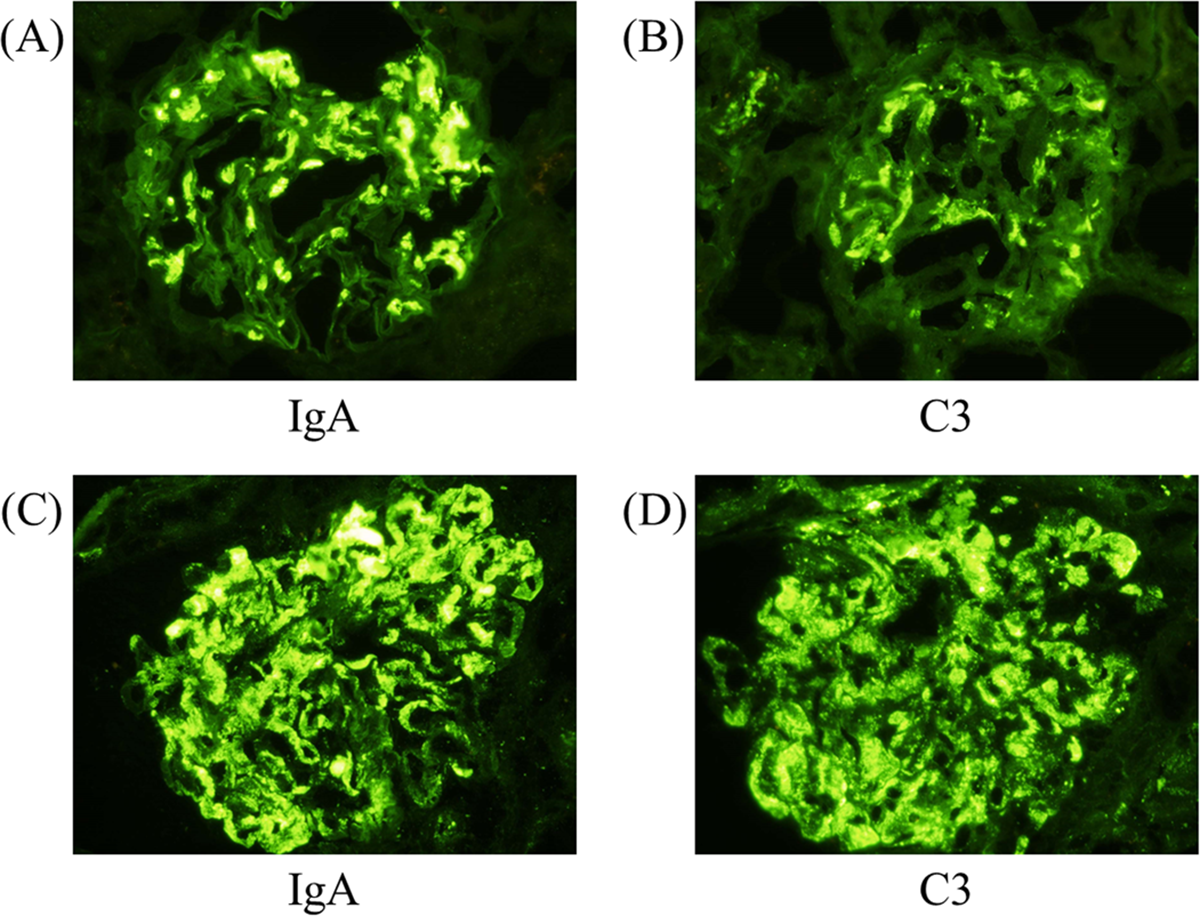
Does an IgA-positive kidney automatically mean primary IgA nephropathy? No. First identify the dominant immunoglobulin and its distribution, then examine the complete light-microscopy, immunofluorescence and electron-microscopy findings. Mesangial IgA-dominant or codominant deposits support IgA nephropathy only after relevant clinical context is considered. Electron-dense mesangial deposits describe the corresponding compartment on electron microscopy. [1]
Actual renal-biopsy immunofluorescence, not skin tissue. In the source's typical IgA nephropathy example, panel A shows mesangial IgA and panel B mesangial C3. Panels C and D show an atypical example with mesangial and capillary staining. All source panels are labeled at original magnification ×400. Kitamura et al., PLOS ONE, 2019, Figure 1, reproduced unmodified under CC BY 4.0. Source and provenance [3]Compare panel A with panel C before reading the distinction
Panel A illustrates mesangial IgA. Panel C includes capillary staining as well. Neither image alone supplies the infection history or systemic findings needed to classify an individual patient's disease.
Palpable purpura with joint or abdominal symptoms makes IgA vasculitis important; a renal IgA stain cannot distinguish that systemic syndrome by itself. Active staphylococcal infection, low C3 and an exudative, neutrophil-rich biopsy raise concern for IgA-dominant infection-related glomerulonephritis. Infection treatment and source control take priority over reflex use of a primary-IgA immunosuppression regimen. Chronic liver disease and other secondary settings also require assessment. [1][3][15]
Strong linear basement-membrane IgG with necrotizing nephritis suggests anti-GBM disease; absence of hemoptysis does not remove the need for urgent assessment. A full-house immune pattern with systemic lupus findings, anti-dsDNA and low complement points toward lupus nephritis. These patterns demand a different assessment from mesangial IgA alone. [12][13]
In lupus nephritis, the full-house pattern identifies the context, but class and activity change treatment. Mesangial-only class I/II disease with low-level proteinuria generally does not require a separate renal induction regimen; immunosuppression is guided by extrarenal lupus. Active proliferative class III/IV nephritis and nephrotic presentations require different assessment. [12]
A membranoproliferative pattern includes capillary-wall remodeling and double contours, sometimes called tram tracks. It describes injury rather than one etiology. Immune-complex drivers include hepatitis-related disease, cryoglobulinemia and autoimmune disease; complement-dominant disease requires a different etiologic assessment. Dense deposit disease is a form of C3 glomerulopathy, not simply an interchangeable name for all membranoproliferative disease. Do not require both C3 and C4 to be low in every example. [16]
Long-standing familial hematuria, sensorineural hearing loss or characteristic basement-membrane abnormalities suggest a COL4-associated disorder. A respiratory illness or an episode of visible blood does not erase that inherited pattern. Genetic evaluation may be appropriate, and a thin basement membrane should not be called universally benign. Follow proteinuria and kidney function even when current filtration is preserved. [10]
Read a kidney specimen, not just an IgA label
Compare A with C. Is the bright staining confined to the mesangial regions, or does it also follow capillary walls?
Kidney biopsy immunofluorescence. A/B show mesangial IgA/C3 in a typical case. C/D show mesangial and capillary IgA/C3 in an atypical case. All original magnifications ×400. Open full-size image. Kitamura et al. (2019), Figure 1. CC BY 4.0. Re-encoded as PNG; image content unchanged.Compare your observation with the reported finding
A and B concentrate the signal in mesangial regions. C and D also outline capillary areas. The second pattern needs clinicopathologic interpretation; IgA positivity alone cannot distinguish primary IgA nephropathy from every secondary or infection-related process.
Follow two outcomes over time
What if urine protein improves while filtration keeps falling? Track both protein excretion and serial eGFR. Proteinuria reflects clinically important barrier injury and kidney stress. eGFR estimates filtration function. The measurements inform each other but are not interchangeable. An improved protein value does not prove that a patient has stopped losing kidney function.
Original schematic using fictional teaching data, not a validated risk calculator. Both examples have protein excretion falling from 1.8 to 0.6 g/day. Example A has sustained eGFR loss, while example B remains comparatively stable. The contrast explains why both outcomes need follow-up.Choose which example needs reassessment for sustained functional loss
Example A falls from eGFR 74 to 52 over two years. The better protein result does not explain away that sustained loss. Example B is more stable, but residual proteinuria still needs risk-directed follow-up.
KDIGO 2025 considers persistent proteinuria of at least 0.5 g/day a progression-risk signal. The treatment goal is proteinuria below 0.5 g/day, ideally below 0.3, with preservation of kidney function. Do not wait for every patient to cross an older 1 g/day trial-oriented threshold before discussing risk. Equally, crossing 0.5 g/day does not automatically select a particular immunosuppressant. Patient characteristics, eligibility, preferences, safety and available therapies still matter. [1]
MEST-C records mesangial hypercellularity, endocapillary hypercellularity, segmental sclerosis, tubular atrophy/interstitial fibrosis, and crescents. It adds prognostic information from sampled tissue. Neither MEST-C nor the International IgAN Prediction Tool predicts which particular immunosuppressant will work. A crescent score with stable eGFR is not the same clinical situation as rapidly progressive loss of filtration. Avoid a single fixed lifetime-risk percentage for all patients. [1]
Check whether a change is sustained and whether an acute reversible cause is present. Volume depletion, obstruction and medications can alter filtration. A modest early eGFR dip after an SGLT2 inhibitor may be compatible with continued treatment when it stabilizes and the patient tolerates therapy. A decline continuing over years on unchanged treatment is not that brief initiation effect. [14]