A stone is not just a stone. The crystal shape, urine pH, and imaging appearance together point to a different metabolic culprit and a different prevention strategy. Get the stone type wrong, and the patient keeps making new ones.
The Four Stones
Match the Crystal to the Culprit
Four stone types, four distinct clue packages. Every board question buries the answer in one of them. Flip each card, then commit the pattern.
A 45-year-old man on a high-protein, low-fluid diet comes to the emergency department with acute, severe right flank pain radiating to the groin for the past 3 hours. He has gross hematuria. He has never had a stone before. Temperature is 37.0 C; he is writhing in distress and unable to find a comfortable position. Examination reveals right costovertebral angle tenderness. His urine pH is 5.1. Serum creatinine is 1.1 mg/dL. A plain abdominal X-ray shows no opacities over the urinary tract.
The X-ray shows nothing. What does that tell you about this stone, and what imaging should you get next?
There are four stone types that matter. Calcium oxalate accounts for about 80 percent of all stones. The other three each carry a board-specific clue set: struvite means urease, uric acid means radiolucent in acidic urine, and cystine means children with a transport defect. The clue in the stem tells you which world you are in. Flip each card.
Calcium OxalateTap to flip · Most common
The default stone
~80% of stones. Crystal shape: envelope or dumbbell. Radiopaque on X-ray (appears white). Forms in normal to acidic urine. Caused by hypocitraturia, malabsorption (Crohn disease), ethylene glycol, or excess vitamin C. Prevention: thiazide diuretic (reduces urinary calcium), potassium citrate (raises urine pH and citrate), high fluid intake.
StruviteTap to flip · Urease
Infection stone
Magnesium ammonium phosphate. Crystal shape: coffin-lid. Radiopaque. Grows into staghorn calculi filling the renal pelvis. Forms in alkaline urine (pH > 7.5) created by urease-producing bacteria: Proteus mirabilis, Klebsiella, or Pseudomonas. Urease splits urea into ammonia, which alkalinizes urine and precipitates the stone. You must treat the infection or the stone regrows.
Uric AcidTap to flip · Radiolucent
Invisible on X-ray
Radiolucent on plain X-ray (not visible on KUB). Crystal shape: rhomboid or needle-like. Forms in acidic urine (pH < 5.5). Associated with gout, high purine diets, myeloproliferative disorders, and Lesch-Nyhan syndrome. Treatment: alkalinize the urine with potassium citrate (goal pH 6.0–6.5), allopurinol to reduce uric acid production, reduce dietary purines.
CystineTap to flip · Children
Transport defect
Crystal shape: hexagonal. Mildly radiopaque. Forms in acidic urine. Caused by a defect in the amino acid transporter for COLA: Cystine, Ornithine, Lysine, and Arginine in the proximal tubule and gut. Autosomal recessive. Recurrent stones in children and young adults is the flag. The sodium cyanide-nitroprusside urine test turns red-purple with cystinuria. Treatment: high fluid intake, alkalinize urine, D-penicillamine or tiopronin if needed.
From the Attending
The mnemonic the boards love for radiolucent stones: "I can't c (see) u (you)" stands for cystine and uric acid. Both are invisible on a plain X-ray. If a stem hands you a negative KUB with a classic stone presentation, your differential immediately narrows to those two. Then let urine pH and demographics sort it: acidic urine plus gout equals uric acid; hexagonal crystals plus a young patient equals cystine.
Clinical Images
Calcium oxalate crystals · envelope shape · tap
Staghorn calculus · struvite · tap
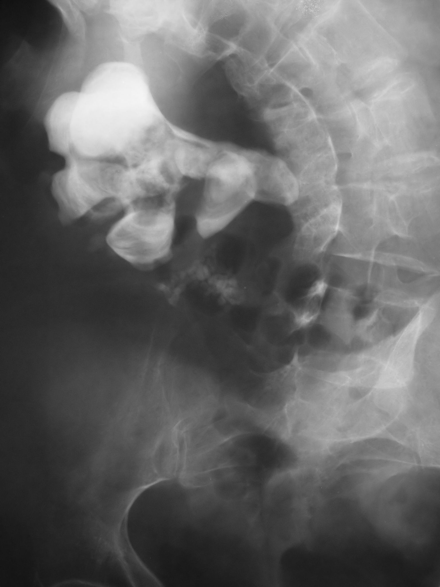
Non-contrast CT · hyperdense ureteral stone · tap
At the Bedside
Pain, Blood, and the Blocked Kidney
The classic presentation is hard to miss. The dangerous complication is not. Post-renal oliguria from obstruction is the reason kidney stones matter beyond pain management.
The classic presentation is acute colicky flank pain. A stone leaving the kidney and entering the ureter generates severe, cramping pain starting in the flank and radiating downward toward the groin as the stone migrates. The pain is colicky because peristaltic waves periodically compress the stone, and ureteral spasm drives the cramping. The patient is usually writhing and cannot find a comfortable position, which is the opposite of the peritoneal pain patient who lies completely still.
Hematuria almost always accompanies the pain. The stone abrades the urothelium. Gross or microscopic hematuria is present in roughly 90 percent of stone episodes. Its absence does not exclude a stone, but its presence in a writhing patient with flank-to-groin pain is essentially a stone until proven otherwise.
No fever, no pyuria. An uncomplicated stone causes no infection and no systemic illness. Fever plus stone is an emergency: an obstructed, infected kidney is sepsis waiting to happen and needs urgent drainage, not just pain control.
Board Trap
The post-renal oliguria vignette is the one that kills. A patient presents with acute flank-to-groin pain plus a sudden drop in urine output. The instinct is to think intrinsic renal failure, but the obstructing stone is the cause. Post-renal acute kidney injury from nephrolithiasis requires either bilateral ureteral obstruction or a stone in the solitary functioning kidney. The mechanism: back-pressure collapses the renal tubules and shuts down glomerular filtration. Workup is non-contrast CT. Not ultrasound, not urinalysis. Non-contrast CT.
Walk the obstruction chain once. Tap each step to reveal the beat.
A 58-year-old man on a high-protein, low-fluid diet develops acute right flank pain radiating to the groin plus his urine output drops from 1200 mL/day to 200 mL/day. What is the mechanism of the oliguria?
Right. The stone occludes the ureter. Hydrostatic pressure builds upstream in the collecting system and proximal tubule. When that pressure exceeds the driving pressure for filtration, GFR drops. This is post-renal AKI, fully reversible once the obstruction is relieved. The key: the BUN and creatinine rise, but the urine sediment is bland (no casts, no protein). Mechanical back-pressure, not tubular death.
The same patient has a plain abdominal X-ray that shows no stone. Can you exclude nephrolithiasis, or do you need more imaging?
Right. Plain X-ray misses uric acid and cystine stones entirely, and misses small calcium stones. Non-contrast CT detects virtually all stones and grades the hydronephrosis. This is the answer every time you are asked what workup to get for a suspected stone. Non-contrast CT, not KUB, not ultrasound (unless pregnant).
Non-contrast CT shows a 4 mm right ureteral stone with mild hydronephrosis and no signs of infection. The patient is in pain but tolerating oral fluids. Best management?
Right. Stones under 5 mm pass spontaneously in most patients. Alpha-blockers relax the ureteral smooth muscle and speed passage. Ketorolac is the analgesic of choice because it also reduces ureteral spasm. Intervention is reserved for stones greater than 10 mm, failure to pass after 4 to 6 weeks, infection, single kidney, or intractable pain. Under 5 mm: push fluids, relax the ureter, control pain, and wait.
From the Attending
Know the intervention thresholds cold. Under 5 mm: watch and wait with an alpha-blocker. 5 to 10 mm: borderline; many pass but intervention is on the table. Over 10 mm: spontaneous passage unlikely; refer for ureteroscopy or extracorporeal shock wave lithotripsy. Staghorn calculus: percutaneous nephrolithotomy, because lithotripsy cannot handle that volume. Infected obstructed kidney at any size: urgent drainage first, stone second.
The Identifier
Which Stone Is This?
Answer four quick clinical questions and the tree lands you on a stone type with the full clue set. Run it until the logic is automatic.
Stone-Type Decision Tree
Tap the answer that fits the stem. The tree routes you to the stone and shows you why.
Is the stone visible on a plain abdominal X-ray (KUB)?
What is the urine pH?
Is there a history of recurrent urinary tract infections, or does the stone fill the renal pelvis in a branching pattern (staghorn)?
What is the patient profile?
Stone Type Identified
Calcium Oxalate
Most common stone (~80%). Radiopaque. Crystal shape: envelope or dumbbell. Urine pH normal to acidic. Causes: hypocitraturia, Crohn disease or other malabsorptive conditions, excess vitamin C, ethylene glycol ingestion. The boards also test calcium phosphate, which is radiopaque and forms in alkaline urine; if the context is distal renal tubular acidosis (type 1 RTA), think calcium phosphate stones in that alkaline, high-calcium milieu.
Prevention: thiazide diuretic (reduces urinary calcium), potassium citrate (raises urine pH and adds citrate, which chelates calcium and inhibits stone nucleation), adequate fluid intake (urine output > 2 L/day), moderate dietary calcium (low-calcium diets paradoxically increase oxalate absorption).
Stone Type Identified
Struvite (Magnesium Ammonium Phosphate)
Radiopaque. Crystal shape: coffin-lid. Forms in alkaline urine (pH > 7.5) from urease-producing bacteria. The mechanism chain: urease-positive bacteria (Proteus mirabilis, Klebsiella, Pseudomonas) hydrolyze urea → releases NH3 → urine pH rises above 7.5 → magnesium, ammonium, and phosphate precipitate together → stone grows rapidly and branches into the collecting system forming a staghorn calculus.
Prevention: eradicate the underlying infection. Acetohydroxamic acid can inhibit urease if infection cannot be cleared. These stones do not dissolve with pH manipulation; they require mechanical removal followed by definitive antibiotics.
Stone Type Identified
Uric Acid
Radiolucent on plain X-ray, but visible on CT (appears hyperdense on non-contrast CT). Crystal shape: rhomboid or needle-like. Urine pH < 5.5. Associated with gout, high-purine diets (red meat, organ meat, shellfish), myeloproliferative disorders with high cell turnover, and Lesch-Nyhan syndrome.
Prevention and treatment: alkalinize the urine with potassium citrate (goal urine pH 6.0 to 6.5), reduce dietary purines, allopurinol to reduce urate production. Unlike calcium stones, uric acid stones can dissolve with medical management (alkalinization alone can chemically dissolve existing stones).
Stone Type Identified
Cystine
Mildly radiopaque (less than calcium; may look faint on X-ray). Crystal shape: hexagonal. Urine pH acidic. Autosomal recessive defect in the COLA transporter (Cystine, Ornithine, Lysine, Arginine) in the proximal tubule and intestine. All four amino acids spill into the urine. Only cystine is insoluble enough to precipitate.
Detection: sodium cyanide-nitroprusside test turns brick-red with cystinuria. Prevention: very high fluid intake, alkalinize urine (cystine is more soluble at pH > 7.0), D-penicillamine or tiopronin (chelate cystine to form more soluble complexes) for refractory cases.
Quick-reference: stone types at a glance. The table locks in the comparisons the boards probe most.
Stone Type
Crystal Shape
Radiopacity
Urine pH
Key Association
Calcium oxalate
Envelope or dumbbell
Radiopaque
Acidic to normal
Most common; hypocitraturia, Crohn, vit C excess
Struvite
Coffin-lid
Radiopaque
Alkaline (>7.5)
Urease bacteria; staghorn calculus
Uric acid
Rhomboid / needle
Radiolucent
Acidic (<5.5)
Gout, high purines; dissolves with alkalinization
Cystine
Hexagonal
Faintly opaque
Acidic
COLA defect; children/young adults
After the Stone Passes
Prevention is Stone-Type Specific
Generic "drink more water" is not enough. After a stone passes, a 24-hour urine collection identifies the metabolic defect and drives targeted prevention. Get the type wrong, and the treatment is wrong.
All stone types share one intervention: high fluid intake. The goal is urine output above 2 liters per day, which dilutes every lithogenic substance regardless of stone type. After that, the strategy diverges. Tap each card for type-specific prevention.
Calcium Oxalate
Targeted prevention?
Thiazide diuretic reduces urinary calcium excretion. Potassium citrate increases urinary citrate (which complexes calcium and raises pH). If hypercalciuria from hyperparathyroidism, treat the primary cause. Surprisingly, do not restrict dietary calcium: low oral calcium means more free oxalate absorbed in the gut, which raises urinary oxalate. Dietary restriction of oxalate-rich foods (spinach, nuts, chocolate) helps only if oxalate is high on 24-hour urine.
Struvite
Targeted prevention?
Eradicate the underlying urease-producing infection. Without killing the bacteria, the stone regrows on residual fragments. Acetohydroxamic acid (urease inhibitor) reduces urine ammonium when infection cannot be cleared. These stones require mechanical removal (percutaneous nephrolithotomy for staghorns) followed by antibiotics; pH manipulation alone cannot dissolve them.
Uric Acid
Targeted prevention?
Alkalinize the urine with potassium citrate (goal urine pH 6.0 to 6.5): cystine and uric acid are far more soluble at higher pH, and existing uric acid stones can actually dissolve. Allopurinol reduces urate production in patients with gout or overproduction. Reduce dietary purines. Avoid dehydration. This is the only stone type that is chemically dissolvable.
Cystine
Targeted prevention?
Very high fluid intake (3+ L/day). Alkalinize urine to above pH 7.0 with potassium citrate (cystine is more soluble in alkaline urine). For refractory patients: D-penicillamine or tiopronin chelate cystine to form mixed disulfide complexes that are far more soluble. These drugs have significant side effects and are reserved for failure of conservative measures.
Board Trap
The calcium restriction trap. A patient with recurrent calcium oxalate stones is counseled to eat a low-calcium diet. This is wrong and makes the situation worse. Gut oxalate needs calcium to bind it for excretion. When dietary calcium is low, free oxalate is absorbed in the colon and excreted by the kidneys, raising urinary oxalate and stone risk. The correct advice: maintain normal dietary calcium, but reduce sodium and animal protein, both of which raise urinary calcium. Restrict oxalate-rich foods only if urinary oxalate is elevated on the 24-hour collection.
The 24-hour urine collection is how you find the defect. It measures calcium, oxalate, urate, citrate, sodium, creatinine, pH, and volume. The results tell you exactly which abnormality is driving stone formation, so you can target it. First-time stone formers over 50 and all recurrent stone formers get this test. It is the only way to match prevention to mechanism.
Prove It
Board Walkthrough
Six original clinical vignettes, 5 dealt per round, answer choices shuffled, never repeating within a round. The glowing clues activate after you answer, showing which features sealed the diagnosis.
Medically reviewed by Kaitlyn Cocuzzo, MD and Fatima Ali, DO · Last reviewed June 2026
Bone Wizardry is an independent educational resource for visual learning in the medical sciences. It is not affiliated with, endorsed by, or sponsored by any licensing or examination board, contains no real or recalled examination questions, and does not guarantee any educational or examination outcome.