Artery of the Ligamentum Teres and Femoral-Head Perfusion
Trace the foveal and retinacular routes, compare growth and injury patterns, and distinguish vascular anatomy from evidence of femoral-head ischemia.
A vessel can enter the femoral head without being its main supply. Start with where the vessel travels, then ask how much reserve that route can actually provide.
Which route reaches which part of the head?
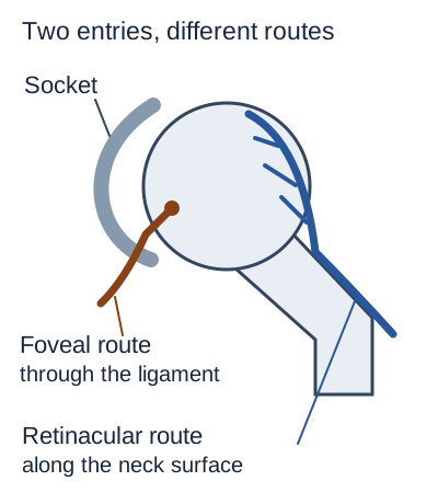
The artery in the ligamentum teres is also called the foveal artery. Its name describes its route through the ligament of the femoral head. It does not mean that this vessel supplies the whole head or can replace every vessel damaged by a fracture.
The ligament extends from the acetabular notch and transverse acetabular ligament region to the fovea capitis, the small nonarticular pit on the head. The ligament is inside the capsule but covered by synovium, so it is described as intra-articular and extrasynovial. Its vessel usually comes from the acetabular branch of the obturator artery, typically from its posterior division; anatomical variation is possible. [4][14]
Trace where each route enters. The small medial pit is not the entire weight-bearing surface. [1][4]
Trace it with a fingertip or your eyes. Follow the brown route from the socket toward the pit. Now follow the blue route upward along the outside of the neck. These are different entries, not two names for the same vessel.
The main adult route comes from the deep branch of the medial femoral circumflex artery. It travels posteriorly and gives rise to retinacular vessels that run beneath synovium along the neck before entering the head. Some important terminal vessels are called lateral epiphyseal vessels; here, lateral describes their location, not origin from the lateral circumflex artery. [1]
The medial circumflex artery commonly arises from the profunda femoris, also called the deep femoral artery. A direct femoral origin is a documented variant. The lateral circumflex artery contributes around the hip, but it should not displace the medial circumflex retinacular system as the usual answer for the dominant adult head supply. [1][5]
Try a changed situation. A vessel is seen traveling inside the ligament toward the pit. Which feature identifies it more directly, its course or the patient's age? The course identifies the foveal route. Age may affect vascular contribution, but it cannot turn that vessel into a retinacular branch.
A compact memory aid is “through the ligament” versus “along the neck.” Keep the actual names attached to those locations. Foveal means the first route; retinacular means the second.
Try it here · Checkpoint 1 of 3
Make your prediction before reading the choices. A first attempt is just a starting point.
Case 6
Show answer and explanations for case 6
A. Lateral femoral circumflex artery with its usual origin (Why this does not fit)
The circumflex name and femoral origin make this plausible. The posterosuperior neck course is the decisive feature favoring the medial rather than lateral circumflex vessel.
Reasoning steps for option A
Why consider lateral femoral circumflex artery with its usual origin?
The circumflex name and femoral origin make this plausible.
How does this discriminator affect the choice of Lateral femoral circumflex artery with its usual origin: The posterosuperior neck course is the decisive feature favoring the medial rather than lateral circumflex vessel?
The posterosuperior neck course is the decisive feature favoring the medial rather than lateral circumflex vessel.
When evaluating Lateral femoral circumflex artery with its usual origin, how does this case-specific rule guide the decision: Distal course can identify an artery despite a variant proximal origin?
Distal course can identify an artery despite a variant proximal origin.
B. Obturator artery with an enlarged acetabular branch (Why this does not fit)
An enlarged acetabular branch could contribute to the head. The observed posterior neck route differs from the characteristic ligament-to-fovea path.
Reasoning steps for option B
Why consider obturator artery with an enlarged acetabular branch?
An enlarged acetabular branch could contribute to the head.
How does this discriminator affect the choice of Obturator artery with an enlarged acetabular branch: The observed posterior neck route differs from the characteristic ligament-to-fovea path?
The observed posterior neck route differs from the characteristic ligament-to-fovea path.
When evaluating Obturator artery with an enlarged acetabular branch, how does this case-specific rule guide the decision: Distal course can identify an artery despite a variant proximal origin?
Distal course can identify an artery despite a variant proximal origin.
C. Profunda femoris artery with a short proximal segment (Why this does not fit)
The profunda often gives rise to the circumflex arteries. The described vessel itself turns around the proximal femur into head-bound branches rather than continuing as the profunda trunk.
Reasoning steps for option C
Why consider profunda femoris artery with a short proximal segment?
The profunda often gives rise to the circumflex arteries.
How does this discriminator affect the choice of Profunda femoris artery with a short proximal segment: The described vessel itself turns around the proximal femur into head-bound branches rather than continuing as the profunda trunk?
The described vessel itself turns around the proximal femur into head-bound branches rather than continuing as the profunda trunk.
When evaluating Profunda femoris artery with a short proximal segment, how does this case-specific rule guide the decision: Distal course can identify an artery despite a variant proximal origin?
Distal course can identify an artery despite a variant proximal origin.
D. Medial femoral circumflex artery with a variant origin (Best answer)
The posterior course and ascending head-bound branches identify the medial circumflex artery. A direct common femoral origin is a documented variant and does not change its distal identity.
Reasoning steps for option D
Which artery matches the posterior course and head-bound branches?
The posterior course and ascending head-bound branches identify the medial circumflex artery.
Does its direct femoral origin change that identification?
A direct common femoral origin is a documented variant and does not change its distal identity.
When evaluating Medial femoral circumflex artery with a variant origin, how does this case-specific rule guide the decision: Distal course can identify an artery despite a variant proximal origin?
Distal course can identify an artery despite a variant proximal origin.
Takeaway: Distal course can identify an artery despite a variant proximal origin.
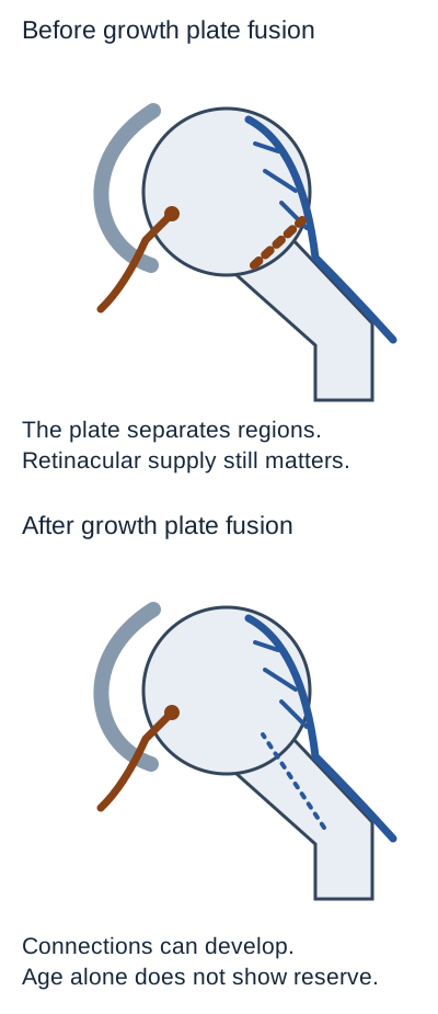
Do not substitute an age slogan for the vascular map. The growing head has an epiphysis, while the neck includes the metaphysis; an open growth plate separates these regions. Their connections change during development, and the retinacular supply is important in children as well as adults. [2]
The dashed brown line is the open growth plate, not a blocked artery. The lower dotted vessel indicates possible connections after fusion, not guaranteed rescue. [2][4]
Compare the two drawings. Find the growth plate in the upper view. Explain why a vessel supplying the neck cannot automatically be counted as a vessel crossing into the epiphysis. Then compare the lower view, where fusion permits a different pattern of connections.
Developmental injection work describes changing metaphyseal, lateral epiphyseal and ligamentous contributions. It does not support a universal rule that the foveal artery is the main childhood supply, becomes unimportant on a particular birthday, and disappears in every adult. The drawing deliberately omits percentages and exact age cutoffs. [2]
Adult ligament specimens can contain small arteries, veins and nerve bundles. Visible stump bleeding has also been documented during adult hip surgery; presence and absence both occur. [14] This establishes that the ligament is not simply an empty cord. It does not establish how much living femoral-head tissue those arteries perfuse, because a vessel within a ligament can nourish the ligament itself. [4]
Predict what a tracer result would establish. If injected dye reaches a small area near the fovea, it demonstrates a patent route under that experiment's conditions. It does not establish enough living blood flow to support the weight-bearing head after a different route is lost.
That distinction is vascular reserve, the ability of remaining routes to meet tissue needs. A collateral connection is useful anatomy, but its presence alone cannot quantify reserve. An inferior gluteal connection with the medial circumflex system has been demonstrated in dissections, so dismissing all gluteal contributions would also be too absolute. [3]
Apply the distinction. A child's radiograph shows head collapse while the artery near the fovea appears patent on another study. These findings can coexist. Patency of one route does not prove adequate delivery throughout the head.
Try it here · Checkpoint 2 of 3
Make your prediction before reading the choices. A first attempt is just a starting point.
Case 22
Show answer and explanations for case 22
A. The ligamentous route supplied half of head perfusion in each group (Why this does not fit)
The one-half fractions describe the proportion of patients with visible stump bleeding. Head perfusion was not measured, so those fractions cannot be reassigned to blood-flow contribution.
Reasoning steps for option A
Why consider the ligamentous route supplied half of head perfusion in each group?
The one-half fractions describe the proportion of patients with visible stump bleeding.
How does this discriminator affect the choice of The ligamentous route supplied half of head perfusion in each group: Head perfusion was not measured, so those fractions cannot be reassigned to blood-flow contribution?
Head perfusion was not measured, so those fractions cannot be reassigned to blood-flow contribution.
When evaluating The ligamentous route supplied half of head perfusion in each group, how does this case-specific rule guide the decision: A proportion of patients with bleeding is not a proportion of head blood flow?
A proportion of patients with bleeding is not a proportion of head blood flow.
B. The older group had twice the probability of foveal vessel patency (Why this does not fit)
There were twice as many positive observations in the older group, but also twice as many patients. The observed proportion is identical rather than doubled.
Reasoning steps for option B
Why consider the older group had twice the probability of foveal vessel patency?
There were twice as many positive observations in the older group, but also twice as many patients.
How does this discriminator affect the choice of The older group had twice the probability of foveal vessel patency: The observed proportion is identical rather than doubled?
The observed proportion is identical rather than doubled.
When evaluating The older group had twice the probability of foveal vessel patency, how does this case-specific rule guide the decision: A proportion of patients with bleeding is not a proportion of head blood flow?
A proportion of patients with bleeding is not a proportion of head blood flow.
C. Observed stump bleeding occurred in half of each age group (Best answer)
Both fractions equal one half, so these grouped observations do not separate bleeding status by the two age groups. They measure patient counts with stump bleeding, not the fraction of head perfusion provided by the foveal route.
Reasoning steps for option C
What are the two observed proportions?
Both fractions equal one half, so these grouped observations do not separate bleeding status by the two age groups.
Why are those proportions not head-flow fractions?
They measure patient counts with stump bleeding, not the fraction of head perfusion provided by the foveal route.
When evaluating Observed stump bleeding occurred in half of each age group, how does this case-specific rule guide the decision: A proportion of patients with bleeding is not a proportion of head blood flow?
A proportion of patients with bleeding is not a proportion of head blood flow.
D. The younger group had twice the reserve after retinacular interruption (Why this does not fit)
A larger reserve in younger patients might be hypothesized. Neither group underwent a reserve measurement, and equal stump-bleeding proportions do not establish that proposed difference.
Reasoning steps for option D
Why consider the younger group had twice the reserve after retinacular interruption?
A larger reserve in younger patients might be hypothesized.
How does this discriminator affect the choice of The younger group had twice the reserve after retinacular interruption: Neither group underwent a reserve measurement, and equal stump-bleeding proportions do not establish that proposed difference?
Neither group underwent a reserve measurement, and equal stump-bleeding proportions do not establish that proposed difference.
When evaluating The younger group had twice the reserve after retinacular interruption, how does this case-specific rule guide the decision: A proportion of patients with bleeding is not a proportion of head blood flow?
A proportion of patients with bleeding is not a proportion of head blood flow.
Takeaway: A proportion of patients with bleeding is not a proportion of head blood flow.
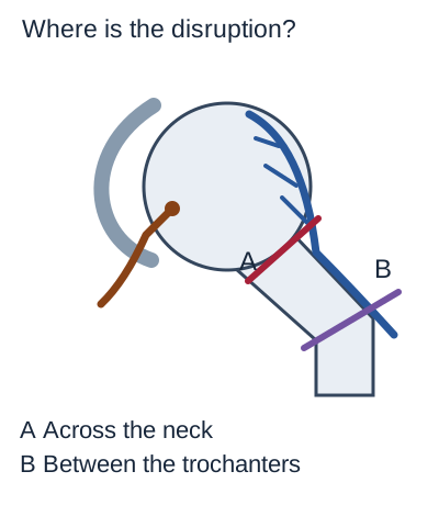
A displaced intracapsular femoral-neck fracture can disrupt the retinacular vessels on the neck. Loss of this supply can contribute to nonunion or later osteonecrosis, which means death of bone tissue from inadequate perfusion. A normal foot pulse does not exclude that local problem. [1][7]
Compare the fracture plane with the route on the neck. Displacement across the neck can threaten head perfusion even when distal pulses remain normal. [1][7]
Compare A with B before looking at the outcome. A crosses the neck near the head-bound vessels. B lies farther down in the trochanteric region, where head perfusion is generally less directly threatened. Location and displacement matter together; “hip fracture” alone is not precise enough to predict vascular injury.
Explore the remaining connections
This experiment changes one connection at a time in a simplified adult hip. Choose an interruption, compare its diagram with the starting view, and name the route that remains. Open either explanation immediately; no answer is required.
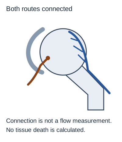
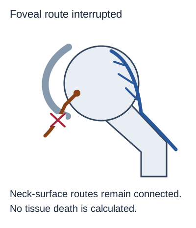
Connection is not a flow measurement. This is a qualitative connection experiment, not a clinical flow simulator. [1][3][4]Compare an interrupted foveal routeNeck-surface routes remain connected. This is a qualitative connection experiment, not a clinical flow simulator. [1][3][4]
The cross marks the foveal interruption. The neck-surface routes remain drawn as continuous because the experiment has not disrupted them. This does not guarantee normal tissue perfusion, but it does explain why loss of the foveal route alone is not equivalent to loss of the main adult retinacular supply.
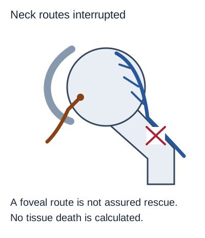
Compare interrupted neck-surface routesA foveal route is not assured rescue. This is a qualitative connection experiment, not a clinical flow simulator. [1][3][4]
The cross now marks the connection feeding the neck-surface routes. A foveal route remains, but that observation does not establish adequate reserve for the head. Preserving a small alternative route cannot be treated as proof that osteonecrosis is prevented.
Compare or reset. Both experiments can stay open for comparison. Close each one to return to the starting view. The images show connections rather than blood-flow fractions, and their unchanged bone shading deliberately avoids predicting a territory or time of tissue death.
The complete result is also worth stating without operating the controls. Foveal interruption leaves the modeled retinacular connection intact; retinacular interruption removes the usual dominant adult route. Neither state, by itself, supplies enough information to calculate a patient's remaining perfusion. [1][3][4]
Now change the injury. A dashboard impact produces a posterior hip dislocation rather than a neck fracture. Stretching or tearing around the hip can still threaten vessels and the sciatic nerve. A traumatic dislocation requires emergency assessment and reduction by trained clinicians; restoring alignment does not erase the need to assess the injury and follow for later complications. [8]
Try it here · Checkpoint 3 of 3
Make your prediction before reading the choices. A first attempt is just a starting point.
Case 16
Show answer and explanations for case 16
A. Its distal pulses indicate poorer perfusion of the entire affected leg (Why this does not fit)
Both patients have normal distal pulses, so the supplied pulse finding does not discriminate between them. The relevant distinction is local head-bound vascular anatomy and displacement.
Reasoning steps for option A
Why consider its distal pulses indicate poorer perfusion of the entire affected leg?
Both patients have normal distal pulses, so the supplied pulse finding does not discriminate between them.
How does this discriminator affect the choice of Its distal pulses indicate poorer perfusion of the entire affected leg: The relevant distinction is local head-bound vascular anatomy and displacement?
The relevant distinction is local head-bound vascular anatomy and displacement.
When evaluating Its distal pulses indicate poorer perfusion of the entire affected leg, how does this case-specific rule guide the decision: Compare fracture location and displacement, not just the diagnosis of hip fracture?
Compare fracture location and displacement, not just the diagnosis of hip fracture.
B. Its fracture plane crosses the ligamentum teres at the foveal attachment (Why this does not fit)
The foveal attachment lies on the head and is not the subcapital neck fracture plane. The increased concern is disruption of the retinacular supply along the neck.
Reasoning steps for option B
Why consider its fracture plane crosses the ligamentum teres at the foveal attachment?
The foveal attachment lies on the head and is not the subcapital neck fracture plane.
How does this discriminator affect the choice of Its fracture plane crosses the ligamentum teres at the foveal attachment: The increased concern is disruption of the retinacular supply along the neck?
The increased concern is disruption of the retinacular supply along the neck.
When evaluating Its fracture plane crosses the ligamentum teres at the foveal attachment, how does this case-specific rule guide the decision: Compare fracture location and displacement, not just the diagnosis of hip fracture?
Compare fracture location and displacement, not just the diagnosis of hip fracture.
C. Its fracture plane and displacement more directly threaten head-bound retinacular vessels (Best answer)
The subcapital injury crosses the intracapsular neck where head-bound vessels ascend. The intertrochanteric injury is farther from that route, although no fracture pattern gives an absolute guarantee against complications.
Reasoning steps for option C
Which fracture lies closer to ascending head-bound vessels?
The subcapital injury crosses the intracapsular neck where head-bound vessels ascend.
Why is the second fracture different without being risk-free?
The intertrochanteric injury is farther from that route, although no fracture pattern gives an absolute guarantee against complications.
When evaluating Its fracture plane and displacement more directly threaten head-bound retinacular vessels, how does this case-specific rule guide the decision: Compare fracture location and displacement, not just the diagnosis of hip fracture?
Compare fracture location and displacement, not just the diagnosis of hip fracture.
D. Its blood supply normally depends on the lateral circumflex descending branch (Why this does not fit)
The descending lateral circumflex branch is not the usual dominant route to the adult head. The injury comparison turns on the medial circumflex retinacular route instead.
Reasoning steps for option D
Why consider its blood supply normally depends on the lateral circumflex descending branch?
The descending lateral circumflex branch is not the usual dominant route to the adult head.
How does this discriminator affect the choice of Its blood supply normally depends on the lateral circumflex descending branch: The injury comparison turns on the medial circumflex retinacular route instead?
The injury comparison turns on the medial circumflex retinacular route instead.
When evaluating Its blood supply normally depends on the lateral circumflex descending branch, how does this case-specific rule guide the decision: Compare fracture location and displacement, not just the diagnosis of hip fracture?
Compare fracture location and displacement, not just the diagnosis of hip fracture.
Takeaway: Compare fracture location and displacement, not just the diagnosis of hip fracture.
Look for the structural change before naming an artery. In a child, weeks of limping or knee pain with limited hip abduction and internal rotation should direct attention to the hip, even if the knee examination is normal. Perthes disease is childhood osteonecrosis of the femoral head, not a diagnosis made from the site of pain alone. [6]
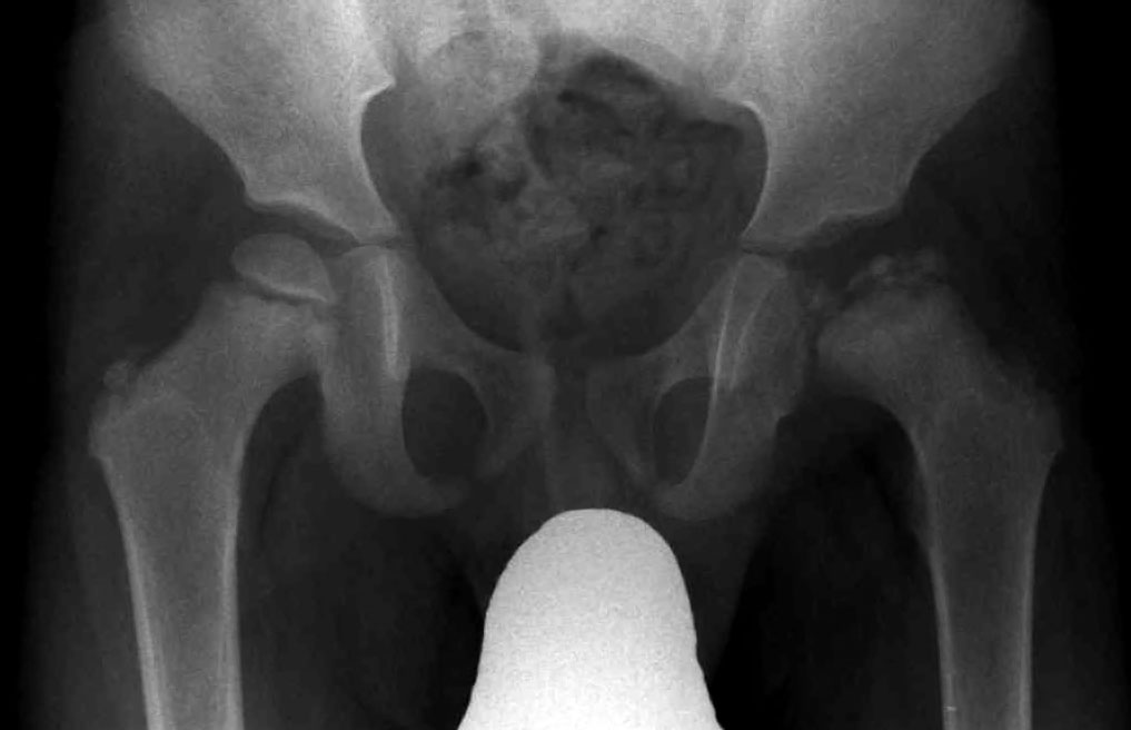
Compare femoral-head contours and density: the flatter, irregular head on the displayed right contrasts with the smoother opposite side. Confirm laterality from the radiograph marker before assigning a patient side. Image: J. Lengerke, public domain. [12]
Compare the two head outlines. On the right side of this displayed radiograph, the head is flatter and more irregular than the opposite head. Its density is also uneven. Trace the smoother outline on the other side, then identify where the abnormal outline departs from that curve.
Separate the observation from the inference
Observation is the changed shape and density of the head. The image shows Perthes disease. The image does not show which named arterial trunk was first affected, and the image alone cannot establish age, symptoms or stage.
Perthes disease involves interruption of head perfusion, but its initiating cause is incompletely understood. “A child has Perthes disease” does not prove “the medial circumflex artery is occluded,” and it does not prove isolated failure of the foveal artery. A source of blood supply and a demonstrated cause of disease are different claims. [6]
Use a contrasting presentation. An adolescent has hip or knee pain, a leg that rests externally rotated, and imaging that shows displacement through the growth plate. This points toward slipped capital femoral epiphysis, or SCFE, rather than diagnosing Perthes disease from limp alone. Suspected SCFE warrants urgent clinical assessment; confirmed SCFE requires avoiding weight bearing while orthopedic treatment is arranged. [9]
The practical comparison is the structure affected. Perthes changes the developing head through osteonecrosis; SCFE displaces the head relative to the neck through the physis. Both can involve vascular complications, but they are not interchangeable labels for a painful pediatric hip.
Is this a vessel problem, a nerve problem, or insufficient evidence?
A pelvis that drops on the unsupported side during single-leg stance indicates impaired abductor support on the stance side. The superior gluteal nerve supplies gluteus medius, gluteus minimus and tensor fasciae latae, but pain, tendon injury and other problems can also impair that support. A Trendelenburg sign alone is not a test of the superior gluteal artery, nor proof of a nerve lesion. [11]
Sort the evidence by what it measures. Weakness and electromyography help localize a motor deficit. Head collapse suggests structural bone failure. A visible lumen, dye injection or a large foot pulse answers a different question from adequate delivery to head tissue.
For example, weakness of all three superior-gluteal-innervated muscles with matching denervation is more specific than an isolated pelvic drop. Conversely, delayed groin pain and head collapse after a neck injury favor a vascular bone complication rather than explaining the findings as isolated abductor denervation.
Systemic disease also changes the reasoning. Sickle cell disease can cause femoral-head osteonecrosis without a traumatic tear of a named hip artery. When the history supports a systemic process, do not force the explanation into the same large-vessel injury used for a displaced fracture. [10]
Test the limit of a reassuring result. An adult's imaging shows a continuous ligamentum teres. What remains unknown after a femoral-neck fracture? Ligament continuity does not measure the small vessel's patency or prove adequate head perfusion. Even direct visualization of a small artery would not, by itself, quantify reserve. [4][13]
Ligament signal on MR arthrography can also overlap between intact and partially torn tissue. Interpret the structural appearance in context rather than treating it as a direct measurement of the artery inside. [13]
A practical decision sequence
First locate the structure actually demonstrated by the stem, examination or image. Then trace the relevant route and account for development, displacement or systemic disease. Finally stop at the conclusion the evidence supports, rather than converting a possible mechanism into a proven artery occlusion.
Use the same discipline when two reassuring facts appear together. Normal distal pulses plus restored joint alignment are valuable findings, but neither independently establishes that the small head-bound vessels remained intact. Their combination does not create a measurement that was never made.
Try the sequence without help. A patient has preserved ankle strength, a healed neck fracture and later flattening of the femoral head. Decide whether the central problem is motor localization or bone perfusion, then explain why fracture union did not settle the second question.
Practice with new presentations
Use the supplied findings to choose the best answer. The questions distinguish a likely mechanism from a demonstrated lesion; every option has an explanation you can revisit. Three selected cases also appear beside the relevant teaching sections.
Case 1
Show answer and explanations for case 1
A. Acetabular branches traveling within the ligamentum teres (Why this does not fit)
The foveal route does enter the head and is therefore a tempting choice. However, this fracture crosses the neck rather than the ligament, placing the retinacular supply at more direct risk.
Reasoning steps for option A
Why consider acetabular branches traveling within the ligamentum teres?
The foveal route does enter the head and is therefore a tempting choice.
How does this discriminator affect the choice of Acetabular branches traveling within the ligamentum teres: this fracture crosses the neck rather than the ligament, placing the retinacular supply at more direct risk?
However, this fracture crosses the neck rather than the ligament, placing the retinacular supply at more direct risk.
When evaluating Acetabular branches traveling within the ligamentum teres, how does this case-specific rule guide the decision: Locate the fracture before using a distal pulse to judge the head supply?
Locate the fracture before using a distal pulse to judge the head supply.
B. Descending branches of the lateral femoral circumflex artery (Why this does not fit)
The lateral circumflex system supplies structures around the proximal femur. Its descending branches are not the main route ascending the intracapsular neck to the head.
Reasoning steps for option B
Why consider descending branches of the lateral femoral circumflex artery?
The lateral circumflex system supplies structures around the proximal femur.
How does this discriminator affect the choice of Descending branches of the lateral femoral circumflex artery: Its descending branches are not the main route ascending the intracapsular neck to the head?
Its descending branches are not the main route ascending the intracapsular neck to the head.
When evaluating Descending branches of the lateral femoral circumflex artery, how does this case-specific rule guide the decision: Locate the fracture before using a distal pulse to judge the head supply?
Locate the fracture before using a distal pulse to judge the head supply.
C. Retinacular branches ascending along the femoral neck at the head-neck junction (Best answer)
The subcapital fracture lies beside the ascending head-bound vessels. Distal pulses assess a different vascular territory and do not exclude damage to these branches.
Reasoning steps for option C
Which part of the vascular map lies beside a subcapital fracture?
The subcapital fracture lies beside the ascending head-bound vessels.
Why do the foot pulses not settle that regional risk?
Distal pulses assess a different vascular territory and do not exclude damage to these branches.
When evaluating Retinacular branches ascending along the femoral neck at the head-neck junction, how does this case-specific rule guide the decision: Locate the fracture before using a distal pulse to judge the head supply?
Locate the fracture before using a distal pulse to judge the head supply.
D. Perforating branches entering the posterior femoral shaft (Why this does not fit)
Perforating arteries are important branches of the profunda femoris. Their shaft territory does not match a displaced subcapital injury threatening the head.
Reasoning steps for option D
Why consider perforating branches entering the posterior femoral shaft?
Perforating arteries are important branches of the profunda femoris.
How does this discriminator affect the choice of Perforating branches entering the posterior femoral shaft: Their shaft territory does not match a displaced subcapital injury threatening the head?
Their shaft territory does not match a displaced subcapital injury threatening the head.
When evaluating Perforating branches entering the posterior femoral shaft, how does this case-specific rule guide the decision: Locate the fracture before using a distal pulse to judge the head supply?
Locate the fracture before using a distal pulse to judge the head supply.
Takeaway: Locate the fracture before using a distal pulse to judge the head supply.
A. Femoral-head ischemic injury without identification of one occluded arterial trunk (Best answer)
The chronic limp, hip restriction and epiphyseal changes support Perthes disease. That syndrome establishes ischemic head injury but does not identify which named arterial trunk first became obstructed.
Reasoning steps for option A
What process fits the hip restriction and epiphyseal changes?
The chronic limp, hip restriction and epiphyseal changes support Perthes disease.
What arterial conclusion does the diagnosis still leave unproven?
That syndrome establishes ischemic head injury but does not identify which named arterial trunk first became obstructed.
When evaluating Femoral-head ischemic injury without identification of one occluded arterial trunk, how does this case-specific rule guide the decision: Recognizing Perthes disease does not identify a specific occluded artery?
Recognizing Perthes disease does not identify a specific occluded artery.
B. Foveal-artery disruption caused by displacement through the proximal femoral physis (Why this does not fit)
A physeal displacement can threaten vessels and produce hip or knee pain. The radiographs instead show head sclerosis and flattening without displacement through the physis.
Reasoning steps for option B
Why consider foveal-artery disruption caused by displacement through the proximal femoral physis?
A physeal displacement can threaten vessels and produce hip or knee pain.
How does this discriminator affect the choice of Foveal-artery disruption caused by displacement through the proximal femoral physis: The radiographs instead show head sclerosis and flattening without displacement through the physis?
The radiographs instead show head sclerosis and flattening without displacement through the physis.
When evaluating Foveal-artery disruption caused by displacement through the proximal femoral physis, how does this case-specific rule guide the decision: Recognizing Perthes disease does not identify a specific occluded artery?
Recognizing Perthes disease does not identify a specific occluded artery.
C. Medial circumflex occlusion demonstrated by the femoral epiphyseal contour change (Why this does not fit)
The medial circumflex system is important to head perfusion. An altered contour shows a structural consequence, not an angiographic demonstration of occlusion in that arterial trunk.
Reasoning steps for option C
Why consider medial circumflex occlusion demonstrated by the femoral epiphyseal contour change?
The medial circumflex system is important to head perfusion.
How does this discriminator affect the choice of Medial circumflex occlusion demonstrated by the femoral epiphyseal contour change: An altered contour shows a structural consequence, not an angiographic demonstration of occlusion in that arterial trunk?
An altered contour shows a structural consequence, not an angiographic demonstration of occlusion in that arterial trunk.
When evaluating Medial circumflex occlusion demonstrated by the femoral epiphyseal contour change, how does this case-specific rule guide the decision: Recognizing Perthes disease does not identify a specific occluded artery?
Recognizing Perthes disease does not identify a specific occluded artery.
D. Obturator-artery occlusion established by ischemia limited to the femoral epiphysis (Why this does not fit)
The obturator artery is associated with the foveal route. The radiographic distribution alone cannot establish its occlusion, and a small foveal route is not equivalent to the whole epiphyseal supply.
Reasoning steps for option D
Why consider obturator-artery occlusion established by ischemia limited to the femoral epiphysis?
The obturator artery is associated with the foveal route.
How does this discriminator affect the choice of Obturator-artery occlusion established by ischemia limited to the femoral epiphysis: The radiographic distribution alone cannot establish its occlusion, and a small foveal route is not equivalent to the whole epiphyseal supply?
The radiographic distribution alone cannot establish its occlusion, and a small foveal route is not equivalent to the whole epiphyseal supply.
When evaluating Obturator-artery occlusion established by ischemia limited to the femoral epiphysis, how does this case-specific rule guide the decision: Recognizing Perthes disease does not identify a specific occluded artery?
Recognizing Perthes disease does not identify a specific occluded artery.
Takeaway: Recognizing Perthes disease does not identify a specific occluded artery.
A. Fragmentation of the femoral epiphysis with a preserved head-neck relationship (Why this does not fit)
Fragmentation can occur with childhood femoral-head osteonecrosis. The adolescent rotational pattern favors physeal displacement rather than the preserved head-neck relationship in this option.
Reasoning steps for option A
Why consider fragmentation of the femoral epiphysis with a preserved head-neck relationship?
Fragmentation can occur with childhood femoral-head osteonecrosis.
How does this discriminator affect the choice of Fragmentation of the femoral epiphysis with a preserved head-neck relationship: The adolescent rotational pattern favors physeal displacement rather than the preserved head-neck relationship in this option?
The adolescent rotational pattern favors physeal displacement rather than the preserved head-neck relationship in this option.
When evaluating Fragmentation of the femoral epiphysis with a preserved head-neck relationship, how does this case-specific rule guide the decision: An adolescent rotational deformity can signal a physeal slip rather than head fragmentation?
An adolescent rotational deformity can signal a physeal slip rather than head fragmentation.
B. A transverse fracture through the femoral neck below the growth plate (Why this does not fit)
A neck fracture can threaten retinacular vessels. The gradual atraumatic presentation and rotational restriction in an adolescent favor a physeal slip over an acute transverse neck fracture.
Reasoning steps for option B
Why consider a transverse fracture through the femoral neck below the growth plate?
A neck fracture can threaten retinacular vessels.
How does this discriminator affect the choice of A transverse fracture through the femoral neck below the growth plate: The gradual atraumatic presentation and rotational restriction in an adolescent favor a physeal slip over an acute transverse neck fracture?
The gradual atraumatic presentation and rotational restriction in an adolescent favor a physeal slip over an acute transverse neck fracture.
When evaluating A transverse fracture through the femoral neck below the growth plate, how does this case-specific rule guide the decision: An adolescent rotational deformity can signal a physeal slip rather than head fragmentation?
An adolescent rotational deformity can signal a physeal slip rather than head fragmentation.
C. Superolateral displacement of the entire head beyond the acetabular rim (Why this does not fit)
A dislocated head can impair perfusion and hip motion. Displacement out of the socket is less consistent with the gradual atraumatic adolescent presentation than a proximal physeal slip.
Reasoning steps for option C
Why consider superolateral displacement of the entire head beyond the acetabular rim?
A dislocated head can impair perfusion and hip motion.
How does this discriminator affect the choice of Superolateral displacement of the entire head beyond the acetabular rim: Displacement out of the socket is less consistent with the gradual atraumatic adolescent presentation than a proximal physeal slip?
Displacement out of the socket is less consistent with the gradual atraumatic adolescent presentation than a proximal physeal slip.
When evaluating Superolateral displacement of the entire head beyond the acetabular rim, how does this case-specific rule guide the decision: An adolescent rotational deformity can signal a physeal slip rather than head fragmentation?
An adolescent rotational deformity can signal a physeal slip rather than head fragmentation.
D. Posteroinferior displacement of the femoral epiphysis relative to the neck (Best answer)
The adolescent presentation with external rotation and restricted internal rotation suggests SCFE. Displacement through the open proximal femoral physis accounts for the deformity and potential vascular injury.
Reasoning steps for option D
What explains the adolescent rotational pattern?
The adolescent presentation with external rotation and restricted internal rotation suggests SCFE.
What structural change and vascular concern follow from that diagnosis?
Displacement through the open proximal femoral physis accounts for the deformity and potential vascular injury.
When evaluating Posteroinferior displacement of the femoral epiphysis relative to the neck, how does this case-specific rule guide the decision: An adolescent rotational deformity can signal a physeal slip rather than head fragmentation?
An adolescent rotational deformity can signal a physeal slip rather than head fragmentation.
Takeaway: An adolescent rotational deformity can signal a physeal slip rather than head fragmentation.
A. Persistent complete femoral-artery interruption despite normal distal circulation (Why this does not fit)
A proximal arterial interruption is a possible major trauma complication. Persistent complete interruption does not fit the preserved distal circulation as well as a local injury to small head-bound vessels.
Reasoning steps for option A
Why consider persistent complete femoral-artery interruption despite normal distal circulation?
A proximal arterial interruption is a possible major trauma complication.
How does this discriminator affect the choice of Persistent complete femoral-artery interruption despite normal distal circulation: Persistent complete interruption does not fit the preserved distal circulation as well as a local injury to small head-bound vessels?
Persistent complete interruption does not fit the preserved distal circulation as well as a local injury to small head-bound vessels.
When evaluating Persistent complete femoral-artery interruption despite normal distal circulation, how does this case-specific rule guide the decision: Restored alignment does not prove that the original vascular injury has resolved?
Restored alignment does not prove that the original vascular injury has resolved.
B. Initial damage to head-bound vessels despite restoration of joint alignment (Best answer)
A dislocation can injure vessels even without a neck fracture. Reduction corrects alignment, but neither alignment nor distal pulses demonstrates that the original vascular injury was absent.
Reasoning steps for option B
Can a dislocation injure vessels without a neck fracture?
A dislocation can injure vessels even without a neck fracture.
What does successful reduction fail to demonstrate?
Reduction corrects alignment, but neither alignment nor distal pulses demonstrates that the original vascular injury was absent.
When evaluating Initial damage to head-bound vessels despite restoration of joint alignment, how does this case-specific rule guide the decision: Restored alignment does not prove that the original vascular injury has resolved?
Restored alignment does not prove that the original vascular injury has resolved.
C. Progressive sciatic denervation causing primary ischemia of the epiphyseal bone (Why this does not fit)
Sciatic injury is an important complication of posterior dislocation. Its motor and sensory consequences do not directly account for primary ischemic necrosis of the femoral head.
Reasoning steps for option C
Why consider progressive sciatic denervation causing primary ischemia of the epiphyseal bone?
Sciatic injury is an important complication of posterior dislocation.
How does this discriminator affect the choice of Progressive sciatic denervation causing primary ischemia of the epiphyseal bone: Its motor and sensory consequences do not directly account for primary ischemic necrosis of the femoral head?
Its motor and sensory consequences do not directly account for primary ischemic necrosis of the femoral head.
When evaluating Progressive sciatic denervation causing primary ischemia of the epiphyseal bone, how does this case-specific rule guide the decision: Restored alignment does not prove that the original vascular injury has resolved?
Restored alignment does not prove that the original vascular injury has resolved.
D. Redislocation of the head demonstrated by the concentric post-reduction image (Why this does not fit)
Redislocation would alter the relationship of the head to the socket. The supplied image instead documents restored alignment, which can coexist with a vascular complication from the initial injury.
Reasoning steps for option D
Why consider redislocation of the head demonstrated by the concentric post-reduction image?
Redislocation would alter the relationship of the head to the socket.
How does this discriminator affect the choice of Redislocation of the head demonstrated by the concentric post-reduction image: The supplied image instead documents restored alignment, which can coexist with a vascular complication from the initial injury?
The supplied image instead documents restored alignment, which can coexist with a vascular complication from the initial injury.
When evaluating Redislocation of the head demonstrated by the concentric post-reduction image, how does this case-specific rule guide the decision: Restored alignment does not prove that the original vascular injury has resolved?
Restored alignment does not prove that the original vascular injury has resolved.
Takeaway: Restored alignment does not prove that the original vascular injury has resolved.
A. Medial circumflex artery through its retinacular branches (Why this does not fit)
The medial circumflex system supplies much of the head. Its usual retinacular route travels along the neck, not within the described band from socket to fovea.
Reasoning steps for option A
Why consider medial circumflex artery through its retinacular branches?
The medial circumflex system supplies much of the head.
How does this discriminator affect the choice of Medial circumflex artery through its retinacular branches: Its usual retinacular route travels along the neck, not within the described band from socket to fovea?
Its usual retinacular route travels along the neck, not within the described band from socket to fovea.
When evaluating Medial circumflex artery through its retinacular branches, how does this case-specific rule guide the decision: A vessel inside the ligamentum teres is identified by its route to the fovea?
A vessel inside the ligamentum teres is identified by its route to the fovea.
B. Obturator artery through its acetabular branch within the ligamentum teres (Best answer)
The band and its attachment identify the ligamentum teres and fovea. The vessel in that route usually arises from the acetabular branch of the obturator artery.
Reasoning steps for option B
Which ligament is identified by the two attachment sites?
The band and its attachment identify the ligamentum teres and fovea.
Which usual parent supplies the vessel inside it?
The vessel in that route usually arises from the acetabular branch of the obturator artery.
When evaluating Obturator artery through its acetabular branch within the ligamentum teres, how does this case-specific rule guide the decision: A vessel inside the ligamentum teres is identified by its route to the fovea?
A vessel inside the ligamentum teres is identified by its route to the fovea.
C. Lateral circumflex artery through its descending branch (Why this does not fit)
The lateral circumflex artery contributes to proximal femoral circulation. Its descending branch does not match the vessel running inside the ligamentum teres.
Reasoning steps for option C
Why consider lateral circumflex artery through its descending branch?
The lateral circumflex artery contributes to proximal femoral circulation.
How does this discriminator affect the choice of Lateral circumflex artery through its descending branch: Its descending branch does not match the vessel running inside the ligamentum teres?
Its descending branch does not match the vessel running inside the ligamentum teres.
When evaluating Lateral circumflex artery through its descending branch, how does this case-specific rule guide the decision: A vessel inside the ligamentum teres is identified by its route to the fovea?
A vessel inside the ligamentum teres is identified by its route to the fovea.
D. Superior gluteal artery through its muscular branches (Why this does not fit)
The superior gluteal artery supplies the abductor region. A vessel within the band attached to the fovea is better identified by the obturator acetabular route.
Reasoning steps for option D
Why consider superior gluteal artery through its muscular branches?
The superior gluteal artery supplies the abductor region.
How does this discriminator affect the choice of Superior gluteal artery through its muscular branches: A vessel within the band attached to the fovea is better identified by the obturator acetabular route?
A vessel within the band attached to the fovea is better identified by the obturator acetabular route.
When evaluating Superior gluteal artery through its muscular branches, how does this case-specific rule guide the decision: A vessel inside the ligamentum teres is identified by its route to the fovea?
A vessel inside the ligamentum teres is identified by its route to the fovea.
Takeaway: A vessel inside the ligamentum teres is identified by its route to the fovea.
A. Deep branch of the medial femoral circumflex artery (Best answer)
Lateral epiphyseal describes the terminal vessels and their location. Their posterosuperior retinacular route ordinarily traces back to the deep medial circumflex branch.
Reasoning steps for option A
What does lateral epiphyseal describe?
Lateral epiphyseal describes the terminal vessels and their location.
Where does the posterosuperior retinacular route usually originate?
Their posterosuperior retinacular route ordinarily traces back to the deep medial circumflex branch.
When evaluating Deep branch of the medial femoral circumflex artery, how does this case-specific rule guide the decision: The word lateral in lateral epiphyseal does not name the lateral circumflex artery?
The word lateral in lateral epiphyseal does not name the lateral circumflex artery.
B. Descending branch of the lateral femoral circumflex artery (Why this does not fit)
The word lateral makes this choice attractive. However, terminal location does not establish a lateral circumflex origin, and the described retinacular route is usually medial circumflex.
Reasoning steps for option B
Why consider descending branch of the lateral femoral circumflex artery?
The word lateral makes this choice attractive.
How does this discriminator affect the choice of Descending branch of the lateral femoral circumflex artery: terminal location does not establish a lateral circumflex origin, and the described retinacular route is usually medial circumflex?
However, terminal location does not establish a lateral circumflex origin, and the described retinacular route is usually medial circumflex.
When evaluating Descending branch of the lateral femoral circumflex artery, how does this case-specific rule guide the decision: The word lateral in lateral epiphyseal does not name the lateral circumflex artery?
The word lateral in lateral epiphyseal does not name the lateral circumflex artery.
C. Acetabular branch of the obturator artery (Why this does not fit)
The acetabular branch can reach the femoral head. It travels through the ligament to the fovea rather than ascending the posterosuperior neck as described.
Reasoning steps for option C
Why consider acetabular branch of the obturator artery?
The acetabular branch can reach the femoral head.
How does this discriminator affect the choice of Acetabular branch of the obturator artery: It travels through the ligament to the fovea rather than ascending the posterosuperior neck as described?
It travels through the ligament to the fovea rather than ascending the posterosuperior neck as described.
When evaluating Acetabular branch of the obturator artery, how does this case-specific rule guide the decision: The word lateral in lateral epiphyseal does not name the lateral circumflex artery?
The word lateral in lateral epiphyseal does not name the lateral circumflex artery.
D. Superficial branch of the superior gluteal artery (Why this does not fit)
A gluteal artery can participate in regional circulation. The usual source of these named retinacular vessels is the deep medial circumflex system, not the superficial superior gluteal branch.
Reasoning steps for option D
Why consider superficial branch of the superior gluteal artery?
A gluteal artery can participate in regional circulation.
How does this discriminator affect the choice of Superficial branch of the superior gluteal artery: The usual source of these named retinacular vessels is the deep medial circumflex system, not the superficial superior gluteal branch?
The usual source of these named retinacular vessels is the deep medial circumflex system, not the superficial superior gluteal branch.
When evaluating Superficial branch of the superior gluteal artery, how does this case-specific rule guide the decision: The word lateral in lateral epiphyseal does not name the lateral circumflex artery?
The word lateral in lateral epiphyseal does not name the lateral circumflex artery.
Takeaway: The word lateral in lateral epiphyseal does not name the lateral circumflex artery.
A. Metaphyseal branches derived mainly from femoral shaft perforators (Why this does not fit)
Metaphyseal connections can contribute after skeletal maturity. They are not the usual dominant adult head supply identified by this isolated ligament injury.
Reasoning steps for option A
Why consider metaphyseal branches derived mainly from femoral shaft perforators?
Metaphyseal connections can contribute after skeletal maturity.
How does this discriminator affect the choice of Metaphyseal branches derived mainly from femoral shaft perforators: They are not the usual dominant adult head supply identified by this isolated ligament injury?
They are not the usual dominant adult head supply identified by this isolated ligament injury.
When evaluating Metaphyseal branches derived mainly from femoral shaft perforators, how does this case-specific rule guide the decision: An isolated ligament injury need not interrupt the major adult neck-surface supply?
An isolated ligament injury need not interrupt the major adult neck-surface supply.
B. Foveal vessels derived mainly from the obturator acetabular branch (Why this does not fit)
This is the route associated with the injured ligament. It does not best explain the separate major supply that can remain despite the ligament tear.
Reasoning steps for option B
Why consider foveal vessels derived mainly from the obturator acetabular branch?
This is the route associated with the injured ligament.
How does this discriminator affect the choice of Foveal vessels derived mainly from the obturator acetabular branch: It does not best explain the separate major supply that can remain despite the ligament tear?
It does not best explain the separate major supply that can remain despite the ligament tear.
When evaluating Foveal vessels derived mainly from the obturator acetabular branch, how does this case-specific rule guide the decision: An isolated ligament injury need not interrupt the major adult neck-surface supply?
An isolated ligament injury need not interrupt the major adult neck-surface supply.
C. Retinacular vessels derived mainly from the medial circumflex system (Best answer)
The tear concerns the ligament-to-fovea route rather than the neck-surface route. Intact retinacular vessels can preserve the usual major adult supply, although the contour alone is not a direct flow measurement.
Reasoning steps for option C
Which route does the isolated ligament tear involve?
The tear concerns the ligament-to-fovea route rather than the neck-surface route.
Which major adult supply can remain separate from that tear?
Intact retinacular vessels can preserve the usual major adult supply, although the contour alone is not a direct flow measurement.
When evaluating Retinacular vessels derived mainly from the medial circumflex system, how does this case-specific rule guide the decision: An isolated ligament injury need not interrupt the major adult neck-surface supply?
An isolated ligament injury need not interrupt the major adult neck-surface supply.
D. Muscular branches derived mainly from the superior gluteal system (Why this does not fit)
The superior gluteal artery serves the abductor region. An intact muscular supply does not substitute for the retinacular route to the adult head.
Reasoning steps for option D
Why consider muscular branches derived mainly from the superior gluteal system?
The superior gluteal artery serves the abductor region.
How does this discriminator affect the choice of Muscular branches derived mainly from the superior gluteal system: An intact muscular supply does not substitute for the retinacular route to the adult head?
An intact muscular supply does not substitute for the retinacular route to the adult head.
When evaluating Muscular branches derived mainly from the superior gluteal system, how does this case-specific rule guide the decision: An isolated ligament injury need not interrupt the major adult neck-surface supply?
An isolated ligament injury need not interrupt the major adult neck-surface supply.
Takeaway: An isolated ligament injury need not interrupt the major adult neck-surface supply.
A. Microscopic confirmation of synovium around the ligamentous tissue (Why this does not fit)
A synovial covering helps identify the tissue and its relationship to the joint. It does not establish that the vessels deliver sufficient blood to the head.
Reasoning steps for option A
Why consider microscopic confirmation of synovium around the ligamentous tissue?
A synovial covering helps identify the tissue and its relationship to the joint.
How does this discriminator affect the choice of Microscopic confirmation of synovium around the ligamentous tissue: It does not establish that the vessels deliver sufficient blood to the head?
It does not establish that the vessels deliver sufficient blood to the head.
When evaluating Microscopic confirmation of synovium around the ligamentous tissue, how does this case-specific rule guide the decision: A vessel seen in a tissue section is not proof of adequate perfusion elsewhere?
A vessel seen in a tissue section is not proof of adequate perfusion elsewhere.
B. Anatomic confirmation of collagen at the ligament attachment (Why this does not fit)
Collagen at the attachment is consistent with a ligament. Structural attachment does not measure the physiologic adequacy of its vascular contribution.
Reasoning steps for option B
Why consider anatomic confirmation of collagen at the ligament attachment?
Collagen at the attachment is consistent with a ligament.
How does this discriminator affect the choice of Anatomic confirmation of collagen at the ligament attachment: Structural attachment does not measure the physiologic adequacy of its vascular contribution?
Structural attachment does not measure the physiologic adequacy of its vascular contribution.
When evaluating Anatomic confirmation of collagen at the ligament attachment, how does this case-specific rule guide the decision: A vessel seen in a tissue section is not proof of adequate perfusion elsewhere?
A vessel seen in a tissue section is not proof of adequate perfusion elsewhere.
C. Radiographic confirmation of a closed proximal femoral growth plate (Why this does not fit)
A closed growth plate establishes skeletal maturity and potential connections. It still does not quantify head perfusion after loss of the dominant route.
Reasoning steps for option C
Why consider radiographic confirmation of a closed proximal femoral growth plate?
A closed growth plate establishes skeletal maturity and potential connections.
How does this discriminator affect the choice of Radiographic confirmation of a closed proximal femoral growth plate: It still does not quantify head perfusion after loss of the dominant route?
It still does not quantify head perfusion after loss of the dominant route.
When evaluating Radiographic confirmation of a closed proximal femoral growth plate, how does this case-specific rule guide the decision: A vessel seen in a tissue section is not proof of adequate perfusion elsewhere?
A vessel seen in a tissue section is not proof of adequate perfusion elsewhere.
D. Physiologic head perfusion through the remaining ligamentous route (Best answer)
Histology establishes vessels within the ligament, not adequate flow to the head. Testing the rescue proposal requires a functional assessment of tissue perfusion through that route.
Reasoning steps for option D
What does the ligament histology establish?
Histology establishes vessels within the ligament, not adequate flow to the head.
What functional evidence would test the proposed rescue capacity?
Testing the rescue proposal requires a functional assessment of tissue perfusion through that route.
When evaluating Physiologic head perfusion through the remaining ligamentous route, how does this case-specific rule guide the decision: A vessel seen in a tissue section is not proof of adequate perfusion elsewhere?
A vessel seen in a tissue section is not proof of adequate perfusion elsewhere.
Takeaway: A vessel seen in a tissue section is not proof of adequate perfusion elsewhere.
A. A gluteal connection reaches the head through the ligamentum teres (Why this does not fit)
A ligamentous route could also enter the head. The observed passage along ascending neck-surface vessels localizes this connection to the retinacular network instead.
Reasoning steps for option A
Why consider a gluteal connection reaches the head through the ligamentum teres?
A ligamentous route could also enter the head.
How does this discriminator affect the choice of A gluteal connection reaches the head through the ligamentum teres: The observed passage along ascending neck-surface vessels localizes this connection to the retinacular network instead?
The observed passage along ascending neck-surface vessels localizes this connection to the retinacular network instead.
When evaluating A gluteal connection reaches the head through the ligamentum teres, how does this case-specific rule guide the decision: An anastomosis shows a connection, not a guaranteed capacity to rescue injured tissue?
An anastomosis shows a connection, not a guaranteed capacity to rescue injured tissue.
B. A gluteal connection reaches the medial circumflex retinacular network (Best answer)
The contrast is observed passing into the ascending neck-surface network. This is evidence of an anastomotic connection, not proof that the network has an exclusively gluteal origin in every person.
Reasoning steps for option B
Where does the injected gluteal contrast arrive?
The contrast is observed passing into the ascending neck-surface network.
What is established by that observed passage?
This is evidence of an anastomotic connection, not proof that the network has an exclusively gluteal origin in every person.
When evaluating A gluteal connection reaches the medial circumflex retinacular network, how does this case-specific rule guide the decision: An anastomosis shows a connection, not a guaranteed capacity to rescue injured tissue?
An anastomosis shows a connection, not a guaranteed capacity to rescue injured tissue.
C. A muscular gluteal branch directly measures living head perfusion (Why this does not fit)
The injected vessel can demonstrate an anatomic pathway. Cadaveric contrast filling does not directly measure physiologic perfusion of living head tissue.
Reasoning steps for option C
Why consider a muscular gluteal branch directly measures living head perfusion?
The injected vessel can demonstrate an anatomic pathway.
How does this discriminator affect the choice of A muscular gluteal branch directly measures living head perfusion: Cadaveric contrast filling does not directly measure physiologic perfusion of living head tissue?
Cadaveric contrast filling does not directly measure physiologic perfusion of living head tissue.
When evaluating A muscular gluteal branch directly measures living head perfusion, how does this case-specific rule guide the decision: An anastomosis shows a connection, not a guaranteed capacity to rescue injured tissue?
An anastomosis shows a connection, not a guaranteed capacity to rescue injured tissue.
D. A collateral channel establishes sufficient reserve after neck fracture (Why this does not fit)
A collateral connection could contribute to remaining supply. The experiment does not establish that this route can meet tissue demand after a specific clinical fracture.
Reasoning steps for option D
Why consider a collateral channel establishes sufficient reserve after neck fracture?
A collateral connection could contribute to remaining supply.
How does this discriminator affect the choice of A collateral channel establishes sufficient reserve after neck fracture: The experiment does not establish that this route can meet tissue demand after a specific clinical fracture?
The experiment does not establish that this route can meet tissue demand after a specific clinical fracture.
When evaluating A collateral channel establishes sufficient reserve after neck fracture, how does this case-specific rule guide the decision: An anastomosis shows a connection, not a guaranteed capacity to rescue injured tissue?
An anastomosis shows a connection, not a guaranteed capacity to rescue injured tissue.
Takeaway: An anastomosis shows a connection, not a guaranteed capacity to rescue injured tissue.
A. The foveal depression interrupting the retinacular vessel pathway (Why this does not fit)
The fovea is the ligament attachment and foveal entry region. It does not explain why a metaphyseal injection stops at the growth plate while a retinacular injection reaches the epiphysis.
Reasoning steps for option A
Why consider the foveal depression interrupting the retinacular vessel pathway?
The fovea is the ligament attachment and foveal entry region.
How does this discriminator affect the choice of The foveal depression interrupting the retinacular vessel pathway: It does not explain why a metaphyseal injection stops at the growth plate while a retinacular injection reaches the epiphysis?
It does not explain why a metaphyseal injection stops at the growth plate while a retinacular injection reaches the epiphysis.
When evaluating The foveal depression interrupting the retinacular vessel pathway, how does this case-specific rule guide the decision: Neck filling is not necessarily epiphyseal filling when the growth plate remains open?
Neck filling is not necessarily epiphyseal filling when the growth plate remains open.
B. The acetabular labrum separating the femoral head from its neck (Why this does not fit)
The labrum is at the acetabular rim rather than between the femoral epiphysis and metaphysis. Its location does not match the observed contrast boundary.
Reasoning steps for option B
Why consider the acetabular labrum separating the femoral head from its neck?
The labrum is at the acetabular rim rather than between the femoral epiphysis and metaphysis.
How does this discriminator affect the choice of The acetabular labrum separating the femoral head from its neck: Its location does not match the observed contrast boundary?
Its location does not match the observed contrast boundary.
When evaluating The acetabular labrum separating the femoral head from its neck, how does this case-specific rule guide the decision: Neck filling is not necessarily epiphyseal filling when the growth plate remains open?
Neck filling is not necessarily epiphyseal filling when the growth plate remains open.
C. The open physis separating metaphyseal and epiphyseal regions (Best answer)
The growth plate separates the regions supplied by the two injections. Retinacular vessels can reach the epiphysis without relying on a vessel to cross directly from the metaphysis through that plate.
Reasoning steps for option C
What separates the two vascular regions before fusion?
The growth plate separates the regions supplied by the two injections.
How can a retinacular vessel reach the epiphysis independently?
Retinacular vessels can reach the epiphysis without relying on a vessel to cross directly from the metaphysis through that plate.
When evaluating The open physis separating metaphyseal and epiphyseal regions, how does this case-specific rule guide the decision: Neck filling is not necessarily epiphyseal filling when the growth plate remains open?
Neck filling is not necessarily epiphyseal filling when the growth plate remains open.
D. The posterior capsule blocking vessels already inside the epiphysis (Why this does not fit)
The capsule surrounds the hip and is crossed by head-bound vessels. It is not the internal boundary between metaphyseal and epiphyseal regions identified by this experiment.
Reasoning steps for option D
Why consider the posterior capsule blocking vessels already inside the epiphysis?
The capsule surrounds the hip and is crossed by head-bound vessels.
How does this discriminator affect the choice of The posterior capsule blocking vessels already inside the epiphysis: It is not the internal boundary between metaphyseal and epiphyseal regions identified by this experiment?
It is not the internal boundary between metaphyseal and epiphyseal regions identified by this experiment.
When evaluating The posterior capsule blocking vessels already inside the epiphysis, how does this case-specific rule guide the decision: Neck filling is not necessarily epiphyseal filling when the growth plate remains open?
Neck filling is not necessarily epiphyseal filling when the growth plate remains open.
Takeaway: Neck filling is not necessarily epiphyseal filling when the growth plate remains open.
A. Systemic microvascular ischemia affecting both femoral heads (Best answer)
Sickle cell disease provides a systemic setting for ischemic bone injury. Bilateral osteonecrosis without local trauma is better explained by that process than by a single mechanically disrupted arterial branch.
Reasoning steps for option A
What systemic context can produce this ischemic bone injury?
Sickle cell disease provides a systemic setting for ischemic bone injury.
Why does the bilateral pattern matter?
Bilateral osteonecrosis without local trauma is better explained by that process than by a single mechanically disrupted arterial branch.
When evaluating Systemic microvascular ischemia affecting both femoral heads, how does this case-specific rule guide the decision: A systemic cause can injure head perfusion without a named artery being torn?
A systemic cause can injure head perfusion without a named artery being torn.
B. An isolated foveal-vessel tear at one ligament attachment (Why this does not fit)
A focal tear could affect one small route after local injury. It does not account well for bilateral atraumatic head osteonecrosis in a child with sickle cell disease.
Reasoning steps for option B
Why consider an isolated foveal-vessel tear at one ligament attachment?
A focal tear could affect one small route after local injury.
How does this discriminator affect the choice of An isolated foveal-vessel tear at one ligament attachment: It does not account well for bilateral atraumatic head osteonecrosis in a child with sickle cell disease?
It does not account well for bilateral atraumatic head osteonecrosis in a child with sickle cell disease.
When evaluating An isolated foveal-vessel tear at one ligament attachment, how does this case-specific rule guide the decision: A systemic cause can injure head perfusion without a named artery being torn?
A systemic cause can injure head perfusion without a named artery being torn.
C. A unilateral retinacular tear from displaced neck fragments (Why this does not fit)
Displaced neck fragments can disrupt retinacular vessels. No fracture is present, and a unilateral local tear does not fit the bilateral pattern.
Reasoning steps for option C
Why consider a unilateral retinacular tear from displaced neck fragments?
Displaced neck fragments can disrupt retinacular vessels.
How does this discriminator affect the choice of A unilateral retinacular tear from displaced neck fragments: No fracture is present, and a unilateral local tear does not fit the bilateral pattern?
No fracture is present, and a unilateral local tear does not fit the bilateral pattern.
When evaluating A unilateral retinacular tear from displaced neck fragments, how does this case-specific rule guide the decision: A systemic cause can injure head perfusion without a named artery being torn?
A systemic cause can injure head perfusion without a named artery being torn.
D. A mechanical physeal slip producing secondary vessel stretching (Why this does not fit)
A physeal slip can threaten perfusion in an adolescent. The imaging specifically excludes physeal displacement and instead shows bilateral ischemic injury in a systemic disease context.
Reasoning steps for option D
Why consider a mechanical physeal slip producing secondary vessel stretching?
A physeal slip can threaten perfusion in an adolescent.
How does this discriminator affect the choice of A mechanical physeal slip producing secondary vessel stretching: The imaging specifically excludes physeal displacement and instead shows bilateral ischemic injury in a systemic disease context?
The imaging specifically excludes physeal displacement and instead shows bilateral ischemic injury in a systemic disease context.
When evaluating A mechanical physeal slip producing secondary vessel stretching, how does this case-specific rule guide the decision: A systemic cause can injure head perfusion without a named artery being torn?
A systemic cause can injure head perfusion without a named artery being torn.
Takeaway: A systemic cause can injure head perfusion without a named artery being torn.
A. Proximal obturator nerve injury (Why this does not fit)
The obturator nerve is relevant to medial-thigh muscle function. Normal adductor testing and denervation of the named abductor group do not fit its motor territory.
Reasoning steps for option A
Why consider proximal obturator nerve injury?
The obturator nerve is relevant to medial-thigh muscle function.
How does this discriminator affect the choice of Proximal obturator nerve injury: Normal adductor testing and denervation of the named abductor group do not fit its motor territory?
Normal adductor testing and denervation of the named abductor group do not fit its motor territory.
When evaluating Proximal obturator nerve injury, how does this case-specific rule guide the decision: Use the shared motor distribution, not pelvic drop alone, to localize a nerve lesion?
Use the shared motor distribution, not pelvic drop alone, to localize a nerve lesion.
B. Superior gluteal nerve injury affecting the hip abductor muscle group (Best answer)
The three denervated muscles share the superior gluteal nerve. Preserved quadriceps and adductors support this regional motor localization rather than a femoral or obturator lesion.
Reasoning steps for option B
Which nerve do all three denervated muscles share?
The three denervated muscles share the superior gluteal nerve.
How do the spared muscle groups narrow the localization?
Preserved quadriceps and adductors support this regional motor localization rather than a femoral or obturator lesion.
When evaluating Superior gluteal nerve injury affecting the hip abductor muscle group, how does this case-specific rule guide the decision: Use the shared motor distribution, not pelvic drop alone, to localize a nerve lesion?
Use the shared motor distribution, not pelvic drop alone, to localize a nerve lesion.
C. Proximal femoral nerve injury (Why this does not fit)
The femoral nerve can affect function around the hip and knee. Preserved quadriceps with selective denervation of gluteus medius, gluteus minimus and tensor fasciae latae argues against it.
Reasoning steps for option C
Why consider proximal femoral nerve injury?
The femoral nerve can affect function around the hip and knee.
How does this discriminator affect the choice of Proximal femoral nerve injury: Preserved quadriceps with selective denervation of gluteus medius, gluteus minimus and tensor fasciae latae argues against it?
Preserved quadriceps with selective denervation of gluteus medius, gluteus minimus and tensor fasciae latae argues against it.
When evaluating Proximal femoral nerve injury, how does this case-specific rule guide the decision: Use the shared motor distribution, not pelvic drop alone, to localize a nerve lesion?
Use the shared motor distribution, not pelvic drop alone, to localize a nerve lesion.
D. Proximal sciatic nerve injury (Why this does not fit)
The sciatic nerve is vulnerable in some hip injuries. Its motor distribution does not include this three-muscle superior gluteal pattern.
Reasoning steps for option D
Why consider proximal sciatic nerve injury?
The sciatic nerve is vulnerable in some hip injuries.
How does this discriminator affect the choice of Proximal sciatic nerve injury: Its motor distribution does not include this three-muscle superior gluteal pattern?
Its motor distribution does not include this three-muscle superior gluteal pattern.
When evaluating Proximal sciatic nerve injury, how does this case-specific rule guide the decision: Use the shared motor distribution, not pelvic drop alone, to localize a nerve lesion?
Use the shared motor distribution, not pelvic drop alone, to localize a nerve lesion.
Takeaway: Use the shared motor distribution, not pelvic drop alone, to localize a nerve lesion.
A. Denervation of the hip abductor muscle group (Why this does not fit)
Denervation could impair abduction and produce pelvic drop. Here, normal electromyography and a demonstrated tendon tear point to a mechanical rather than primary nerve problem.
Reasoning steps for option A
Why consider denervation of the hip abductor muscle group?
Denervation could impair abduction and produce pelvic drop.
How does this discriminator affect the choice of Denervation of the hip abductor muscle group: Here, normal electromyography and a demonstrated tendon tear point to a mechanical rather than primary nerve problem?
Here, normal electromyography and a demonstrated tendon tear point to a mechanical rather than primary nerve problem.
When evaluating Denervation of the hip abductor muscle group, how does this case-specific rule guide the decision: A Trendelenburg pattern can result from tendon failure without a nerve or artery lesion?
A Trendelenburg pattern can result from tendon failure without a nerve or artery lesion.
B. Ischemic collapse of the femoral-head support surface (Why this does not fit)
Head collapse can cause pain and poor hip function. The head contour is preserved, while a specific abductor tendon lesion is demonstrated.
Reasoning steps for option B
Why consider ischemic collapse of the femoral-head support surface?
Head collapse can cause pain and poor hip function.
How does this discriminator affect the choice of Ischemic collapse of the femoral-head support surface: The head contour is preserved, while a specific abductor tendon lesion is demonstrated?
The head contour is preserved, while a specific abductor tendon lesion is demonstrated.
When evaluating Ischemic collapse of the femoral-head support surface, how does this case-specific rule guide the decision: A Trendelenburg pattern can result from tendon failure without a nerve or artery lesion?
A Trendelenburg pattern can result from tendon failure without a nerve or artery lesion.
C. Displacement of an intracapsular femoral-neck fracture (Why this does not fit)
A displaced neck fracture could prevent effective weight bearing. MRI shows no fracture and instead identifies a tendon injury explaining the stance deficit.
Reasoning steps for option C
Why consider displacement of an intracapsular femoral-neck fracture?
A displaced neck fracture could prevent effective weight bearing.
How does this discriminator affect the choice of Displacement of an intracapsular femoral-neck fracture: MRI shows no fracture and instead identifies a tendon injury explaining the stance deficit?
MRI shows no fracture and instead identifies a tendon injury explaining the stance deficit.
When evaluating Displacement of an intracapsular femoral-neck fracture, how does this case-specific rule guide the decision: A Trendelenburg pattern can result from tendon failure without a nerve or artery lesion?
A Trendelenburg pattern can result from tendon failure without a nerve or artery lesion.
D. Mechanical failure of the hip abductor attachment (Best answer)
The tendon tear provides a direct mechanical reason for ineffective abductor force. Normal electromyography and preserved head structure favor that explanation over denervation or head osteonecrosis.
Reasoning steps for option D
What demonstrated lesion prevents transmission of abductor force?
The tendon tear provides a direct mechanical reason for ineffective abductor force.
Which findings weigh against a primary nerve or bone mechanism?
Normal electromyography and preserved head structure favor that explanation over denervation or head osteonecrosis.
When evaluating Mechanical failure of the hip abductor attachment, how does this case-specific rule guide the decision: A Trendelenburg pattern can result from tendon failure without a nerve or artery lesion?
A Trendelenburg pattern can result from tendon failure without a nerve or artery lesion.
Takeaway: A Trendelenburg pattern can result from tendon failure without a nerve or artery lesion.
A. Left superior gluteal nerve supplying the stance-side hip abductors (Best answer)
The unsupported right side drops because the left stance-side abductors are not supporting the pelvis. Denervation of left medius and tensor fasciae latae localizes the deficit to the left superior gluteal nerve.
Reasoning steps for option A
Which side must support the pelvis during left-leg stance?
The unsupported right side drops because the left stance-side abductors are not supporting the pelvis.
Which nerve matches the affected left muscles?
Denervation of left medius and tensor fasciae latae localizes the deficit to the left superior gluteal nerve.
When evaluating Left superior gluteal nerve supplying the stance-side hip abductors, how does this case-specific rule guide the decision: The weak abductors are on the stance side, opposite the side of pelvic drop?
The weak abductors are on the stance side, opposite the side of pelvic drop.
B. Right superior gluteal nerve (Why this does not fit)
The side of pelvic drop makes this tempting. However, the support task is performed by the left stance-side muscles, and the denervation findings are also on the left.
Reasoning steps for option B
Why consider right superior gluteal nerve?
The side of pelvic drop makes this tempting.
How does this discriminator affect the choice of Right superior gluteal nerve: the support task is performed by the left stance-side muscles, and the denervation findings are also on the left?
However, the support task is performed by the left stance-side muscles, and the denervation findings are also on the left.
When evaluating Right superior gluteal nerve, how does this case-specific rule guide the decision: The weak abductors are on the stance side, opposite the side of pelvic drop?
The weak abductors are on the stance side, opposite the side of pelvic drop.
C. Left obturator nerve (Why this does not fit)
A left-sided nerve lesion fits the side of abnormal testing. The preserved adduction and affected medius and tensor fasciae latae favor superior gluteal rather than obturator distribution.
Reasoning steps for option C
Why consider left obturator nerve?
A left-sided nerve lesion fits the side of abnormal testing.
How does this discriminator affect the choice of Left obturator nerve: The preserved adduction and affected medius and tensor fasciae latae favor superior gluteal rather than obturator distribution?
The preserved adduction and affected medius and tensor fasciae latae favor superior gluteal rather than obturator distribution.
When evaluating Left obturator nerve, how does this case-specific rule guide the decision: The weak abductors are on the stance side, opposite the side of pelvic drop?
The weak abductors are on the stance side, opposite the side of pelvic drop.
D. Right obturator nerve (Why this does not fit)
A contralateral nerve lesion can seem plausible when the right pelvis drops. It matches neither the tested side nor the affected abductor muscle distribution.
Reasoning steps for option D
Why consider right obturator nerve?
A contralateral nerve lesion can seem plausible when the right pelvis drops.
How does this discriminator affect the choice of Right obturator nerve: It matches neither the tested side nor the affected abductor muscle distribution?
It matches neither the tested side nor the affected abductor muscle distribution.
When evaluating Right obturator nerve, how does this case-specific rule guide the decision: The weak abductors are on the stance side, opposite the side of pelvic drop?
The weak abductors are on the stance side, opposite the side of pelvic drop.
Takeaway: The weak abductors are on the stance side, opposite the side of pelvic drop.
A. Persistent femoral-neck nonunion with separation at the original fracture (Why this does not fit)
Nonunion is a possible complication of a vascularly compromised neck fracture. Documented union and a continuous neck localize the new structural problem to the head instead.
Reasoning steps for option A
Why consider persistent femoral-neck nonunion with separation at the original fracture?
Nonunion is a possible complication of a vascularly compromised neck fracture.
How does this discriminator affect the choice of Persistent femoral-neck nonunion with separation at the original fracture: Documented union and a continuous neck localize the new structural problem to the head instead?
Documented union and a continuous neck localize the new structural problem to the head instead.
When evaluating Persistent femoral-neck nonunion with separation at the original fracture, how does this case-specific rule guide the decision: Union of the neck does not prove long-term viability of the head?
Union of the neck does not prove long-term viability of the head.
B. Isolated sciatic neuropathy with preserved femoral-head bone viability (Why this does not fit)
A sciatic neuropathy may affect function after hip trauma. It does not explain the new radiographic flattening and collapse of the head.
Reasoning steps for option B
Why consider isolated sciatic neuropathy with preserved femoral-head bone viability?
A sciatic neuropathy may affect function after hip trauma.
How does this discriminator affect the choice of Isolated sciatic neuropathy with preserved femoral-head bone viability: It does not explain the new radiographic flattening and collapse of the head?
It does not explain the new radiographic flattening and collapse of the head.
When evaluating Isolated sciatic neuropathy with preserved femoral-head bone viability, how does this case-specific rule guide the decision: Union of the neck does not prove long-term viability of the head?
Union of the neck does not prove long-term viability of the head.
C. Osteonecrosis following injury to the head-bound vascular supply (Best answer)
The new lesion is collapse of the head rather than persistence of a neck fracture gap. A prior displaced neck fracture can injure head perfusion, and later osteonecrosis can occur despite union.
Reasoning steps for option C
Where is the new structural failure after documented union?
The new lesion is collapse of the head rather than persistence of a neck fracture gap.
Why can a prior neck injury still matter?
A prior displaced neck fracture can injure head perfusion, and later osteonecrosis can occur despite union.
When evaluating Osteonecrosis following injury to the head-bound vascular supply, how does this case-specific rule guide the decision: Union of the neck does not prove long-term viability of the head?
Union of the neck does not prove long-term viability of the head.
D. An isolated abductor tendon tear with preserved head architecture (Why this does not fit)
An abductor tendon tear may cause lateral pain and impaired stance. It does not account for the demonstrated loss of femoral-head architecture.
Reasoning steps for option D
Why consider an isolated abductor tendon tear with preserved head architecture?
An abductor tendon tear may cause lateral pain and impaired stance.
How does this discriminator affect the choice of An isolated abductor tendon tear with preserved head architecture: It does not account for the demonstrated loss of femoral-head architecture?
It does not account for the demonstrated loss of femoral-head architecture.
When evaluating An isolated abductor tendon tear with preserved head architecture, how does this case-specific rule guide the decision: Union of the neck does not prove long-term viability of the head?
Union of the neck does not prove long-term viability of the head.
Takeaway: Union of the neck does not prove long-term viability of the head.
A. Reduced delivery through the major adult retinacular supply (Best answer)
The course identifies the deep medial circumflex system leading to retinacular vessels. An intact ligamentum teres does not establish enough alternative supply to compensate for damage to this route.
Reasoning steps for option A
Which route matches the posterior course around obturator externus?
The course identifies the deep medial circumflex system leading to retinacular vessels.
Does an intact ligament establish adequate replacement supply?
An intact ligamentum teres does not establish enough alternative supply to compensate for damage to this route.
When evaluating Reduced delivery through the major adult retinacular supply, how does this case-specific rule guide the decision: Protecting the ligament does not replace protecting the major retinacular route?
Protecting the ligament does not replace protecting the major retinacular route.
B. Reduced delivery limited to the foveal ligamentous route (Why this does not fit)
The ligamentous route enters at the fovea rather than ascending the neck. The described posterior vessel is upstream of the retinacular system, not limited to the foveal route.
Reasoning steps for option B
Why consider reduced delivery limited to the foveal ligamentous route?
The ligamentous route enters at the fovea rather than ascending the neck.
How does this discriminator affect the choice of Reduced delivery limited to the foveal ligamentous route: The described posterior vessel is upstream of the retinacular system, not limited to the foveal route?
The described posterior vessel is upstream of the retinacular system, not limited to the foveal route.
When evaluating Reduced delivery limited to the foveal ligamentous route, how does this case-specific rule guide the decision: Protecting the ligament does not replace protecting the major retinacular route?
Protecting the ligament does not replace protecting the major retinacular route.
C. Reduced innervation of the medial-thigh adductor group (Why this does not fit)
Obturator externus provides an anatomical landmark here. Naming that tendon does not make the observed blood vessel the obturator nerve supplying the adductors.
Reasoning steps for option C
Why consider reduced innervation of the medial-thigh adductor group?
Obturator externus provides an anatomical landmark here.
How does this discriminator affect the choice of Reduced innervation of the medial-thigh adductor group: Naming that tendon does not make the observed blood vessel the obturator nerve supplying the adductors?
Naming that tendon does not make the observed blood vessel the obturator nerve supplying the adductors.
When evaluating Reduced innervation of the medial-thigh adductor group, how does this case-specific rule guide the decision: Protecting the ligament does not replace protecting the major retinacular route?
Protecting the ligament does not replace protecting the major retinacular route.
D. Reduced arterial delivery confined to the posterior femoral shaft (Why this does not fit)
The profunda perforators supply the posterior shaft region. The observed entry into the capsule and ascent along the neck identify a head-bound route instead.
Reasoning steps for option D
Why consider reduced arterial delivery confined to the posterior femoral shaft?
The profunda perforators supply the posterior shaft region.
How does this discriminator affect the choice of Reduced arterial delivery confined to the posterior femoral shaft: The observed entry into the capsule and ascent along the neck identify a head-bound route instead?
The observed entry into the capsule and ascent along the neck identify a head-bound route instead.
When evaluating Reduced arterial delivery confined to the posterior femoral shaft, how does this case-specific rule guide the decision: Protecting the ligament does not replace protecting the major retinacular route?
Protecting the ligament does not replace protecting the major retinacular route.
Takeaway: Protecting the ligament does not replace protecting the major retinacular route.
A. Ligament signal establishes foveal artery occlusion but not its proximal origin (Why this does not fit)
The distinction between occlusion and origin sounds cautious. However, ligament signal is not itself a demonstration of occlusion, so even the first part of that conclusion is unsupported.
Reasoning steps for option A
Why consider ligament signal establishes foveal artery occlusion but not its proximal origin?
The distinction between occlusion and origin sounds cautious.
How does this discriminator affect the choice of Ligament signal establishes foveal artery occlusion but not its proximal origin: ligament signal is not itself a demonstration of occlusion, so even the first part of that conclusion is unsupported?
However, ligament signal is not itself a demonstration of occlusion, so even the first part of that conclusion is unsupported.
When evaluating Ligament signal establishes foveal artery occlusion but not its proximal origin, how does this case-specific rule guide the decision: A structural ligament image is not a perfusion study?
A structural ligament image is not a perfusion study.
B. Ligament signal establishes retinacular injury but not the size of the affected territory (Why this does not fit)
Retinacular injury can impair head perfusion. The image concerns the ligamentum teres, a different route, and does not show retinacular vessel damage.
Reasoning steps for option B
Why consider ligament signal establishes retinacular injury but not the size of the affected territory?
Retinacular injury can impair head perfusion.
How does this discriminator affect the choice of Ligament signal establishes retinacular injury but not the size of the affected territory: The image concerns the ligamentum teres, a different route, and does not show retinacular vessel damage?
The image concerns the ligamentum teres, a different route, and does not show retinacular vessel damage.
When evaluating Ligament signal establishes retinacular injury but not the size of the affected territory, how does this case-specific rule guide the decision: A structural ligament image is not a perfusion study?
A structural ligament image is not a perfusion study.
C. Ligament signal establishes inadequate head reserve but not the presence of a tear (Why this does not fit)
Inadequate reserve is a functional vascular conclusion. Neither ligament signal nor uncertainty about tearing establishes the head blood flow that would support that conclusion.
Reasoning steps for option C
Why consider ligament signal establishes inadequate head reserve but not the presence of a tear?
Inadequate reserve is a functional vascular conclusion.
How does this discriminator affect the choice of Ligament signal establishes inadequate head reserve but not the presence of a tear: Neither ligament signal nor uncertainty about tearing establishes the head blood flow that would support that conclusion?
Neither ligament signal nor uncertainty about tearing establishes the head blood flow that would support that conclusion.
When evaluating Ligament signal establishes inadequate head reserve but not the presence of a tear, how does this case-specific rule guide the decision: A structural ligament image is not a perfusion study?
A structural ligament image is not a perfusion study.
D. Ligament signal alone does not establish arterial patency or head perfusion (Best answer)
Structural ligament signal can overlap in intact and partially torn ligaments. Without a vascular measurement, this finding does not establish either artery occlusion or ischemic delivery to head tissue.
Reasoning steps for option D
What can ligament signal tell you about structural injury?
Structural ligament signal can overlap in intact and partially torn ligaments.
What measurement is missing for the vascular conclusion?
Without a vascular measurement, this finding does not establish either artery occlusion or ischemic delivery to head tissue.
When evaluating Ligament signal alone does not establish arterial patency or head perfusion, how does this case-specific rule guide the decision: A structural ligament image is not a perfusion study?
A structural ligament image is not a perfusion study.
Takeaway: A structural ligament image is not a perfusion study.
A. The two routes enter separately, with the ligamentous route supplying the larger demonstrated distribution (Why this does not fit)
Separate entry routes fit the different responses to interruption. However, the larger tracer distribution persisted without the ligamentous route and was lost when the neck-surface route was interrupted.
Reasoning steps for option A
Why consider the two routes enter separately, with the ligamentous route supplying the larger demonstrated distribution?
Separate entry routes fit the different responses to interruption.
How does this discriminator affect the choice of The two routes enter separately, with the ligamentous route supplying the larger demonstrated distribution: the larger tracer distribution persisted without the ligamentous route and was lost when the neck-surface route was interrupted?
However, the larger tracer distribution persisted without the ligamentous route and was lost when the neck-surface route was interrupted.
When evaluating The two routes enter separately, with the ligamentous route supplying the larger demonstrated distribution, how does this case-specific rule guide the decision: Use contrasting interventions to distinguish separate routes from routes arranged in series?
Use contrasting interventions to distinguish separate routes from routes arranged in series.
B. The two routes enter separately, with the retinacular route supplying the larger demonstrated distribution (Best answer)
The two different interruption results show separate entries rather than two names for one route. The larger distribution depends on the neck-surface pathway in this model, without quantifying living tissue viability.
Reasoning steps for option B
What do the contrasting interruptions show about route arrangement?
The two different interruption results show separate entries rather than two names for one route.
Which route accounts for the larger modeled distribution?
The larger distribution depends on the neck-surface pathway in this model, without quantifying living tissue viability.
When evaluating The two routes enter separately, with the retinacular route supplying the larger demonstrated distribution, how does this case-specific rule guide the decision: Use contrasting interventions to distinguish separate routes from routes arranged in series?
Use contrasting interventions to distinguish separate routes from routes arranged in series.
C. The two routes enter in series, with the ligamentous route upstream of the neck-surface distribution (Why this does not fit)
A serial arrangement would make the upstream route necessary for the downstream distribution. The neck-surface pattern persists when the ligamentous route is interrupted, contradicting that arrangement.
Reasoning steps for option C
Why consider the two routes enter in series, with the ligamentous route upstream of the neck-surface distribution?
A serial arrangement would make the upstream route necessary for the downstream distribution.
How does this discriminator affect the choice of The two routes enter in series, with the ligamentous route upstream of the neck-surface distribution: The neck-surface pattern persists when the ligamentous route is interrupted, contradicting that arrangement?
The neck-surface pattern persists when the ligamentous route is interrupted, contradicting that arrangement.
When evaluating The two routes enter in series, with the ligamentous route upstream of the neck-surface distribution, how does this case-specific rule guide the decision: Use contrasting interventions to distinguish separate routes from routes arranged in series?
Use contrasting interventions to distinguish separate routes from routes arranged in series.
D. The two routes enter in series, with the neck-surface route upstream of the foveal entry region (Why this does not fit)
A serial arrangement would make foveal filling depend on the neck-surface route. The model instead retains a foveal entry region after interruption of the neck-surface route.
Reasoning steps for option D
Why consider the two routes enter in series, with the neck-surface route upstream of the foveal entry region?
A serial arrangement would make foveal filling depend on the neck-surface route.
How does this discriminator affect the choice of The two routes enter in series, with the neck-surface route upstream of the foveal entry region: The model instead retains a foveal entry region after interruption of the neck-surface route?
The model instead retains a foveal entry region after interruption of the neck-surface route.
When evaluating The two routes enter in series, with the neck-surface route upstream of the foveal entry region, how does this case-specific rule guide the decision: Use contrasting interventions to distinguish separate routes from routes arranged in series?
Use contrasting interventions to distinguish separate routes from routes arranged in series.
Takeaway: Use contrasting interventions to distinguish separate routes from routes arranged in series.
A. A small nonarticular pit on the femoral head (Best answer)
The traced ligament is the ligamentum teres, which inserts at the fovea capitis. Its attachment is a nonarticular pit rather than the broad cartilage-covered weight-bearing surface.
Reasoning steps for option A
Where does the ligamentum teres insert on the femur?
The traced ligament is the ligamentum teres, which inserts at the fovea capitis.
How does that site differ from the broad articular surface?
Its attachment is a nonarticular pit rather than the broad cartilage-covered weight-bearing surface.
When evaluating A small nonarticular pit on the femoral head, how does this case-specific rule guide the decision: The ligament ends at the foveal pit, not the broad articular dome?
The ligament ends at the foveal pit, not the broad articular dome.
B. A broad articular ridge at the femoral head-neck junction (Why this does not fit)
The head-neck junction is relevant to the retinacular routes and hip shape. It is not the small attachment pit reached by the ligamentum teres.
Reasoning steps for option B
Why consider a broad articular ridge at the femoral head-neck junction?
The head-neck junction is relevant to the retinacular routes and hip shape.
How does this discriminator affect the choice of A broad articular ridge at the femoral head-neck junction: It is not the small attachment pit reached by the ligamentum teres?
It is not the small attachment pit reached by the ligamentum teres.
When evaluating A broad articular ridge at the femoral head-neck junction, how does this case-specific rule guide the decision: The ligament ends at the foveal pit, not the broad articular dome?
The ligament ends at the foveal pit, not the broad articular dome.
C. A muscular attachment prominence at the greater trochanter (Why this does not fit)
The greater trochanter anchors abductor tendons and other structures. A ligament traveling from the acetabular region to the head does not insert at that lateral prominence.
Reasoning steps for option C
Why consider a muscular attachment prominence at the greater trochanter?
The greater trochanter anchors abductor tendons and other structures.
How does this discriminator affect the choice of A muscular attachment prominence at the greater trochanter: A ligament traveling from the acetabular region to the head does not insert at that lateral prominence?
A ligament traveling from the acetabular region to the head does not insert at that lateral prominence.
When evaluating A muscular attachment prominence at the greater trochanter, how does this case-specific rule guide the decision: The ligament ends at the foveal pit, not the broad articular dome?
The ligament ends at the foveal pit, not the broad articular dome.
D. A fibrocartilaginous rim along the acetabular margin (Why this does not fit)
The acetabular labrum is at the socket margin. The question asks for the femoral insertion of the ligament, which is the foveal pit on the head.
Reasoning steps for option D
Why consider a fibrocartilaginous rim along the acetabular margin?
The acetabular labrum is at the socket margin.
How does this discriminator affect the choice of A fibrocartilaginous rim along the acetabular margin: The question asks for the femoral insertion of the ligament, which is the foveal pit on the head?
The question asks for the femoral insertion of the ligament, which is the foveal pit on the head.
When evaluating A fibrocartilaginous rim along the acetabular margin, how does this case-specific rule guide the decision: The ligament ends at the foveal pit, not the broad articular dome?
The ligament ends at the foveal pit, not the broad articular dome.
Takeaway: The ligament ends at the foveal pit, not the broad articular dome.
A. Foveal artery enlargement creating a new ligament attachment (Why this does not fit)
The ligamentous route enters at the fovea and does not need to traverse the former growth plate. Enlargement there does not match the location of the newly demonstrated channels.
Reasoning steps for option A
Why consider foveal artery enlargement creating a new ligament attachment?
The ligamentous route enters at the fovea and does not need to traverse the former growth plate.
How does this discriminator affect the choice of Foveal artery enlargement creating a new ligament attachment: Enlargement there does not match the location of the newly demonstrated channels?
Enlargement there does not match the location of the newly demonstrated channels.
When evaluating Foveal artery enlargement creating a new ligament attachment, how does this case-specific rule guide the decision: After physeal fusion, a new connection need not represent enlargement of the foveal route?
After physeal fusion, a new connection need not represent enlargement of the foveal route.
B. Growth plate fusion permitting metaphyseal-epiphyseal connections (Best answer)
The observed change occurs at the former growth plate rather than inside the ligament. Fusion can permit vascular connections between the neck metaphysis and head epiphysis that were separated in the earlier specimen.
Reasoning steps for option B
Where is the new vascular connection after skeletal maturity?
The observed change occurs at the former growth plate rather than inside the ligament.
What developmental change can permit that connection?
Fusion can permit vascular connections between the neck metaphysis and head epiphysis that were separated in the earlier specimen.
When evaluating Growth plate fusion permitting metaphyseal-epiphyseal connections, how does this case-specific rule guide the decision: After physeal fusion, a new connection need not represent enlargement of the foveal route?
After physeal fusion, a new connection need not represent enlargement of the foveal route.
C. Lateral circumflex replacement of the posterior retinacular system (Why this does not fit)
A change in arterial dominance would require evidence about the sources and distributions. The stated contrast is a new passage across the former growth plate, not replacement of the posterior network.
Reasoning steps for option C
Why consider lateral circumflex replacement of the posterior retinacular system?
A change in arterial dominance would require evidence about the sources and distributions.
How does this discriminator affect the choice of Lateral circumflex replacement of the posterior retinacular system: The stated contrast is a new passage across the former growth plate, not replacement of the posterior network?
The stated contrast is a new passage across the former growth plate, not replacement of the posterior network.
When evaluating Lateral circumflex replacement of the posterior retinacular system, how does this case-specific rule guide the decision: After physeal fusion, a new connection need not represent enlargement of the foveal route?
After physeal fusion, a new connection need not represent enlargement of the foveal route.
D. Synovial membrane fusion creating direct neck-to-head bone continuity (Why this does not fit)
The relevant separating structure is the growth plate inside the proximal femur. The synovial membrane lines joint-related surfaces and is not the site of the demonstrated metaphyseal-epiphyseal continuity.
Reasoning steps for option D
Why consider synovial membrane fusion creating direct neck-to-head bone continuity?
The relevant separating structure is the growth plate inside the proximal femur.
How does this discriminator affect the choice of Synovial membrane fusion creating direct neck-to-head bone continuity: The synovial membrane lines joint-related surfaces and is not the site of the demonstrated metaphyseal-epiphyseal continuity?
The synovial membrane lines joint-related surfaces and is not the site of the demonstrated metaphyseal-epiphyseal continuity.
When evaluating Synovial membrane fusion creating direct neck-to-head bone continuity, how does this case-specific rule guide the decision: After physeal fusion, a new connection need not represent enlargement of the foveal route?
After physeal fusion, a new connection need not represent enlargement of the foveal route.
Takeaway: After physeal fusion, a new connection need not represent enlargement of the foveal route.
A. Superior gluteal nerve injury and disruption of head-bound retinacular vessels (Why this does not fit)
The vascular component fits a possible complication of dislocation. The superior gluteal nerve supplies proximal abductors rather than the ankle dorsiflexors and plantarflexors.
Reasoning steps for option A
Why consider superior gluteal nerve injury and disruption of head-bound retinacular vessels?
The vascular component fits a possible complication of dislocation.
How does this discriminator affect the choice of Superior gluteal nerve injury affecting the hip abductor muscle group and disruption of head-bound retinacular vessels: The superior gluteal nerve supplies proximal abductors rather than the ankle dorsiflexors and plantarflexors?
The superior gluteal nerve supplies proximal abductors rather than the ankle dorsiflexors and plantarflexors.
When evaluating Superior gluteal nerve injury affecting the hip abductor muscle group and disruption of head-bound retinacular vessels, how does this case-specific rule guide the decision: One traumatic event can produce distinct nerve and vascular injuries that require separate localization?
One traumatic event can produce distinct nerve and vascular injuries that require separate localization.
B. Sciatic nerve injury and disruption confined to posterior shaft perforators (Why this does not fit)
The nerve component fits the distal deficits. Injury confined to shaft perforators does not best explain osteonecrosis localized to the femoral head.
Reasoning steps for option B
Why consider sciatic nerve injury and disruption confined to posterior shaft perforators?
The nerve component fits the distal deficits.
How does this discriminator affect the choice of Sciatic nerve injury and disruption confined to posterior shaft perforators: Injury confined to shaft perforators does not best explain osteonecrosis localized to the femoral head?
Injury confined to shaft perforators does not best explain osteonecrosis localized to the femoral head.
When evaluating Sciatic nerve injury and disruption confined to posterior shaft perforators, how does this case-specific rule guide the decision: One traumatic event can produce distinct nerve and vascular injuries that require separate localization?
One traumatic event can produce distinct nerve and vascular injuries that require separate localization.
C. Femoral nerve injury and disruption confined to the ligamentous foveal route (Why this does not fit)
The femoral nerve does not account for the combined ankle deficits. A foveal-route injury alone is also not the usual explanation for loss of the major adult head supply after dislocation.
Reasoning steps for option C
Why consider femoral nerve injury and disruption confined to the ligamentous foveal route?
The femoral nerve does not account for the combined ankle deficits.
How does this discriminator affect the choice of Femoral nerve injury and disruption confined to the ligamentous foveal route: A foveal-route injury alone is also not the usual explanation for loss of the major adult head supply after dislocation?
A foveal-route injury alone is also not the usual explanation for loss of the major adult head supply after dislocation.
When evaluating Femoral nerve injury and disruption confined to the ligamentous foveal route, how does this case-specific rule guide the decision: One traumatic event can produce distinct nerve and vascular injuries that require separate localization?
One traumatic event can produce distinct nerve and vascular injuries that require separate localization.
D. Sciatic nerve injury and disruption of head-bound retinacular vessels (Best answer)
The combined distal ankle deficits fit a sciatic distribution in the setting of posterior dislocation. The later head osteonecrosis is a separate vascular complication involving the head-bound supply.
Reasoning steps for option D
Which nerve distribution fits combined ankle deficits after posterior dislocation?
The combined distal ankle deficits fit a sciatic distribution in the setting of posterior dislocation.
What separate mechanism explains the later head lesion?
The later head osteonecrosis is a separate vascular complication involving the head-bound supply.
When evaluating Sciatic nerve injury and disruption of head-bound retinacular vessels, how does this case-specific rule guide the decision: One traumatic event can produce distinct nerve and vascular injuries that require separate localization?
One traumatic event can produce distinct nerve and vascular injuries that require separate localization.
Takeaway: One traumatic event can produce distinct nerve and vascular injuries that require separate localization.
A. Sclerosis and fragmentation of the femoral epiphysis (Best answer)
The examination redirects attention from the normal knee to the hip. Epiphyseal sclerosis and fragmentation provide structural evidence for a head bone process rather than isolated motor denervation.
Reasoning steps for option A
Where do the painful passive restrictions redirect the examination?
The examination redirects attention from the normal knee to the hip.
What type of finding favors head bone injury over isolated denervation?
Epiphyseal sclerosis and fragmentation provide structural evidence for a head bone process rather than isolated motor denervation.
When evaluating Sclerosis and fragmentation of the femoral epiphysis, how does this case-specific rule guide the decision: A normal knee examination can redirect the search, but a head finding is needed to support a head diagnosis?
A normal knee examination can redirect the search, but a head finding is needed to support a head diagnosis.
B. Denervation of gluteus medius and tensor fasciae latae (Why this does not fit)
This would support a superior gluteal motor lesion. It does not provide the head bone finding needed to favor an ischemic epiphyseal process.
Reasoning steps for option B
Why consider denervation of gluteus medius and tensor fasciae latae?
This would support a superior gluteal motor lesion.
How does this discriminator affect the choice of Denervation of gluteus medius and tensor fasciae latae: It does not provide the head bone finding needed to favor an ischemic epiphyseal process?
It does not provide the head bone finding needed to favor an ischemic epiphyseal process.
When evaluating Denervation of gluteus medius and tensor fasciae latae, how does this case-specific rule guide the decision: A normal knee examination can redirect the search, but a head finding is needed to support a head diagnosis?
A normal knee examination can redirect the search, but a head finding is needed to support a head diagnosis.
C. Weak abduction with preserved passive hip rotation (Why this does not fit)
This pattern would fit a motor deficit more directly than the painful passive restriction already described. It does not establish structural ischemic injury in the epiphysis.
Reasoning steps for option C
Why consider weak abduction with preserved passive hip rotation?
This pattern would fit a motor deficit more directly than the painful passive restriction already described.
How does this discriminator affect the choice of Weak abduction with preserved passive hip rotation: It does not establish structural ischemic injury in the epiphysis?
It does not establish structural ischemic injury in the epiphysis.
When evaluating Weak abduction with preserved passive hip rotation, how does this case-specific rule guide the decision: A normal knee examination can redirect the search, but a head finding is needed to support a head diagnosis?
A normal knee examination can redirect the search, but a head finding is needed to support a head diagnosis.
D. Reduced abductor recruitment with normal epiphyseal contour (Why this does not fit)
Reduced recruitment would add evidence about muscle activation. A normal contour supplies no positive radiographic evidence for the ischemic head process being considered.
Reasoning steps for option D
Why consider reduced abductor recruitment with normal epiphyseal contour?
Reduced recruitment would add evidence about muscle activation.
How does this discriminator affect the choice of Reduced abductor recruitment with normal epiphyseal contour: A normal contour supplies no positive radiographic evidence for the ischemic head process being considered?
A normal contour supplies no positive radiographic evidence for the ischemic head process being considered.
When evaluating Reduced abductor recruitment with normal epiphyseal contour, how does this case-specific rule guide the decision: A normal knee examination can redirect the search, but a head finding is needed to support a head diagnosis?
A normal knee examination can redirect the search, but a head finding is needed to support a head diagnosis.
Takeaway: A normal knee examination can redirect the search, but a head finding is needed to support a head diagnosis.