Deep Femoral Artery: Trace the Supply, Find the Injury
Trace the profunda femoris from thigh muscles to hip vessels, test collateral routes, and recognize deep arterial injury even when foot pulses remain present.
A patient can lose blood into the thigh while the foot still has a pulse. A different patient can have a threatened femoral head while the thigh muscles remain perfused. The deep femoral artery, also called the profunda femoris, helps explain both situations: it is a major thigh supplier, the usual parent of important hip branches, and a source of collateral routes. Learn to follow the actual vessel from its origin to the tissue in question rather than choose a name from symptoms alone.
By the end, you should be able to trace the profunda and its branches, predict what a particular interruption leaves supplied, and distinguish a regional vascular injury from a nerve or bone diagnosis. The comparisons below are anatomy exercises, not substitutes for clinical assessment.
Where does the profunda go?
Start with a prediction: if a vessel continues all the way through the adductor hiatus to the knee, are you following the profunda or the main femoral artery? Keep that destination in mind while tracing the diagram.
Below the inguinal ligament, the femoral artery crosses the femoral triangle. The ligament forms its superior boundary, sartorius its lateral boundary, and adductor longus its medial boundary. Proximally, the femoral nerve, artery and vein lie in that lateral-to-medial order; the nerve is outside the femoral sheath. These are orientation relationships, not a fixed-distance guide for needle placement. Usually the largest femoral branch, the profunda leaves the posterolateral femoral artery a few centimeters below the ligament, but its height and direction vary. [1][12]
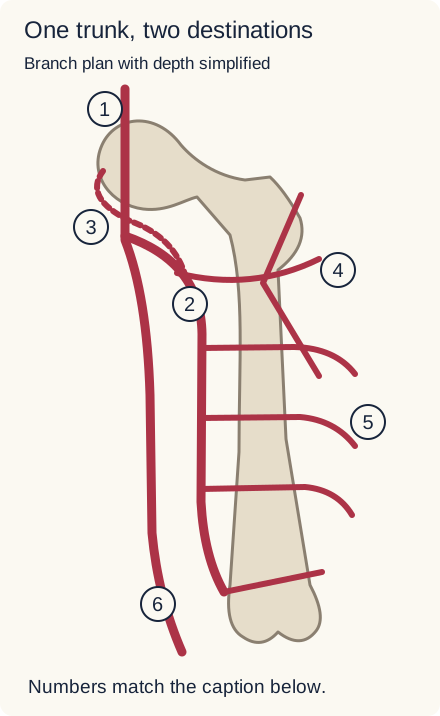
Trace the numbered routes. 1: proximal femoral inflow. 2: profunda. 3: medial circumflex route passing posteriorly. 4: lateral circumflex branches. 5: posteriorly directed perforators. 6: continuing femoral route toward the knee. Depth and branch spacing are simplified. [1][2]
In clinical vascular language, the segment before this division is often called the common femoral artery. The continuing vessel is often called the superficial femoral artery, although it is not a superficial skin vessel. That continuing femoral artery enters the adductor canal, passes through the distal adductor hiatus, and becomes the popliteal artery. The profunda does not undergo that name change. [13]
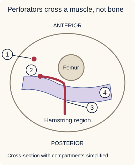
The profunda turns deep, passing behind the femoral vessels and then behind adductor longus. Farther down it lies on adductor magnus near the medial side of the femur. Its perforating arteries cross adductor magnus near the femoral attachment to reach posterior muscles. The classic description is three perforating branches plus a terminal continuation, often called a fourth perforator; individual counts and positions vary. The small perforating passages are not the same structure as the distal adductor hiatus. [2][11]
In this simplified cross-section, 1 is the continuing femoral artery, 2 the profunda, 3 a perforator and 4 adductor magnus. Trace the red branch across the muscle band into the posterior compartment, not through the femur. Nerves and veins are omitted. [2][13]
Trace, then compare: follow route 2 to route 5, and separately follow route 6 toward the knee. The first path supplies deep thigh tissues, including the hamstrings through perforators. Muscular and circumflex branches also contribute to adductors and the anterior/lateral thigh. These territories overlap with neighboring arterial sources; the profunda is important, not an exclusive supply to every thigh muscle. [9]
Transfer: a shaft-level drill can injure a perforator near adductor magnus while leaving the main femoral route intact. Predict a deep bleeding site without assuming that the pedal pulse must disappear. Conversely, a patent profunda cannot prove that the separate main route to the knee is unobstructed.
Try it here · Checkpoint 1 of 3
Make your prediction before reading the choices. A first attempt is just a starting point.
Case 4
Show answer and explanations for case 4
A. Obturator membrane near the superior pubic ramus (Why this does not fit)
Obturator vessels pass through the obturator canal at the membrane margin. That proximal pelvic route does not explain a shaft-level vessel reaching the hamstrings. Localize the vessel by both bone level and supplied compartment.
Reasoning steps for option A
Evidence check for dfa-04 option 1 (Obturator membrane near the superior pubic ramus): which stem facts require explanation?
Medial shaft cortex near linea aspera; Small branch reaches hamstrings; main femoral artery intact. Candidate under review: Obturator membrane near the superior pubic ramus.
Anatomic discriminator for dfa-04 option 1 (Obturator membrane near the superior pubic ramus): what supplied feature separates this choice?
That proximal pelvic route does not explain a shaft-level vessel reaching the hamstrings.
B. Adductor magnus near its femoral attachment (Best answer)
Perforating branches cross adductor magnus close to the femur to reach posterior thigh muscles. The drill location and hamstring distribution identify this route. These small passages are different from the distal adductor hiatus for the main femoral vessels.
Reasoning steps for option B
Evidence check for dfa-04 option 2 (Adductor magnus near its femoral attachment): which stem facts require explanation?
Medial shaft cortex near linea aspera; Small branch reaches hamstrings; main femoral artery intact. Candidate under review: Adductor magnus near its femoral attachment.
Anatomic discriminator for dfa-04 option 2 (Adductor magnus near its femoral attachment): what supplied feature separates this choice?
The drill location and hamstring distribution identify this route.
C. Sartorius near the femoral triangle (Why this does not fit)
Sartorius forms a boundary of the femoral triangle and covers distal femoral structures. It is not the deep medial-to-posterior passage taken by perforating arteries near the femoral shaft. A superficial landmark does not account for the posterior destination.
Reasoning steps for option C
Evidence check for dfa-04 option 3 (Sartorius near the femoral triangle): which stem facts require explanation?
Medial shaft cortex near linea aspera; Small branch reaches hamstrings; main femoral artery intact. Candidate under review: Sartorius near the femoral triangle.
Anatomic discriminator for dfa-04 option 3 (Sartorius near the femoral triangle): what supplied feature separates this choice?
It is not the deep medial-to-posterior passage taken by perforating arteries near the femoral shaft.
D. Adductor longus near its proximal origin (Why this does not fit)
Adductor longus overlies much of the profunda course. The posteriorly directed vessels near the linea aspera traverse adductor magnus rather than the proximal longus origin. Distinguish a muscle covering the trunk from the muscle traversed by its branches.
Reasoning steps for option D
Evidence check for dfa-04 option 4 (Adductor longus near its proximal origin): which stem facts require explanation?
Medial shaft cortex near linea aspera; Small branch reaches hamstrings; main femoral artery intact. Candidate under review: Adductor longus near its proximal origin.
Anatomic discriminator for dfa-04 option 4 (Adductor longus near its proximal origin): what supplied feature separates this choice?
The posteriorly directed vessels near the linea aspera traverse adductor magnus rather than the proximal longus origin.
Takeaway: Perforators cross adductor magnus; the main femoral artery uses the distal adductor hiatus.
Look for the final vessel, not just its parent. The profunda commonly gives rise to the medial and lateral circumflex femoral arteries. The lateral circumflex system runs laterally, deep to sartorius and rectus femoris, and has ascending, transverse and descending branches. It is important for anterior/lateral thigh tissues and proximal connections. The medial circumflex system follows a different route toward the posterior femoral neck. Either circumflex artery may have a separate femoral origin. [1][14]
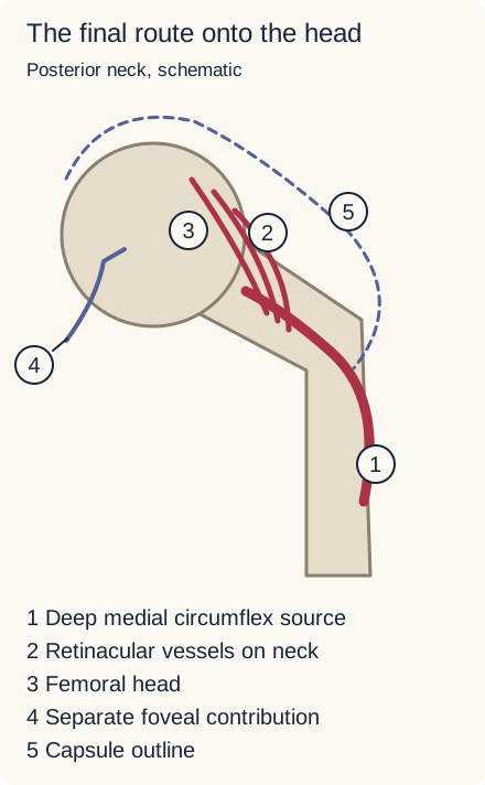
The dominant adult route is the deep medial circumflex contribution followed by ascending retinacular vessels on the neck. A separate small foveal route is shown, not a second equivalent supply. The capsule outline is simplified. [3][10]
In adults, the deep branch of the medial circumflex femoral artery usually provides the dominant supply to the head through subsynovial retinacular vessels, especially along the posterosuperior neck. They approach the head along the surface of the neck rather than arriving through the center of the shaft. This explains why the position of a femoral-neck fracture matters. Proximal circumflex or gluteal connections can remain open while the final retinacular vessels are damaged farther along the route. [3]
Use the picture: trace from number 1 through number 2 to the head. Now place an imaginary interruption halfway along number 2. Blood reaching the parent artery is not evidence that it can cross that interruption. The distinction is between a supplied parent vessel and a perfused target tissue, not between a universally correct branch name and a universally incorrect trunk name.
The ligamentum teres carries a variable contribution toward the fovea, commonly from an acetabular branch of the obturator artery, with other described contributions. In the adult, its presence does not establish enough supply to rescue the entire head after retinacular interruption. Likewise, lateral circumflex and gluteal contributions are not zero simply because medial circumflex supply is dominant. [3][10]
Change the origin: suppose angiography shows the medial circumflex artery arising directly from the common femoral artery before an isolated profunda-origin occlusion. That particular head-supply route may remain perfused. If it instead arises downstream of the occlusion, its direct inflow is threatened. Follow the observed origins before predicting the affected tissue.
Transfer: a prompt asking for the main adult artery supplying the head usually seeks the medial circumflex artery. A prompt demonstrating a proximal profunda lesion can legitimately implicate its downstream hip branches. Choose the level of specificity justified by the evidence; a small affected region alone does not prove where the initiating lesion lies.
Try it here · Checkpoint 2 of 3
Make your prediction before reading the choices. A first attempt is just a starting point.
Case 1
Show answer and explanations for case 1
A. Inferior gluteal artery (Why this does not fit)
The inferior gluteal artery participates in proximal hip collateral connections. An anastomotic contributor is not the usual principal source of these ascending capsular vessels. Separate a connection around the hip from the final route onto the head.
Reasoning steps for option A
Evidence check for dfa-01 option 1 (Inferior gluteal artery): which stem facts require explanation?
Anatomic discriminator for dfa-01 option 1 (Inferior gluteal artery): what supplied feature separates this choice?
An anastomotic contributor is not the usual principal source of these ascending capsular vessels.
B. Medial circumflex femoral artery (Best answer)
The deep branch supplies most adult femoral-head perfusion through retinacular vessels. A displaced intracapsular fracture can interrupt those vessels while distal leg inflow remains patent. A pedal signal assesses a different arterial pathway from the damaged femoral-head supply.
Reasoning steps for option B
Evidence check for dfa-01 option 2 (Medial circumflex femoral artery): which stem facts require explanation?
Anatomic discriminator for dfa-01 option 2 (Medial circumflex femoral artery): what supplied feature separates this choice?
A displaced intracapsular fracture can interrupt those vessels while distal leg inflow remains patent.
C. Lateral circumflex femoral artery (Why this does not fit)
This artery supplies the lateral proximal thigh and participates in hip anastomoses. It is not the usual dominant source of the posterosuperior retinacular vessels described here. The precise neck location matters more than the shared circumflex name.
Reasoning steps for option C
Evidence check for dfa-01 option 3 (Lateral circumflex femoral artery): which stem facts require explanation?
Anatomic discriminator for dfa-01 option 3 (Lateral circumflex femoral artery): what supplied feature separates this choice?
It is not the usual dominant source of the posterosuperior retinacular vessels described here.
D. Obturator artery (Why this does not fit)
Its acetabular contribution can reach the head through the ligamentum teres. That foveal route is not the dominant posterior-neck route identified at operation. Do not assume a patent ligament vessel replaces disrupted retinacular perfusion.
Reasoning steps for option D
Evidence check for dfa-01 option 4 (Obturator artery): which stem facts require explanation?
Fracture risk is not the same as a Perthes diagnosis
Compare two mechanisms: a displaced fracture can physically interrupt vessels; an ischemic bone disorder can be identified without demonstrating a particular arterial tear or thrombus. Both involve blood supply, but they support different conclusions.
A displaced intracapsular, including subcapital, femoral-neck fracture can disrupt the retinacular vessels and threaten the femoral head. Important later concerns include osteonecrosis and nonunion. A fracture radiograph does not prove that all head tissue is already dead, and a normal foot pulse does not establish head viability. Extracapsular intertrochanteric fractures lie in a different relationship to the neck vessels, but they can still cause major blood loss or injure deep vessels through a displaced fragment or instrumentation. [3][8][9]
For older adults with displaced femoral-neck fractures, AAOS guidance favors arthroplasty over fixation. That is an age- and fracture-specific clinical recommendation, not a rule that every hip fracture or every younger patient requires replacement. Anatomy explains part of the concern; patient characteristics and the fracture pattern still determine the clinical decision. [6]
Compare the tissue affected. Perthes disease is an ischemic disorder of the developing capital femoral epiphysis. A child, typically between 4 and 10 years old, may have a persistent limp and groin, thigh or referred knee pain. A normal knee examination should prompt attention to the hip. Reduced abduction and internal rotation support hip involvement; evolving radiographs can show epiphyseal sclerosis, fragmentation and flattening. The initiating cause of the blood-supply loss is not established by the diagnosis. [5]
In the immature proximal femur, the open physis can separate epiphyseal and metaphyseal vascular territories. Consequently, good metaphyseal perfusion is not proof of adequate epiphyseal perfusion. Developmental studies support the importance of the retinacular routes but do not support a universal timetable in which one large ligamentum teres artery predictably disappears at a particular birthday. Vessel arrangements and contributions vary. [4]
Make a bounded conclusion: in a child with established Perthes disease, say that the epiphysis has suffered ischemic injury. Do not claim that the radiograph proves a medial circumflex clot, that the profunda has been excluded as a possible upstream contributor, or that an isolated examination finding identifies a torn artery. Forced external rotation with hip flexion also should not be treated as a unique sign of Perthes; hip disorders share examination findings.
Transfer: contrast a documented posterior-neck vessel cutoff after trauma with epiphyseal fragmentation during an atraumatic childhood limp. The first localizes a visible vascular lesion. The second supports a bone disorder and an ischemic mechanism while leaving the initiating vascular event uncertain.
Test what an arterial connection can bypass
Predict before comparing: if the continuing femoral artery is obstructed after the profunda origin, can blood still reach distal arteries through deep-thigh connections? The answer depends on patent inflow, a connected path, suitable outflow and enough capacity for the tissue demand. The existence of a named anastomosis does not establish all four.
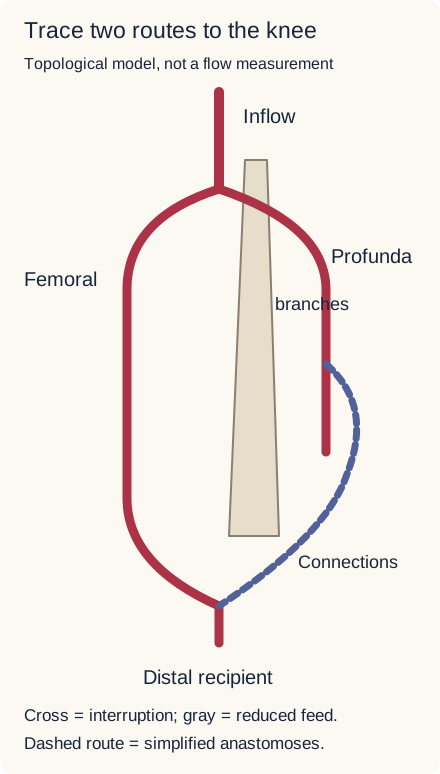
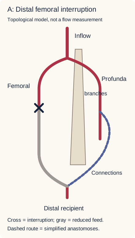
Two simplified paths reach a distal recipient: the direct femoral route and a profunda-fed collateral route. The dashed path represents multiple muscular and periarticular connections, not one named artery. Pelvic contributions and resistance are omitted. [1][2]
In chronic femoral occlusion, profunda muscular and perforating connections may enlarge and help reconstitute distal femoral or popliteal circulation. Resting viability can persist while exercise produces ischemic symptoms because a detour may have less reserve than the original artery. A new profunda-origin stenosis can therefore worsen an already collateral-dependent limb even when the old femoral obstruction is unchanged. Abrupt occlusion, poor inflow or inadequate distal filling can defeat that protection. A suddenly cold, painful or numb limb requires emergency vascular assessment rather than reassurance from possible collateral routes. [1][2][15]
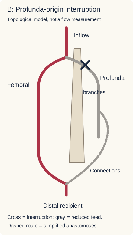
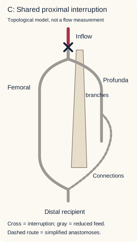
Change one interruption site. First predict which direct route remains connected in A, B and C. Open each comparison to inspect the changed vessel paths. Crosses mark the interrupted site; gray identifies reduced direct feed rather than a measured amount of tissue perfusion.
Compare A: interruption after the profunda origin
The direct femoral segment is interrupted, but the model retains a profunda-fed detour. Adequacy of that detour must be assessed, not assumed.Compare B: interruption at the profunda origin
The continuing femoral route remains connected while the profunda-fed detour loses its direct input. Other sources are not modeled.Compare C: interruption before both branches
Both modeled direct routes lose their shared inflow. Unshown pelvic connections may contribute clinically but cannot be presumed adequate.
The visible result: A preserves the modeled profunda source; B preserves the modeled main femoral source; C compromises both. This comparison is fully usable without animation or scripting. In a patient, compare these anatomical possibilities with actual distal perfusion, symptoms and imaging rather than treat the diagram as a flow simulator.
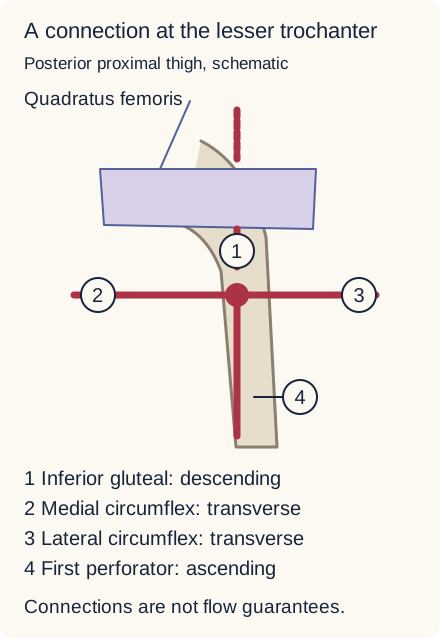
The cruciate network links an inferior gluteal contribution with profunda-related branches below quadratus femoris near the lesser trochanter. Branch positions are schematic. [10]
The conventional cruciate connection includes the descending inferior gluteal, transverse medial circumflex, transverse lateral circumflex and ascending first perforating contributions. It links internal iliac and external iliac-derived systems. The more proximal trochanteric network uses ascending circumflex and gluteal contributions around the hip; it is not the same four-part connection. Neither network guarantees rescue beyond disrupted retinacular vessels. [10]
Transfer: identify the source entering a detour, then the place where blood rejoins the downstream circulation. A proximal block can deprive the detour of inflow; a distal interruption beyond its connections can isolate the target. A viable resting foot is evidence about present adequacy, not a promise about future exercise or a new arterial lesion.
A pulse does not exclude a deep arterial leak
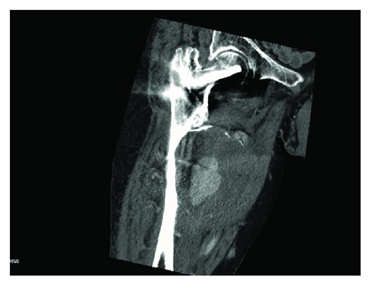
Look at the actual scan. Before reading the interpretation, locate the proximal femur and hardware, the large collection on its medial side, and the brighter contrast-containing region within that collection. How does that last finding change the meaning of a postoperative mass?
Actual patient CT, Figure 4 from Vande Voorde, Dauwe and Van Oost (2018), reproduced unchanged under the publication's Creative Commons Attribution License. The article identifies active extravasation within a hematoma and medial cortical scalloping. Source and license statement. [8]
Interpretation: a contrast-containing region communicating with an injured artery is not simply proof of an old, inactive hematoma. A pseudoaneurysm is a contained arterial wall disruption with a space that remains connected to the arterial lumen. Surrounding tissue contains the blood; it is not an intact arterial wall expanded through all layers. A hematoma can coexist with the pseudoaneurysm. Full vascular imaging defines the connection and treatment anatomy; this single teaching image is not a complete diagnostic study. [8]
Profunda or perforator injury can follow a deep wound, a fracture fragment, or a drill, screw or retractor during fixation. Presentation can be delayed. Increasing thigh size, pain or unexplained anemia deserves evaluation even if the foot is warm and pedal signals remain present, because the separate continuing femoral artery may still supply the leg. Do not presume a deep postoperative mass is safe to drain or biopsy before an arterial connection has been assessed. [2][8]
Choose the sequence from stability. In a stable patient who needs arterial imaging, CT angiography can define the lesion when examination or ultrasound leaves important uncertainty. In penetrating trauma, rapidly expanding hematoma, pulsatile bleeding, a pulse deficit, or a vascular bruit/thrill are hard arterial signs requiring urgent surgical assessment. Do not delay hemorrhage control for routine screening or imaging in a deteriorating patient. [7]
Transfer: distinguish two independent questions: is blood reaching the foot, and is blood escaping into the thigh? A distal pressure measurement helps assess the first. It does not, by itself, answer the second.
Try it here · Checkpoint 3 of 3
Make your prediction before reading the choices. A first attempt is just a starting point.
Case 23
Show answer and explanations for case 23
A. Obtain serial ankle pressure ratios before escalating the evaluation (Why this does not fit)
Ankle pressure indices help selected patients without hard vascular signs. An expanding hematoma and pulsatile bleeding are not a screening-negative presentation even with a distal signal. A distal-flow measurement does not assess the full hemorrhage risk.
Reasoning steps for option A
Evidence check for dfa-23 option 1 (Obtain serial ankle pressure ratios before escalating the evaluation): which stem facts require explanation?
Rapidly expanding hematoma and pulsatile bleeding; Falling blood pressure with detectable pedal signal. Candidate under review: Obtain serial ankle pressure ratios before escalating the evaluation.
Anatomic discriminator for dfa-23 option 1 (Obtain serial ankle pressure ratios before escalating the evaluation): what supplied feature separates this choice?
An expanding hematoma and pulsatile bleeding are not a screening-negative presentation even with a distal signal.
B. Perform duplex characterization before obtaining a vascular consultation (Why this does not fit)
Duplex can characterize selected stable vascular lesions. The active hemorrhage and hemodynamic deterioration make delayed characterization the wrong priority. Select the diagnostic sequence according to current stability and injury signs.
Reasoning steps for option B
Evidence check for dfa-23 option 2 (Perform duplex characterization before obtaining a vascular consultation): which stem facts require explanation?
Rapidly expanding hematoma and pulsatile bleeding; Falling blood pressure with detectable pedal signal. Candidate under review: Perform duplex characterization before obtaining a vascular consultation.
Anatomic discriminator for dfa-23 option 2 (Perform duplex characterization before obtaining a vascular consultation): what supplied feature separates this choice?
The active hemorrhage and hemodynamic deterioration make delayed characterization the wrong priority.
C. Complete CT angiography before arranging definitive hemorrhage control (Why this does not fit)
CT angiography is useful when vascular imaging is required in a suitable patient. Hard arterial signs and worsening circulation already establish the need for urgent operative assessment; routine imaging must not delay control. Use imaging to resolve uncertainty, not to postpone action on an established emergency.
Reasoning steps for option C
Evidence check for dfa-23 option 3 (Complete CT angiography before arranging definitive hemorrhage control): which stem facts require explanation?
Rapidly expanding hematoma and pulsatile bleeding; Falling blood pressure with detectable pedal signal. Candidate under review: Complete CT angiography before arranging definitive hemorrhage control.
Anatomic discriminator for dfa-23 option 3 (Complete CT angiography before arranging definitive hemorrhage control): what supplied feature separates this choice?
Hard arterial signs and worsening circulation already establish the need for urgent operative assessment; routine imaging must not delay control.
D. Arrange immediate surgical hemorrhage control while resuscitation proceeds (Best answer)
Rapidly expanding hematoma and pulsatile bleeding are hard signs of arterial injury. Deteriorating circulation requires immediate hemorrhage-control and resuscitation priorities despite residual pedal flow. A side-branch injury can threaten life without eliminating the distal pulse.
Reasoning steps for option D
Evidence check for dfa-23 option 4 (Arrange immediate surgical hemorrhage control while resuscitation proceeds): which stem facts require explanation?
Rapidly expanding hematoma and pulsatile bleeding; Falling blood pressure with detectable pedal signal. Candidate under review: Arrange immediate surgical hemorrhage control while resuscitation proceeds.
Anatomic discriminator for dfa-23 option 4 (Arrange immediate surgical hemorrhage control while resuscitation proceeds): what supplied feature separates this choice?
Match the finding to the tissue, then test the cause
Try this contrast: one patient has poor enhancement of the femoral head on vascular imaging. Another has pelvic descent during single-leg stance with normal arterial perfusion. The first is a perfusion finding; the second is a functional finding. They should not automatically receive the same kind of explanation.
Gluteal vessels are genuine contributors to hip-region circulation. The inferior gluteal system supplies much of gluteus maximus and participates in proximal thigh connections; the superior gluteal system supplies the abductor region, including gluteus medius and minimus. Yet a Trendelenburg pattern is not a diagnosis of superior gluteal arterial occlusion. Pain, tendon injury and motor dysfunction can impair pelvic support. [9]
The superior gluteal nerve supplies medius, minimus and tensor fasciae latae. If the left support-side abductors fail, the unsupported right pelvis can descend. Electrodiagnostic abnormalities in that shared muscle group, with preserved arterial perfusion, support a neural localization rather than an arterial one. The inferior gluteal nerve instead supplies gluteus maximus, important for powerful hip extension. A nerve and an artery can share a regional name without being interchangeable causes of weakness. [9]
Use three questions: what tissue is abnormal, what kind of evidence demonstrates that abnormality, and which anatomical route best accounts for it? A direct angiographic cutoff permits more precise vascular localization than pain or a limp alone. Conversely, a patent proximal artery is less informative about a distal tissue if the final vessels have not been assessed.
Final transfer: explain why these can all be true: profunda bleeding with a pedal pulse, head ischemia with normally enhancing thigh muscles, and abductor weakness with patent gluteal arteries. They concern different structures and different measurements. The most useful answer is the narrowest conclusion supported by the supplied evidence, not the narrowest artery name available.
Apply the anatomy to a new case
Cover the options first. Name the threatened tissue, sketch the relevant route, and identify what the stem actually demonstrates. After choosing, compare the competing options and explain which specific finding rules each one out.
Case 2
Show answer and explanations for case 2
A. Loss of leg filling from interruption of the popliteal continuation (Why this does not fit)
The femoral artery continues into the popliteal artery after the adductor hiatus. The patent distal femoral artery is separate from the occluded profunda origin. Do not substitute the profunda for the main continuation to the knee.
Reasoning steps for option A
Evidence check for dfa-02 option 1 (Loss of leg filling from interruption of the popliteal continuation): which stem facts require explanation?
Profunda origin occluded; Posterior-neck artery has a patent separate common femoral origin. Candidate under review: Loss of leg filling from interruption of the popliteal continuation.
Anatomic discriminator for dfa-02 option 1 (Loss of leg filling from interruption of the popliteal continuation): what supplied feature separates this choice?
The patent distal femoral artery is separate from the occluded profunda origin.
B. Preserved head filling through a separate medial circumflex origin (Best answer)
The medial circumflex artery has variable origins, including the common femoral artery. Its demonstrated origin is upstream of the profunda lesion, so this route can still receive blood. Trace the observed branch rather than impose the usual branching pattern.
Reasoning steps for option B
Evidence check for dfa-02 option 2 (Preserved head filling through a separate medial circumflex origin): which stem facts require explanation?
Profunda origin occluded; Posterior-neck artery has a patent separate common femoral origin. Candidate under review: Preserved head filling through a separate medial circumflex origin.
Anatomic discriminator for dfa-02 option 2 (Preserved head filling through a separate medial circumflex origin): what supplied feature separates this choice?
Its demonstrated origin is upstream of the profunda lesion, so this route can still receive blood.
C. Preserved head filling through a separate inferior gluteal origin (Why this does not fit)
Gluteal branches can contribute to hip circulation. The described artery arises from the common femoral artery, not the internal iliac system. Both origin and course must fit the selected vessel.
Reasoning steps for option C
Evidence check for dfa-02 option 3 (Preserved head filling through a separate inferior gluteal origin): which stem facts require explanation?
Profunda origin occluded; Posterior-neck artery has a patent separate common femoral origin. Candidate under review: Preserved head filling through a separate inferior gluteal origin.
Anatomic discriminator for dfa-02 option 3 (Preserved head filling through a separate inferior gluteal origin): what supplied feature separates this choice?
The described artery arises from the common femoral artery, not the internal iliac system.
D. Loss of head filling from interruption of the obturator artery (Why this does not fit)
Obturator branches can contribute to foveal circulation. No obturator interruption is shown, and the angiogram directly demonstrates a perfused posterior-neck route. A proposed explanation must account for the vessel actually opacified.
Reasoning steps for option D
Evidence check for dfa-02 option 4 (Loss of head filling from interruption of the obturator artery): which stem facts require explanation?
Profunda origin occluded; Posterior-neck artery has a patent separate common femoral origin. Candidate under review: Loss of head filling from interruption of the obturator artery.
Anatomic discriminator for dfa-02 option 4 (Loss of head filling from interruption of the obturator artery): what supplied feature separates this choice?
No obturator interruption is shown, and the angiogram directly demonstrates a perfused posterior-neck route.
Takeaway: An observed variant takes priority over a memorized parent vessel.
A. Obturator artery within the obturator canal (Why this does not fit)
The obturator artery supplies medial-thigh structures and can be injured near the pelvis. A leaking longitudinal vessel behind adductor longus follows the profunda course rather than the obturator canal. Medial pain alone does not identify the obturator artery.
Reasoning steps for option A
Evidence check for dfa-03 option 1 (Obturator artery within the obturator canal): which stem facts require explanation?
Falling hemoglobin and deep thigh leak; Patent main femoral-to-popliteal artery. Candidate under review: Obturator artery within the obturator canal.
Anatomic discriminator for dfa-03 option 1 (Obturator artery within the obturator canal): what supplied feature separates this choice?
A leaking longitudinal vessel behind adductor longus follows the profunda course rather than the obturator canal.
B. Profunda femoris artery in the deep thigh (Best answer)
The profunda descends behind adductor longus and supplies deep muscular branches. A profunda leak can produce substantial thigh blood loss while the separate distal femoral pathway maintains ankle pressure. Preserved distal inflow does not exclude a bleeding side branch.
Reasoning steps for option B
Evidence check for dfa-03 option 2 (Profunda femoris artery in the deep thigh): which stem facts require explanation?
Falling hemoglobin and deep thigh leak; Patent main femoral-to-popliteal artery. Candidate under review: Profunda femoris artery in the deep thigh.
Anatomic discriminator for dfa-03 option 2 (Profunda femoris artery in the deep thigh): what supplied feature separates this choice?
A profunda leak can produce substantial thigh blood loss while the separate distal femoral pathway maintains ankle pressure.
C. Superior gluteal artery above piriformis (Why this does not fit)
This artery can bleed after pelvic or upper gluteal injury. The demonstrated leak is within the deep thigh rather than above piriformis. The wound level and imaging territory distinguish neighboring arterial systems.
Reasoning steps for option C
Evidence check for dfa-03 option 3 (Superior gluteal artery above piriformis): which stem facts require explanation?
Falling hemoglobin and deep thigh leak; Patent main femoral-to-popliteal artery. Candidate under review: Superior gluteal artery above piriformis.
Anatomic discriminator for dfa-03 option 3 (Superior gluteal artery above piriformis): what supplied feature separates this choice?
The demonstrated leak is within the deep thigh rather than above piriformis.
D. Femoral artery within the adductor canal (Why this does not fit)
Injury here can cause major hemorrhage and threaten distal inflow. Imaging instead shows an intact femoral-to-popliteal lumen and a separate vessel behind adductor longus. Use the demonstrated course rather than blood loss alone to localize injury.
Reasoning steps for option D
Evidence check for dfa-03 option 4 (Femoral artery within the adductor canal): which stem facts require explanation?
Falling hemoglobin and deep thigh leak; Patent main femoral-to-popliteal artery. Candidate under review: Femoral artery within the adductor canal.
Anatomic discriminator for dfa-03 option 4 (Femoral artery within the adductor canal): what supplied feature separates this choice?
Imaging instead shows an intact femoral-to-popliteal lumen and a separate vessel behind adductor longus.
Takeaway: A deep arterial leak and a well-perfused foot can coexist.
A. Collateral flow meets resting demand but has limited reserve (Best answer)
Enlarged profunda connections can deliver blood beyond a chronic femoral occlusion. That supply can maintain resting needs yet remain insufficient when working muscles require more flow. Anatomical continuity does not guarantee normal flow reserve.
Reasoning steps for option A
Evidence check for dfa-05 option 1 (Collateral flow meets resting demand but has limited reserve): which stem facts require explanation?
Chronic adductor-canal femoral occlusion; Patent profunda with delayed distal filling; Symptoms only with exercise. Candidate under review: Collateral flow meets resting demand but has limited reserve.
Anatomic discriminator for dfa-05 option 1 (Collateral flow meets resting demand but has limited reserve): what supplied feature separates this choice?
That supply can maintain resting needs yet remain insufficient when working muscles require more flow.
B. Hip-joint loading produces referred pain despite normal perfusion (Why this does not fit)
Hip disorders can cause referred lower-limb pain. The described delayed distal arterial filling is not evidence of normal arterial delivery, and no hip findings support this alternative. A musculoskeletal cause needs supporting findings rather than an assumption that resting viability means normal perfusion.
Reasoning steps for option B
Evidence check for dfa-05 option 2 (Hip-joint loading produces referred pain despite normal perfusion): which stem facts require explanation?
Chronic adductor-canal femoral occlusion; Patent profunda with delayed distal filling; Symptoms only with exercise. Candidate under review: Hip-joint loading produces referred pain despite normal perfusion.
Anatomic discriminator for dfa-05 option 2 (Hip-joint loading produces referred pain despite normal perfusion): what supplied feature separates this choice?
The described delayed distal arterial filling is not evidence of normal arterial delivery, and no hip findings support this alternative.
C. Activity-dependent nerve-root compression limits muscle recruitment (Why this does not fit)
Spinal stenosis can produce exertional leg symptoms. The angiogram directly demonstrates a restricted arterial route with delayed distal filling; no positional or neurological pattern is supplied. Use objective vascular findings when distinguishing causes of exertional pain.
Reasoning steps for option C
Evidence check for dfa-05 option 3 (Activity-dependent nerve-root compression limits muscle recruitment): which stem facts require explanation?
Chronic adductor-canal femoral occlusion; Patent profunda with delayed distal filling; Symptoms only with exercise. Candidate under review: Activity-dependent nerve-root compression limits muscle recruitment.
Anatomic discriminator for dfa-05 option 3 (Activity-dependent nerve-root compression limits muscle recruitment): what supplied feature separates this choice?
The angiogram directly demonstrates a restricted arterial route with delayed distal filling; no positional or neurological pattern is supplied.
D. Elevated venous pressure limits capillary drainage during exertion (Why this does not fit)
Venous obstruction can impair drainage and produce exertional discomfort. The demonstrated abnormality is arterial obstruction with collateral reconstitution, not a venous outflow lesion. Distinguish arterial supply limitation from venous drainage limitation.
Reasoning steps for option D
Evidence check for dfa-05 option 4 (Elevated venous pressure limits capillary drainage during exertion): which stem facts require explanation?
Chronic adductor-canal femoral occlusion; Patent profunda with delayed distal filling; Symptoms only with exercise. Candidate under review: Elevated venous pressure limits capillary drainage during exertion.
Anatomic discriminator for dfa-05 option 4 (Elevated venous pressure limits capillary drainage during exertion): what supplied feature separates this choice?
The demonstrated abnormality is arterial obstruction with collateral reconstitution, not a venous outflow lesion.
Takeaway: Collateral supply can preserve resting viability without restoring exercise capacity.
A. It removes distal muscular branches while preserving profunda inflow (Why this does not fit)
Distal branch loss can reduce perfusion in selected tissues. A thrombosis extending across the profunda origin does not preserve its direct common femoral input. The branch origin, not only the length of thrombus, determines which routes lose inflow.
Reasoning steps for option A
Evidence check for dfa-06 option 1 (It removes distal muscular branches while preserving profunda inflow): which stem facts require explanation?
Chronic distal femoral occlusion with profunda-dependent distal filling; New common femoral thrombosis crosses profunda origin. Candidate under review: It removes distal muscular branches while preserving profunda inflow.
Anatomic discriminator for dfa-06 option 1 (It removes distal muscular branches while preserving profunda inflow): what supplied feature separates this choice?
A thrombosis extending across the profunda origin does not preserve its direct common femoral input.
B. It interrupts proximal inflow to the established collateral network (Best answer)
The profunda can feed distal collateral perfusion around a chronic femoral occlusion. A lesion across its origin threatens the input to that detour as well as the main route. Identify the arterial source of a collateral pathway before judging protection.
Reasoning steps for option B
Evidence check for dfa-06 option 2 (It interrupts proximal inflow to the established collateral network): which stem facts require explanation?
Chronic distal femoral occlusion with profunda-dependent distal filling; New common femoral thrombosis crosses profunda origin. Candidate under review: It interrupts proximal inflow to the established collateral network.
Anatomic discriminator for dfa-06 option 2 (It interrupts proximal inflow to the established collateral network): what supplied feature separates this choice?
A lesion across its origin threatens the input to that detour as well as the main route.
C. It narrows the collateral channels without changing their arterial source (Why this does not fit)
Disease within established collateral channels could reduce their capacity. The demonstrated thrombosis crosses the feeding origin rather than selectively narrowing the channels farther downstream. Distinguish loss of a source from resistance within the route.
Reasoning steps for option C
Evidence check for dfa-06 option 3 (It narrows the collateral channels without changing their arterial source): which stem facts require explanation?
Chronic distal femoral occlusion with profunda-dependent distal filling; New common femoral thrombosis crosses profunda origin. Candidate under review: It narrows the collateral channels without changing their arterial source.
Anatomic discriminator for dfa-06 option 3 (It narrows the collateral channels without changing their arterial source): what supplied feature separates this choice?
The demonstrated thrombosis crosses the feeding origin rather than selectively narrowing the channels farther downstream.
D. It blocks the distal recipient where the established collaterals rejoin (Why this does not fit)
A new distal outflow obstruction could defeat otherwise adequate collateral inflow. The stated new lesion is at the common femoral/profunda origin, not where the distal branches rejoin. Locate the new lesion relative to both ends of the detour.
Reasoning steps for option D
Evidence check for dfa-06 option 4 (It blocks the distal recipient where the established collaterals rejoin): which stem facts require explanation?
Chronic distal femoral occlusion with profunda-dependent distal filling; New common femoral thrombosis crosses profunda origin. Candidate under review: It blocks the distal recipient where the established collaterals rejoin.
Anatomic discriminator for dfa-06 option 4 (It blocks the distal recipient where the established collaterals rejoin): what supplied feature separates this choice?
The stated new lesion is at the common femoral/profunda origin, not where the distal branches rejoin.
Takeaway: A useful collateral network still requires a patent source of inflow.
A. Proximal network filling indicates maintained retinacular delivery to the head (Why this does not fit)
Proximal anastomoses can provide alternate connections around an upstream lesion. Here the final retinacular vessels crossing the fracture do not fill, so proximal network patency does not establish delivery to the head. Follow flow all the way to the target tissue.
Reasoning steps for option A
Evidence check for dfa-07 option 1 (Proximal network filling indicates maintained retinacular delivery to the head): which stem facts require explanation?
Patent proximal circumflex connections; Absent filling in retinacular vessels across fracture. Candidate under review: Proximal network filling indicates maintained retinacular delivery to the head.
Anatomic discriminator for dfa-07 option 1 (Proximal network filling indicates maintained retinacular delivery to the head): what supplied feature separates this choice?
Here the final retinacular vessels crossing the fracture do not fill, so proximal network patency does not establish delivery to the head.
B. Preserved circumflex filling places the fracture outside the joint capsule (Why this does not fit)
Extracapsular fractures have a different relationship to retinacular vessels. The stem documents an intracapsular fracture with a distal vascular interruption; proximal circumflex patency does not relocate that fracture. Do not infer bone location from a patent parent vessel.
Reasoning steps for option B
Evidence check for dfa-07 option 2 (Preserved circumflex filling places the fracture outside the joint capsule): which stem facts require explanation?
Patent proximal circumflex connections; Absent filling in retinacular vessels across fracture. Candidate under review: Preserved circumflex filling places the fracture outside the joint capsule.
Anatomic discriminator for dfa-07 option 2 (Preserved circumflex filling places the fracture outside the joint capsule): what supplied feature separates this choice?
The stem documents an intracapsular fracture with a distal vascular interruption; proximal circumflex patency does not relocate that fracture.
C. Absent distal filling identifies established head necrosis at presentation (Why this does not fit)
Interrupted perfusion can lead to osteonecrosis. An acute vascular deficit does not establish the extent of irreversible tissue injury at that instant. Separate present perfusion risk from established tissue death.
Reasoning steps for option C
Evidence check for dfa-07 option 3 (Absent distal filling identifies established head necrosis at presentation): which stem facts require explanation?
Patent proximal circumflex connections; Absent filling in retinacular vessels across fracture. Candidate under review: Absent distal filling identifies established head necrosis at presentation.
Anatomic discriminator for dfa-07 option 3 (Absent distal filling identifies established head necrosis at presentation): what supplied feature separates this choice?
An acute vascular deficit does not establish the extent of irreversible tissue injury at that instant.
D. Retinacular interruption threatens head perfusion despite proximal network filling (Best answer)
The retinacular vessels are the final route from proximal circumflex connections onto the head. A break beyond the anastomoses can prevent those connections from providing adequate head perfusion. An interruption must lie within a bypassable segment for a connection to protect the target.
Reasoning steps for option D
Evidence check for dfa-07 option 4 (Retinacular interruption threatens head perfusion despite proximal network filling): which stem facts require explanation?
Patent proximal circumflex connections; Absent filling in retinacular vessels across fracture. Candidate under review: Retinacular interruption threatens head perfusion despite proximal network filling.
Anatomic discriminator for dfa-07 option 4 (Retinacular interruption threatens head perfusion despite proximal network filling): what supplied feature separates this choice?
A break beyond the anastomoses can prevent those connections from providing adequate head perfusion.
Takeaway: A collateral connection proximal to a damaged final vessel does not necessarily protect the target tissue.
A. CT angiography of the affected thigh (Best answer)
CT angiography can define arterial patency, a leaking branch and its relationship to a deep collection. The incomplete ultrasound, falling hemoglobin and postoperative location leave arterial injury unresolved. Choose an arterial study before considering an invasive procedure on the collection.
Reasoning steps for option A
Evidence check for dfa-08 option 1 (CT angiography of the affected thigh): which stem facts require explanation?
Progressive swelling and serial hemoglobin decline; Stable circulation; ultrasound cannot assess arterial relation. Candidate under review: CT angiography of the affected thigh.
Anatomic discriminator for dfa-08 option 1 (CT angiography of the affected thigh): what supplied feature separates this choice?
The incomplete ultrasound, falling hemoglobin and postoperative location leave arterial injury unresolved.
B. CT venography of the affected thigh (Why this does not fit)
Venous imaging can investigate proximal thrombosis causing postoperative swelling. It is not the targeted study for an incompletely assessed arterial lesion associated with falling hemoglobin. The choice of contrast timing should match the unresolved vascular concern.
Reasoning steps for option B
Evidence check for dfa-08 option 2 (CT venography of the affected thigh): which stem facts require explanation?
Progressive swelling and serial hemoglobin decline; Stable circulation; ultrasound cannot assess arterial relation. Candidate under review: CT venography of the affected thigh.
Anatomic discriminator for dfa-08 option 2 (CT venography of the affected thigh): what supplied feature separates this choice?
It is not the targeted study for an incompletely assessed arterial lesion associated with falling hemoglobin.
C. Repeat plain radiography of the affected thigh (Why this does not fit)
Radiographs assess alignment, implants and bone changes after fixation. They cannot adequately establish whether a deep collection communicates with an artery. A satisfactory implant image does not exclude adjacent vascular injury.
Reasoning steps for option C
Evidence check for dfa-08 option 3 (Repeat plain radiography of the affected thigh): which stem facts require explanation?
Progressive swelling and serial hemoglobin decline; Stable circulation; ultrasound cannot assess arterial relation. Candidate under review: Repeat plain radiography of the affected thigh.
Anatomic discriminator for dfa-08 option 3 (Repeat plain radiography of the affected thigh): what supplied feature separates this choice?
They cannot adequately establish whether a deep collection communicates with an artery.
D. Unenhanced MRI of the affected thigh (Why this does not fit)
MRI can characterize many soft-tissue masses and hematomas. The immediate question is active arterial communication rather than tissue characterization alone. Define the vascular relationship before treating this as an isolated mass.
Reasoning steps for option D
Evidence check for dfa-08 option 4 (Unenhanced MRI of the affected thigh): which stem facts require explanation?
Progressive swelling and serial hemoglobin decline; Stable circulation; ultrasound cannot assess arterial relation. Candidate under review: Unenhanced MRI of the affected thigh.
Anatomic discriminator for dfa-08 option 4 (Unenhanced MRI of the affected thigh): what supplied feature separates this choice?
The immediate question is active arterial communication rather than tissue characterization alone.
Takeaway: A stable patient with an incompletely assessed deep collection still needs an appropriate arterial evaluation.
A. The injured branch is connected directly to the external iliac vein (Why this does not fit)
An arteriovenous fistula can follow arterial access. The angiogram describes a wall defect without early venous filling or a fistulous connection. Do not add a second injury that is not demonstrated.
Reasoning steps for option A
Evidence check for dfa-09 option 1 (The injured branch is connected directly to the external iliac vein): which stem facts require explanation?
Injured branch behind adductor longus with perforators; Other branch continues through adductor canal. Candidate under review: The injured branch is connected directly to the external iliac vein.
Anatomic discriminator for dfa-09 option 1 (The injured branch is connected directly to the external iliac vein): what supplied feature separates this choice?
The angiogram describes a wall defect without early venous filling or a fistulous connection.
B. The uninjured branch provides the principal foveal route to the head (Why this does not fit)
The foveal route reaches the head through the ligamentum teres. An artery entering the adductor canal is not that route, and head perfusion does not account for pedal flow. Match the preserved function to the artery reaching that territory.
Reasoning steps for option B
Evidence check for dfa-09 option 2 (The uninjured branch provides the principal foveal route to the head): which stem facts require explanation?
Injured branch behind adductor longus with perforators; Other branch continues through adductor canal. Candidate under review: The uninjured branch provides the principal foveal route to the head.
Anatomic discriminator for dfa-09 option 2 (The uninjured branch provides the principal foveal route to the head): what supplied feature separates this choice?
An artery entering the adductor canal is not that route, and head perfusion does not account for pedal flow.
C. The uninjured branch provides the direct arterial route to the knee (Best answer)
The branch entering the adductor canal is the continuing femoral artery. It can preserve distal flow despite a separate profunda wall injury. A palpable distal pulse does not prove that every proximal branch is intact.
Reasoning steps for option C
Evidence check for dfa-09 option 3 (The uninjured branch provides the direct arterial route to the knee): which stem facts require explanation?
Injured branch behind adductor longus with perforators; Other branch continues through adductor canal. Candidate under review: The uninjured branch provides the direct arterial route to the knee.
Anatomic discriminator for dfa-09 option 3 (The uninjured branch provides the direct arterial route to the knee): what supplied feature separates this choice?
It can preserve distal flow despite a separate profunda wall injury.
D. The injured branch is the main continuation into the popliteal artery (Why this does not fit)
The main femoral continuation supplies the distal leg through the popliteal artery. The injured branch instead follows the profunda course behind adductor longus. Identify which branch remains continuous with the knee vessels.
Reasoning steps for option D
Evidence check for dfa-09 option 4 (The injured branch is the main continuation into the popliteal artery): which stem facts require explanation?
Injured branch behind adductor longus with perforators; Other branch continues through adductor canal. Candidate under review: The injured branch is the main continuation into the popliteal artery.
Anatomic discriminator for dfa-09 option 4 (The injured branch is the main continuation into the popliteal artery): what supplied feature separates this choice?
The injured branch instead follows the profunda course behind adductor longus.
Takeaway: Branch anatomy explains why an access injury can spare the distal pulse.
A. Displacement of the capital epiphysis across the growth plate (Why this does not fit)
A slipped capital epiphysis is an important hip disorder that can cause referred knee pain and restricted rotation. The image describes sclerosis and fragmentation without a displaced physeal slip. Use the structural radiographic change rather than hip pain alone.
Reasoning steps for option A
Evidence check for dfa-10 option 1 (Displacement of the capital epiphysis across the growth plate): which stem facts require explanation?
Childhood limp with normal knee examination; Hip restriction and epiphyseal sclerosis/fragmentation. Candidate under review: Displacement of the capital epiphysis across the growth plate.
Anatomic discriminator for dfa-10 option 1 (Displacement of the capital epiphysis across the growth plate): what supplied feature separates this choice?
The image describes sclerosis and fragmentation without a displaced physeal slip.
B. Transient synovial inflammation without injury to the epiphysis (Why this does not fit)
Transient synovitis can produce childhood hip pain, restricted motion and limping. Persistent symptoms plus epiphyseal sclerosis and fragmentation indicate structural bone disease rather than an isolated transient effusion. Imaging evidence of bone injury changes the interpretation of shared symptoms.
Reasoning steps for option B
Evidence check for dfa-10 option 2 (Transient synovial inflammation without injury to the epiphysis): which stem facts require explanation?
Childhood limp with normal knee examination; Hip restriction and epiphyseal sclerosis/fragmentation. Candidate under review: Transient synovial inflammation without injury to the epiphysis.
Anatomic discriminator for dfa-10 option 2 (Transient synovial inflammation without injury to the epiphysis): what supplied feature separates this choice?
Persistent symptoms plus epiphyseal sclerosis and fragmentation indicate structural bone disease rather than an isolated transient effusion.
C. Ischemic injury and remodeling of the capital femoral epiphysis (Best answer)
This pattern fits Perthes disease in a child with a persistent limp and restricted hip motion. Epiphyseal sclerosis and fragmentation support ischemic injury, but they do not establish an initiating occlusion of a named artery. Separate recognition of the bone process from unsupported vascular localization.
Reasoning steps for option C
Evidence check for dfa-10 option 3 (Ischemic injury and remodeling of the capital femoral epiphysis): which stem facts require explanation?
Childhood limp with normal knee examination; Hip restriction and epiphyseal sclerosis/fragmentation. Candidate under review: Ischemic injury and remodeling of the capital femoral epiphysis.
Anatomic discriminator for dfa-10 option 3 (Ischemic injury and remodeling of the capital femoral epiphysis): what supplied feature separates this choice?
Epiphyseal sclerosis and fragmentation support ischemic injury, but they do not establish an initiating occlusion of a named artery.
D. Primary osteonecrosis centered in the femoral shaft diaphysis (Why this does not fit)
Ischemia can affect different bone territories depending on the vascular lesion. The abnormalities here are in the capital epiphysis, not the shaft. Localize the demonstrated bone territory before naming its mechanism.
Reasoning steps for option D
Evidence check for dfa-10 option 4 (Primary osteonecrosis centered in the femoral shaft diaphysis): which stem facts require explanation?
Childhood limp with normal knee examination; Hip restriction and epiphyseal sclerosis/fragmentation. Candidate under review: Primary osteonecrosis centered in the femoral shaft diaphysis.
Anatomic discriminator for dfa-10 option 4 (Primary osteonecrosis centered in the femoral shaft diaphysis): what supplied feature separates this choice?
The abnormalities here are in the capital epiphysis, not the shaft.
Takeaway: Perthes identifies an ischemic epiphyseal disorder, not a proven occlusion of one named artery.
A. Reduced epiphyseal filling with preserved metaphyseal filling (Best answer)
The open physis can separate metaphyseal and epiphyseal vascular territories. With no crossing channel shown, retinacular interruption can affect the epiphysis while the metaphysis continues to fill. Adjacent bone regions need not share an effective arterial bypass.
Reasoning steps for option A
Evidence check for dfa-11 option 1 (Reduced epiphyseal filling with preserved metaphyseal filling): which stem facts require explanation?
Open physis without observed transphyseal channels; Metaphyseal filling preserved; retinacular route interrupted. Candidate under review: Reduced epiphyseal filling with preserved metaphyseal filling.
Anatomic discriminator for dfa-11 option 1 (Reduced epiphyseal filling with preserved metaphyseal filling): what supplied feature separates this choice?
With no crossing channel shown, retinacular interruption can affect the epiphysis while the metaphysis continues to fill.
B. Preserved epiphyseal filling with preserved metaphyseal filling (Why this does not fit)
Both could remain supplied if an adequate alternate epiphyseal route bypassed the interruption. No transphyseal route is demonstrated, so preserved metaphyseal filling cannot establish this complete rescue. Require evidence for the proposed bypass.
Reasoning steps for option B
Evidence check for dfa-11 option 2 (Preserved epiphyseal filling with preserved metaphyseal filling): which stem facts require explanation?
Open physis without observed transphyseal channels; Metaphyseal filling preserved; retinacular route interrupted. Candidate under review: Preserved epiphyseal filling with preserved metaphyseal filling.
Anatomic discriminator for dfa-11 option 2 (Preserved epiphyseal filling with preserved metaphyseal filling): what supplied feature separates this choice?
No transphyseal route is demonstrated, so preserved metaphyseal filling cannot establish this complete rescue.
C. Reduced epiphyseal filling with reduced metaphyseal filling (Why this does not fit)
Both regions could lose tracer if their shared proximal source were interrupted. The study instead preserves separate metaphyseal injection below the physis. Do not extend a selective retinacular interruption to an independently perfused compartment.
Reasoning steps for option C
Evidence check for dfa-11 option 3 (Reduced epiphyseal filling with reduced metaphyseal filling): which stem facts require explanation?
Open physis without observed transphyseal channels; Metaphyseal filling preserved; retinacular route interrupted. Candidate under review: Reduced epiphyseal filling with reduced metaphyseal filling.
Anatomic discriminator for dfa-11 option 3 (Reduced epiphyseal filling with reduced metaphyseal filling): what supplied feature separates this choice?
The study instead preserves separate metaphyseal injection below the physis.
D. Preserved epiphyseal filling with reduced metaphyseal filling (Why this does not fit)
This pattern could follow a selective metaphyseal interruption with intact epiphyseal inflow. The intervention does the reverse: it interrupts the retinacular route while metaphyseal filling is demonstrated. Keep the intervention and the predicted territory in the same orientation.
Reasoning steps for option D
Evidence check for dfa-11 option 4 (Preserved epiphyseal filling with reduced metaphyseal filling): which stem facts require explanation?
Open physis without observed transphyseal channels; Metaphyseal filling preserved; retinacular route interrupted. Candidate under review: Preserved epiphyseal filling with reduced metaphyseal filling.
Anatomic discriminator for dfa-11 option 4 (Preserved epiphyseal filling with reduced metaphyseal filling): what supplied feature separates this choice?
The intervention does the reverse: it interrupts the retinacular route while metaphyseal filling is demonstrated.
Takeaway: A patent metaphyseal circulation does not prove an adequate epiphyseal supply across an open physis.
A. Obturator contribution; whole-head rescue is established (Why this does not fit)
The obturator contribution correctly identifies a common source of the foveal ligament vessel. The error is treating its patency as proof of adequate whole-head perfusion despite impaired retinacular filling. Correct source identification does not establish sufficient functional reserve.
Reasoning steps for option A
Evidence check for dfa-12 option 1 (Obturator contribution; whole-head rescue is established): which stem facts require explanation?
Vessel travels in ligament attached to fovea; Retinacular filling is impaired. Candidate under review: Obturator contribution; whole-head rescue is established.
Anatomic discriminator for dfa-12 option 1 (Obturator contribution; whole-head rescue is established): what supplied feature separates this choice?
The error is treating its patency as proof of adequate whole-head perfusion despite impaired retinacular filling.
B. Inferior gluteal contribution; whole-head rescue is established (Why this does not fit)
The inferior gluteal artery participates in hip-region anastomoses. It is not the common foveal source described, and a local connection would not establish adequate head rescue. Check both anatomical identity and the claimed functional consequence.
Reasoning steps for option B
Evidence check for dfa-12 option 2 (Inferior gluteal contribution; whole-head rescue is established): which stem facts require explanation?
Vessel travels in ligament attached to fovea; Retinacular filling is impaired. Candidate under review: Inferior gluteal contribution; whole-head rescue is established.
Anatomic discriminator for dfa-12 option 2 (Inferior gluteal contribution; whole-head rescue is established): what supplied feature separates this choice?
It is not the common foveal source described, and a local connection would not establish adequate head rescue.
C. Inferior gluteal contribution; whole-head rescue remains uncertain (Why this does not fit)
Inferior gluteal branches can contribute to proximal hip connections. That does not make the inferior gluteal artery the common source of the foveal ligament vessel shown here. A correct caution about perfusion cannot correct a mismatched anatomical route.
Reasoning steps for option C
Evidence check for dfa-12 option 3 (Inferior gluteal contribution; whole-head rescue remains uncertain): which stem facts require explanation?
Vessel travels in ligament attached to fovea; Retinacular filling is impaired. Candidate under review: Inferior gluteal contribution; whole-head rescue remains uncertain.
Anatomic discriminator for dfa-12 option 3 (Inferior gluteal contribution; whole-head rescue remains uncertain): what supplied feature separates this choice?
That does not make the inferior gluteal artery the common source of the foveal ligament vessel shown here.
D. Obturator contribution; whole-head rescue remains uncertain (Best answer)
An obturator acetabular branch commonly contributes to the ligamentum teres vessel. Its variable contribution does not establish adequate replacement for disrupted retinacular inflow. A visible foveal vessel is not a perfusion test of the entire head.
Reasoning steps for option D
Evidence check for dfa-12 option 4 (Obturator contribution; whole-head rescue remains uncertain): which stem facts require explanation?
Vessel travels in ligament attached to fovea; Retinacular filling is impaired. Candidate under review: Obturator contribution; whole-head rescue remains uncertain.
Anatomic discriminator for dfa-12 option 4 (Obturator contribution; whole-head rescue remains uncertain): what supplied feature separates this choice?
Its variable contribution does not establish adequate replacement for disrupted retinacular inflow.
Takeaway: The ligamentum teres route contributes variably and is not a reliable substitute for retinacular supply.
A. Medial circumflex femoral artery (Why this does not fit)
The medial circumflex artery supplies the posterior-neck retinacular route. That is the separate vessel in this study, not the lateral vessel being mapped for the pedicle. The important head supply does not replace the separate lateral arterial territory.
Reasoning steps for option A
Evidence check for dfa-13 option 1 (Medial circumflex femoral artery): which stem facts require explanation?
Lateral course deep to sartorius and rectus femoris; Ascending, transverse and descending branches. Candidate under review: Medial circumflex femoral artery.
Anatomic discriminator for dfa-13 option 1 (Medial circumflex femoral artery): what supplied feature separates this choice?
That is the separate vessel in this study, not the lateral vessel being mapped for the pedicle.
B. Lateral circumflex femoral artery (Best answer)
The lateral circumflex system passes laterally and gives ascending, transverse and descending branches. Those findings identify the artery whose descending branch is being mapped. Preoperative imaging defines the actual pedicle rather than a presumed fixed origin.
Reasoning steps for option B
Evidence check for dfa-13 option 2 (Lateral circumflex femoral artery): which stem facts require explanation?
Lateral course deep to sartorius and rectus femoris; Ascending, transverse and descending branches. Candidate under review: Lateral circumflex femoral artery.
Anatomic discriminator for dfa-13 option 2 (Lateral circumflex femoral artery): what supplied feature separates this choice?
Those findings identify the artery whose descending branch is being mapped.
C. Obturator artery (Why this does not fit)
The obturator artery supplies part of the medial thigh and hip. It does not usually form the described lateral ascending, transverse and descending branch system. The lateral course and branch pattern distinguish it from a medial-thigh contributor.
Reasoning steps for option C
Evidence check for dfa-13 option 3 (Obturator artery): which stem facts require explanation?
Lateral course deep to sartorius and rectus femoris; Ascending, transverse and descending branches. Candidate under review: Obturator artery.
Anatomic discriminator for dfa-13 option 3 (Obturator artery): what supplied feature separates this choice?
It does not usually form the described lateral ascending, transverse and descending branch system.
D. Inferior gluteal artery (Why this does not fit)
The inferior gluteal artery supplies the buttock and participates in proximal thigh connections. It does not pass laterally under sartorius and rectus femoris in the pattern described. Origin, course and target should all agree.
Reasoning steps for option D
Evidence check for dfa-13 option 4 (Inferior gluteal artery): which stem facts require explanation?
Lateral course deep to sartorius and rectus femoris; Ascending, transverse and descending branches. Candidate under review: Inferior gluteal artery.
Anatomic discriminator for dfa-13 option 4 (Inferior gluteal artery): what supplied feature separates this choice?
It does not pass laterally under sartorius and rectus femoris in the pattern described.
Takeaway: The lateral circumflex system must be distinguished from the posterior-neck medial circumflex system.
A. The main artery renamed popliteal at the adductor hiatus (Why this does not fit)
The main femoral continuation becomes popliteal after the hiatus. The specimen shows that continuation separately from the terminal profunda vessel. A shared distal direction does not make the two arteries continuous.
Reasoning steps for option A
Evidence check for dfa-14 option 1 (The main artery renamed popliteal at the adductor hiatus): which stem facts require explanation?
Three separate profunda perforators; Terminal profunda also crosses magnus; main femoral artery is separate. Candidate under review: The main artery renamed popliteal at the adductor hiatus.
Anatomic discriminator for dfa-14 option 1 (The main artery renamed popliteal at the adductor hiatus): what supplied feature separates this choice?
The specimen shows that continuation separately from the terminal profunda vessel.
B. A descending genicular branch arising within the adductor canal (Why this does not fit)
The descending genicular artery arises from the distal femoral system. The specimen directly traces this vessel to the profunda instead. An observed vessel origin outweighs proximity to a familiar distal branch.
Reasoning steps for option B
Evidence check for dfa-14 option 2 (A descending genicular branch arising within the adductor canal): which stem facts require explanation?
Three separate profunda perforators; Terminal profunda also crosses magnus; main femoral artery is separate. Candidate under review: A descending genicular branch arising within the adductor canal.
Anatomic discriminator for dfa-14 option 2 (A descending genicular branch arising within the adductor canal): what supplied feature separates this choice?
The specimen directly traces this vessel to the profunda instead.
C. A fourth perforating artery formed by the terminal continuation (Best answer)
Three named perforators and a terminal continuation are a common descriptive arrangement. The terminal profunda crossing to the posterior compartment is often called the fourth perforator. Counts vary; identify continuity and destination rather than demand four separate origins.
Reasoning steps for option C
Evidence check for dfa-14 option 3 (A fourth perforating artery formed by the terminal continuation): which stem facts require explanation?
Three separate profunda perforators; Terminal profunda also crosses magnus; main femoral artery is separate. Candidate under review: A fourth perforating artery formed by the terminal continuation.
Anatomic discriminator for dfa-14 option 3 (A fourth perforating artery formed by the terminal continuation): what supplied feature separates this choice?
The terminal profunda crossing to the posterior compartment is often called the fourth perforator.
D. An ascending circumflex branch returning to the femoral head (Why this does not fit)
Ascending circumflex branches supply proximal hip-region structures. This vessel continues distally and crosses toward the hamstrings, away from the head. Direction and destination separate a terminal perforator from a circumflex branch.
Reasoning steps for option D
Evidence check for dfa-14 option 4 (An ascending circumflex branch returning to the femoral head): which stem facts require explanation?
Three separate profunda perforators; Terminal profunda also crosses magnus; main femoral artery is separate. Candidate under review: An ascending circumflex branch returning to the femoral head.
Anatomic discriminator for dfa-14 option 4 (An ascending circumflex branch returning to the femoral head): what supplied feature separates this choice?
This vessel continues distally and crosses toward the hamstrings, away from the head.
Takeaway: The terminal profunda may be described as a fourth perforator; it is not the popliteal continuation.
The obturator nerve supplies much of the thigh adductor group. Adductor testing is normal, whereas the abductor group is abnormal. Contrast the tested muscle groups before selecting a nearby nerve.
Reasoning steps for option A
Evidence check for dfa-15 option 1 (Left obturator nerve): which stem facts require explanation?
Right pelvis descends during left-leg stance; Left medius, minimus and tensor fasciae latae abnormalities; Preserved arterial perfusion. Candidate under review: Left obturator nerve.
Anatomic discriminator for dfa-15 option 1 (Left obturator nerve): what supplied feature separates this choice?
Adductor testing is normal, whereas the abductor group is abnormal.
B. Right superior gluteal nerve (Why this does not fit)
Superior gluteal dysfunction can cause contralateral pelvic descent during single-leg stance. The supported leg is left and the left muscles have abnormal testing, so the lesion is not on the descending right side. Localize the weak support side rather than the visibly lower side.
Reasoning steps for option B
Evidence check for dfa-15 option 2 (Right superior gluteal nerve): which stem facts require explanation?
Right pelvis descends during left-leg stance; Left medius, minimus and tensor fasciae latae abnormalities; Preserved arterial perfusion. Candidate under review: Right superior gluteal nerve.
Anatomic discriminator for dfa-15 option 2 (Right superior gluteal nerve): what supplied feature separates this choice?
The supported leg is left and the left muscles have abnormal testing, so the lesion is not on the descending right side.
C. Left superior gluteal nerve (Best answer)
This nerve supplies medius, minimus and tensor fasciae latae. Left abductor weakness explains descent of the unsupported right pelvis, and preserved arterial perfusion argues against substituting an arterial diagnosis. A gait pattern indicates functional failure; additional evidence identifies its cause.
Reasoning steps for option C
Evidence check for dfa-15 option 3 (Left superior gluteal nerve): which stem facts require explanation?
Right pelvis descends during left-leg stance; Left medius, minimus and tensor fasciae latae abnormalities; Preserved arterial perfusion. Candidate under review: Left superior gluteal nerve.
Anatomic discriminator for dfa-15 option 3 (Left superior gluteal nerve): what supplied feature separates this choice?
Left abductor weakness explains descent of the unsupported right pelvis, and preserved arterial perfusion argues against substituting an arterial diagnosis.
D. Left inferior gluteal nerve (Why this does not fit)
This nerve supplies gluteus maximus and affects powerful hip extension. The abnormal muscles here are the abductor group supplied by another nerve. Use the shared innervation of affected muscles rather than the general buttock location.
Reasoning steps for option D
Evidence check for dfa-15 option 4 (Left inferior gluteal nerve): which stem facts require explanation?
Right pelvis descends during left-leg stance; Left medius, minimus and tensor fasciae latae abnormalities; Preserved arterial perfusion. Candidate under review: Left inferior gluteal nerve.
Anatomic discriminator for dfa-15 option 4 (Left inferior gluteal nerve): what supplied feature separates this choice?
The abnormal muscles here are the abductor group supplied by another nerve.
Takeaway: Pelvic descent identifies failed support by the stance-side abductors, not a named arterial occlusion.
A. Articular branches of the obturator and popliteal arteries (Why this does not fit)
Both parent systems have articular branches, but at different joints. A popliteal articular branch is not a conventional member of this proximal-thigh four-part network. Do not combine branches from different joint levels.
Reasoning steps for option A
Evidence check for dfa-16 option 1 (Articular branches of the obturator and popliteal arteries): which stem facts require explanation?
Network below quadratus femoris near lesser trochanter; Inferior gluteal and first perforating contributions already identified. Candidate under review: Articular branches of the obturator and popliteal arteries.
Anatomic discriminator for dfa-16 option 1 (Articular branches of the obturator and popliteal arteries): what supplied feature separates this choice?
A popliteal articular branch is not a conventional member of this proximal-thigh four-part network.
B. Ascending branches of the medial and lateral circumflex arteries (Why this does not fit)
Ascending circumflex contributions are associated with the more proximal trochanteric network. The connection described below quadratus femoris with the first perforator is the cruciate network. Similar parent vessels can supply different anastomoses through different branches.
Reasoning steps for option B
Evidence check for dfa-16 option 2 (Ascending branches of the medial and lateral circumflex arteries): which stem facts require explanation?
Network below quadratus femoris near lesser trochanter; Inferior gluteal and first perforating contributions already identified. Candidate under review: Ascending branches of the medial and lateral circumflex arteries.
Anatomic discriminator for dfa-16 option 2 (Ascending branches of the medial and lateral circumflex arteries): what supplied feature separates this choice?
The connection described below quadratus femoris with the first perforator is the cruciate network.
C. Transverse branches of the medial and lateral circumflex arteries (Best answer)
These transverse circumflex contributions meet the inferior gluteal and first perforating contributions in the cruciate network. The stated level and the two identified contributors distinguish this from the more proximal trochanteric network. Locate the network before naming its remaining branches.
Reasoning steps for option C
Evidence check for dfa-16 option 3 (Transverse branches of the medial and lateral circumflex arteries): which stem facts require explanation?
Network below quadratus femoris near lesser trochanter; Inferior gluteal and first perforating contributions already identified. Candidate under review: Transverse branches of the medial and lateral circumflex arteries.
Anatomic discriminator for dfa-16 option 3 (Transverse branches of the medial and lateral circumflex arteries): what supplied feature separates this choice?
The stated level and the two identified contributors distinguish this from the more proximal trochanteric network.
D. Descending branches of the superior gluteal and genicular arteries (Why this does not fit)
Gluteal and genicular networks can participate in collateral routes at different levels. They are not the two transverse circumflex contributions completing this identified connection. A limb-level detour may include multiple networks without making their members interchangeable.
Reasoning steps for option D
Evidence check for dfa-16 option 4 (Descending branches of the superior gluteal and genicular arteries): which stem facts require explanation?
Network below quadratus femoris near lesser trochanter; Inferior gluteal and first perforating contributions already identified. Candidate under review: Descending branches of the superior gluteal and genicular arteries.
Anatomic discriminator for dfa-16 option 4 (Descending branches of the superior gluteal and genicular arteries): what supplied feature separates this choice?
They are not the two transverse circumflex contributions completing this identified connection.
Takeaway: The cruciate connection uses transverse circumflex branches plus inferior gluteal and first perforating contributions.
The inferior gluteal artery exits below piriformis and contributes to the cruciate connection. Its descending route matches the observed connection with the first profunda perforator. This is an internal iliac contribution to an external iliac-derived thigh network.
Reasoning steps for option A
Evidence check for dfa-17 option 1 (Inferior gluteal artery): which stem facts require explanation?
Internal iliac injection; vessel exits below piriformis; Connection reaches first profunda perforator near lesser trochanter. Candidate under review: Inferior gluteal artery.
Anatomic discriminator for dfa-17 option 1 (Inferior gluteal artery): what supplied feature separates this choice?
Its descending route matches the observed connection with the first profunda perforator.
B. Internal pudendal artery (Why this does not fit)
The internal pudendal artery exits below piriformis before entering the perineal route. The observed vessel instead descends into the buttock and proximal-thigh connection with the first perforator. Use destination as well as a shared exit landmark.
Reasoning steps for option B
Evidence check for dfa-17 option 2 (Internal pudendal artery): which stem facts require explanation?
Internal iliac injection; vessel exits below piriformis; Connection reaches first profunda perforator near lesser trochanter. Candidate under review: Internal pudendal artery.
Anatomic discriminator for dfa-17 option 2 (Internal pudendal artery): what supplied feature separates this choice?
The observed vessel instead descends into the buttock and proximal-thigh connection with the first perforator.
C. Obturator artery (Why this does not fit)
The obturator artery links pelvic and medial-thigh circulation. It passes through the obturator canal, not below piriformis toward the cruciate network. A possible collateral contributor must still follow the displayed route.
Reasoning steps for option C
Evidence check for dfa-17 option 3 (Obturator artery): which stem facts require explanation?
Internal iliac injection; vessel exits below piriformis; Connection reaches first profunda perforator near lesser trochanter. Candidate under review: Obturator artery.
Anatomic discriminator for dfa-17 option 3 (Obturator artery): what supplied feature separates this choice?
It passes through the obturator canal, not below piriformis toward the cruciate network.
D. Superior gluteal artery (Why this does not fit)
The superior gluteal artery contributes to proximal hip circulation. Its pelvic exit is above piriformis, unlike the observed vessel. The exit level distinguishes the gluteal arteries before considering their anastomoses.
Reasoning steps for option D
Evidence check for dfa-17 option 4 (Superior gluteal artery): which stem facts require explanation?
Internal iliac injection; vessel exits below piriformis; Connection reaches first profunda perforator near lesser trochanter. Candidate under review: Superior gluteal artery.
Anatomic discriminator for dfa-17 option 4 (Superior gluteal artery): what supplied feature separates this choice?
Its pelvic exit is above piriformis, unlike the observed vessel.
Takeaway: The inferior gluteal artery connects the internal iliac system with the proximal profunda network.
A. Acute thigh expansion compresses otherwise patent arterial pathways (Why this does not fit)
A large expanding collection can compress neighboring structures and impair perfusion. The supplied imaging instead demonstrates an intraluminal common femoral occlusion, without an expanding compressive lesion. Distinguish arterial occlusion from external compression using the demonstrated lesion.
Reasoning steps for option A
Evidence check for dfa-18 option 1 (Acute thigh expansion compresses otherwise patent arterial pathways): which stem facts require explanation?
Acute common femoral occlusion; Little distal filling with cool foot and progressive sensory change. Candidate under review: Acute thigh expansion compresses otherwise patent arterial pathways.
Anatomic discriminator for dfa-18 option 1 (Acute thigh expansion compresses otherwise patent arterial pathways): what supplied feature separates this choice?
The supplied imaging instead demonstrates an intraluminal common femoral occlusion, without an expanding compressive lesion.
B. Chronic compensated occlusion coexists with a nonvascular pain source (Why this does not fit)
A chronic compensated obstruction can coexist with musculoskeletal or neurological pain. Sudden coldness, absent pedal signals and worsening sensation with little distal filling show inadequate arterial perfusion now. Do not classify a changing ischemic examination as a stable compensated state.
Reasoning steps for option B
Evidence check for dfa-18 option 2 (Chronic compensated occlusion coexists with a nonvascular pain source): which stem facts require explanation?
Acute common femoral occlusion; Little distal filling with cool foot and progressive sensory change. Candidate under review: Chronic compensated occlusion coexists with a nonvascular pain source.
Anatomic discriminator for dfa-18 option 2 (Chronic compensated occlusion coexists with a nonvascular pain source): what supplied feature separates this choice?
Sudden coldness, absent pedal signals and worsening sensation with little distal filling show inadequate arterial perfusion now.
C. Abrupt distal outflow loss prevents flow beyond the popliteal artery (Why this does not fit)
An acute distal arterial occlusion can threaten the foot even with patent proximal inflow. The demonstrated lesion here is at the common femoral level and compromises the source to the thigh pathways. The same threatened tissue can result from lesions at different levels.
Reasoning steps for option C
Evidence check for dfa-18 option 3 (Abrupt distal outflow loss prevents flow beyond the popliteal artery): which stem facts require explanation?
Acute common femoral occlusion; Little distal filling with cool foot and progressive sensory change. Candidate under review: Abrupt distal outflow loss prevents flow beyond the popliteal artery.
Anatomic discriminator for dfa-18 option 3 (Abrupt distal outflow loss prevents flow beyond the popliteal artery): what supplied feature separates this choice?
The demonstrated lesion here is at the common femoral level and compromises the source to the thigh pathways.
D. Abrupt proximal inflow loss leaves inadequate collateral perfusion (Best answer)
Collateral benefit depends on a feeding source and enough functional capacity. This acute proximal lesion yields little distal filling and already accompanies neurological compromise. An anatomical possibility of collateral supply does not establish adequate current perfusion.
Reasoning steps for option D
Evidence check for dfa-18 option 4 (Abrupt proximal inflow loss leaves inadequate collateral perfusion): which stem facts require explanation?
Acute common femoral occlusion; Little distal filling with cool foot and progressive sensory change. Candidate under review: Abrupt proximal inflow loss leaves inadequate collateral perfusion.
Anatomic discriminator for dfa-18 option 4 (Abrupt proximal inflow loss leaves inadequate collateral perfusion): what supplied feature separates this choice?
This acute proximal lesion yields little distal filling and already accompanies neurological compromise.
Takeaway: Collateral anatomy cannot justify delay when actual distal perfusion and neurological function are deteriorating.
A. Lateral to the femoral artery and outside the femoral sheath (Best answer)
The femoral nerve supplies quadriceps and carries the saphenous sensory pathway. That motor and sensory pattern localizes the affected nerve, which lies lateral to the artery and outside the femoral sheath proximally. Identify the affected structure from function before using the triangle relationships.
Reasoning steps for option A
Evidence check for dfa-19 option 1 (Lateral to the femoral artery and outside the femoral sheath): which stem facts require explanation?
Quadriceps weakness and medial lower-leg sensory reduction; Preserved adduction and femoral arterial flow. Candidate under review: Lateral to the femoral artery and outside the femoral sheath.
Anatomic discriminator for dfa-19 option 1 (Lateral to the femoral artery and outside the femoral sheath): what supplied feature separates this choice?
That motor and sensory pattern localizes the affected nerve, which lies lateral to the artery and outside the femoral sheath proximally.
B. Lateral to the femoral artery and inside the femoral sheath (Why this does not fit)
The lateral relationship fits the femoral nerve in the proximal triangle. The femoral sheath surrounds vascular compartments, not the femoral nerve. Being near the artery does not mean sharing its fascial compartment.
Reasoning steps for option B
Evidence check for dfa-19 option 2 (Lateral to the femoral artery and inside the femoral sheath): which stem facts require explanation?
Quadriceps weakness and medial lower-leg sensory reduction; Preserved adduction and femoral arterial flow. Candidate under review: Lateral to the femoral artery and inside the femoral sheath.
Anatomic discriminator for dfa-19 option 2 (Lateral to the femoral artery and inside the femoral sheath): what supplied feature separates this choice?
The femoral sheath surrounds vascular compartments, not the femoral nerve.
C. Medial to the femoral artery and outside the femoral sheath (Why this does not fit)
Some neural structures serving the thigh have medial courses. The combined quadriceps and medial-leg sensory pattern identifies the femoral nerve, which is lateral to the artery at this level. Use the supplied motor and sensory distribution to avoid a neighboring nerve.
Reasoning steps for option C
Evidence check for dfa-19 option 3 (Medial to the femoral artery and outside the femoral sheath): which stem facts require explanation?
Quadriceps weakness and medial lower-leg sensory reduction; Preserved adduction and femoral arterial flow. Candidate under review: Medial to the femoral artery and outside the femoral sheath.
Anatomic discriminator for dfa-19 option 3 (Medial to the femoral artery and outside the femoral sheath): what supplied feature separates this choice?
The combined quadriceps and medial-leg sensory pattern identifies the femoral nerve, which is lateral to the artery at this level.
D. Medial to the femoral artery and inside the femoral sheath (Why this does not fit)
The femoral vein lies medial to the artery within the sheath proximally. A venous structure does not account for the localized quadriceps and saphenous sensory changes with preserved perfusion. Do not substitute an adjacent vascular compartment for the affected nerve.
Reasoning steps for option D
Evidence check for dfa-19 option 4 (Medial to the femoral artery and inside the femoral sheath): which stem facts require explanation?
Quadriceps weakness and medial lower-leg sensory reduction; Preserved adduction and femoral arterial flow. Candidate under review: Medial to the femoral artery and inside the femoral sheath.
Anatomic discriminator for dfa-19 option 4 (Medial to the femoral artery and inside the femoral sheath): what supplied feature separates this choice?
A venous structure does not account for the localized quadriceps and saphenous sensory changes with preserved perfusion.
Takeaway: The femoral nerve lies lateral to the artery and outside the femoral sheath; neurological effects do not imply profunda occlusion.
A. Reduced profunda inflow impairs the established collateral bypass route (Best answer)
The profunda feeds muscular and perforating connections that can supply arteries beyond a chronic femoral obstruction. New origin stenosis reduces that collateral source while the old main-route lesion remains unchanged. Disease in the collateral feeder can worsen perfusion without progression at the original blockage.
Reasoning steps for option A
Evidence check for dfa-20 option 1 (Reduced profunda inflow impairs the established collateral bypass route): which stem facts require explanation?
Unchanged chronic superficial femoral occlusion; New profunda-origin stenosis with reduced distal collateral filling. Candidate under review: Reduced profunda inflow impairs the established collateral bypass route.
Anatomic discriminator for dfa-20 option 1 (Reduced profunda inflow impairs the established collateral bypass route): what supplied feature separates this choice?
New origin stenosis reduces that collateral source while the old main-route lesion remains unchanged.
B. New obstruction of shared iliac inflow reduces both femoral pathways (Why this does not fit)
A new iliac lesion could compromise both major thigh arterial pathways. The angiogram instead places the new stenosis at the origin of the deep-thigh perforating supplier. Do not extend a demonstrated branch-origin lesion into an unreported proximal vessel.
Reasoning steps for option B
Evidence check for dfa-20 option 2 (New obstruction of shared iliac inflow reduces both femoral pathways): which stem facts require explanation?
Unchanged chronic superficial femoral occlusion; New profunda-origin stenosis with reduced distal collateral filling. Candidate under review: New obstruction of shared iliac inflow reduces both femoral pathways.
Anatomic discriminator for dfa-20 option 2 (New obstruction of shared iliac inflow reduces both femoral pathways): what supplied feature separates this choice?
The angiogram instead places the new stenosis at the origin of the deep-thigh perforating supplier.
C. New obstruction of the distal recipient limits flow beyond the detour (Why this does not fit)
A distal outflow lesion could reduce perfusion even with an adequate collateral source. The described new abnormality is proximal in the artery feeding the perforators, not at the distal recipient. Locate the change at the source, channel or recipient of the collateral route.
Reasoning steps for option C
Evidence check for dfa-20 option 3 (New obstruction of the distal recipient limits flow beyond the detour): which stem facts require explanation?
Unchanged chronic superficial femoral occlusion; New profunda-origin stenosis with reduced distal collateral filling. Candidate under review: New obstruction of the distal recipient limits flow beyond the detour.
Anatomic discriminator for dfa-20 option 3 (New obstruction of the distal recipient limits flow beyond the detour): what supplied feature separates this choice?
The described new abnormality is proximal in the artery feeding the perforators, not at the distal recipient.
D. Progression within the main femoral obstruction reduces direct distal flow (Why this does not fit)
Progression of the original obstructed segment could worsen limb perfusion. The original obstruction is unchanged, whereas reduced collateral filling accompanies a new feeder stenosis. Use the interval imaging difference rather than attribute every change to the known original lesion.
Reasoning steps for option D
Evidence check for dfa-20 option 4 (Progression within the main femoral obstruction reduces direct distal flow): which stem facts require explanation?
Unchanged chronic superficial femoral occlusion; New profunda-origin stenosis with reduced distal collateral filling. Candidate under review: Progression within the main femoral obstruction reduces direct distal flow.
Anatomic discriminator for dfa-20 option 4 (Progression within the main femoral obstruction reduces direct distal flow): what supplied feature separates this choice?
The original obstruction is unchanged, whereas reduced collateral filling accompanies a new feeder stenosis.
Takeaway: New disease in the profunda can worsen a limb already dependent on its collateral supply.
A. Acetabular wear and prosthetic dislocation (Why this does not fit)
These are relevant considerations after arthroplasty. They are not the vascular complications of retaining an injured femoral head and neck with fixation. Separate risks of the proposed treatment from risks posed by the original fracture.
Reasoning steps for option A
Evidence check for dfa-21 option 1 (Acetabular wear and prosthetic dislocation): which stem facts require explanation?
Older adult with displaced intracapsular fracture; Decision between fixation and arthroplasty. Candidate under review: Acetabular wear and prosthetic dislocation.
Anatomic discriminator for dfa-21 option 1 (Acetabular wear and prosthetic dislocation): what supplied feature separates this choice?
They are not the vascular complications of retaining an injured femoral head and neck with fixation.
B. Femoral shaft malrotation and shortening (Why this does not fit)
These are important mechanical complications of some femoral fractures and their fixation. They do not directly express failure of blood supply to the intracapsular head-and-neck region. Match the requested vascular consequence to the fracture location.
Reasoning steps for option B
Evidence check for dfa-21 option 2 (Femoral shaft malrotation and shortening): which stem facts require explanation?
Older adult with displaced intracapsular fracture; Decision between fixation and arthroplasty. Candidate under review: Femoral shaft malrotation and shortening.
Anatomic discriminator for dfa-21 option 2 (Femoral shaft malrotation and shortening): what supplied feature separates this choice?
They do not directly express failure of blood supply to the intracapsular head-and-neck region.
C. Femoral-head osteonecrosis and femoral-neck nonunion (Best answer)
Displacement can disrupt retinacular supply and compromise bone viability and healing. Those risks are relevant when deciding whether to retain the head and neck; older-adult guidance favors arthroplasty for displaced fractures. This does not establish existing necrosis or dictate replacement for every age and fracture pattern.
Reasoning steps for option C
Evidence check for dfa-21 option 3 (Femoral-head osteonecrosis and femoral-neck nonunion): which stem facts require explanation?
Older adult with displaced intracapsular fracture; Decision between fixation and arthroplasty. Candidate under review: Femoral-head osteonecrosis and femoral-neck nonunion.
Anatomic discriminator for dfa-21 option 3 (Femoral-head osteonecrosis and femoral-neck nonunion): what supplied feature separates this choice?
Those risks are relevant when deciding whether to retain the head and neck; older-adult guidance favors arthroplasty for displaced fractures.
D. Venous thrombosis and pulmonary embolism (Why this does not fit)
Hip fracture and immobility increase venous thromboembolic concern. Those complications do not specifically arise from loss of femoral-head arterial perfusion or explain the requested fixation-versus-replacement rationale. Arterial bone viability and venous thrombosis are different problems.
Reasoning steps for option D
Evidence check for dfa-21 option 4 (Venous thrombosis and pulmonary embolism): which stem facts require explanation?
Older adult with displaced intracapsular fracture; Decision between fixation and arthroplasty. Candidate under review: Venous thrombosis and pulmonary embolism.
Anatomic discriminator for dfa-21 option 4 (Venous thrombosis and pulmonary embolism): what supplied feature separates this choice?
Those complications do not specifically arise from loss of femoral-head arterial perfusion or explain the requested fixation-versus-replacement rationale.
Takeaway: Vascular risk informs treatment selection, but neither necrosis nor one operation is automatic for every fracture.
A. Contained arterial wall disruption with ongoing luminal communication (Best answer)
A pseudoaneurysm forms when blood passes through a wall defect into a contained space communicating with the artery. The contrast-filled sac and surrounding hematoma fit that process, without early venous filling to suggest a fistula. A contained lesion remains an arterial problem rather than a routine collection.
Reasoning steps for option A
Evidence check for dfa-22 option 1 (Contained arterial wall disruption with ongoing luminal communication): which stem facts require explanation?
Arterial wall defect with filling sac; Surrounding hematoma; no early venous opacification. Candidate under review: Contained arterial wall disruption with ongoing luminal communication.
Anatomic discriminator for dfa-22 option 1 (Contained arterial wall disruption with ongoing luminal communication): what supplied feature separates this choice?
The contrast-filled sac and surrounding hematoma fit that process, without early venous filling to suggest a fistula.
B. Dilation of an artery with continuity of all wall layers (Why this does not fit)
A true aneurysm is dilation involving the arterial wall layers. The focal defect leading into a hematoma-supported sac after instrumentation favors a pseudoaneurysm instead. Separate wall dilation from a cavity formed outside a disrupted wall.
Reasoning steps for option B
Evidence check for dfa-22 option 2 (Dilation of an artery with continuity of all wall layers): which stem facts require explanation?
Arterial wall defect with filling sac; Surrounding hematoma; no early venous opacification. Candidate under review: Dilation of an artery with continuity of all wall layers.
Anatomic discriminator for dfa-22 option 2 (Dilation of an artery with continuity of all wall layers): what supplied feature separates this choice?
The focal defect leading into a hematoma-supported sac after instrumentation favors a pseudoaneurysm instead.
C. Direct arterial communication into an adjacent draining vein (Why this does not fit)
An arteriovenous fistula can follow trauma or instrumentation. A fistula would support early venous opacification, whereas the study shows a contained sac without that finding. Trace where the contrast goes after it leaves the artery.
Reasoning steps for option C
Evidence check for dfa-22 option 3 (Direct arterial communication into an adjacent draining vein): which stem facts require explanation?
Arterial wall defect with filling sac; Surrounding hematoma; no early venous opacification. Candidate under review: Direct arterial communication into an adjacent draining vein.
Anatomic discriminator for dfa-22 option 3 (Direct arterial communication into an adjacent draining vein): what supplied feature separates this choice?
A fistula would support early venous opacification, whereas the study shows a contained sac without that finding.
D. Organizing extravascular blood without continuing arterial communication (Why this does not fit)
An organizing hematoma can persist after surgery. Direct arterial-phase filling through a communicating defect demonstrates an ongoing vascular connection. A mass can contain old blood and still have an active arterial source.
Reasoning steps for option D
Evidence check for dfa-22 option 4 (Organizing extravascular blood without continuing arterial communication): which stem facts require explanation?
Arterial wall defect with filling sac; Surrounding hematoma; no early venous opacification. Candidate under review: Organizing extravascular blood without continuing arterial communication.
Anatomic discriminator for dfa-22 option 4 (Organizing extravascular blood without continuing arterial communication): what supplied feature separates this choice?
Direct arterial-phase filling through a communicating defect demonstrates an ongoing vascular connection.
Takeaway: An arterially filling sac communicating through a wall defect is a pseudoaneurysm, not an isolated hematoma.
A. Common femoral artery proximal to the profunda origin (Why this does not fit)
A common femoral obstruction would affect inflow to both major femoral routes. Normal profunda and circumflex-origin filling do not support that proximal localization. Use preserved downstream territories to constrain the possible lesion.
Reasoning steps for option A
Evidence check for dfa-24 option 1 (Common femoral artery proximal to the profunda origin): which stem facts require explanation?
Patent profunda, circumflex origins and thigh enhancement; Posterosuperior neck cutoff with reduced head enhancement. Candidate under review: Common femoral artery proximal to the profunda origin.
Anatomic discriminator for dfa-24 option 1 (Common femoral artery proximal to the profunda origin): what supplied feature separates this choice?
Normal profunda and circumflex-origin filling do not support that proximal localization.
B. First perforating artery distal to its adductor passage (Why this does not fit)
The first perforator supplies posterior thigh structures and proximal connections. Its interruption does not match the directly visualized neck cutoff with reduced head enhancement and preserved thigh enhancement. A nearby collateral contributor is not the same as the final injured route.
Reasoning steps for option B
Evidence check for dfa-24 option 2 (First perforating artery distal to its adductor passage): which stem facts require explanation?
Patent profunda, circumflex origins and thigh enhancement; Posterosuperior neck cutoff with reduced head enhancement. Candidate under review: First perforating artery distal to its adductor passage.
Anatomic discriminator for dfa-24 option 2 (First perforating artery distal to its adductor passage): what supplied feature separates this choice?
Its interruption does not match the directly visualized neck cutoff with reduced head enhancement and preserved thigh enhancement.
C. Profunda femoris trunk proximal to its circumflex branches (Why this does not fit)
A profunda trunk lesion could affect circumflex and muscular downstream branches. Those vessels fill normally in this angiogram, placing the demonstrated problem farther distally. Parent-vessel disease requires evidence that fits the observed branch filling.
Reasoning steps for option C
Evidence check for dfa-24 option 3 (Profunda femoris trunk proximal to its circumflex branches): which stem facts require explanation?
Patent profunda, circumflex origins and thigh enhancement; Posterosuperior neck cutoff with reduced head enhancement. Candidate under review: Profunda femoris trunk proximal to its circumflex branches.
Anatomic discriminator for dfa-24 option 3 (Profunda femoris trunk proximal to its circumflex branches): what supplied feature separates this choice?
Those vessels fill normally in this angiogram, placing the demonstrated problem farther distally.
D. Retinacular vessels distal to the medial circumflex origin (Best answer)
The posterosuperior neck vessels carry the dominant route toward the head. Their focal interruption explains reduced head enhancement with preserved proximal branches and thigh muscle perfusion. Localized tissue impairment is supported here by direct vascular localization, not inferred from symptoms alone.
Reasoning steps for option D
Evidence check for dfa-24 option 4 (Retinacular vessels distal to the medial circumflex origin): which stem facts require explanation?
Patent profunda, circumflex origins and thigh enhancement; Posterosuperior neck cutoff with reduced head enhancement. Candidate under review: Retinacular vessels distal to the medial circumflex origin.
Anatomic discriminator for dfa-24 option 4 (Retinacular vessels distal to the medial circumflex origin): what supplied feature separates this choice?
Their focal interruption explains reduced head enhancement with preserved proximal branches and thigh muscle perfusion.
Takeaway: Use actual vessel filling and tissue enhancement to justify how specifically an injury is localized.
A. Continuing circumflex inflow but reduced inflow to all perforators (Why this does not fit)
A lesion in the profunda after circumflex origins but before all perforators could produce that distribution. The proposed position is within one perforator, beyond the origins of the other branches. Match the predicted territory to the actual catheter position.
Reasoning steps for option A
Evidence check for dfa-25 option 1 (Continuing circumflex inflow but reduced inflow to all perforators): which stem facts require explanation?
Patent parent, circumflex and other perforating origins; Proposed occlusion is within one injured perforator without reflux. Candidate under review: Continuing circumflex inflow but reduced inflow to all perforators.
Anatomic discriminator for dfa-25 option 1 (Continuing circumflex inflow but reduced inflow to all perforators): what supplied feature separates this choice?
The proposed position is within one perforator, beyond the origins of the other branches.
B. Reduced inflow to circumflex and all perforating branches (Why this does not fit)
A proximal profunda-origin interruption could directly affect all these downstream branches in the usual arrangement. The selected catheter position is distal within one injured perforator, not at the profunda origin. Contrast a parent-level interruption with a branch-level interruption.
Reasoning steps for option B
Evidence check for dfa-25 option 2 (Reduced inflow to circumflex and all perforating branches): which stem facts require explanation?
Patent parent, circumflex and other perforating origins; Proposed occlusion is within one injured perforator without reflux. Candidate under review: Reduced inflow to circumflex and all perforating branches.
Anatomic discriminator for dfa-25 option 2 (Reduced inflow to circumflex and all perforating branches): what supplied feature separates this choice?
The selected catheter position is distal within one injured perforator, not at the profunda origin.
C. Reduced circumflex inflow but continuing inflow to other perforators (Why this does not fit)
Selective circumflex obstruction could spare profunda perforating branches. The catheter is in a perforator, not a circumflex artery, and no parent-trunk reflux is observed. Do not transfer a branch-selective intervention to a different branch family.
Reasoning steps for option C
Evidence check for dfa-25 option 3 (Reduced circumflex inflow but continuing inflow to other perforators): which stem facts require explanation?
Patent parent, circumflex and other perforating origins; Proposed occlusion is within one injured perforator without reflux. Candidate under review: Reduced circumflex inflow but continuing inflow to other perforators.
Anatomic discriminator for dfa-25 option 3 (Reduced circumflex inflow but continuing inflow to other perforators): what supplied feature separates this choice?
The catheter is in a perforator, not a circumflex artery, and no parent-trunk reflux is observed.
D. Continuing inflow to circumflex and uninvolved perforating branches (Best answer)
A catheter within one perforator beyond its origin can isolate that branch while leaving the parent and other branch origins patent. Occlusion at the profunda origin would interrupt a broader set of direct routes. The anatomical advantage does not guarantee procedural safety or adequate collateral perfusion.
Reasoning steps for option D
Evidence check for dfa-25 option 4 (Continuing inflow to circumflex and uninvolved perforating branches): which stem facts require explanation?
Patent parent, circumflex and other perforating origins; Proposed occlusion is within one injured perforator without reflux. Candidate under review: Continuing inflow to circumflex and uninvolved perforating branches.
Anatomic discriminator for dfa-25 option 4 (Continuing inflow to circumflex and uninvolved perforating branches): what supplied feature separates this choice?
Occlusion at the profunda origin would interrupt a broader set of direct routes.
Takeaway: Selectivity depends on the catheter position relative to the demonstrated branch origins.
A. The lateral proximal thigh through the separate lateral circumflex artery (Why this does not fit)
The lateral circumflex artery supplies lateral and anterior proximal-thigh structures. Its separately demonstrated patent origin is not downstream of the profunda occlusion. A usual profunda branch may have an independent source in a particular patient.
Reasoning steps for option A
Evidence check for dfa-26 option 1 (The lateral proximal thigh through the separate lateral circumflex artery): which stem facts require explanation?
Both circumflex arteries arise separately and fill normally; Profunda occlusion lies upstream of perforators; Main femoral continuation is patent. Candidate under review: The lateral proximal thigh through the separate lateral circumflex artery.
Anatomic discriminator for dfa-26 option 1 (The lateral proximal thigh through the separate lateral circumflex artery): what supplied feature separates this choice?
Its separately demonstrated patent origin is not downstream of the profunda occlusion.
B. The posterior thigh through the profunda perforating branches (Best answer)
The perforators are downstream of the specified occluded profunda segment. They lose that direct inflow, whereas the circumflex vessels and main femoral continuation remain patent. Loss of a direct route predicts risk, not inevitable complete necrosis in an interconnected circulation.
Reasoning steps for option B
Evidence check for dfa-26 option 2 (The posterior thigh through the profunda perforating branches): which stem facts require explanation?
Both circumflex arteries arise separately and fill normally; Profunda occlusion lies upstream of perforators; Main femoral continuation is patent. Candidate under review: The posterior thigh through the profunda perforating branches.
Anatomic discriminator for dfa-26 option 2 (The posterior thigh through the profunda perforating branches): what supplied feature separates this choice?
They lose that direct inflow, whereas the circumflex vessels and main femoral continuation remain patent.
C. The distal leg through the femoral-to-popliteal continuation (Why this does not fit)
The continuing femoral artery is the main direct route toward the popliteal artery. That route is explicitly patent and separate from the blocked profunda segment. Do not transfer a side-branch lesion to the patent main continuation.
Reasoning steps for option C
Evidence check for dfa-26 option 3 (The distal leg through the femoral-to-popliteal continuation): which stem facts require explanation?
Both circumflex arteries arise separately and fill normally; Profunda occlusion lies upstream of perforators; Main femoral continuation is patent. Candidate under review: The distal leg through the femoral-to-popliteal continuation.
Anatomic discriminator for dfa-26 option 3 (The distal leg through the femoral-to-popliteal continuation): what supplied feature separates this choice?
That route is explicitly patent and separate from the blocked profunda segment.
D. The femoral head through the separate medial circumflex artery (Why this does not fit)
The medial circumflex artery supplies the dominant retinacular route to the adult head. Its separate common femoral origin fills normally upstream of the isolated profunda lesion. The observed variant separates this direct supply from the obstructed segment.
Reasoning steps for option D
Evidence check for dfa-26 option 4 (The femoral head through the separate medial circumflex artery): which stem facts require explanation?
Both circumflex arteries arise separately and fill normally; Profunda occlusion lies upstream of perforators; Main femoral continuation is patent. Candidate under review: The femoral head through the separate medial circumflex artery.
Anatomic discriminator for dfa-26 option 4 (The femoral head through the separate medial circumflex artery): what supplied feature separates this choice?
Its separate common femoral origin fills normally upstream of the isolated profunda lesion.
Takeaway: Observed branch origins determine which direct supply is interrupted; collateral adequacy remains a separate question.