
Choose an answer, then open any option to work its reasoning.
MSK · Physiology
Bone Physiology and the RANKL Axis
Three cells run the skeleton: the osteoblast builds, the osteoclast eats, and the osteocyte listens to the load. The leash between builder and eater is the RANKL and OPG axis, and every drug that matters for the boards grabs that leash at a named point: denosumab at RANKL, bisphosphonates at the eater, teriparatide at the builder. Add Wolff's law, the remodeling cycle, the calcium axis, the matrix, and the marker split, and you own the physiology behind every osteoporosis stem.
What this page makes you able to do
- Name the cell by its job: builder expresses RANKL, eater expresses RANK, sensor secretes sclerostin
- Predict the bone-density direction when any hormone or drug pulls the RANKL and OPG leash
- Sequence the remodeling cycle and read formation versus resorption markers
- Explain the mineral recipe: collagen scaffold, hydroxyapatite fill, and the ALP brake on pyrophosphate
- Dr. Fatima Ali, DOPsychiatry residentPrimary reviewer
Last reviewed
The pearl
The axis is the answer key: the builder expresses RANKL, the eater expresses RANK, and OPG is the decoy. Estrogen protects by raising OPG, so menopause unmasks RANKL; PTH, glucocorticoids, and cytokines pull the RANKL side; and the boards ask which drug grabs which point of the leash.
Prove it
Opening question
Answer before you read anything, then keep the leash in mind through every section.
A 64-year-old woman comes to the office for follow-up of osteoporosis diagnosed after a fragility wrist fracture. She went through menopause at age 51 and has never taken estrogen. Dual-energy x-ray absorptiometry shows a femoral neck T-score of -2.9, and serum calcium, phosphate, and 25-hydroxyvitamin D are normal. Which of the following treatments most directly interrupts the RANKL-RANK interaction that drives her bone loss?
- Why this is rightHer bone loss is the menopause story: estrogen off, OPG down, and RANKL binding RANK unopposed. Denosumab is the monoclonal antibody that binds RANKL itself, blocking the signal before it reaches the osteoclast, so osteoclastogenesis falls. Rule: denosumab is osteoprotegerin in a syringe; it grabs the leash at the exact point menopause lets go.
- Why this failsAlendronate is a pyrophosphate analog that binds hydroxyapatite and poisons the osteoclast from within. It is an excellent antiresorptive, but it works downstream of the signal, not on the RANKL-RANK interaction itself. Rule: bisphosphonates kill the eater; they do not touch the leash.
- Why this failsTeriparatide is anabolic: intermittent PTH1 receptor activation pushes osteoblasts to build. It does not block RANKL, and its role is the patient with severe osteoporosis or a bisphosphonate failure, not the first-line answer to the RANKL question. Rule: teriparatide builds bone; it never neutralizes the resorption signal.
- Why this failsCalcitonin from thyroid C cells inhibits osteoclast activity, but its effect is weak and transient, and it plays a minor role in adult calcium homeostasis. It does not target the RANKL-RANK interaction and is not a first-line osteoporosis therapy. Rule: calcitonin is the footnote of the axis, not the leash.
Work the reasoning
Estrogen upregulates OPG, the decoy that mops up RANKL. After menopause, OPG falls and RANKL acts unopposed, osteoclast number and activity climb, and resorption runs ahead of formation.
Normal calcium, phosphate, and 25-hydroxyvitamin D push out osteomalacia and hyperparathyroidism. Fragility fracture plus a T-score of -2.9 with normal minerals is the estrogen and OPG story.
Denosumab binds RANKL; bisphosphonates poison the osteoclast at the mineral surface; teriparatide stimulates the osteoblast through the PTH1 receptor. The stem asks for the direct interruption of the signal: that is denosumab.
The answer is A: denosumab, the monoclonal antibody that binds RANKL and blocks osteoclast activation at the source.
THE TWO BUILDERS
The Two Builders: Who Pours Bone and Who Eats It
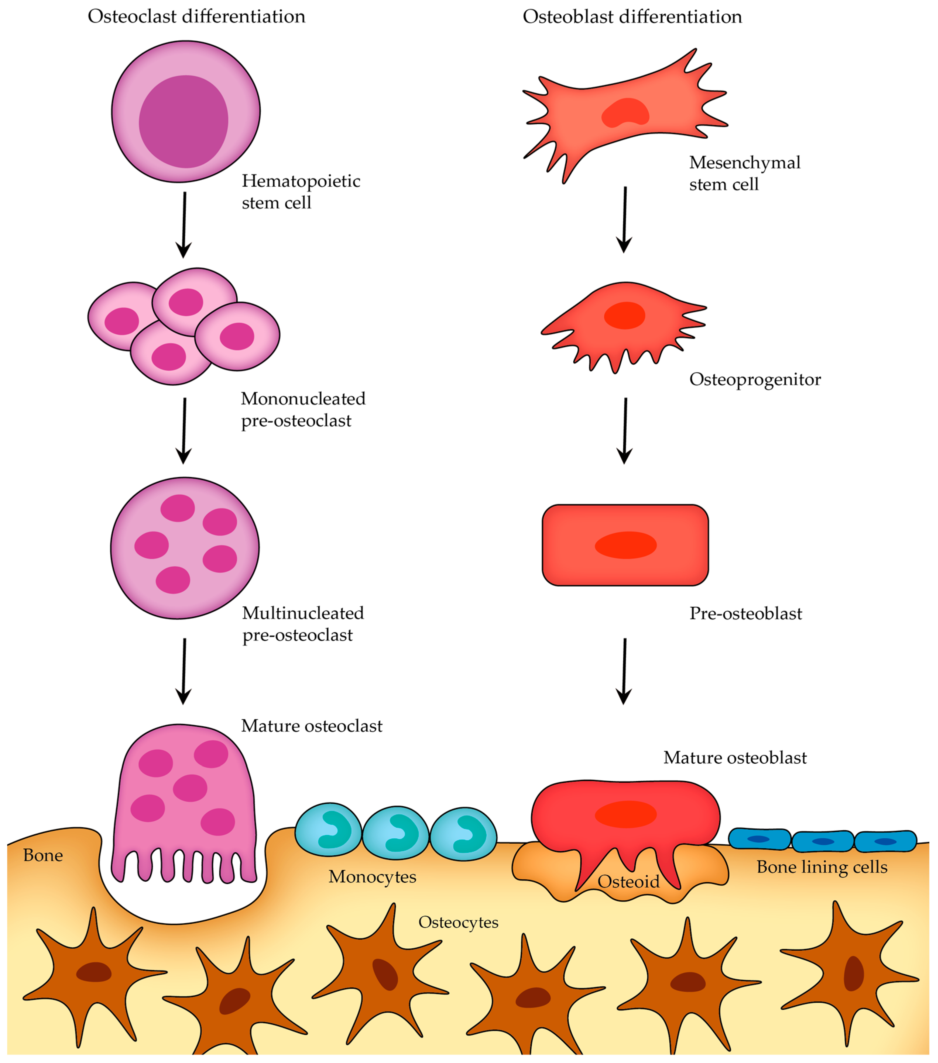
Osteoblasts lay down matrix and express RANKL. Osteoclasts dissolve mineral with carbonic anhydrase II and an H+ ATPase and express RANK. The handshake between the two lineages is the axis this page runs on.
The osteoblast is the builder: it secretes type I collagen and osteocalcin, drives mineralization with alkaline phosphatase, and carries PTH receptors, which is why intermittent PTH (teriparatide) can make it build. Its fate is retirement: buried in its own matrix it becomes an osteocyte, and flattened on a resting surface it becomes a lining cell. The osteoblast also expresses RANKL, the signal that tells osteoclasts to work: the builder holds the leash on the eater.
The osteoclast is the eater and a stranger in the bone family: it is a multinucleated giant cell from the monocyte and macrophage lineage, not from osteogenic stem cells. It expresses RANK, seals a resorption lacuna, and acidifies it: carbonic anhydrase II makes protons and a vacuolar H+ ATPase pumps them in, dissolving hydroxyapatite, while collagenases chew the organic matrix. When the acid machinery fails, bone is never resorbed, and the skeleton turns to marble that breaks: that is osteopetrosis (carbonic anhydrase II or CLCN7 defects), with marrow failure, pancytopenia, and Erlenmeyer flask femurs.
Flip between the two cells and hold the discriminator.
THE HANDSHAKE
The Handshake: RANKL, RANK, and the OPG Decoy
One membrane signal, one receptor, and one decoy set the level of resorption at any moment. Every hormone and drug on this page moves one side of that balance.
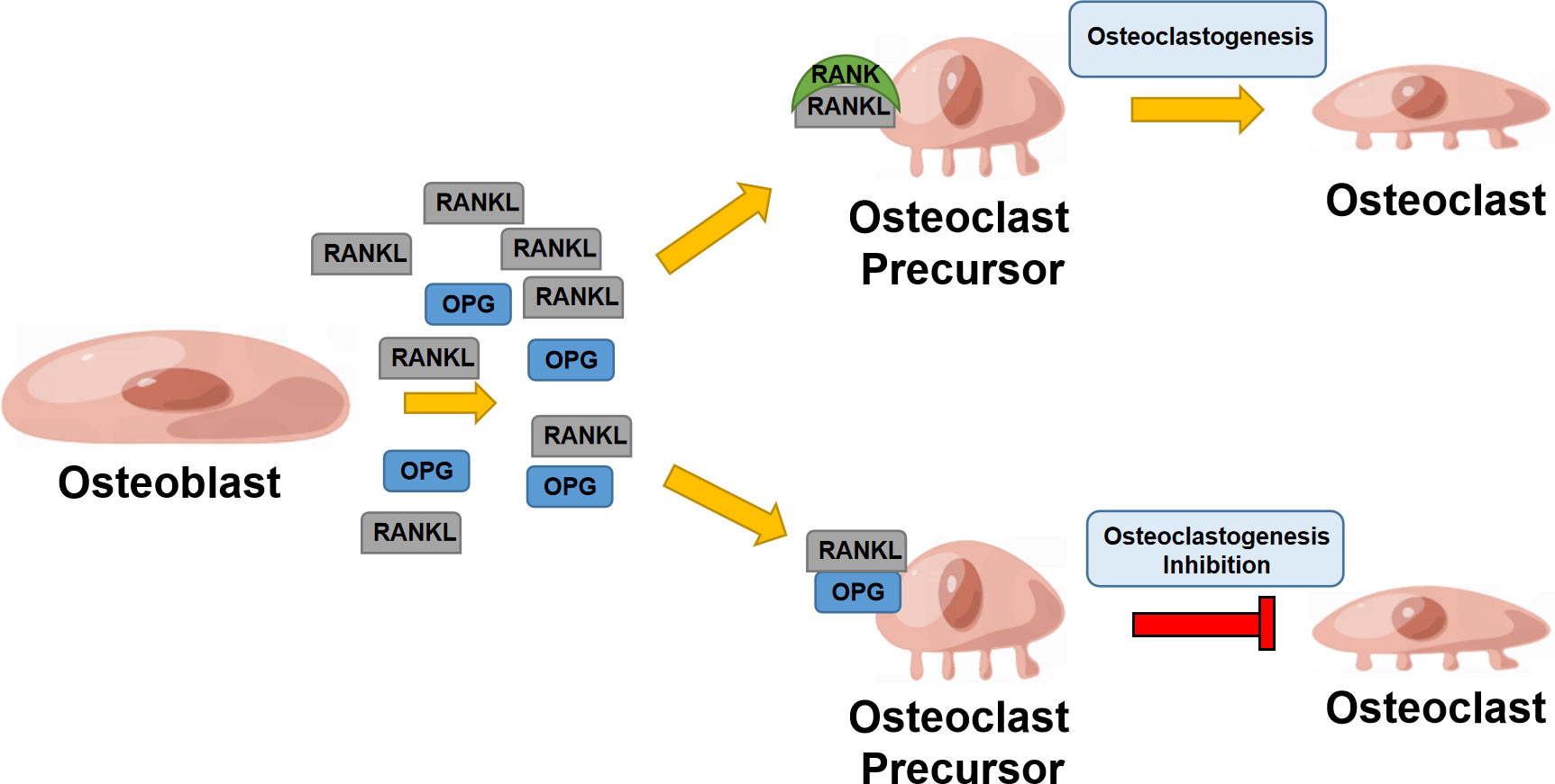
RANKL sits on osteoblasts and marrow stromal cells and binds RANK on osteoclast precursors and mature osteoclasts, driving differentiation, fusion, and activation. OPG, osteoprotegerin, is the soluble decoy made by the same osteoblast line: it intercepts RANKL before RANK can, and the axis goes quiet. The ratio of RANKL to OPG is the rheostat of bone resorption.
The regulators are the boards' favorite mechanism stems. Estrogen upregulates OPG, which is why menopause is the classic bone-loss story: estrogen off, OPG down, RANKL unopposed. Sustained PTH, 1,25-(OH)2D at high levels, glucocorticoids, and IL-1, IL-6, and TNF all pull the RANKL side and cost bone. The drugs grab named points: denosumab is a monoclonal antibody against RANKL (OPG in a syringe), bisphosphonates kill the osteoclast at the mineral surface, and teriparatide and abaloparatide are anabolic PTH analogs that build through the osteoblast.
Commit to the axis map before the reveal.
Which of the following statements about the RANKL-RANK-OPG system is correct?
A. OPG is the decoy. Osteoblasts make both the signal (RANKL) and the brake (OPG), and OPG intercepts RANKL before it reaches RANK. B. RANK on osteoblasts. RANK sits on the osteoclast and its precursors; the osteoblast is the RANKL side. C. Osteoclasts secrete RANKL. RANKL comes from osteoblasts and stromal cells, not from the eater. D. OPG activates RANK. OPG does the opposite: it binds RANKL and blocks the signal. Rule: the builder expresses RANKL, the eater expresses RANK, and OPG is the decoy in between.
THE REMODELING UNIT
The Remodeling Unit: Osteocyte, Lining Cell, Builder, Eater
Four cells share one job posting: replace a packet of old bone with new. The osteocyte is the foreman, the osteoclast digs, the osteoblast fills, and the lining cell watches the finished surface.
The osteocyte is the most abundant bone cell: a former osteoblast buried in the lacuna it made, reaching its neighbors through canaliculi. It is the skeleton's strain gauge: fluid shear from mechanical loading is the signal, and the osteocyte translates load into local remodeling decisions, which is Wolff's law at the cellular level. It also secretes sclerostin, the brake on osteoblast Wnt signaling; unloading raises sclerostin and suppresses formation, and romosozumab, the sclerostin inhibitor, releases the brake.
The lining cell is the quiet surface made visible: a flat, quiescent osteoblast covering resting bone. It maintains the surface and re-enters the story when a new cycle activates. The osteoclast opens the unit by digging, and the osteoblast closes it by filling: resorption is coupled to formation, so what is dug is filled, and the surface returns to rest under lining cells until the next activation.
Tap each cell on the remodeling unit and read its job.
Tap a cell.
THE LEVER LAB
The Lever Lab: Hormones and Drugs on the Axis
Every lever moves RANKL or OPG, and the direction of the bone-density needle is the board answer. Know which side each hormone and drug pulls before you touch the dock.
Start with the two natural levers boards love. Estrogen upregulates OPG, so estrogen loss is RANKL unopposed: postmenopausal osteoporosis in one sentence, and the steepest loss sits in the first years after menopause in trabecular-rich sites. Sustained high PTH (primary or secondary hyperparathyroidism) upregulates RANKL on osteoblasts and stromal cells, so calcium comes out of the skeleton, the kidney keeps calcium and dumps phosphate, and 1-alpha-hydroxylase makes more 1,25-(OH)2D.
The drug levers grab named points. Denosumab, a monoclonal antibody against RANKL, blocks osteoclastogenesis the way OPG would; it is given as a subcutaneous injection every 6 months, and stopping it without follow-on treatment allows resorption to rebound. Bisphosphonates are pyrophosphate analogs that bind hydroxyapatite and inhibit the osteoclast, promoting its apoptosis. Teriparatide (recombinant PTH 1-34, 20 mcg subcutaneously once daily) is the anabolic lever: intermittent PTH1 receptor activation favors formation, while sustained PTH favors resorption, and romosozumab, the sclerostin inhibitor, releases the osteocyte brake on Wnt signaling.
Dock the levers and score the direction: check each bone-loss lever and each bone-gain lever, then read the tally.
Net score 0
THE CYCLE
The Cycle: Activation to Quiescence
Five phases, one unit, about 10 percent of the skeleton remodeled every year. Resorption is coupled to formation, which is why antiresorptive drugs lower both marker families.
The remodeling cycle is the basic unit of bone maintenance. Activation prepares the surface and recruits osteoclast precursors: the RANKL handshake opens the unit. Resorption follows as osteoclasts acidify a sealed lacuna and chew collagen over about 3 weeks, leaving a pit. Reversal clears the pit floor as the osteoclasts die off. Formation lets osteoblasts lay down osteoid that mineralizes over about 3 months, filling the pit with lamellar bone. Quiescence returns the surface to rest under flat lining cells.
Two facts make the cycle board-gold. First, coupling: formation follows resorption in the same unit, so what is dug is filled, and an antiresorptive slows the whole unit, dropping NTX and CTX and, with them, P1NP and bone ALP: that coordinated decline is coupling, not treatment failure. Second, uncoupling: menopause and glucocorticoids let resorption keep digging while formation lags, so each cycle nets lost bone, and teriparatide shifts the unit the other way, which is why P1NP rises early after it is started.
Put the five phases of the remodeling cycle in order.
THE MINERAL RECIPE
The Mineral Recipe: Matrix, ALP, and the Calcium Axis
Bone is a composite: type I collagen scaffold, hydroxyapatite fill, and a mineralization checklist that runs on calcium, phosphate, and alkaline phosphatase. The calcium axis decides who clears the checklist.
About 90 percent of the organic matrix is type I collagen, the scaffold that carries tension; the mineral that fills it is hydroxyapatite, Ca10(PO4)6(OH)2, which carries compression. The noncollagenous proteins organize the interface: osteocalcin, a vitamin K dependent Gla protein made by osteoblasts, plus osteonectin and osteopontin. Osteoid is the newly laid, unmineralized matrix, and it mineralizes only when calcium, phosphate, and alkaline phosphatase are all on the checklist. ALP's job is to cleave pyrophosphate, a natural inhibitor of hydroxyapatite deposition; when ALP is deficient, hypophosphatasia, pyrophosphate accumulates and mineralization fails even with normal calcium and phosphate, giving a rickets-like picture with a low serum ALP, the opposite direction of the high ALP seen in rickets and Paget disease.
The calcium axis runs the supply line. Low serum calcium raises PTH, which activates osteoclasts through RANKL, activates renal 1-alpha-hydroxylase to make 1,25-(OH)2D, increases renal calcium reabsorption, and increases renal phosphate excretion. 1,25-(OH)2D then raises intestinal calcium and phosphate absorption and is permissive for mineralization. Calcitonin from thyroid C cells lowers calcium and inhibits osteoclasts, but its adult role is minor: thyroidectomy does not derange calcium, and that is the board's proof.
Serum calcium falls. Which lever is the body's first response?
Serum calcium falls. Which chain is the body's first response?
THE MARKER SPLIT
The Marker Split: Formation vs Resorption
Formation markers are the builder's receipts; resorption markers are the eater's debris. The boards plant the split in one sentence: a rising NTX with a stable P1NP is a resorption story.
Formation markers come from osteoblast work: bone alkaline phosphatase, the bone isoenzyme released during osteoblast activity; osteocalcin, the vitamin K dependent Gla protein; and P1NP, the N-terminal propeptide of type I procollagen, cleaved off as collagen is assembled. Resorption markers come from the osteoclast: NTX and CTX, the telopeptide fragments of type I collagen released as the matrix is chewed; pyridinoline crosslinks; and TRAP5b, the tartrate-resistant acid phosphatase isoform released by active osteoclasts.
The clinical patterns are the payoff. Start teriparatide and P1NP and bone ALP rise early: the marker story proves the drug is building. Start alendronate or denosumab and NTX and CTX fall, and because of coupling, P1NP follows: a coordinated decline, not a failure. In Paget disease, a surged osteoclast front is coupled to a disorganized osteoblast response, so both families rise together, and the serum ALP becomes a disease monitor.
Tap each letter and hold the builder-eater split.
OPA NCT
OOsteocalcin: vitamin K dependent Gla protein made by osteoblasts, a formation marker
PP1NP: N-terminal propeptide of type I procollagen, cleaved as collagen is assembled, a formation marker
ABone ALP: the bone isoenzyme of alkaline phosphatase from active osteoblasts, a formation marker
NNTX: N-terminal telopeptide of type I collagen released as osteoclasts chew matrix, a resorption marker
CCTX: C-terminal telopeptide of type I collagen, the classic serum resorption marker
TTRAP5b: tartrate-resistant acid phosphatase 5b released by active osteoclasts, a resorption marker
Open each card and hold the drug map and the law.
Bone remodels along lines of mechanical stress: load lands, bone forms; load stops, bone resorbs. The osteocyte is the strain gauge, and unloading raises sclerostin to brake formation. Astronauts, bedbound patients, and immobilized limbs lose bone by this pathway, and the prescription is weight-bearing load, not extra calcium.
Monoclonal antibody against RANKL, given as a subcutaneous injection of 60 mg every 6 months. It mimics OPG, blocking osteoclastogenesis. Correct calcium and vitamin D status before starting, because potent antiresorptives can drop serum calcium, and plan follow-on treatment when stopping, because RANKL is unmasked when the drug wears off.
Recombinant human PTH 1-34, 20 mcg subcutaneously once daily, typically used for up to 2 years. Intermittent PTH1 receptor activation is anabolic: osteoblast recruitment and survival win, and P1NP rises early. It is the builder's lever and is followed by an antiresorptive to hold the gain.
Pyrophosphate analogs that bind hydroxyapatite at the resorption surface; nitrogen-containing agents such as alendronate inhibit farnesyl pyrophosphate synthase in the mevalonate pathway, disrupting the osteoclast cytoskeleton and promoting apoptosis. The antiresorptive workhorse: alendronate 70 mg orally once weekly, with the dose taken on an empty stomach to avoid esophagitis.
Monoclonal antibody against sclerostin, the osteocyte brake on Wnt signaling. It releases the brake so osteoblasts build, and it also suppresses osteoclast activity, a dual anabolic and antiresorptive profile given as a monthly subcutaneous injection for 12 months, then followed by an antiresorptive.
Peptide from thyroid C cells that lowers serum calcium and inhibits osteoclast activity, but with a weak and transient adult effect: thyroidectomy does not derange calcium. Its niche is refractory Paget disease and some hypercalcemia settings, not first-line osteoporosis therapy.
Prove it
Walkthrough: pull the levers case by case
Original practice scenarios, one at a time, never repeated until the bank is exhausted. Choose an answer, then open any option to work its reasoning.
Clinical walkthrough
