A deep buttock injury produces bleeding, painful swelling, and new foot weakness. The inferior gluteal artery may be the bleeding source while the nearby sciatic nerve explains the weakness. Those are related findings, but an artery and a nerve are not interchangeable answers.
This artery is easiest to understand by following three relationships. It leaves the pelvis below piriformis, supplies the deep surface of gluteus maximus and neighboring tissues, and connects pelvic inflow with vessels around the proximal femur. Its collateral contribution matters even though the medial femoral circumflex artery usually provides the principal retinacular supply to the femoral head.
Use piriformis as a landmark, then follow the destination Trace the vessel by relationships Read the question, predict, then check the reasoning.
Which pelvic parent is the usual starting point for this route?
Reveal this step The inferior gluteal artery usually arises from the anterior internal iliac division.
Why should the expected branch point guide rather than settle identification?
Reveal this step Inferior gluteal origin and course can vary.
Which side of piriformis places the vessel on its usual buttock route?
Reveal this step It exits the greater sciatic foramen inferior to piriformis.
What separates this artery from another vessel sharing the infrapiriform exit?
Reveal this step The inferior gluteal artery continues into the buttock rather than returning toward the perineum.
Start this reasoning again Sources [1] [9]
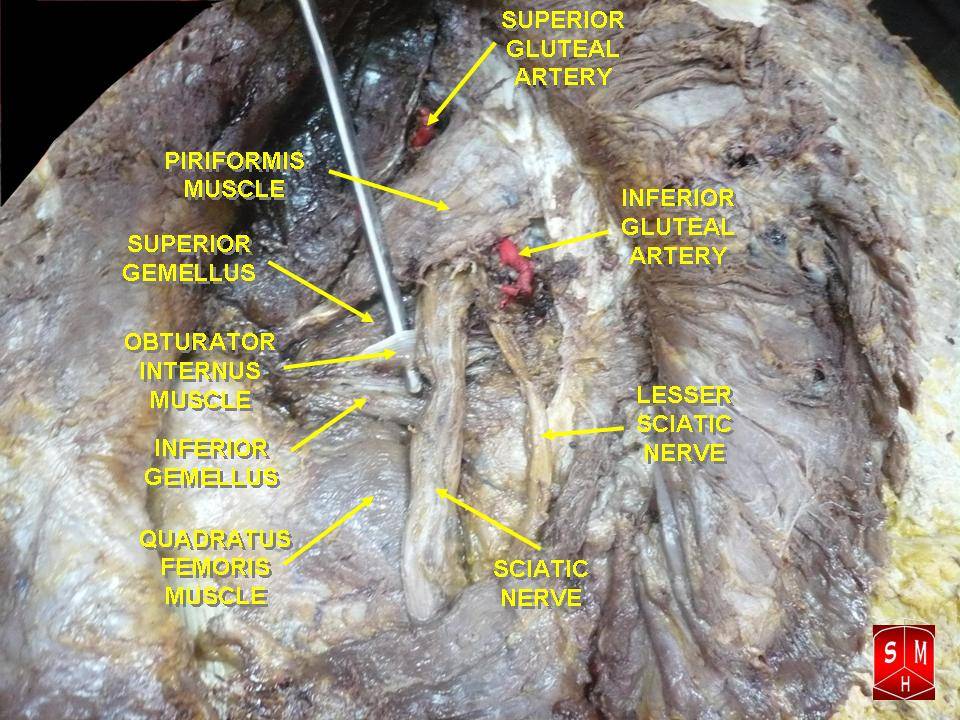
The inferior gluteal artery usually arises from the anterior division of the internal iliac artery. It passes out of the pelvis through the greater sciatic foramen inferior to piriformis. The superior gluteal artery usually arises from the posterior division and passes superior to piriformis. This is a useful default map, not a promise that every branching pattern will be identical. Angiography and operative exposure should establish the actual origin. [1]
Open whole image Use piriformis first: the inferior gluteal artery passes below it, while the superior gluteal artery passes above it. Image: Anatomist90. CC BY-SA 3.0 . Original source .
Whole image Close
Apply: Use piriformis as a landmark then follow the destination Angiography after a deep buttock laceration shows active leakage from a branch of the anterior internal iliac division. It exits below piriformis and spreads deep to gluteus maximus. Which artery is injured?
Inferior gluteal artery Superior gluteal artery Internal pudendal artery
Try again Read the worked solution Inferior gluteal artery Best fit. The origin, infrapiriform exit, and gluteus maximus destination identify it together. [1]
Superior gluteal artery Compare this alternative. Its usual exit is above piriformis and its usual origin is the posterior division. [1]
Internal pudendal artery Compare this alternative. It also exits below piriformis but returns through the lesser foramen toward the perineum. [1]
Pelvis to buttock, organized around piriformis. The labels describe usual relationships and can be read vertically on a small screen. Above piriformis Superior gluteal artery, veins, and nerve leave the greater sciatic foramen. The artery supplies superficial and deep gluteal territories; the nerve supplies gluteus medius, gluteus minimus, and tensor fasciae latae.
At piriformis Piriformis occupies part of the greater sciatic foramen. It separates the usual superior gluteal passage from the larger inferior passage.
Below piriformis Inferior gluteal vessels and nerve enter the buttock. The sciatic nerve and posterior femoral cutaneous nerve also pass through this region. Internal pudendal vessels and the pudendal nerve leave here, curve near the ischial spine, and enter the perineum through the lesser sciatic foramen.
Below piriformis therefore narrows the location without uniquely naming an artery. Follow where the vessel goes. A branch spreading on the deep surface of gluteus maximus favors inferior gluteal supply. A vessel turning around the ischial spine toward the lesser foramen favors the internal pudendal artery. A vessel in the anterior thigh arising from profunda femoris belongs to another arterial route, even if it ultimately communicates with the gluteal circulation. [1] [2]
The sciatic nerve commonly emerges below piriformis, but its divisions and relationship to the muscle vary. The nerve is much larger than the small arterial branch that accompanies it. The artery to the sciatic nerve is a branch associated with the inferior gluteal circulation; it is not a reason to rename the entire inferior gluteal artery as the nerve's exclusive blood supply.
Try it here · Checkpoint 1 of 3
Make your prediction before reading the choices. A first attempt is just a starting point.
Case 1
Show answer and explanations for case 1
A. Inferior gluteal artery (Best answer)
Reason through this option Read the question, predict, then check the reasoning.
Which pelvic division gives the leaking branch?
Reveal this step The branch comes from the anterior division of the internal iliac artery.
Which route and destination identify it?
Reveal this step It exits below piriformis and spreads deep to gluteus maximus, identifying the inferior gluteal artery.
Start this reasoning again Sources [1]
Read the complete explanation The origin, infrapiriform exit, and gluteus maximus destination identify it together.
B. Superior gluteal artery (Why this does not fit)
Reason through this option Read the question, predict, then check the reasoning.
Why can the superior gluteal artery seem plausible?
Reveal this step It is another internal iliac branch supplying the gluteal region.
Which landmark rules it out?
Reveal this step The superior gluteal artery usually exits above piriformis, not below it.
Start this reasoning again Sources [1]
Read the complete explanation Its usual exit is above piriformis and its usual origin is the posterior division.
C. Internal pudendal artery (Why this does not fit)
Reason through this option Read the question, predict, then check the reasoning.
Why is the internal pudendal artery nearby?
Reveal this step It also leaves the pelvis below piriformis.
Which destination distinguishes it?
Reveal this step It curves toward the lesser sciatic foramen and perineum rather than spreading deep to gluteus maximus.
Start this reasoning again Sources [1]
Read the complete explanation It also exits below piriformis but returns through the lesser foramen toward the perineum.
D. Medial femoral circumflex artery (Why this does not fit)
Reason through this option Read the question, predict, then check the reasoning.
Why might a circumflex vessel enter consideration?
Reveal this step Circumflex femoral branches communicate with vessels around the posterior hip.
Which route excludes this option?
Reveal this step The medial femoral circumflex artery approaches from femoral circulation, not the anterior internal iliac division.
Start this reasoning again Sources [1]
Read the complete explanation It approaches from the femoral circulation rather than this internal iliac route.
Takeaway: Use origin, passage, and destination together.
Case sources: [1]
Save this spot
Try a question on this topic A 67-year-old man suddenly develops vertigo, vomiting, and loss of hearing in his right ear. Examination shows right facial weakness involving the forehead and mouth, right-sided limb ataxia, and decreased pain sensation on the left side of the body. Diffusion-weighted MRI shows an infarct of the right lateral caudal pons and adjacent anterior inferior cerebellum. He was speaking normally immediately before the event and has no prior history of hearing impairment or chronic ear infection. Bedside testing suggests sensorineural rather than conductive hearing loss on the right. His tongue protrudes in the midline, and there is no hoarseness or palatal asymmetry. The simultaneous onset of hearing, facial, and long-tract sensory findings prompts urgent vascular imaging rather than treatment for an isolated ear disorder.
Which artery is most likely occluded?
A. Anterior inferior cerebellar artery B. Posterior inferior cerebellar artery C. Superior cerebellar artery D. Anterior spinal artery E. Posterior cerebral artery
Choose an answer before revealing the reasoning.
Get one of these every morning · Practice more in the QBank
Separate the muscle's artery from its motor nerve Separate blood supply from motor function Read the question, predict, then check the reasoning.
Does reaching gluteus maximus prove a single exclusive arterial supply?
Reveal this step No. Superior gluteal branches also contribute to this muscle.
Which finding favors motor denervation over arterial injury?
Reveal this step Weak powerful hip extension without hemorrhage favors inferior gluteal nerve dysfunction.
Which muscle group is tested when the opposite pelvis drops during stance?
Reveal this step The stance-side hip abductors are failing to stabilize the pelvis.
Why can a buttock hematoma produce foot weakness without becoming a nerve?
Reveal this step The expanding vascular mass can compress the nearby sciatic nerve.
Start this reasoning again Sources [1] [2]
Inferior gluteal branches supply much of gluteus maximus, adjacent buttock skin, deep lateral rotators, proximal posterior thigh tissues, and vessels around the hip. Distribution overlaps with superior gluteal and femoral branches. Gluteus maximus does not belong exclusively to one artery, and the superior gluteal artery is not limited to the abductors. Its superficial branches also contribute to gluteus maximus and overlying tissues. [1]
Apply: Separate the muscle's artery from its motor nerve A patient has difficulty rising from a low chair after an isolated nerve injury in the deep buttock. Hip abduction is strong, and imaging shows no hematoma or perfusion defect. Which nerve is most likely affected?
Inferior gluteal nerve Superior gluteal nerve Obturator nerve
Try again Read the worked solution Inferior gluteal nerve Best fit. Weak powerful hip extension with preserved abduction fits gluteus maximus denervation. [2]
Superior gluteal nerve Compare this alternative. Its abductor targets remain strong in this stem. [2]
Obturator nerve Compare this alternative. It chiefly serves the medial thigh adductors, not gluteus maximus. [2]
Gluteus maximus is particularly useful for powerful hip extension, such as rising from a low chair or climbing a steep step. Its motor nerve is the inferior gluteal nerve, usually L5 through S2. An isolated nerve lesion can impair that task without hemorrhage or a perfusion defect. Pain, tendon injury, muscle damage, and ischemia can also limit extension, so the observed action is not by itself an arterial angiogram. [2]
Gluteus medius and minimus stabilize the pelvis during single-leg support. Weakness on the stance side can allow the opposite side of the pelvis to drop, producing a Trendelenburg sign. The superior gluteal nerve supplies these muscles and tensor fasciae latae. A motor deficit after a local procedure raises concern for that nerve or the muscles themselves. A superior gluteal arterial lesion becomes a stronger answer only when vascular evidence, such as active contrast leakage or a documented perfusion injury, is added.
Posterior thigh skin requires another distinction. The posterior femoral cutaneous nerve supplies much of that cutaneous territory. The sciatic nerve supplies hamstrings and, through its tibial and common fibular divisions, most muscles below the knee and broad distal sensory territories. A buttock hematoma can compress the sciatic nerve and produce foot weakness or distal paresthesia, but posterior thigh numbness alone should not be assigned automatically to the sciatic nerve. Nearby nerves can be affected together in a large injury. [2]
Save this spot
The buttock connects pelvic and femoral inflow Build the posterior collateral junction Read the question, predict, then check the reasoning.
Which pelvic branch descends toward the cruciate connection?
Reveal this step A descending inferior gluteal branch approaches the posterior proximal femur.
Which circumflex components meet that pelvic branch near the lesser trochanter?
Reveal this step Transverse medial and lateral circumflex femoral branches contribute.
Which profunda branch supplies the inferior limb of the classic junction?
Reveal this step The ascending branch of the first perforating artery completes it.
What does seeing this connection establish about threatened tissue?
Reveal this step It establishes an alternative route, not adequate collateral flow.
Start this reasoning again Sources [1] [3]
Explore the reasoning
The buttock connects pelvic and femoral inflow
Work through the reasoning
Which section principle should anchor this decision?
The buttock connects pelvic and femoral inflow The classic cruciate anastomosis lies posterior to the proximal femur near the level of the lesser trochanter.
Show all answers
Focus on one step
Earlier step Following step
What should be established first? The buttock connects pelvic and femoral inflow The classic cruciate anastomosis lies posterior to the proximal femur near the level of the lesser trochanter.
What second distinction prevents the shortcut? The arrangement gives blood alternative paths between internal iliac and profunda femoris territories.
What should carry into the next case? The cruciate anastomosis joins pelvic, circumflex, and perforating arterial routes.
Use the section like a decision map: identify the finding, then follow only the supported relationship to the conclusion.
Where the comparison stops. This organizes reasoning; it does not replace the patient history, examination, source-specific criteria, or appropriate diagnostic testing.
The buttock connects pelvic and femoral inflow Ascending branch of the first profunda femoris perforator The cruciate anastomosis joins pelvic circumflex and perforating The cruciate anastomosis joins pelvic, circumflex, and perforating arterial routes.
Sources [1]
Compare both answers Ascending branch of the first profunda femoris perforator
Why it fits. This is the inferior contributor to the classic cruciate anastomosis.
This answer best fits the stated findings and the section mechanism.
The cruciate anastomosis joins pelvic, circumflex, and perforating arterial routes.
Ascending branch of the lateral femoral circumflex artery
Why it is tempting. This contributes to the trochanteric region. The classic cruciate connection instead uses the transverse circumflex branch and the ascending first perforator.
Compare the stated findings with the section mechanism before choosing the alternative.
The cruciate anastomosis joins pelvic, circumflex, and perforating arterial routes.
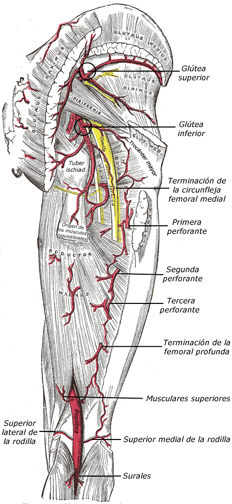
The classic cruciate anastomosis lies posterior to the proximal femur near the level of the lesser trochanter. Its named contributors are a descending inferior gluteal branch, transverse branches of the medial and lateral circumflex femoral arteries, and an ascending branch of the first perforating artery from profunda femoris. The arrangement gives blood alternative paths between internal iliac and profunda femoris territories. It does not guarantee sufficient perfusion after every occlusion. [1] [3]
Open whole image Follow the posterior arterial network toward the proximal femur before reasoning through collateral connections. Image: Henry Gray. Public domain . Original source .
Whole image Close
Apply: The buttock connects pelvic and femoral inflow During posterior hip dissection, a descending pelvic arterial branch joins transverse circumflex femoral branches near the lesser trochanter. Which additional branch completes the classic cruciate connection?
Ascending branch of the first profunda femoris perforator Ascending branch of the lateral femoral circumflex artery Deep branch of the superior gluteal artery
Try again Read the worked solution Ascending branch of the first profunda femoris perforator Best fit. This is the inferior contributor to the classic cruciate anastomosis. [1]
Ascending branch of the lateral femoral circumflex artery Compare this alternative. This contributes to the trochanteric region. The classic cruciate connection instead uses the transverse circumflex branch and the ascending first perforator. [1]
Deep branch of the superior gluteal artery Compare this alternative. This is associated with the trochanteric network. It is not the classic inferior contributor joining the transverse circumflex branches near the lesser trochanter. [1]
Two connected routes near the hip. These are a schematic of connections, not a scale drawing or a claim of equal flow. Pelvic route. Internal iliac artery supplies inferior gluteal artery below piriformis.Femoral route. Femoral artery supplies profunda femoris, usually the circumflex femoral arteries, and perforating branches.Posterior connection. Inferior gluteal branches communicate with circumflex and perforating branches around the proximal femur.Head-directed route. The medial femoral circumflex artery gives rise to retinacular vessels that approach the femoral head along the neck.The medial femoral circumflex artery commonly arises from profunda femoris, but direct origin from the femoral artery is a recognized variant. Parent vessel identification and territory identification answer different questions. An artery can have a variant parent while preserving its characteristic course around the proximal femur. Do not reject a medial circumflex vessel simply because it does not arise where a diagram placed it.
Cadaveric studies demonstrate direct anastomoses between the inferior gluteal artery and the medial femoral circumflex system near the posterior hip. A 16-dissection study identified the inferior gluteal connection through a piriformis branch as an important collateral route to the femoral head circulation. That finding corrects the claim that inferior gluteal supply is merely an irrelevant twig. It does not make the inferior gluteal artery the usual dominant head artery, nor does it prove that a collateral can rescue every disrupted retinacular vessel. [3]
For interpretation, distinguish a named anatomical connection from demonstrated adequate flow. Existing stenosis, trauma to multiple vessels, tissue pressure, anatomic variation, and the speed of an occlusion all influence collateral performance. A patent connection on a dissection or scan explains a possible route; perfusion of the threatened tissue remains the clinical question.
Save this spot
Why a preserved buttock circulation may not protect the femoral head Keep collateral routes in proportion Read the question, predict, then check the reasoning.
Which vessels are most exposed by a displaced intracapsular neck fracture?
Reveal this step Head-directed retinacular vessels from the medial femoral circumflex system are most vulnerable.
Does an inferior gluteal connection make that artery the principal head supply?
Reveal this step No. The connection is collateral to the usual dominant retinacular route.
What does a normal foot pulse fail to test after hip trauma?
Reveal this step It does not test perfusion through the small head-directed vessels.
Which imaging distinction separates Perthes disease from SCFE?
Reveal this step Perthes affects the femoral head, while SCFE displaces the proximal femoral physis.
Start this reasoning again Sources [1] [3] [4] [5] [6]
The adult femoral head receives its principal arterial supply through retinacular branches associated predominantly with the medial femoral circumflex artery. Their course along the femoral neck makes them vulnerable in displaced intracapsular neck fractures and hip dislocation. A normal distal pulse or viable gluteus maximus does not establish that those small head-directed vessels remain intact. The foot and the femoral head are supplied through different downstream pathways. [3]
Apply: Why a preserved buttock circulation may not protect the femoral A displaced intracapsular femoral neck fracture is followed by femoral head osteonecrosis. Which arterial pathway was most vulnerable?
Retinacular vessels predominantly from the medial femoral circumflex artery Inferior gluteal descending branches forming the cruciate anastomosis Ascending lateral femoral circumflex contribution near the greater trochanter
Try again Read the worked solution Retinacular vessels predominantly from the medial femoral circumflex artery Best fit. Their course along the neck exposes the principal head supply to displacement. [1] [3]
Inferior gluteal descending branches forming the cruciate anastomosis Compare this alternative. These participate in a proximal posterior femoral collateral network. The displaced intracapsular fracture directly threatens head-directed retinacular vessels along the femoral neck. [1] [3]
Ascending lateral femoral circumflex contribution near the greater trochanter Compare this alternative. This contributes around the proximal femur, but it is not the predominant retinacular head supply most directly endangered by displacement through the neck. [1] [3]
The artery in the ligament of the head of the femur usually reaches the foveal region through an acetabular branch associated with the obturator artery, with variations. Its contribution should not be taught as the sole supply in infancy followed by a universal switch at a fixed birthday. Developmental vascular studies show a more complex arrangement of cervical and epiphyseal vessels. Age changes vulnerability, but a child's age alone cannot identify one transected artery. [1] [4]
Legg-Calvé-Perthes disease is childhood osteonecrosis related to interruption of femoral head perfusion, with the initiating cause not fully understood. A child in the usual school-age range may limp or report hip, thigh, or referred knee pain. Restricted internal rotation and abduction, followed by radiographic sclerosis, fragmentation, or flattening, support the diagnosis. It is inaccurate to present every case as proven occlusion of one named vessel or to equate it with a traumatic arterial laceration. [5]
Slipped capital femoral epiphysis is a different process at the proximal femoral growth plate, generally in an older child or adolescent. Hip or knee pain, an out-toed gait, and loss of internal rotation should prompt assessment. The head and neck lose their normal relationship at the physis; this is not the same mechanism as Perthes fragmentation. Suspected SCFE requires prompt orthopedic evaluation and protection from weight bearing. [6]
After hip trauma, later femoral head collapse suggests osteonecrosis but does not retrospectively prove which exact vessel was severed. The most defensible anatomical answer to a displaced neck fracture question is the vulnerable retinacular supply, predominantly from the medial femoral circumflex artery, unless imaging or operative findings provide a more specific vessel injury.
Try it here · Checkpoint 2 of 3
Make your prediction before reading the choices. A first attempt is just a starting point.
Case 10
Show answer and explanations for case 10
A. Inferior gluteal descending branches forming the cruciate anastomosis (Why this does not fit)
Reason through this option Read the question, predict, then check the reasoning.
What role do inferior gluteal descending branches play?
Reveal this step They help form a posterior collateral network near the proximal femur.
Why are they not the principal vulnerable pathway?
Reveal this step They are collateral contributors, while head-directed retinacular vessels course along the fractured femoral neck.
Start this reasoning again Sources [1] [3]
Read the complete explanation These participate in a proximal posterior femoral collateral network. The displaced intracapsular fracture directly threatens head-directed retinacular vessels along the femoral neck.
B. Ascending lateral femoral circumflex contribution near the greater trochanter (Why this does not fit)
Reason through this option Read the question, predict, then check the reasoning.
Where does the ascending lateral circumflex contribution run?
Reveal this step It participates around the greater trochanter.
Which vessels are more exposed in an intracapsular neck fracture?
Reveal this step Retinacular branches along the femoral neck are more directly threatened.
Start this reasoning again Sources [1] [3]
Read the complete explanation This contributes around the proximal femur, but it is not the predominant retinacular head supply most directly endangered by displacement through the neck.
C. Obturator contribution through the ligament of the femoral head (Why this does not fit)
Reason through this option Read the question, predict, then check the reasoning.
Why might the ligament artery seem protective?
Reveal this step Its obturator-associated branch can reach the foveal region of the head.
Why is it not the main adult pathway?
Reveal this step Adult head perfusion depends principally on medial circumflex retinacular vessels.
Start this reasoning again Sources [1] [3]
Read the complete explanation The ligament artery can contribute to head perfusion, but the principal adult retinacular route exposed by a displaced neck fracture is predominantly medial femoral circumflex.
D. Retinacular vessels predominantly from the medial femoral circumflex artery (Best answer)
Reason through this option Read the question, predict, then check the reasoning.
Where do the vulnerable vessels travel?
Reveal this step Retinacular vessels ascend along the femoral neck toward the head.
Which artery predominantly supplies them?
Reveal this step They arise predominantly from the medial femoral circumflex artery, explaining osteonecrosis after displacement.
Start this reasoning again Sources [1] [3]
Read the complete explanation Their course along the neck exposes the principal head supply to displacement.
Takeaway: The head's vulnerable microvascular route matters more than a preserved distal pulse.
Case sources: [1] [3]
Save this spot
Recognize bleeding that can masquerade as sciatica Treat the mass as vascular first Read the question, predict, then check the reasoning.
Does absent visible pulsation exclude a post-traumatic pseudoaneurysm?
Reveal this step No. A gluteal pseudoaneurysm can present as a nonpulsatile mass.
Why do preserved pedal pulses fail to clear the buttock artery?
Reveal this step Distal limb flow can persist despite injury to an internal iliac branch.
Which stable-patient study can map both arterial leakage and hematoma extent?
Reveal this step CT angiography can define the vascular lesion and surrounding hematoma.
What unresolved problem can remain after embolization stops the leakage?
Reveal this step The existing hematoma can continue compressing muscle or nerve.
Start this reasoning again Sources [7] [8]
Explore the reasoning
Recognize bleeding that can masquerade as sciatica
Work through the reasoning
Which section principle should anchor this decision?
Recognize bleeding that can masquerade as sciatica Penetrating injury, blunt trauma, or a procedure can damage a gluteal arterial wall.
Show all answers
Focus on one step
Earlier step Following step
What should be established first? Recognize bleeding that can masquerade as sciatica Penetrating injury, blunt trauma, or a procedure can damage a gluteal arterial wall.
What second distinction prevents the shortcut? Active hemorrhage may produce an expanding buttock hematoma and falling hemoglobin.
What should carry into the next case? Use direct vascular evidence to name the vessel and assess motor injury separately.
Use the section like a decision map: identify the finding, then follow only the supported relationship to the conclusion.
Where the comparison stops. This organizes reasoning; it does not replace the patient history, examination, source-specific criteria, or appropriate diagnostic testing.
Recognize bleeding that can masquerade as sciatica Superior gluteal arterial injury with possible associated nerve Use direct vascular evidence to name the vessel Use direct vascular evidence to name the vessel and assess motor injury separately.
Sources [1] [2]
Compare both answers Superior gluteal arterial injury with possible associated nerve or muscle injury
Why it fits. The vascular image identifies the artery; the motor deficit requires assessment of neighboring motor structures.
This answer best fits the stated findings and the section mechanism.
Use direct vascular evidence to name the vessel and assess motor injury separately.
Only the superior gluteal nerve is injured because weakness is present
Why it is tempting. An isolated nerve injury cannot explain active arterial contrast extravasation. The motor deficit deserves assessment, but it does not erase the documented vascular injury.
Compare the stated findings with the section mechanism before choosing the alternative.
Use direct vascular evidence to name the vessel and assess motor injury separately.
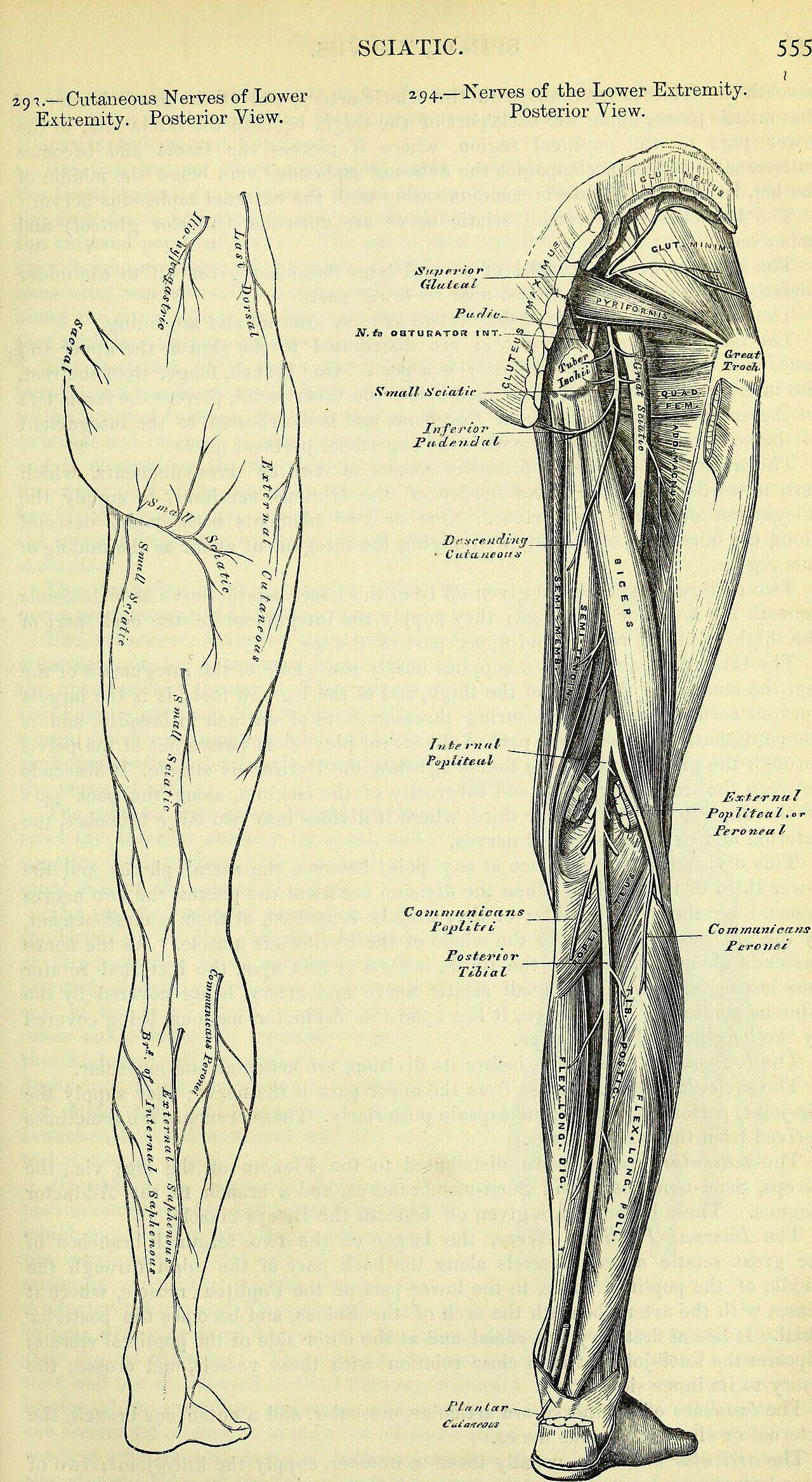
Penetrating injury, blunt trauma, or a procedure can damage a gluteal arterial wall. Active hemorrhage may produce an expanding buttock hematoma and falling hemoglobin. A contained communication with the arterial lumen can form a pseudoaneurysm that presents later as pain or swelling. It may compress the sciatic nerve and produce distal weakness or paresthesia. Published cases demonstrate that such a mass can be nonpulsatile and that distal pulses can remain present. Those findings do not exclude a gluteal arterial injury. [7] [8]
Open whole image The artery and sciatic nerve share a crowded region, explaining how a vascular mass can create neurologic symptoms. Image: Internet Archive Book Images. No known copyright restrictions . Original source .
Whole image Close
Apply: Recognize bleeding that can masquerade as sciatica A patient has active contrast extravasation above piriformis after pelvic trauma and new abductor weakness. Which interpretation is most defensible?
Superior gluteal arterial injury with possible associated nerve or muscle injury Only the superior gluteal nerve is injured because weakness is present An internal pudendal arterial injury alone explains the entire pattern
Try again Read the worked solution Superior gluteal arterial injury with possible associated nerve or muscle injury Best fit. The vascular image identifies the artery; the motor deficit requires assessment of neighboring motor structures. [1] [2]
Only the superior gluteal nerve is injured because weakness is present Compare this alternative. An isolated nerve injury cannot explain active arterial contrast extravasation. The motor deficit deserves assessment, but it does not erase the documented vascular injury. [1] [2]
An internal pudendal arterial injury alone explains the entire pattern Compare this alternative. The internal pudendal artery usually passes below piriformis before entering the perineum. The imaged bleeding is above piriformis and is associated with abductor rather than perineal dysfunction. [1] [2]
A painful mass after buttock trauma should therefore not be aspirated as a presumed abscess without considering a vascular cause. In a stable patient, Doppler ultrasound or CT angiography can establish arterial flow, the extent of the hematoma, and the vessel involved. Catheter angiography can combine diagnosis with selective embolization. Hemodynamic instability, rapidly progressive neurologic loss, or concern for compartment syndrome requires urgent specialist management rather than a routine sciatica pathway. [7] [8]
Hemostasis and decompression solve different problems. Embolization can stop a bleeding artery; a large compressive hematoma or compartment syndrome may still require surgical treatment. Conversely, opening a vascular mass before controlling its arterial communication can precipitate severe bleeding. The exact order and method depend on physiology, imaging, and the treating trauma, vascular, and surgical teams. Case reports support recognition of this complication, not a single mandatory procedure for every patient.
When answering a localization question, state the evidence at the right level. Contrast extravasation from a branch below piriformis continuing into gluteus maximus identifies the inferior gluteal artery. Isolated loss of powerful hip extension suggests its motor nerve or muscle. Contralateral pelvic drop during stance suggests abductor dysfunction. Foot weakness beside an expanding buttock hematoma suggests a compressive sciatic complication. Keeping those observations separate makes the final explanation more precise.
Try it here · Checkpoint 3 of 3
Make your prediction before reading the choices. A first attempt is just a starting point.
Case 23
Show answer and explanations for case 23
A. Only the superior gluteal nerve is injured because weakness is present (Why this does not fit)
Reason through this option Read the question, predict, then check the reasoning.
Can nerve injury explain abductor weakness?
Reveal this step Yes. The superior gluteal nerve supplies the main hip abductors.
What finding proves a vascular component?
Reveal this step Active contrast extravasation above piriformis directly demonstrates arterial injury.
Start this reasoning again Sources [1] [2]
Read the complete explanation An isolated nerve injury cannot explain active arterial contrast extravasation. The motor deficit deserves assessment, but it does not erase the documented vascular injury.
B. An internal pudendal arterial injury alone explains the entire pattern (Why this does not fit)
Reason through this option Read the question, predict, then check the reasoning.
Where does the internal pudendal artery exit?
Reveal this step It exits below piriformis before turning toward the perineum.
Why does it not fit the complete pattern?
Reveal this step The bleeding is above piriformis, and abductor dysfunction lies in the superior gluteal territory.
Start this reasoning again Sources [1] [2]
Read the complete explanation The internal pudendal artery usually passes below piriformis before entering the perineum. The imaged bleeding is above piriformis and is associated with abductor rather than perineal dysfunction.
C. Superior gluteal arterial injury with possible associated nerve or muscle injury (Best answer)
Reason through this option Read the question, predict, then check the reasoning.
Which artery normally passes above piriformis?
Reveal this step The superior gluteal artery uses the suprapiriform route.
How should the weakness be interpreted?
Reveal this step It suggests associated superior gluteal nerve or abductor muscle injury alongside the demonstrated arterial lesion.
Start this reasoning again Sources [1] [2]
Read the complete explanation The vascular image identifies the artery; the motor deficit requires assessment of neighboring motor structures.
D. The weakness alone proves inferior gluteal arterial injury (Why this does not fit)
Reason through this option Read the question, predict, then check the reasoning.
Does weakness alone identify an artery?
Reveal this step No. Weakness primarily localizes motor nerve or muscle dysfunction.
Which landmark contradicts inferior gluteal injury?
Reveal this step The active extravasation lies above piriformis, while the inferior gluteal artery usually passes below it.
Start this reasoning again Sources [1] [2]
Read the complete explanation The image is above piriformis, and weakness alone cannot name an artery.
Takeaway: Use direct vascular evidence to name the vessel and assess motor injury separately.
Case sources: [1] [2]
Save this spot
Try these without looking During posterior hip dissection a descending pelvic arterial branch joins transverse circumflex femoral branches near the lesser trochanter Which additional branch completes the classic cruciate connection? The cruciate anastomosis joins pelvic, circumflex, and perforating arterial routes.
Revisit this explanation [1]
A patient has active contrast extravasation above piriformis after pelvic trauma and new abductor weakness Which interpretation is most defensible? Use direct vascular evidence to name the vessel and assess motor injury separately.
Revisit this explanation [1] [2]
What prevents this lesson from becoming a one-clue shortcut? Use the complete clinical or study context, then apply the named test, anatomical relation, or guideline only within its validated conditions.
Revisit this explanation [1] [2]
Practice the vessel, nerve, and collateral distinctions
Case 2
Show answer and explanations for case 2
A. Inferior gluteal artery (Why this does not fit)
Reason through this option Read the question, predict, then check the reasoning.
Why is an inferior gluteal vessel plausible?
Reveal this step It supplies the buttock through the greater sciatic foramen.
Which relationship excludes it?
Reveal this step Its usual exit is inferior to piriformis, while this vessel passes superior to the muscle.
Start this reasoning again Sources [1]
Read the complete explanation Its usual passage is inferior to piriformis.
B. Internal pudendal artery (Why this does not fit)
Reason through this option Read the question, predict, then check the reasoning.
Where does the internal pudendal artery initially exit?
Reveal this step It exits the greater sciatic foramen below piriformis.
Where does it then travel?
Reveal this step It turns around the ischial spine toward the lesser foramen and perineum, unlike this gluteal branch pattern.
Start this reasoning again Sources [1]
Read the complete explanation It usually passes below piriformis before entering the perineum.
C. Obturator artery (Why this does not fit)
Reason through this option Read the question, predict, then check the reasoning.
Which opening carries the obturator artery?
Reveal this step It passes through the obturator canal toward the medial thigh.
Why does that exclude it here?
Reveal this step The described vessel uses the greater sciatic foramen and divides within the gluteal region.
Start this reasoning again Sources [1]
Read the complete explanation It reaches the medial thigh through the obturator canal.
D. Superior gluteal artery (Best answer)
Reason through this option Read the question, predict, then check the reasoning.
Which landmark defines the superior gluteal route?
Reveal this step The vessel exits the greater sciatic foramen above piriformis.
Does its branching territory fit?
Reveal this step Yes. Superficial and deep gluteal branches are characteristic of the superior gluteal artery.
Start this reasoning again Sources [1]
Read the complete explanation Its usual course above piriformis and gluteal distribution fit the operative description.
Takeaway: Piriformis distinguishes the usual superior and inferior gluteal exits.
Case sources: [1]
Case 3
Show answer and explanations for case 3
A. Profunda femoris artery (Why this does not fit)
Reason through this option Read the question, predict, then check the reasoning.
Why can profunda femoris seem connected?
Reveal this step Its branches join pelvic vessels around the proximal femur.
Is it the usual proximal parent?
Reveal this step No. It belongs to femoral circulation and is not the usual origin of the inferior gluteal artery.
Start this reasoning again Sources [1]
Read the complete explanation The profunda supplies major thigh branches and communicates with pelvic circulation, but it is not the usual parent of the inferior gluteal artery.
B. External iliac artery (Why this does not fit)
Reason through this option Read the question, predict, then check the reasoning.
What circulation follows the external iliac artery?
Reveal this step It continues as the femoral artery below the inguinal ligament.
Why does that route not fit?
Reveal this step The inferior gluteal artery usually arises within internal iliac pelvic circulation.
Start this reasoning again Sources [1]
Read the complete explanation This continues into the femoral circulation rather than normally giving the inferior gluteal artery.
C. Anterior division of internal iliac (Best answer)
Reason through this option Read the question, predict, then check the reasoning.
Which internal iliac division is the usual parent?
Reveal this step The anterior division is the usual parent of the inferior gluteal artery.
What confirms the named vessel despite variation?
Reveal this step Angiography should confirm the actual origin and infrapiriform gluteal course.
Start this reasoning again Sources [1]
Read the complete explanation This is the usual origin, although the actual branching pattern must be confirmed.
D. Posterior division of internal iliac (Why this does not fit)
Reason through this option Read the question, predict, then check the reasoning.
Which gluteal artery commonly arises posteriorly?
Reveal this step The superior gluteal artery commonly arises from the posterior internal iliac division.
Why is that distinction decisive?
Reveal this step The inferior gluteal artery usually traces to the anterior division instead.
Start this reasoning again Sources [1]
Read the complete explanation This more commonly gives the superior gluteal artery.
Takeaway: A usual origin guides catheter planning but does not replace angiographic confirmation.
Case sources: [1]
Case 4
Show answer and explanations for case 4
A. Lateral circumflex femoral artery (Why this does not fit)
Reason through this option Read the question, predict, then check the reasoning.
Why might a femoral branch seem regionally relevant?
Reveal this step Lateral circumflex branches participate in arterial networks around the proximal femur.
Which route rules it out?
Reveal this step It approaches from the thigh and does not exit below piriformis around the ischial spine.
Start this reasoning again Sources [1]
Read the complete explanation It arises in the femoral circulation and does not follow this pelvic passage.
B. Internal pudendal artery (Best answer)
Reason through this option Read the question, predict, then check the reasoning.
Which vessel exits below piriformis near the ischial spine?
Reveal this step The internal pudendal artery follows that initial pelvic exit.
Which turn confirms its identity?
Reveal this step It curves around the ischial spine and enters the lesser sciatic foramen toward the perineum.
Start this reasoning again Sources [1]
Read the complete explanation This return into the perineum is characteristic of its course.
C. Inferior gluteal artery (Why this does not fit)
Reason through this option Read the question, predict, then check the reasoning.
Why is the inferior gluteal artery plausible?
Reveal this step It also exits the greater sciatic foramen below piriformis.
Which destination separates the vessels?
Reveal this step Inferior gluteal branches spread through the buttock instead of reentering through the lesser sciatic foramen.
Start this reasoning again Sources [1]
Read the complete explanation It remains distributed to the buttock and adjacent posterior tissues rather than following this perineal route.
D. Superior gluteal artery (Why this does not fit)
Reason through this option Read the question, predict, then check the reasoning.
Where does the superior gluteal artery exit?
Reveal this step It usually exits the greater sciatic foramen above piriformis.
Does that match the described landmark?
Reveal this step No. The injured vessel passes below piriformis and around the ischial spine.
Start this reasoning again Sources [1]
Read the complete explanation It usually exits above piriformis and does not take the described return route.
Takeaway: Below piriformis is shared by several structures; destination resolves the vessel.
Case sources: [1]
Case 5
Show answer and explanations for case 5
A. Inferior gluteal nerve (Best answer)
Reason through this option Read the question, predict, then check the reasoning.
Which muscle powers rising from a low chair?
Reveal this step Gluteus maximus supplies the powerful hip extension needed for that action.
Which nerve supplies that muscle?
Reveal this step The inferior gluteal nerve supplies gluteus maximus, fitting isolated extension weakness without bleeding.
Start this reasoning again Sources [2]
Read the complete explanation Weak powerful hip extension with preserved abduction fits gluteus maximus denervation.
B. Superior gluteal nerve (Why this does not fit)
Reason through this option Read the question, predict, then check the reasoning.
Which large abductor stabilizes the pelvis during stance?
Reveal this step Gluteus medius provides important stance-side abductor stabilization.
Which deeper gluteal muscle shares that stabilizing role?
Reveal this step Gluteus minimus also contributes to abductor stabilization.
Which additional muscle shares superior gluteal innervation?
Reveal this step Tensor fasciae latae also receives superior gluteal motor supply.
Which preserved action argues against this nerve lesion?
Reveal this step Strong hip abduction supports preserved superior gluteal motor function.
Start this reasoning again Sources [2]
Read the complete explanation Its abductor targets remain strong in this stem.
C. Obturator nerve (Why this does not fit)
Reason through this option Read the question, predict, then check the reasoning.
What is the obturator nerve's main hip action territory?
Reveal this step It mainly supplies medial thigh muscles that adduct the hip.
Why does the task not fit?
Reveal this step Rising from a chair tests powerful hip extension, not primarily thigh adduction.
Start this reasoning again Sources [2]
Read the complete explanation It chiefly serves the medial thigh adductors, not gluteus maximus.
D. Femoral nerve (Why this does not fit)
Reason through this option Read the question, predict, then check the reasoning.
What major action depends on the femoral nerve?
Reveal this step Its quadriceps territory primarily extends the knee.
Which localization is more specific here?
Reveal this step An isolated deep buttock lesion impairing hip extension localizes to the inferior gluteal nerve.
Start this reasoning again Sources [2]
Read the complete explanation It chiefly affects knee extension and anterior thigh function.
Takeaway: An extension deficit is a motor finding; it does not by itself identify an arterial injury.
Case sources: [2]
Case 6
Show answer and explanations for case 6
A. Left superior gluteal nerve (Why this does not fit)
Reason through this option Read the question, predict, then check the reasoning.
Which side contains the weak abductors in Trendelenburg testing?
Reveal this step Weakness is on the stance side, which is the right side here.
Why is the left nerve not responsible?
Reveal this step The left pelvis drops because right abductors fail to stabilize it, not because left abductors are weak.
Start this reasoning again Sources [2]
Read the complete explanation The supporting right abductors, rather than left swing-side abductors, are being tested.
B. Right inferior gluteal nerve (Why this does not fit)
Reason through this option Read the question, predict, then check the reasoning.
What muscle does the inferior gluteal nerve chiefly supply?
Reveal this step It supplies gluteus maximus for powerful hip extension.
Which deficit points elsewhere?
Reveal this step Contralateral pelvic drop during right stance reflects right hip abductor weakness.
Start this reasoning again Sources [2]
Read the complete explanation Gluteus maximus weakness chiefly impairs powerful extension rather than this abductor stabilization.
C. Right posterior femoral cutaneous nerve (Why this does not fit)
Reason through this option Read the question, predict, then check the reasoning.
What modality does the posterior femoral cutaneous nerve carry?
Reveal this step It carries cutaneous sensation from much of the posterior thigh.
Can that sensory nerve explain pelvic drop?
Reveal this step No. Pelvic stabilization requires motor input to the hip abductors.
Start this reasoning again Sources [2]
Read the complete explanation This is a sensory nerve and cannot directly denervate the abductors.
D. Right superior gluteal nerve (Best answer)
Reason through this option Read the question, predict, then check the reasoning.
Which muscles stabilize the pelvis during right stance?
Reveal this step The right gluteus medius and minimus hold the opposite pelvis level.
Which nerve supplies those abductors?
Reveal this step The right superior gluteal nerve supplies them, so its injury produces a left pelvic drop.
Start this reasoning again Sources [2]
Read the complete explanation Right stance-side abductor weakness allows the opposite pelvis to drop.
Takeaway: A Trendelenburg sign localizes stance-side abductor dysfunction, not a named bleeding artery.
Case sources: [2]
Case 7
Show answer and explanations for case 7
A. Superior gluteal nerve (Why this does not fit)
Reason through this option Read the question, predict, then check the reasoning.
What deficit follows superior gluteal nerve injury?
Reveal this step Hip abductor weakness and pelvic instability are expected.
Why do the foot findings not fit?
Reveal this step This nerve does not provide the tibial and common fibular motor pathways controlling the foot.
Start this reasoning again Sources [2] [7]
Read the complete explanation It supplies hip abductors rather than distal leg muscles.
B. Posterior femoral cutaneous nerve (Why this does not fit)
Reason through this option Read the question, predict, then check the reasoning.
Why is this nerve nearby?
Reveal this step The posterior femoral cutaneous nerve also passes through the infrapiriform region.
Which tissue function rules it out?
Reveal this step It is sensory to posterior thigh skin and cannot directly cause dorsiflexion and plantar flexion weakness.
Start this reasoning again Sources [2] [7]
Read the complete explanation It supplies skin and does not explain the motor loss.
C. Sciatic nerve (Best answer)
Reason through this option Read the question, predict, then check the reasoning.
Which nerve carries both major distal motor divisions?
Reveal this step The sciatic nerve contains the tibial and common fibular components.
How can the hematoma affect it?
Reveal this step An expanding infrapiriform hematoma can compress this nearby nerve and weaken both ankle movements.
Start this reasoning again Sources [2] [7]
Read the complete explanation Its tibial and common fibular divisions carry the motor pathways for these distal actions.
D. Inferior gluteal nerve (Why this does not fit)
Reason through this option Read the question, predict, then check the reasoning.
What motor territory belongs to the inferior gluteal nerve?
Reveal this step It supplies gluteus maximus for powerful hip extension.
Why are bilateral ankle actions different?
Reveal this step Dorsiflexion and plantar flexion travel through sciatic divisions, not the inferior gluteal nerve.
Start this reasoning again Sources [2] [7]
Read the complete explanation Its principal target is gluteus maximus, not the ankle muscles.
Takeaway: A gluteal arterial hematoma can cause a secondary sciatic neuropathy.
Case sources: [2] [7]
Case 8
Show answer and explanations for case 8
A. Deep fibular nerve (Why this does not fit)
Reason through this option Read the question, predict, then check the reasoning.
What area does the deep fibular nerve chiefly serve?
Reveal this step It supplies anterior leg muscles and a small first web space sensory territory.
Why does posterior thigh numbness not fit?
Reveal this step That cutaneous field lies proximal and posterior, outside the deep fibular distribution.
Start this reasoning again Sources [2]
Read the complete explanation Its sensory field is a small dorsal first web space in the foot.
B. Posterior femoral cutaneous nerve (Best answer)
Reason through this option Read the question, predict, then check the reasoning.
Which nerve carries posterior thigh skin sensation?
Reveal this step The posterior femoral cutaneous nerve supplies much of that cutaneous territory.
Why can strength remain normal?
Reveal this step It is a cutaneous nerve, so an isolated lesion need not impair ankle or toe muscles.
Start this reasoning again Sources [2]
Read the complete explanation It supplies the posterior thigh cutaneous territory.
C. Inferior gluteal nerve (Why this does not fit)
Reason through this option Read the question, predict, then check the reasoning.
What does the inferior gluteal nerve control?
Reveal this step It provides motor supply to gluteus maximus.
Does it carry posterior thigh skin sensation?
Reveal this step No. The posterior femoral cutaneous nerve carries that sensory territory.
Start this reasoning again Sources [2]
Read the complete explanation It is motor to gluteus maximus rather than the posterior thigh skin.
D. Femoral nerve (Why this does not fit)
Reason through this option Read the question, predict, then check the reasoning.
Which skin territory is associated with the femoral nerve?
Reveal this step Its sensory branches serve the anterior thigh and medial leg.
Which location excludes it?
Reveal this step The sensory loss is confined to the posterior thigh.
Start this reasoning again Sources [2]
Read the complete explanation Its cutaneous supply is mainly anterior thigh and, through saphenous, medial leg.
Takeaway: Posterior thigh cutaneous sensation should not be assigned automatically to the sciatic nerve.
Case sources: [2]
Case 9
Show answer and explanations for case 9
A. Ascending branch of the first profunda femoris perforator (Best answer)
Reason through this option Read the question, predict, then check the reasoning.
Which profunda branch approaches the posterior connection?
Reveal this step An ascending branch from the first perforating artery reaches the proximal posterior thigh.
What network does it complete?
Reveal this step It joins inferior gluteal and transverse circumflex branches in the classic cruciate anastomosis.
Start this reasoning again Sources [1]
Read the complete explanation This is the inferior contributor to the classic cruciate anastomosis.
B. Ascending branch of the lateral femoral circumflex artery (Why this does not fit)
Reason through this option Read the question, predict, then check the reasoning.
Why is the lateral circumflex artery plausible?
Reveal this step Its transverse branch contributes to the cruciate connection.
Which lateral branch is not the missing component?
Reveal this step The ascending lateral circumflex branch belongs near the trochanteric network, not the named first perforator contribution.
Start this reasoning again Sources [1]
Read the complete explanation This contributes to the trochanteric region. The classic cruciate connection instead uses the transverse circumflex branch and the ascending first perforator.
C. Deep branch of the superior gluteal artery (Why this does not fit)
Reason through this option Read the question, predict, then check the reasoning.
Why can another gluteal branch seem nearby?
Reveal this step Superior gluteal branches also occupy the gluteal region.
Which pelvic contribution is actually named?
Reveal this step The classic cruciate anastomosis uses a descending inferior gluteal branch, not the deep superior gluteal branch.
Start this reasoning again Sources [1]
Read the complete explanation This is associated with the trochanteric network. It is not the classic inferior contributor joining the transverse circumflex branches near the lesser trochanter.
D. Acetabular branch of the obturator artery (Why this does not fit)
Reason through this option Read the question, predict, then check the reasoning.
Where does the acetabular obturator branch travel?
Reveal this step It reaches the hip through the acetabular route and ligament of the femoral head.
Why is it not the cruciate component?
Reveal this step The posterior lesser trochanter connection instead receives the ascending first perforating branch.
Start this reasoning again Sources [1]
Read the complete explanation This can contribute to the artery in the ligament of the head. That intra-articular route is distinct from the posterior cruciate connection.
Takeaway: The cruciate anastomosis joins pelvic, circumflex, and perforating arterial routes.
Case sources: [1]
Case 11
Show answer and explanations for case 11
A. The inferior gluteal artery must have been the only vessel injured (Why this does not fit)
Reason through this option Read the question, predict, then check the reasoning.
Can collapse identify one gluteal vessel with certainty?
Reveal this step No. Delayed osteonecrosis shows failed head perfusion, not a uniquely proven arterial laceration.
Which pathway is usually more central?
Reveal this step Medial circumflex retinacular supply is usually principal for the adult femoral head.
Start this reasoning again Sources [3]
Read the complete explanation Collapse alone cannot identify one specific torn artery.
B. A patent artery in the ligament of the head guarantees complete protection (Why this does not fit)
Reason through this option Read the question, predict, then check the reasoning.
Does the ligament artery reach part of the head?
Reveal this step It can contribute to the foveal region.
Can its patency guarantee full protection?
Reveal this step No. It cannot reliably replace disrupted principal retinacular perfusion.
Start this reasoning again Sources [3]
Read the complete explanation That vessel cannot be assumed to replace disrupted retinacular supply.
C. Head-directed retinacular perfusion may have been disrupted despite patent distal limb arteries (Best answer)
Reason through this option Read the question, predict, then check the reasoning.
What does femoral head collapse suggest after dislocation?
Reveal this step It suggests osteonecrosis from disrupted head-directed perfusion.
Why can pedal pulses remain normal?
Reveal this step Distal limb arteries and small retinacular head vessels are separate downstream pathways.
Start this reasoning again Sources [3]
Read the complete explanation The head's small vessels can be injured independently of the main route to the foot.
D. Normal foot pulses exclude femoral head ischemia (Why this does not fit)
Reason through this option Read the question, predict, then check the reasoning.
What do normal foot pulses establish?
Reveal this step They establish continued distal arterial flow to the limb.
What do they fail to test?
Reveal this step They do not establish patency of retinacular vessels supplying the femoral head.
Start this reasoning again Sources [3]
Read the complete explanation They assess a different downstream arterial territory.
Takeaway: Normal foot circulation does not establish femoral head viability.
Case sources: [3]
Case 12
Show answer and explanations for case 12
A. The normal sole source of femoral head blood (Why this does not fit)
Reason through this option Read the question, predict, then check the reasoning.
Does an inferior gluteal connection reach head circulation?
Reveal this step Yes. It can communicate with the medial circumflex system near the posterior hip.
Is it normally the sole source?
Reveal this step No. Principal adult head supply is predominantly retinacular flow from the medial femoral circumflex artery.
Start this reasoning again Sources [3]
Read the complete explanation The medial femoral circumflex retinacular route usually remains dominant.
B. A potentially important collateral to the head circulation (Best answer)
Reason through this option Read the question, predict, then check the reasoning.
What systems does this branch connect?
Reveal this step It connects inferior gluteal pelvic inflow with the medial femoral circumflex system.
How should its role be described?
Reveal this step It is a potentially important collateral route to femoral head circulation.
Start this reasoning again Sources [3]
Read the complete explanation Cadaveric studies demonstrate a meaningful anatomical connection, though its ability to maintain flow varies.
C. An irrelevant vessel that never contributes to head perfusion (Why this does not fit)
Reason through this option Read the question, predict, then check the reasoning.
Has a direct anastomosis been demonstrated?
Reveal this step Yes. Dissection shows an inferior gluteal connection near piriformis.
Why is calling it irrelevant incorrect?
Reveal this step The connection can provide an alternative route to the medial circumflex head circulation.
Start this reasoning again Sources [3]
Read the complete explanation This contradicts demonstrated inferior gluteal to medial circumflex anastomoses.
D. Proof that medial circumflex vessels can always be divided safely (Why this does not fit)
Reason through this option Read the question, predict, then check the reasoning.
Does anatomical connection prove adequate collateral flow?
Reveal this step No. A visible route does not establish sufficient perfusion under every condition.
Can medial circumflex vessels therefore be divided safely?
Reveal this step No. They remain the principal retinacular source and cannot be assumed expendable.
Start this reasoning again Sources [3]
Read the complete explanation A collateral connection does not guarantee sufficient flow after division.
Takeaway: Collateral importance and dominant supply can both be true.
Case sources: [3]
Case 13
Show answer and explanations for case 13
A. Medial femoral circumflex artery (Best answer)
Reason through this option Read the question, predict, then check the reasoning.
Which vessel commonly arises from profunda femoris?
Reveal this step The medial femoral circumflex artery commonly has that origin.
Which destination confirms it?
Reveal this step Its posterior femoral neck course gives retinacular branches to the femoral head.
Start this reasoning again Sources [1] [3]
Read the complete explanation Its usual origin and posterior neck-directed course fit the description.
B. Inferior gluteal artery (Why this does not fit)
Reason through this option Read the question, predict, then check the reasoning.
Why can the inferior gluteal artery seem related?
Reveal this step It communicates with circumflex vessels near the posterior hip.
Which parent route rules it out?
Reveal this step The inferior gluteal artery arises from internal iliac circulation, not profunda femoris.
Start this reasoning again Sources [1] [3]
Read the complete explanation Its usual parent is internal iliac and its route enters the buttock below piriformis.
C. Obturator artery (Why this does not fit)
Reason through this option Read the question, predict, then check the reasoning.
How can the obturator artery contribute to the head?
Reveal this step An acetabular branch can travel through the ligament of the femoral head.
Why does this angiographic route differ?
Reveal this step The vessel arises from profunda and reaches the posterior neck through the medial circumflex route.
Start this reasoning again Sources [1] [3]
Read the complete explanation Its usual pelvic course is through the obturator canal.
D. Superior gluteal artery (Why this does not fit)
Reason through this option Read the question, predict, then check the reasoning.
Where does the superior gluteal artery originate?
Reveal this step It usually arises from the posterior division of the internal iliac artery.
Why does profunda origin exclude it?
Reveal this step Profunda origin and posterior neck retinacular branches identify a femoral circumflex vessel.
Start this reasoning again Sources [1] [3]
Read the complete explanation It generally leaves the pelvis above piriformis.
Takeaway: Trace the vessel's course as well as its parent.
Case sources: [1] [3]
Case 14
Show answer and explanations for case 14
A. It cannot be the medial femoral circumflex artery (Why this does not fit)
Reason through this option Read the question, predict, then check the reasoning.
Is profunda origin absolutely required for this name?
Reveal this step No. Direct origin from the femoral artery is a recognized variant.
What feature preserves the identification?
Reveal this step Its characteristic course around the proximal femur matters despite a variant parent.
Start this reasoning again Sources [1] [3]
Read the complete explanation An origin variant does not erase the vessel's characteristic course and branches.
B. The femoral head therefore has no retinacular supply (Why this does not fit)
Reason through this option Read the question, predict, then check the reasoning.
Does parent variation erase distal branches?
Reveal this step No. Origin and supplied territory are separate anatomical questions.
What supply can remain intact?
Reveal this step The variant medial circumflex artery can still provide retinacular branches to the head.
Start this reasoning again Sources [1] [3]
Read the complete explanation The observed vessel may still provide that supply.
C. The inferior gluteal artery must also arise from the femoral artery (Why this does not fit)
Reason through this option Read the question, predict, then check the reasoning.
Are the two arteries required to share an origin?
Reveal this step No. A medial circumflex variant does not relocate the inferior gluteal parent.
What is the inferior gluteal usual source?
Reveal this step It usually arises from the anterior division of the internal iliac artery.
Start this reasoning again Sources [1] [3]
Read the complete explanation One variant does not establish the origin of another artery.
D. This is a recognized origin variant that should guide the operation (Best answer)
Reason through this option Read the question, predict, then check the reasoning.
Is direct femoral origin anatomically recognized?
Reveal this step Yes. The medial femoral circumflex artery can arise directly from the femoral artery.
What should the surgeon do with that finding?
Reveal this step Use the documented variant course to plan and protect the vessel during the operation.
Start this reasoning again Sources [1] [3]
Read the complete explanation The artery can retain its characteristic territory despite a different proximal origin.
Takeaway: A vessel's identity depends on its full course and distribution, not one expected branch point.
Case sources: [1] [3]
Case 15
Show answer and explanations for case 15
A. A gluteal pseudoaneurysm (Why this does not fit)
Reason through this option Read the question, predict, then check the reasoning.
What change after trauma would favor a gluteal pseudoaneurysm?
Reveal this step Progressive enlargement of a post-traumatic buttock mass favors pseudoaneurysm.
Which finding localizes this case away from a vascular mass?
Reveal this step Femoral head fragmentation localizes the process to bone.
Start this reasoning again Sources [5]
Read the complete explanation This would be better supported by a traumatic buttock mass or vascular imaging findings.
B. A simple knee sprain (Why this does not fit)
Reason through this option Read the question, predict, then check the reasoning.
Why can knee discomfort mislead localization?
Reveal this step Hip disease can refer pain to the knee.
Which examination finding excludes a simple knee sprain?
Reveal this step Restricted hip rotation localizes the disorder to the hip.
Start this reasoning again Sources [5]
Read the complete explanation The abnormal hip examination and imaging identify the hip as the source.
C. Legg-Calvé-Perthes disease (Best answer)
Reason through this option Read the question, predict, then check the reasoning.
What does the gradual childhood limp suggest about tempo?
Reveal this step The limp suggests a chronic pediatric hip process.
Which examination finding localizes the process to the hip?
Reveal this step Restricted internal rotation localizes the process to the hip.
Which femoral-head density change supports the chronic process?
Reveal this step Increased femoral-head density is sclerosis.
What does accompanying femoral-head fragmentation support in this child?
Reveal this step This pattern supports Legg-Calve-Perthes disease.
Start this reasoning again Sources [5]
Read the complete explanation The gradual childhood presentation and head fragmentation fit this osteonecrotic process.
D. An isolated inferior gluteal nerve lesion (Why this does not fit)
Reason through this option Read the question, predict, then check the reasoning.
What would inferior gluteal nerve injury impair?
Reveal this step It would weaken gluteus maximus and powerful hip extension.
Why does it not explain this case?
Reveal this step A motor nerve lesion does not produce femoral head sclerosis and fragmentation.
Start this reasoning again Sources [5]
Read the complete explanation That could impair extension but would not explain femoral head sclerosis and fragmentation.
Takeaway: A child's knee pain may arise from hip disease.
Case sources: [5]
Case 16
Show answer and explanations for case 16
A. Posterior femoral cutaneous neuropathy (Why this does not fit)
Reason through this option Read the question, predict, then check the reasoning.
What deficit follows posterior femoral cutaneous neuropathy?
Reveal this step It causes sensory loss or pain in posterior thigh skin.
Why does the imaging exclude it?
Reveal this step A sensory neuropathy cannot produce displacement through the proximal femoral physis.
Start this reasoning again Sources [5] [6]
Read the complete explanation A cutaneous neuropathy cannot produce the radiographic physeal abnormality.
B. Slipped capital femoral epiphysis (Best answer)
Reason through this option Read the question, predict, then check the reasoning.
Which age group makes SCFE the leading physeal process?
Reveal this step SCFE most characteristically presents during adolescence.
Which examination pattern localizes the problem to the proximal hip?
Reveal this step An out-toed gait with lost internal rotation localizes the proximal hip.
Which structural finding confirms the process?
Reveal this step Displacement at the proximal femoral physis identifies slipped capital femoral epiphysis.
Start this reasoning again Sources [5] [6]
Read the complete explanation Physeal displacement in an adolescent with this gait and motion pattern fits SCFE.
C. Perthes disease (Why this does not fit)
Reason through this option Read the question, predict, then check the reasoning.
Why can Perthes disease seem plausible initially?
Reveal this step Perthes disease can also present with a childhood limp.
Which finding identifies the adolescent disorder here?
Reveal this step Displacement at the proximal femoral physis indicates SCFE.
Where is the structural problem in Perthes disease instead?
Reveal this step Perthes disease affects femoral head ossification.
Start this reasoning again Sources [5] [6]
Read the complete explanation Perthes is a perfusion-related head process, not the stated physeal displacement.
D. Isolated inferior gluteal artery stenosis (Why this does not fit)
Reason through this option Read the question, predict, then check the reasoning.
What might inferior gluteal arterial disease affect?
Reveal this step It could affect buttock perfusion or collateral circulation.
Why does it not explain the radiograph?
Reveal this step Isolated stenosis does not create mechanical displacement at the proximal femoral growth plate.
Start this reasoning again Sources [5] [6]
Read the complete explanation That does not explain the structural slip at the growth plate.
Takeaway: Keep adolescent physeal slipping separate from childhood femoral head fragmentation.
Case sources: [5] [6]
Case 17
Show answer and explanations for case 17
A. Developing hips have a complex vascular pattern, and a fixed sole-supply rule is unsupported (Best answer)
Reason through this option Read the question, predict, then check the reasoning.
Is one artery universally the sole toddler supply?
Reveal this step No. Developing proximal femoral circulation includes changing cervical and epiphyseal vascular patterns.
What correction follows?
Reveal this step A fixed age-based sole-supply rule is unsupported by developmental anatomy.
Start this reasoning again Sources [4]
Read the complete explanation Developmental studies do not justify assigning all toddler head perfusion to this one artery.
B. The inferior gluteal artery is the sole head supply in every toddler (Why this does not fit)
Reason through this option Read the question, predict, then check the reasoning.
Can inferior gluteal flow contribute near the head?
Reveal this step It can provide collateral communication around the posterior hip.
Does that make it the universal sole supply?
Reveal this step No. A collateral contribution is not exclusive head perfusion in every toddler.
Start this reasoning again Sources [4]
Read the complete explanation Replacing one absolute rule with another is unsupported.
C. No artery reaches the developing head before adolescence (Why this does not fit)
Reason through this option Read the question, predict, then check the reasoning.
Is the developing femoral head avascular?
Reveal this step No. Development requires cervical and epiphyseal arterial supply.
Why is adolescence not the starting point?
Reveal this step Vessels reach and supply the proximal femur well before adolescence.
Start this reasoning again Sources [4]
Read the complete explanation The developing head requires and has a vascular supply.
D. All retinacular vessels disappear at birth (Why this does not fit)
Reason through this option Read the question, predict, then check the reasoning.
Do retinacular pathways vanish at birth?
Reveal this step No. Cervical vessels remain part of the evolving proximal femoral circulation.
What error does this claim repeat?
Reveal this step It replaces complex developmental change with an unsupported universal transition.
Start this reasoning again Sources [4]
Read the complete explanation Developmental evidence does not support this claim.
Takeaway: Age affects vascular anatomy without creating a universal single-vessel rule.
Case sources: [4]
Case 18
Show answer and explanations for case 18
A. Retinacular branches of the medial femoral circumflex artery (Why this does not fit)
Reason through this option Read the question, predict, then check the reasoning.
Where do medial circumflex retinacular branches travel?
Reveal this step They ascend along the femoral neck toward the head.
Why does the ligament route differ?
Reveal this step A vessel entering from the acetabular side follows the obturator-associated ligament route instead.
Start this reasoning again Sources [1] [4]
Read the complete explanation These approach the head along the femoral neck. The vessel in the stem enters from the acetabular side through the ligament of the head.
B. Ascending branch of the lateral femoral circumflex artery (Why this does not fit)
Reason through this option Read the question, predict, then check the reasoning.
Where is the ascending lateral circumflex branch found?
Reveal this step It contributes around the lateral proximal femur and greater trochanter.
Does it enter through the femoral head ligament?
Reveal this step No. That ligament vessel commonly comes from an acetabular obturator branch.
Start this reasoning again Sources [1] [4]
Read the complete explanation This contributes to the trochanteric arterial network. It is not the usual route entering the ligament from the acetabular side.
C. Descending branch of the inferior gluteal artery (Why this does not fit)
Reason through this option Read the question, predict, then check the reasoning.
What territory does a descending inferior gluteal branch approach?
Reveal this step It descends toward posterior proximal femoral collateral networks.
Which direction excludes it here?
Reveal this step The observed vessel enters the ligament from the acetabular side, not from the buttock.
Start this reasoning again Sources [1] [4]
Read the complete explanation The descending inferior gluteal branch contributes to the cruciate connection. That posterior extra-articular route is distinct from the small foveal vessel described.
D. Acetabular branch of the obturator artery (Best answer)
Reason through this option Read the question, predict, then check the reasoning.
Which artery commonly supplies the acetabular ligament route?
Reveal this step An acetabular branch associated with the obturator artery commonly does.
Where can this small vessel contribute?
Reveal this step It travels in the ligament of the head toward the foveal region.
Start this reasoning again Sources [1] [4]
Read the complete explanation This is a common route to the artery in the ligament of the head, with variations.
Takeaway: Identify the ligament artery without overstating how much head perfusion it supplies.
Case sources: [1] [4]
Case 19
Show answer and explanations for case 19
A. Simple lumbar radiculopathy as the sole diagnosis (Why this does not fit)
Reason through this option Read the question, predict, then check the reasoning.
Why can foot paresthesia resemble radiculopathy?
Reveal this step Both lumbar root disease and sciatic compression can produce distal sensory symptoms.
Which local finding demands another diagnosis?
Reveal this step A growing post-traumatic buttock mass suggests local vascular injury compressing the sciatic nerve.
Start this reasoning again Sources [7] [8]
Read the complete explanation A growing local mass after trauma requires investigation beyond the spine.
B. Isolated hamstring strain requiring no imaging (Why this does not fit)
Reason through this option Read the question, predict, then check the reasoning.
Could trauma cause a hamstring strain nearby?
Reveal this step Yes. Posterior hip and thigh pain can arise from muscle injury.
Which pattern makes no imaging unsafe?
Reveal this step Progressive mass enlargement with neurologic symptoms raises concern for ongoing arterial communication.
Start this reasoning again Sources [7] [8]
Read the complete explanation The enlarging mass and neurologic symptoms exceed a routine strain pattern.
C. Gluteal arterial pseudoaneurysm (Best answer)
Reason through this option Read the question, predict, then check the reasoning.
What delayed vascular lesion follows arterial wall injury?
Reveal this step A contained arterial communication can form a pseudoaneurysm.
How does it explain the paresthesia?
Reveal this step The enlarging gluteal mass can compress the nearby sciatic nerve even without visible pulsation.
Start this reasoning again Sources [7] [8]
Read the complete explanation A delayed arterial communication can be nonpulsatile and compress the sciatic nerve.
D. Uncomplicated abscess established by lack of pulsation (Why this does not fit)
Reason through this option Read the question, predict, then check the reasoning.
Does absent visible pulsation exclude arterial flow?
Reveal this step No. A gluteal pseudoaneurysm can present as a nonpulsatile mass.
Why should aspiration wait?
Reveal this step Puncturing an unrecognized vascular mass can cause severe bleeding.
Start this reasoning again Sources [7] [8]
Read the complete explanation Lack of pulsation does not establish infection or exclude a vascular lesion.
Takeaway: A nonpulsatile post-traumatic buttock mass can still communicate with an artery.
Case sources: [7] [8]
Case 20
Show answer and explanations for case 20
A. Noncontrast CT of the pelvis alone (Why this does not fit)
Reason through this option Read the question, predict, then check the reasoning.
What can noncontrast CT show?
Reveal this step It can show the size and location of a hematoma.
What crucial feature may it not define?
Reveal this step Without vascular contrast, it may not localize active arterial leakage or the responsible vessel.
Start this reasoning again Sources [7] [8]
Read the complete explanation Noncontrast CT can demonstrate a hematoma, but it cannot adequately characterize arterial contrast leakage or the arterial connection of a pseudoaneurysm.
B. CT angiography (Best answer)
Reason through this option Read the question, predict, then check the reasoning.
Which sign can CT angiography show during active bleeding?
Reveal this step CT angiography can show active contrast extravasation.
Which contained arterial lesion can it characterize?
Reveal this step CT angiography can characterize a pseudoaneurysm.
What surrounding injury can it map?
Reveal this step CT angiography can map the hematoma extent.
Why does that fit this stable patient?
Reveal this step The vascular map can guide targeted management.
Start this reasoning again Sources [7] [8]
Read the complete explanation It can show contrast leakage, a pseudoaneurysm, and the extent of the hematoma.
C. Plain pelvic radiography alone (Why this does not fit)
Reason through this option Read the question, predict, then check the reasoning.
What does plain pelvic radiography evaluate well?
Reveal this step It evaluates bones and gross alignment.
Why is it insufficient for this problem?
Reveal this step It cannot map active arterial bleeding within a gluteal hematoma.
Start this reasoning again Sources [7] [8]
Read the complete explanation Radiographs may identify an associated fracture, but they do not define the bleeding arterial lumen or the extent of soft-tissue contrast extravasation.
D. Elective lumbar MRI focused on degenerative radiculopathy (Why this does not fit)
Reason through this option Read the question, predict, then check the reasoning.
Why might lumbar MRI seem relevant?
Reveal this step Radiculopathy can cause leg pain or neurologic symptoms.
Which urgent pattern redirects imaging?
Reveal this step Falling hemoglobin and a large buttock hematoma require vascular imaging of the bleeding site.
Start this reasoning again Sources [7] [8]
Read the complete explanation This can investigate a spinal cause of leg symptoms, but it would delay and misdirect evaluation of the demonstrated expanding buttock hematoma and falling hemoglobin.
Takeaway: Match the diagnostic study to the suspected vascular mechanism.
Case sources: [7] [8]
Case 21
Show answer and explanations for case 21
A. Distal arterial pulsation persists, but the gluteal vascular lesion remains real (Best answer)
Reason through this option Read the question, predict, then check the reasoning.
What do symmetrical pedal pulses demonstrate?
Reveal this step They demonstrate preserved pulsatile flow in distal limb arteries.
What do they not negate?
Reveal this step They do not negate a confirmed inferior gluteal pseudoaneurysm in a separate branch territory.
Start this reasoning again Sources [7]
Read the complete explanation Internal iliac branch injury can coexist with patent femoral and distal limb routes.
B. The pseudoaneurysm diagnosis is impossible (Why this does not fit)
Reason through this option Read the question, predict, then check the reasoning.
Can a branch lesion coexist with distal flow?
Reveal this step Yes. Gluteal arterial injury does not require occlusion of the main distal limb pathway.
Why is the diagnosis still valid?
Reveal this step The pseudoaneurysm has been directly confirmed despite preserved pedal pulses.
Start this reasoning again Sources [7]
Read the complete explanation Published cases and anatomy support preserved distal pulses.
C. The sciatic nerve cannot be compressed (Why this does not fit)
Reason through this option Read the question, predict, then check the reasoning.
What structure can an expanding buttock mass compress?
Reveal this step It can compress the nearby sciatic nerve.
Do pedal pulses assess nerve compression?
Reveal this step No. They assess distal arterial flow, not pressure on neural tissue.
Start this reasoning again Sources [7]
Read the complete explanation Distal pulses do not measure local mass effect on a nerve.
D. The patient cannot lose additional blood (Why this does not fit)
Reason through this option Read the question, predict, then check the reasoning.
What process causes blood loss here?
Reveal this step Blood can continue entering the pseudoaneurysm or surrounding hematoma from the injured branch.
Can distal pulses measure that loss?
Reveal this step No. Preserved pulses do not establish hemostasis or stable hemoglobin.
Start this reasoning again Sources [7]
Read the complete explanation A patent distal circulation does not stop bleeding from a pelvic branch.
Takeaway: A distal pulse is not a screen that excludes every proximal branch injury.
Case sources: [7]
Case 22
Show answer and explanations for case 22
A. Whether normal pulses eliminate the pressure problem (Why this does not fit)
Reason through this option Read the question, predict, then check the reasoning.
What do normal distal pulses assess?
Reveal this step They assess distal arterial pulsation.
Can they exclude dangerous buttock pressure?
Reveal this step No. Compartment pressure and sciatic compression can persist despite palpable pulses.
Start this reasoning again Sources [7] [8]
Read the complete explanation Normal pulses do not exclude local compression.
B. Whether an isolated skin sensory test can replace surgical review (Why this does not fit)
Reason through this option Read the question, predict, then check the reasoning.
What can a skin sensory test reveal?
Reveal this step It can identify part of a peripheral sensory deficit.
Why can it not replace urgent review?
Reveal this step Progressive sciatic weakness and severe tension suggest a deep pressure emergency requiring comprehensive assessment.
Start this reasoning again Sources [7] [8]
Read the complete explanation A limited sensory test cannot evaluate a threatened deep compartment.
C. Whether the patient can simply wait for the mass to disappear (Why this does not fit)
Reason through this option Read the question, predict, then check the reasoning.
What problem did embolization solve?
Reveal this step It controlled ongoing arterial leakage.
Why is passive waiting unsafe?
Reveal this step The retained hematoma can continue compressing nerve and muscle despite hemostasis.
Start this reasoning again Sources [7] [8]
Read the complete explanation Progressive weakness and tension require prompt assessment rather than unmonitored waiting.
D. Compressive hematoma or compartment syndrome (Best answer)
Reason through this option Read the question, predict, then check the reasoning.
Which findings indicate persistent pressure injury?
Reveal this step Severe buttock tension and progressive sciatic weakness indicate ongoing compression.
What complication needs urgent assessment?
Reveal this step A compressive hematoma or gluteal compartment syndrome may require treatment beyond embolization.
Start this reasoning again Sources [7] [8]
Read the complete explanation Hemostasis does not necessarily relieve existing pressure on muscle and nerve.
Takeaway: Stopping blood flow and relieving tissue pressure are distinct treatment goals.
Case sources: [7] [8]
Case 24
Show answer and explanations for case 24
A. Profunda femoris perforators are the muscle's only arterial supply (Why this does not fit)
Reason through this option Read the question, predict, then check the reasoning.
Why might profunda perforators seem relevant?
Reveal this step Femoral branches can contribute to posterior thigh and proximal femoral networks.
Why are they not the only supply?
Reveal this step Gluteal arteries directly contribute blood to gluteus maximus.
Start this reasoning again Sources [1]
Read the complete explanation Profunda branches can participate in regional circulation, but they do not replace the substantial superior and inferior gluteal contributions to gluteus maximus.
B. Superior gluteal branches also contribute to gluteus maximus supply (Best answer)
Reason through this option Read the question, predict, then check the reasoning.
Is inferior gluteal supply important to gluteus maximus?
Reveal this step Yes. It supplies much of the muscle's deep surface.
What corrects the exclusivity claim?
Reveal this step Superficial branches of the superior gluteal artery also contribute to gluteus maximus and overlying tissues.
Start this reasoning again Sources [1]
Read the complete explanation The muscle has overlapping arterial inputs, relevant to pedicle assessment.
C. Only the superior gluteal artery supplies the muscle (Why this does not fit)
Reason through this option Read the question, predict, then check the reasoning.
Does the superior gluteal artery contribute?
Reveal this step Yes. Its superficial branches can supply part of gluteus maximus.
Why is exclusive superior supply also wrong?
Reveal this step Inferior gluteal branches provide substantial supply to the muscle, so the territories overlap.
Start this reasoning again Sources [1]
Read the complete explanation That discards the substantial inferior gluteal contribution.
D. Motor nerve supply determines a single exclusive arterial source (Why this does not fit)
Reason through this option Read the question, predict, then check the reasoning.
What does motor innervation identify?
Reveal this step It identifies the nerve controlling muscle contraction.
Can motor innervation determine arterial supply?
Reveal this step No. Neural and vascular maps answer different anatomical questions.
Why is one exclusive artery still unsupported?
Reveal this step Arterial territories within gluteus maximus can overlap.
Start this reasoning again Sources [1]
Read the complete explanation Nerves and arteries have different distributions and cannot be substituted.
Takeaway: Overlapping arterial territories require individual pedicle assessment.
Case sources: [1]
Case 25
Show answer and explanations for case 25
A. An alternative anatomical route exists, but its perfusion adequacy needs assessment (Best answer)
Reason through this option Read the question, predict, then check the reasoning.
What does the visible connection establish anatomically?
Reveal this step It establishes a route between inferior gluteal pelvic inflow and circumflex femoral circulation.
What remains clinically unknown?
Reveal this step Its capacity to deliver adequate perfusion under the patient's disease conditions still requires assessment.
Start this reasoning again Sources [1] [3]
Read the complete explanation Presence of a collateral does not quantify the flow available to threatened tissue.
B. All pelvic arterial occlusions will be asymptomatic (Why this does not fit)
Reason through this option Read the question, predict, then check the reasoning.
What does the visible connection establish?
Reveal this step It establishes route anatomy without quantifying flow capacity.
What lesion feature can restrict collateral inflow?
Reveal this step Severe stenosis can limit flow entering the collateral route.
What local condition can reduce tissue delivery?
Reveal this step Elevated tissue pressure can impair perfusion despite the connection.
What broader context can overwhelm one collateral?
Reveal this step Extensive arterial disease can limit the route's protective capacity.
Start this reasoning again Sources [1] [3]
Read the complete explanation Collateral capacity and disease extent vary.
C. The femoral head is immune to future osteonecrosis (Why this does not fit)
Reason through this option Read the question, predict, then check the reasoning.
Does a collateral reach head circulation potentially?
Reveal this step Yes. Inferior gluteal communication can support the medial circumflex system.
Why does it not guarantee immunity?
Reveal this step A visible route does not prove adequate flow if retinacular perfusion later becomes compromised.
Start this reasoning again Sources [1] [3]
Read the complete explanation Retinacular disruption or other perfusion failure can still occur.
D. The connection proves an inferior gluteal nerve lesion (Why this does not fit)
Reason through this option Read the question, predict, then check the reasoning.
What type of structure is demonstrated?
Reveal this step The angiogram demonstrates an arterial connection.
Can that diagnose a nerve lesion?
Reveal this step No. Inferior gluteal nerve injury requires neurologic evidence, not merely vascular anatomy.
Start this reasoning again Sources [1] [3]
Read the complete explanation An arterial image does not establish motor nerve injury.
Takeaway: Anatomical continuity is not the same as sufficient tissue perfusion.
Case sources: [1] [3]