Trace the superior gluteal vessels above piriformis, distinguish abductor dysfunction from painful hip restriction, and protect femoral head perfusion.
A patient with a painful hip, a patient with a denervated abductor, and a patient with a gluteal arterial injury can all have difficulty using the hip. They do not necessarily have the same damaged structure. Start with three separate questions: where does the vessel travel, which tissue receives its blood, and what evidence identifies the cause of lost function?
Which structure actually passes above piriformis?
The superior gluteal artery is usually the large terminal branch of the posterior division of the internal iliac artery. The iliolumbar and lateral sacral arteries are its familiar posterior-division neighbors. The inferior gluteal and internal pudendal arteries usually arise from the anterior division. This is a useful starting map, not a substitute for an individual's vascular imaging: origin and branching patterns vary. [14]
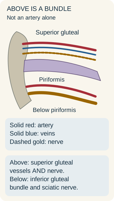
The superior gluteal artery reaches the buttock through the greater sciatic foramen above piriformis. It does not travel alone. Superior gluteal veins and the superior gluteal nerve also use this space. The bony sciatic notch and the sacrospinous and sacrotuberous ligaments form the surrounding foraminal boundaries; piriformis separates the superior and inferior routes. [2][3]
Use piriformis to locate a neurovascular bundle, then identify the individual structure from its function or imaging. Original Bone Wizardry schematic. [2][3][14]
\1 follow each line above the muscle band. Before naming the injured structure, decide what a contrast leak, impaired motor activation, or venous bleeding would identify.
\1 the route identifies a bundle. Arterial-phase extravasation identifies an artery; denervation identifies a motor pathway. Saying that the artery is the only structure above piriformis would erase the very neighbor that explains a neural deficit.
Different exits lead to different territories
Route
Structures to keep together
Useful distinction
RouteAbove piriformis
Structures to keep togetherSuperior gluteal artery, veins, and nerve
Useful distinctionGluteal perfusion and abductor activation are separate jobs.
RouteBelow piriformis
Structures to keep togetherInferior gluteal vessels and nerve; sciatic and posterior femoral cutaneous nerves; pudendal nerve and internal pudendal vessels; nerves to obturator internus and quadratus femoris
Useful distinctionSharing an exit does not mean sharing a final target.
RouteObturator canal
Structures to keep togetherObturator neurovascular structures
Useful distinctionThink medial thigh rather than the suprapiriform bundle.
The pudendal nerve and internal pudendal vessels leave below piriformis, turn around the ischial spine near the sacrospinous ligament, and enter the perineum through the lesser sciatic foramen. They do not remain in the gluteal muscle compartment. [18][19]
\1 bleeding above piriformis plus intact external iliac and femoral arteries still fits superior gluteal injury. Trace its parent toward the internal iliac, not toward the profunda femoris.
Try it here · Checkpoint 1 of 3
Make your prediction before reading the choices. A first attempt is just a starting point.
Case 1
Show answer and explanations for case 1
A. External iliac artery (Why this does not fit)
The external iliac supplies the lower limb through the femoral artery. Its continuation does not account for a branch leaving the pelvis above piriformis.
Reasoning steps for option A
In the 42-year-old presentation, which localization detail should be tested first against External iliac artery?
Start from the stem localization: After a pelvic fracture, a 42-year-old patient develops an enlarging upper-buttock hematoma. That finding must be anatomically compatible with External iliac artery before the option can survive.
After that localization in the 42-year-old presentation, what independent discriminator should be checked for External iliac artery?
Use the separate clinical or imaging observation: CT angiography shows active extravasation from a vessel that leaves the pelvis immediately superior to piriformis, then courses along the lateral ilium. The expected territory or function of External iliac artery must fit that second observation as well.
B. Profunda femoris artery (Why this does not fit)
The profunda supplies deep thigh tissues and commonly gives the circumflex femoral arteries. That thigh origin does not match the demonstrated pelvic exit.
Reasoning steps for option B
In the 42-year-old presentation, which localization detail should be tested first against Profunda femoris artery?
Start from the stem localization: After a pelvic fracture, a 42-year-old patient develops an enlarging upper-buttock hematoma. That finding must be anatomically compatible with Profunda femoris artery before the option can survive.
After that localization in the 42-year-old presentation, what independent discriminator should be checked for Profunda femoris artery?
Use the separate clinical or imaging observation: CT angiography shows active extravasation from a vessel that leaves the pelvis immediately superior to piriformis, then courses along the lateral ilium. The expected territory or function of Profunda femoris artery must fit that second observation as well.
C. Internal iliac artery (Best answer)
The superior gluteal artery usually arises from the posterior division of the internal iliac. The suprapiriform exit identifies the injured branch, so tracing proximally leads to the internal iliac.
Reasoning steps for option C
In the 42-year-old presentation, which localization detail should be tested first against Internal iliac artery?
Start from the stem localization: After a pelvic fracture, a 42-year-old patient develops an enlarging upper-buttock hematoma. That finding must be anatomically compatible with Internal iliac artery before the option can survive.
After that localization in the 42-year-old presentation, what independent discriminator should be checked for Internal iliac artery?
Use the separate clinical or imaging observation: CT angiography shows active extravasation from a vessel that leaves the pelvis immediately superior to piriformis, then courses along the lateral ilium. The expected territory or function of Internal iliac artery must fit that second observation as well.
D. Obturator artery (Why this does not fit)
The obturator artery supplies the medial thigh through the obturator canal. It is another pelvic branch, but it is not the parent of the vessel shown above piriformis.
Reasoning steps for option D
In the 42-year-old presentation, which localization detail should be tested first against Obturator artery?
Start from the stem localization: After a pelvic fracture, a 42-year-old patient develops an enlarging upper-buttock hematoma. That finding must be anatomically compatible with Obturator artery before the option can survive.
After that localization in the 42-year-old presentation, what independent discriminator should be checked for Obturator artery?
Use the separate clinical or imaging observation: CT angiography shows active extravasation from a vessel that leaves the pelvis immediately superior to piriformis, then courses along the lateral ilium. The expected territory or function of Obturator artery must fit that second observation as well.
Takeaway: Use the observed course to identify the branch before inferring its parent vessel.
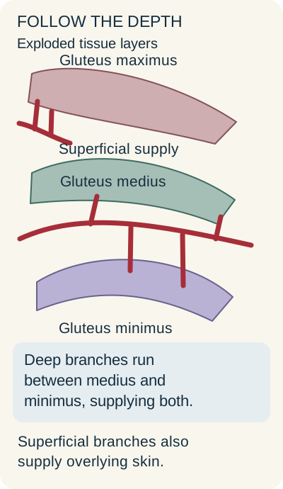
Just outside the pelvis, the superior gluteal artery distributes superficial and deep branches. The superficial route supplies gluteus maximus and gives perforators to overlying skin and fat. The deep route travels between gluteus medius and minimus and supplies these major abductors. Contributions also reach tensor fasciae latae. The muscle territories overlap with neighboring arteries, so an artery's principal territory is not an exclusive ownership boundary. [1][10][12]
Trace the red deep route between medius and minimus; the superficial branch instead supports maximus and cutaneous perforators. Original Bone Wizardry schematic. [1][10][12]
\1 cover the caption and identify which red route lies between medius and minimus. Predict which tissues would be most directly affected by a selective interruption there, compared with an interruption confined to a cutaneous perforator.
\1 a deep branch lesion threatens abductor perfusion; a superficial perforator lesion threatens its skin and fat territory. The latter does not automatically establish denervation or a deep abductor infarct.
Gluteus maximus receives important inferior gluteal supply as well as superior gluteal contributions. The inferior gluteal artery exits below piriformis, near the inferior gluteal nerve and sciatic pathway. The inferior gluteal nerve supplies maximus, which is particularly important for powerful hip extension, such as rising from a low seat. Do not infer a specific motor deficit from an arterial name alone. [3][12][16]
Why can an occluded artery coexist with normal strength? Regional anastomoses can maintain enough blood flow to the distal muscle. In a cadaveric angiographic study, the effect of superior gluteal occlusion differed with the collateral routes preserved by different acetabular exposures. An extensive exposure that interrupts those remaining routes can change the outcome. A visible connection demonstrates a possible route, not a guarantee of adequate perfusion in every patient. [4]
\1 normal muscle enhancement despite a confirmed main-trunk occlusion supports effective alternative inflow. New poor enhancement after additional dissection raises a different concern: the previously functioning collateral routes may have been lost.
Why does the opposite side of the pelvis fall?
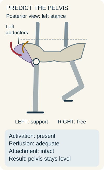
During left-leg stance, the left abductors counter the tendency of body weight to lower the unsupported right hemipelvis. If the left abductor apparatus cannot provide adequate force, the right side can descend. Name the supporting leg before naming the deficient side. This is a functional observation, not a diagnosis of superior gluteal arterial occlusion. [2][11][16]
Change one requirement for abductor function
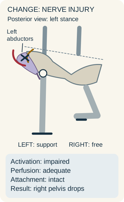
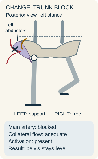
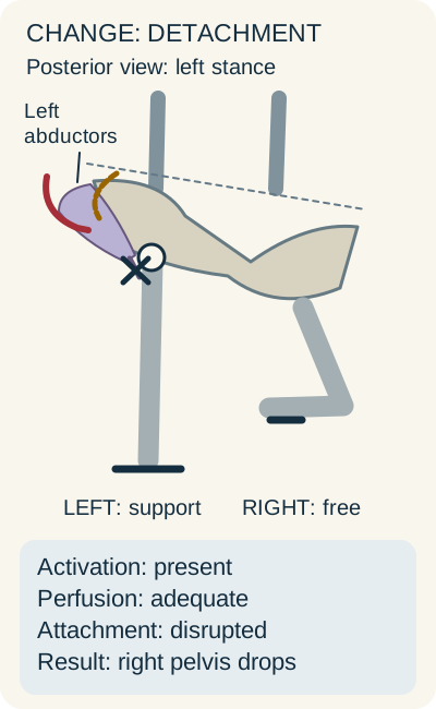
The reference below has adequate perfusion, motor activation, and tendon attachment. Predict the result of each change before opening its comparison. A black X marks the interrupted structure. Red lines show blood supply; the dashed gold line represents the motor pathway. These are anatomical teaching states, not a patient-specific vascular simulation.
With functioning left abductors, intact attachment, and adequate perfusion, the unsupported right hemipelvis is held level. Original Bone Wizardry schematic. [2][4][11][16]Test interruption of the left motor nerve
Disabling the left superior gluteal motor pathway impairs left abductor force. The right, unsupported hemipelvis falls despite preserved blood flow. Original Bone Wizardry schematic. [2][4][11][16]
The right side descends because the left abductors lose adequate activation, even though muscle blood flow is preserved.
Test arterial occlusion with adequate collateral flow
Blocking the main superior gluteal artery while preserving adequate collateral perfusion and motor activation need not produce pelvic descent. The dashed red alternate path represents the stipulated collateral supply. Original Bone Wizardry schematic. [2][4][11][16]
The stipulated collateral route supplies enough blood, and the nerve still activates the muscle. The pelvis can therefore remain level. Occlusion without adequate collateral flow would be a different state.
Test detachment of the left abductor insertion
A detached abductor insertion prevents effective force transfer despite preserved nerve activation and muscle perfusion; the unsupported right hemipelvis falls. Original Bone Wizardry schematic. [2][4][11][16]
The muscle remains perfused and activated, but its force cannot be transferred effectively through the disrupted attachment. The right side can again descend.
Close any comparison to reset it. The reference stays available, and the complete outcomes below remain readable without opening a control or running JavaScript.
The same sign can arise from different failures
Changed requirement
Observation in this example
Explanation
Changed requirementMotor activation lost
Observation in this exampleRight pelvic descent during left stance
ExplanationLeft abductor force is insufficient despite perfusion.
ExplanationMuscle perfusion and activation are retained.
Changed requirementAttachment disrupted
Observation in this exampleRight pelvic descent during left stance
ExplanationViable muscle cannot transmit its force normally.
The superior gluteal nerve activates medius, minimus, and tensor fasciae latae. Selective denervation with relatively preserved passive motion supports a neural deficit. Tendon imaging, muscle enhancement, and motor testing help distinguish other causes. Pain can inhibit abductor effort and can coexist with weakness; these categories are not mutually exclusive. [2][4][11][16]
\1 a retracted medius tendon with normal muscle enhancement and preserved motor recruitment points to a force-transmission problem. A painful restriction of passive hip rotation points toward joint assessment rather than proof that the superior gluteal artery is blocked.
Is the injured tissue muscle or femoral head?
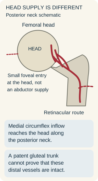
A femoral head lesion asks a different vascular question from a medius or minimus lesion. The medial circumflex femoral artery, commonly arising from the profunda femoris, gives the major deep retinacular pathway around the posterior femoral neck into the head. Its origin can vary; an imaged direct femoral origin takes precedence over a memorized usual branch tree. [5]
Follow the main medial circumflex pathway along the neck. Dashed left: the small foveal route. Dashed right: an inferior gluteal collateral connection. Accessory routes do not guarantee adequate head perfusion. Original Bone Wizardry schematic. [5][6][7]
\1 trace from the lower right arterial inflow toward the head. Now imagine an intracapsular neck fracture interrupting the small retinacular vessels near the head. Would patency of a distant gluteal trunk prove that this final pathway is intact?
\1 no. A patent upstream or neighboring artery cannot bridge every downstream vascular injury. This explains why normal gluteal perfusion can coexist with femoral head osteonecrosis.
The artery in the ligamentum teres enters through the fovea and has a variable contribution. It is not accurate to say that this artery predictably disappears after infancy or that it reliably rescues the entire head. In the developing hip, the open physis limits direct vascular continuity between the neck's metaphyseal circulation and the capital epiphysis. A perfused neck therefore does not prove a perfused head. [7]
Also avoid the opposite absolute: that gluteal arteries have no relationship to head circulation. An anatomical injection study demonstrated an inferior gluteal connection to the medial circumflex femoral pathway near the obturator externus tendon. That is a meaningful collateral connection, not proof that it will protect every head after trauma. The principal retinacular source remains the best answer when the question specifically asks for that source. [5][6]
\1 distinguish the named branch from its parent. A question about a retinacular supplier favors the medial circumflex femoral artery; a question about loss of several profunda-dependent territories may require a more proximal answer. Never reject the profunda simply because it is one level upstream.
Try it here · Checkpoint 2 of 3
Make your prediction before reading the choices. A first attempt is just a starting point.
Case 13
Show answer and explanations for case 13
A. Superficial superior gluteal branches to maximus (Why this does not fit)
These branches principally perfuse gluteus maximus and overlying tissues. Their disruption would not directly explain an isolated head lesion following an intracapsular neck fracture.
Reasoning steps for option A
In the 71-year-old presentation, which localization detail should be tested first against Superficial superior gluteal branches to maximus?
Start from the stem localization: A 71-year-old patient sustains a displaced intracapsular femoral neck fracture. That finding must be anatomically compatible with Superficial superior gluteal branches to maximus before the option can survive.
After that localization in the 71-year-old presentation, what independent discriminator should be checked for Superficial superior gluteal branches to maximus?
Use the separate clinical or imaging observation: CT angiography shows patent superior and inferior gluteal trunks. The expected territory or function of Superficial superior gluteal branches to maximus must fit that second observation as well.
B. Internal pudendal branches to the perineum (Why this does not fit)
These branches supply perineal structures after their sciatic-foramen route. They do not run along the femoral neck where the fracture threatens head perfusion.
Reasoning steps for option B
In the 71-year-old presentation, which localization detail should be tested first against Internal pudendal branches to the perineum?
Start from the stem localization: A 71-year-old patient sustains a displaced intracapsular femoral neck fracture. That finding must be anatomically compatible with Internal pudendal branches to the perineum before the option can survive.
After that localization in the 71-year-old presentation, what independent discriminator should be checked for Internal pudendal branches to the perineum?
Use the separate clinical or imaging observation: CT angiography shows patent superior and inferior gluteal trunks. The expected territory or function of Internal pudendal branches to the perineum must fit that second observation as well.
C. Obturator muscular branches to the adductors (Why this does not fit)
These branches supply the medial thigh. Muscular adductor perfusion is not the retinacular pathway vulnerable at the fractured femoral neck.
Reasoning steps for option C
In the 71-year-old presentation, which localization detail should be tested first against Obturator muscular branches to the adductors?
Start from the stem localization: A 71-year-old patient sustains a displaced intracapsular femoral neck fracture. That finding must be anatomically compatible with Obturator muscular branches to the adductors before the option can survive.
After that localization in the 71-year-old presentation, what independent discriminator should be checked for Obturator muscular branches to the adductors?
Use the separate clinical or imaging observation: CT angiography shows patent superior and inferior gluteal trunks. The expected territory or function of Obturator muscular branches to the adductors must fit that second observation as well.
D. Medial circumflex retinacular branches along the neck (Best answer)
These subsynovial branches provide a major route into the femoral head. A displaced intracapsular fracture can disrupt them even when regional gluteal trunks remain patent.
Reasoning steps for option D
In the 71-year-old presentation, which localization detail should be tested first against Medial circumflex retinacular branches along the neck?
Start from the stem localization: A 71-year-old patient sustains a displaced intracapsular femoral neck fracture. That finding must be anatomically compatible with Medial circumflex retinacular branches along the neck before the option can survive.
After that localization in the 71-year-old presentation, what independent discriminator should be checked for Medial circumflex retinacular branches along the neck?
Use the separate clinical or imaging observation: CT angiography shows patent superior and inferior gluteal trunks. The expected territory or function of Medial circumflex retinacular branches along the neck must fit that second observation as well.
Takeaway: Proximal arterial patency does not guarantee perfusion beyond a local retinacular disruption.
What does a painful pediatric hip actually establish?
Consider a school-age child with an atraumatic limp, referred thigh or knee pain, and painful limitation of hip abduction and internal rotation. If radiographs show characteristic capital epiphyseal sclerosis, fragmentation, or flattening, Perthes disease belongs near the center of the assessment. It is childhood osteonecrosis of the femoral head with an incompletely understood initiating cause. The presentation does not prove that a named artery has undergone a discrete thrombosis. [7][8]
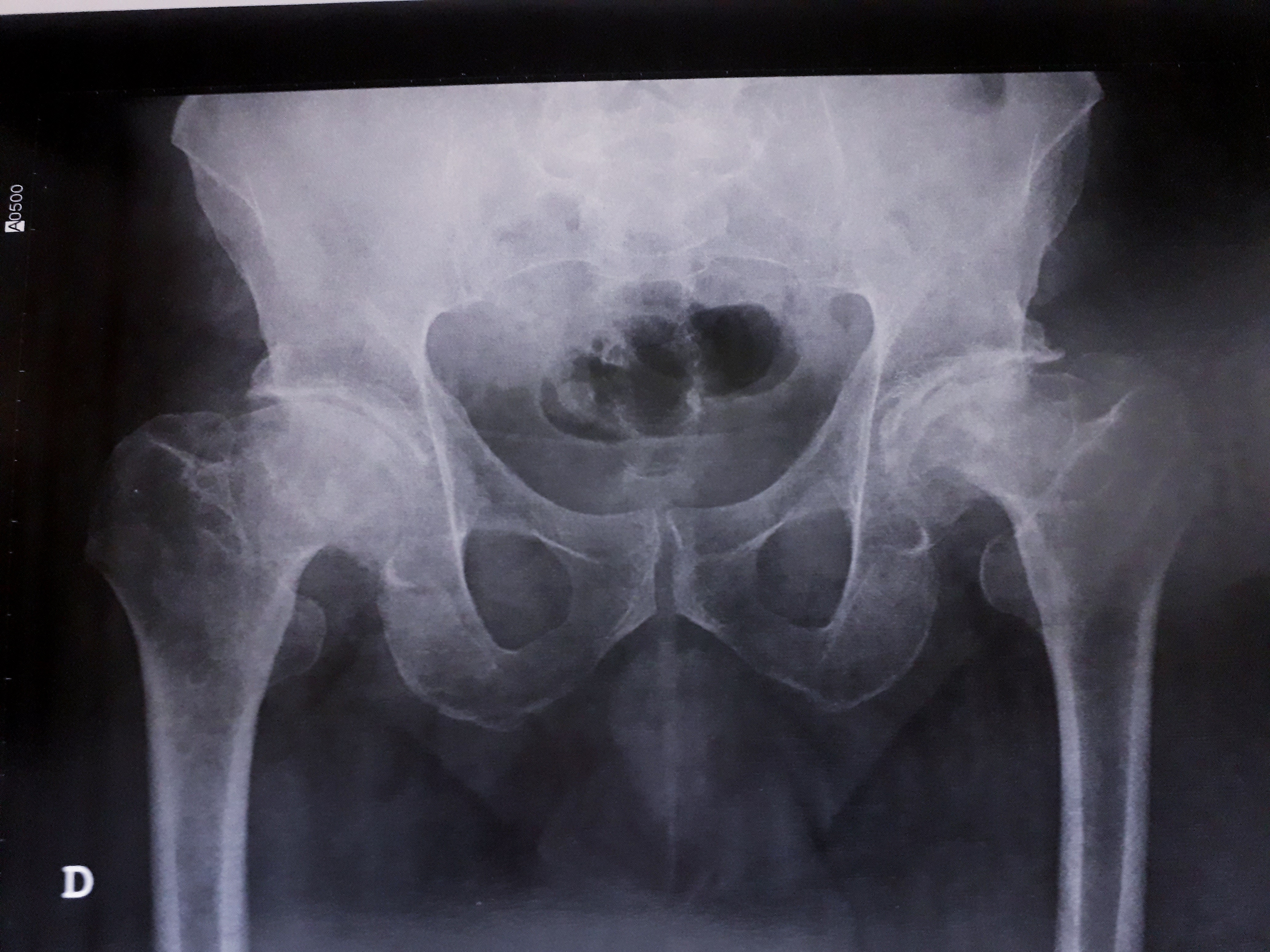
Adult residual deformity, not a new childhood presentation. This radiograph shows a 52-year-old man with Perthes sequelae. Use the film to observe altered femoral-head shape, not to infer the patient's childhood arterial anatomy. Photograph by Andreia Flor, 4 May 2020; original image unmodified, CC BY-SA 4.0. [13]
\1 inspect the femoral-head contours. Name the abnormal tissue first. Then ask which additional information would be needed to diagnose a newly symptomatic child.
\1 head deformity concerns bone and joint architecture. Age, symptoms, examination, and appropriate imaging still matter. Joint-space widening alone is nonspecific, and this adult image cannot establish a new pediatric diagnosis or a specific occluded vessel.
Pain and guarding can limit passive abduction or internal rotation. That is not the same measurement as painless failure to generate abductor force. A painful joint can also inhibit muscle activity, so painful restriction and functional weakness can coexist. The useful distinction is between evidence for the affected tissue and an unsupported leap from the word abduction to an arterial diagnosis. [8][16]
Change the age and rotation pattern
An adolescent, particularly with obesity, who has hip or referred knee pain and external rotation during attempted hip flexion requires assessment for slipped capital femoral epiphysis. This pattern should not be automatically assigned to Perthes disease. Suspected slipped epiphysis warrants stopping weight bearing and urgent orthopedic assessment; repeated forced hip rotation is not an appropriate learning exercise on the patient. [9]
\1 in a child with a radiographically localized head lesion, choose the retinacular supply when asked about the head's principal arterial pathway. In a patient with painless weakness and selective medius-minimus denervation, localize the superior gluteal motor pathway instead.
Which observations should change the safety assessment?
A gluteal vascular injury may present as obvious bleeding, a deep expanding hematoma, or a delayed hemoglobin decline after pelvic or proximal femoral surgery. The superior gluteal artery can be difficult to control near the greater sciatic notch, and its close relationship to nerve branches means that a combined vascular and motor injury is possible. An apparently satisfactory bony screw position does not prove that adjacent soft tissues are clear. [2][3][10][11]
Compare perfusion with blood loss
A patient has normal foot pulses but arterial extravasation above piriformis. Predict whether the pulses rule out a significant gluteal bleed.
\1 they do not. The patent external iliac and femoral pathway can maintain distal pulses while a separate internal iliac branch loses blood into the buttock.
Compare a mass with a routine collection
A deep postoperative mass enlarges while hemoglobin falls. Predict whether lack of obvious pulsatility makes aspiration safe.
\1 it does not establish a nonvascular collection. Assess for an arterial communication before sampling or drainage. In a stable patient, vascular imaging can define the lesion and guide specialist care.
Hemodynamic instability requires immediate resuscitation and hemorrhage-control assessment; do not delay that care for a routine imaging sequence. Treatment of a confirmed gluteal arterial lesion depends on the injury and available expertise. Published cases include successful endovascular treatment, but a case report does not establish one mandatory treatment for every patient. [10][15]
Keep the collateral networks distinct
Superior gluteal branches contribute to regional and trochanteric connections. The classic cruciate anastomosis instead combines the descending inferior gluteal contribution, transverse medial and lateral circumflex femoral branches, and the ascending first perforating branch of the profunda femoris. The superior gluteal artery is not the standard gluteal limb of that four-part network. [6][17]
\1 starting below piriformis, follow inferior gluteal inflow toward the transverse circumflex branches and first perforator behind the proximal femur. The named partners identify a cruciate connection, not the perineal pudendal route or a knee-level genicular network. As with the head-supply example, a potential collateral route must not be mistaken for proof of adequate perfusion after a particular injury.
\1 interpret an abnormal image or examination in this order: identify the tissue, identify the disrupted function or flow, and then use the demonstrated course to choose the relevant artery or nerve. That approach preserves the anatomy without assigning every painful or weak hip to the same vessel.
Try it here · Checkpoint 3 of 3
Make your prediction before reading the choices. A first attempt is just a starting point.
Case 19
Show answer and explanations for case 19
A. Arterial-phase imaging before sampling the gluteal collection (Best answer)
A postoperative collection with ongoing blood loss can communicate with a gluteal artery. Define the vascular anatomy urgently before a proposed needle intervention; deep location can make pulsatility difficult to detect.
Reasoning steps for option A
In the 55-year-old presentation, which localization detail should be tested first against Arterial-phase imaging before sampling the gluteal collection?
Start from the stem localization: Three weeks after pelvic surgery, a 55-year-old patient has an enlarging deep buttock mass and a falling hemoglobin concentration. That finding must be anatomically compatible with Arterial-phase imaging before sampling the gluteal collection before the option can survive.
After that localization in the 55-year-old presentation, what independent discriminator should be checked for Arterial-phase imaging before sampling the gluteal collection?
Use the separate clinical or imaging observation: The skin is not erythematous, and the mass is not clearly pulsatile. The expected territory or function of Arterial-phase imaging before sampling the gluteal collection must fit that second observation as well.
B. Image-guided aspiration for bacterial culture and cell counting (Why this does not fit)
Aspiration can help characterize an infected or nonvascular fluid collection. A vascular communication must be excluded first in this enlarging collection with falling hemoglobin.
Reasoning steps for option B
In the 55-year-old presentation, which localization detail should be tested first against Image-guided aspiration for bacterial culture and cell counting?
Start from the stem localization: Three weeks after pelvic surgery, a 55-year-old patient has an enlarging deep buttock mass and a falling hemoglobin concentration. That finding must be anatomically compatible with Image-guided aspiration for bacterial culture and cell counting before the option can survive.
After that localization in the 55-year-old presentation, what independent discriminator should be checked for Image-guided aspiration for bacterial culture and cell counting?
Use the separate clinical or imaging observation: The skin is not erythematous, and the mass is not clearly pulsatile. The expected territory or function of Image-guided aspiration for bacterial culture and cell counting must fit that second observation as well.
C. Nonangiographic MRI to characterize a solid soft-tissue tumor (Why this does not fit)
MRI can help characterize a nonvascular soft-tissue mass. The postoperative timing and ongoing blood loss prioritize assessment of an arterial communication before a tumor-focused study.
Reasoning steps for option C
In the 55-year-old presentation, which localization detail should be tested first against Nonangiographic MRI to characterize a solid soft-tissue tumor?
Start from the stem localization: Three weeks after pelvic surgery, a 55-year-old patient has an enlarging deep buttock mass and a falling hemoglobin concentration. That finding must be anatomically compatible with Nonangiographic MRI to characterize a solid soft-tissue tumor before the option can survive.
After that localization in the 55-year-old presentation, what independent discriminator should be checked for Nonangiographic MRI to characterize a solid soft-tissue tumor?
Use the separate clinical or imaging observation: The skin is not erythematous, and the mass is not clearly pulsatile. The expected territory or function of Nonangiographic MRI to characterize a solid soft-tissue tumor must fit that second observation as well.
D. Serial hemoglobin testing without immediate vascular imaging (Why this does not fit)
Serial values quantify the trend in blood loss. They do not identify or control its source in an enlarging postoperative gluteal mass.
Reasoning steps for option D
In the 55-year-old presentation, which localization detail should be tested first against Serial hemoglobin testing without immediate vascular imaging?
Start from the stem localization: Three weeks after pelvic surgery, a 55-year-old patient has an enlarging deep buttock mass and a falling hemoglobin concentration. That finding must be anatomically compatible with Serial hemoglobin testing without immediate vascular imaging before the option can survive.
After that localization in the 55-year-old presentation, what independent discriminator should be checked for Serial hemoglobin testing without immediate vascular imaging?
Use the separate clinical or imaging observation: The skin is not erythematous, and the mass is not clearly pulsatile. The expected territory or function of Serial hemoglobin testing without immediate vascular imaging must fit that second observation as well.
Takeaway: An enlarging postoperative gluteal collection with anemia warrants vascular assessment before aspiration, even without obvious pulsatility.
Use the location, tissue findings, and vascular or motor evidence together. Each original case has one best answer and an explanation for every option.
Case 2
Show answer and explanations for case 2
A. Inferior gluteal artery (Why this does not fit)
The inferior gluteal neurovascular structures are associated with gluteus maximus. Preserved maximus function and denervated medius and minimus instead localize the injury to the superior gluteal nerve.
Reasoning steps for option A
In the 46-year-old presentation, which localization detail should be tested first against Inferior gluteal artery?
Start from the stem localization: Six weeks after posterior pelvic fixation, a 46-year-old patient has weak hip abduction with nearly full painless passive hip motion. That finding must be anatomically compatible with Inferior gluteal artery before the option can survive.
After that localization in the 46-year-old presentation, what independent discriminator should be checked for Inferior gluteal artery?
Use the separate clinical or imaging observation: Electromyography shows denervation of gluteus medius and minimus; quadriceps, gluteus maximus, and distal leg muscles are spared. The expected territory or function of Inferior gluteal artery must fit that second observation as well.
B. Superior gluteal artery (Best answer)
The superior gluteal nerve and vessels travel together above piriformis and across the lateral ilium. Selective denervation of medius and minimus identifies this bundle; the arterial name does not mean the artery caused the denervation.
Reasoning steps for option B
In the 46-year-old presentation, which localization detail should be tested first against Superior gluteal artery?
Start from the stem localization: Six weeks after posterior pelvic fixation, a 46-year-old patient has weak hip abduction with nearly full painless passive hip motion. That finding must be anatomically compatible with Superior gluteal artery before the option can survive.
After that localization in the 46-year-old presentation, what independent discriminator should be checked for Superior gluteal artery?
Use the separate clinical or imaging observation: Electromyography shows denervation of gluteus medius and minimus; quadriceps, gluteus maximus, and distal leg muscles are spared. The expected territory or function of Superior gluteal artery must fit that second observation as well.
C. Medial circumflex femoral artery (Why this does not fit)
The medial circumflex femoral artery is important for femoral head perfusion. A femoral head perfusion deficit does not produce this selective electromyographic nerve distribution.
Reasoning steps for option C
In the 46-year-old presentation, which localization detail should be tested first against Medial circumflex femoral artery?
Start from the stem localization: Six weeks after posterior pelvic fixation, a 46-year-old patient has weak hip abduction with nearly full painless passive hip motion. That finding must be anatomically compatible with Medial circumflex femoral artery before the option can survive.
After that localization in the 46-year-old presentation, what independent discriminator should be checked for Medial circumflex femoral artery?
Use the separate clinical or imaging observation: Electromyography shows denervation of gluteus medius and minimus; quadriceps, gluteus maximus, and distal leg muscles are spared. The expected territory or function of Medial circumflex femoral artery must fit that second observation as well.
D. Internal pudendal artery (Why this does not fit)
The internal pudendal artery accompanies the pudendal nerve toward the perineum. The affected gluteal muscles are outside that nerve distribution.
Reasoning steps for option D
In the 46-year-old presentation, which localization detail should be tested first against Internal pudendal artery?
Start from the stem localization: Six weeks after posterior pelvic fixation, a 46-year-old patient has weak hip abduction with nearly full painless passive hip motion. That finding must be anatomically compatible with Internal pudendal artery before the option can survive.
After that localization in the 46-year-old presentation, what independent discriminator should be checked for Internal pudendal artery?
Use the separate clinical or imaging observation: Electromyography shows denervation of gluteus medius and minimus; quadriceps, gluteus maximus, and distal leg muscles are spared. The expected territory or function of Internal pudendal artery must fit that second observation as well.
Takeaway: Electromyographic denervation localizes a neural lesion; the adjacent vessel is identified by shared anatomy.
The abductors of the supporting hip resist downward rotation of the unsupported hemipelvis. Right-sided pelvic descent during left stance therefore indicates inadequate left abductor force.
Reasoning steps for option A
In the 58-year-old presentation, which localization detail should be tested first against Left gluteus medius and minimus?
Start from the stem localization: A 58-year-old patient has painless weakness after a left lateral hip operation. That finding must be anatomically compatible with Left gluteus medius and minimus before the option can survive.
After that localization in the 58-year-old presentation, what independent discriminator should be checked for Left gluteus medius and minimus?
Use the separate clinical or imaging observation: When standing on the left leg with the right foot off the ground, the right iliac crest falls. The expected territory or function of Left gluteus medius and minimus must fit that second observation as well.
B. Right gluteus medius and minimus (Why this does not fit)
Right abductors stabilize the pelvis during right-leg stance. The pelvis stays level in that condition; the abnormal test loads the left hip.
Reasoning steps for option B
In the 58-year-old presentation, which localization detail should be tested first against Right gluteus medius and minimus?
Start from the stem localization: A 58-year-old patient has painless weakness after a left lateral hip operation. That finding must be anatomically compatible with Right gluteus medius and minimus before the option can survive.
After that localization in the 58-year-old presentation, what independent discriminator should be checked for Right gluteus medius and minimus?
Use the separate clinical or imaging observation: When standing on the left leg with the right foot off the ground, the right iliac crest falls. The expected territory or function of Right gluteus medius and minimus must fit that second observation as well.
C. Left gluteus maximus and hamstrings (Why this does not fit)
These muscles are major hip extensors rather than the principal frontal-plane pelvic stabilizers. The observed failure is pelvic control in single-leg stance, not loss of hip extension.
Reasoning steps for option C
In the 58-year-old presentation, which localization detail should be tested first against Left gluteus maximus and hamstrings?
Start from the stem localization: A 58-year-old patient has painless weakness after a left lateral hip operation. That finding must be anatomically compatible with Left gluteus maximus and hamstrings before the option can survive.
After that localization in the 58-year-old presentation, what independent discriminator should be checked for Left gluteus maximus and hamstrings?
Use the separate clinical or imaging observation: When standing on the left leg with the right foot off the ground, the right iliac crest falls. The expected territory or function of Left gluteus maximus and hamstrings must fit that second observation as well.
D. Right adductor longus and brevis (Why this does not fit)
These muscles adduct the right thigh through obturator motor supply. They do not provide the stance-side abductor torque missing in the left-leg test.
Reasoning steps for option D
In the 58-year-old presentation, which localization detail should be tested first against Right adductor longus and brevis?
Start from the stem localization: A 58-year-old patient has painless weakness after a left lateral hip operation. That finding must be anatomically compatible with Right adductor longus and brevis before the option can survive.
After that localization in the 58-year-old presentation, what independent discriminator should be checked for Right adductor longus and brevis?
Use the separate clinical or imaging observation: When standing on the left leg with the right foot off the ground, the right iliac crest falls. The expected territory or function of Right adductor longus and brevis must fit that second observation as well.
Takeaway: Name the supporting leg first: pelvic descent is on the opposite side of an inadequate stance-side abductor apparatus.
A. Superficial branch of the superior gluteal artery (Why this does not fit)
The superficial branch supplies gluteus maximus and overlying tissues. Preserved maximus enhancement and the deeper intermuscular termination do not match its principal course.
Reasoning steps for option A
In the 52-year-old presentation, which localization detail should be tested first against Superficial branch of the superior gluteal artery?
Start from the stem localization: Following an acetabular operation, a 52-year-old patient has poor contrast enhancement of gluteus medius and minimus while gluteus maximus enhancement remains preserved. That finding must be anatomically compatible with Superficial branch of the superior gluteal artery before the option can survive.
After that localization in the 52-year-old presentation, what independent discriminator should be checked for Superficial branch of the superior gluteal artery?
Use the separate clinical or imaging observation: The feeding trunk remains patent at its pelvic exit, but a distal branch ends abruptly in the plane between medius and minimus. The expected territory or function of Superficial branch of the superior gluteal artery must fit that second observation as well.
B. Deep branch of the medial circumflex femoral artery (Why this does not fit)
This branch travels toward the posterior femoral neck and retinacular supply. The affected muscle plane is between medius and minimus rather than along the femoral neck.
Reasoning steps for option B
In the 52-year-old presentation, which localization detail should be tested first against Deep branch of the medial circumflex femoral artery?
Start from the stem localization: Following an acetabular operation, a 52-year-old patient has poor contrast enhancement of gluteus medius and minimus while gluteus maximus enhancement remains preserved. That finding must be anatomically compatible with Deep branch of the medial circumflex femoral artery before the option can survive.
After that localization in the 52-year-old presentation, what independent discriminator should be checked for Deep branch of the medial circumflex femoral artery?
Use the separate clinical or imaging observation: The feeding trunk remains patent at its pelvic exit, but a distal branch ends abruptly in the plane between medius and minimus. The expected territory or function of Deep branch of the medial circumflex femoral artery must fit that second observation as well.
C. Descending branch of the inferior gluteal artery (Why this does not fit)
Inferior gluteal branches chiefly support maximus and adjacent posterior tissues. This is a suprapiriform trunk with a deficit in the deep abductor plane.
Reasoning steps for option C
In the 52-year-old presentation, which localization detail should be tested first against Descending branch of the inferior gluteal artery?
Start from the stem localization: Following an acetabular operation, a 52-year-old patient has poor contrast enhancement of gluteus medius and minimus while gluteus maximus enhancement remains preserved. That finding must be anatomically compatible with Descending branch of the inferior gluteal artery before the option can survive.
After that localization in the 52-year-old presentation, what independent discriminator should be checked for Descending branch of the inferior gluteal artery?
Use the separate clinical or imaging observation: The feeding trunk remains patent at its pelvic exit, but a distal branch ends abruptly in the plane between medius and minimus. The expected territory or function of Descending branch of the inferior gluteal artery must fit that second observation as well.
D. Deep branch of the superior gluteal artery (Best answer)
The deep branch distributes vessels between medius and minimus. Its selective interruption matches reduced enhancement in those muscles despite preserved superficial territory.
Reasoning steps for option D
In the 52-year-old presentation, which localization detail should be tested first against Deep branch of the superior gluteal artery?
Start from the stem localization: Following an acetabular operation, a 52-year-old patient has poor contrast enhancement of gluteus medius and minimus while gluteus maximus enhancement remains preserved. That finding must be anatomically compatible with Deep branch of the superior gluteal artery before the option can survive.
After that localization in the 52-year-old presentation, what independent discriminator should be checked for Deep branch of the superior gluteal artery?
Use the separate clinical or imaging observation: The feeding trunk remains patent at its pelvic exit, but a distal branch ends abruptly in the plane between medius and minimus. The expected territory or function of Deep branch of the superior gluteal artery must fit that second observation as well.
Takeaway: Combine the tissue enhancement pattern with the branch plane rather than treating every gluteal artery as interchangeable.
A. Medial circumflex femoral artery (Why this does not fit)
The medial circumflex femoral artery supplies important retinacular vessels of the femoral head. It does not explain a cutaneous perforator traced to a suprapiriform pelvic trunk.
Reasoning steps for option A
In the 49-year-old presentation, which localization detail should be tested first against Medial circumflex femoral artery?
Start from the stem localization: A 49-year-old patient undergoes upper-buttock soft-tissue reconstruction. That finding must be anatomically compatible with Medial circumflex femoral artery before the option can survive.
After that localization in the 49-year-old presentation, what independent discriminator should be checked for Medial circumflex femoral artery?
Use the separate clinical or imaging observation: The selected skin-and-fat perforator passes through gluteus maximus into a superficial arterial network, while proximal tracing reaches a trunk emerging above piriformis. The expected territory or function of Medial circumflex femoral artery must fit that second observation as well.
B. Inferior gluteal artery (Why this does not fit)
Inferior gluteal perforators can supply buttock tissue through maximus. The demonstrated origin above rather than below piriformis distinguishes the selected superior gluteal perforator.
Reasoning steps for option B
In the 49-year-old presentation, which localization detail should be tested first against Inferior gluteal artery?
Start from the stem localization: A 49-year-old patient undergoes upper-buttock soft-tissue reconstruction. That finding must be anatomically compatible with Inferior gluteal artery before the option can survive.
After that localization in the 49-year-old presentation, what independent discriminator should be checked for Inferior gluteal artery?
Use the separate clinical or imaging observation: The selected skin-and-fat perforator passes through gluteus maximus into a superficial arterial network, while proximal tracing reaches a trunk emerging above piriformis. The expected territory or function of Inferior gluteal artery must fit that second observation as well.
C. Obturator artery (Why this does not fit)
The obturator artery reaches the medial thigh through the obturator canal. That course differs from the observed intramuscular gluteal perforator and pelvic exit.
Reasoning steps for option C
In the 49-year-old presentation, which localization detail should be tested first against Obturator artery?
Start from the stem localization: A 49-year-old patient undergoes upper-buttock soft-tissue reconstruction. That finding must be anatomically compatible with Obturator artery before the option can survive.
After that localization in the 49-year-old presentation, what independent discriminator should be checked for Obturator artery?
Use the separate clinical or imaging observation: The selected skin-and-fat perforator passes through gluteus maximus into a superficial arterial network, while proximal tracing reaches a trunk emerging above piriformis. The expected territory or function of Obturator artery must fit that second observation as well.
D. Superior gluteal artery (Best answer)
Superficial superior gluteal branches supply maximus and perforators to the overlying skin and fat. Both the perforator course and its suprapiriform parent establish the flap inflow without requiring muscle resection.
Reasoning steps for option D
In the 49-year-old presentation, which localization detail should be tested first against Superior gluteal artery?
Start from the stem localization: A 49-year-old patient undergoes upper-buttock soft-tissue reconstruction. That finding must be anatomically compatible with Superior gluteal artery before the option can survive.
After that localization in the 49-year-old presentation, what independent discriminator should be checked for Superior gluteal artery?
Use the separate clinical or imaging observation: The selected skin-and-fat perforator passes through gluteus maximus into a superficial arterial network, while proximal tracing reaches a trunk emerging above piriformis. The expected territory or function of Superior gluteal artery must fit that second observation as well.
Takeaway: A vessel can supply both muscle and overlying skin; preserving a muscle and its nerve separates flap perfusion from motor sacrifice.
Gluteus maximus generates powerful hip extension from a flexed position. A local inferior gluteal nerve injury with sciatic sparing preferentially impairs that action.
Reasoning steps for option A
In the 36-year-old presentation, which localization detail should be tested first against Rising from a low chair?
Start from the stem localization: A 36-year-old patient sustains a deep gluteal wound that damages an artery and the small motor nerve emerging with it below piriformis. That finding must be anatomically compatible with Rising from a low chair before the option can survive.
After that localization in the 36-year-old presentation, what independent discriminator should be checked for Rising from a low chair?
Use the separate clinical or imaging observation: The sciatic nerve is intact. The expected territory or function of Rising from a low chair must fit that second observation as well.
B. Holding the pelvis level on one leg (Why this does not fit)
This task mainly tests the stance-side abductor apparatus supplied by the superior gluteal nerve. The described injury is below piriformis and affects the inferior gluteal motor pathway.
Reasoning steps for option B
In the 36-year-old presentation, which localization detail should be tested first against Holding the pelvis level on one leg?
Start from the stem localization: A 36-year-old patient sustains a deep gluteal wound that damages an artery and the small motor nerve emerging with it below piriformis. That finding must be anatomically compatible with Holding the pelvis level on one leg before the option can survive.
After that localization in the 36-year-old presentation, what independent discriminator should be checked for Holding the pelvis level on one leg?
Use the separate clinical or imaging observation: The sciatic nerve is intact. The expected territory or function of Holding the pelvis level on one leg must fit that second observation as well.
C. Straightening the knee against resistance (Why this does not fit)
Resisted knee extension mainly tests quadriceps supplied through femoral motor fibers. That pathway is separate from the small infrapiriform motor nerve injured in this wound.
Reasoning steps for option C
In the 36-year-old presentation, which localization detail should be tested first against Straightening the knee against resistance?
Start from the stem localization: A 36-year-old patient sustains a deep gluteal wound that damages an artery and the small motor nerve emerging with it below piriformis. That finding must be anatomically compatible with Straightening the knee against resistance before the option can survive.
After that localization in the 36-year-old presentation, what independent discriminator should be checked for Straightening the knee against resistance?
Use the separate clinical or imaging observation: The sciatic nerve is intact. The expected territory or function of Straightening the knee against resistance must fit that second observation as well.
D. Bringing the thighs together against resistance (Why this does not fit)
Resisted hip adduction chiefly tests obturator-innervated adductors. The obturator pathway travels through a different pelvic exit from this gluteal wound.
Reasoning steps for option D
In the 36-year-old presentation, which localization detail should be tested first against Bringing the thighs together against resistance?
Start from the stem localization: A 36-year-old patient sustains a deep gluteal wound that damages an artery and the small motor nerve emerging with it below piriformis. That finding must be anatomically compatible with Bringing the thighs together against resistance before the option can survive.
After that localization in the 36-year-old presentation, what independent discriminator should be checked for Bringing the thighs together against resistance?
Use the separate clinical or imaging observation: The sciatic nerve is intact. The expected territory or function of Bringing the thighs together against resistance must fit that second observation as well.
Takeaway: Inferior gluteal nerve injury impairs powerful hip extension; do not assign every gluteal deficit to the abductors.
The obturator canal transmits the obturator neurovascular structures toward the medial thigh. Perineal sensory loss and the turn around the ischial spine instead identify the pudendal pathway.
Reasoning steps for option A
In the 33-year-old presentation, which localization detail should be tested first against Obturator canal?
Start from the stem localization: After a deep wound near the ischial spine, a 33-year-old patient has perineal sensory loss and bleeding from a vessel that curves around the sacrospinous ligament. That finding must be anatomically compatible with Obturator canal before the option can survive.
After that localization in the 33-year-old presentation, what independent discriminator should be checked for Obturator canal?
Use the separate clinical or imaging observation: Imaging traces the vessel out of the pelvis below piriformis. The expected territory or function of Obturator canal must fit that second observation as well.
B. Lesser sciatic foramen (Best answer)
The pudendal nerve and internal pudendal vessels leave through the greater sciatic foramen, curve around the ischial spine, and enter the perineum through the lesser sciatic foramen. The clinical territory and observed bend specify this second opening.
Reasoning steps for option B
In the 33-year-old presentation, which localization detail should be tested first against Lesser sciatic foramen?
Start from the stem localization: After a deep wound near the ischial spine, a 33-year-old patient has perineal sensory loss and bleeding from a vessel that curves around the sacrospinous ligament. That finding must be anatomically compatible with Lesser sciatic foramen before the option can survive.
After that localization in the 33-year-old presentation, what independent discriminator should be checked for Lesser sciatic foramen?
Use the separate clinical or imaging observation: Imaging traces the vessel out of the pelvis below piriformis. The expected territory or function of Lesser sciatic foramen must fit that second observation as well.
C. Suprapiriform part of the greater sciatic foramen (Why this does not fit)
The superior gluteal neurovascular bundle takes this route into the buttock. It does not follow the ischial-spine bend to supply the perineum.
Reasoning steps for option C
In the 33-year-old presentation, which localization detail should be tested first against Suprapiriform part of the greater sciatic foramen?
Start from the stem localization: After a deep wound near the ischial spine, a 33-year-old patient has perineal sensory loss and bleeding from a vessel that curves around the sacrospinous ligament. That finding must be anatomically compatible with Suprapiriform part of the greater sciatic foramen before the option can survive.
After that localization in the 33-year-old presentation, what independent discriminator should be checked for Suprapiriform part of the greater sciatic foramen?
Use the separate clinical or imaging observation: Imaging traces the vessel out of the pelvis below piriformis. The expected territory or function of Suprapiriform part of the greater sciatic foramen must fit that second observation as well.
D. Femoral ring (Why this does not fit)
The femoral ring is the proximal opening of the femoral canal. It is not the continuation of the pudendal bundle after its infrapiriform exit.
Reasoning steps for option D
In the 33-year-old presentation, which localization detail should be tested first against Femoral ring?
Start from the stem localization: After a deep wound near the ischial spine, a 33-year-old patient has perineal sensory loss and bleeding from a vessel that curves around the sacrospinous ligament. That finding must be anatomically compatible with Femoral ring before the option can survive.
After that localization in the 33-year-old presentation, what independent discriminator should be checked for Femoral ring?
Use the separate clinical or imaging observation: Imaging traces the vessel out of the pelvis below piriformis. The expected territory or function of Femoral ring must fit that second observation as well.
Takeaway: Follow the complete route: leaving the pelvis below piriformis does not mean every structure remains in the buttock.
A. Deep gluteal abductor territory (Why this does not fit)
The deep superior gluteal artery supplies medius and minimus. The obturator-canal injury and preserved pelvic stability favor the medial thigh rather than this gluteal territory.
Reasoning steps for option A
In the 41-year-old presentation, which localization detail should be tested first against Deep gluteal abductor territory?
Start from the stem localization: A 41-year-old patient has a pelvic fragment injure a nerve and artery together in the obturator canal. That finding must be anatomically compatible with Deep gluteal abductor territory before the option can survive.
After that localization in the 41-year-old presentation, what independent discriminator should be checked for Deep gluteal abductor territory?
Use the separate clinical or imaging observation: Thigh adduction is weak, knee extension is normal, and pelvic stability is preserved during single-leg stance. The expected territory or function of Deep gluteal abductor territory must fit that second observation as well.
B. Superficial gluteal extensor territory (Why this does not fit)
Gluteal arterial branches supply maximus and overlying tissue. The damaged canal and adductor deficit are remote from those gluteal branches.
Reasoning steps for option B
In the 41-year-old presentation, which localization detail should be tested first against Superficial gluteal extensor territory?
Start from the stem localization: A 41-year-old patient has a pelvic fragment injure a nerve and artery together in the obturator canal. That finding must be anatomically compatible with Superficial gluteal extensor territory before the option can survive.
After that localization in the 41-year-old presentation, what independent discriminator should be checked for Superficial gluteal extensor territory?
Use the separate clinical or imaging observation: Thigh adduction is weak, knee extension is normal, and pelvic stability is preserved during single-leg stance. The expected territory or function of Superficial gluteal extensor territory must fit that second observation as well.
C. Medial thigh adductor territory (Best answer)
The obturator neurovascular structures reach the medial thigh through the obturator canal. Weak adduction with preserved quadriceps and pelvic stability matches this local territory.
Reasoning steps for option C
In the 41-year-old presentation, which localization detail should be tested first against Medial thigh adductor territory?
Start from the stem localization: A 41-year-old patient has a pelvic fragment injure a nerve and artery together in the obturator canal. That finding must be anatomically compatible with Medial thigh adductor territory before the option can survive.
After that localization in the 41-year-old presentation, what independent discriminator should be checked for Medial thigh adductor territory?
Use the separate clinical or imaging observation: Thigh adduction is weak, knee extension is normal, and pelvic stability is preserved during single-leg stance. The expected territory or function of Medial thigh adductor territory must fit that second observation as well.
D. Perineal soft-tissue territory (Why this does not fit)
Internal pudendal vessels reach the perineum by the sciatic foramina. They do not accompany the obturator nerve in the canal identified on imaging.
Reasoning steps for option D
In the 41-year-old presentation, which localization detail should be tested first against Perineal soft-tissue territory?
Start from the stem localization: A 41-year-old patient has a pelvic fragment injure a nerve and artery together in the obturator canal. That finding must be anatomically compatible with Perineal soft-tissue territory before the option can survive.
After that localization in the 41-year-old presentation, what independent discriminator should be checked for Perineal soft-tissue territory?
Use the separate clinical or imaging observation: Thigh adduction is weak, knee extension is normal, and pelvic stability is preserved during single-leg stance. The expected territory or function of Perineal soft-tissue territory must fit that second observation as well.
Takeaway: Match a pelvic exit with its target compartment before interpreting a nearby muscle deficit.
A. Recanalization restores flow through the original gluteal trunk (Why this does not fit)
Recanalization can restore downstream inflow after some arterial occlusions. Serial imaging still shows a complete main-trunk occlusion, so this cannot explain the maintained muscle perfusion.
Reasoning steps for option A
In the 64-year-old presentation, which localization detail should be tested first against Recanalization restores flow through the original gluteal trunk?
Start from the stem localization: A 64-year-old patient has a completely occluded right superior gluteal main trunk on two CT angiograms six months apart. That finding must be anatomically compatible with Recanalization restores flow through the original gluteal trunk before the option can survive.
After that localization in the 64-year-old presentation, what independent discriminator should be checked for Recanalization restores flow through the original gluteal trunk?
Use the separate clinical or imaging observation: Arterial and delayed images confirm the interruption. The expected territory or function of Recanalization restores flow through the original gluteal trunk must fit that second observation as well.
B. Regional collateral vessels preserve the distal muscle perfusion (Best answer)
Alternative arterial connections can carry flow into a territory whose usual trunk is occluded. Preserved muscle enhancement on both studies despite confirmed trunk occlusion supports effective collateral inflow.
Reasoning steps for option B
In the 64-year-old presentation, which localization detail should be tested first against Regional collateral vessels preserve the distal muscle perfusion?
Start from the stem localization: A 64-year-old patient has a completely occluded right superior gluteal main trunk on two CT angiograms six months apart. That finding must be anatomically compatible with Regional collateral vessels preserve the distal muscle perfusion before the option can survive.
After that localization in the 64-year-old presentation, what independent discriminator should be checked for Regional collateral vessels preserve the distal muscle perfusion?
Use the separate clinical or imaging observation: Arterial and delayed images confirm the interruption. The expected territory or function of Regional collateral vessels preserve the distal muscle perfusion must fit that second observation as well.
C. Transient arterial spasm accounts for the apparent trunk cutoff (Why this does not fit)
A transient narrowing could mimic interruption on a single examination. The same established complete occlusion on serial studies argues against a transient spasm.
Reasoning steps for option C
In the 64-year-old presentation, which localization detail should be tested first against Transient arterial spasm accounts for the apparent trunk cutoff?
Start from the stem localization: A 64-year-old patient has a completely occluded right superior gluteal main trunk on two CT angiograms six months apart. That finding must be anatomically compatible with Transient arterial spasm accounts for the apparent trunk cutoff before the option can survive.
After that localization in the 64-year-old presentation, what independent discriminator should be checked for Transient arterial spasm accounts for the apparent trunk cutoff?
Use the separate clinical or imaging observation: Arterial and delayed images confirm the interruption. The expected territory or function of Transient arterial spasm accounts for the apparent trunk cutoff must fit that second observation as well.
D. Slow contrast arrival creates a false impression of occlusion (Why this does not fit)
Poor timing can make a patent vessel appear unopacified. The complete occlusion is confirmed on arterial and delayed images while distal muscle enhancement is retained.
Reasoning steps for option D
In the 64-year-old presentation, which localization detail should be tested first against Slow contrast arrival creates a false impression of occlusion?
Start from the stem localization: A 64-year-old patient has a completely occluded right superior gluteal main trunk on two CT angiograms six months apart. That finding must be anatomically compatible with Slow contrast arrival creates a false impression of occlusion before the option can survive.
After that localization in the 64-year-old presentation, what independent discriminator should be checked for Slow contrast arrival creates a false impression of occlusion?
Use the separate clinical or imaging observation: Arterial and delayed images confirm the interruption. The expected territory or function of Slow contrast arrival creates a false impression of occlusion must fit that second observation as well.
Takeaway: Arterial interruption does not establish muscle ischemia without considering collateral flow and the observed tissue response.
A. A new occlusion of the superior gluteal main trunk (Why this does not fit)
A new main-trunk occlusion can reduce downstream perfusion when alternative inflow is inadequate. This trunk was already chronically occluded before surgery, when the abductors still enhanced; a newly lost alternative route better explains the change.
Reasoning steps for option A
In the 67-year-old presentation, which localization detail should be tested first against A new occlusion of the superior gluteal main trunk?
Start from the stem localization: Before a complex acetabular reconstruction, a 67-year-old patient has chronic superior gluteal occlusion with preserved abductor enhancement. That finding must be anatomically compatible with A new occlusion of the superior gluteal main trunk before the option can survive.
After that localization in the 67-year-old presentation, what independent discriminator should be checked for A new occlusion of the superior gluteal main trunk?
Use the separate clinical or imaging observation: After extensive lateral soft-tissue dissection, the same muscles enhance poorly and become weak. The expected territory or function of A new occlusion of the superior gluteal main trunk must fit that second observation as well.
B. A new interruption of femoral head retinacular vessels (Why this does not fit)
Retinacular injury can impair head perfusion after hip surgery. The new imaging deficit is in medius and minimus rather than the femoral head, so a head-specific route does not explain the observed tissue pattern.
Reasoning steps for option B
In the 67-year-old presentation, which localization detail should be tested first against A new interruption of femoral head retinacular vessels?
Start from the stem localization: Before a complex acetabular reconstruction, a 67-year-old patient has chronic superior gluteal occlusion with preserved abductor enhancement. That finding must be anatomically compatible with A new interruption of femoral head retinacular vessels before the option can survive.
After that localization in the 67-year-old presentation, what independent discriminator should be checked for A new interruption of femoral head retinacular vessels?
Use the separate clinical or imaging observation: After extensive lateral soft-tissue dissection, the same muscles enhance poorly and become weak. The expected territory or function of A new interruption of femoral head retinacular vessels must fit that second observation as well.
C. An isolated failure of superior gluteal motor conduction (Why this does not fit)
A nerve can lose conduction despite remaining anatomically continuous, producing abductor weakness. A motor conduction defect alone does not explain the accompanying new loss of muscle enhancement.
Reasoning steps for option C
In the 67-year-old presentation, which localization detail should be tested first against An isolated failure of superior gluteal motor conduction?
Start from the stem localization: Before a complex acetabular reconstruction, a 67-year-old patient has chronic superior gluteal occlusion with preserved abductor enhancement. That finding must be anatomically compatible with An isolated failure of superior gluteal motor conduction before the option can survive.
After that localization in the 67-year-old presentation, what independent discriminator should be checked for An isolated failure of superior gluteal motor conduction?
Use the separate clinical or imaging observation: After extensive lateral soft-tissue dissection, the same muscles enhance poorly and become weak. The expected territory or function of An isolated failure of superior gluteal motor conduction must fit that second observation as well.
D. An interruption of collateral arterial inflow to the abductors (Best answer)
With the main superior gluteal trunk already occluded, adequate muscle perfusion depends on remaining arterial routes. Extensive dissection can interrupt those routes, explaining the transition from preserved to poor enhancement and the resulting weakness.
Reasoning steps for option D
In the 67-year-old presentation, which localization detail should be tested first against An interruption of collateral arterial inflow to the abductors?
Start from the stem localization: Before a complex acetabular reconstruction, a 67-year-old patient has chronic superior gluteal occlusion with preserved abductor enhancement. That finding must be anatomically compatible with An interruption of collateral arterial inflow to the abductors before the option can survive.
After that localization in the 67-year-old presentation, what independent discriminator should be checked for An interruption of collateral arterial inflow to the abductors?
Use the separate clinical or imaging observation: After extensive lateral soft-tissue dissection, the same muscles enhance poorly and become weak. The expected territory or function of An interruption of collateral arterial inflow to the abductors must fit that second observation as well.
Takeaway: An intact motor nerve cannot preserve muscle function when the remaining blood supply has been interrupted.
A. Superior gluteal artery (Why this does not fit)
This artery is an important source for the hip abductor muscles. Painful restriction does not localize the lesion to those muscles when imaging places it in the femoral head.
Reasoning steps for option A
In the 6-year-old presentation, which localization detail should be tested first against Superior gluteal artery?
Start from the stem localization: A 6-year-old boy has three months of an atraumatic limp and intermittent knee pain. That finding must be anatomically compatible with Superior gluteal artery before the option can survive.
After that localization in the 6-year-old presentation, what independent discriminator should be checked for Superior gluteal artery?
Use the separate clinical or imaging observation: The knee examination is normal. The expected territory or function of Superior gluteal artery must fit that second observation as well.
B. Inferior gluteal artery (Why this does not fit)
This artery supplies gluteal tissue and can contribute collateral inflow to the medial circumflex femoral artery. The question asks for the principal named source of the retinacular pathway, not a regional collateral contributor.
Reasoning steps for option B
In the 6-year-old presentation, which localization detail should be tested first against Inferior gluteal artery?
Start from the stem localization: A 6-year-old boy has three months of an atraumatic limp and intermittent knee pain. That finding must be anatomically compatible with Inferior gluteal artery before the option can survive.
After that localization in the 6-year-old presentation, what independent discriminator should be checked for Inferior gluteal artery?
Use the separate clinical or imaging observation: The knee examination is normal. The expected territory or function of Inferior gluteal artery must fit that second observation as well.
C. Medial circumflex femoral artery (Best answer)
Its deep branch supplies the major posterosuperior retinacular pathway to the femoral head. The age, course, and epiphyseal changes favor Perthes disease; the arterial answer describes relevant supply, not proof of a discrete arterial occlusion.
Reasoning steps for option C
In the 6-year-old presentation, which localization detail should be tested first against Medial circumflex femoral artery?
Start from the stem localization: A 6-year-old boy has three months of an atraumatic limp and intermittent knee pain. That finding must be anatomically compatible with Medial circumflex femoral artery before the option can survive.
After that localization in the 6-year-old presentation, what independent discriminator should be checked for Medial circumflex femoral artery?
Use the separate clinical or imaging observation: The knee examination is normal. The expected territory or function of Medial circumflex femoral artery must fit that second observation as well.
D. Lateral circumflex femoral artery (Why this does not fit)
The lateral circumflex contributes around the proximal femur and to regional anastomoses. It is not the dominant source of the posterosuperior retinacular pathway implicated by the head lesion.
Reasoning steps for option D
In the 6-year-old presentation, which localization detail should be tested first against Lateral circumflex femoral artery?
Start from the stem localization: A 6-year-old boy has three months of an atraumatic limp and intermittent knee pain. That finding must be anatomically compatible with Lateral circumflex femoral artery before the option can survive.
After that localization in the 6-year-old presentation, what independent discriminator should be checked for Lateral circumflex femoral artery?
Use the separate clinical or imaging observation: The knee examination is normal. The expected territory or function of Lateral circumflex femoral artery must fit that second observation as well.
Takeaway: Localize the diseased tissue on imaging before selecting its blood supply; Perthes disease does not prove that a named artery was ligated or thrombosed.
A. Stop weight bearing and obtain urgent orthopedic evaluation (Best answer)
This adolescent pattern raises concern for slipped capital femoral epiphysis. Protecting the hip from further loading and arranging urgent assessment is appropriate; repeated forced motion is not a diagnostic exercise.
Reasoning steps for option A
In the 14-year-old presentation, which localization detail should be tested first against Stop weight bearing and obtain urgent orthopedic evaluation?
Start from the stem localization: A 14-year-old adolescent with obesity has six weeks of thigh and knee pain. That finding must be anatomically compatible with Stop weight bearing and obtain urgent orthopedic evaluation before the option can survive.
After that localization in the 14-year-old presentation, what independent discriminator should be checked for Stop weight bearing and obtain urgent orthopedic evaluation?
Use the separate clinical or imaging observation: The knee examination is normal. The expected territory or function of Stop weight bearing and obtain urgent orthopedic evaluation must fit that second observation as well.
B. Continue weight bearing and start resisted abductor exercise (Why this does not fit)
Abductor rehabilitation can be appropriate for selected muscular conditions. The age and obligatory external rotation raise a physeal concern that requires protection and assessment before exercise.
Reasoning steps for option B
In the 14-year-old presentation, which localization detail should be tested first against Continue weight bearing and start resisted abductor exercise?
Start from the stem localization: A 14-year-old adolescent with obesity has six weeks of thigh and knee pain. That finding must be anatomically compatible with Continue weight bearing and start resisted abductor exercise before the option can survive.
After that localization in the 14-year-old presentation, what independent discriminator should be checked for Continue weight bearing and start resisted abductor exercise?
Use the separate clinical or imaging observation: The knee examination is normal. The expected territory or function of Continue weight bearing and start resisted abductor exercise must fit that second observation as well.
C. Permit unrestricted activity and reassess the knee in six weeks (Why this does not fit)
Referred knee pain can originate in the hip. The abnormal hip examination means a normal knee examination is not a basis for delaying hip evaluation.
Reasoning steps for option C
In the 14-year-old presentation, which localization detail should be tested first against Permit unrestricted activity and reassess the knee in six weeks?
Start from the stem localization: A 14-year-old adolescent with obesity has six weeks of thigh and knee pain. That finding must be anatomically compatible with Permit unrestricted activity and reassess the knee in six weeks before the option can survive.
After that localization in the 14-year-old presentation, what independent discriminator should be checked for Permit unrestricted activity and reassess the knee in six weeks?
Use the separate clinical or imaging observation: The knee examination is normal. The expected territory or function of Permit unrestricted activity and reassess the knee in six weeks must fit that second observation as well.
D. Force hip internal rotation and repeat strength testing (Why this does not fit)
A strength test does not resolve a possible mechanically unstable proximal femoral physis. Forceful examination can add stress without safely establishing the diagnosis.
Reasoning steps for option D
In the 14-year-old presentation, which localization detail should be tested first against Force hip internal rotation and repeat strength testing?
Start from the stem localization: A 14-year-old adolescent with obesity has six weeks of thigh and knee pain. That finding must be anatomically compatible with Force hip internal rotation and repeat strength testing before the option can survive.
After that localization in the 14-year-old presentation, what independent discriminator should be checked for Force hip internal rotation and repeat strength testing?
Use the separate clinical or imaging observation: The knee examination is normal. The expected territory or function of Force hip internal rotation and repeat strength testing must fit that second observation as well.
Takeaway: An adolescent with referred knee pain and external rotation during hip flexion needs evaluation for a slipped epiphysis, not automatic labeling as Perthes disease.
A. Retrograde filling through an inferior gluteal anastomosis (Why this does not fit)
An inferior gluteal connection can support the medial circumflex pathway in some hips. Such a collateral route is not the most direct explanation when the normal antegrade inflow is demonstrably outside the treated branch.
Reasoning steps for option A
In the 62-year-old presentation, which localization detail should be tested first against Retrograde filling through an inferior gluteal anastomosis?
Start from the stem localization: Preoperative angiography in a 62-year-old patient shows a medial circumflex femoral artery arising directly from the common femoral artery. That finding must be anatomically compatible with Retrograde filling through an inferior gluteal anastomosis before the option can survive.
After that localization in the 62-year-old presentation, what independent discriminator should be checked for Retrograde filling through an inferior gluteal anastomosis?
Use the separate clinical or imaging observation: A separate profunda femoris branch supplying a thigh tumor is then embolized distal to that origin. The expected territory or function of Retrograde filling through an inferior gluteal anastomosis must fit that second observation as well.
B. Antegrade filling through the intact medial circumflex artery (Best answer)
A directly arising medial circumflex artery remains connected to common femoral inflow. Embolizing a separate profunda branch does not interrupt the imaged artery that supplies the major retinacular pathway.
Reasoning steps for option B
In the 62-year-old presentation, which localization detail should be tested first against Antegrade filling through the intact medial circumflex artery?
Start from the stem localization: Preoperative angiography in a 62-year-old patient shows a medial circumflex femoral artery arising directly from the common femoral artery. That finding must be anatomically compatible with Antegrade filling through the intact medial circumflex artery before the option can survive.
After that localization in the 62-year-old presentation, what independent discriminator should be checked for Antegrade filling through the intact medial circumflex artery?
Use the separate clinical or imaging observation: A separate profunda femoris branch supplying a thigh tumor is then embolized distal to that origin. The expected territory or function of Antegrade filling through the intact medial circumflex artery must fit that second observation as well.
C. Foveal filling through the obturator acetabular arterial branch (Why this does not fit)
The foveal route can contribute to head perfusion. Its contribution need not replace the retinacular pathway here because the medial circumflex artery remains patent.
Reasoning steps for option C
In the 62-year-old presentation, which localization detail should be tested first against Foveal filling through the obturator acetabular arterial branch?
Start from the stem localization: Preoperative angiography in a 62-year-old patient shows a medial circumflex femoral artery arising directly from the common femoral artery. That finding must be anatomically compatible with Foveal filling through the obturator acetabular arterial branch before the option can survive.
After that localization in the 62-year-old presentation, what independent discriminator should be checked for Foveal filling through the obturator acetabular arterial branch?
Use the separate clinical or imaging observation: A separate profunda femoris branch supplying a thigh tumor is then embolized distal to that origin. The expected territory or function of Foveal filling through the obturator acetabular arterial branch must fit that second observation as well.
D. Ascending filling through the lateral circumflex arterial branch (Why this does not fit)
Lateral circumflex branches contribute to the proximal femoral vascular network. The directly demonstrated intact medial circumflex route, rather than an alternative contributor, most directly explains preserved head perfusion.
Reasoning steps for option D
In the 62-year-old presentation, which localization detail should be tested first against Ascending filling through the lateral circumflex arterial branch?
Start from the stem localization: Preoperative angiography in a 62-year-old patient shows a medial circumflex femoral artery arising directly from the common femoral artery. That finding must be anatomically compatible with Ascending filling through the lateral circumflex arterial branch before the option can survive.
After that localization in the 62-year-old presentation, what independent discriminator should be checked for Ascending filling through the lateral circumflex arterial branch?
Use the separate clinical or imaging observation: A separate profunda femoris branch supplying a thigh tumor is then embolized distal to that origin. The expected territory or function of Ascending filling through the lateral circumflex arterial branch must fit that second observation as well.
Takeaway: Use a patient's demonstrated vascular origin rather than assuming that every medial circumflex artery arises from the profunda.
A. Accessory inflow persists but does not compensate adequately (Best answer)
A patent foveal branch provides some inflow without necessarily perfusing the entire head sufficiently. Persistent poor enhancement of the weight-bearing head shows inadequate compensation after the neck injury.
Reasoning steps for option A
In the 73-year-old presentation, which localization detail should be tested first against Accessory inflow persists but does not compensate adequately?
Start from the stem localization: After a displaced femoral neck injury, a 73-year-old patient has a patent small foveal artery but markedly reduced enhancement of the weight-bearing femoral head. That finding must be anatomically compatible with Accessory inflow persists but does not compensate adequately before the option can survive.
After that localization in the 73-year-old presentation, what independent discriminator should be checked for Accessory inflow persists but does not compensate adequately?
Use the separate clinical or imaging observation: Gluteal muscle enhancement is normal. The expected territory or function of Accessory inflow persists but does not compensate adequately must fit that second observation as well.
B. Retinacular inflow persists but contrast arrival is globally delayed (Why this does not fit)
Delayed systemic contrast delivery can make multiple tissues appear poorly enhanced. Normal gluteal enhancement with a localized head deficit after neck injury favors a regional supply problem rather than global delay.
Reasoning steps for option B
In the 73-year-old presentation, which localization detail should be tested first against Retinacular inflow persists but contrast arrival is globally delayed?
Start from the stem localization: After a displaced femoral neck injury, a 73-year-old patient has a patent small foveal artery but markedly reduced enhancement of the weight-bearing femoral head. That finding must be anatomically compatible with Retinacular inflow persists but contrast arrival is globally delayed before the option can survive.
After that localization in the 73-year-old presentation, what independent discriminator should be checked for Retinacular inflow persists but contrast arrival is globally delayed?
Use the separate clinical or imaging observation: Gluteal muscle enhancement is normal. The expected territory or function of Retinacular inflow persists but contrast arrival is globally delayed must fit that second observation as well.
C. Gluteal inflow fails and secondarily limits the head perfusion (Why this does not fit)
Gluteal arteries can contribute to proximal femoral collateral pathways. The gluteal muscles enhance normally, so the observed data do not identify gluteal inflow failure as the primary explanation.
Reasoning steps for option C
In the 73-year-old presentation, which localization detail should be tested first against Gluteal inflow fails and secondarily limits the head perfusion?
Start from the stem localization: After a displaced femoral neck injury, a 73-year-old patient has a patent small foveal artery but markedly reduced enhancement of the weight-bearing femoral head. That finding must be anatomically compatible with Gluteal inflow fails and secondarily limits the head perfusion before the option can survive.
After that localization in the 73-year-old presentation, what independent discriminator should be checked for Gluteal inflow fails and secondarily limits the head perfusion?
Use the separate clinical or imaging observation: Gluteal muscle enhancement is normal. The expected territory or function of Gluteal inflow fails and secondarily limits the head perfusion must fit that second observation as well.
D. Accessory inflow fails while the main retinacular supply persists (Why this does not fit)
Loss of the small foveal route alone would leave the main retinacular route as the major source. The imaged foveal vessel is patent and the weight-bearing head remains poorly perfused, opposing this allocation of the injury.
Reasoning steps for option D
In the 73-year-old presentation, which localization detail should be tested first against Accessory inflow fails while the main retinacular supply persists?
Start from the stem localization: After a displaced femoral neck injury, a 73-year-old patient has a patent small foveal artery but markedly reduced enhancement of the weight-bearing femoral head. That finding must be anatomically compatible with Accessory inflow fails while the main retinacular supply persists before the option can survive.
After that localization in the 73-year-old presentation, what independent discriminator should be checked for Accessory inflow fails while the main retinacular supply persists?
Use the separate clinical or imaging observation: Gluteal muscle enhancement is normal. The expected territory or function of Accessory inflow fails while the main retinacular supply persists must fit that second observation as well.
Takeaway: A small patent vessel is not proof that the entire downstream organ has adequate blood flow.
A. Antegrade filling crosses the blocked medial circumflex segment (Why this does not fit)
Restored antegrade filling would require a patent route through the occluded segment. The preparation blocks that segment and injects the inferior gluteal artery instead.
Reasoning steps for option A
In the 68-year-old presentation, which localization detail should be tested first against Antegrade filling crosses the blocked medial circumflex segment?
Start from the stem localization: A perfused hip preparation from a 68-year-old adult donor has intact distal retinacular vessels. That finding must be anatomically compatible with Antegrade filling crosses the blocked medial circumflex segment before the option can survive.
After that localization in the 68-year-old presentation, what independent discriminator should be checked for Antegrade filling crosses the blocked medial circumflex segment?
Use the separate clinical or imaging observation: The medial circumflex femoral artery is blocked proximal to its connection with an inferior gluteal branch, yet injection through the inferior gluteal artery still fills the distal retinacular vessels. The expected territory or function of Antegrade filling crosses the blocked medial circumflex segment must fit that second observation as well.
B. Foveal filling occurs without any distal retinacular connection (Why this does not fit)
An isolated foveal route would introduce material at the fovea rather than demonstrate the specified distal retinacular filling. The observed filling follows the intact anastomosis into the retinacular vessels.
Reasoning steps for option B
In the 68-year-old presentation, which localization detail should be tested first against Foveal filling occurs without any distal retinacular connection?
Start from the stem localization: A perfused hip preparation from a 68-year-old adult donor has intact distal retinacular vessels. That finding must be anatomically compatible with Foveal filling occurs without any distal retinacular connection before the option can survive.
After that localization in the 68-year-old presentation, what independent discriminator should be checked for Foveal filling occurs without any distal retinacular connection?
Use the separate clinical or imaging observation: The medial circumflex femoral artery is blocked proximal to its connection with an inferior gluteal branch, yet injection through the inferior gluteal artery still fills the distal retinacular vessels. The expected territory or function of Foveal filling occurs without any distal retinacular connection must fit that second observation as well.
C. Collateral filling bypasses the proximal medial circumflex block (Best answer)
The inferior gluteal connection joins the medial circumflex pathway beyond the experimental block. Injection through that connection can reach the intact distal retinacular vessels without traversing the blocked proximal segment.
Reasoning steps for option C
In the 68-year-old presentation, which localization detail should be tested first against Collateral filling bypasses the proximal medial circumflex block?
Start from the stem localization: A perfused hip preparation from a 68-year-old adult donor has intact distal retinacular vessels. That finding must be anatomically compatible with Collateral filling bypasses the proximal medial circumflex block before the option can survive.
After that localization in the 68-year-old presentation, what independent discriminator should be checked for Collateral filling bypasses the proximal medial circumflex block?
Use the separate clinical or imaging observation: The medial circumflex femoral artery is blocked proximal to its connection with an inferior gluteal branch, yet injection through the inferior gluteal artery still fills the distal retinacular vessels. The expected territory or function of Collateral filling bypasses the proximal medial circumflex block must fit that second observation as well.
D. Direct superior gluteal filling bypasses the distal retinacular vessels (Why this does not fit)
Superior gluteal branches take part in regional collateral circulation. This experiment instead injects the inferior gluteal route and demonstrates filling through, not around, the intact retinacular vessels.
Reasoning steps for option D
In the 68-year-old presentation, which localization detail should be tested first against Direct superior gluteal filling bypasses the distal retinacular vessels?
Start from the stem localization: A perfused hip preparation from a 68-year-old adult donor has intact distal retinacular vessels. That finding must be anatomically compatible with Direct superior gluteal filling bypasses the distal retinacular vessels before the option can survive.
After that localization in the 68-year-old presentation, what independent discriminator should be checked for Direct superior gluteal filling bypasses the distal retinacular vessels?
Use the separate clinical or imaging observation: The medial circumflex femoral artery is blocked proximal to its connection with an inferior gluteal branch, yet injection through the inferior gluteal artery still fills the distal retinacular vessels. The expected territory or function of Direct superior gluteal filling bypasses the distal retinacular vessels must fit that second observation as well.
Takeaway: A demonstrated anastomosis establishes an available route, not universal clinical protection or a bypass around every distal injury.
A. A bridging arterial ring across the epiphyseal growth plate (Why this does not fit)
A ring connecting both sides of the physis would permit direct compensation from metaphyseal inflow. The persistent separation of neck and head filling is not explained by such an effective bridge.
Reasoning steps for option A
In the 7-year-old presentation, which localization detail should be tested first against A bridging arterial ring across the epiphyseal growth plate?
Start from the stem localization: A 7-year-old child has a perfused hip specimen in which metaphyseal vessels fill the femoral neck normally. That finding must be anatomically compatible with A bridging arterial ring across the epiphyseal growth plate before the option can survive.
After that localization in the 7-year-old presentation, what independent discriminator should be checked for A bridging arterial ring across the epiphyseal growth plate?
Use the separate clinical or imaging observation: Retinacular entry to the capital epiphysis is interrupted, and the head fills poorly despite a patent metaphyseal circulation. The expected territory or function of A bridging arterial ring across the epiphyseal growth plate must fit that second observation as well.
B. A shared medullary arterial supply to the neck and head (Why this does not fit)
An unrestricted shared medullary route would link metaphyseal filling to epiphyseal filling. The open physis limits that communication in the developing hip.
Reasoning steps for option B
In the 7-year-old presentation, which localization detail should be tested first against A shared medullary arterial supply to the neck and head?
Start from the stem localization: A 7-year-old child has a perfused hip specimen in which metaphyseal vessels fill the femoral neck normally. That finding must be anatomically compatible with A shared medullary arterial supply to the neck and head before the option can survive.
After that localization in the 7-year-old presentation, what independent discriminator should be checked for A shared medullary arterial supply to the neck and head?
Use the separate clinical or imaging observation: Retinacular entry to the capital epiphysis is interrupted, and the head fills poorly despite a patent metaphyseal circulation. The expected territory or function of A shared medullary arterial supply to the neck and head must fit that second observation as well.
C. Restricted vascular continuity across the open growth plate (Best answer)
The developing physis restricts direct vascular communication between metaphysis and epiphysis. Consequently, perfusion of the neck does not guarantee head perfusion after retinacular entry is interrupted.
Reasoning steps for option C
In the 7-year-old presentation, which localization detail should be tested first against Restricted vascular continuity across the open growth plate?
Start from the stem localization: A 7-year-old child has a perfused hip specimen in which metaphyseal vessels fill the femoral neck normally. That finding must be anatomically compatible with Restricted vascular continuity across the open growth plate before the option can survive.
After that localization in the 7-year-old presentation, what independent discriminator should be checked for Restricted vascular continuity across the open growth plate?
Use the separate clinical or imaging observation: Retinacular entry to the capital epiphysis is interrupted, and the head fills poorly despite a patent metaphyseal circulation. The expected territory or function of Restricted vascular continuity across the open growth plate must fit that second observation as well.
D. A foveal artery replacing the interrupted retinacular supply (Why this does not fit)
An effective replacement route could maintain epiphyseal filling despite interrupted retinacular entry. The head fills poorly in this preparation, so adequate foveal replacement does not explain the observation.
Reasoning steps for option D
In the 7-year-old presentation, which localization detail should be tested first against A foveal artery replacing the interrupted retinacular supply?
Start from the stem localization: A 7-year-old child has a perfused hip specimen in which metaphyseal vessels fill the femoral neck normally. That finding must be anatomically compatible with A foveal artery replacing the interrupted retinacular supply before the option can survive.
After that localization in the 7-year-old presentation, what independent discriminator should be checked for A foveal artery replacing the interrupted retinacular supply?
Use the separate clinical or imaging observation: Retinacular entry to the capital epiphysis is interrupted, and the head fills poorly despite a patent metaphyseal circulation. The expected territory or function of A foveal artery replacing the interrupted retinacular supply must fit that second observation as well.
Takeaway: In a developing hip, neck perfusion and head perfusion are not interchangeable observations.
A. Superior gluteal arterial occlusion (Why this does not fit)
An arterial lesion could impair muscle function if effective tissue perfusion were lost. Normal muscle enhancement and a demonstrated tendon discontinuity favor a mechanical force-transmission defect instead.
Reasoning steps for option A
In the 59-year-old presentation, which localization detail should be tested first against Superior gluteal arterial occlusion?
Start from the stem localization: A 59-year-old patient develops lateral hip pain and pelvic descent on the opposite side during stance. That finding must be anatomically compatible with Superior gluteal arterial occlusion before the option can survive.
After that localization in the 59-year-old presentation, what independent discriminator should be checked for Superior gluteal arterial occlusion?
Use the separate clinical or imaging observation: MRI shows a retracted gluteus medius tendon at the greater trochanter. The expected territory or function of Superior gluteal arterial occlusion must fit that second observation as well.
B. Superior gluteal nerve injury (Why this does not fit)
This nerve activates medius and minimus and can cause a similar stance deficit. Preserved recruitment without denervation and the retracted tendon identify a non-neural explanation.
Reasoning steps for option B
In the 59-year-old presentation, which localization detail should be tested first against Superior gluteal nerve injury?
Start from the stem localization: A 59-year-old patient develops lateral hip pain and pelvic descent on the opposite side during stance. That finding must be anatomically compatible with Superior gluteal nerve injury before the option can survive.
After that localization in the 59-year-old presentation, what independent discriminator should be checked for Superior gluteal nerve injury?
Use the separate clinical or imaging observation: MRI shows a retracted gluteus medius tendon at the greater trochanter. The expected territory or function of Superior gluteal nerve injury must fit that second observation as well.
C. Femoral head retinacular interruption (Why this does not fit)
Retinacular interruption threatens head viability and can cause painful hip disease. The demonstrated abnormality is a detached abductor tendon rather than a head perfusion lesion.
Reasoning steps for option C
In the 59-year-old presentation, which localization detail should be tested first against Femoral head retinacular interruption?
Start from the stem localization: A 59-year-old patient develops lateral hip pain and pelvic descent on the opposite side during stance. That finding must be anatomically compatible with Femoral head retinacular interruption before the option can survive.
After that localization in the 59-year-old presentation, what independent discriminator should be checked for Femoral head retinacular interruption?
Use the separate clinical or imaging observation: MRI shows a retracted gluteus medius tendon at the greater trochanter. The expected territory or function of Femoral head retinacular interruption must fit that second observation as well.
D. Abductor tendon force-transmission failure (Best answer)
A viable, innervated muscle cannot apply its normal force through a detached tendon. The retracted tendon, preserved perfusion, and preserved recruitment together explain the stance deficit.
Reasoning steps for option D
In the 59-year-old presentation, which localization detail should be tested first against Abductor tendon force-transmission failure?
Start from the stem localization: A 59-year-old patient develops lateral hip pain and pelvic descent on the opposite side during stance. That finding must be anatomically compatible with Abductor tendon force-transmission failure before the option can survive.
After that localization in the 59-year-old presentation, what independent discriminator should be checked for Abductor tendon force-transmission failure?
Use the separate clinical or imaging observation: MRI shows a retracted gluteus medius tendon at the greater trochanter. The expected territory or function of Abductor tendon force-transmission failure must fit that second observation as well.
Takeaway: A Trendelenburg pattern identifies inadequate abductor function, not a unique nerve, artery, or tendon diagnosis.
A. Loss of motor activation through the superior gluteal nerve (Why this does not fit)
Denervation can reduce abductor force and produce pelvic descent. Preserved recruitment argues against that explanation and does not account for the displaced insertion site.
Reasoning steps for option A
In the 60-year-old presentation, which localization detail should be tested first against Loss of motor activation through the superior gluteal nerve?
Start from the stem localization: After a hip injury, a 60-year-old patient cannot keep the opposite side of the pelvis level during single-leg stance. That finding must be anatomically compatible with Loss of motor activation through the superior gluteal nerve before the option can survive.
After that localization in the 60-year-old presentation, what independent discriminator should be checked for Loss of motor activation through the superior gluteal nerve?
Use the separate clinical or imaging observation: CT shows a displaced greater trochanteric fragment with the abductor tendons still attached to that fragment. The expected territory or function of Loss of motor activation through the superior gluteal nerve must fit that second observation as well.
B. Loss of effective abductor attachment and leverage (Best answer)
Abductor force must pass through an appropriately positioned tendon-bone attachment to stabilize the pelvis. A displaced trochanteric fragment disrupts that effective attachment despite viable, innervated muscles.
Reasoning steps for option B
In the 60-year-old presentation, which localization detail should be tested first against Loss of effective abductor attachment and leverage?
Start from the stem localization: After a hip injury, a 60-year-old patient cannot keep the opposite side of the pelvis level during single-leg stance. That finding must be anatomically compatible with Loss of effective abductor attachment and leverage before the option can survive.
After that localization in the 60-year-old presentation, what independent discriminator should be checked for Loss of effective abductor attachment and leverage?
Use the separate clinical or imaging observation: CT shows a displaced greater trochanteric fragment with the abductor tendons still attached to that fragment. The expected territory or function of Loss of effective abductor attachment and leverage must fit that second observation as well.
C. Loss of arterial inflow through the deep superior gluteal branch (Why this does not fit)
Reduced arterial inflow can compromise the abductor muscles themselves. Normal enhancement argues against a perfusion explanation for the structurally demonstrated attachment problem.
Reasoning steps for option C
In the 60-year-old presentation, which localization detail should be tested first against Loss of arterial inflow through the deep superior gluteal branch?
Start from the stem localization: After a hip injury, a 60-year-old patient cannot keep the opposite side of the pelvis level during single-leg stance. That finding must be anatomically compatible with Loss of arterial inflow through the deep superior gluteal branch before the option can survive.
After that localization in the 60-year-old presentation, what independent discriminator should be checked for Loss of arterial inflow through the deep superior gluteal branch?
Use the separate clinical or imaging observation: CT shows a displaced greater trochanteric fragment with the abductor tendons still attached to that fragment. The expected territory or function of Loss of arterial inflow through the deep superior gluteal branch must fit that second observation as well.
D. Loss of head perfusion through the foveal arterial branch (Why this does not fit)
The foveal branch contributes blood to the femoral head. Its interruption would not explain the displaced abductor insertion with normal muscle perfusion and activation.
Reasoning steps for option D
In the 60-year-old presentation, which localization detail should be tested first against Loss of head perfusion through the foveal arterial branch?
Start from the stem localization: After a hip injury, a 60-year-old patient cannot keep the opposite side of the pelvis level during single-leg stance. That finding must be anatomically compatible with Loss of head perfusion through the foveal arterial branch before the option can survive.
After that localization in the 60-year-old presentation, what independent discriminator should be checked for Loss of head perfusion through the foveal arterial branch?
Use the separate clinical or imaging observation: CT shows a displaced greater trochanteric fragment with the abductor tendons still attached to that fragment. The expected territory or function of Loss of head perfusion through the foveal arterial branch must fit that second observation as well.
Takeaway: Blood supply, nerve activation, and a functioning attachment are separate requirements for effective abductor force.
A. Collateral connections have stopped blood loss from the gluteal defect (Why this does not fit)
Collateral circulation can preserve perfusion beyond a damaged arterial segment. It does not establish hemostasis, and arterial-phase extravasation shows that blood loss is continuing.
Reasoning steps for option A
In the 38-year-old presentation, which localization detail should be tested first against Collateral connections have stopped blood loss from the gluteal defect?
Start from the stem localization: After a penetrating gluteal injury, a 38-year-old patient develops anemia and a large deep hematoma. That finding must be anatomically compatible with Collateral connections have stopped blood loss from the gluteal defect before the option can survive.
After that localization in the 38-year-old presentation, what independent discriminator should be checked for Collateral connections have stopped blood loss from the gluteal defect?
Use the separate clinical or imaging observation: The dorsalis pedis and posterior tibial pulses are normal on that side. The expected territory or function of Collateral connections have stopped blood loss from the gluteal defect must fit that second observation as well.
B. Preserved distal pressure excludes a significant arterial hemorrhage (Why this does not fit)
A pulse examination establishes detectable distal arterial pulsation, not the absence of a separate bleeding branch. The documented anemia and arterial extravasation remain concerning despite palpable foot pulses.
Reasoning steps for option B
In the 38-year-old presentation, which localization detail should be tested first against Preserved distal pressure excludes a significant arterial hemorrhage?
Start from the stem localization: After a penetrating gluteal injury, a 38-year-old patient develops anemia and a large deep hematoma. That finding must be anatomically compatible with Preserved distal pressure excludes a significant arterial hemorrhage before the option can survive.
After that localization in the 38-year-old presentation, what independent discriminator should be checked for Preserved distal pressure excludes a significant arterial hemorrhage?
Use the separate clinical or imaging observation: The dorsalis pedis and posterior tibial pulses are normal on that side. The expected territory or function of Preserved distal pressure excludes a significant arterial hemorrhage must fit that second observation as well.
C. A separate patent pathway maintains direct arterial inflow to the foot (Best answer)
The external iliac and femoral pathway provides the main direct inflow to the distal limb. The bleeding suprapiriform branch belongs to the internal iliac circulation, so its injury can coexist with preserved foot pulses.
Reasoning steps for option C
In the 38-year-old presentation, which localization detail should be tested first against A separate patent pathway maintains direct arterial inflow to the foot?
Start from the stem localization: After a penetrating gluteal injury, a 38-year-old patient develops anemia and a large deep hematoma. That finding must be anatomically compatible with A separate patent pathway maintains direct arterial inflow to the foot before the option can survive.
After that localization in the 38-year-old presentation, what independent discriminator should be checked for A separate patent pathway maintains direct arterial inflow to the foot?
Use the separate clinical or imaging observation: The dorsalis pedis and posterior tibial pulses are normal on that side. The expected territory or function of A separate patent pathway maintains direct arterial inflow to the foot must fit that second observation as well.
D. Preserved pulses identify the contrast leak as a venous injury (Why this does not fit)
Both arterial and venous injuries can occur while distal pulses remain palpable. Pulse preservation cannot override direct arterial-phase localization of the leak to the gluteal branch.
Reasoning steps for option D
In the 38-year-old presentation, which localization detail should be tested first against Preserved pulses identify the contrast leak as a venous injury?
Start from the stem localization: After a penetrating gluteal injury, a 38-year-old patient develops anemia and a large deep hematoma. That finding must be anatomically compatible with Preserved pulses identify the contrast leak as a venous injury before the option can survive.
After that localization in the 38-year-old presentation, what independent discriminator should be checked for Preserved pulses identify the contrast leak as a venous injury?
Use the separate clinical or imaging observation: The dorsalis pedis and posterior tibial pulses are normal on that side. The expected territory or function of Preserved pulses identify the contrast leak as a venous injury must fit that second observation as well.
Takeaway: Normal foot pulses do not exclude a major gluteal arterial injury because the bleeding branch and the main distal inflow follow different routes.
A. Posterior division of the internal iliac artery (Best answer)
The superior gluteal, iliolumbar, and lateral sacral branches usually share posterior-division inflow. A cutoff there explains their common loss of filling while the demonstrated anterior-division branches remain patent.
Reasoning steps for option A
In the 44-year-old presentation, which localization detail should be tested first against Posterior division of the internal iliac artery?
Start from the stem localization: Selective angiography after pelvic trauma in a 44-year-old patient shows an abrupt arterial cutoff. That finding must be anatomically compatible with Posterior division of the internal iliac artery before the option can survive.
After that localization in the 44-year-old presentation, what independent discriminator should be checked for Posterior division of the internal iliac artery?
Use the separate clinical or imaging observation: The superior gluteal, iliolumbar, and lateral sacral branches do not fill from their usual origin. The expected territory or function of Posterior division of the internal iliac artery must fit that second observation as well.
B. Main trunk of the internal iliac artery (Why this does not fit)
The main internal iliac trunk supplies both divisions. A complete cutoff proximal to both divisions would not explain preserved direct filling of the anterior branches shown here.
Reasoning steps for option B
In the 44-year-old presentation, which localization detail should be tested first against Main trunk of the internal iliac artery?
Start from the stem localization: Selective angiography after pelvic trauma in a 44-year-old patient shows an abrupt arterial cutoff. That finding must be anatomically compatible with Main trunk of the internal iliac artery before the option can survive.
After that localization in the 44-year-old presentation, what independent discriminator should be checked for Main trunk of the internal iliac artery?
Use the separate clinical or imaging observation: The superior gluteal, iliolumbar, and lateral sacral branches do not fill from their usual origin. The expected territory or function of Main trunk of the internal iliac artery must fit that second observation as well.
C. Anterior division of the internal iliac artery (Why this does not fit)
The inferior gluteal and internal pudendal branches usually receive anterior-division inflow. Those branches still fill, whereas the posterior group is affected.
Reasoning steps for option C
In the 44-year-old presentation, which localization detail should be tested first against Anterior division of the internal iliac artery?
Start from the stem localization: Selective angiography after pelvic trauma in a 44-year-old patient shows an abrupt arterial cutoff. That finding must be anatomically compatible with Anterior division of the internal iliac artery before the option can survive.
After that localization in the 44-year-old presentation, what independent discriminator should be checked for Anterior division of the internal iliac artery?
Use the separate clinical or imaging observation: The superior gluteal, iliolumbar, and lateral sacral branches do not fill from their usual origin. The expected territory or function of Anterior division of the internal iliac artery must fit that second observation as well.
D. Superior gluteal artery beyond its pelvic exit (Why this does not fit)
A distal superior gluteal interruption would affect its own downstream territory. It would not account for simultaneous loss of iliolumbar and lateral sacral filling at their origins.
Reasoning steps for option D
In the 44-year-old presentation, which localization detail should be tested first against Superior gluteal artery beyond its pelvic exit?
Start from the stem localization: Selective angiography after pelvic trauma in a 44-year-old patient shows an abrupt arterial cutoff. That finding must be anatomically compatible with Superior gluteal artery beyond its pelvic exit before the option can survive.
After that localization in the 44-year-old presentation, what independent discriminator should be checked for Superior gluteal artery beyond its pelvic exit?
Use the separate clinical or imaging observation: The superior gluteal, iliolumbar, and lateral sacral branches do not fill from their usual origin. The expected territory or function of Superior gluteal artery beyond its pelvic exit must fit that second observation as well.
Takeaway: Several affected branches with a spared neighboring division localize an injury to their shared upstream segment.
A. Superior gluteal artery and cruciate anastomosis (Why this does not fit)
The superior gluteal artery participates in regional collateral circulation. Its suprapiriform exit does not match the traced vessel, and it is not the standard gluteal limb of the described cruciate network.
Reasoning steps for option A
In the 65-year-old presentation, which localization detail should be tested first against Superior gluteal artery and cruciate anastomosis?
Start from the stem localization: During posterior hip surgery in a 65-year-old patient, a vessel leaving the pelvis below piriformis is traced behind the proximal femur. That finding must be anatomically compatible with Superior gluteal artery and cruciate anastomosis before the option can survive.
After that localization in the 65-year-old presentation, what independent discriminator should be checked for Superior gluteal artery and cruciate anastomosis?
Use the separate clinical or imaging observation: Its descending branch joins transverse circumflex femoral branches plus an ascending first perforating branch of the profunda femoris. The expected territory or function of Superior gluteal artery and cruciate anastomosis must fit that second observation as well.
B. Inferior gluteal artery and cruciate anastomosis (Best answer)
The inferior gluteal descending branch joins transverse circumflex branches and the ascending first perforator in the classic cruciate anastomosis. Both the infrapiriform exit and the specified partners support this pairing.
Reasoning steps for option B
In the 65-year-old presentation, which localization detail should be tested first against Inferior gluteal artery and cruciate anastomosis?
Start from the stem localization: During posterior hip surgery in a 65-year-old patient, a vessel leaving the pelvis below piriformis is traced behind the proximal femur. That finding must be anatomically compatible with Inferior gluteal artery and cruciate anastomosis before the option can survive.
After that localization in the 65-year-old presentation, what independent discriminator should be checked for Inferior gluteal artery and cruciate anastomosis?
Use the separate clinical or imaging observation: Its descending branch joins transverse circumflex femoral branches plus an ascending first perforating branch of the profunda femoris. The expected territory or function of Inferior gluteal artery and cruciate anastomosis must fit that second observation as well.
C. Superior gluteal artery and trochanteric anastomosis (Why this does not fit)
Superior gluteal branches contribute around the greater trochanter. The below-piriformis exit and first-perforating connection instead identify the inferior gluteal contribution to the cruciate network.
Reasoning steps for option C
In the 65-year-old presentation, which localization detail should be tested first against Superior gluteal artery and trochanteric anastomosis?
Start from the stem localization: During posterior hip surgery in a 65-year-old patient, a vessel leaving the pelvis below piriformis is traced behind the proximal femur. That finding must be anatomically compatible with Superior gluteal artery and trochanteric anastomosis before the option can survive.
After that localization in the 65-year-old presentation, what independent discriminator should be checked for Superior gluteal artery and trochanteric anastomosis?
Use the separate clinical or imaging observation: Its descending branch joins transverse circumflex femoral branches plus an ascending first perforating branch of the profunda femoris. The expected territory or function of Superior gluteal artery and trochanteric anastomosis must fit that second observation as well.
D. Inferior gluteal artery and genicular anastomosis (Why this does not fit)
The inferior gluteal artery supplies gluteal tissues and proximal collateral connections. The named partners lie near the proximal femur, not around the knee where the genicular network is located.
Reasoning steps for option D
In the 65-year-old presentation, which localization detail should be tested first against Inferior gluteal artery and genicular anastomosis?
Start from the stem localization: During posterior hip surgery in a 65-year-old patient, a vessel leaving the pelvis below piriformis is traced behind the proximal femur. That finding must be anatomically compatible with Inferior gluteal artery and genicular anastomosis before the option can survive.
After that localization in the 65-year-old presentation, what independent discriminator should be checked for Inferior gluteal artery and genicular anastomosis?
Use the separate clinical or imaging observation: Its descending branch joins transverse circumflex femoral branches plus an ascending first perforating branch of the profunda femoris. The expected territory or function of Inferior gluteal artery and genicular anastomosis must fit that second observation as well.
Takeaway: Distinguish a general collateral contribution from membership in a specifically named arterial network.
A. Femoral neurovascular structures beneath the inguinal ligament (Why this does not fit)
These structures supply anterior thigh function and distal arterial inflow. Their location and expected quadriceps deficit do not match the lateral iliac screw head or selective abductor denervation.
Reasoning steps for option A
In the 47-year-old presentation, which localization detail should be tested first against Femoral neurovascular structures beneath the inguinal ligament?
Start from the stem localization: After iliosacral screw placement, a 47-year-old patient develops abductor weakness and a deep gluteal hematoma. That finding must be anatomically compatible with Femoral neurovascular structures beneath the inguinal ligament before the option can survive.
After that localization in the 47-year-old presentation, what independent discriminator should be checked for Femoral neurovascular structures beneath the inguinal ligament?
Use the separate clinical or imaging observation: CT shows that the screw remains within the planned bony corridor, but its head compresses soft tissues against the outer ilium. The expected territory or function of Femoral neurovascular structures beneath the inguinal ligament must fit that second observation as well.
B. Pudendal neurovascular structures near the ischial spine (Why this does not fit)
This bundle supplies perineal tissues after turning around the ischial spine. Its territory and location do not explain medius and minimus denervation at the outer ilium.
Reasoning steps for option B
In the 47-year-old presentation, which localization detail should be tested first against Pudendal neurovascular structures near the ischial spine?
Start from the stem localization: After iliosacral screw placement, a 47-year-old patient develops abductor weakness and a deep gluteal hematoma. That finding must be anatomically compatible with Pudendal neurovascular structures near the ischial spine before the option can survive.
After that localization in the 47-year-old presentation, what independent discriminator should be checked for Pudendal neurovascular structures near the ischial spine?
Use the separate clinical or imaging observation: CT shows that the screw remains within the planned bony corridor, but its head compresses soft tissues against the outer ilium. The expected territory or function of Pudendal neurovascular structures near the ischial spine must fit that second observation as well.
C. Obturator neurovascular structures within the obturator canal (Why this does not fit)
This bundle primarily serves the medial thigh compartment. A canal lesion would favor an adductor deficit rather than this lateral gluteal pattern.
Reasoning steps for option C
In the 47-year-old presentation, which localization detail should be tested first against Obturator neurovascular structures within the obturator canal?
Start from the stem localization: After iliosacral screw placement, a 47-year-old patient develops abductor weakness and a deep gluteal hematoma. That finding must be anatomically compatible with Obturator neurovascular structures within the obturator canal before the option can survive.
After that localization in the 47-year-old presentation, what independent discriminator should be checked for Obturator neurovascular structures within the obturator canal?
Use the separate clinical or imaging observation: CT shows that the screw remains within the planned bony corridor, but its head compresses soft tissues against the outer ilium. The expected territory or function of Obturator neurovascular structures within the obturator canal must fit that second observation as well.
D. Superior gluteal neurovascular branches along the outer ilium (Best answer)
Superior gluteal nerve and arterial branches lie near the lateral iliac surface and provide abductor activation and perfusion. Screw-head contact can injure them even when the osseous trajectory is acceptable, explaining both the hematoma and denervation.
Reasoning steps for option D
In the 47-year-old presentation, which localization detail should be tested first against Superior gluteal neurovascular branches along the outer ilium?
Start from the stem localization: After iliosacral screw placement, a 47-year-old patient develops abductor weakness and a deep gluteal hematoma. That finding must be anatomically compatible with Superior gluteal neurovascular branches along the outer ilium before the option can survive.
After that localization in the 47-year-old presentation, what independent discriminator should be checked for Superior gluteal neurovascular branches along the outer ilium?
Use the separate clinical or imaging observation: CT shows that the screw remains within the planned bony corridor, but its head compresses soft tissues against the outer ilium. The expected territory or function of Superior gluteal neurovascular branches along the outer ilium must fit that second observation as well.
Takeaway: An acceptable bony screw position does not establish clearance from neighboring soft-tissue neurovascular structures.
A. Quadriceps denervation with normal gluteal motor units (Why this does not fit)
Quadriceps denervation localizes to a femoral motor pathway or its proximal supply. The vessel injured above piriformis travels with the superior gluteal nerve rather than the femoral nerve.
Reasoning steps for option A
In the 76-year-old presentation, which localization detail should be tested first against Quadriceps denervation with normal gluteal motor units?
Start from the stem localization: Four days after cephalomedullary fixation of a proximal femoral fracture, a 76-year-old patient develops increasing buttock swelling and a hemoglobin decline despite transfusion. That finding must be anatomically compatible with Quadriceps denervation with normal gluteal motor units before the option can survive.
After that localization in the 76-year-old presentation, what independent discriminator should be checked for Quadriceps denervation with normal gluteal motor units?
Use the separate clinical or imaging observation: CT angiography traces the bleeding vessel back to the pelvis above piriformis. The expected territory or function of Quadriceps denervation with normal gluteal motor units must fit that second observation as well.
B. Hamstring denervation with intact hip abductor recruitment (Why this does not fit)
The hamstrings chiefly receive motor supply through the sciatic pathway. That pathway normally leaves below piriformis and is not the motor companion of the demonstrated suprapiriform vessel.
Reasoning steps for option B
In the 76-year-old presentation, which localization detail should be tested first against Hamstring denervation with intact hip abductor recruitment?
Start from the stem localization: Four days after cephalomedullary fixation of a proximal femoral fracture, a 76-year-old patient develops increasing buttock swelling and a hemoglobin decline despite transfusion. That finding must be anatomically compatible with Hamstring denervation with intact hip abductor recruitment before the option can survive.
After that localization in the 76-year-old presentation, what independent discriminator should be checked for Hamstring denervation with intact hip abductor recruitment?
Use the separate clinical or imaging observation: CT angiography traces the bleeding vessel back to the pelvis above piriformis. The expected territory or function of Hamstring denervation with intact hip abductor recruitment must fit that second observation as well.
C. Gluteus medius and minimus denervation with intact quadriceps recruitment (Best answer)
The superior gluteal nerve accompanies the superior gluteal vessels and activates medius and minimus. Tracing the bleeding artery above piriformis identifies that bundle; accompanying nerve injury would produce this abductor pattern despite restored muscle perfusion.
Reasoning steps for option C
In the 76-year-old presentation, which localization detail should be tested first against Gluteus medius and minimus denervation with intact quadriceps recruitment?
Start from the stem localization: Four days after cephalomedullary fixation of a proximal femoral fracture, a 76-year-old patient develops increasing buttock swelling and a hemoglobin decline despite transfusion. That finding must be anatomically compatible with Gluteus medius and minimus denervation with intact quadriceps recruitment before the option can survive.
After that localization in the 76-year-old presentation, what independent discriminator should be checked for Gluteus medius and minimus denervation with intact quadriceps recruitment?
Use the separate clinical or imaging observation: CT angiography traces the bleeding vessel back to the pelvis above piriformis. The expected territory or function of Gluteus medius and minimus denervation with intact quadriceps recruitment must fit that second observation as well.
D. Adductor denervation with normal gluteal motor units (Why this does not fit)
The principal medial thigh adductors receive obturator motor supply. The obturator nerve uses the obturator canal rather than the suprapiriform route demonstrated in this patient.
Reasoning steps for option D
In the 76-year-old presentation, which localization detail should be tested first against Adductor denervation with normal gluteal motor units?
Start from the stem localization: Four days after cephalomedullary fixation of a proximal femoral fracture, a 76-year-old patient develops increasing buttock swelling and a hemoglobin decline despite transfusion. That finding must be anatomically compatible with Adductor denervation with normal gluteal motor units before the option can survive.
After that localization in the 76-year-old presentation, what independent discriminator should be checked for Adductor denervation with normal gluteal motor units?
Use the separate clinical or imaging observation: CT angiography traces the bleeding vessel back to the pelvis above piriformis. The expected territory or function of Adductor denervation with normal gluteal motor units must fit that second observation as well.
Takeaway: Trace the injured artery to its neighboring motor nerve, then predict the muscle distribution; restored blood flow does not establish restored nerve function.