A patient's upper lip is numb after an infraorbital block, but a maxillary incisor still responds to pulp testing. The lip and the pulp are different targets. A successful skin block does not establish anesthesia of every dental branch that separated earlier in the canal.
Start every localization with the tissue being tested. Tooth pulp, buccal gingiva, palatal mucosa, and facial skin all carry sensation through the maxillary division of the trigeminal nerve, but they reach it by different routes. The familiar anterior, middle, and posterior superior alveolar map remains useful when treated as a usual pattern with overlapping and additional inputs.
Trace V2 from the skull to the upper jaw Trace V2 before naming a branch Read the question, predict, then check the reasoning.
Which opening brings V2 from the cranial cavity into the pterygopalatine fossa?
Reveal this step Foramen rotundum is the skull-base passage for V2.
Which opening carries the infraorbital continuation into the orbit?
Reveal this step The inferior orbital fissure leads to the infraorbital groove and canal.
Why can a canal lesion affect teeth and facial skin together?
Reveal this step Dental fibers may still share the proximal infraorbital route before separating.
What does the infraorbital foramen mark?
Reveal this step It marks the facial exit of distal cutaneous branches, not a palatal or dental exit.
Start this reasoning again Sources [1] [2] [4]
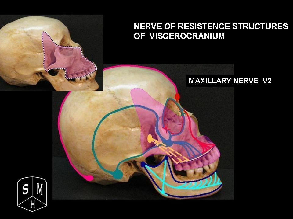
V2 is the sensory maxillary division of cranial nerve V. It passes through foramen rotundum into the pterygopalatine fossa. Its infraorbital continuation enters the orbit through the inferior orbital fissure, travels in the infraorbital groove and canal, and emerges through the infraorbital foramen onto the face. Foramen rotundum, inferior orbital fissure, and infraorbital foramen are successive landmarks, not interchangeable names for the same opening. [1] [2]
Open whole image Trace V2 toward the midface before separating its infraorbital, dental, and palatal branches. Image: Anatomist90. CC BY-SA 3.0 . Original source .
Whole image Close
Apply: Trace V2 from the skull to the upper jaw Foramen rotundum is narrowed by a skull-base lesion. Which sensory pattern best fits isolated involvement of the structure passing through it?
Upper jaw, palate, and midface sensory impairment Isolated forehead and corneal sensory loss Lower lip and chin sensory loss alone
Try again Read the worked solution Upper jaw, palate, and midface sensory impairment Best fit. V2 passes through foramen rotundum and supplies these territories. [1] [2]
Isolated forehead and corneal sensory loss Compare this alternative. That is primarily V1 territory through the superior orbital fissure. [1] [2]
Lower lip and chin sensory loss alone Compare this alternative. This fits distal V3 mental territory more closely. [1] [2]
A branching route rather than a flat tooth chart. Read downward from the skull; branches leave before the nerve reaches facial skin. Foramen rotundum. V2 enters the pterygopalatine fossa.Pterygopalatine fossa. Posterior superior alveolar branches head toward the posterior maxilla. Connections with the pterygopalatine ganglion lead to palatal and nasal sensory branches.Inferior orbital fissure, groove, and canal. The infraorbital nerve travels anteriorly. Anterior superior alveolar and variable middle superior alveolar branches descend toward the dental plexus.Infraorbital foramen. Terminal branches distribute to the lower eyelid, side of the nose, upper lip, and nearby cheek skin.The posterior superior alveolar nerve usually leaves proximal to the main infraorbital canal course and enters foramina on the posterior maxilla. The anterior superior alveolar nerve usually leaves the infraorbital nerve within the canal and follows the anterior maxillary wall. The middle superior alveolar nerve is variable in presence and origin, often arising from the infraorbital nerve around the groove or canal. These branches join a superior dental plexus rather than remaining isolated wires ending at one tooth. [2] [3] [4]
V2 sensory fibers associated with the pterygopalatine ganglion pass through its connections without their sensory cell bodies synapsing there. Those cell bodies are in the trigeminal ganglion. Parasympathetic fibers can synapse in the pterygopalatine ganglion and then travel with branches toward glands. Distinguishing sensory from autonomic traffic prevents the mistaken conclusion that V2 must be a facial motor nerve because it carries fibers associated with lacrimal or nasal secretion. [1]
Try it here · Checkpoint 1 of 3
Make your prediction before reading the choices. A first attempt is just a starting point.
Case 13
Show answer and explanations for case 13
A. Inferior orbital fissure (Best answer)
Reason through this option Read the question, predict, then check the reasoning.
Where does the route begin before entering the orbit?
Reveal this step The infraorbital continuation lies in the pterygopalatine region.
Which opening leads it into the orbital floor?
Reveal this step It enters through the inferior orbital fissure before reaching the groove.
Start this reasoning again Sources [1] [2]
Read the complete explanation This links the pterygopalatine region with the infraorbital route.
B. Superior orbital fissure (Why this does not fit)
Reason through this option Read the question, predict, then check the reasoning.
Why does the superior orbital fissure sound plausible?
Reveal this step It is another communication with the orbit.
What structures distinguish it?
Reveal this step It carries V1-related and ocular motor structures, not the V2 infraorbital continuation.
Start this reasoning again Sources [1] [2]
Read the complete explanation This transmits V1-related and ocular motor structures rather than the infraorbital continuation.
C. Foramen ovale (Why this does not fit)
Reason through this option Read the question, predict, then check the reasoning.
Which trigeminal division uses foramen ovale?
Reveal this step V3 exits the skull through foramen ovale.
Why does it not match the orbital floor route?
Reveal this step The infraorbital continuation belongs to V2 and enters through the inferior orbital fissure.
Start this reasoning again Sources [1] [2]
Read the complete explanation This is the V3 skull exit.
D. Incisive foramen (Why this does not fit)
Reason through this option Read the question, predict, then check the reasoning.
Where is the incisive foramen?
Reveal this step It is in the anterior hard palate behind the incisors.
Which nerve uses that opening instead?
Reveal this step The nasopalatine nerve uses it, not the infraorbital continuation.
Start this reasoning again Sources [1] [2]
Read the complete explanation This is an anterior palatal opening for the nasopalatine route.
Takeaway: The inferior orbital fissure is upstream of the infraorbital groove and canal.
Case sources: [1] [2]
Save this spot
Try a question on this topic A 54-year-old woman is referred for progressive difficulty swallowing solid foods, hoarseness, and right shoulder weakness developing over four months. Examination reveals an absent gag reflex on the right, leftward deviation of the uvula on phonation, and weakness of the right sternocleidomastoid and trapezius against resistance. Tongue protrusion and lateral movement are completely normal. Facial sensation, facial expression, and bilateral hearing are intact. MRI of the skull base demonstrates a 2.4 cm heterogeneously enhancing mass centered at the right jugular foramen with extension into the posterior cranial fossa.
Which structures pass through the jugular foramen and are being compressed by this mass to produce the patient's deficits?
A. CN IX (glossopharyngeal), CN X (vagus), and CN XI (spinal accessory), along with the sigmoid sinus continuing as the internal jugular vein B. CN VII (facial), CN VIII (vestibulocochlear), and CN IX (glossopharyngeal) C. CN X (vagus), CN XI (spinal accessory), and CN XII (hypoglossal) D. CN VIII (vestibulocochlear), CN IX (glossopharyngeal), and CN X (vagus) E. CN IX (glossopharyngeal), CN X (vagus), CN XI (spinal accessory), and CN XII (hypoglossal)
Choose an answer before revealing the reasoning.
Get one of these every morning · Practice more in the QBank
Use the tooth map without making it absolute Predict the usual dental route Read the question, predict, then check the reasoning.
Which named branch is the usual starting prediction for an incisor pulp?
Reveal this step ASA is the principal named anterior dental route, while additional inputs can occur.
What changes when a distinct MSA is absent?
Reveal this step Adjacent ASA and PSA fibers can still supply the premolar plexus.
Which named branch is the usual starting prediction for molar pulp?
Reveal this step PSA usually supplies molar territory, with variable first-molar contributions from the surrounding plexus.
What can residual first-molar sensitivity establish?
Reveal this step It identifies incomplete tissue coverage, not a proven individual branching pattern.
Start this reasoning again Sources [2] [3] [4] [11]
Explore the reasoning
Use the tooth map without making it absolute
Work through the reasoning
Which section principle should anchor this decision?
Use the tooth map without making it absolute Usual superior alveolar contributions on one side of the upper dental arch.
Show all answers
Focus on one step
Earlier step Following step
What should be established first? Use the tooth map without making it absolute Usual superior alveolar contributions on one side of the upper dental arch.
What second distinction prevents the shortcut? This is an anterior-to-posterior comparison, not a claim of exclusive innervation.
What should carry into the next case? Identify the usual superior alveolar route without claiming exclusive innervation.
Use the section like a decision map: identify the finding, then follow only the supported relationship to the conclusion.
Where the comparison stops. This organizes reasoning; it does not replace the patient history, examination, source-specific criteria, or appropriate diagnostic testing.
Use the tooth map without making it absolute Anterior superior alveolar nerve Identify the usual superior alveolar route without claiming Identify the usual superior alveolar route without claiming exclusive innervation.
Sources [2] [4] [6]
Compare both answers Anterior superior alveolar nerve
Why it fits. ASA is the principal named superior alveolar route for incisors, while additional inputs can occur.
This answer best fits the stated findings and the section mechanism.
Identify the usual superior alveolar route without claiming exclusive innervation.
Middle superior alveolar nerve
Why it is tempting. MSA is more closely associated with premolars and variable first molar contribution.
Compare the stated findings with the section mechanism before choosing the alternative.
Identify the usual superior alveolar route without claiming exclusive innervation.
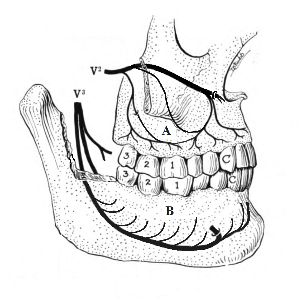
Usual superior alveolar contributions on one side of the upper dental arch. This is an anterior-to-posterior comparison, not a claim of exclusive innervation. Incisors and canine The anterior superior alveolar nerve is the principal named superior alveolar route. Premolars The middle superior alveolar nerve contributes when present. Anterior and posterior superior alveolar branches can provide the territory when a separate middle branch is absent. First molar The posterior superior alveolar nerve usually contributes to molar supply. The mesiobuccal root often receives additional middle or anterior plexus input and is a common site of incomplete anesthesia after a posterior superior alveolar block. Second and third molars The posterior superior alveolar route is the usual named molar pathway. ASA, MSA, and PSA are convenient abbreviations for these three superior alveolar branches. Their distributions also include alveolar bone, periodontal tissues, buccal gingiva, and parts of the maxillary sinus, with differing branch patterns. A question about the maxillary canine's usual superior alveolar nerve points to ASA. A question about an entire cheek or a palatal incision asks something different. [2] [3]
Open whole image Use the upper dental plexus as a territory map, not as proof that one named branch exclusively owns a tooth. Image: Lesion. CC BY-SA 3.0 . Original source .
Whole image Close
Apply: Use the tooth map without making it absolute A patient needs treatment of a fractured maxillary central incisor. Which named superior alveolar branch usually supplies its pulp?
Anterior superior alveolar nerve Middle superior alveolar nerve Posterior superior alveolar nerve
Try again Read the worked solution Anterior superior alveolar nerve Best fit. ASA is the principal named superior alveolar route for incisors, while additional inputs can occur. [2] [4] [6]
Middle superior alveolar nerve Compare this alternative. MSA is more closely associated with premolars and variable first molar contribution. [2] [4] [6]
Posterior superior alveolar nerve Compare this alternative. PSA is the usual molar route rather than the principal incisor branch. [2] [4] [6]
The first molar exception must be worded carefully. Residual pain in its mesiobuccal region after a PSA block is compatible with additional innervation, often described through MSA or the surrounding plexus. It does not prove that a distinct MSA exists in that person. A primary dissection study found a middle branch in only seven of nineteen heads and documented substantial variation in other alveolar branches. Those data demonstrate variability; they should not be converted into a universal prevalence for all populations. [4]
Similarly, an absent MSA does not mean premolars have no sensation. Neighboring superior alveolar fibers distribute through the plexus. The clinical response to a block depends on the anatomy, local anesthetic spread, inflammation, dose, timing, and technique. One failed injection is not a dissection and cannot establish a unique branching pattern.
Do not confuse the palatal root of a molar with the palatal gingiva beside it. The first describes a root containing a pulp canal. The second is a mucosal and periodontal surface. In the traditional superior alveolar map, the palatal root of the first molar usually receives PSA contribution, while posterior palatal soft tissue is chiefly a greater palatine territory. Additional palatal dental inputs are real, as the next section explains, but the two tissues still require separate assessment.
Save this spot
The palate has its own routes and dental connections Separate palate from pulp Read the question, predict, then check the reasoning.
Which route best predicts sensation just behind the central incisors?
Reveal this step The nasopalatine nerve reaches the anterior hard palate through the incisive canal.
Which route best predicts posterior hard-palate sensation beside molars?
Reveal this step The greater palatine nerve emerges posteriorly and travels anteriorly beneath the mucoperiosteum.
Which route chiefly serves the soft palate?
Reveal this step The lesser palatine nerves are the principal V2 sensory routes for that tissue.
How should modern palatal dental evidence change the map?
Reveal this step Allow overlapping dental input without promising universal prevalence or block success.
Start this reasoning again Sources [2] [5] [6] [7] [11]
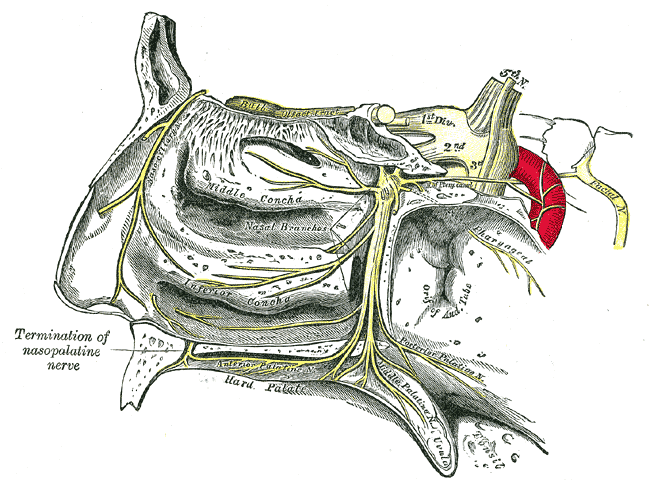
The nasopalatine nerve travels through the nasal cavity along the septum and reaches the anterior hard palate through the incisive canal and foramen behind the central incisors. It supplies the anterior palatal mucosa and communicates with greater palatine branches. The greater palatine nerve descends through the greater palatine canal and emerges onto the posterior hard palate, then runs anteriorly beneath the mucoperiosteum. The lesser palatine nerves primarily supply soft palatal sensory territories. Greater and lesser refer to different routes and distributions, not simply stronger and weaker versions of one dental nerve. [2] [5]
Open whole image Follow the palatine branches toward the palate and locate the anterior nasopalatine termination. Historical labels are retained; the adjacent teaching uses current nerve names. Image: Henry Vandyke Carter. Public domain . Original source .
Whole image Close
Apply: The palate has its own routes and dental connections A palatal incision is planned immediately behind the maxillary central incisors. Which sensory nerve is the principal local target?
Nasopalatine nerve Posterior superior alveolar nerve Lesser palatine nerve
Try again Read the worked solution Nasopalatine nerve Best fit. It emerges through the incisive foramen into the anterior hard palate. [2] [5]
Posterior superior alveolar nerve Compare this alternative. Its principal course is along the posterior maxilla to molar and buccal territories. [2] [5]
Lesser palatine nerve Compare this alternative. It principally serves soft palate sensory territories. [2] [5]
For planning a palatal incision, think from front to back. An incision immediately behind the incisors principally concerns nasopalatine supply. Posterior hard palatal gingiva beside molars principally concerns greater palatine supply. Soft palate sensation principally concerns lesser palatine branches, with other sensory contributions depending on the precise region. The transition around the canine and premolar area overlaps, so a rigid line between colored territories is misleading.
The greater palatine foramen lies in the posterior hard palate near the molar region, but its exact position and the nerve's branching vary. A primary dissection series documented branching within the canal in some specimens, before the nerve reached the palate. Surgical planning should account for the associated artery and the actual foramen rather than relying on one universal distance from a particular tooth. [5]
Older teaching often states that palatal nerves never supply teeth. That is too absolute. A small clinical study showed that some central incisors still responsive after ASA anesthesia became unresponsive after a nasopalatine block. This supports additional input in some patients, but its selected sample and testing method do not establish a population-wide percentage or guaranteed success. [6]
A 2025 anatomical and radiologic investigation provided more direct evidence. Using eight cadaveric maxillae with injection, micro-CT, dissection, and histology methods, investigators demonstrated palatal alveolar pathways through which greater palatine and nasopalatine branches contribute to maxillary dental and periodontal supply. These findings supplement the superior alveolar map. They do not relocate the ASA or MSA nerve trunks into the palate, and an anatomical connection alone does not establish the efficacy of a particular injection technique. [7]
The practical distinction is therefore between a usual target and an exclusive territory. A greater palatine block is commonly selected for posterior palatal soft tissue. It should not be assumed to provide complete molar pulpal anesthesia in every patient. Conversely, persistent dental sensation is not evidence that a palatal contribution is anatomically impossible.
Try it here · Checkpoint 2 of 3
Make your prediction before reading the choices. A first attempt is just a starting point.
Case 7
Show answer and explanations for case 7
A. Infraorbital terminal labial branch (Why this does not fit)
Reason through this option Read the question, predict, then check the reasoning.
Where do terminal infraorbital labial branches end?
Reveal this step They distribute to upper lip and adjacent facial tissue.
What tissue remains painful here?
Reveal this step Posterior palatal gingiva beside a molar, not facial skin.
Start this reasoning again Sources [2] [5]
Read the complete explanation It supplies upper lip tissue rather than posterior hard palate.
B. Inferior alveolar nerve (Why this does not fit)
Reason through this option Read the question, predict, then check the reasoning.
Which jaw does the inferior alveolar nerve serve?
Reveal this step It supplies mandibular teeth through V3.
Why is it excluded?
Reveal this step The painful flap is on the maxillary palate.
Start this reasoning again Sources [2] [5]
Read the complete explanation It belongs to mandibular dental sensation.
C. Greater palatine nerve (Best answer)
Reason through this option Read the question, predict, then check the reasoning.
Which tissue is not anesthetized?
Reveal this step Posterior hard palatal mucosa and gingiva remain painful.
Which local nerve chiefly supplies it?
Reveal this step The greater palatine nerve runs anteriorly across the posterior hard palate.
Start this reasoning again Sources [2] [5]
Read the complete explanation It chiefly supplies posterior hard palatal mucosa and palatal gingiva.
D. Lesser palatine nerve (Why this does not fit)
Reason through this option Read the question, predict, then check the reasoning.
Why is lesser palatine a nearby possibility?
Reveal this step It also descends through a palatine route.
Which tissue boundary separates it?
Reveal this step Lesser palatine chiefly serves soft palate, while this flap is on hard palate.
Start this reasoning again Sources [2] [5]
Read the complete explanation Its main territory is soft palate, posterior to the hard palatal target.
Takeaway: Pulpal anesthesia does not guarantee palatal flap anesthesia.
Case sources: [2] [5]
Save this spot
Where does a midface injury interrupt the route? Localize by tissue and level Read the question, predict, then check the reasoning.
What does upper-lip loss with spared tooth sensation suggest?
Reveal this step A distal infraorbital cutaneous injury after the dental branches separated.
What does combined upper-lip and anterior-tooth loss suggest?
Reveal this step A more proximal infraorbital canal lesion can interrupt both fiber groups.
What does tooth, palate, and midface loss suggest together?
Reveal this step A proximal V2 lesion before those peripheral territories separate.
Which nearby nerve supplies sensation near the nasal tip?
Reveal this step The external nasal branch of the anterior ethmoidal nerve comes from V1.
Start this reasoning again Sources [1] [2] [8] [10]
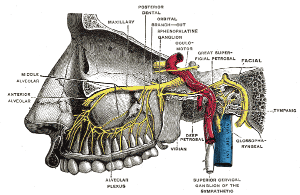
The distal infraorbital branches supply the lower eyelid, lateral nose, upper lip, and nearby cheek. A small injury after the dental branches have separated can affect facial skin while sparing tooth sensation. A fracture through the infraorbital groove or canal may affect the infraorbital nerve before ASA or MSA branches have left and can therefore affect both facial and dental sensation. The phrase infraorbital nerve injury is incomplete without a location. [1] [2] [8]
Open whole image The branch point matters: a lesion before dental branches separate can affect teeth and facial territory together. Image: Henry Vandyke Carter. Public domain . Original source .
Whole image Close
Apply: Where does a midface injury interrupt the route A patient feels light touch normally over the upper lip but cannot smile symmetrically after a facial procedure. Which neural function is primarily impaired?
Facial nerve motor output Greater palatine sensation V2 dental sensation
Try again Read the worked solution Facial nerve motor output Best fit. Facial expression depends on VII, while preserved touch indicates a different sensory pathway remains functional. [1] [2]
Greater palatine sensation Compare this alternative. This concerns the palate and does not power the upper lip muscles. [1] [2]
V2 dental sensation Compare this alternative. A dental sensory lesion would not directly paralyze facial expression. [1] [2]
A lesion at the pterygopalatine fossa or foramen rotundum can combine sensory loss in several V2 branches, including maxillary teeth, palate, and midface. An isolated PSA lesion would not explain upper lip numbness. An isolated infraorbital terminal branch injury would not explain extensive palatal sensory loss. Compare both sides and test distinct tissues rather than asking only whether the cheek feels different.
V1 provides sensation to the forehead, upper eyelid, and cornea, with nasal territory including the external nasal branch of the anterior ethmoidal nerve toward the tip. [10] V2 supplies much of the side of the nose and upper lip. V3 supplies mandibular teeth and, through the mental nerve, lower lip and chin sensation. The borders overlap, but upper lip plus upper tooth findings differ meaningfully from chin plus lower tooth findings. [1] [2]
Facial expression uses cranial nerve VII. A patient who feels an upper lip touch but cannot produce a symmetric smile has a motor problem requiring a different localization from an isolated V2 sensory deficit. Mastication uses V3 motor fibers, not V2. Palatal sensation and palatal motor function are also separate examinations. These distinctions prevent a nearby sensory nerve from becoming the answer to every symptom around the mouth.
Save this spot
Test the tissue the procedure will actually touch Test the tissue that matters Read the question, predict, then check the reasoning.
Does a numb upper lip prove that incisor pulp is anesthetized?
Reveal this step No. Cutaneous and pulpal endpoints require separate assessment.
Does a numb molar pulp prove that palatal flap tissue is anesthetized?
Reveal this step No. Posterior palatal mucosa has a distinct usual target.
What should persistent sensation make the clinician identify first?
Reveal this step Identify the exact tissue that still responds.
What timing question comes before declaring coverage incomplete?
Reveal this step Confirm that the expected onset interval has elapsed.
What is the first action when systemic warning signs appear during injection?
Reveal this step Stop anesthetic administration immediately.
Start this reasoning again Sources [2] [8] [9]
Explore the reasoning
Test the tissue the procedure will actually touch
Work through the reasoning
Which section principle should anchor this decision?
Test the tissue the procedure will actually touch Begin with the intended procedure.
Show all answers
Focus on one step
Earlier step Following step
What should be established first? Test the tissue the procedure will actually touch Begin with the intended procedure.
What second distinction prevents the shortcut? Pulp treatment, buccal soft-tissue manipulation, and a palatal flap can require different anesthetic coverage even around the same tooth.
What should carry into the next case? A partially effective block can reveal a coverage gap without proving the exact anatomical variant.
Use the section like a decision map: identify the finding, then follow only the supported relationship to the conclusion.
Where the comparison stops. This organizes reasoning; it does not replace the patient history, examination, source-specific criteria, or appropriate diagnostic testing.
Test the tissue the procedure will actually touch Additional superior dental plexus input may remain unblocked A partially effective block can reveal a coverage A partially effective block can reveal a coverage gap without proving the exact anatomical variant.
Sources [2] [4] [9]
Compare both answers Additional superior dental plexus input may remain unblocked
Why it fits. The mesiobuccal first molar region commonly has variable input beyond the PSA route.
This answer best fits the stated findings and the section mechanism.
A partially effective block can reveal a coverage gap without proving the exact anatomical variant.
All palatal nerves must have been anesthetized
Why it is tempting. The PSA block and residual pulpal response do not establish palatal soft-tissue coverage.
Compare the stated findings with the section mechanism before choosing the alternative.
A partially effective block can reveal a coverage gap without proving the exact anatomical variant.
Begin with the intended procedure. Pulp treatment, buccal soft-tissue manipulation, and a palatal flap can require different anesthetic coverage even around the same tooth. A tooth-specific buccal infiltration can reach terminal dental fibers without producing a complete proximal V2 block. A regional block may cover a wider field but still leave one root or tissue sensitive. The name of the injection does not substitute for testing its effect. [2] [9]
Apply: Test the tissue the procedure will actually touch After a PSA block, testing shows anesthesia of most maxillary molar territory, but the first molar's mesiobuccal component remains sensitive. Which explanation is best?
Additional superior dental plexus input may remain unblocked All palatal nerves must have been anesthetized The response proves that a distinct MSA exists
Try again Read the worked solution Additional superior dental plexus input may remain unblocked Best fit. The mesiobuccal first molar region commonly has variable input beyond the PSA route. [2] [4] [9]
All palatal nerves must have been anesthetized Compare this alternative. The PSA block and residual pulpal response do not establish palatal soft-tissue coverage. [2] [4] [9]
The response proves that a distinct MSA exists Compare this alternative. A block response cannot establish a unique branching anatomy. [2] [4] [9]
After a PSA block, residual sensitivity at the first molar's mesiobuccal component can require supplemental anesthesia. Do not conclude that the entire injection failed when other molar territories are anesthetized. After an infraorbital block, a numb upper lip demonstrates effect on cutaneous fibers, while an appropriate pulp assessment checks a different target. After a greater palatine block, posterior palatal tissue anesthesia does not prove the pulp is ready for instrumentation.
When anesthesia is incomplete, reassess the exact tooth and tissue, the waiting interval, local inflammation, the administered dose, and the probable nerve routes. Additional infiltration or another selected technique may be appropriate, but repeated injections require accounting for the cumulative dose. Avoid explaining every failure as unusual anatomy; common problems with delivery or inflammation also matter.
AAPD guidance emphasizes dose calculation using body weight in children, documentation of the agent and total dose, aspiration before delivery, slow injection, and observation for adverse effects. Unexpected neurologic or cardiovascular symptoms during injection require stopping administration and assessing for systemic toxicity. The anatomy lesson does not provide a drug-specific dosing protocol; the relevant preparation, patient factors, and clinical guidance determine that calculation. [9]
After treatment, a numb lip or cheek remains vulnerable to biting even when pulpal anesthesia has waned. Explain the expected numbness and protection of soft tissues to the patient or caregiver. New sensory loss that persists beyond the expected anesthetic duration should be documented and evaluated rather than assigned a fictional guaranteed recovery date. The same standard applies after a fracture or operation. Record the injured location and observed course instead of claiming that a two-week-old fracture must already be healed.
Try it here · Checkpoint 3 of 3
Make your prediction before reading the choices. A first attempt is just a starting point.
Case 10
Show answer and explanations for case 10
A. The pulp test must be disregarded because the lip is numb (Why this does not fit)
Reason through this option Read the question, predict, then check the reasoning.
What does lip numbness actually verify?
Reveal this step It verifies anesthesia of cutaneous infraorbital endpoints.
Why must the pulp test still count?
Reveal this step Dental fibers can branch proximally and require their own functional assessment.
Start this reasoning again Sources [2] [8] [9]
Read the complete explanation Lip numbness does not guarantee dental anesthesia.
B. The patient has lost all V2 sensation (Why this does not fit)
Reason through this option Read the question, predict, then check the reasoning.
Would complete V2 loss preserve incisor pulp sensation?
Reveal this step No. Maxillary dental sensation also travels through V2.
What finding disproves complete loss?
Reveal this step The incisor still responds to appropriate pulp testing.
Start this reasoning again Sources [2] [8] [9]
Read the complete explanation The persistent pulp response directly contradicts that conclusion.
C. The maxillary tooth must be supplied only by V3 (Why this does not fit)
Reason through this option Read the question, predict, then check the reasoning.
Why might V3 be considered?
Reveal this step V3 carries dental sensation in the mandible.
Which anatomical boundary rejects it?
Reveal this step This tooth is maxillary and retains V2 dental supply despite an incomplete block.
Start this reasoning again Sources [2] [8] [9]
Read the complete explanation An incomplete V2 block does not change the tooth's cranial nerve division.
D. Cutaneous anesthesia is present, but pulpal anesthesia is incomplete (Best answer)
Reason through this option Read the question, predict, then check the reasoning.
Which endpoint is anesthetized?
Reveal this step The upper lip cutaneous endpoint is anesthetized.
Which endpoint remains functional?
Reveal this step Incisor pulp sensation remains, proving incomplete dental coverage.
Start this reasoning again Sources [2] [8] [9]
Read the complete explanation The dental branch and skin endpoints need separate assessment.
Takeaway: Proceed according to tissue testing, not an assumed effect of the block's name.
Case sources: [2] [8] [9]
Save this spot
Try these without looking A patient needs treatment of a fractured maxillary central incisor Which named superior alveolar branch usually supplies its pulp? Identify the usual superior alveolar route without claiming exclusive innervation.
Revisit this explanation [2] [4] [6]
After a PSA block testing shows anesthesia of most maxillary molar territory but the first molar's mesiobuccal component remains sensitive Which explanation is best? A partially effective block can reveal a coverage gap without proving the exact anatomical variant.
Revisit this explanation [2] [4] [9]
What prevents this lesson from becoming a one-clue shortcut? Use the complete clinical or study context, then apply the named test, anatomical relation, or guideline only within its validated conditions.
Revisit this explanation [2] [4] [9]
Practice separating tooth, palate, and skin
Case 1
Show answer and explanations for case 1
A. Anterior superior alveolar nerve (Best answer)
Reason through this option Read the question, predict, then check the reasoning.
Which tooth is being treated?
Reveal this step A maxillary central incisor, within the anterior dental territory.
Which named branch usually reaches that pulp?
Reveal this step The anterior superior alveolar nerve is the principal route.
Start this reasoning again Sources [2] [4] [6]
Read the complete explanation ASA is the principal named superior alveolar route for incisors, while additional inputs can occur.
B. Middle superior alveolar nerve (Why this does not fit)
Reason through this option Read the question, predict, then check the reasoning.
Why might the middle branch sound plausible?
Reveal this step It joins the same superior dental plexus when present.
What landmark separates its usual territory?
Reveal this step The MSA chiefly descends toward premolars, not central incisors.
Start this reasoning again Sources [2] [4] [6]
Read the complete explanation MSA is more closely associated with premolars and variable first molar contribution.
C. Posterior superior alveolar nerve (Why this does not fit)
Reason through this option Read the question, predict, then check the reasoning.
Where does the posterior branch usually distribute?
Reveal this step It approaches the posterior maxilla and chiefly supplies molars.
Why is it not the principal incisor answer?
Reveal this step The central incisor lies in the anterior ASA territory.
Start this reasoning again Sources [2] [4] [6]
Read the complete explanation PSA is the usual molar route rather than the principal incisor branch.
D. Inferior alveolar nerve (Why this does not fit)
Reason through this option Read the question, predict, then check the reasoning.
Which trigeminal division contains the inferior alveolar nerve?
Reveal this step It is a V3 branch serving mandibular teeth.
What feature excludes it here?
Reveal this step The treated tooth is maxillary and therefore supplied through V2 routes.
Start this reasoning again Sources [2] [4] [6]
Read the complete explanation This V3 branch supplies mandibular rather than maxillary teeth.
Takeaway: Identify the usual superior alveolar route without claiming exclusive innervation.
Case sources: [2] [4] [6]
Case 2
Show answer and explanations for case 2
A. Posterior superior alveolar nerve (Why this does not fit)
Reason through this option Read the question, predict, then check the reasoning.
Why is PSA a nearby dental possibility?
Reveal this step It contributes to the same superior dental plexus.
What course distinguishes it from the described branch?
Reveal this step PSA approaches from the posterior maxilla rather than descending from the infraorbital nerve to premolars.
Start this reasoning again Sources [2] [4]
Read the complete explanation PSA usually approaches from the posterior maxilla after branching more proximally.
B. Greater palatine nerve (Why this does not fit)
Reason through this option Read the question, predict, then check the reasoning.
Where does the greater palatine nerve travel?
Reveal this step It descends through the greater palatine canal to the hard palate.
Why does that route not match?
Reveal this step The observed branch arises from the infraorbital nerve and enters the premolar plexus.
Start this reasoning again Sources [2] [4]
Read the complete explanation This reaches the palate through the greater palatine canal rather than the described infraorbital origin.
C. Nasopalatine nerve (Why this does not fit)
Reason through this option Read the question, predict, then check the reasoning.
What landmarks identify the nasopalatine route?
Reveal this step It follows the nasal septum and incisive canal.
What landmark identifies this specimen instead?
Reveal this step Its infraorbital origin and premolar destination identify a superior alveolar branch.
Start this reasoning again Sources [2] [4]
Read the complete explanation This follows the nasal septum and incisive canal.
D. Middle superior alveolar nerve (Best answer)
Reason through this option Read the question, predict, then check the reasoning.
Which territory does the observed branch enter?
Reveal this step It enters the premolar dental plexus.
Which variable branch has that origin and territory?
Reveal this step The middle superior alveolar nerve may descend from the infraorbital nerve to premolars.
Start this reasoning again Sources [2] [4]
Read the complete explanation Its position and premolar connection define the middle branch when present.
Takeaway: MSA is a variable anatomical branch, not a guaranteed structure in every patient.
Case sources: [2] [4]
Case 3
Show answer and explanations for case 3
A. Mental nerve (Why this does not fit)
Reason through this option Read the question, predict, then check the reasoning.
Why is the mental nerve a dental-sounding distractor?
Reveal this step It is a terminal branch associated with the mandibular alveolar route.
Which tissue landmark excludes it?
Reveal this step It supplies lower lip and chin skin, not maxillary second molar pulp.
Start this reasoning again Sources [2] [3]
Read the complete explanation This V3 branch supplies lower lip and chin sensation, not upper molar pulp.
B. Lesser palatine nerve (Why this does not fit)
Reason through this option Read the question, predict, then check the reasoning.
Where do lesser palatine nerves distribute?
Reveal this step They principally supply soft palatal sensation.
Why is that not the requested target?
Reveal this step The question asks for molar pulp through a superior alveolar route, not soft palate.
Start this reasoning again Sources [2] [3]
Read the complete explanation This is chiefly a soft palatal sensory route rather than the usual superior alveolar molar target.
C. Posterior superior alveolar nerve (Best answer)
Reason through this option Read the question, predict, then check the reasoning.
Where is the treated tooth located?
Reveal this step The second molar is in the posterior maxillary dental territory.
Which named branch usually supplies that territory?
Reveal this step The posterior superior alveolar nerve is the usual molar target.
Start this reasoning again Sources [2] [3]
Read the complete explanation PSA supplies the usual molar superior alveolar pathway.
D. Anterior superior alveolar nerve (Why this does not fit)
Reason through this option Read the question, predict, then check the reasoning.
Which teeth are chiefly associated with ASA?
Reveal this step ASA chiefly supplies incisors and the canine through the anterior plexus.
What separates this case?
Reveal this step A second molar lies posteriorly in the usual PSA territory.
Start this reasoning again Sources [2] [3]
Read the complete explanation ASA chiefly serves the anterior dental plexus.
Takeaway: Tooth pulp and nearby palatal surfaces should be assessed separately.
Case sources: [2] [3]
Case 4
Show answer and explanations for case 4
A. All palatal nerves must have been anesthetized (Why this does not fit)
Reason through this option Read the question, predict, then check the reasoning.
What tissue was shown to remain sensitive?
Reveal this step A first molar pulpal component remained sensitive.
Does that establish palatal soft-tissue anesthesia?
Reveal this step No. Dental pulp and palatal mucosa require separate testing and routes.
Start this reasoning again Sources [2] [4] [9]
Read the complete explanation The PSA block and residual pulpal response do not establish palatal soft-tissue coverage.
B. Additional superior dental plexus input may remain unblocked (Best answer)
Reason through this option Read the question, predict, then check the reasoning.
Which first molar region often has overlapping input?
Reveal this step The mesiobuccal component may receive input beyond the PSA route.
What best explains selective residual sensitivity?
Reveal this step Unblocked fibers from the variable superior dental plexus can preserve sensation there.
Start this reasoning again Sources [2] [4] [9]
Read the complete explanation The mesiobuccal first molar region commonly has variable input beyond the PSA route.
C. The response proves that a distinct MSA exists (Why this does not fit)
Reason through this option Read the question, predict, then check the reasoning.
Why does MSA seem plausible?
Reveal this step A middle branch can contribute to the first molar mesiobuccal region.
What anatomical alternative can preserve sensation?
Reveal this step Superior dental plexus overlap can preserve sensation.
What procedural alternative can produce the same response?
Reveal this step Incomplete anesthetic delivery can produce the same response.
Start this reasoning again Sources [2] [4] [9]
Read the complete explanation A block response cannot establish a unique branching anatomy.
D. The first molar must be innervated by V3 (Why this does not fit)
Reason through this option Read the question, predict, then check the reasoning.
Which division supplies maxillary dental sensation?
Reveal this step The superior alveolar system arises from V2.
Does incomplete PSA anesthesia shift the tooth to V3?
Reveal this step No. It indicates overlapping V2 input, not mandibular innervation.
Start this reasoning again Sources [2] [4] [9]
Read the complete explanation Variable superior alveolar contributions remain within V2.
Takeaway: A partially effective block can reveal a coverage gap without proving the exact anatomical variant.
Case sources: [2] [4] [9]
Case 5
Show answer and explanations for case 5
A. Nasopalatine nerve (Best answer)
Reason through this option Read the question, predict, then check the reasoning.
Where is the incision?
Reveal this step It is on anterior hard palate immediately behind the central incisors.
Which nerve emerges there?
Reveal this step The nasopalatine nerve reaches this region through the incisive foramen.
Start this reasoning again Sources [2] [5]
Read the complete explanation It emerges through the incisive foramen into the anterior hard palate.
B. Posterior superior alveolar nerve (Why this does not fit)
Reason through this option Read the question, predict, then check the reasoning.
Why might PSA sound relevant?
Reveal this step It supplies posterior maxillary dental and buccal tissues.
What separates the planned tissue?
Reveal this step The incision is anterior palatal mucosa, not posterior molar or buccal territory.
Start this reasoning again Sources [2] [5]
Read the complete explanation Its principal course is along the posterior maxilla to molar and buccal territories.
C. Lesser palatine nerve (Why this does not fit)
Reason through this option Read the question, predict, then check the reasoning.
Which palate region chiefly receives lesser palatine nerves?
Reveal this step They principally serve the soft palate.
Which landmark places this incision elsewhere?
Reveal this step The incisors and incisive foramen mark anterior hard palate.
Start this reasoning again Sources [2] [5]
Read the complete explanation It principally serves soft palate sensory territories.
D. Mental nerve (Why this does not fit)
Reason through this option Read the question, predict, then check the reasoning.
Where does the mental nerve emerge?
Reveal this step It emerges on the mandible to supply lower lip and chin.
Why is it anatomically remote?
Reveal this step The procedure is on maxillary anterior hard palate within V2 territory.
Start this reasoning again Sources [2] [5]
Read the complete explanation It supplies the lower lip and chin rather than the hard palate.
Takeaway: The tissue and the side of the dental arch determine the target.
Case sources: [2] [5]
Case 6
Show answer and explanations for case 6
A. Any infraorbital lesion can affect skin only (Why this does not fit)
Reason through this option Read the question, predict, then check the reasoning.
Do infraorbital fibers include dental fibers proximally?
Reveal this step Yes. Superior alveolar branches can leave during the groove and canal course.
Why is skin-only loss not inevitable?
Reveal this step A proximal canal lesion can occur before dental and cutaneous fibers separate.
Start this reasoning again Sources [1] [2] [4]
Read the complete explanation The proximal canal contains fibers destined for dental branches.
B. The mental nerve supplies both territories (Why this does not fit)
Reason through this option Read the question, predict, then check the reasoning.
What is the mental nerve territory?
Reveal this step It supplies lower lip and chin through V3.
Which findings exclude it?
Reveal this step Upper lip and maxillary anterior tooth deficits localize to V2.
Start this reasoning again Sources [1] [2] [4]
Read the complete explanation Mental territory is mandibular lower lip and chin.
C. A lesser palatine lesion accounts for the upper lip deficit (Why this does not fit)
Reason through this option Read the question, predict, then check the reasoning.
Where do lesser palatine branches distribute?
Reveal this step They principally supply soft palatal tissue.
Can that route explain upper lip skin loss?
Reveal this step No. It does not travel to infraorbital facial branches.
Start this reasoning again Sources [1] [2] [4]
Read the complete explanation Lesser palatine branches do not supply upper lip skin.
D. Injury proximal to the departure of affected alveolar branches (Best answer)
Reason through this option Read the question, predict, then check the reasoning.
Which structure is injured?
Reveal this step The infraorbital nerve is injured within its canal.
How can one lesion affect lip and teeth?
Reveal this step It interrupts shared proximal fibers before affected alveolar branches depart toward the anterior teeth.
Start this reasoning again Sources [1] [2] [4]
Read the complete explanation A canal lesion can interrupt cutaneous and dental fibers before they separate.
Takeaway: State where along the infraorbital route an injury lies.
Case sources: [1] [2] [4]
Case 8
Show answer and explanations for case 8
A. Facial nerve motor branches (Why this does not fit)
Reason through this option Read the question, predict, then check the reasoning.
What function do facial nerve branches provide?
Reveal this step They provide motor output for facial expression.
Why do they not unify these findings?
Reveal this step The combined deficit is sensory rather than facial motor.
Start this reasoning again Sources [1] [2]
Read the complete explanation They control facial expression and do not provide these general sensory pathways.
B. Maxillary division of trigeminal nerve (Best answer)
Reason through this option Read the question, predict, then check the reasoning.
What does the breadth of sensory loss imply?
Reveal this step The deficit spans multiple distal V2 territories.
Where do their sensory routes converge proximally?
Reveal this step They converge in V2 within the pterygopalatine fossa before peripheral branching.
Start this reasoning again Sources [1] [2]
Read the complete explanation Multiple V2 branch territories are affected before their peripheral separation.
C. Posterior superior alveolar nerve alone (Why this does not fit)
Reason through this option Read the question, predict, then check the reasoning.
Why might PSA fit part of the pattern?
Reveal this step It supplies posterior maxillary dental territory.
Which deficits extend beyond PSA?
Reveal this step Hard palate and upper lip loss require other V2 branches.
Start this reasoning again Sources [1] [2]
Read the complete explanation This cannot explain the palate and upper lip deficits together.
D. Infraorbital terminal branches alone (Why this does not fit)
Reason through this option Read the question, predict, then check the reasoning.
What do terminal infraorbital branches explain?
Reveal this step They explain upper lip and nearby midface sensation.
Why are they too distal?
Reveal this step They cannot account for broad palatal and dental deficits together.
Start this reasoning again Sources [1] [2]
Read the complete explanation These cannot explain extensive palatal sensory loss.
Takeaway: Several separated V2 territories point to a more proximal lesion.
Case sources: [1] [2]
Case 9
Show answer and explanations for case 9
A. Anterior superior alveolar nerve (Best answer)
Reason through this option Read the question, predict, then check the reasoning.
What tissue remains sensitive?
Reveal this step The lateral incisor pulp remains sensitive during instrumentation.
Which usual route must be assessed separately?
Reveal this step The ASA route to anterior tooth pulp may remain active despite palatal numbness.
Start this reasoning again Sources [2] [6] [9]
Read the complete explanation ASA is the principal superior alveolar incisor route, and palatal numbness does not establish its blockade.
B. Lesser palatine nerve (Why this does not fit)
Reason through this option Read the question, predict, then check the reasoning.
Where do lesser palatine nerves principally distribute?
Reveal this step They supply the soft palate.
Why can they not explain this pulp response?
Reveal this step The tested tooth is an anterior incisor with usual ASA supply.
Start this reasoning again Sources [2] [6] [9]
Read the complete explanation It is chiefly a soft palate route and does not explain the usual anterior tooth pathway.
C. Long buccal nerve (Why this does not fit)
Reason through this option Read the question, predict, then check the reasoning.
Why might long buccal seem orally relevant?
Reveal this step It supplies buccal soft tissue near mandibular molars.
Which landmarks exclude it?
Reveal this step This is maxillary incisor pulp, not mandibular molar cheek tissue.
Start this reasoning again Sources [2] [6] [9]
Read the complete explanation This V3 branch supplies cheek and mandibular molar buccal soft tissues.
D. Mental nerve (Why this does not fit)
Reason through this option Read the question, predict, then check the reasoning.
What does the mental nerve supply?
Reveal this step It supplies lower lip and chin skin.
What distinguishes the painful target?
Reveal this step The target is upper lateral incisor pulp within V2 dental territory.
Start this reasoning again Sources [2] [6] [9]
Read the complete explanation It supplies lower lip and chin sensation.
Takeaway: Testing one tissue cannot substitute for testing the intended dental target.
Case sources: [2] [6] [9]
Case 11
Show answer and explanations for case 11
A. Exclusive replacement by the greater palatine nerve in every such specimen (Why this does not fit)
Reason through this option Read the question, predict, then check the reasoning.
Can palatal branches contribute to some teeth?
Reveal this step Yes, but contribution does not imply exclusive replacement.
What remains the expected plexus source?
Reveal this step Adjacent superior alveolar fibers can supply premolars without a distinct MSA.
Start this reasoning again Sources [2] [4]
Read the complete explanation Palatal dental contributions can exist, but absence of a distinct MSA does not establish exclusive replacement by one palatal nerve. Adjacent superior alveolar contributions remain the expected explanation.
B. A required single direct V2 branch bypassing the superior dental plexus (Why this does not fit)
Reason through this option Read the question, predict, then check the reasoning.
Does an absent MSA require a new direct V2 branch?
Reveal this step No. Named branch absence does not erase the superior dental plexus.
What existing routes can fill the territory?
Reveal this step ASA and PSA contributions can overlap within the premolar plexus.
Start this reasoning again Sources [2] [4]
Read the complete explanation V2 is the common proximal division, but an absent separate MSA does not require a new direct branch. Existing ASA and PSA fibers can supply the intact plexus.
C. Contributions from adjacent ASA and PSA branches (Best answer)
Reason through this option Read the question, predict, then check the reasoning.
What structure remains intact?
Reveal this step The premolar superior dental plexus remains intact.
How can it receive fibers without MSA?
Reveal this step Neighboring ASA and PSA branches can contribute overlapping fibers.
Start this reasoning again Sources [2] [4]
Read the complete explanation A plexus can supply the premolars without a distinct middle branch.
D. Normal absence of all premolar sensory fibers (Why this does not fit)
Reason through this option Read the question, predict, then check the reasoning.
What is absent in the cadaver?
Reveal this step Only a separately identifiable MSA branch is absent.
Does that mean sensory fibers are absent?
Reveal this step No. The intact plexus can carry premolar sensation from adjacent branches.
Start this reasoning again Sources [2] [4]
Read the complete explanation Absence of the named branch does not imply an insensate tooth.
Takeaway: Absence of a discrete branch differs from absence of its usual territory's innervation.
Case sources: [2] [4]
Case 12
Show answer and explanations for case 12
A. Isolated forehead and corneal sensory loss (Why this does not fit)
Reason through this option Read the question, predict, then check the reasoning.
Which division supplies forehead and cornea?
Reveal this step V1 supplies those territories.
Which opening separates V1 from this lesion?
Reveal this step V1 uses the superior orbital fissure, not foramen rotundum.
Start this reasoning again Sources [1] [2]
Read the complete explanation That is primarily V1 territory through the superior orbital fissure.
B. Upper jaw, palate, and midface sensory impairment (Best answer)
Reason through this option Read the question, predict, then check the reasoning.
Which nerve passes through foramen rotundum?
Reveal this step The maxillary division V2 passes through it.
What does the combined sensory deficit localize?
Reveal this step The deficit localizes to the V2 distribution.
Start this reasoning again Sources [1] [2]
Read the complete explanation V2 passes through foramen rotundum and supplies these territories.
C. Lower lip and chin sensory loss alone (Why this does not fit)
Reason through this option Read the question, predict, then check the reasoning.
Which nerve supplies lower lip and chin?
Reveal this step The distal V3 mental nerve supplies them.
Which skull opening carries V3 instead?
Reveal this step V3 exits through foramen ovale rather than foramen rotundum.
Start this reasoning again Sources [1] [2]
Read the complete explanation This fits distal V3 mental territory more closely.
D. Mandibular tooth sensory loss with weakness of mastication (Why this does not fit)
Reason through this option Read the question, predict, then check the reasoning.
Which division supplies mandibular teeth and mastication?
Reveal this step Both functions depend on V3.
Why is that pattern excluded?
Reveal this step An isolated foramen rotundum lesion affects sensory V2, not V3 through foramen ovale.
Start this reasoning again Sources [1] [2]
Read the complete explanation This combines V3 sensory and motor functions. V3 passes through foramen ovale, whereas the isolated foramen rotundum lesion affects V2.
Takeaway: Match skull opening to trigeminal division before following distal branches.
Case sources: [1] [2]
Case 14
Show answer and explanations for case 14
A. All upper teeth normally receive V1 sensation (Why this does not fit)
Reason through this option Read the question, predict, then check the reasoning.
Which division normally supplies upper teeth?
Reveal this step Upper dental sensation travels through V2 superior alveolar branches.
Does intact tooth sensation imply V1 supply?
Reveal this step No. It reflects sparing of proximal V2 dental branches.
Start this reasoning again Sources [1] [2] [8]
Read the complete explanation Upper teeth are primarily V2 territories.
B. The infraorbital nerve never carries fibers destined for teeth (Why this does not fit)
Reason through this option Read the question, predict, then check the reasoning.
Can the proximal infraorbital nerve carry dental-bound fibers?
Reveal this step Yes. ASA and variable MSA branches arise during its proximal course.
Why are teeth spared here?
Reveal this step The laceration affects a distal labial branch after those fibers departed.
Start this reasoning again Sources [1] [2] [8]
Read the complete explanation Its proximal course gives rise to superior alveolar branches.
C. Normal tooth sensation excludes any infraorbital branch injury (Why this does not fit)
Reason through this option Read the question, predict, then check the reasoning.
What finding localizes the injury distally?
Reveal this step Upper lip sensation is reduced while tooth sensation remains intact.
Does intact pulp exclude a distal labial injury?
Reveal this step No. It supports separation of dental fibers before the facial lesion.
Start this reasoning again Sources [1] [2] [8]
Read the complete explanation It is compatible with the distal lesion specified.
D. The dental branches have already separated proximally (Best answer)
Reason through this option Read the question, predict, then check the reasoning.
Where is the damaged branch?
Reveal this step It is damaged after emerging onto the face.
Why can teeth remain normal?
Reveal this step Superior alveolar branches separated proximally before the terminal labial branch was injured.
Start this reasoning again Sources [1] [2] [8]
Read the complete explanation A distal cutaneous branch lesion can spare the superior alveolar pathways.
Takeaway: Distal branch localization explains selective skin loss.
Case sources: [1] [2] [8]
Case 15
Show answer and explanations for case 15
A. Mental nerve (Why this does not fit)
Reason through this option Read the question, predict, then check the reasoning.
What cutaneous territory does the mental nerve serve?
Reveal this step It serves the lower lip and chin.
Which facial landmark excludes it?
Reveal this step The deficit is at the nasal tip, well above mandibular territory.
Start this reasoning again Sources [1] [2] [10]
Read the complete explanation It supplies the lower lip and chin.
B. Greater palatine nerve (Why this does not fit)
Reason through this option Read the question, predict, then check the reasoning.
Where does the greater palatine nerve run?
Reveal this step It runs on the hard palate with the palatine neurovascular bundle.
Can it explain isolated external nasal tip loss?
Reveal this step No. Its territory is palatal, not external nasal skin.
Start this reasoning again Sources [1] [2] [10]
Read the complete explanation It supplies palatal pathways rather than the external nasal tip.
C. External nasal branch of the anterior ethmoidal nerve, from V1 (Best answer)
Reason through this option Read the question, predict, then check the reasoning.
Which territory is selectively affected?
Reveal this step Sensation near the nasal tip is reduced.
Which non-V2 route reaches that landmark?
Reveal this step The external nasal branch of the anterior ethmoidal nerve carries V1 sensation there.
Start this reasoning again Sources [1] [2] [10]
Read the complete explanation V1 contributes to the nasal tip, unlike the claim that all external nose sensation is V2.
D. Posterior superior alveolar nerve (Why this does not fit)
Reason through this option Read the question, predict, then check the reasoning.
Where does PSA chiefly distribute?
Reveal this step It chiefly supplies posterior maxillary molar and adjacent buccal territories.
Why is normal tooth sensation informative?
Reveal this step It supports sparing of dental V2 routes while a nasal V1 branch is affected.
Start this reasoning again Sources [1] [2] [10]
Read the complete explanation Its principal territory is the maxillary molar region.
Takeaway: Nasal sensation is divided between trigeminal branches and should not be treated as a single V2 patch.
Case sources: [1] [2] [10]
Case 16
Show answer and explanations for case 16
A. Greater palatine sensation (Why this does not fit)
Reason through this option Read the question, predict, then check the reasoning.
What does greater palatine sensation test?
Reveal this step It tests sensory function on posterior hard palate.
Why can it not explain an asymmetric smile?
Reveal this step Palatal sensory fibers do not activate facial expression muscles.
Start this reasoning again Sources [1] [2]
Read the complete explanation This concerns the palate and does not power the upper lip muscles.
B. Facial nerve motor output (Best answer)
Reason through this option Read the question, predict, then check the reasoning.
Which function is abnormal?
Reveal this step Voluntary facial expression is abnormal while upper lip touch remains intact.
Which nerve supplies that motor function?
Reveal this step Cranial nerve VII supplies facial motor output for smiling.
Start this reasoning again Sources [1] [2]
Read the complete explanation Facial expression depends on VII, while preserved touch indicates a different sensory pathway remains functional.
C. V2 dental sensation (Why this does not fit)
Reason through this option Read the question, predict, then check the reasoning.
What would V2 dental dysfunction alter?
Reveal this step It would alter sensation from maxillary teeth or related V2 tissues.
Why does it not localize smile weakness?
Reveal this step V2 is sensory and does not power facial expression muscles.
Start this reasoning again Sources [1] [2]
Read the complete explanation A dental sensory lesion would not directly paralyze facial expression.
D. Nasopalatine sensation (Why this does not fit)
Reason through this option Read the question, predict, then check the reasoning.
Which tissue does the nasopalatine nerve supply?
Reveal this step It supplies anterior palatal sensory territory.
What separates the observed deficit?
Reveal this step The deficit is motor asymmetry of the face, not anterior palatal numbness.
Start this reasoning again Sources [1] [2]
Read the complete explanation This concerns anterior palate rather than smile motor control.
Takeaway: Sensation and facial expression use different cranial nerve pathways.
Case sources: [1] [2]
Case 17
Show answer and explanations for case 17
A. Mental nerve (Best answer)
Reason through this option Read the question, predict, then check the reasoning.
Which skin regions are numb?
Reveal this step The lower lip and chin are numb after a mandibular procedure.
Which distal nerve matches both landmarks?
Reveal this step The mental nerve carries V3 sensation to both regions.
Start this reasoning again Sources [2]
Read the complete explanation It is the distal V3 cutaneous route to lower lip and chin.
B. Infraorbital nerve (Why this does not fit)
Reason through this option Read the question, predict, then check the reasoning.
Why is infraorbital a nearby facial distractor?
Reveal this step It supplies lip and adjacent facial skin.
Which lip distinguishes its territory?
Reveal this step Infraorbital branches supply upper lip, while the deficit involves lower lip and chin.
Start this reasoning again Sources [2]
Read the complete explanation Its terminal branches chiefly serve upper lip and adjacent midface.
C. ASA nerve (Why this does not fit)
Reason through this option Read the question, predict, then check the reasoning.
What is the ASA target territory?
Reveal this step ASA chiefly supplies maxillary incisors and canine through the anterior dental plexus.
Why does it not match?
Reveal this step The finding is mandibular cutaneous loss at the chin, not upper tooth sensation.
Start this reasoning again Sources [2]
Read the complete explanation It is associated with upper anterior teeth rather than chin skin.
D. Greater palatine nerve (Why this does not fit)
Reason through this option Read the question, predict, then check the reasoning.
Where is greater palatine sensation located?
Reveal this step It is located on posterior hard palate within V2 territory.
Which anatomical boundary excludes it?
Reveal this step The lower lip and chin are mandibular V3 territory.
Start this reasoning again Sources [2]
Read the complete explanation The greater palatine nerve serves hard-palate sensory territory in V2. It does not account for lower-lip and chin sensory loss after a mandibular procedure.
Takeaway: Use upper versus lower facial territory before choosing a dental branch.
Case sources: [2]
Case 18
Show answer and explanations for case 18
A. Anterior superior alveolar nerve trunk (Why this does not fit)
Reason through this option Read the question, predict, then check the reasoning.
Why might ASA seem plausible near incisors?
Reveal this step It is the usual superior alveolar route to incisor pulp.
Which course separates it from this branch?
Reveal this step ASA follows the anterior maxillary wall, not the incisive canal toward the septum.
Start this reasoning again Sources [1] [2] [7]
Read the complete explanation ASA follows an anterior maxillary canal route and should not be redrawn as the main incisive canal nerve.
B. Lesser palatine nerve (Why this does not fit)
Reason through this option Read the question, predict, then check the reasoning.
Which openings carry lesser palatine nerves?
Reveal this step They reach the soft palate through lesser palatine openings.
What landmark conflicts with that route?
Reveal this step This branch occupies the incisive canal at the anterior palate.
Start this reasoning again Sources [1] [2] [7]
Read the complete explanation It reaches soft palate through lesser palatine openings.
C. Infraorbital terminal labial branch (Why this does not fit)
Reason through this option Read the question, predict, then check the reasoning.
Where does an infraorbital labial branch emerge?
Reveal this step It emerges onto the face through the infraorbital foramen.
Why is the traced branch different?
Reveal this step It links the palate to the nasal septum through the incisive canal.
Start this reasoning again Sources [1] [2] [7]
Read the complete explanation It leaves onto the face through the infraorbital foramen.
D. Nasopalatine nerve (Best answer)
Reason through this option Read the question, predict, then check the reasoning.
Which two regions does the traced route connect?
Reveal this step It connects the anterior hard palate and nasal septum.
Which nerve follows the incisive canal between them?
Reveal this step The nasopalatine nerve follows this characteristic route.
Start this reasoning again Sources [1] [2] [7]
Read the complete explanation Its route links the septum and anterior palate through the incisive canal.
Takeaway: Names of injection techniques do not change the anatomical route of the nerve trunks.
Case sources: [1] [2] [7]
Case 19
Show answer and explanations for case 19
A. Infraorbital foramen (Why this does not fit)
Reason through this option Read the question, predict, then check the reasoning.
Where is the infraorbital foramen?
Reveal this step It lies on the anterior maxilla below the orbit.
Why is it not the graft landmark?
Reveal this step The graft is on posterior hard palate, away from the facial foramen.
Start this reasoning again Sources [5]
Read the complete explanation It lies on the anterior maxilla below the orbit, away from this graft field.
B. Foramen rotundum (Why this does not fit)
Reason through this option Read the question, predict, then check the reasoning.
Where is foramen rotundum located?
Reveal this step It is a skull-base opening carrying V2.
Why is it not the local surgical landmark?
Reveal this step It is remote from the posterior palatal graft field.
Start this reasoning again Sources [5]
Read the complete explanation It is a skull-base opening, not the local palatal graft landmark.
C. Greater palatine foramen and its bundle (Best answer)
Reason through this option Read the question, predict, then check the reasoning.
Which bundle crosses the posterior hard palate?
Reveal this step The greater palatine nerve and artery emerge there.
Why must the foramen be assessed directly?
Reveal this step Its position and branching vary rather than maintaining one fixed tooth distance.
Start this reasoning again Sources [5]
Read the complete explanation The foramen's position and nerve branching vary, with the artery nearby.
D. Mental foramen (Why this does not fit)
Reason through this option Read the question, predict, then check the reasoning.
Which jaw contains the mental foramen?
Reveal this step The mental foramen lies on the mandible.
What excludes it from this operation?
Reveal this step The graft field is posterior maxillary hard palate.
Start this reasoning again Sources [5]
Read the complete explanation This is a mandibular landmark and does not define the posterior hard palate.
Takeaway: A usual molar relationship guides orientation but does not replace individual anatomical assessment.
Case sources: [5]
Case 20
Show answer and explanations for case 20
A. Infraorbital superior labial branches (Why this does not fit)
Reason through this option Read the question, predict, then check the reasoning.
Where do superior labial branches distribute?
Reveal this step They supply upper lip and nearby facial tissue.
Which tissue boundary excludes them?
Reveal this step The procedure is confined to soft palate, not facial skin.
Start this reasoning again Sources [1] [2]
Read the complete explanation They serve upper lip tissue rather than the soft palate.
B. Lesser palatine nerves (Best answer)
Reason through this option Read the question, predict, then check the reasoning.
Which tissue requires sensory coverage?
Reveal this step Only the soft palate requires coverage.
Which branches principally serve that tissue?
Reveal this step The lesser palatine nerves are the most direct V2 sensory routes.
Start this reasoning again Sources [1] [2]
Read the complete explanation They principally serve soft palatal sensory territories.
C. ASA branches (Why this does not fit)
Reason through this option Read the question, predict, then check the reasoning.
Which territory does ASA principally serve?
Reveal this step ASA principally serves anterior maxillary teeth and related tissues.
Why is it not the selected target?
Reveal this step No anterior tooth or dental instrumentation is planned.
Start this reasoning again Sources [1] [2]
Read the complete explanation Their principal dental territory is the anterior maxilla.
D. PSA branches (Why this does not fit)
Reason through this option Read the question, predict, then check the reasoning.
Which territory does PSA principally serve?
Reveal this step PSA principally serves maxillary molars and adjacent posterior buccal tissues.
What separates the procedure?
Reveal this step It involves soft palate without molar or hard-palate instrumentation.
Start this reasoning again Sources [1] [2]
Read the complete explanation Their principal dental territory is the molar region.
Takeaway: Soft palate and hard palate are distinct sensory targets.
Case sources: [1] [2]
Case 21
Show answer and explanations for case 21
A. Additional dental input has been demonstrated, but block success is not universal (Best answer)
Reason through this option Read the question, predict, then check the reasoning.
Has palatal dental input been observed?
Reveal this step Yes. Clinical and anatomical evidence supports additional nasopalatine input in some incisors.
Does that guarantee every block succeeds?
Reveal this step No. Variable anatomy and limited clinical evidence prevent a universal efficacy claim.
Start this reasoning again Sources [6] [7]
Read the complete explanation Clinical and anatomical studies support a palatal contribution without guaranteeing reliable anesthesia in every patient.
B. Every central incisor is supplied exclusively by nasopalatine (Why this does not fit)
Reason through this option Read the question, predict, then check the reasoning.
What is the usual incisor pathway?
Reveal this step ASA remains the principal named superior alveolar route.
Why is exclusive nasopalatine supply too strong?
Reveal this step Additional palatal input supplements rather than universally replaces the ASA pathway.
Start this reasoning again Sources [6] [7]
Read the complete explanation That overstates the evidence and ignores the usual ASA pathway.
C. A successful response proves the ASA trunk runs through the incisive canal (Why this does not fit)
Reason through this option Read the question, predict, then check the reasoning.
Can a successful palatal block reveal functional contribution?
Reveal this step Yes. It may show that palatal fibers influence pulp sensation.
Does it relocate the ASA trunk?
Reveal this step No. ASA retains its anterior maxillary course outside the incisive canal.
Start this reasoning again Sources [6] [7]
Read the complete explanation Clinical effect does not relocate the trunk anatomically.
D. A negative response proves no palatal dental fibers exist in humans (Why this does not fit)
Reason through this option Read the question, predict, then check the reasoning.
Can one negative block exclude an anatomical pathway?
Reveal this step No. One response cannot exclude an anatomical pathway.
What procedural factor can change that response?
Reveal this step Anesthetic delivery can change the observed response.
What interpretive factor can also change it?
Reveal this step The chosen sensory endpoint can change the result.
What evidence remains despite that response?
Reveal this step Documented anatomical palatal dental fibers still exist in humans.
Start this reasoning again Sources [6] [7]
Read the complete explanation An individual block response cannot overturn the anatomical findings.
Takeaway: Evidence for additional innervation requires qualified clinical interpretation.
Case sources: [6] [7]
Case 22
Show answer and explanations for case 22
A. A greater palatine block is guaranteed to anesthetize all molar pulps (Why this does not fit)
Reason through this option Read the question, predict, then check the reasoning.
What does the cadaveric finding establish?
Reveal this step It establishes anatomical connectivity from palatal branches toward dental tissues.
Why does it not guarantee a block?
Reveal this step Anatomical pathways alone do not prove consistent anesthetic spread or complete pulpal effect.
Start this reasoning again Sources [7]
Read the complete explanation Anatomical connectivity does not establish universal block efficacy.
B. The superior alveolar nerves normally run as large trunks across the hard palate (Why this does not fit)
Reason through this option Read the question, predict, then check the reasoning.
What structures were seen entering palatal alveolar foramina?
Reveal this step Small branches associated with greater palatine pathways entered them.
Does that move superior alveolar trunks onto the palate?
Reveal this step No. Additional small routes do not replace their established maxillary courses.
Start this reasoning again Sources [7]
Read the complete explanation The new pathways do not alter the known routes of those trunks.
C. The PSA nerve has no role in molar sensation (Why this does not fit)
Reason through this option Read the question, predict, then check the reasoning.
Can an additional route eliminate a usual route?
Reveal this step No. Converging sensory supply can include both pathways.
Which usual molar route remains relevant?
Reveal this step PSA remains a major superior alveolar contributor to molar sensation.
Start this reasoning again Sources [7]
Read the complete explanation Additional palatal contribution does not eliminate the superior alveolar contribution.
D. Palatal routes can contribute to maxillary dental and periodontal supply (Best answer)
Reason through this option Read the question, predict, then check the reasoning.
Where did the observed branches travel?
Reveal this step They entered palatal alveolar pathways toward teeth and periodontal structures.
What conclusion matches that anatomy?
Reveal this step Palatal routes can supplement maxillary dental and periodontal innervation.
Start this reasoning again Sources [7]
Read the complete explanation The study demonstrates additional anatomical pathways alongside the superior alveolar system.
Takeaway: Update the map without turning a small anatomical study into an unsupported treatment guarantee.
Case sources: [7]
Case 23
Show answer and explanations for case 23
A. Only whether the upper lip is numb (Why this does not fit)
Reason through this option Read the question, predict, then check the reasoning.
What does upper lip numbness assess?
Reveal this step It assesses a cutaneous endpoint of the block.
What essential risk does it not quantify?
Reveal this step It does not quantify cumulative anesthetic exposure or remaining safe dose.
Start this reasoning again Sources [9]
Read the complete explanation Skin anesthesia does not determine pulpal coverage or systemic dose exposure.
B. Only whether a distinct MSA is presumed present (Why this does not fit)
Reason through this option Read the question, predict, then check the reasoning.
Why might MSA variation explain sensitivity?
Reveal this step Variable dental supply can contribute to incomplete anesthesia.
What local context can also reduce anesthetic reliability?
Reveal this step Inflamed tissue can reduce local anesthetic reliability.
What timing check precedes reinjection?
Reveal this step Confirm that the expected onset interval has elapsed.
Why is anatomy still insufficient for safety?
Reveal this step An anatomical guess cannot establish cumulative drug exposure.
Start this reasoning again Sources [9]
Read the complete explanation An anatomical hypothesis cannot replace dose accounting.
C. The agent, concentration, cumulative dose, body weight, and current response (Best answer)
Reason through this option Read the question, predict, then check the reasoning.
Which patient factor sets dose limits?
Reveal this step Body weight helps determine the patient-specific maximum dose.
Why must the anesthetic agent be identified?
Reveal this step Each agent has its own dosing limit.
How does concentration enter dose accounting?
Reveal this step Concentration converts delivered volume into drug mass.
Which exposure value must be totaled?
Reveal this step The cumulative delivered dose must be calculated before supplementation.
Start this reasoning again Sources [9]
Read the complete explanation These determine whether supplementation can remain within an appropriate patient-specific dose limit.
D. Only the number of punctures made (Why this does not fit)
Reason through this option Read the question, predict, then check the reasoning.
Does puncture count equal drug exposure?
Reveal this step No. Puncture count does not measure administered drug.
Which drug detail must the record identify?
Reveal this step The record must identify the anesthetic agent.
Which delivery details determine the drug amount?
Reveal this step Concentration converts cumulative volume into drug mass.
What must replace the puncture shortcut?
Reveal this step Compare cumulative dose with the weight-based limit before supplementing.
Start this reasoning again Sources [9]
Read the complete explanation Puncture count does not establish the amount of drug administered.
Takeaway: Supplemental anesthesia requires both anatomical reasoning and cumulative dose assessment.
Case sources: [9]
Case 24
Show answer and explanations for case 24
A. Repeat a palatal block immediately (Why this does not fit)
Reason through this option Read the question, predict, then check the reasoning.
Do these symptoms indicate an uncovered palatal route?
Reveal this step No. They are systemic neurologic warning signs.
Why is another block unsafe?
Reveal this step Additional anesthetic could worsen suspected toxicity.
Start this reasoning again Sources [9]
Read the complete explanation Changing injection location does not address possible systemic toxicity.
B. Stop anesthetic administration and assess for systemic toxicity with emergency support as indicated (Best answer)
Reason through this option Read the question, predict, then check the reasoning.
What pattern do the new symptoms suggest?
Reveal this step They suggest possible local anesthetic systemic toxicity.
What action immediately limits further exposure?
Reveal this step Stop anesthetic administration immediately.
What care must follow the stop?
Reveal this step Assess urgently with emergency support as indicated.
Start this reasoning again Sources [9]
Read the complete explanation These neurologic symptoms can signal local anesthetic toxicity and require prompt assessment.
C. Continue injection until the planned cartridge is empty (Why this does not fit)
Reason through this option Read the question, predict, then check the reasoning.
What would continuing the cartridge do?
Reveal this step It would increase systemic anesthetic exposure.
Why is that dangerous now?
Reveal this step The patient already has neurologic toxicity warning signs.
Start this reasoning again Sources [9]
Read the complete explanation Further exposure could worsen a toxic reaction.
D. Interpret the symptoms as proof of a successful V2 block (Why this does not fit)
Reason through this option Read the question, predict, then check the reasoning.
What does a successful V2 block normally cause?
Reveal this step It causes regional sensory loss in targeted V2 tissues.
Why do these findings not fit?
Reveal this step Systemic neurologic warning signs are not regional sensory endpoints.
Start this reasoning again Sources [9]
Read the complete explanation A regional sensory effect does not explain generalized neurologic symptoms.
Takeaway: Unexpected systemic symptoms override completion of an injection plan.
Case sources: [9]
Case 25
Show answer and explanations for case 25
A. Soft-tissue anesthesia can outlast dental anesthesia, so protect the lip until sensation returns (Best answer)
Reason through this option Read the question, predict, then check the reasoning.
Can soft-tissue numbness persist after tooth comfort returns?
Reveal this step Yes. Cutaneous and pulpal anesthetic effects can end at different times.
What prevents injury during that interval?
Reveal this step Protect the numb lip from biting until sensation returns.
Start this reasoning again Sources [9]
Read the complete explanation The sensory deficit increases the risk of self-injury even after treatment is complete.
B. Lip numbness proves irreversible V2 damage (Why this does not fit)
Reason through this option Read the question, predict, then check the reasoning.
Is temporary numbness expected after local anesthesia?
Reveal this step Yes. Soft-tissue anesthesia commonly persists after treatment.
What would be needed before suspecting lasting injury?
Reveal this step Persistence beyond the expected duration requires documentation and evaluation, not an immediate irreversible diagnosis.
Start this reasoning again Sources [9]
Read the complete explanation Expected temporary block effects do not establish permanent injury.
C. Chewing the lip accelerates nerve recovery (Why this does not fit)
Reason through this option Read the question, predict, then check the reasoning.
Does chewing stimulate nerve recovery?
Reveal this step No. It has no therapeutic role in anesthetic recovery.
What tissue consequence is likely instead?
Reveal this step Reduced sensation permits repeated biting and traumatic lip injury.
Start this reasoning again Sources [9]
Read the complete explanation It risks tissue trauma and has no therapeutic role.
D. Normal tooth comfort means the lip must already have normal sensation (Why this does not fit)
Reason through this option Read the question, predict, then check the reasoning.
Does tooth comfort test upper lip sensation?
Reveal this step No. Pulp and facial soft tissue are different sensory endpoints.
Why can the lip remain numb?
Reveal this step Soft-tissue anesthesia may outlast the dental effect even around the same procedure.
Start this reasoning again Sources [9]
Read the complete explanation Pulpal and soft-tissue anesthetic durations differ.
Takeaway: Aftercare should address the specific numb soft tissues.
Case sources: [9]