Medial Circumflex Femoral Artery: Routes to the Femoral Head
Trace femoral head blood supply, distinguish pediatric hip disease and fracture patterns, and explain vascular risk without treating one artery as the only route.
A warm foot does not prove that a femoral head is receiving blood. The important question is whether blood can still reach the head through the small vessels along the femoral neck. The medial circumflex femoral artery provides the dominant route, not the only possible route. Learn the route first, then use age, injury location, and imaging to predict what has failed. [1][4]
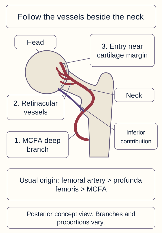
Start at the femoral artery in the groin. The profunda femoris, also called the deep femoral artery, usually gives rise to the medial circumflex femoral artery (MCFA). The MCFA can instead arise directly from the femoral artery. This is an origin variation, not a change in its identity or destination. An angiogram showing the actual origin should take precedence over the usual branching pattern. [1][15]
The MCFA passes between iliopsoas and pectineus toward the posterior hip. Its deep branch passes behind the obturator externus tendon, then toward the capsule, where branches travel along the posterosuperior femoral neck beneath synovial folds. These retinacular vessels enter near the head's cartilage margin. An inferior retinacular contribution is also important; the drawing simplifies a three-dimensional, variable vascular system. [1][2]
Trace the dominant route to its final entry points. Numbered labels distinguish the parent branch from terminal neck vessels. The simplified view does not show every muscle or vascular connection. [1][2] Bone Wizardry; Original construction. Open full-size image.
Trace it: follow the numbered route from the parent artery to a vessel beside the neck, then to the femoral head. Now place a fingertip across the final neck segment. Blood can still reach the thigh while its route into the head has been interrupted. That spatial separation explains why a normal distal pulse cannot exclude retinacular injury.
The MCFA and its retinacular branches are not interchangeable answer labels. When a question asks for the parent of the head-supplying branches, name the MCFA. When an operation describes torn vessels directly against the intracapsular neck, localize the lesion to the retinacular system. Profunda is farther upstream: damage there can matter, but a patent profunda does not prove that its distal branches remain connected. [1][13]
For a new patient, compare the structure actually injured with the tissue at risk. The lateral circumflex femoral artery has major anterior and lateral thigh and trochanteric territories, with variable contributions near the head. The obturator artery supplies a foveal route through the ligament of the femoral head. Gluteal arteries supply gluteal tissues and have periarticular connections. Do not erase those contributions to make MCFA dominance easier to remember. [4][15][16]
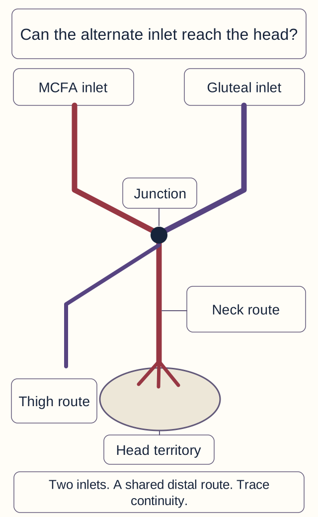
Can a bypass still reach its destination?
A collateral connection is another route into a vascular network. It helps only if the remaining route reaches the threatened tissue. A connection beside the hip cannot restore a vessel that has been torn farther along the neck. This is why preserved limb circulation and impaired head circulation can coexist. Conversely, interruption of one proximal artery does not prove that every downstream territory has lost all supply. [4]
A conceptual network, not a measurement of blood flow. A junction permits an alternate route only if the required downstream vessels remain connected. The diagram omits other surviving retinacular and foveal contributions. [4][16] Bone Wizardry; Original construction. Open full-size image.
The classic cruciate anastomosis lies posterior to the proximal femur near the lesser trochanter. It links the descending inferior gluteal branch, transverse branches of the medial and lateral circumflex femoral arteries, and the ascending branch of the first profunda perforator. This can connect internal iliac inflow with thigh vessels. The superior gluteal artery is associated with the more superior trochanteric network, rather than replacing the inferior gluteal member of the classic four-part cruciate arrangement. Real periarticular networks communicate and vary. [14]
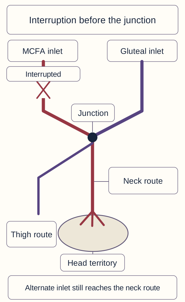
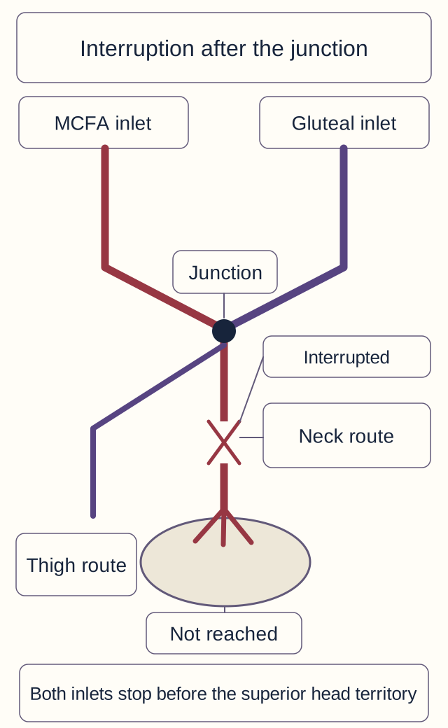
Test the two injury sites: on the diagram, predict whether the collateral route can reach the head when the interruption is before, or after, its junction with the head-supplying route. Choose either experiment below to display that interrupted network. These are conceptual connectivity tests, not measurements of human blood flow.
Test an interruption before the collateral junction
A conceptual network, not a measurement of blood flow. A junction permits an alternate route only if the required downstream vessels remain connected. The diagram omits other surviving retinacular and foveal contributions. [4][16] Bone Wizardry; Original construction. Open full-size image.
The alternate inlet reaches the downstream junction and an intact neck route. Some head perfusion is anatomically possible. Vessel size, pressure, and tissue injury still determine whether that supply is adequate.
Test an interruption after the collateral junction
A conceptual network, not a measurement of blood flow. A junction permits an alternate route only if the required downstream vessels remain connected. The diagram omits other surviving retinacular and foveal contributions. [4][16] Bone Wizardry; Original construction. Open full-size image.
The alternate inlet reaches the junction but stops at the interrupted neck route. Inflow upstream of a tear is not flow beyond it. Other surviving retinacular or foveal vessels can modify the result; this model does not predict inevitable whole-head necrosis.
The essential result remains the same with either experiment closed: collateral inflow and distal continuity are separate requirements. Injection studies have demonstrated inferior gluteal connections with the MCFA near obturator externus that can feed the epiphyseal route. They refute an absolute claim of no backup, but they do not establish dependable rescue after every fracture or dislocation. [4]
Age adds another boundary. During growth, the open physis separates much of the metaphyseal and epiphyseal circulation. Epiphyseal retinacular vessels approach around its periphery, rather than simply traversing the growth plate. With fusion, intraosseous connections can develop across the former physeal region; the adult physeal scar is not an impermeable wall. MCFA dominance persists despite those connections. [2][3]
The artery in the ligamentum teres has a variable, generally limited territory. Its importance does not follow a universal rule that it feeds toddlers and disappears in schoolchildren. Developmental specimen studies describe a changing contribution, including contribution later in childhood. In an adult, a patent foveal artery is not dependable replacement for the retinacular supply to the weight-bearing dome. Apply this to a new fracture by asking which routes remain connected, not whether a particular birthday has passed. [3][16]
Try it here · Checkpoint 1 of 3
Make your prediction before reading the choices. A first attempt is just a starting point.
Case 24
Show answer and explanations for case 24
A. Inferior gluteal trunk before the posterior connection (Why this does not fit)
A proximal inferior gluteal lesion could prevent that collateral source from entering the circumflex system. The injection already demonstrates filling through the posterior connection and into the neck. Use the last demonstrated patent segment to place an interruption downstream rather than upstream.
Reasoning steps for option A
For the inferior gluteal trunk before the posterior conn alternative, what would interruption before a collateral junction prevent?
A proximal inferior gluteal lesion could prevent that collateral source from entering the circumflex system.
For the inferior gluteal trunk before the posterior conn alternative, which lesion fits the last patent segment shown by the injection?
The injection already demonstrates filling through the posterior connection and into the neck.
For the inferior gluteal trunk before the posterior conn alternative, how does the position of an interruption limit collateral rescue?
Use the last demonstrated patent segment to place an interruption downstream rather than upstream.
B. Retinacular neck route beyond the posterior connection (Best answer)
Collateral contrast reaches the neck but fails to enter the superior epiphyseal vessels. An interruption beyond the junction explains preserved upstream and thigh perfusion with failure of the terminal head route. Collateral inflow cannot compensate for every lesion distal to its entry point.
Reasoning steps for option B
For the retinacular neck route beyond the posterior conn alternative, where does contrast stop after reaching the neck?
Collateral contrast reaches the neck but fails to enter the superior epiphyseal vessels.
For the retinacular neck route beyond the posterior conn alternative, which lesion fits the last patent segment shown by the injection?
An interruption beyond the junction explains preserved upstream and thigh perfusion with failure of the terminal head route.
For the retinacular neck route beyond the posterior conn alternative, how does the position of an interruption limit collateral rescue?
Collateral inflow cannot compensate for every lesion distal to its entry point.
C. Profunda origin before the circumflex branch separates (Why this does not fit)
A profunda origin lesion can impair its downstream circumflex supply in usual anatomy. The profunda is stated to be patent, and the demonstrated failure occurs after collateral contrast reaches the neck. Prefer the observed distal filling boundary over an assumed upstream occlusion.
Reasoning steps for option C
For the profunda origin before the circumflex branch sep alternative, what could a proximal profunda lesion affect in usual anatomy?
A profunda origin lesion can impair its downstream circumflex supply in usual anatomy.
For the profunda origin before the circumflex branch sep alternative, which lesion fits the last patent segment shown by the injection?
The profunda is stated to be patent, and the demonstrated failure occurs after collateral contrast reaches the neck.
For the profunda origin before the circumflex branch sep alternative, how does the position of an interruption limit collateral rescue?
Prefer the observed distal filling boundary over an assumed upstream occlusion.
D. Foveal branch before entry through the ligamentum teres (Why this does not fit)
Loss of the foveal route can reduce a limited contribution to the head. The tested route follows the posterior neck toward superior epiphyseal entries rather than the ligamentum teres. Interpret a perfusion study within the vascular route that was actually injected.
Reasoning steps for option D
For the foveal branch before entry through the ligamentu alternative, what contribution can be lost through a foveal lesion?
Loss of the foveal route can reduce a limited contribution to the head.
For the foveal branch before entry through the ligamentu alternative, which lesion fits the last patent segment shown by the injection?
The tested route follows the posterior neck toward superior epiphyseal entries rather than the ligamentum teres.
For the foveal branch before entry through the ligamentu alternative, how does the position of an interruption limit collateral rescue?
Interpret a perfusion study within the vascular route that was actually injected.
Takeaway: The location where contrast stops can identify a distal retinacular interruption despite patent collateral inflow.
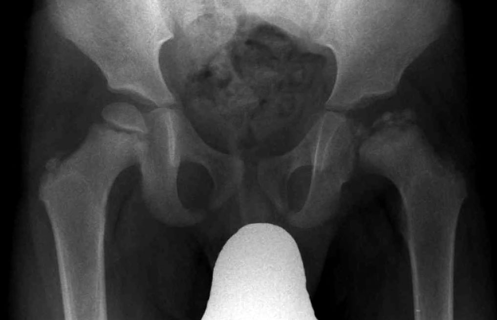
A seven-year-old has eight weeks of limping and activity-related knee pain. The knee examination is normal, but hip abduction and internal rotation are limited. The normal knee redirects examination toward the hip; it does not make the pain imaginary. In a younger child, this time course and a femoral epiphyseal abnormality raise concern for Legg-Calve-Perthes disease, an idiopathic osteonecrosis process affecting the capital epiphysis. This does not prove that the main MCFA trunk is occluded. [5]
Compare the two heads: identify the less rounded, structurally abnormal epiphysis in the radiograph, then describe its contour rather than naming an artery. The image is an independent teaching example, not a radiograph belonging to the synthetic child above. Loss of height, increased density, and fragmentation can appear as Perthes evolves. Early radiographs may be unrevealing; joint-space widening alone is nonspecific and does not establish the diagnosis. [5]
Perthes usually presents between ages four and ten. During necrosis and fragmentation, the head is mechanically vulnerable; subsequent reossification and remodeling do not guarantee restoration of a perfect sphere. Treatment seeks a mobile, contained head that can remodel within the acetabulum. Observation, activity modification, motion-preserving therapy, and selected operative containment depend on age, extent of involvement, and specialist assessment. Neither bracing nor surgery is required for every child. [5]
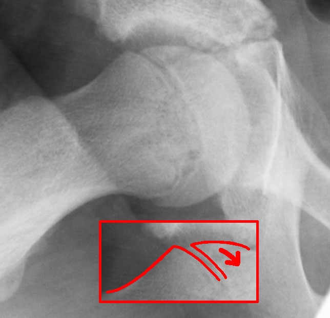
Now change the patient to a thirteen-year-old with a higher body mass, several weeks of knee pain, out-toeing, and obligatory external rotation when the hip flexes. This pattern raises concern for slipped capital femoral epiphysis (SCFE). The capital epiphysis is displaced posteriorly and inferiorly relative to the neck; equivalently, the neck is anterior and superior relative to the epiphysis. The reference structure determines the wording. [6][7]
Locate the physis and compare head-neck alignment with the epiphyseal abnormality in the Perthes example. This image does not prescribe a safe positioning technique for an acute patient. The red inset and arrow belong to the source image and were not added for this lesson. [6][7] Dr. Jochen Lengerke; CC0 1.0. Original source. No changes. Open full-size image.
Locate the failure: identify the head-neck relationship in the example, then contrast displacement at the physis with fragmentation within the epiphysis. A slip can stretch, kink, or disrupt adjacent retinacular vessels. The explanation is not that head-supplying arteries normally run straight through the physis. Stable SCFE means weight bearing is possible, with or without crutches; unstable SCFE means it is not possible even with crutches and carries greater osteonecrosis risk. Do not ask the patient to walk merely to demonstrate this classification. [6][7]
Suspected SCFE requires immediate protection from weight bearing and urgent orthopaedic assessment. For an acute or unstable suspected slip, obtain an AP pelvis and a cross-table lateral without forcing the hip into frog-leg positioning. Avoid forceful manipulation. Stable slips are commonly stabilized in situ; unstable slips need specialist planning rather than a one-procedure rule. A febrile, acutely unwell child with a painful hip also needs urgent assessment for infection, not reassurance from a vascular explanation. [6][7][17]
For transfer, distinguish pain-limited passive hip motion from true abductor weakness. A pelvic drop caused by weak abductors is a different finding from a child resisting painful abduction. The latter does not localize disease to the superior gluteal artery. A compensatory lumbar curve is similarly not a map of the artery supplying the femoral head.
Try it here · Checkpoint 2 of 3
Make your prediction before reading the choices. A first attempt is just a starting point.
Case 4
Show answer and explanations for case 4
A. AP pelvis and a forced frog-leg lateral view (Why this does not fit)
A frog-leg lateral can show chronic stable slips in patients who have continued to bear weight. The acute deterioration and inability to bear weight make forced positioning unsafe in this presentation. The appropriate view depends on clinical stability, not merely on which view can display a slip.
Reasoning steps for option A
For the ap pelvis and a forced frogleg lateral view alternative, in which clinical setting can a frog-leg view display a slip?
A frog-leg lateral can show chronic stable slips in patients who have continued to bear weight.
For the ap pelvis and a forced frogleg lateral view alternative, which imaging plan fits the acute unstable presentation without unnecessary loading or positioning?
The acute deterioration and inability to bear weight make forced positioning unsafe in this presentation.
For the ap pelvis and a forced frogleg lateral view alternative, what should be protected during evaluation of a suspected acute slip?
The appropriate view depends on clinical stability, not merely on which view can display a slip.
B. Standing AP pelvis and a weight-bearing comparison view (Why this does not fit)
Weight-bearing films can answer alignment questions in other hip conditions. This patient has an acutely unstable suspected slip and should remain protected from weight bearing. Do not reproduce a dangerous load just to obtain a comparison image.
Reasoning steps for option B
For the standing ap pelvis and a weightbearing compariso alternative, what can weight-bearing films assess in other hip disorders?
Weight-bearing films can answer alignment questions in other hip conditions.
For the standing ap pelvis and a weightbearing compariso alternative, which imaging plan fits the acute unstable presentation without unnecessary loading or positioning?
This patient has an acutely unstable suspected slip and should remain protected from weight bearing.
For the standing ap pelvis and a weightbearing compariso alternative, what should be protected during evaluation of a suspected acute slip?
Do not reproduce a dangerous load just to obtain a comparison image.
C. Isolated knee radiographs before hip imaging (Why this does not fit)
Referred knee pain can initially resemble a knee disorder. The normal knee, external hip rotation, and inability to bear weight localize the current problem to the hip. The painful location and the diseased joint need not be the same.
Reasoning steps for option C
For the isolated knee radiographs before hip imaging alternative, why might knee imaging initially seem relevant?
Referred knee pain can initially resemble a knee disorder.
For the isolated knee radiographs before hip imaging alternative, which imaging plan fits the acute unstable presentation without unnecessary loading or positioning?
The normal knee, external hip rotation, and inability to bear weight localize the current problem to the hip.
For the isolated knee radiographs before hip imaging alternative, what should be protected during evaluation of a suspected acute slip?
The painful location and the diseased joint need not be the same.
D. AP pelvis and a cross-table lateral view (Best answer)
The acute inability to bear weight supports an unstable suspected SCFE. AP and cross-table lateral imaging can assess the hip without forcing it into a frog-leg position that could worsen retinacular compromise. Protect the limb and obtain urgent orthopaedic assessment while imaging a suspected acute slip.
Reasoning steps for option D
For the ap pelvis and a crosstable lateral view alternative, what does inability to bear weight suggest about this slip?
The acute inability to bear weight supports an unstable suspected SCFE.
For the ap pelvis and a crosstable lateral view alternative, which imaging plan fits the acute unstable presentation without unnecessary loading or positioning?
AP and cross-table lateral imaging can assess the hip without forcing it into a frog-leg position that could worsen retinacular compromise.
For the ap pelvis and a crosstable lateral view alternative, what should be protected during evaluation of a suspected acute slip?
Protect the limb and obtain urgent orthopaedic assessment while imaging a suspected acute slip.
Takeaway: Acute or unstable suspected SCFE requires protection from weight bearing and imaging that avoids forced frog-leg positioning.
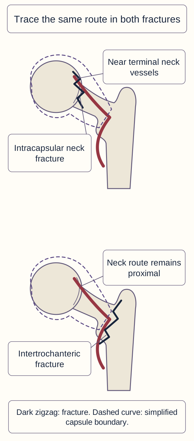
Two patients can both have a shortened, externally rotated leg after a fall yet have different vascular risks. Locate the fracture before inferring the complication. A displaced subcapital or transcervical fracture crosses the intracapsular neck beside the retinacular vessels. An intertrochanteric fracture lies farther from the head and is usually extracapsular. The capsule and blood route, not simply the label hip fracture, explain the difference. [9][13]
Locate each fracture relative to the terminal neck vessels. This drawing marks fracture level, not displacement severity. Intracapsular displacement increases vascular risk; an extracapsular injury can still be mechanically unstable. [1][9][13] Bone Wizardry; Original construction. Open full-size image.
Trace and compare: trace the same neck vessel on both drawings. Identify which fracture separates the vessel-bearing neck from the head. Displacement can disrupt that supply, increasing the risk of osteonecrosis. It also creates mechanical and biological difficulties for union. These are risks, not proof that the entire head has already died. An intertrochanteric fracture generally has less direct risk to head perfusion, but it is still a serious injury requiring treatment. [9][13]
For an older adult with a displaced femoral neck fracture, arthroplasty is generally favored over internal fixation. Replacing the head avoids depending on an injured head-neck unit to unite and remain viable. Total versus hemiarthroplasty depends on function, health, acetabular disease, and complication risk. The decision is not justified by a claim that all retinacular flow is certainly absent or that the ligamentum teres has ceased to exist. [8]
For a younger adult with a reconstructible displaced neck fracture, urgent specialist care generally prioritizes anatomic reduction and stable fixation to preserve the native head. Intertrochanteric fractures are generally fixed rather than treated as though their heads are nonviable. Nondisplaced neck fractures require their own assessment; do not apply the older-adult displaced-fracture recommendation indiscriminately. [8][9][13]
For transfer, consider a healed fracture followed months later by head collapse. Union describes continuity across the fracture. Osteonecrosis describes survival of the head's bone. A fracture can unite while a previously ischemic segment later collapses, so union and perfusion cannot substitute for one another in follow-up. [13]
Try it here · Checkpoint 3 of 3
Make your prediction before reading the choices. A first attempt is just a starting point.
Case 18
Show answer and explanations for case 18
A. Lower direct head vascular risk; unstable load transmission (Best answer)
The fracture lies distal to the principal retinacular neck route. The disrupted buttress and displacement under load establish mechanical instability despite the relatively lower direct threat to head perfusion. Vascular risk and fracture stability are separate dimensions of a proximal femoral injury.
Reasoning steps for option A
For the lower direct head vascular risk unstable load tr alternative, where is this fracture relative to the retinacular neck route?
The fracture lies distal to the principal retinacular neck route.
For the lower direct head vascular risk unstable load tr alternative, which pairing accounts for both the intact neck route and the disrupted load-bearing buttress?
The disrupted buttress and displacement under load establish mechanical instability despite the relatively lower direct threat to head perfusion.
For the lower direct head vascular risk unstable load tr alternative, why must perfusion risk and mechanical stability be judged separately?
Vascular risk and fracture stability are separate dimensions of a proximal femoral injury.
B. Lower direct head vascular risk; stable load transmission (Why this does not fit)
The trochanteric level is relatively less disruptive of head perfusion than a displaced intracapsular neck fracture. The disrupted buttress and displacement under load do not support mechanical stability. Preserved head circulation does not mean that a fracture can safely transmit load.
Reasoning steps for option B
For the lower direct head vascular risk stable load tran alternative, how does a trochanteric level compare with an intracapsular fracture?
The trochanteric level is relatively less disruptive of head perfusion than a displaced intracapsular neck fracture.
For the lower direct head vascular risk stable load tran alternative, which pairing accounts for both the intact neck route and the disrupted load-bearing buttress?
The disrupted buttress and displacement under load do not support mechanical stability.
For the lower direct head vascular risk stable load tran alternative, why must perfusion risk and mechanical stability be judged separately?
Preserved head circulation does not mean that a fracture can safely transmit load.
C. Higher direct head vascular risk; unstable load transmission (Why this does not fit)
Loss of the buttress and displacement under load do establish an unstable fracture. An intact neck beyond the trochanteric fracture is relatively less directly threatening to the retinacular head supply than the comparator. Separate the severity of mechanical instability from the location of the head-supplying vessels.
Reasoning steps for option C
For the higher direct head vascular risk unstable load t alternative, what do buttress loss and displacement establish?
Loss of the buttress and displacement under load do establish an unstable fracture.
For the higher direct head vascular risk unstable load t alternative, which pairing accounts for both the intact neck route and the disrupted load-bearing buttress?
An intact neck beyond the trochanteric fracture is relatively less directly threatening to the retinacular head supply than the comparator.
For the higher direct head vascular risk unstable load t alternative, why must perfusion risk and mechanical stability be judged separately?
Separate the severity of mechanical instability from the location of the head-supplying vessels.
D. Higher direct head vascular risk; stable load transmission (Why this does not fit)
A fracture near the hip can raise both biological and mechanical concerns. The supplied anatomy instead supports a relatively lower direct head vascular risk and an unstable buttress. Use the fracture level for the vascular comparison and the supporting structure for the stability assessment.
Reasoning steps for option D
For the higher direct head vascular risk stable load tra alternative, can a proximal femoral fracture raise more than one kind of concern?
A fracture near the hip can raise both biological and mechanical concerns.
For the higher direct head vascular risk stable load tra alternative, which pairing accounts for both the intact neck route and the disrupted load-bearing buttress?
The supplied anatomy instead supports a relatively lower direct head vascular risk and an unstable buttress.
For the higher direct head vascular risk stable load tra alternative, why must perfusion risk and mechanical stability be judged separately?
Use the fracture level for the vascular comparison and the supporting structure for the stability assessment.
Takeaway: A trochanteric fracture can have relatively lower direct head ischemia risk yet remain mechanically unstable and require fixation.
A dashboard impact can drive a flexed femur posteriorly out of the acetabulum. The typical posteriorly dislocated hip is flexed, adducted, internally rotated, and shortened. A fractured proximal femur more often rests externally rotated, but posture is not a substitute for imaging and a trauma assessment. Associated acetabular, femoral, and other injuries can change the reduction plan. [10]
Use the route drawing again: imagine the head leaving its socket while vessels remain attached along the neck. Tension, distortion, and tearing can impair retinacular circulation even though femoral and pedal arterial flow continues. Normal foot pulses answer a question about the limb's main circulation, not the survival of the femoral head. [1][10]
After initial trauma stabilization, dislocation requires urgent reduction by an appropriately equipped clinical team. Earlier reduction is associated with lower osteonecrosis risk. Six hours is a frequently reported observational comparison threshold, not permission to wait, and reduction before it does not guarantee a normal outcome. Record neurovascular findings before and after reduction, confirm alignment, assess associated fragments or fractures, and arrange follow-up. [10][11]
A separate injury can affect the sciatic nerve, particularly its common fibular division. New weakness of ankle dorsiflexion and eversion or dorsal-foot sensory loss requires neurologic localization, not an assumption that the MCFA directly powers those muscles. A warm foot does not exclude a nerve injury either. Superior gluteal dysfunction instead affects hip abductors; inferior gluteal dysfunction affects gluteus maximus. The superior gluteal vessels pass above piriformis, while the inferior gluteal vessels pass below it and participate in the periarticular circulation. [10][15][19]
In posterior acetabular exposure, such as a Kocher-Langenbeck approach, the deep MCFA and its relationship to obturator externus deserve attention. The tendon and surrounding soft tissues can protect the vessel. A vessel-preserving surgical dislocation is not equivalent to uncontrolled traumatic dislocation. Likewise, an anterior approach is not a blanket guarantee of head perfusion. The practical distinction is which tissues and vessels remain intact, not the approach name alone. Apply that distinction when evaluating an operative description with preserved versus detached short-rotator tissues. [1][2][4]
What fails before the joint surface collapses?
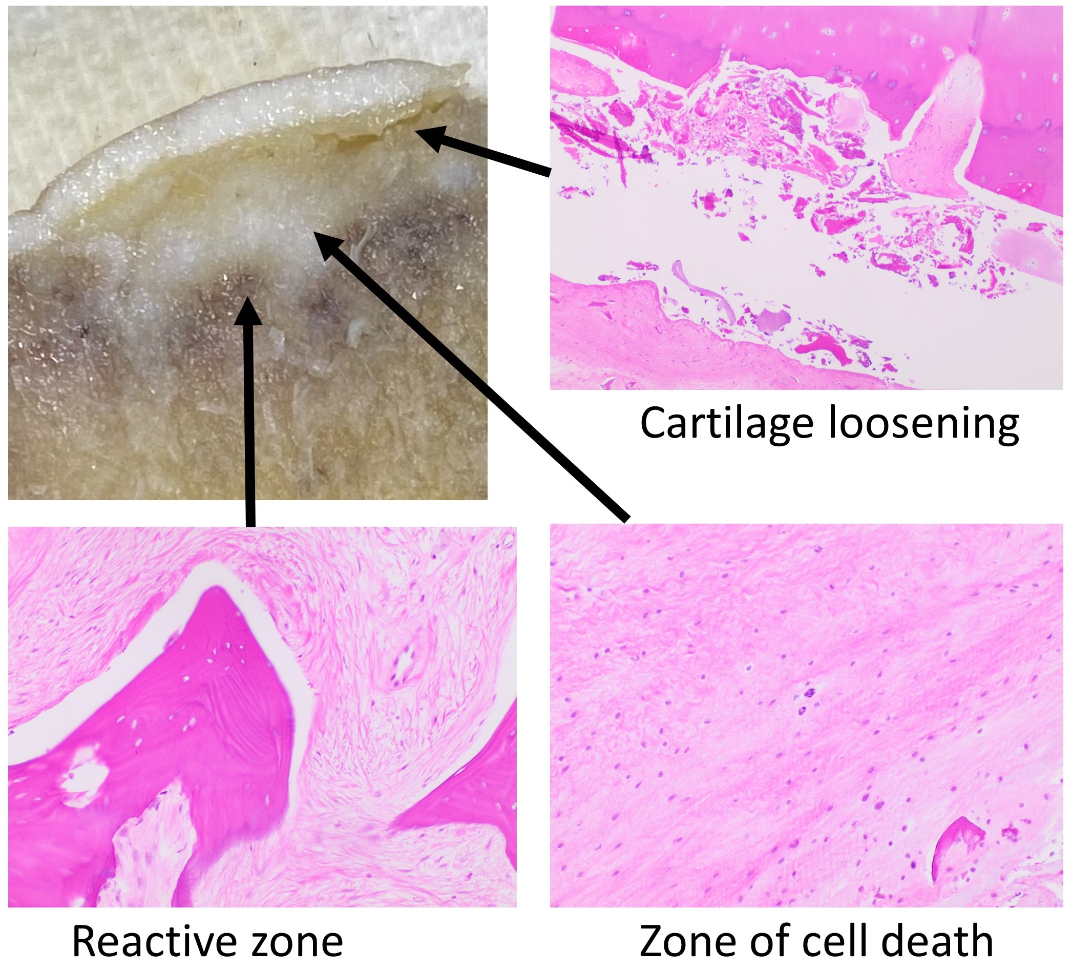
After ischemic injury, bone and marrow cells can die while the joint surface still appears relatively preserved. Histology may show empty osteocyte lacunae and necrotic marrow. Those findings describe osteonecrosis; they do not identify which named artery was injured. Interpretation depends on the overall tissue pattern, because an isolated empty lacuna is not enough to establish the diagnosis. [12][18]
Use the source labels to compare necrotic bone and adjacent reactive tissue. The original montage is retained; no additional labels or crops were added. [18] Mikael Häggström; CC0 1.0. Original source. No changes. Open full-size image.
Compare tissue compartments: look for the source image's examples of nonviable bone and marrow, then ask what happens when the bone beneath cartilage cannot support repeated loading. Ischemic bone initially retains structure, but repair and mechanical stress can lead to a subchondral fracture, a crescent-shaped radiographic line, and collapse of the articular contour. Secondary degenerative arthritis can follow. This is a progression, not a change that must be visible immediately after injury. [12][13]
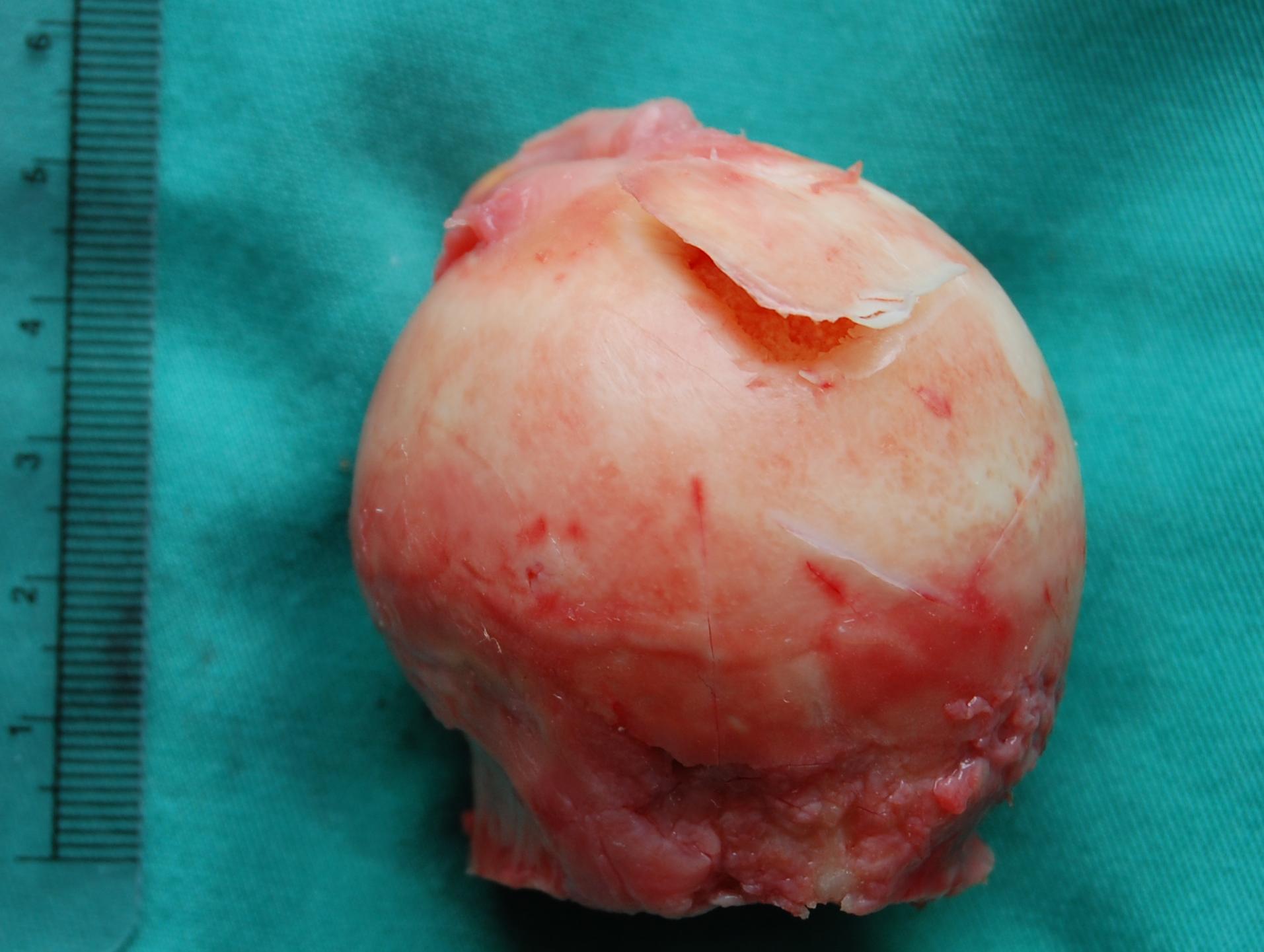
The cartilage flap illustrates a surface consequence of underlying bone injury. The source does not establish which artery initiated the lesion. [12] Steven Fruitsmaak; CC BY-SA 3.0. Original source. No changes. Open full-size image.
The gross specimen illustrates a cartilage flap over underlying osteonecrosis. Describe the damaged support and surface contour before assigning a cause. The specimen's source does not establish a traumatic MCFA lesion, and it does not belong to any of this lesson's synthetic cases. A recognizable endpoint should not be used as proof of an unseen initiating event.
Persistent groin pain with a relevant history deserves reassessment even when plain radiographs are normal. MRI can identify osteonecrosis before typical radiographic changes. Later sclerosis, a subchondral crescent, or collapse may be visible on radiographs. Select the investigation for the actual question: early tissue injury is different from a retained fracture fragment immediately after reduction. [12]
Now change the history: a patient has bilateral groin pain after prolonged systemic corticosteroid exposure and no hip trauma. Osteonecrosis is still possible without a mechanically transected MCFA. Other recognized associations include substantial alcohol exposure and sickle cell disease. Recognizing an ischemic endpoint does not establish a single universal arterial cause. Treatment depends on the cause, symptoms, stage, and extent of damage; early head-preserving options and treatment of advanced collapse are not interchangeable. [12]
Bring the lesson together with three questions: where is the lesion, which route still reaches the head, and what does the available evidence actually establish? Those questions distinguish a painful hip from an abductor deficit, a displaced neck fracture from an intertrochanteric fracture, and possible ischemic injury from proven structural collapse.
Independent clinical practice
Commit to an answer from the stem before inspecting the choices. Then compare the strongest alternative using a finding that was actually supplied. These original educational cases are separate from the guided tracing activities; difficulty and exam equivalence have not been measured.
Case 1
Show answer and explanations for case 1
A. Main femoral artery in the femoral triangle (Why this does not fit)
The femoral artery is upstream of several hip and limb territories. Normal limb perfusion with delayed segmental head collapse favors a more local injury than loss of the main femoral inflow. Distinguish regional branch injury from interruption of the limb's principal artery.
Reasoning steps for option A
For the main femoral artery in the femoral triangle alternative, which territories lie downstream of the main femoral artery?
The femoral artery is upstream of several hip and limb territories.
For the main femoral artery in the femoral triangle alternative, which vascular location best fits union with isolated head collapse and preserved limb flow?
Normal limb perfusion with delayed segmental head collapse favors a more local injury than loss of the main femoral inflow.
For the main femoral artery in the femoral triangle alternative, which endpoints must remain separate during fracture follow-up?
Distinguish regional branch injury from interruption of the limb's principal artery.
B. Retinacular vessels along the intracapsular neck (Best answer)
The displaced neck fracture lies beside vessels that enter the femoral head. Damage there can impair head viability despite fracture union and continued blood flow to the rest of the limb. A united neck and palpable pulses do not prove that the head's retinacular circulation survived.
Reasoning steps for option B
For the retinacular vessels along the intracapsular neck alternative, where is the displaced fracture relative to the head-supplying vessels?
The displaced neck fracture lies beside vessels that enter the femoral head.
For the retinacular vessels along the intracapsular neck alternative, which vascular location best fits union with isolated head collapse and preserved limb flow?
Damage there can impair head viability despite fracture union and continued blood flow to the rest of the limb.
For the retinacular vessels along the intracapsular neck alternative, which endpoints must remain separate during fracture follow-up?
A united neck and palpable pulses do not prove that the head's retinacular circulation survived.
C. First perforating branch within adductor magnus (Why this does not fit)
Profunda perforators supply the thigh and participate in collateral connections. A neck-level injury followed by isolated head collapse is better localized to the terminal neck vessels than to a thigh perforator. Follow the lesion to the threatened tissue rather than choosing any upstream contributor.
Reasoning steps for option C
For the first perforating branch within adductor magnus alternative, what is the role of a profunda perforator?
Profunda perforators supply the thigh and participate in collateral connections.
For the first perforating branch within adductor magnus alternative, which vascular location best fits union with isolated head collapse and preserved limb flow?
A neck-level injury followed by isolated head collapse is better localized to the terminal neck vessels than to a thigh perforator.
For the first perforating branch within adductor magnus alternative, which endpoints must remain separate during fracture follow-up?
Follow the lesion to the threatened tissue rather than choosing any upstream contributor.
D. Acetabular branch within the ligamentum teres (Why this does not fit)
The foveal artery does enter the femoral head and can be injured around the joint. Its limited territory is a weaker explanation than injury to the dominant retinacular route directly beside the displaced neck fracture. A vessel reaching the head is not necessarily its principal weight-bearing supply.
Reasoning steps for option D
For the acetabular branch within the ligamentum teres alternative, does a foveal vessel enter the head?
The foveal artery does enter the femoral head and can be injured around the joint.
For the acetabular branch within the ligamentum teres alternative, which vascular location best fits union with isolated head collapse and preserved limb flow?
Its limited territory is a weaker explanation than injury to the dominant retinacular route directly beside the displaced neck fracture.
For the acetabular branch within the ligamentum teres alternative, which endpoints must remain separate during fracture follow-up?
A vessel reaching the head is not necessarily its principal weight-bearing supply.
Takeaway: Fracture union and distal arterial flow can coexist with delayed osteonecrosis from retinacular injury.
A. Its retinacular inflow is supplied through the separate origin (Best answer)
The angiogram directly shows an origin outside the occluded profunda segment. Contrast reaches the neck vessels through that patent branch, so the usual profunda origin cannot be assumed in this patient. Use demonstrated branching anatomy to interpret the effect of a proximal occlusion.
Reasoning steps for option A
For the its retinacular inflow is supplied through the s alternative, where does this patient's circumflex vessel originate?
The angiogram directly shows an origin outside the occluded profunda segment.
For the its retinacular inflow is supplied through the s alternative, which route actually fills the neck vessels on this angiogram?
Contrast reaches the neck vessels through that patent branch, so the usual profunda origin cannot be assumed in this patient.
For the its retinacular inflow is supplied through the s alternative, how should demonstrated anatomy affect an occlusion assessment?
Use demonstrated branching anatomy to interpret the effect of a proximal occlusion.
B. Its retinacular inflow is lost because the profunda is occluded (Why this does not fit)
The MCFA commonly originates from profunda, making profunda disease relevant in usual anatomy. Here the MCFA has a separate patent origin and the downstream neck vessels actually fill. A population's usual branching pattern cannot override the patient's demonstrated vessels.
Reasoning steps for option B
For the its retinacular inflow is lost because the profu alternative, why would profunda disease matter in the usual branching pattern?
The MCFA commonly originates from profunda, making profunda disease relevant in usual anatomy.
For the its retinacular inflow is lost because the profu alternative, which route actually fills the neck vessels on this angiogram?
Here the MCFA has a separate patent origin and the downstream neck vessels actually fill.
For the its retinacular inflow is lost because the profu alternative, how should demonstrated anatomy affect an occlusion assessment?
A population's usual branching pattern cannot override the patient's demonstrated vessels.
C. Its epiphyseal inflow is supplied through the foveal artery alone (Why this does not fit)
The ligamentum teres can carry a separate foveal supply. The observed contrast route is through the circumflex and retinacular vessels, not an isolated foveal pathway. Identify the route that the study demonstrates rather than substituting another possible inlet.
Reasoning steps for option C
For the its epiphyseal inflow is supplied through the fo alternative, what other inlet can the ligament of the head provide?
The ligamentum teres can carry a separate foveal supply.
For the its epiphyseal inflow is supplied through the fo alternative, which route actually fills the neck vessels on this angiogram?
The observed contrast route is through the circumflex and retinacular vessels, not an isolated foveal pathway.
For the its epiphyseal inflow is supplied through the fo alternative, how should demonstrated anatomy affect an occlusion assessment?
Identify the route that the study demonstrates rather than substituting another possible inlet.
D. Its epiphyseal inflow depends on reversed inferior gluteal flow (Why this does not fit)
Inferior gluteal connections can provide collateral inflow to the head-supplying system. A patent direct origin and antegrade filling already account for this angiographic pattern without requiring gluteal reversal. Do not infer an unobserved collateral mechanism when a demonstrated route explains perfusion.
Reasoning steps for option D
For the its epiphyseal inflow depends on reversed inferi alternative, could a gluteal connection provide an alternate inlet?
Inferior gluteal connections can provide collateral inflow to the head-supplying system.
For the its epiphyseal inflow depends on reversed inferi alternative, which route actually fills the neck vessels on this angiogram?
A patent direct origin and antegrade filling already account for this angiographic pattern without requiring gluteal reversal.
For the its epiphyseal inflow depends on reversed inferi alternative, how should demonstrated anatomy affect an occlusion assessment?
Do not infer an unobserved collateral mechanism when a demonstrated route explains perfusion.
Takeaway: An origin variant can preserve a downstream route that an assumed standard branching pattern would place at risk.
A. Greater in B because the trochanters supply the entire head (Why this does not fit)
The trochanteric region has vascular connections and a fracture there can be serious. The head's dominant terminal vessels travel along the neck, which is directly separated in A rather than B. A vascular region near the head is not the same as the terminal route into the head.
Reasoning steps for option A
For the greater in b because the trochanters supply the alternative, why is the trochanteric region relevant to proximal femoral perfusion?
The trochanteric region has vascular connections and a fracture there can be serious.
For the greater in b because the trochanters supply the alternative, which supplied feature distinguishes the retinacular risk in A from B?
The head's dominant terminal vessels travel along the neck, which is directly separated in A rather than B.
For the greater in b because the trochanters supply the alternative, what information should drive a vascular comparison between fractures?
A vascular region near the head is not the same as the terminal route into the head.
B. Similar because both fractures shorten the injured limb (Why this does not fit)
Either fracture can produce shortening and a rotated resting posture. Those external signs do not identify whether the intracapsular retinacular route has been disrupted. Use fracture anatomy and displacement rather than a shared limb posture to compare vascular risk.
Reasoning steps for option B
For the similar because both fractures shorten the injur alternative, can both fracture levels cause a shortened limb?
Either fracture can produce shortening and a rotated resting posture.
For the similar because both fractures shorten the injur alternative, which supplied feature distinguishes the retinacular risk in A from B?
Those external signs do not identify whether the intracapsular retinacular route has been disrupted.
For the similar because both fractures shorten the injur alternative, what information should drive a vascular comparison between fractures?
Use fracture anatomy and displacement rather than a shared limb posture to compare vascular risk.
C. Greater in A because displacement threatens neck retinacular vessels (Best answer)
A has a displaced intracapsular neck fracture, whereas B has an intertrochanteric fracture. A's fracture directly separates the vessel-bearing neck from the head and is more likely to compromise retinacular flow. Higher risk is not certainty of osteonecrosis, and lower head risk does not make the other fracture harmless.
Reasoning steps for option C
For the greater in a because displacement threatens neck alternative, how do the two fracture levels differ?
A has a displaced intracapsular neck fracture, whereas B has an intertrochanteric fracture.
For the greater in a because displacement threatens neck alternative, which supplied feature distinguishes the retinacular risk in A from B?
A's fracture directly separates the vessel-bearing neck from the head and is more likely to compromise retinacular flow.
For the greater in a because displacement threatens neck alternative, what information should drive a vascular comparison between fractures?
Higher risk is not certainty of osteonecrosis, and lower head risk does not make the other fracture harmless.
D. Similar because both injuries interrupt the femoral artery (Why this does not fit)
Large-vessel injury could threaten perfusion after severe trauma. Neither description establishes a femoral trunk injury, and the relevant difference is the fracture's relationship to small neck vessels. Do not substitute an assumed trunk injury for the local anatomy actually described.
Reasoning steps for option D
For the similar because both injuries interrupt the femo alternative, could a major arterial injury threaten a limb after trauma?
Large-vessel injury could threaten perfusion after severe trauma.
For the similar because both injuries interrupt the femo alternative, which supplied feature distinguishes the retinacular risk in A from B?
Neither description establishes a femoral trunk injury, and the relevant difference is the fracture's relationship to small neck vessels.
For the similar because both injuries interrupt the femo alternative, what information should drive a vascular comparison between fractures?
Do not substitute an assumed trunk injury for the local anatomy actually described.
Takeaway: Displaced intracapsular neck fractures pose greater direct danger to retinacular flow than uncomplicated intertrochanteric fractures.
A. Shear failure through the proximal femoral physis (Why this does not fit)
SCFE can cause a limp, referred knee pain, and restricted hip rotation. The younger age and epiphyseal fragmentation without physeal displacement favor a different process. Distinguish injury within the epiphysis from separation across its growth plate.
Reasoning steps for option A
For the shear failure through the proximal femoral physi alternative, can a physeal slip cause referred knee pain?
SCFE can cause a limp, referred knee pain, and restricted hip rotation.
For the shear failure through the proximal femoral physi alternative, which process explains fragmentation within the epiphysis without displacement through the physis?
The younger age and epiphyseal fragmentation without physeal displacement favor a different process.
For the shear failure through the proximal femoral physi alternative, how should clinical localization be combined with structural imaging?
Distinguish injury within the epiphysis from separation across its growth plate.
B. Idiopathic osteonecrosis of the capital femoral epiphysis (Best answer)
The age, persistent atraumatic limp, and hip-limited motion fit a childhood hip disorder rather than an isolated knee lesion. Epiphyseal sclerosis and fragmentation without a slip support Perthes disease. This identifies a bone process but does not prove occlusion of the main MCFA trunk.
Reasoning steps for option B
For the idiopathic osteonecrosis of the capital femoral alternative, what clinical pattern is suggested by this child's age and course?
The age, persistent atraumatic limp, and hip-limited motion fit a childhood hip disorder rather than an isolated knee lesion.
For the idiopathic osteonecrosis of the capital femoral alternative, which process explains fragmentation within the epiphysis without displacement through the physis?
Epiphyseal sclerosis and fragmentation without a slip support Perthes disease.
For the idiopathic osteonecrosis of the capital femoral alternative, how should clinical localization be combined with structural imaging?
This identifies a bone process but does not prove occlusion of the main MCFA trunk.
C. Transient inflammation with preserved epiphyseal structure (Why this does not fit)
Transient hip synovitis can cause an atraumatic limp and restricted motion. Two months of symptoms with structural epiphyseal fragmentation is not explained by a transient effusion alone. A nonspecific symptom pattern must be interpreted alongside the demonstrated bone changes.
Reasoning steps for option C
For the transient inflammation with preserved epiphyseal alternative, can transient synovitis cause a limp?
Transient hip synovitis can cause an atraumatic limp and restricted motion.
For the transient inflammation with preserved epiphyseal alternative, which process explains fragmentation within the epiphysis without displacement through the physis?
Two months of symptoms with structural epiphyseal fragmentation is not explained by a transient effusion alone.
For the transient inflammation with preserved epiphyseal alternative, how should clinical localization be combined with structural imaging?
A nonspecific symptom pattern must be interpreted alongside the demonstrated bone changes.
D. Traumatic separation of the intracapsular femoral neck (Why this does not fit)
A neck fracture can impair head perfusion and eventually produce collapse. There is no traumatic event or described neck fracture, while the abnormality is centered within the epiphysis. Do not infer a fracture mechanism simply because osteonecrosis is possible after fractures.
Reasoning steps for option D
For the traumatic separation of the intracapsular femora alternative, can a neck fracture lead to head ischemia?
A neck fracture can impair head perfusion and eventually produce collapse.
For the traumatic separation of the intracapsular femora alternative, which process explains fragmentation within the epiphysis without displacement through the physis?
There is no traumatic event or described neck fracture, while the abnormality is centered within the epiphysis.
For the traumatic separation of the intracapsular femora alternative, how should clinical localization be combined with structural imaging?
Do not infer a fracture mechanism simply because osteonecrosis is possible after fractures.
E. Developmental loss of acetabular coverage (Why this does not fit)
Poor acetabular coverage can cause abnormal loading and gait. The supplied abnormality is epiphyseal sclerosis and fragmentation rather than an uncovered or displaced head. Localize the radiographic abnormality before selecting a developmental diagnosis.
Reasoning steps for option E
For the developmental loss of acetabular coverage alternative, can poor acetabular coverage affect gait?
Poor acetabular coverage can cause abnormal loading and gait.
For the developmental loss of acetabular coverage alternative, which process explains fragmentation within the epiphysis without displacement through the physis?
The supplied abnormality is epiphyseal sclerosis and fragmentation rather than an uncovered or displaced head.
For the developmental loss of acetabular coverage alternative, how should clinical localization be combined with structural imaging?
Localize the radiographic abnormality before selecting a developmental diagnosis.
Takeaway: Perthes is an idiopathic epiphyseal osteonecrosis process, not proof of a physeal slip or a named arterial trunk occlusion.
A. A has greater osteonecrosis risk because symptoms lasted longer (Why this does not fit)
A longer symptom duration can accompany a chronic slip and still requires treatment. The supplied distinction that most directly identifies the high-risk unstable category is B's inability to bear weight even with crutches. Duration alone does not replace the clinical stability classification.
Reasoning steps for option A
For the a has greater osteonecrosis risk because symptom alternative, does a chronic symptom history still require treatment?
A longer symptom duration can accompany a chronic slip and still requires treatment.
For the a has greater osteonecrosis risk because symptom alternative, does a matching angle erase the difference in clinical stability?
The supplied distinction that most directly identifies the high-risk unstable category is B's inability to bear weight even with crutches.
For the a has greater osteonecrosis risk because symptom alternative, which assessment defines stable versus unstable SCFE?
Duration alone does not replace the clinical stability classification.
B. Their osteonecrosis risks are identical because the angles match (Why this does not fit)
Radiographic severity helps characterize a slip. Similar angles do not erase the difference between a clinically stable and unstable presentation. Structural magnitude and clinical stability are related but noninterchangeable assessments.
Reasoning steps for option B
For the their osteonecrosis risks are identical because alternative, what does a slip angle help characterize?
Radiographic severity helps characterize a slip.
For the their osteonecrosis risks are identical because alternative, does a matching angle erase the difference in clinical stability?
Similar angles do not erase the difference between a clinically stable and unstable presentation.
For the their osteonecrosis risks are identical because alternative, which assessment defines stable versus unstable SCFE?
Structural magnitude and clinical stability are related but noninterchangeable assessments.
C. B has greater osteonecrosis risk because the slip is unstable (Best answer)
A could bear weight with crutches, whereas B could not. B therefore meets the clinical unstable category, which is associated with greater risk to femoral head viability. Determine stability from the history without provoking additional weight bearing.
Reasoning steps for option C
For the b has greater osteonecrosis risk because the sli alternative, how do the two patients differ in weight-bearing ability?
A could bear weight with crutches, whereas B could not.
For the b has greater osteonecrosis risk because the sli alternative, does a matching angle erase the difference in clinical stability?
B therefore meets the clinical unstable category, which is associated with greater risk to femoral head viability.
For the b has greater osteonecrosis risk because the sli alternative, which assessment defines stable versus unstable SCFE?
Determine stability from the history without provoking additional weight bearing.
D. Their risks are similar because both are unstable once crutches are needed (Why this does not fit)
Use of crutches indicates that symptoms affect walking. A can bear weight with crutches and is classified as stable; B cannot, which is the unstable category. Stability classification depends on the ability to bear weight with or without support, not simply on whether support is used.
Reasoning steps for option D
For the their risks are similar because both are unstabl alternative, what does the use of crutches tell you by itself?
Use of crutches indicates that symptoms affect walking.
For the their risks are similar because both are unstabl alternative, does a matching angle erase the difference in clinical stability?
A can bear weight with crutches and is classified as stable; B cannot, which is the unstable category.
For the their risks are similar because both are unstabl alternative, which assessment defines stable versus unstable SCFE?
Stability classification depends on the ability to bear weight with or without support, not simply on whether support is used.
Takeaway: Weight-bearing ability classifies SCFE stability; a similar radiographic angle does not imply a similar vascular risk.
A. The open physis separates circulations that can communicate after fusion (Best answer)
The growing specimen has separate metaphyseal and peripheral epiphyseal filling patterns. The mature specimen demonstrates connections after fusion, contradicting an impermeable adult physeal barrier. Developmental vascular boundaries should not be treated as permanent adult anatomy.
Reasoning steps for option A
For the the open physis separates circulations that can alternative, how do the two injections fill the growing specimen?
The growing specimen has separate metaphyseal and peripheral epiphyseal filling patterns.
For the the open physis separates circulations that can alternative, which explanation accounts for peripheral epiphyseal filling and later intraosseous communication?
The mature specimen demonstrates connections after fusion, contradicting an impermeable adult physeal barrier.
For the the open physis separates circulations that can alternative, what developmental boundary should not be carried unchanged into adult anatomy?
Developmental vascular boundaries should not be treated as permanent adult anatomy.
B. The physeal scar permanently blocks metaphyseal access to the head (Why this does not fit)
An open growth plate can separate much of the developing circulation. Communicating channels in the mature specimen directly contradict a permanently blocking scar. Distinguish the open physis from the fused adult region.
Reasoning steps for option B
For the the physeal scar permanently blocks metaphyseal alternative, can an open growth plate separate developing vascular compartments?
An open growth plate can separate much of the developing circulation.
For the the physeal scar permanently blocks metaphyseal alternative, which explanation accounts for peripheral epiphyseal filling and later intraosseous communication?
Communicating channels in the mature specimen directly contradict a permanently blocking scar.
For the the physeal scar permanently blocks metaphyseal alternative, what developmental boundary should not be carried unchanged into adult anatomy?
Distinguish the open physis from the fused adult region.
C. Epiphyseal retinacular vessels pass directly through the open physis (Why this does not fit)
Retinacular vessels provide an important epiphyseal route during growth. The demonstrated peripheral filling with absent transphyseal metaphyseal filling supports an approach around the physeal boundary. A vessel supplying an epiphysis need not cross through its growth plate.
Reasoning steps for option C
For the epiphyseal retinacular vessels pass directly thr alternative, do retinacular vessels contribute to the developing epiphysis?
Retinacular vessels provide an important epiphyseal route during growth.
For the epiphyseal retinacular vessels pass directly thr alternative, which explanation accounts for peripheral epiphyseal filling and later intraosseous communication?
The demonstrated peripheral filling with absent transphyseal metaphyseal filling supports an approach around the physeal boundary.
For the epiphyseal retinacular vessels pass directly thr alternative, what developmental boundary should not be carried unchanged into adult anatomy?
A vessel supplying an epiphysis need not cross through its growth plate.
D. The ligamentum teres replaces retinacular supply when fusion occurs (Why this does not fit)
The ligamentum teres can contribute to head circulation. Neither the peripheral injection result nor the mature intraosseous connections demonstrate replacement of the retinacular system. Additional connections do not imply disappearance of the dominant head-supplying route.
Reasoning steps for option D
For the the ligamentum teres replaces retinacular supply alternative, can the ligament of the head contribute blood?
The ligamentum teres can contribute to head circulation.
For the the ligamentum teres replaces retinacular supply alternative, which explanation accounts for peripheral epiphyseal filling and later intraosseous communication?
Neither the peripheral injection result nor the mature intraosseous connections demonstrate replacement of the retinacular system.
For the the ligamentum teres replaces retinacular supply alternative, what developmental boundary should not be carried unchanged into adult anatomy?
Additional connections do not imply disappearance of the dominant head-supplying route.
Takeaway: The open physis and the fused adult physeal region have different vascular implications.
A. The foveal study proves adequate perfusion of the entire head (Why this does not fit)
The observed foveal flow establishes that one arterial route is patent. The same study documents reduced perfusion in the superior weight-bearing segment, so whole-head adequacy cannot be inferred. Patency of one inlet does not establish adequate delivery to every tissue territory.
Reasoning steps for option A
For the the foveal study proves adequate perfusion of th alternative, what does observed foveal flow establish?
The observed foveal flow establishes that one arterial route is patent.
For the the foveal study proves adequate perfusion of th alternative, how can foveal filling coexist with poor perfusion of the superior segment?
The same study documents reduced perfusion in the superior weight-bearing segment, so whole-head adequacy cannot be inferred.
For the the foveal study proves adequate perfusion of th alternative, what is the difference between a patent inlet and adequate territorial coverage?
Patency of one inlet does not establish adequate delivery to every tissue territory.
B. The superior segment is supplied primarily by the femoral shaft (Why this does not fit)
Intraosseous communications can exist after physeal fusion. Those connections do not make the shaft the principal substitute for an injured retinacular supply to the superior head. Adult vascular communication does not eliminate the importance of the neck vessels.
Reasoning steps for option B
For the the superior segment is supplied primarily by th alternative, can adult intraosseous communications exist?
Intraosseous communications can exist after physeal fusion.
For the the superior segment is supplied primarily by th alternative, how can foveal filling coexist with poor perfusion of the superior segment?
Those connections do not make the shaft the principal substitute for an injured retinacular supply to the superior head.
For the the superior segment is supplied primarily by th alternative, what is the difference between a patent inlet and adequate territorial coverage?
Adult vascular communication does not eliminate the importance of the neck vessels.
C. The foveal artery should be absent after childhood (Why this does not fit)
The contribution of the ligamentum teres changes during development. Visible adult flow directly disproves absence, and the clinical issue is its limited territory rather than its existence. Avoid universal age-based disappearance rules for a variable vessel.
Reasoning steps for option C
For the the foveal artery should be absent after childho alternative, does the foveal contribution change during development?
The contribution of the ligamentum teres changes during development.
For the the foveal artery should be absent after childho alternative, how can foveal filling coexist with poor perfusion of the superior segment?
Visible adult flow directly disproves absence, and the clinical issue is its limited territory rather than its existence.
For the the foveal artery should be absent after childho alternative, what is the difference between a patent inlet and adequate territorial coverage?
Avoid universal age-based disappearance rules for a variable vessel.
D. Foveal flow does not replace the injured retinacular territory (Best answer)
The study demonstrates a patent foveal route and a separate poorly perfused superior segment. Limited foveal supply can coexist with inadequate retinacular delivery to the weight-bearing head after a neck injury. Interpret both the continuity and the territory of each surviving route.
Reasoning steps for option D
For the foveal flow does not replace the injured retinac alternative, which two perfusion territories are demonstrated?
The study demonstrates a patent foveal route and a separate poorly perfused superior segment.
For the foveal flow does not replace the injured retinac alternative, how can foveal filling coexist with poor perfusion of the superior segment?
Limited foveal supply can coexist with inadequate retinacular delivery to the weight-bearing head after a neck injury.
For the foveal flow does not replace the injured retinac alternative, what is the difference between a patent inlet and adequate territorial coverage?
Interpret both the continuity and the territory of each surviving route.
Takeaway: A patent foveal artery cannot be assumed to rescue a poorly perfused weight-bearing head.
A. Loss of the dominant superior retinacular route (Why this does not fit)
Surgery around the head can threaten its blood supply. The operation specifically preserves the neck retinacula and superior head perfusion is demonstrated afterward. Localize the divided structure rather than treating every intra-articular intervention as the same vascular injury.
Reasoning steps for option A
For the loss of the dominant superior retinacular route alternative, can an operation near the head endanger its circulation?
Surgery around the head can threaten its blood supply.
For the loss of the dominant superior retinacular route alternative, which route is divided and which route is explicitly preserved here?
The operation specifically preserves the neck retinacula and superior head perfusion is demonstrated afterward.
For the loss of the dominant superior retinacular route alternative, how should the operative site determine the predicted vascular consequence?
Localize the divided structure rather than treating every intra-articular intervention as the same vascular injury.
B. Foveal loss with preserved retinacular inflow (Best answer)
The divided ligament carries a foveal arterial contribution rather than the dominant neck route. Preserved retinacular tissue and superior perfusion are therefore compatible with loss of that more limited contribution. A foveal lesion and a retinacular lesion have different implications for the weight-bearing head.
Reasoning steps for option B
For the foveal loss with preserved retinacular inflow alternative, which route is carried in the divided ligament?
The divided ligament carries a foveal arterial contribution rather than the dominant neck route.
For the foveal loss with preserved retinacular inflow alternative, which route is divided and which route is explicitly preserved here?
Preserved retinacular tissue and superior perfusion are therefore compatible with loss of that more limited contribution.
For the foveal loss with preserved retinacular inflow alternative, how should the operative site determine the predicted vascular consequence?
A foveal lesion and a retinacular lesion have different implications for the weight-bearing head.
C. Loss of the main profunda supply to the thigh (Why this does not fit)
Profunda is an important upstream source of circumflex arteries. Dividing an intra-articular ligament does not describe injury to the profunda trunk in the proximal thigh. Separate a terminal intra-articular route from its region's major extracapsular vessels.
Reasoning steps for option C
For the loss of the main profunda supply to the thigh alternative, where is profunda in the arterial hierarchy?
Profunda is an important upstream source of circumflex arteries.
For the loss of the main profunda supply to the thigh alternative, which route is divided and which route is explicitly preserved here?
Dividing an intra-articular ligament does not describe injury to the profunda trunk in the proximal thigh.
For the loss of the main profunda supply to the thigh alternative, how should the operative site determine the predicted vascular consequence?
Separate a terminal intra-articular route from its region's major extracapsular vessels.
D. Loss of the gluteal connection at the posterior neck (Why this does not fit)
The inferior gluteal artery can join the MCFA near the posterior hip. The described intervention is in the ligament of the head, not the posterior extracapsular anastomotic region. Use the physical location of the intervention to predict which connection is interrupted.
Reasoning steps for option D
For the loss of the gluteal connection at the posterior alternative, where can an inferior gluteal connection join the circumflex system?
The inferior gluteal artery can join the MCFA near the posterior hip.
For the loss of the gluteal connection at the posterior alternative, which route is divided and which route is explicitly preserved here?
The described intervention is in the ligament of the head, not the posterior extracapsular anastomotic region.
For the loss of the gluteal connection at the posterior alternative, how should the operative site determine the predicted vascular consequence?
Use the physical location of the intervention to predict which connection is interrupted.
Takeaway: The ligamentum teres and the neck retinacula carry different vascular contributions.
A. Trochanteric anastomosis centered above the femoral neck (Why this does not fit)
The trochanteric network also connects vessels around the proximal femur. The lesser-trochanter level and first perforating contribution identify the lower classic cruciate arrangement rather than the more superior network. Use the level and branch pattern together when naming a periarticular connection.
Reasoning steps for option A
For the trochanteric anastomosis centered above the femo alternative, does a trochanteric network also connect proximal femoral vessels?
The trochanteric network also connects vessels around the proximal femur.
For the trochanteric anastomosis centered above the femo alternative, which network matches the lesser-trochanter level and the observed contributors?
The lesser-trochanter level and first perforating contribution identify the lower classic cruciate arrangement rather than the more superior network.
For the trochanteric anastomosis centered above the femo alternative, which anatomical details identify a collateral network beyond its general function?
Use the level and branch pattern together when naming a periarticular connection.
B. Foveal pathway through the ligamentum teres (Why this does not fit)
A foveal vessel can connect pelvic arterial supply to a small head territory. The observed route is posterior in the proximal thigh and joins circumflex and perforating branches, not the ligament of the head. An intra-articular head inlet is not the same pathway as a thigh collateral network.
Reasoning steps for option B
For the foveal pathway through the ligamentum teres alternative, where does the foveal route enter the head?
A foveal vessel can connect pelvic arterial supply to a small head territory.
For the foveal pathway through the ligamentum teres alternative, which network matches the lesser-trochanter level and the observed contributors?
The observed route is posterior in the proximal thigh and joins circumflex and perforating branches, not the ligament of the head.
For the foveal pathway through the ligamentum teres alternative, which anatomical details identify a collateral network beyond its general function?
An intra-articular head inlet is not the same pathway as a thigh collateral network.
C. Cruciate anastomosis involving the inferior gluteal artery (Best answer)
The descending vessel below piriformis is consistent with inferior gluteal inflow. Its connection with transverse circumflex branches and the first perforator at this level identifies the cruciate anastomosis. Such a route can support thigh inflow without proving intact terminal vessels inside the hip capsule.
Reasoning steps for option C
For the cruciate anastomosis involving the inferior glut alternative, what does a descending vessel below piriformis suggest?
The descending vessel below piriformis is consistent with inferior gluteal inflow.
For the cruciate anastomosis involving the inferior glut alternative, which network matches the lesser-trochanter level and the observed contributors?
Its connection with transverse circumflex branches and the first perforator at this level identifies the cruciate anastomosis.
For the cruciate anastomosis involving the inferior glut alternative, which anatomical details identify a collateral network beyond its general function?
Such a route can support thigh inflow without proving intact terminal vessels inside the hip capsule.
D. Genicular anastomosis surrounding the distal femur (Why this does not fit)
Genicular networks can contribute to collateral circulation farther down the limb. The stated level is near the lesser trochanter, with gluteal and proximal circumflex contributors rather than knee branches. A collateral function alone does not identify the anatomical site of the network.
Reasoning steps for option D
For the genicular anastomosis surrounding the distal fem alternative, can genicular vessels form collateral pathways?
Genicular networks can contribute to collateral circulation farther down the limb.
For the genicular anastomosis surrounding the distal fem alternative, which network matches the lesser-trochanter level and the observed contributors?
The stated level is near the lesser trochanter, with gluteal and proximal circumflex contributors rather than knee branches.
For the genicular anastomosis surrounding the distal fem alternative, which anatomical details identify a collateral network beyond its general function?
A collateral function alone does not identify the anatomical site of the network.
Takeaway: Identify the cruciate connection from both its proximal posterior thigh location and its contributing branches.
A. Inferior gluteal flow reaching the epiphyseal vessels (Best answer)
The injected material is observed passing from the inferior gluteal artery into the epiphyseal route. That finding demonstrates an anatomical alternative inlet beyond the clamped proximal segment. An anatomical connection does not by itself prove adequate perfusion after every living patient's traumatic injury.
Reasoning steps for option A
For the inferior gluteal flow reaching the epiphyseal ve alternative, what route did the injection actually demonstrate?
The injected material is observed passing from the inferior gluteal artery into the epiphyseal route.
For the inferior gluteal flow reaching the epiphyseal ve alternative, what does filling beyond this clamp establish, and what does it leave unmeasured?
That finding demonstrates an anatomical alternative inlet beyond the clamped proximal segment.
For the inferior gluteal flow reaching the epiphyseal ve alternative, how should an anatomical observation be distinguished from a clinical guarantee?
An anatomical connection does not by itself prove adequate perfusion after every living patient's traumatic injury.
B. The inferior gluteal artery supplies the head in every hip (Why this does not fit)
The experiment demonstrates that a gluteal route can contribute to head supply. A single supplied specimen and a successful injection do not establish a universal dominant source. Do not convert the presence of a connection into a population-wide guarantee.
Reasoning steps for option B
For the the inferior gluteal artery supplies the head in alternative, does the experiment show a gluteal contribution?
The experiment demonstrates that a gluteal route can contribute to head supply.
For the the inferior gluteal artery supplies the head in alternative, what does filling beyond this clamp establish, and what does it leave unmeasured?
A single supplied specimen and a successful injection do not establish a universal dominant source.
For the the inferior gluteal artery supplies the head in alternative, how should an anatomical observation be distinguished from a clinical guarantee?
Do not convert the presence of a connection into a population-wide guarantee.
C. The foveal artery is the necessary intermediate connection (Why this does not fit)
The ligamentum teres provides another possible inlet to the head. The described injection is traced beside obturator externus into epiphyseal vessels rather than through the fovea. Use the observed path instead of assuming that every pelvic contribution uses the ligamentum teres.
Reasoning steps for option C
For the the foveal artery is the necessary intermediate alternative, could the ligament of the head be a separate inlet?
The ligamentum teres provides another possible inlet to the head.
For the the foveal artery is the necessary intermediate alternative, what does filling beyond this clamp establish, and what does it leave unmeasured?
The described injection is traced beside obturator externus into epiphyseal vessels rather than through the fovea.
For the the foveal artery is the necessary intermediate alternative, how should an anatomical observation be distinguished from a clinical guarantee?
Use the observed path instead of assuming that every pelvic contribution uses the ligamentum teres.
D. The clamp had to be distal to the epiphyseal vessel origins (Why this does not fit)
A clamp's position changes which routes can fill downstream vessels. The stem places it proximal to the posterior connection, which permits the observed alternative inlet to bypass it. Interpret collateral filling relative to the lesion and junction positions.
Reasoning steps for option D
For the the clamp had to be distal to the epiphyseal ves alternative, why does clamp position matter?
A clamp's position changes which routes can fill downstream vessels.
For the the clamp had to be distal to the epiphyseal ves alternative, what does filling beyond this clamp establish, and what does it leave unmeasured?
The stem places it proximal to the posterior connection, which permits the observed alternative inlet to bypass it.
For the the clamp had to be distal to the epiphyseal ves alternative, how should an anatomical observation be distinguished from a clinical guarantee?
Interpret collateral filling relative to the lesion and junction positions.
Takeaway: Demonstrated gluteal connections refute an absolute no-backup model without guaranteeing rescue after trauma.
A. Primary superior gluteal denervation explains the examination (Why this does not fit)
Superior gluteal nerve dysfunction can cause abductor weakness and an abnormal gait. Preserved abductor force with pain during passive motion and an epiphyseal lesion favors a painful hip rather than primary denervation. Differentiate true weakness from pain-limited joint motion.
Reasoning steps for option A
For the primary superior gluteal denervation explains th alternative, what kind of deficit can superior gluteal dysfunction cause?
Superior gluteal nerve dysfunction can cause abductor weakness and an abnormal gait.
For the primary superior gluteal denervation explains th alternative, do retained abductor force and painful passive motion fit a primary muscle or nerve deficit?
Preserved abductor force with pain during passive motion and an epiphyseal lesion favors a painful hip rather than primary denervation.
For the primary superior gluteal denervation explains th alternative, what should be separated when pain limits an action?
Differentiate true weakness from pain-limited joint motion.
B. Primary inferior gluteal ischemia explains the examination (Why this does not fit)
The inferior gluteal artery supplies important gluteal tissue and can contribute around the hip. The examination identifies painful passive hip motion, not a demonstrated gluteus maximus ischemic deficit. A painful action is not a direct map of the artery supplying the muscle that performs it.
Reasoning steps for option B
For the primary inferior gluteal ischemia explains the e alternative, what tissues does inferior gluteal perfusion serve?
The inferior gluteal artery supplies important gluteal tissue and can contribute around the hip.
For the primary inferior gluteal ischemia explains the e alternative, do retained abductor force and painful passive motion fit a primary muscle or nerve deficit?
The examination identifies painful passive hip motion, not a demonstrated gluteus maximus ischemic deficit.
For the primary inferior gluteal ischemia explains the e alternative, what should be separated when pain limits an action?
A painful action is not a direct map of the artery supplying the muscle that performs it.
C. The lumbar curve identifies the head-supplying artery (Why this does not fit)
Painful gait can be accompanied by compensatory spinal findings. The curve does not establish a named vascular lesion, while the local hip examination and imaging identify a joint-centered problem. Use compensatory findings as context rather than an arterial localization test.
Reasoning steps for option C
For the the lumbar curve identifies the headsupplying ar alternative, can a painful gait produce compensatory spinal findings?
Painful gait can be accompanied by compensatory spinal findings.
For the the lumbar curve identifies the headsupplying ar alternative, do retained abductor force and painful passive motion fit a primary muscle or nerve deficit?
The curve does not establish a named vascular lesion, while the local hip examination and imaging identify a joint-centered problem.
For the the lumbar curve identifies the headsupplying ar alternative, what should be separated when pain limits an action?
Use compensatory findings as context rather than an arterial localization test.
D. Pain-limited hip motion with preserved abductor strength (Best answer)
Passive hip motion reproduces pain and an epiphyseal abnormality is present. Preserved abductor force makes a primary motor deficit less explanatory than pain-limited motion from the hip. A normal knee with knee pain should prompt examination of the hip rather than a muscle-artery shortcut.
Reasoning steps for option D
For the painlimited hip motion with preserved abductor s alternative, what happens during passive motion of this hip?
Passive hip motion reproduces pain and an epiphyseal abnormality is present.
For the painlimited hip motion with preserved abductor s alternative, do retained abductor force and painful passive motion fit a primary muscle or nerve deficit?
Preserved abductor force makes a primary motor deficit less explanatory than pain-limited motion from the hip.
For the painlimited hip motion with preserved abductor s alternative, what should be separated when pain limits an action?
A normal knee with knee pain should prompt examination of the hip rather than a muscle-artery shortcut.
Takeaway: Painful passive abduction is not equivalent to abductor weakness or superior gluteal vascular injury.
A. Observe the limb until pedal perfusion begins to decline (Why this does not fit)
Pedal perfusion is an important part of the trauma examination. Normal foot circulation does not exclude ongoing compromise of the separate retinacular head supply. A local hip vascular threat should not be monitored solely through a distal pulse.
Reasoning steps for option A
For the observe the limb until pedal perfusion begins to alternative, why are pedal pulses included in the trauma examination?
Pedal perfusion is an important part of the trauma examination.
For the observe the limb until pedal perfusion begins to alternative, which option addresses the established dislocation without waiting for a different endpoint?
Normal foot circulation does not exclude ongoing compromise of the separate retinacular head supply.
For the observe the limb until pedal perfusion begins to alternative, which findings should and should not delay urgent reduction?
A local hip vascular threat should not be monitored solely through a distal pulse.
B. Proceed with urgent reduction and repeat neurovascular assessment (Best answer)
The posture and radiograph identify a posterior hip dislocation after initial stabilization. Urgent reduction addresses the distorted joint and retinacular tissues, while repeat examination detects associated neurovascular changes. A normal distal pulse is not a reason to delay reduction of a traumatic hip dislocation.
Reasoning steps for option B
For the proceed with urgent reduction and repeat neurova alternative, what injury has been established after initial stabilization?
The posture and radiograph identify a posterior hip dislocation after initial stabilization.
For the proceed with urgent reduction and repeat neurova alternative, which option addresses the established dislocation without waiting for a different endpoint?
Urgent reduction addresses the distorted joint and retinacular tissues, while repeat examination detects associated neurovascular changes.
For the proceed with urgent reduction and repeat neurova alternative, which findings should and should not delay urgent reduction?
A normal distal pulse is not a reason to delay reduction of a traumatic hip dislocation.
C. Wait until six hours after injury before attempting reduction (Why this does not fit)
Six hours is a commonly reported threshold in outcome comparisons. It is not a treatment target or an allowed delay when reduction can be undertaken safely now. An observational cutoff should not be converted into permission to prolong ischemic risk.
Reasoning steps for option C
For the wait until six hours after injury before attempt alternative, what does a six-hour threshold represent in outcome reports?
Six hours is a commonly reported threshold in outcome comparisons.
For the wait until six hours after injury before attempt alternative, which option addresses the established dislocation without waiting for a different endpoint?
It is not a treatment target or an allowed delay when reduction can be undertaken safely now.
For the wait until six hours after injury before attempt alternative, which findings should and should not delay urgent reduction?
An observational cutoff should not be converted into permission to prolong ischemic risk.
D. Obtain elective perfusion MRI before treating the dislocation (Why this does not fit)
MRI can answer selected questions about femoral head tissue injury. It should not defer treatment of the established acute dislocation when the prepared team can proceed. Choose tests and treatment in an order that addresses the immediate emergency.
Reasoning steps for option D
For the obtain elective perfusion mri before treating th alternative, what can MRI assess in selected head injuries?
MRI can answer selected questions about femoral head tissue injury.
For the obtain elective perfusion mri before treating th alternative, which option addresses the established dislocation without waiting for a different endpoint?
It should not defer treatment of the established acute dislocation when the prepared team can proceed.
For the obtain elective perfusion mri before treating th alternative, which findings should and should not delay urgent reduction?
Choose tests and treatment in an order that addresses the immediate emergency.
Takeaway: Treat an established traumatic hip dislocation urgently even when foot pulses remain normal.
A. Femoral nerve as it crosses beneath the inguinal ligament (Why this does not fit)
Femoral nerve injury can follow trauma around the anterior hip and produce lower-limb weakness. It would emphasize knee extension and the patellar reflex rather than the supplied dorsiflexion and eversion pattern. Use the affected actions and reflexes to localize a traumatic nerve deficit.
Reasoning steps for option A
For the femoral nerve as it crosses beneath the inguinal alternative, why might an anterior hip nerve enter the differential?
Femoral nerve injury can follow trauma around the anterior hip and produce lower-limb weakness.
For the femoral nerve as it crosses beneath the inguinal alternative, which neural structure matches the distal pattern and the hip-level injury?
It would emphasize knee extension and the patellar reflex rather than the supplied dorsiflexion and eversion pattern.
For the femoral nerve as it crosses beneath the inguinal alternative, how should motor, sensory, and anatomical findings be combined?
Use the affected actions and reflexes to localize a traumatic nerve deficit.
B. Superior gluteal nerve as it passes above piriformis (Why this does not fit)
Superior gluteal injury can impair hip abduction after pelvic or hip trauma. The observed deficit is in distal dorsiflexors and evertors, not a primarily weak hip abductor group. A hip-level injury can affect a nerve serving distant muscles, so identify the actual motor distribution.
Reasoning steps for option B
For the superior gluteal nerve as it passes above pirifo alternative, what muscle function is associated with the superior gluteal nerve?
Superior gluteal injury can impair hip abduction after pelvic or hip trauma.
For the superior gluteal nerve as it passes above pirifo alternative, which neural structure matches the distal pattern and the hip-level injury?
The observed deficit is in distal dorsiflexors and evertors, not a primarily weak hip abductor group.
For the superior gluteal nerve as it passes above pirifo alternative, how should motor, sensory, and anatomical findings be combined?
A hip-level injury can affect a nerve serving distant muscles, so identify the actual motor distribution.
C. Common fibular division of the sciatic nerve at the hip (Best answer)
The distal motor and sensory findings emphasize the common fibular distribution. The posterior hip injury and absence of a fibular-head lesion favor that division of the sciatic nerve at the hip. Normal pedal pulses do not exclude a clinically important nerve injury.
Reasoning steps for option C
For the common fibular division of the sciatic nerve at alternative, which distal functional distribution is most affected?
The distal motor and sensory findings emphasize the common fibular distribution.
For the common fibular division of the sciatic nerve at alternative, which neural structure matches the distal pattern and the hip-level injury?
The posterior hip injury and absence of a fibular-head lesion favor that division of the sciatic nerve at the hip.
For the common fibular division of the sciatic nerve at alternative, how should motor, sensory, and anatomical findings be combined?
Normal pedal pulses do not exclude a clinically important nerve injury.
D. Tibial sciatic division as it passes behind the hip (Why this does not fit)
The tibial division is part of the sciatic nerve and can be injured in hip trauma. Relatively preserved plantar flexion with weak dorsiflexion and eversion points more strongly to the other division. Distinguish sciatic divisions using the supplied distal muscle pattern.
Reasoning steps for option D
For the tibial sciatic division as it passes behind the alternative, could a hip injury affect the tibial division?
The tibial division is part of the sciatic nerve and can be injured in hip trauma.
For the tibial sciatic division as it passes behind the alternative, which neural structure matches the distal pattern and the hip-level injury?
Relatively preserved plantar flexion with weak dorsiflexion and eversion points more strongly to the other division.
For the tibial sciatic division as it passes behind the alternative, how should motor, sensory, and anatomical findings be combined?
Distinguish sciatic divisions using the supplied distal muscle pattern.
E. Obturator nerve as it enters the obturator canal (Why this does not fit)
Obturator injury can occur around the pelvis and affect medial thigh function. It does not explain the described distal foot motor and sensory distribution. Nerve proximity to the hip is insufficient without a matching functional pattern.
Reasoning steps for option E
For the obturator nerve as it enters the obturator canal alternative, can the obturator nerve be injured around the pelvis?
Obturator injury can occur around the pelvis and affect medial thigh function.
For the obturator nerve as it enters the obturator canal alternative, which neural structure matches the distal pattern and the hip-level injury?
It does not explain the described distal foot motor and sensory distribution.
For the obturator nerve as it enters the obturator canal alternative, how should motor, sensory, and anatomical findings be combined?
Nerve proximity to the hip is insufficient without a matching functional pattern.
Takeaway: Posterior hip trauma with dorsiflexion and eversion weakness suggests a common fibular-predominant sciatic injury.
A. Concentric joint reduction with femoral head viability still uncertain (Best answer)
The postreduction radiographs demonstrate a concentrically located femoral head. They establish alignment rather than survival of all tissue injured during the dislocation; distal pulses assess a separate circulation. Early reduction reduces risk but does not replace follow-up for delayed head complications.
Reasoning steps for option A
For the concentric joint reduction with femoral head via alternative, what do the postreduction radiographs demonstrate?
The postreduction radiographs demonstrate a concentrically located femoral head.
For the concentric joint reduction with femoral head via alternative, which endpoint was measured directly, and which outcome remains uncertain?
They establish alignment rather than survival of all tissue injured during the dislocation; distal pulses assess a separate circulation.
For the concentric joint reduction with femoral head via alternative, why should mechanical alignment and tissue viability be assessed separately?
Early reduction reduces risk but does not replace follow-up for delayed head complications.
B. Confirmed retinacular reperfusion with delayed collapse excluded (Why this does not fit)
Restoring the hip relationship can relieve distortion of retinacular tissues. Plain radiographic congruence does not directly establish patency of those vessels. Do not treat an alignment study as a direct vascular measurement.
Reasoning steps for option B
For the confirmed retinacular reperfusion with delayed c alternative, how can restoring the hip relationship affect vessel distortion?
Restoring the hip relationship can relieve distortion of retinacular tissues.
For the confirmed retinacular reperfusion with delayed c alternative, which endpoint was measured directly, and which outcome remains uncertain?
Plain radiographic congruence does not directly establish patency of those vessels.
For the confirmed retinacular reperfusion with delayed c alternative, why should mechanical alignment and tissue viability be assessed separately?
Do not treat an alignment study as a direct vascular measurement.
C. Confirmed femoral head viability despite uncertain reduction (Why this does not fit)
Head viability is an important biological goal of prompt reduction. The films already establish congruence but do not establish viability, reversing the two endpoints. Identify which outcome the available study actually measures.
Reasoning steps for option C
For the confirmed femoral head viability despite uncerta alternative, what biological outcome is a goal of prompt reduction?
Head viability is an important biological goal of prompt reduction.
For the confirmed femoral head viability despite uncerta alternative, which endpoint was measured directly, and which outcome remains uncertain?
The films already establish congruence but do not establish viability, reversing the two endpoints.
For the confirmed femoral head viability despite uncerta alternative, why should mechanical alignment and tissue viability be assessed separately?
Identify which outcome the available study actually measures.
D. Confirmed foveal perfusion despite uncertain joint alignment (Why this does not fit)
The foveal route can contribute to femoral head perfusion. No foveal perfusion study was performed, whereas concentric alignment is explicitly shown. Do not infer a specific patent arterial inlet from a postreduction alignment image.
Reasoning steps for option D
For the confirmed foveal perfusion despite uncertain joi alternative, can the foveal route contribute to head perfusion?
The foveal route can contribute to femoral head perfusion.
For the confirmed foveal perfusion despite uncertain joi alternative, which endpoint was measured directly, and which outcome remains uncertain?
No foveal perfusion study was performed, whereas concentric alignment is explicitly shown.
For the confirmed foveal perfusion despite uncertain joi alternative, why should mechanical alignment and tissue viability be assessed separately?
Do not infer a specific patent arterial inlet from a postreduction alignment image.
Takeaway: Prompt reduction is important but does not eliminate the need to assess later femoral head complications.
A. Protected weight bearing without operative stabilization (Why this does not fit)
Nonoperative care can be appropriate for selected patients whose condition or goals make surgery unsuitable. This patient is an operative candidate with a completely displaced neck fracture and prior independent ambulation. Base nonoperative selection on the whole clinical situation, not age alone.
Reasoning steps for option A
For the protected weight bearing without operative stabi alternative, when can nonoperative fracture care be appropriate?
Nonoperative care can be appropriate for selected patients whose condition or goals make surgery unsuitable.
For the protected weight bearing without operative stabi alternative, which strategy best fits this older operative candidate with complete displacement and osteoporosis?
This patient is an operative candidate with a completely displaced neck fracture and prior independent ambulation.
For the protected weight bearing without operative stabi alternative, how should patient context alter the reconstruction objective?
Base nonoperative selection on the whole clinical situation, not age alone.
B. Valgus osteotomy with fixation of the native neck (Why this does not fit)
Valgus osteotomy can help selected head-preserving reconstructions, particularly in salvage of a neck nonunion. This is an acute completely displaced fracture in an older osteoporotic operative candidate, for whom arthroplasty is generally preferred. Distinguish a selected salvage procedure from the recommended primary treatment category.
Reasoning steps for option B
For the valgus osteotomy with fixation of the native nec alternative, in which selected setting might a valgus osteotomy help?
Valgus osteotomy can help selected head-preserving reconstructions, particularly in salvage of a neck nonunion.
For the valgus osteotomy with fixation of the native nec alternative, which strategy best fits this older operative candidate with complete displacement and osteoporosis?
This is an acute completely displaced fracture in an older osteoporotic operative candidate, for whom arthroplasty is generally preferred.
For the valgus osteotomy with fixation of the native nec alternative, how should patient context alter the reconstruction objective?
Distinguish a selected salvage procedure from the recommended primary treatment category.
C. Primary hip arthroplasty after perioperative assessment (Best answer)
The patient is an older adult with a displaced intracapsular neck fracture and poor bone quality. Arthroplasty avoids reliance on the fractured head-neck unit to unite and remain viable, addressing important failure risks of fixation in this setting. Choose the type of arthroplasty separately according to function, health, and joint disease.
Reasoning steps for option C
For the primary hip arthroplasty after perioperative ass alternative, what features of this head-neck unit make failure a concern?
The patient is an older adult with a displaced intracapsular neck fracture and poor bone quality.
For the primary hip arthroplasty after perioperative ass alternative, which strategy best fits this older operative candidate with complete displacement and osteoporosis?
Arthroplasty avoids reliance on the fractured head-neck unit to unite and remain viable, addressing important failure risks of fixation in this setting.
For the primary hip arthroplasty after perioperative ass alternative, how should patient context alter the reconstruction objective?
Choose the type of arthroplasty separately according to function, health, and joint disease.
D. Reduction and screw fixation as a head-preserving strategy (Why this does not fit)
Preserving the native head with reduction and fixation is particularly important in younger patients. In this older patient with complete displacement and osteoporosis, guideline-supported care generally favors arthroplasty over fixation. An appropriate strategy for a young adult is not automatically the preferred strategy for an older adult.
Reasoning steps for option D
For the reduction and screw fixation as a headpreserving alternative, for whom is native-head preservation especially important?
Preserving the native head with reduction and fixation is particularly important in younger patients.
For the reduction and screw fixation as a headpreserving alternative, which strategy best fits this older operative candidate with complete displacement and osteoporosis?
In this older patient with complete displacement and osteoporosis, guideline-supported care generally favors arthroplasty over fixation.
For the reduction and screw fixation as a headpreserving alternative, how should patient context alter the reconstruction objective?
An appropriate strategy for a young adult is not automatically the preferred strategy for an older adult.
Takeaway: Older-adult displaced femoral neck fractures generally favor arthroplasty without requiring proof that the whole head is already necrotic.
A. Replace the head with a hemiarthroplasty as the initial objective (Why this does not fit)
Hemiarthroplasty can avoid dependence on a damaged head-neck unit, especially in older patients. This young patient has reconstructible anatomy and no established joint destruction, favoring an attempt to preserve the native head. Do not apply the usual older-adult replacement strategy solely because a neck fracture is displaced.
Reasoning steps for option A
For the replace the head with a hemiarthroplasty as the alternative, what dependence can hemiarthroplasty avoid?
Hemiarthroplasty can avoid dependence on a damaged head-neck unit, especially in older patients.
For the replace the head with a hemiarthroplasty as the alternative, which objective best fits a healthy young adult without established joint destruction?
This young patient has reconstructible anatomy and no established joint destruction, favoring an attempt to preserve the native head.
For the replace the head with a hemiarthroplasty as the alternative, what does a head-preserving objective promise, and what does it not promise?
Do not apply the usual older-adult replacement strategy solely because a neck fracture is displaced.
B. Replace both joint surfaces with a total hip arthroplasty (Why this does not fit)
Total arthroplasty can address established destructive joint disease and selected fracture presentations. The supplied young age, reconstructibility, and absence of preexisting arthritic destruction favor head-preserving reconstruction here. The decision to replace a young native joint requires more than recognition of vascular risk.
Reasoning steps for option B
For the replace both joint surfaces with a total hip art alternative, which joint conditions can total arthroplasty address?
Total arthroplasty can address established destructive joint disease and selected fracture presentations.
For the replace both joint surfaces with a total hip art alternative, which objective best fits a healthy young adult without established joint destruction?
The supplied young age, reconstructibility, and absence of preexisting arthritic destruction favor head-preserving reconstruction here.
For the replace both joint surfaces with a total hip art alternative, what does a head-preserving objective promise, and what does it not promise?
The decision to replace a young native joint requires more than recognition of vascular risk.
C. Fuse the hip to eliminate motion across the fracture (Why this does not fit)
Arthrodesis can be considered in unusual salvage settings. A reconstructible acute neck fracture in this young patient is not a salvage situation requiring loss of joint motion. Choose an objective proportionate to the demonstrated injury and available reconstruction.
Reasoning steps for option C
For the fuse the hip to eliminate motion across the frac alternative, in what broad setting might arthrodesis be considered?
Arthrodesis can be considered in unusual salvage settings.
For the fuse the hip to eliminate motion across the frac alternative, which objective best fits a healthy young adult without established joint destruction?
A reconstructible acute neck fracture in this young patient is not a salvage situation requiring loss of joint motion.
For the fuse the hip to eliminate motion across the frac alternative, what does a head-preserving objective promise, and what does it not promise?
Choose an objective proportionate to the demonstrated injury and available reconstruction.
D. Restore alignment and provide stable head-preserving fixation (Best answer)
The fracture is displaced but the young patient's head and neck remain reconstructible. Urgent specialist reduction and stable fixation aim to preserve the native head while limiting avoidable mechanical and vascular injury. Preservation is an objective, not a promise that osteonecrosis or nonunion cannot later develop.
Reasoning steps for option D
For the restore alignment and provide stable headpreserv alternative, what remains reconstructible in this patient?
The fracture is displaced but the young patient's head and neck remain reconstructible.
For the restore alignment and provide stable headpreserv alternative, which objective best fits a healthy young adult without established joint destruction?
Urgent specialist reduction and stable fixation aim to preserve the native head while limiting avoidable mechanical and vascular injury.
For the restore alignment and provide stable headpreserv alternative, what does a head-preserving objective promise, and what does it not promise?
Preservation is an objective, not a promise that osteonecrosis or nonunion cannot later develop.
Takeaway: A reconstructible displaced neck fracture in a young adult generally calls for a head-preserving operative strategy.
A. Repeat plain radiographs after additional symptoms develop (Why this does not fit)
Later radiographs can demonstrate sclerosis, a crescent, or collapse. The clinical concern is a symptomatic early lesion that may already be detectable before those structural changes appear. Do not require visible collapse before investigating a plausible early osteonecrosis process.
Reasoning steps for option A
For the repeat plain radiographs after additional sympto alternative, what can later plain radiographs show?
Later radiographs can demonstrate sclerosis, a crescent, or collapse.
For the repeat plain radiographs after additional sympto alternative, which test addresses suspected early intraosseous injury after normal radiographs?
The clinical concern is a symptomatic early lesion that may already be detectable before those structural changes appear.
For the repeat plain radiographs after additional sympto alternative, how should the suspected tissue problem determine the imaging choice?
Do not require visible collapse before investigating a plausible early osteonecrosis process.
B. MRI of the affected hip without routine contrast (Best answer)
The history and persistent hip symptoms justify assessment for post-traumatic head injury despite normal radiographs. MRI can demonstrate early osteonecrosis before plain films show characteristic changes. Match the imaging method to early bone viability rather than waiting for a late contour abnormality.
Reasoning steps for option B
For the mri of the affected hip without routine contrast alternative, why does this history justify further head assessment?
The history and persistent hip symptoms justify assessment for post-traumatic head injury despite normal radiographs.
For the mri of the affected hip without routine contrast alternative, which test addresses suspected early intraosseous injury after normal radiographs?
MRI can demonstrate early osteonecrosis before plain films show characteristic changes.
For the mri of the affected hip without routine contrast alternative, how should the suspected tissue problem determine the imaging choice?
Match the imaging method to early bone viability rather than waiting for a late contour abnormality.
C. Ultrasound of the hip joint for an effusion (Why this does not fit)
Ultrasound can demonstrate joint fluid and guide selected procedures. It does not provide the same assessment of an early intraosseous osteonecrosis pattern that is the stated concern. A test for fluid is not a substitute for a test of the suspected bone abnormality.
Reasoning steps for option C
For the ultrasound of the hip joint for an effusion alternative, what can hip ultrasound demonstrate?
Ultrasound can demonstrate joint fluid and guide selected procedures.
For the ultrasound of the hip joint for an effusion alternative, which test addresses suspected early intraosseous injury after normal radiographs?
It does not provide the same assessment of an early intraosseous osteonecrosis pattern that is the stated concern.
For the ultrasound of the hip joint for an effusion alternative, how should the suspected tissue problem determine the imaging choice?
A test for fluid is not a substitute for a test of the suspected bone abnormality.
D. CT angiography of the lower-limb arterial runoff (Why this does not fit)
Arterial imaging can assess major-vessel injury or occlusive disease. The present question concerns early femoral head tissue injury rather than a described defect in limb arterial runoff. A vascular cause does not mean that large-vessel angiography is the best test for its bone consequence.
Reasoning steps for option D
For the ct angiography of the lowerlimb arterial runoff alternative, what can arterial runoff imaging assess?
Arterial imaging can assess major-vessel injury or occlusive disease.
For the ct angiography of the lowerlimb arterial runoff alternative, which test addresses suspected early intraosseous injury after normal radiographs?
The present question concerns early femoral head tissue injury rather than a described defect in limb arterial runoff.
For the ct angiography of the lowerlimb arterial runoff alternative, how should the suspected tissue problem determine the imaging choice?
A vascular cause does not mean that large-vessel angiography is the best test for its bone consequence.
E. CT of the pelvis to define an acute displaced fragment (Why this does not fit)
CT is valuable for characterizing fractures or retained fragments after trauma. Eight months later the stated concern is early osteonecrosis with unrevealing radiographs, not an acute fragment requiring definition. Choose between structural fragment assessment and early marrow assessment using the clinical question.
Reasoning steps for option E
For the ct of the pelvis to define an acute displaced fr alternative, which trauma questions are well suited to CT?
CT is valuable for characterizing fractures or retained fragments after trauma.
For the ct of the pelvis to define an acute displaced fr alternative, which test addresses suspected early intraosseous injury after normal radiographs?
Eight months later the stated concern is early osteonecrosis with unrevealing radiographs, not an acute fragment requiring definition.
For the ct of the pelvis to define an acute displaced fr alternative, how should the suspected tissue problem determine the imaging choice?
Choose between structural fragment assessment and early marrow assessment using the clinical question.
Takeaway: Normal radiographs do not exclude early osteonecrosis; persistent appropriate suspicion favors MRI.
A. Uniform joint-space loss before any subchondral injury (Why this does not fit)
Articular cartilage loss can eventually narrow joint space in degenerative arthritis. The supplied early injury is in the bone supporting relatively preserved cartilage, so uniform cartilage loss need not be the first structural event. Identify which tissue compartment fails before predicting the next visible change.
Reasoning steps for option A
For the uniform jointspace loss before any subchondral i alternative, what tissue change eventually narrows joint space in arthritis?
Articular cartilage loss can eventually narrow joint space in degenerative arthritis.
For the uniform jointspace loss before any subchondral i alternative, which structural failure follows from nonviable subchondral support?
The supplied early injury is in the bone supporting relatively preserved cartilage, so uniform cartilage loss need not be the first structural event.
For the uniform jointspace loss before any subchondral i alternative, why does the injured tissue compartment determine the expected progression?
Identify which tissue compartment fails before predicting the next visible change.
B. Epiphyseal displacement through an open growth plate (Why this does not fit)
A physeal slip can disturb head-neck alignment and threaten blood supply in adolescents. The described finding is a subchondral necrotic segment, not a supplied open physis with a shear injury. Do not replace an ischemic bone process with a growth-plate diagnosis without the necessary anatomy.
Reasoning steps for option B
For the epiphyseal displacement through an open growth p alternative, what kind of process produces a physeal slip?
A physeal slip can disturb head-neck alignment and threaten blood supply in adolescents.
For the epiphyseal displacement through an open growth p alternative, which structural failure follows from nonviable subchondral support?
The described finding is a subchondral necrotic segment, not a supplied open physis with a shear injury.
For the epiphyseal displacement through an open growth p alternative, why does the injured tissue compartment determine the expected progression?
Do not replace an ischemic bone process with a growth-plate diagnosis without the necessary anatomy.
C. Subchondral fracture followed by articular contour collapse (Best answer)
Nonviable subchondral bone supports the initially retained cartilage surface. As the affected region weakens during loading and repair, a subchondral fracture and collapse can develop before advanced secondary arthritis. An apparently retained cartilage surface does not establish that its underlying support remains viable.
Reasoning steps for option C
For the subchondral fracture followed by articular conto alternative, which tissue supports the initially retained cartilage?
Nonviable subchondral bone supports the initially retained cartilage surface.
For the subchondral fracture followed by articular conto alternative, which structural failure follows from nonviable subchondral support?
As the affected region weakens during loading and repair, a subchondral fracture and collapse can develop before advanced secondary arthritis.
For the subchondral fracture followed by articular conto alternative, why does the injured tissue compartment determine the expected progression?
An apparently retained cartilage surface does not establish that its underlying support remains viable.
D. Periosteal bridging callus across the joint surface (Why this does not fit)
Bridging callus is associated with repair across an appropriate fracture interface. The lesion is a necrotic subchondral segment beneath articular cartilage, not a described periosteal shaft-fracture repair site. The location and tissue environment determine the expected structural consequence.
Reasoning steps for option D
For the periosteal bridging callus across the joint surf alternative, where does bridging callus usually address an injury?
Bridging callus is associated with repair across an appropriate fracture interface.
For the periosteal bridging callus across the joint surf alternative, which structural failure follows from nonviable subchondral support?
The lesion is a necrotic subchondral segment beneath articular cartilage, not a described periosteal shaft-fracture repair site.
For the periosteal bridging callus across the joint surf alternative, why does the injured tissue compartment determine the expected progression?
The location and tissue environment determine the expected structural consequence.
Takeaway: Death of subchondral bone can precede fracture and collapse beneath an initially preserved cartilage surface.
A. Improve foveal artery flow by stretching the ligament of the head (Why this does not fit)
The ligament of the head carries a vascular contribution. The demonstrated benefit is improved acetabular coverage of a vulnerable head, not measured restoration of foveal arterial flow. Do not substitute an unmeasured vascular effect for the structural purpose of containment.
Reasoning steps for option A
For the improve foveal artery flow by stretching the lig alternative, does the ligament of the head carry a vascular contribution?
The ligament of the head carries a vascular contribution.
For the improve foveal artery flow by stretching the lig alternative, which objective explains the benefit of improved acetabular coverage in this child?
The demonstrated benefit is improved acetabular coverage of a vulnerable head, not measured restoration of foveal arterial flow.
For the improve foveal artery flow by stretching the lig alternative, how does containment differ from reopening an artery or fixing a slip?
Do not substitute an unmeasured vascular effect for the structural purpose of containment.
B. Keep the vulnerable head covered while repair and remodeling occur (Best answer)
The epiphysis is structurally vulnerable during fragmentation after osteonecrosis. Improved coverage within the acetabulum can help preserve its shape during repair, making the demonstrated congruence relevant to containment. Treatment selection still depends on age, extent of involvement, and motion rather than a universal requirement for surgery.
Reasoning steps for option B
For the keep the vulnerable head covered while repair an alternative, why is a fragmenting epiphysis mechanically vulnerable?
The epiphysis is structurally vulnerable during fragmentation after osteonecrosis.
For the keep the vulnerable head covered while repair an alternative, which objective explains the benefit of improved acetabular coverage in this child?
Improved coverage within the acetabulum can help preserve its shape during repair, making the demonstrated congruence relevant to containment.
For the keep the vulnerable head covered while repair an alternative, how does containment differ from reopening an artery or fixing a slip?
Treatment selection still depends on age, extent of involvement, and motion rather than a universal requirement for surgery.
C. Prevent posterior displacement by permanently fusing the physis (Why this does not fit)
Stabilizing a failing physis is central to treatment of a slip. This child's problem is fragmentation within the epiphysis, not posterior physeal displacement requiring stabilization. Distinguish containment of an ischemic head from fixation of a physeal slip.
Reasoning steps for option C
For the prevent posterior displacement by permanently fu alternative, what does stabilization of a failing physis treat?
Stabilizing a failing physis is central to treatment of a slip.
For the prevent posterior displacement by permanently fu alternative, which objective explains the benefit of improved acetabular coverage in this child?
This child's problem is fragmentation within the epiphysis, not posterior physeal displacement requiring stabilization.
For the prevent posterior displacement by permanently fu alternative, how does containment differ from reopening an artery or fixing a slip?
Distinguish containment of an ischemic head from fixation of a physeal slip.
D. Achieve immediate joint unloading through femoral head replacement (Why this does not fit)
Arthroplasty can treat advanced painful collapse in appropriate mature patients. A child with a containable, mobile head has potential for repair and remodeling; replacement is not the proposed containment mechanism. Do not substitute an adult joint-replacement objective for preservation of a developing epiphysis.
Reasoning steps for option D
For the achieve immediate joint unloading through femora alternative, which mature-joint problem can arthroplasty address?
Arthroplasty can treat advanced painful collapse in appropriate mature patients.
For the achieve immediate joint unloading through femora alternative, which objective explains the benefit of improved acetabular coverage in this child?
A child with a containable, mobile head has potential for repair and remodeling; replacement is not the proposed containment mechanism.
For the achieve immediate joint unloading through femora alternative, how does containment differ from reopening an artery or fixing a slip?
Do not substitute an adult joint-replacement objective for preservation of a developing epiphysis.
Takeaway: Containment seeks favorable mechanical support for a vulnerable epiphysis during repair; it does not identify or reopen a particular artery.
A. The epiphysis is anterosuperior to the neck; peripheral retinacular vessels are at risk (Why this does not fit)
Peripheral retinacular vessels are vulnerable in a slip. Reversing the stated reference frame places the epiphysis posterior and inferior, not anterior and superior, to the neck. Name both the reference structure and the direction when describing a slip.
Reasoning steps for option A
For the the epiphysis is anterosuperior to the neck peri alternative, are peripheral retinacular vessels relevant in a slip?
Peripheral retinacular vessels are vulnerable in a slip.
For the the epiphysis is anterosuperior to the neck peri alternative, does the option correctly combine the reference frame and the epiphyseal vascular route?
Reversing the stated reference frame places the epiphysis posterior and inferior, not anterior and superior, to the neck.
For the the epiphysis is anterosuperior to the neck peri alternative, why must direction and developmental vascular anatomy both be checked?
Name both the reference structure and the direction when describing a slip.
B. The epiphysis is posteroinferior to the neck; shaft nutrient vessels are at risk (Why this does not fit)
Posteroinferior displacement correctly reverses the image description. The head's vulnerable epiphyseal supply approaches along the peripheral neck retinacula, not primarily through the shaft nutrient route across an open physis. Correct displacement terminology does not by itself identify the relevant vascular compartment.
Reasoning steps for option B
For the the epiphysis is posteroinferior to the neck sha alternative, does posteroinferior correctly reverse the supplied direction?
Posteroinferior displacement correctly reverses the image description.
For the the epiphysis is posteroinferior to the neck sha alternative, does the option correctly combine the reference frame and the epiphyseal vascular route?
The head's vulnerable epiphyseal supply approaches along the peripheral neck retinacula, not primarily through the shaft nutrient route across an open physis.
For the the epiphysis is posteroinferior to the neck sha alternative, why must direction and developmental vascular anatomy both be checked?
Correct displacement terminology does not by itself identify the relevant vascular compartment.
C. The epiphysis is anterosuperior to the neck; shaft nutrient vessels are at risk (Why this does not fit)
A metaphyseal or shaft arterial lesion can affect developing bone. Both the relative direction and the principal threatened epiphyseal route differ from those established in this slip. Use the observed geometry and the developmental blood-supply boundary together.
Reasoning steps for option C
For the the epiphysis is anterosuperior to the neck shaf alternative, can a shaft or metaphyseal lesion affect developing bone?
A metaphyseal or shaft arterial lesion can affect developing bone.
For the the epiphysis is anterosuperior to the neck shaf alternative, does the option correctly combine the reference frame and the epiphyseal vascular route?
Both the relative direction and the principal threatened epiphyseal route differ from those established in this slip.
For the the epiphysis is anterosuperior to the neck shaf alternative, why must direction and developmental vascular anatomy both be checked?
Use the observed geometry and the developmental blood-supply boundary together.
D. Posteroinferior epiphysis; peripheral retinacular vessels at risk (Best answer)
The neck is anterior and superior to the seated epiphysis, so the epiphysis is posterior and inferior relative to the neck. This altered relationship can distort vessels approaching the epiphysis around the peripheral neck and physis. The vessels need not traverse the growth plate for a physeal slip to threaten them.
Reasoning steps for option D
For the posteroinferior epiphysis peripheral retinacular alternative, where is the epiphysis when the neck is anterior and superior to it?
The neck is anterior and superior to the seated epiphysis, so the epiphysis is posterior and inferior relative to the neck.
For the posteroinferior epiphysis peripheral retinacular alternative, does the option correctly combine the reference frame and the epiphyseal vascular route?
This altered relationship can distort vessels approaching the epiphysis around the peripheral neck and physis.
For the posteroinferior epiphysis peripheral retinacular alternative, why must direction and developmental vascular anatomy both be checked?
The vessels need not traverse the growth plate for a physeal slip to threaten them.
Takeaway: State the reference frame explicitly and distinguish peripheral epiphyseal vessels from transphyseal flow.
A. Deep medial circumflex branch and its retinacular continuation (Best answer)
The deep MCFA has an important relationship to obturator externus near the posterior hip. Preserving those tissues and the posterosuperior neck protects the route that continues into head-supplying retinacular vessels. The tissues preserved during an approach matter more than assuming every posterior exposure interrupts head perfusion.
Reasoning steps for option A
For the deep medial circumflex branch and its retinacula alternative, which artery has an important relationship to obturator externus?
The deep MCFA has an important relationship to obturator externus near the posterior hip.
For the deep medial circumflex branch and its retinacula alternative, which route continues through the posterior tissues being preserved?
Preserving those tissues and the posterosuperior neck protects the route that continues into head-supplying retinacular vessels.
For the deep medial circumflex branch and its retinacula alternative, why do the actual retained tissues matter more than an approach label?
The tissues preserved during an approach matter more than assuming every posterior exposure interrupts head perfusion.
B. Ascending lateral circumflex branch and its trochanteric continuation (Why this does not fit)
The lateral circumflex ascending branch contributes around the proximal femur. The specified obturator externus and posterior-neck relationship fits the deep medial circumflex route more directly. Distinguish anterior or lateral periarticular contributions from the protected posterior head-supplying course.
Reasoning steps for option B
For the ascending lateral circumflex branch and its troc alternative, does the lateral circumflex artery contribute around the proximal femur?
The lateral circumflex ascending branch contributes around the proximal femur.
For the ascending lateral circumflex branch and its troc alternative, which route continues through the posterior tissues being preserved?
The specified obturator externus and posterior-neck relationship fits the deep medial circumflex route more directly.
For the ascending lateral circumflex branch and its troc alternative, why do the actual retained tissues matter more than an approach label?
Distinguish anterior or lateral periarticular contributions from the protected posterior head-supplying course.
C. Acetabular obturator branch and its foveal continuation (Why this does not fit)
The obturator artery can provide a foveal route through the ligament of the head. The described protection is at posterior extracapsular and neck tissues, not within the ligamentum teres. Two vessels can reach the head through different surgical territories.
Reasoning steps for option C
For the acetabular obturator branch and its foveal conti alternative, where can an obturator contribution reach the head?
The obturator artery can provide a foveal route through the ligament of the head.
For the acetabular obturator branch and its foveal conti alternative, which route continues through the posterior tissues being preserved?
The described protection is at posterior extracapsular and neck tissues, not within the ligamentum teres.
For the acetabular obturator branch and its foveal conti alternative, why do the actual retained tissues matter more than an approach label?
Two vessels can reach the head through different surgical territories.
D. First profunda perforator and its posterior thigh continuation (Why this does not fit)
The first perforator contributes to proximal thigh perfusion and collateral connections. It does not match the protected continuation along the posterosuperior neck into the head. A proximal collateral contributor is not interchangeable with the final epiphyseal route.
Reasoning steps for option D
For the first profunda perforator and its posterior thig alternative, what is the role of the first perforator near the thigh?
The first perforator contributes to proximal thigh perfusion and collateral connections.
For the first profunda perforator and its posterior thig alternative, which route continues through the posterior tissues being preserved?
It does not match the protected continuation along the posterosuperior neck into the head.
For the first profunda perforator and its posterior thig alternative, why do the actual retained tissues matter more than an approach label?
A proximal collateral contributor is not interchangeable with the final epiphyseal route.
Takeaway: Obturator externus and posterior neck tissues are landmarks for protecting the deep MCFA and its retinacular continuation.
A. Bilateral traumatic retinacular transection at the femoral neck (Why this does not fit)
Traumatic retinacular injury can lead to osteonecrosis after a neck fracture or dislocation. There is no such injury, while bilateral lesions and a systemic exposure support a nontraumatic process. An ischemic endpoint does not prove physical transection of a named artery.
Reasoning steps for option A
For the bilateral traumatic retinacular transection at t alternative, can traumatic retinacular injury cause osteonecrosis?
Traumatic retinacular injury can lead to osteonecrosis after a neck fracture or dislocation.
For the bilateral traumatic retinacular transection at t alternative, which explanation fits bilateral osteonecrosis without a fracture or dislocation?
There is no such injury, while bilateral lesions and a systemic exposure support a nontraumatic process.
For the bilateral traumatic retinacular transection at t alternative, why does an ischemic endpoint not establish a single arterial cause?
An ischemic endpoint does not prove physical transection of a named artery.
B. Bilateral mechanical separation through an open proximal physis (Why this does not fit)
A physeal slip can alter head-neck alignment and endanger epiphyseal blood flow. The adult patient has no described open physis or displacement, and the MRI pattern is osteonecrosis. Do not import an adolescent shear mechanism into an adult without matching anatomy.
Reasoning steps for option B
For the bilateral mechanical separation through an open alternative, how can a physeal slip threaten epiphyseal flow?
A physeal slip can alter head-neck alignment and endanger epiphyseal blood flow.
For the bilateral mechanical separation through an open alternative, which explanation fits bilateral osteonecrosis without a fracture or dislocation?
The adult patient has no described open physis or displacement, and the MRI pattern is osteonecrosis.
For the bilateral mechanical separation through an open alternative, why does an ischemic endpoint not establish a single arterial cause?
Do not import an adolescent shear mechanism into an adult without matching anatomy.
C. Corticosteroid-associated nontraumatic osteonecrosis (Best answer)
Corticosteroid exposure is a recognized association with femoral head osteonecrosis. Bilateral disease without a fracture or dislocation supports a systemic nontraumatic process rather than two assumed arterial tears. The association guides causal assessment but does not by itself identify every microscopic mechanism in this individual.
Reasoning steps for option C
For the corticosteroidassociated nontraumatic osteonecro alternative, which recognized exposure is present in this history?
Corticosteroid exposure is a recognized association with femoral head osteonecrosis.
For the corticosteroidassociated nontraumatic osteonecro alternative, which explanation fits bilateral osteonecrosis without a fracture or dislocation?
Bilateral disease without a fracture or dislocation supports a systemic nontraumatic process rather than two assumed arterial tears.
For the corticosteroidassociated nontraumatic osteonecro alternative, why does an ischemic endpoint not establish a single arterial cause?
The association guides causal assessment but does not by itself identify every microscopic mechanism in this individual.
D. Bilateral primary cartilage wear with secondary marrow change (Why this does not fit)
Degenerative joint disease can cause pain and changes in subchondral bone. The stated MRI interpretation, initially nondiagnostic radiographs, and relevant systemic exposure favor osteonecrosis rather than primary cartilage loss. Match the imaging pattern and risk history before assigning the underlying joint process.
Reasoning steps for option D
For the bilateral primary cartilage wear with secondary alternative, can degenerative disease affect subchondral bone?
Degenerative joint disease can cause pain and changes in subchondral bone.
For the bilateral primary cartilage wear with secondary alternative, which explanation fits bilateral osteonecrosis without a fracture or dislocation?
The stated MRI interpretation, initially nondiagnostic radiographs, and relevant systemic exposure favor osteonecrosis rather than primary cartilage loss.
For the bilateral primary cartilage wear with secondary alternative, why does an ischemic endpoint not establish a single arterial cause?
Match the imaging pattern and risk history before assigning the underlying joint process.
Takeaway: Osteonecrosis is a tissue endpoint with traumatic and nontraumatic causes, not a synonym for an MCFA trunk tear.