
Choose an answer, then open any option to work its reasoning.
MUSCULOSKELETAL / LOWER EXTREMITY
Anterior Knee Pain: Patella, Bump, Cyst, and the Extensor Gap
From movie-goer knees to Osgood-Schlatter bumps to the extensor gap that cannot wait: the front-of-knee clinic in one screen. Know what self-limits, what splints, and what goes to the OR tonight.
What this page makes you able to do
- Sort anterior knee pain into four doors: pain without findings, bump, cyst, and gap
- Treat patellofemoral pain with physical therapy and quadriceps strengthening first, and recognize when it is not PFPS
- Distinguish Osgood-Schlatter from Sinding-Larsen-Johansson by site and age, and know that both self-limit
- Work a suddenly swollen calf after a knee effusion as ruptured Baker cyst versus DVT
- Identify the extensor gap, patella alta and baja, and the rupture that needs the OR
- Dr. Fatima Ali, DOPsychiatry residentPrimary reviewer
Last reviewed
The pearl
Ask one question at the front of the knee: bump, gap, cyst, or pain without findings? A tender tibial tubercle bump in a 13-year-old is Osgood-Schlatter and self-limits. A palpable gap with failed active extension is a ruptured extensor tendon and goes to the OR. Popliteal fullness is a Baker cyst; a suddenly swollen calf with a chronic knee effusion is its rupture, pseudothrombophlebitis, and the calf Doppler exists to exclude the clot. Stairs pain with a normal exam is patellofemoral pain, and physical therapy is the treatment, not surgery.
Prove it
Opening question
Answer before you read anything, then keep the bump-gap-cyst-pain reflex in mind through every section.
A 16-year-old girl who runs cross-country comes to the office because of 5 months of pain in the front of her left knee. The pain is worst going down stairs, squatting, and after sitting through a two-hour movie. She denies locking, catching, giving way, and trauma. Temperature is 36.8 C. Examination shows no effusion, full range of motion, a positive J sign as the knee extends, and pain with patellar compression; the joint line is nontender and ligament testing is stable. Radiographs of the knee are normal.Which of the following is the most likely diagnosis?
- Why this is rightAnterior knee pain with stairs, squatting, and long sitting, a normal exam except for J sign and compression pain, and no effusion is the patellofemoral fingerprint. Nothing else here explains the pattern, and the plain film is normal by definition. Rule: stairs plus sitting plus no effusion is PFPS until proven otherwise.
- Why this failsA dislocation is an acute event: the patella pops laterally, the knee locks or gives way, and an effusion follows. Nothing here is acute, locked, or effused. Rule: recurrent lateral popping with hemarthrosis is instability, not this picture.
- Why this failsOsgood-Schlatter is tibial tubercle apophysitis: the pain and the bump sit on the tubercle, below the patella, in a younger, growth-spurt athlete. This patient has no tubercle tenderness and is 16 with a normal exam. Rule: the bump at the tubercle is the clue, and it is not here.
- Why this failsPes anserine bursitis sits on the medial tibia, 4 to 5 cm below the joint line, and follows overuse in middle-aged patients with OA. The pain here is anterior, patellar, and activity-specific. Rule: medial and below the joint line is pes anserine territory, not anterior patellar pain.
- Why this failsPrepatellar bursitis is a swollen, tender bursa over the kneecap in people who kneel, often with erythema and warmth if infected. This runner has no prepatellar swelling at all. Rule: swelling over the patella, not pain behind it, is the bursitis story.
Work the reasoning
Both load the patellofemoral joint with the knee bent: flexion increases the contact pressure between the patella and the trochlea, and the quadriceps must work against it.
As the knee extends, the patella lateralizes and then snaps back, a tracking abnormality that supports maltracking without an effusion or instability.
Physical therapy with quadriceps and hip strengthening plus McConnell taping; surgery is the last resort, and the boards punish the stem that offers it first.
The answer is A: patellofemoral pain syndrome. Stairs, squatting, long sitting, and a J sign with no effusion is PFPS, and physical therapy comes first.
STAIRS AND THE MOVIE THEATER
The Patellofemoral Pair: Pain Versus Instability
One is the most common cause of anterior knee pain in active young people and needs physical therapy; the other is a lateral pop that dislocates the kneecap. They share the patella and nothing else.
Patellofemoral pain syndrome (PFPS) is anterior knee pain made worse by stairs, squatting, and long sitting, the movie-theater knee that aches after a film. The exam is famously quiet: no effusion, full range of motion, and findings you provoke, a J sign as the patella lateralizes during extension, tilt, and pain with patellar compression. Plain films are normal, and that is part of the diagnosis.
Treatment is the ladder, not the knife. Physical therapy with quadriceps and hip strengthening is first-line, with McConnell taping as a bridge; NSAIDs are adjuncts, and surgery is the last resort. The boards grade the stem that offers patellar realignment first as wrong.
Patellar dislocation is the other member of the pair: an acute lateral pop, classically in a young woman with a high Q angle, that leaves a hemarthrosis and a positive apprehension sign. The medial patellofemoral ligament (MPFL) tears, and recurrent dislocations follow. First dislocation gets reduction and bracing plus PT; recurrent instability may need MPFL reconstruction.
Flip between the pair and hold the reflex.
THE 13-YEAR-OLD'S TIBIA
The Apophysitis Bump: Traction Where the Tendon Pulls
A painful bump at a tendon insertion in a growth-spurt athlete is traction apophysitis, and it self-limits when the skeleton matures. The site names the disease.
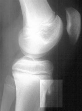
Osgood-Schlatter disease is apophysitis of the tibial tubercle, where the patellar tendon inserts, in active 10-to-15-year-olds. The hallmark is a tender bony bump below the kneecap that hurts with jumping, sprinting, and kneeling. The knee itself is normal: no effusion, full extension, stable ligaments. Radiographs are optional and are obtained to exclude a tumor or avulsion when the story is atypical; when taken, they may show irregular ossification or fragmentation of the tubercle apophysis.
Management is activity modification and ice; the disease is self-limited and resolves as the apophysis fuses. No surgery is offered for the bump itself, and the boards punish immobilization and excision.
Its family shares the mechanism. Sinding-Larsen-Johansson disease is the same traction apophysitis at the inferior pole of the patella. Sever disease is calcaneal apophysitis at the Achilles insertion, heel pain in 8-to-12-year-olds, cross-referenced on the peds hip and foot page.
Tap each letter to reveal the apophysitis family.
O-S S-L-J S
OOsgood-Schlatter: tibial tubercle apophysitis, age 10 to 15, worse with jumping, self-limited
SSymptoms: tender bony bump below the patella, no effusion, normal knee exam
SSinding-Larsen-Johansson: inferior patellar pole apophysitis, same family, older adolescents
LLocation is everything: tubercle means Osgood, patellar pole means SLJ, heel means Sever
JJumping and kneeling reproduce the pain; the apophysis is under traction
SSever: calcaneal apophysitis at the Achilles insertion, heel pain in 8-to-12-year-olds
SWELLING BEHIND THE KNEE
The Popliteal Cyst: Fullness Behind, and the Calf That Fakes a Clot
A Baker cyst is popliteal fossa fullness that follows a knee effusion, and it is usually a finding, not a disease. When it ruptures, the calf screams clot and there is none.
A Baker (popliteal) cyst is a distended gastrocnemius-semimembranosus bursa that communicates with the joint: fluid escapes the knee through the posterior capsule. It follows anything that makes an effusion, osteoarthritis, meniscal tears, and rheumatoid arthritis most often, and it presents as fullness or a mass in the popliteal fossa, tight with full flexion. Ultrasound confirms the cystic nature and excludes a popliteal aneurysm. Asymptomatic or mildly symptomatic cysts are observed and the knee is treated; the cyst shrinks when the effusion stops.
The board moment is the ruptured cyst. Sudden calf pain, swelling, and tenderness in a patient with a known knee effusion is pseudothrombophlebitis: the cyst has dissected down the calf. The exam shows calf swelling without the hard cord, erythema, or fever of a clot. Calf vein ultrasound excludes deep vein thrombosis (cross-ref the cardio venous page), and the process is self-limited: rest, elevation, and treating the knee. Anticoagulation is the classic wrong answer.
On rounds, the resident presents the swollen calf with the chronic knee effusion.
You
The Doppler showed no clot, so I have been looking for another cause of the calf swelling.
Attending
Why did we order the Doppler at all?
You
To rule out DVT. The knee effusion plus sudden calf pain is a ruptured Baker cyst until the veins say otherwise.
Attending
Exactly. The ultrasound is to exclude the clot, not to find it. Pseudothrombophlebitis treats the knee.
A swollen calf with a knee effusion is a ruptured Baker cyst until the Doppler says otherwise. No clot, no anticoagulation.
Run the posterior-knee decision tool: mass or calf, and which door fits.
A posterior knee or calf presentation. Which door fits?
CAN'T EXTEND, LOOK FOR THE GAP
The Extensor Gap: Which Tendon Snapped, and Who Goes to the OR
The extensor mechanism is one chain, patella in the middle: quadriceps tendon above, patellar tendon below. When the chain breaks, active extension fails and the patella sits too high or too low. Surgery is the treatment.
Patellar tendon rupture strikes younger patients, jumpers, and knees after corticosteroid injection. The patient hears or feels a pop, the knee gives way, and active extension is lost. The patella rides high (patella alta), and there is a palpable gap below the patella.
Quadriceps tendon rupture is the over-40 version, often with chronic disease, CKD, diabetes, or steroid use. The gap sits above the patella, the patella rides low (patella baja), and active extension fails with an extensor lag. Both need surgical repair, and the boards reward the surgeon who recognizes the gap.
Patellar fracture completes the differential: a direct blow to the kneecap with a tender patella, a positive grind, and a fracture on radiograph. Nondisplaced, extensor-intact fractures are treated conservatively; displaced or extensor-disrupted fractures are fixed.
From the Attending
Cannot extend the knee? Palpate for the gap before you order anything. A gap below the patella in a young jumper is a patellar tendon rupture. A gap above it in a 70-year-old with diabetes is the quadriceps tendon. High patella, low patella, same rule: the gap is the diagnosis, and the OR is the treatment.
Put the extensor mechanism workup in order.
PES ANSERINE AND THE OA KNEE
The Medial Corner: Below the Joint Line Is the Bursa
The pes anserinus is the conjoined sartorius, gracilis, and semitendinosus insertion on the medial tibia. When the bursa under it inflames, the pain sits below the joint line, and the knee itself is fine.
Pes anserine bursitis is pain and tenderness on the medial tibia, 4 to 5 cm below the joint line, where the three tendons insert. It follows overuse and malalignment: osteoarthritis, overweight, valgus knees, and a sudden increase in activity. Stairs and prolonged walking reproduce it, and the exam is remarkable for how localized it is, no effusion, no instability, no joint-line tenderness.
Treatment is conservative: activity modification, ice, quadriceps and hip strengthening, and weight loss. A local corticosteroid injection is offered for persistent pain, and the boards accept it once the knee itself has been treated. The differential lives at the joint line: MCL sprain (valgus stress pain at the joint line, cross-ref the knee structure page) and medial meniscus tear (joint-line tenderness, McMurray, mechanical symptoms, same cross-ref).
Work the medial knee pain case: reveal each hint, then commit.
A 61-year-old woman with knee osteoarthritis and a body mass index of 33 comes to the office because of 6 weeks of medial knee pain below the joint line, worse with stairs and a new walking program. Examination shows tenderness 4 cm below the medial joint line, a small effusion, and painless valgus stress at 30 degrees. Which of the following is the most likely diagnosis?
RED FLAGS IN FRONT
The Locked and the Hot: When the Front-of-Knee Clinic Stops
Two presentations break every algorithm: the knee that will not unlock and the knee that is hot, swollen, and febrile. One is a meniscus trapped in the joint; the other is septic until aspirated.
A locked knee, stuck in slight flexion with a block to full extension, is a bucket-handle meniscus tear until proven otherwise: the displaced fragment wedges between the condyles. It follows a twist, it effuses, and it needs the knee structure page for the meniscus workup.
An acute hot swollen knee is septic arthritis until the aspirate says no. Fever, warmth, effusion, and refusal to bear weight are the Kocher flags, and the aspirate with Gram stain and culture is the workup, not imaging first. Cross-ref MSK infections for the Kocher criteria in the child.
Prepatellar bursitis sits in front of the patella in people who kneel, roofers and floor installers. The bursa swells and hurts, and the joint itself stays quiet with full range of motion. The split is sterile versus infected: erythema, warmth, fever, and overlying cellulitis point to infection, and the aspirate settles it. And the chronic ACL knee shows its hand as quadriceps atrophy on inspection, a cross-ref to the ligament page.
Tick every red flag that pushes you toward aspiration of the hot knee.
WHICH STRUCTURE, WHICH TEST
The Runner's Knee Screen: Test to Structure, Front to Back
Every structure at the front and sides of the knee has one test the boards reuse. Run the screen in order and the map writes itself: front, outside, inside, deep.
The patellofemoral joint answers to Clarke compression and the J sign, pain with loading. The patellar tendon answers to inferior pole tenderness and the decline squat. The iliotibial band answers to Ober test, lateral knee pain at the epicondyle in runners, the lateral-hip family that lives on the hip pages. The meniscus answers to joint-line tenderness and McMurray, the MCL to valgus stress at 30 degrees, and the ACL to the Lachman test. Each test isolates one structure, and the full ligament and meniscus battery lives on the knee structure page.
Pick the pairing that the screen actually uses.
During the runner's knee screen, a 22-year-old woman has lateral knee pain at the femoral epicondyle that appears when she runs downhill and reproduces with the Ober test. Which structure does this finding localize?
A. Iliotibial band. Lateral knee pain at the epicondyle, worse downhill, with a positive Ober test is IT band friction syndrome, the runner's lateral knee pain. B. Medial meniscus. The meniscus lives at the joint line on the inside and answers to McMurray, not to a positive Ober. C. ACL. The ACL answers to Lachman and the pivot shift, an instability story, not a lateral friction pain. D. Patellar tendon. The tendon answers to inferior pole tenderness and the decline squat, anterior rather than lateral. Rule: lateral plus Ober is the iliotibial band, every time.
BUMP, GAP, CYST, OR PAIN
The One-Screen Discriminator: Four Doors, One Reflex
Age, mechanism, and one exam finding route every front-of-knee story. The bump is an apophysis, the gap is a rupture, the cyst is a Baker, and the pain without findings is PFPS.
Run the doors in order. Bump: a tender prominence at a tendon insertion in a child or teen is apophysitis, Osgood-Schlatter below the patella and Sinding-Larsen-Johansson at its pole, both self-limited.
Gap: a palpable defect with failed active extension is a ruptured extensor tendon, patellar in the young jumper and quadriceps over 40, both surgical. Cyst: popliteal fullness with a knee effusion is a Baker cyst; sudden calf pain with that effusion is its rupture, and the Doppler excludes the clot.
Pain without findings: stairs, squatting, and long sitting with a quiet exam is patellofemoral pain, and physical therapy is the answer.
From the Attending
The front-of-knee stem hands you one physical finding, and that finding is the whole question. A bump means apophysitis and patience. A gap means rupture and the OR. A popliteal mass means the knee made it. Pain with stairs and a normal exam means PT, not imaging and not surgery. Name the door, and the treatment names itself.
The Front-of-Knee Stage
Four doors, one reflex
Jules, 16, cross-country runner: stairs hurt, the exam is quiet.
The patella rides the groove, and every loaded bend reproduces the pain. No effusion, normal film: the pain door.
Marcus, 13, basketball: a tender bump below the kneecap after practice.
The dot rides the patellar tendon down to its insertion. Traction on an open apophysis: the bump door, and it self-limits.
Mrs. Ito, 58, knee OA: fullness behind the knee, then a sudden swollen calf.
The effusion pushes into the fossa, then the cyst tracks down the calf. Pseudothrombophlebitis: the Doppler excludes the clot, and the knee is the disease.
Coach Reyes, 24, jumped and heard a pop: no extension, and a gap below the patella.
The patella rides high and the gap draws itself below it. Failed active extension plus a gap is a rupture, and the OR is the treatment.
Four doors, one screen: name the door and the treatment names itself.
RouteAge and mechanism to door
PatternBump, gap, cyst, or pain
PearlGap goes to the OR; everything else waits
Bump: apophysitis, self-limited. Gap: rupture, surgical. Cyst: Baker, treat the knee. Pain without findings: PFPS, physical therapy first.
Match each presentation to its door.
Tap a presentation, then its door.
Prove it
Walkthrough: the front of the knee in seven cases
Original practice scenarios, one at a time. Choose an answer, then open any option to work its reasoning.
Clinical walkthrough
Reviewed by

Dr. Fatima Ali, DO
Psychiatry resident, PGY-1 · University Hospitals, Columbia
Resident physician whose osteopathic training feeds a whole-system, mechanism-first approach to the subjects students struggle most to reason through alone. Co-founder of Bone Wizardry. Reviews the psychiatry, osteopathic medicine and OMM, clinical-reasoning, and licensing-readiness material, and verifies each page for clinical accuracy.
References
- 1
- 2
- 3
- 4
- 5
Published
Content updated
Medically reviewed
Bone Wizardry is a study resource for medical students. It is not medical advice, and nothing here substitutes for the judgement of a licensed clinician or for the guidelines your program follows.