Bone Tumors: Osteosarcoma, Ewing, and the Pattern Recognition Matrix
Every bone tumor on boards is a pattern of three numbers: the patient's age, the bone zone it lives in, and the way the periosteum reacts. Master the trio and the vignette writes itself: osteosarcoma's sunburst, Ewing's onion-skin, the cartilage arcs, the punched-out skull, and the fallen chip.
What this page makes you able to do
Split the twelve high-yield bone tumors by age, bone zone, and film pattern
Read the six periosteal and matrix patterns and name their owners
Stage and treat the malignant duo: osteosarcoma and Ewing sarcoma
Run the older-adult workup: multiple myeloma versus bone metastases
Every bone tumor on boards is a lock of three numbers: age, zone, and film pattern. Adolescent plus metaphysis plus sunburst and Codman triangle is osteosarcoma; child plus diaphysis plus onion-skin is Ewing until biopsy says otherwise. The pattern names the lineage, the age names the member, and pain at night with a periosteal reaction names the malignancy.
Prove it
Opening question
Answer before you read anything, then keep the age-zone-pattern lock in mind through every section.
A 14-year-old boy comes to the office because of 3 months of right knee pain and a firm mass over the distal thigh. The pain wakes him at night and is not relieved by ibuprofen. Examination shows a warm, tender mass just above the medial femoral condyle. Radiograph of the femur shows a metaphyseal lesion with a sunburst periosteal reaction and a Codman triangle. Chest CT shows a 1.4 cm nodule in the right lower lobe.Which of the following is the most likely diagnosis?
Why this is rightAn adolescent with a metaphyseal sunburst and a Codman triangle has the bone-making sarcoma: osteosarcoma lays down woven bone so fast that spicules radiate perpendicular to the cortex through the lifted periosteum. The 1.4 cm lung nodule is a metastasis, not a second disease; lung is the dominant metastatic site, and chest CT is part of staging before treatment. Rule: adolescent + metaphysis + sunburst and Codman = osteosarcoma, and staging drives neoadjuvant chemotherapy before surgery.
Why this failsEwing lives in the diaphysis and makes onion-skin lamellae, not a metaphyseal sunburst. Its clinical package is the febrile child with malaise and an elevated ESR and WBC, mimicking osteomyelitis. Rule: Ewing is the diaphyseal onion-skin tumor with fever; this film is the metaphyseal bone-maker.
Why this failsInfection can hurt and feel warm, but it does not make a sunburst periosteal reaction with a Codman triangle, and it does not produce a lung nodule. The film pattern is a tumor signature, not pus. Rule: infection never makes sunburst bone; the periosteal reaction is the discriminator.
Why this failsAn osteochondroma is a painless bony projection with cortex and medulla continuous with the host bone, pointing away from the joint. It does not destroy the metaphysis, and a benign exostosis does not metastasize. Rule: painless outgrowth versus painful destructive mass: the adjectives are the diagnosis.
Work the reasoning
The periosteum reacts only when something lifts it. A fast bone-maker like osteosarcoma lays down woven bone so quickly that spicules radiate perpendicular to the cortex, and the triangular cuff at the edge is the Codman triangle. Slow or non-bone-forming tumors never build this.
Ewing claims the diaphysis and lays onion-skin lamellae parallel to the cortex; its patient is often febrile with an elevated ESR and WBC. A metaphyseal sunburst in a 14-year-old is osteosarcoma's signature, not Ewing's.
Lung is the dominant metastatic site for osteosarcoma, so chest CT is a mandatory part of staging. A metastasis means the plan is neoadjuvant chemotherapy (doxorubicin, cisplatin, methotrexate) followed by wide resection, and the histologic response to chemo guides the adjuvant therapy.
The answer is A: osteosarcoma. Adolescent plus metaphyseal sunburst plus Codman triangle equals osteosarcoma, and the lung nodule is a metastasis, not a second disease.
THE PATTERN OF THREE
Meet the Twelve: Age, Zone, and Film Pattern
Every board bone tumor is a locked combination of age, bone zone, and radiograph pattern. Tab the index in your head, then prove it on the matrix.
The skeleton is a moving target, and age is the first lock. Sarcomas track the growth spurt: osteosarcoma peaks from 10 to 25 and Ewing sarcoma from 5 to 20. Giant cell tumor waits for skeletal maturity, so it lives in the 20 to 40 window. Multiple myeloma and bone metastases arrive after 60, and an enchondroma or osteochondroma can sit silently in any decade. The second lock is the zone. Epiphysis: giant cell tumor after maturity. Metaphysis: osteosarcoma, osteochondroma, unicameral cyst. Diaphysis: Ewing. Medulla of flat and axial bones: enchondroma, chondrosarcoma, myeloma, and mets. The third lock is the film: sunburst plus Codman triangle, onion-skin lamellae, rings-and-arcs, punched-out holes, soap-bubble expansion, and the fallen fragment.
The matrix below is the whole game compressed. Malignant flags glue it together: pain at night, systemic symptoms (fever, weight loss), periosteal reaction, and a lesion over 5 cm with deep cortical erosion. A benign-looking pattern with any of those flags is malignant until proven otherwise.
Tumor
Age
Zone
Film pattern
Osteosarcoma
10 to 25
Metaphysis
Sunburst + Codman
Ewing sarcoma
5 to 20
Diaphysis
Onion-skin lamellae
Osteochondroma
Adolescent
Metaphyseal surface
Bony projection away from joint
Enchondroma
Adult
Medulla of small bones
Central rings-and-arcs
Osteoid osteoma
Under 25
Cortex
Nidus under 2 cm + dense rim
Osteoblastoma
Young adult
Posterior spine
Expansile over 2 cm
Chondrosarcoma
Over 40
Pelvis, proximal femur
Painful rings-and-arcs
Multiple myeloma
Over 60
Axial
Punched-out, no rim
Metastases
Over 50
Axial, proximal femur
Blastic or lytic by primary
Giant cell
20 to 40
Epiphysis
Soap-bubble
ABC
Under 20
Metaphysis, spine
Expansile, fluid-fluid levels
UBC
5 to 15
Proximal humerus
Central cyst, fallen fragment
Flip between the two childhood sarcomas before the deep dive.
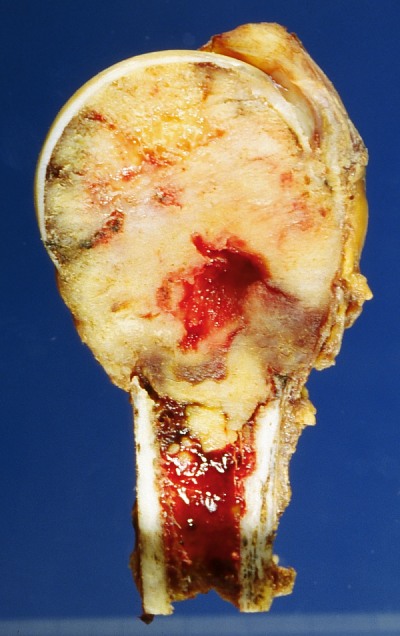
The most common malignant bone tumor in adults. This sawed humerus shows a whitish tumor deposit replacing marrow between the head and the shaft. Metastases seed the axial skeleton and proximal femur because red marrow lives there; prostate, breast, lung, kidney, thyroid, and colon are the primaries boards hand you.
THE DARKROOM
The Pattern Lab: Six Films, Six Owners
Read the periosteal reaction before you read the patient. Each pattern names a lineage, and the lineage narrows the diagnosis.
The radiograph is a language of shadows, and each tumor speaks one dialect. Sunburst spicules plus a Codman triangle mean a bone-maker: osteosarcoma lays woven bone so fast it radiates outward through the lifted periosteum. Onion-skin lamellae stacked parallel to the cortex belong to Ewing's diaphyseal round cell tumor, the one that dresses as osteomyelitis. Rings-and-arcs are calcified cartilage matrix: incidental in a finger bone they are an enchondroma, painful in an adult pelvis they are chondrosarcoma. Punched-out lytic holes with no sclerotic rim are plasma cells dissolving bone with no osteoblastic response, which is why the bone scan is falsely negative in myeloma and the skeletal survey is the right test. Soap-bubble expansion at the epiphysis of a grown skeleton is giant cell tumor. Fallen fragment: a cortical chip settled in a fluid chamber after a fracture is pathognomonic for unicameral bone cyst.
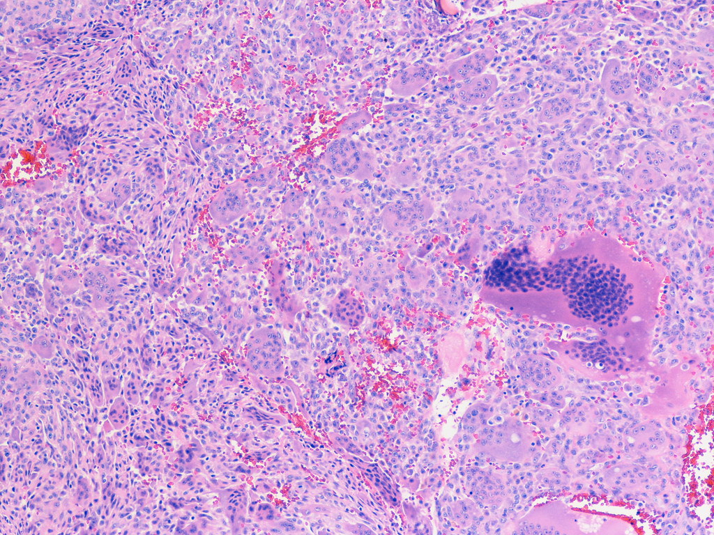
Read the pattern, then confirm with histology. Osteosarcoma: pleomorphic spindle cells producing osteoid and woven bone. Ewing: small round blue cells, CD99 positive, t(11;22) EWSR1-FLI1. Giant cell tumor: neoplastic mononuclear stromal cells plus osteoclast-like giant cells, driven by RANKL, which is why denosumab targets it. The histology names the lineage; age, zone, and symptoms pick the member.
Read the film and commit to the owner.
A 16-year-old girl comes to the office because of 6 weeks of right mid-thigh pain and intermittent low-grade fever. Examination shows mild swelling and tenderness over the femoral shaft without a palpable mass. Radiograph shows a diaphyseal lesion with laminated onion-skin periosteal reaction and moth-eaten destruction. Laboratory studies show an ESR of 58 mm/h and a WBC count of 11,500/mm3. Which of the following is the most likely diagnosis?
A. Ewing sarcoma. Onion-skin lamellae in the diaphysis of a febrile child or adolescent is Ewing until proven otherwise: the small round blue cell tumor with t(11;22) that mimics infection. Biopsy settles it. B. Osteomyelitis. The mimic is real, but layered periosteal new bone along the shaft with moth-eaten destruction is the tumor signature, and a trial of antibiotics delays the sarcoma diagnosis. C. Osteosarcoma. Osteosarcoma is metaphyseal and makes a sunburst, not onion-skin; the diaphysis and lamellae point away from it. D. Fibrous dysplasia. Ground-glass medullary expansion in a younger child does not make layered periosteal reaction or fever. Rule: diaphysis plus onion-skin plus fever in a child is Ewing until the biopsy says otherwise.
The epiphyseal soap bubble under the microscope. Giant cell tumor histology: osteoclast-like giant cells scattered among neoplastic mononuclear stromal cells. The stromal cells secrete RANKL and recruit the giant cells, which is why denosumab, a RANKL inhibitor, is part of the modern treatment.
THE MALIGNANT DUO
Osteosarcoma vs Ewing: Same Skeleton, Opposite Zones
Two childhood sarcomas, two different films, two different patients. Sort every clue into the right column and the pair stops being confusable.
Osteosarcoma is the bone-making sarcoma of the growth spurt: an adolescent, male more often, with a painful knee mass and a metaphyseal sunburst with a Codman triangle around the distal femur or proximal tibia. Skip lesions (a second focus in the same bone) and lung metastases come early, so chest CT is part of staging. Treatment is neoadjuvant chemotherapy with doxorubicin, cisplatin, and methotrexate, then wide resection; the tumor's histologic response to chemo guides the adjuvant therapy. The second peak belongs to older adults with Paget disease or prior radiation, and retinoblastoma (RB1) and Li-Fraumeni raise the risk.
Ewing sarcoma is the round cell tumor that pretends to be infection: a child to young adult with fever, malaise, an elevated ESR and WBC, and a painful diaphyseal lesion with onion-skin lamellae in a long bone, pelvis, or flat bone. Biopsy shows small round blue cells that are CD99 positive with the t(11;22) EWSR1-FLI1 translocation. Treatment is chemotherapy plus local control with surgery or radiation, and staging includes PET or bone marrow evaluation because bone and marrow metastases are the Ewing pattern. The osteomyelitis look-alike is the board trap: infection never makes onion-skin, and a trial of antibiotics wastes the treatment window.
Both sarcomas obey the same workup rule: plain film first, MRI for extent, biopsy before fixation, and the biopsy tract planned by the surgeon who will do the resection. A badly placed biopsy contaminates the compartment and can cost the limb.
Tap the femur zone each tumor claims.
Tap a zone: which tumor claims it?
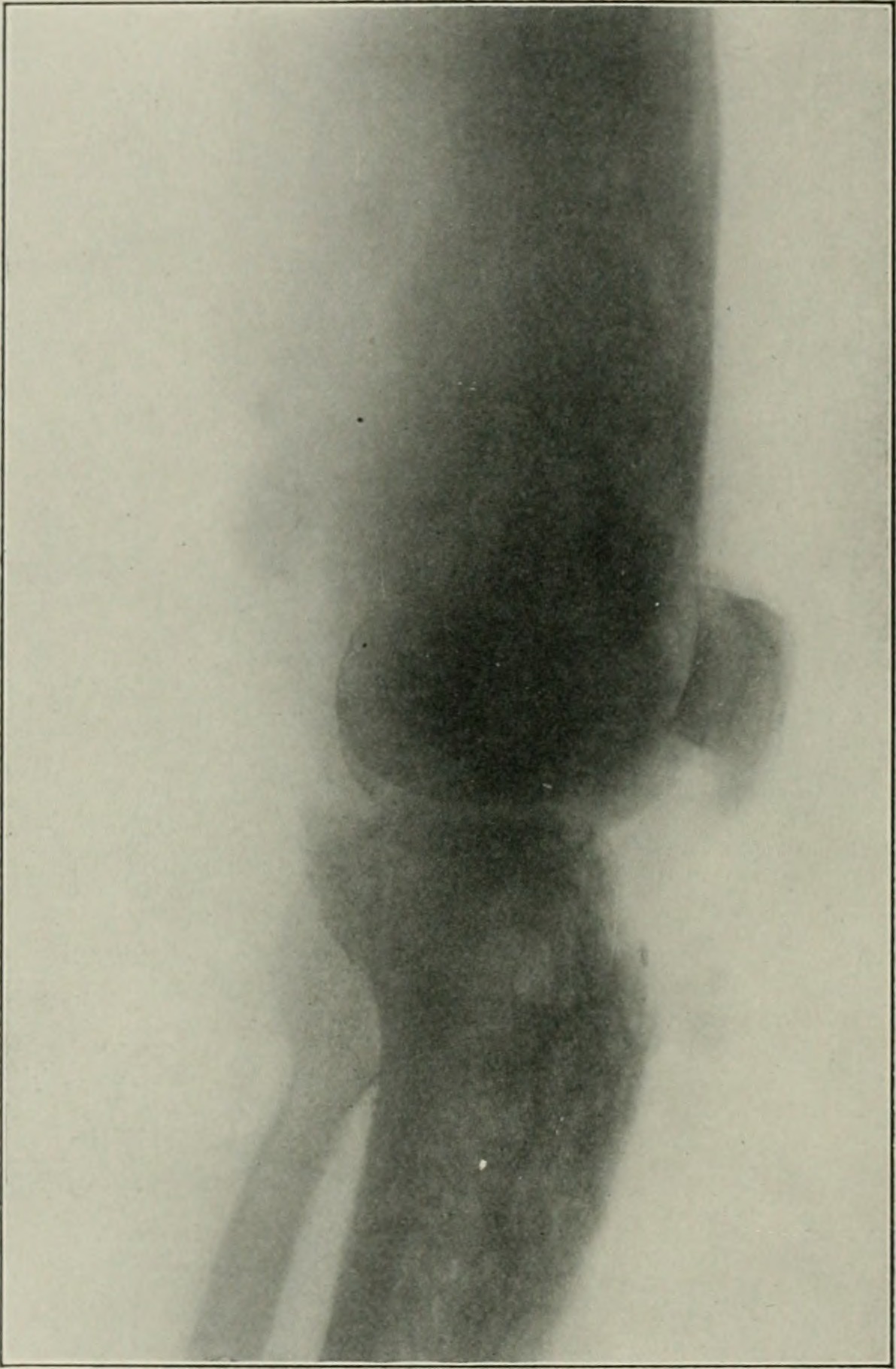
Osteosarcoma at the knee, 1906. This radiograph from the American Practice of Surgery shows the classic territory: an aggressive distal femoral lesion with soft-tissue extension. The metaphysis of the distal femur is the most common home of osteosarcoma in adolescents, and the sunburst is its signature.
THE CARTILAGE LINEAGE
One Cell Line, Three Addresses: Osteochondroma, Enchondroma, Chondrosarcoma
Osteochondroma, enchondroma, and chondrosarcoma share a cell: the chondrocyte. The boards separate them by address, age, and pain.
Osteochondroma is the most common benign bone tumor: an adolescent with a painless bump on the metaphyseal surface pointing away from the joint, with cortex and medulla continuous with the host bone. It stops growing when the skeleton matures, and transformation to chondrosarcoma stays under 1 percent. Hereditary multiple exostoses (EXT1 or EXT2, autosomal dominant) raises that risk, which is why multiple bumps plus a family history get surveillance, and growth after maturity is reimaged.
Enchondroma is the intramedullary cartilage island: an incidental central lucent lesion with rings-and-arcs calcification in a small hand or foot bone, painless unless a pathologic fracture goes through it. Multiple enchondromas with Ollier disease or with hemangiomas in Maffucci syndrome raise transformation risk. The dangerous cousin is chondrosarcoma: an adult over 40 with dull, progressive pain in the pelvis or proximal femur, rings-and-arcs in a lesion over 5 cm, and deep endosteal scalloping. A painful cartilage tumor in an adult is chondrosarcoma until proven otherwise, and the treatment is wide surgical resection because chemotherapy and radiation do not work.
Where does a cartilage lesion go next?
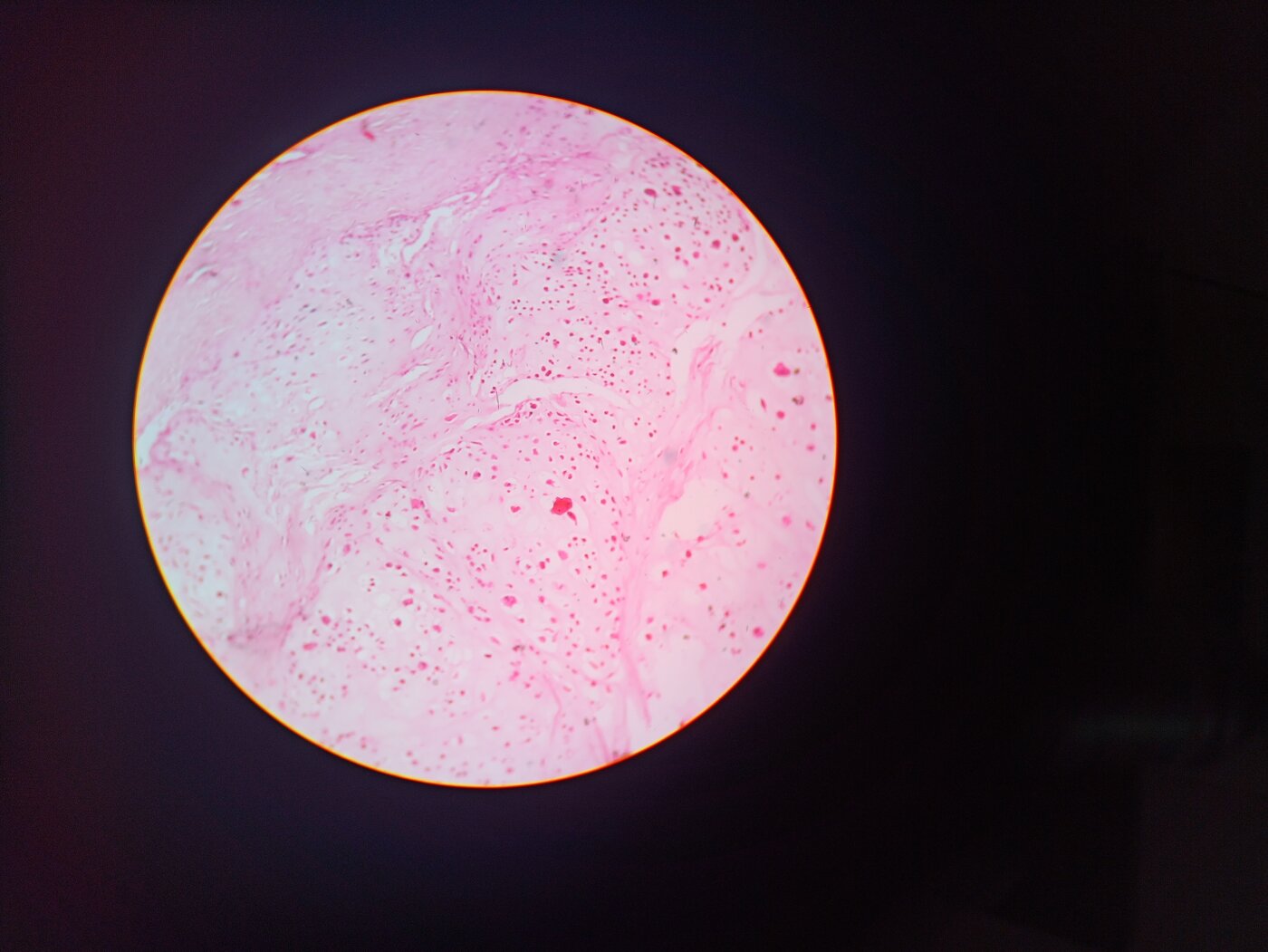
The cartilage cell in its home. Enchondroma histology: lobules of hyaline cartilage with bland chondrocytes sitting in lacunae. When the boards describe neoplastic chondrocytes in lacunae, they are describing the cartilage lineage; in an adult with pain and a pelvic or proximal femoral lesion, that histology is chondrosarcoma.
THE 2 CM LINE
The Hot Nidus and the Bone Cysts
Osteoid osteoma is the tumor that answers to aspirin. Its 2 cm cousin lives in the posterior spine, and a fallen chip names the cyst.
Osteoid osteoma is a benign nidus of woven bone and osteoid that makes prostaglandins, and the prostaglandins make the pain: worse at night, waking the patient, dramatically relieved by aspirin or an NSAID. The patient is a child or young adult under 25, the address is the cortex of the femur or tibia, and the film shows a radiolucent nidus under 2 cm with a dense sclerotic rim. NSAIDs are first-line and often enough; persistent, disabling pain gets CT-guided radiofrequency ablation. The same histology over 2 cm is osteoblastoma, which lives in the posterior elements of the spine, aches without the aspirin response, and is treated with curettage or excision.
The two cysts are split by their fluid. Unicameral bone cyst is one chamber: a child 5 to 15 with a central lucent cyst in the proximal humeral metaphysis, silent until a fall breaks the thinned cortex and the fallen fragment sign proves the chamber is fluid-filled. A fractured UBC is observed; persistent cysts are injected or curetted. Aneurysmal bone cyst is blood-filled: a child or young adult under 20 with an expansile blowout lesion and fluid-fluid levels on MRI, treated with curettage or sclerotherapy, with recurrence the rule rather than the exception.
Check the features that place a nidus on either side of the 2 cm line.
Checked 0
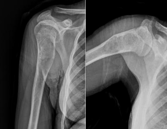
The unicameral bone cyst on film. A central, well-demarcated lucent cyst in the proximal humeral metaphysis of a child, with a pathologic fracture through the thinned cortex. When a cortical chip settles dependently inside the fluid-filled chamber, that fallen fragment sign is pathognomonic.
THE OLDER-ADULT DUO
Myeloma vs Metastases: CRAB or a Traveling Primary
After 60, the bone tumors are systemic: plasma cells or traveling primaries. CRAB picks the plasma cell, the primary site picks the mets, and the film pattern picks the primary.
Multiple myeloma is the most common primary bone malignancy in adults: an older patient with back pain, fatigue, and weight loss, punched-out lytic lesions with no sclerotic rim in the skull and axial skeleton, and the CRAB package (hyperCalcemia, Renal insufficiency, Anemia, Bone lesions). Serum protein electrophoresis shows an M spike, the smear shows rouleaux, and the hypercalcemia rides on a low PTH, because osteoclast activation by myeloma cytokines is the mechanism, not parathyroid disease. The imaging trap is the bone scan: plasma cells suppress osteoblasts, so the bone scan is falsely negative and the skeletal survey is the right test.
Bone metastases are the most common malignant bone lesions in adults, full stop: an older patient with a known or occult primary and axial and proximal femoral lesions. The pattern names the primary: prostate is blastic, breast is mixed, and lung, kidney, thyroid, and colon are lytic. Complications are the board endpoints: pathologic fracture and spinal cord compression, so back pain with leg weakness or urinary retention in a patient with cancer means MRI of the spine now. Treatment is systemic for the primary, radiation for painful lesions, bisphosphonate or denosumab for bone protection, and prophylactic fixation of a femur whose cortex is threatened.
Run the older-adult discriminator.
An older adult with a painful skeleton: which door fits which presentation?
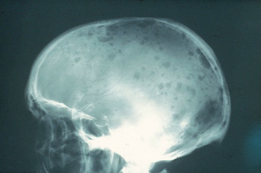
The punched-out skull. Multiple well-demarcated lytic holes in the calvarium with no sclerotic rim: the classic myeloma film. The same plasma cells raise calcium through osteoclast activation, so the bone scan stays cold while the skeleton dissolves.
THE DISCRIMINATOR
The Bone Tumor Discriminator
Age, zone, pattern, and one more fork: the malignant flags. Run the whole matrix as one screen and the vignette writes itself.
The workup order never changes. Plain film first names the pattern. MRI maps the intraosseous and soft-tissue extent. Biopsy before fixation, with the tract planned by the surgeon who will resect, because a contaminated compartment can cost the limb. Chest CT stages the sarcomas, whose lungs are the first stop. Malignant flags upgrade every benign-looking film: pain at night, systemic symptoms, periosteal reaction, and a lesion over 5 cm with deep cortical erosion. A painful enlarging cartilage lesion in an adult, a growing exostosis after maturity, and new pain in Paget bone are all malignant until proven otherwise.
Tap each pattern and name its owner.
SunburstOnionRingsPunchedSoapFallen
SSunburst spicules = the bone-maker: osteosarcoma, metaphysis, adolescent.
OOnion-skin lamellae = Ewing's dinnEr: diaphysis, fever, small round blue cells.
RRings-and-arcs = caRtilage: enchondroma if incidental, chondrosarcoma if painful in an adult.
PPunched-out = Plasma cells: myeloma, no rim, bone scan falsely negative.
SSoap bubble = Giant cell: epiphysis, only after skeletal maturity.
FFallen chip = UBC: one chamber, fluid-filled, silent until fracture.
Open each tumor and hold the whole index.
Age 10 to 25 (second peak with Paget or radiation), metaphysis around the knee, sunburst plus Codman triangle, skip lesions, lung mets at staging. Neoadjuvant chemotherapy (doxorubicin, cisplatin, methotrexate) then wide resection; chemo response guides adjuvant therapy.
Age 5 to 20, diaphysis and flat bones, onion-skin lamellae, fever and elevated ESR and WBC mimicking osteomyelitis. Small round blue cells, CD99 positive, t(11;22) EWSR1-FLI1. Chemotherapy plus local control with surgery or radiation.
Adolescent, metaphyseal surface projection pointing away from the joint, cortex and medulla continuous with host bone. Painless, stops at maturity, transformation under 1 percent; hereditary multiple exostoses (EXT1/EXT2) raises the risk and gets surveillance.
Central medulla of small hand and foot bones, central rings-and-arcs calcification, painless unless pathologic fracture. Ollier disease (multiple) and Maffucci syndrome (with hemangiomas) raise transformation risk.
Child or young adult under 25, cortex of the femur or tibia, nidus under 2 cm with a dense sclerotic rim. Night pain wakes the patient and melts with NSAIDs (prostaglandin-driven). NSAIDs first; CT-guided radiofrequency ablation for persistent disabling pain.
Young adult, posterior elements of the spine, expansile lesion over 2 cm with the same nidus histology as osteoid osteoma. Less predictably NSAID-responsive; curettage or excision, can be locally aggressive.
Adult over 40, medulla of the pelvis and proximal femur, painful enlarging rings-and-arcs with deep endosteal scalloping. Wide surgical resection; chemotherapy and radiation do not work.
Older adult over 60, axial skeleton, punched-out lytic lesions with no rim. CRAB, M spike, rouleaux, low PTH with high calcium. Bone scan is falsely negative; use the skeletal survey.
Older adult, axial skeleton and proximal femur. Prostate blastic, breast mixed, lung/kidney/thyroid/colon lytic. Treat the primary, add bisphosphonate or denosumab, fix threatened bone, and MRI the spine when neurology is involved.
Adult 20 to 40 after skeletal maturity, epiphysis, eccentric soap-bubble lytic lesion abutting subchondral bone. Locally aggressive, recurs after curettage; denosumab targets the RANKL-driven giant cells.
Child or young adult under 20, metaphysis or posterior spine, expansile blowout lesion with fluid-fluid levels on MRI. Blood-filled; curettage or sclerotherapy, recurrence common, can be secondary to another lesion.
Child 5 to 15, central metaphysis of the proximal humerus, one fluid-filled chamber, fallen fragment sign after pathologic fracture. Observe after fracture; inject or curette persistent cysts.
Prove it
Walkthrough: lock the matrix
Seven original cases, one at a time. Choose an answer, then open any option to work its reasoning.
Clinical walkthrough
Choose an answer, then open any option to work its reasoning.
Psychiatry resident, PGY-1 · University Hospitals, Columbia
Resident physician whose osteopathic training feeds a whole-system, mechanism-first approach to the subjects students struggle most to reason through alone. Co-founder of Bone Wizardry. Reviews the psychiatry, osteopathic medicine and OMM, clinical-reasoning, and licensing-readiness material, and verifies each page for clinical accuracy.
Doctor of Osteopathic Medicine, Kansas City University · honored every clinical rotation · 1,000+ tutoring hours · English and Urdu
Bone Wizardry is a study resource for medical students. It is not medical advice, and nothing here substitutes for the judgement of a licensed clinician or for the guidelines your program follows.
 Bone WizardryMEDICAL EDUCATION