Recognize actinic keratosis, assess suspicious change, distinguish epidermal disease from invasion, and choose treatment for lesions and sun-damaged fields.
A rough patch on sun-damaged skin may be an actinic keratosis, but roughness cannot tell you whether abnormal cells have invaded deeper tissue. Begin with three separate questions. What pattern does the lesion fit? Does anything require tissue diagnosis? How much surrounding skin needs treatment? Keeping those decisions separate prevents a familiar-looking patch from receiving the wrong treatment.
Recognize a pattern without closing the differential
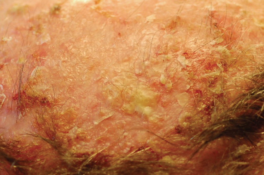
Actinic keratosis (AK) is a UV-associated proliferation of atypical keratinocytes, the cells that form most of the epidermis. Atypical means that their appearance and organization are abnormal. The epidermis is the outer cellular layer of skin. AK often feels gritty, with adherent scale on a pink, skin-colored or pigmented base. It may be easier to feel than see. Common sites include the face, balding scalp, ears, forearms and backs of the hands. Exposure history matters more than what clothing covers the site today. [1][9]
Compare two patches on the same forehead. One is thin and rough, among similar stable patches. The other has developed a firm base and persistent soreness. Both deserve examination, but the second changes the diagnostic question. A person can have ordinary AKs and a different tumor in the same sun-damaged area. Neither a familiar location nor a previous AK diagnosis settles the new finding.
Which forehead patch needs assessment for possible invasion?
The patch with new firmness and persistent soreness needs assessment for possible squamous cell carcinoma.
Morphology supports a differential, not certainty. A pearly border with visible vessels favors basal cell carcinoma, but some basal cell carcinomas are scaly. Waxy, sharply outlined plaques suggest seborrheic keratosis. [17] A stable, flat, uniformly brown macule suggests a solar lentigo, whereas an evolving irregular pigmented lesion needs assessment for melanoma. [22] Psoriasis often forms well-demarcated plaques with white scale and may accompany nail pits or plaques elsewhere. Hands are not an exclusion zone for psoriasis. [10][11][14]
On facial dermoscopy, a red background interrupted by keratotic follicular openings can produce a strawberry-like pattern. This supports AK in context; it does not reveal the basement membrane or exclude another tumor. Bleeding after picking is also nonspecific. Neither a named surface sign nor absent bleeding can certify benign behavior. For transfer, imagine the same rough patch with a new pearly edge. Explain why the differential widens before choosing a destructive treatment. [14][11]
Ultraviolet radiation can damage keratinocyte DNA. UVB produces photoproducts, including bonds between neighboring pyrimidine bases. Nucleotide excision repair cuts out damaged stretches so replacement DNA can be made. The p53 protein has a different job. It regulates the response to damage, including cell-cycle arrest and programmed cell death, called apoptosis. It is not the enzyme that cuts thymine dimers out of DNA. Failure of protective responses can allow damaged clones to persist. [6][7]
Think of repair and quality control as different jobs. Repair machinery replaces a damaged segment. A damage-response regulator helps determine whether the cell should continue dividing. Mutations involving TP53 occur in AK and squamous cell carcinoma, so finding such a mutation does not tell you whether invasion has occurred. Histologic location answers that question. [1]
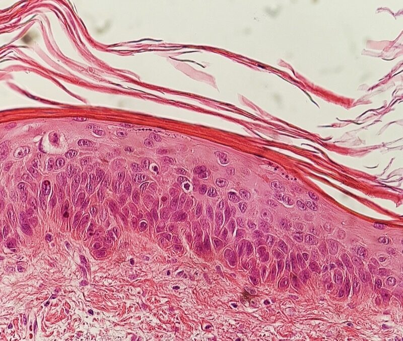
The basement membrane separates epidermis from the supporting dermis. AK typically shows partial-thickness keratinocyte atypia, often most evident basally, but a fixed lower-third rule is too narrow. Squamous cell carcinoma in situ, often called Bowen disease, shows extensive or full-thickness atypia that remains intraepidermal. Invasive squamous cell carcinoma has tumor extending across that boundary into the dermis. Solar elastosis, altered dermal elastic tissue from chronic sun damage, records exposure rather than invasion. [1][3]
Two specimens have atypical cells. Only one contains tumor nests in the dermis. Which establishes invasion?
The specimen with tumor nests extending into the dermis establishes invasion.
Depth across the boundary determines invasion; surface thickness does not. A thick keratin cap may cover intraepidermal disease, while a less dramatic surface can conceal invasion. AK, in situ carcinoma and invasive carcinoma are useful comparisons, not compulsory visible stages through which every lesion passes. [18] AK can persist, regress, recur or progress. For transfer, predict whether full-thickness atypia with an intact boundary proves invasion. It does not, provided the specimen adequately represents the lesion and its base. [1][3]
Epidermal Boundary Lab
Alternative educational schematics, not an inevitable progression or a patient diagnosis. Upper region: epidermis. Lower region: dermis. The heavy line is the basement membrane. Angular enlarged nuclei distinguish atypia without relying on color.
Typical normal. Small regular nuclei remain organized above an intact basement membrane.Partial intraepidermal atypia. Irregular enlarged nuclei occupy part of the epidermis. The boundary remains intact; atypia is not universally confined to the lower third.Full-thickness intraepidermal atypia. Atypical cells span the epidermis but remain above an intact boundary. Full thickness alone is not dermal invasion.Invasion through the basement membrane. Atypical cells cross a breached boundary into dermis. This spatial relationship distinguishes invasion.
Worked comparisons
Typical normal: Small regular nuclei remain organized above an intact basement membrane.
Partial intraepidermal atypia: Irregular enlarged nuclei occupy part of the epidermis. The boundary remains intact; atypia is not universally confined to the lower third.
Full-thickness intraepidermal atypia: Atypical cells span the epidermis but remain above an intact boundary. Full thickness alone is not dermal invasion.
Invasion through the basement membrane: Atypical cells cross a breached boundary into dermis. This spatial relationship distinguishes invasion.
Actinic keratosis with moderate atypia. The source describes atypia spanning approximately half of the spinous layer. A universal lower-third rule would miss this variation. Mikael Häggström; CC0 1.0. Existing reduced-size copy, compared visually with the published original. No new alteration.
Case 2
Show answer and explanations for case 2
A. Invasive squamous carcinoma requiring assessment for dermal tumor (Why this does not fit)
What feature makes a malignant squamous process plausible?
Full-thickness atypia supports squamous carcinoma in situ.
Which supplied feature fails to establish invasion?
The adequately represented boundary remains intact.
What separates in situ from invasive squamous disease?
Dermal invasion requires tumor crossing the epidermal boundary.
Read the complete reasoning
Full-thickness atypia supports squamous carcinoma in situ. The adequately represented boundary remains intact. Dermal invasion requires tumor crossing the epidermal boundary.
B. An inflammatory plaque requiring anti-inflammatory treatment (Why this does not fit)
Why could inflammation enter the clinical differential?
Scaly plaques can represent inflammatory skin disease.
What finding favors neoplasia in this specimen?
Disordered full-thickness keratinocyte atypia indicates a neoplastic process.
How should tissue findings affect a surface differential?
Do not substitute an inflammatory diagnosis for demonstrated epithelial atypia.
Read the complete reasoning
Scaly plaques can represent inflammatory skin disease. Disordered full-thickness keratinocyte atypia indicates a neoplastic process. Do not substitute an inflammatory diagnosis for demonstrated epithelial atypia.
C. Squamous carcinoma in situ requiring treatment of intraepidermal disease (Best answer)
How does specimen B differ from the basal pattern in A?
Atypia extends throughout the epidermal thickness.
Where is that abnormal proliferation confined?
It remains above an intact basement membrane in the sampled lesion.
What treatment distinction follows?
Plan for in situ disease unless invasion is demonstrated or remains inadequately assessed.
Read the complete reasoning
Atypia extends throughout the epidermal thickness. It remains above an intact basement membrane in the sampled lesion. Plan for in situ disease unless invasion is demonstrated or remains inadequately assessed.
D. Typical actinic keratosis requiring treatment of basal atypia (Why this does not fit)
Why does AK remain a close comparison?
AK also contains atypical keratinocytes within the epidermis.
What separates B from the typical pattern in A?
B has disordered maturation throughout the epidermis.
How should full-thickness atypia be interpreted here?
An adequately sampled full-thickness intraepidermal pattern supports carcinoma in situ.
Read the complete reasoning
AK also contains atypical keratinocytes within the epidermis. B has disordered maturation throughout the epidermis. An adequately sampled full-thickness intraepidermal pattern supports carcinoma in situ.
Takeaway: Extent within epidermis and extension into dermis answer different questions.
Field cancerization describes a region containing multiple areas of UV-associated cellular damage, including abnormalities that are not yet clinically apparent. A visible AK is one expression of that field. Clearing one spot can succeed locally while another lesion appears nearby. A new patch elsewhere in the field does not, by itself, establish recurrence of the treated spot or spread from it. [1]
Picture a scalp with several thin AKs and one thicker focus. Lesion-directed treatment targets an individual focus. Field-directed treatment covers a selected region, including visible and subclinical disease. These approaches can be combined. The thicker focus first needs examination for features that warrant biopsy; a large treatment area does not make a suspicious focus less important. [1][3]
A treated spot stays smooth, but new rough patches appear nearby. What did local clearance leave unresolved?
The surrounding damaged field remained capable of producing additional lesions.
Counting spots helps describe burden but is not a treatment algorithm. Extent, site, morphology, prior response, immune status, tolerance and preferences shape the plan. Someone with a few scattered lesions may prefer a procedure. Someone with repeated lesions across a contiguous field may prefer treatment of the region. Difficulty applying medication can change an otherwise reasonable topical plan. [1]
Risk counseling also needs the correct scale. A study reporting people who develop cancer over several years answers a different question from one tracking individual lesions for one year. Estimates vary with population, denominator and follow-up. A denominator is what was counted, such as people or individual lesions. A study of treated people is not a measurement of untreated lesion progression. A raw ratio of affected participants to those enrolled is also distinct from a time-to-event estimate that accounts for follow-up. Multiplying a patient proportion by lesion count mixes those units. Even valid individual-lesion probabilities cannot simply be added to obtain a patient probability, because more than one lesion can progress in the same person. No single fixed percentage reliably predicts an individual AK's future. Regression does not establish that the surrounding field is now normal. For transfer, explain why three new patches after successful spot treatment can justify reconsidering coverage without proving that the original procedure failed. [1][5]
Spot or Field
Coverage only, not efficacy percentages or a prescribing plan. The triangle marks a suspicious growing or indurated lesion: assess it rather than simply adding more empiric destruction.
Untreated. No treatment footprint is selected. Solid circles show visible lesions; dashed patches represent less visible damaged areas.Lesion-directed coverage. Outlined footprints surround selected visible lesions. Damaged areas between them remain outside the outlined treatment area.Field-directed coverage. The outlined footprint includes visible lesions and intervening damaged skin. Coverage does not imply complete cure.
Worked comparisons
Untreated: No treatment footprint is selected. Solid circles show visible lesions; dashed patches represent less visible damaged areas.
Lesion-directed coverage: Outlined footprints surround selected visible lesions. Damaged areas between them remain outside the outlined treatment area.
Field-directed coverage: The outlined footprint includes visible lesions and intervening damaged skin. Coverage does not imply complete cure.
A. The treated lesions persisted, so repeating the same focal treatment addresses the whole problem (Why this does not fit)
Why is incomplete treatment worth considering after cryosurgery?
A treated focus can persist or recur.
What argues against persistence at the treated sites here?
Both mapped treatment sites remain smooth.
How can location clarify post-treatment findings?
Distinguish a persistent treated focus from new lesions elsewhere.
Read the complete reasoning
A treated focus can persist or recur. Both mapped treatment sites remain smooth. Distinguish a persistent treated focus from new lesions elsewhere.
B. The lesions represent dermal spread, so the entire region requires cancer staging (Why this does not fit)
Why should new lesions be examined?
New findings may represent a different diagnosis.
What supplied pattern does not establish dermal spread?
Separate thin rough patches appeared while treated sites stayed clear.
What cannot be inferred from new surface lesions alone?
New AK-like patches do not establish invasive spread.
Read the complete reasoning
New findings may represent a different diagnosis. Separate thin rough patches appeared while treated sites stayed clear. New AK-like patches do not establish invasive spread.
C. The new patches prove continued unprotected exposure, so adherence is the main issue (Why this does not fit)
Why does current UV protection remain relevant?
Reducing further UV exposure helps prevent additional damage.
What other substrate already exists here?
The surrounding region contains longstanding UV-associated damage.
What should not be inferred from new AKs?
New lesions do not by themselves prove inadequate current sun protection.
Read the complete reasoning
Reducing further UV exposure helps prevent additional damage. The surrounding region contains longstanding UV-associated damage. New lesions do not by themselves prove inadequate current sun protection.
D. Local clearance can coexist with field disease, so regional treatment may be useful (Best answer)
What does sustained smoothness at mapped sites suggest?
The targeted lesions have responded locally.
What explains similar new lesions elsewhere in that region?
The surrounding damaged field can produce additional AKs.
What should the next plan reconsider?
Match treatment coverage to both individual lesions and regional burden.
Read the complete reasoning
The targeted lesions have responded locally. The surrounding damaged field can produce additional AKs. Match treatment coverage to both individual lesions and regional burden.
Takeaway: Map location before calling a new lesion recurrence or spread.
Ask whether the specimen can answer the clinical question
A typical thin AK can often be diagnosed clinically. Reassessment becomes important when a lesion persists despite appropriate treatment or becomes increasingly thick, indurated, tender, ulcerated or prone to bleeding. Indurated means unusually firm within or beneath the lesion. These findings raise concern but do not individually prove squamous cell carcinoma. When carcinoma is suspected, obtain tissue that can answer the question before another empiric destructive course. [9][3]
Biopsy is not simply a yes-or-no box. The sample must include sufficient depth and representative tissue. A report describing atypical keratinocytes in a superficial fragment cannot exclude tumor deeper than that fragment. Match the pathology to the examination. If the lesion remains firm and enlarging while the sample lacks its base, further sampling or specialist assessment is needed. The specimen is a window into the lesion; a shallow window cannot show what lies below it. [3]
A superficial specimen shows AK but omits the base of an indurated lesion. Has invasion been excluded?
No. The clinically concerning depth has not been adequately sampled.
A cutaneous horn is a projecting mass of keratin, not a diagnosis of the cells producing it. Its base may contain benign disease, AK or carcinoma. A tender or indurated base needs particular attention, and sampling only the horn tip misses the relevant tissue. Actinic cheilitis is chronic actinic damage of the lip. Persistent focal ulceration or firmness within a scaly lower lip warrants assessment for carcinoma, rather than assuming either harmless chapping or established cancer. [13][8]
A rapidly enlarging crateriform nodule can resemble keratoacanthoma, a squamous proliferation with a central keratin-filled crater. Its possible regression is not a dependable reason to leave a suspected carcinoma undiagnosed. [16] Confirmed invasive disease needs its own risk assessment and definitive treatment plan, usually surgical, rather than an AK field cream alone. For transfer, identify which part of a horn specimen must be represented to assess invasion. [3][13]
Match the treatment to the target and the person
The American Academy of Dermatology strongly recommends UV protection, cryosurgery, topical fluorouracil and imiquimod for AK. Its 2022 focused update added a strong recommendation for tirbanibulin field treatment. Photodynamic therapy and diclofenac have conditional recommendations. Conditional means the balance is more dependent on circumstances and preferences; it does not mean that other therapies must always fail first. Ingenol mebutate is withdrawn and is not a treatment option here. [1][2]
Cryosurgery uses controlled freezing to damage targeted tissue. It can suit discrete lesions, but pain, healing and possible pigment change matter. Fluorouracil, or 5-FU, is an antimetabolite that interferes with nucleic-acid metabolism in proliferating cells. One active metabolite inhibits thymidylate synthase, an enzyme needed to supply thymidine nucleotides for DNA synthesis. [19]
Imiquimod instead activates toll-like receptor 7, or TLR7, in innate immune signaling. Cytokines are signals used by immune cells. The official label distinguishes this known receptor activity from the full mechanism of AK treatment, which is unknown. A change in a laboratory cytokine signal therefore does not establish clinical clearance. [20] Similar redness does not mean these drugs have the same mechanism. [1][9]
Photodynamic therapy, or PDT, combines a sensitizing precursor with appropriate light to produce a photochemical tissue effect. The precursor forms protoporphyrin IX, a photosensitizer. When appropriate light activates it, energy transfer to oxygen produces reactive oxygen species that can injure cells. Oxygen must be present during the photochemical reaction. The LEVULAN KERASTICK label describes this dependency in section 12.1. Sensitizer accumulation alone is not delivery of the light-triggered treatment. [21] Light activation is part of the treatment, not an optional finishing step. This differs from thermal destruction. Clinic-based delivery may suit someone unable to apply medication reliably, although availability and discomfort still matter.
Tirbanibulin inhibits microtubules, cellular structures involved in division. It is another topical option; its US indication covers the face or scalp. The dorsal hands are not included in that US indication. A clinician must assess a new site before extending a prior prescription; clinician-directed off-label prescribing is a separate decision, not automatic permission to reuse a prior prescription. [23][15][1][2]
If the sensitizing preparation is used but light activation is omitted, what essential PDT component is missing?
The light-triggered photochemical reaction is missing.
Redness, crusting and soreness can occur during topical treatment. Severe pain, extensive erosion or signs of infection require contact with the treating clinician and may require interruption or adjustment. More inflammation does not certify better clearance. Explain the expected reaction and how to obtain advice before treatment begins, rather than telling patients to endure every reaction. [12]
Goals of care also matter. For a person with limited life expectancy, stable asymptomatic AKs and a high treatment burden, observation can be reasonable after shared discussion. That choice rests on expected benefit and burden, not a claim that progression is impossible. New symptoms still deserve reassessment. [1]
In the Jansen trial, 5-FU performed best among four studied field treatments in patients with at least five AKs in a contiguous area on the head. The endpoint was a reduction of at least 75% in lesion count 12 months after the end of treatment. Count reduction is not complete clearance. Calculate the decrease from baseline, divide by the baseline count, and multiply by 100. For the stated example, a change from 12 lesions to 3 is a 75% reduction, with three lesions still present. This lesion endpoint does not establish a treatment effect on invasive cancer. [4]
Ahmady's later secondary analysis recorded relatively few invasive cancers and had limited power for treatment comparisons. For transfer, distinguish choosing a therapy for demonstrated AK reduction from promising that it prevents every subsequent carcinoma. [4][5]
Case 15
Show answer and explanations for case 15
A. Discuss clinic-based PDT as a reasonable field option now (Best answer)
What distribution favors regional coverage?
Numerous thin lesions occupy one contiguous field.
What delivery constraint favors a clinic-based option?
Hand tremor makes repeated self-application unreliable.
Does a conditional PDT recommendation require prior topical failure?
PDT selection can be appropriate without first failing a topical course.
Read the complete reasoning
Numerous thin lesions occupy one contiguous field. Hand tremor makes repeated self-application unreliable. PDT selection can be appropriate without first failing a topical course.
B. Try a self-applied fluorouracil course before considering clinic-based treatment (Why this does not fit)
Why is fluorouracil a reasonable therapy to discuss?
It has strong evidence for AK field treatment.
What makes a mandatory trial a poor fit here?
The patient cannot reliably perform the needed self-application.
How should recommendation strength affect sequencing?
A strong recommendation does not create a mandatory trial for every patient.
Read the complete reasoning
It has strong evidence for AK field treatment. The patient cannot reliably perform the needed self-application. A strong recommendation does not create a mandatory trial for every patient.
C. Use lesion-directed freezing of the most visible spots as the sole treatment (Why this does not fit)
What could freezing selected visible spots accomplish?
Cryosurgery can clear individual AKs.
What would treating only the most visible spots leave outside its coverage?
Other lesions and subclinical changes remain across this contiguous field.
Why is clinic-based regional treatment feasible here?
Clinic access permits field treatment despite unreliable self-application.
What should a combined or regional plan match?
Treatment coverage should address the selected field as well as individual lesions.
Read the complete reasoning
Cryosurgery can clear individual AKs. Other lesions and subclinical changes remain across this contiguous field. Clinic access permits field treatment despite unreliable self-application. Treatment coverage should address the selected field as well as individual lesions.
D. Choose a self-applied diclofenac course as the initial field treatment (Why this does not fit)
Why can diclofenac be discussed?
It is a conditionally recommended topical treatment.
What practical problem remains with that selection?
Hand tremor still makes self-application unreliable.
How should first-course treatment be chosen?
Initial therapy should fit the patient rather than an invented clinic-treatment prohibition.
Read the complete reasoning
It is a conditionally recommended topical treatment. Hand tremor still makes self-application unreliable. Initial therapy should fit the patient rather than an invented clinic-treatment prohibition.
Takeaway: A conditional recommendation supports individualized selection, not a compulsory second-line position.
Follow-up checks treatment tolerance, residual lesions, recurrence and new findings elsewhere. A smooth treated field is a useful result, but it does not erase previous UV damage or remove the need to reassess changing skin. Set follow-up according to lesion burden, prior cancers, treatment response and immune status. A changing lesion deserves assessment before the planned routine visit. [8][9]
Solid-organ transplant recipients have increased keratinocyte cancer risk during chronic immunosuppression. Dermatology and the transplant team should coordinate treatment and closer risk-based surveillance. Adjustments to immunosuppression or systemic prevention may be considered for selected high-risk patients, but they require balancing cancer risk against graft rejection and medication harms. A new AK is not permission to stop transplant medication. [1][3]
A transplant recipient develops a tender focus before the next routine visit. Should the existing appointment determine when it is assessed?
No. The new concerning finding should trigger earlier assessment.
UV protection remains useful after decades of exposure. Combine shade, protective clothing and broad-spectrum sunscreen, with reapplication appropriate to exposure and product instructions. Sunscreen supports prevention; it does not replace examination of a persistent abnormal lesion. Greater melanin protection reduces UV injury but does not make a changing lesion irrelevant. For transfer, explain why a patient who starts sun protection today may still develop an AK from previously damaged skin. Prevention reduces further exposure rather than resetting the skin's history. [1][9]
Apply the finding that changes the decision
For each original clinical scenario, state the decision before reading the options. Then identify the finding that defeats the closest alternative. Separate evidence about appearance, tissue depth, treatment coverage and long-term outcomes.
Case 1
Show answer and explanations for case 1
A. Treat the changing focus with cryosurgery (Why this does not fit)
Why might cryosurgery initially fit this scalp?
Cryosurgery can treat discrete clinically typical AKs.
What finding makes destruction premature here?
New underlying firmness with growth raises concern for carcinoma.
What should precede destruction of a suspicious focus?
Establish the diagnosis before empiric destructive treatment.
Read the complete reasoning
Cryosurgery can treat discrete clinically typical AKs. New underlying firmness with growth raises concern for carcinoma. Establish the diagnosis before empiric destructive treatment.
B. Obtain an adequately deep biopsy of the changing focus (Best answer)
How does this focus differ from the surrounding patches?
It has developed progressive firmness and tenderness.
What question must tissue sampling answer?
Sampling must assess whether keratinocyte disease is invasive.
What determines a useful diagnostic sample?
Choose representative tissue deep enough to assess suspected invasion.
Read the complete reasoning
It has developed progressive firmness and tenderness. Sampling must assess whether keratinocyte disease is invasive. Choose representative tissue deep enough to assess suspected invasion.
C. Begin fluorouracil across the affected scalp (Why this does not fit)
Why is field therapy a reasonable later consideration?
Multiple surrounding AKs may benefit from regional treatment.
What does field coverage fail to settle first?
It does not diagnose the growing indurated focus.
How should a suspicious focus affect a field plan?
Resolve suspected carcinoma before treating it as routine AK.
Read the complete reasoning
Multiple surrounding AKs may benefit from regional treatment. It does not diagnose the growing indurated focus. Resolve suspected carcinoma before treating it as routine AK.
D. Reassess the focus after a trial of emollient (Why this does not fit)
What symptom could an emollient improve?
Emollient can soften superficial dryness and scale.
What supplied finding extends beyond superficial dryness?
The lesion is becoming firm beneath the scale.
When is symptomatic skin care insufficient?
Progressive induration needs diagnostic assessment rather than scale care alone.
Read the complete reasoning
Emollient can soften superficial dryness and scale. The lesion is becoming firm beneath the scale. Progressive induration needs diagnostic assessment rather than scale care alone.
Takeaway: A changing focus within an AK field needs its own diagnostic assessment.
A. Arrange further representative sampling that includes the concerning depth (Best answer)
What part of the clinical lesion remains unexplained?
The persistent underlying firmness is not represented by the superficial fragment.
What does the report leave unresolved?
Tumor below the sampled tissue cannot be excluded.
What should clinical-pathologic mismatch trigger?
Obtain tissue adequate to answer the unresolved diagnostic question.
Read the complete reasoning
The persistent underlying firmness is not represented by the superficial fragment. Tumor below the sampled tissue cannot be excluded. Obtain tissue adequate to answer the unresolved diagnostic question.
B. Repeat cryosurgery using the reported AK diagnosis (Why this does not fit)
Why is repeat cryosurgery tempting?
The submitted fragment was interpreted as AK.
Why is that result insufficient for this plaque?
The sample omits the depth responsible for the persistent firmness.
How should limited sampling constrain treatment?
A superficial diagnosis cannot exclude deeper disease it never sampled.
Read the complete reasoning
The submitted fragment was interpreted as AK. The sample omits the depth responsible for the persistent firmness. A superficial diagnosis cannot exclude deeper disease it never sampled.
C. Begin field imiquimod to treat the residual plaque (Why this does not fit)
What makes imiquimod relevant to an AK field?
It is a recommended topical field treatment.
What must be settled before using it for this focus?
Possible invasion remains unresolved beneath the transected sample.
What is the boundary of an empiric field approach?
Field treatment does not replace adequate diagnosis of a suspicious lesion.
Read the complete reasoning
It is a recommended topical field treatment. Possible invasion remains unresolved beneath the transected sample. Field treatment does not replace adequate diagnosis of a suspicious lesion.
D. Review after the next planned surveillance interval (Why this does not fit)
What could justify routine follow-up after some biopsies?
A concordant diagnosis with clinical resolution can support planned surveillance.
What makes this result discordant?
The plaque remains firm while its deeper tissue was not examined.
When should reassessment occur before routine surveillance?
Persistent diagnostic uncertainty about invasion warrants additional assessment.
Read the complete reasoning
A concordant diagnosis with clinical resolution can support planned surveillance. The plaque remains firm while its deeper tissue was not examined. Persistent diagnostic uncertainty about invasion warrants additional assessment.
Takeaway: The word AK on a superficial report does not settle an unsampled base.
A. Chronic UV-associated dysplasia best explains the entire distribution (Why this does not fit)
What supports considering AK on the hands?
The dorsal hands receive occupational UV exposure.
What pattern argues for another unifying process?
Symmetric plaques with nail pits extend beyond the hand lesions.
How should exposure history be weighted?
UV exposure does not override a coherent competing clinical pattern.
Read the complete reasoning
The dorsal hands receive occupational UV exposure. Symmetric plaques with nail pits extend beyond the hand lesions. UV exposure does not override a coherent competing clinical pattern.
B. An inflammatory psoriatic pattern warrants evaluation before destructive treatment (Best answer)
What links the hand lesions to findings elsewhere?
Matching elbow plaques and nail pits support psoriasis.
What does the stable fluctuating course add?
It favors chronic inflammatory disease over a newly evolving focal tumor.
What should precede destruction of uncertain scaly plaques?
Assess the full distribution rather than diagnosing from roughness alone.
Read the complete reasoning
Matching elbow plaques and nail pits support psoriasis. It favors chronic inflammatory disease over a newly evolving focal tumor. Assess the full distribution rather than diagnosing from roughness alone.
C. Multiple in situ squamous carcinomas best explain the symmetric plaques (Why this does not fit)
Why can in situ carcinoma enter this differential?
It can present as a persistent scaly plaque.
What favors psoriasis as the unifying explanation?
Symmetric fluctuating plaques coexist with characteristic nail changes.
How should a focal cancer diagnosis be generalized?
Do not assign one neoplastic diagnosis to a broader inflammatory pattern without support.
Read the complete reasoning
It can present as a persistent scaly plaque. Symmetric fluctuating plaques coexist with characteristic nail changes. Do not assign one neoplastic diagnosis to a broader inflammatory pattern without support.
D. Irritant hand dermatitis best explains the complete skin and nail pattern (Why this does not fit)
Why is irritant dermatitis plausible on working hands?
Occupational exposure can produce chronic hand inflammation.
Which additional findings weaken it as the unifying explanation?
Matching elbow plaques with nail pits favor psoriasis.
What examination improves hand-rash discrimination?
Look beyond the hands for associated skin and nail findings.
Read the complete reasoning
Occupational exposure can produce chronic hand inflammation. Matching elbow plaques with nail pits favor psoriasis. Look beyond the hands for associated skin and nail findings.
Takeaway: Hand location and outdoor work do not exclude psoriasis.
A. Apply field fluorouracil to include the persistent patch (Why this does not fit)
Why might a field drug seem appropriate?
Nearby gritty patches suggest an accompanying AK burden.
What makes this focus diagnostically different?
A translucent raised edge with vessels suggests a competing tumor.
What should a distinct focus within a field receive?
Evaluate its own diagnosis before including it in routine AK treatment.
Read the complete reasoning
Nearby gritty patches suggest an accompanying AK burden. A translucent raised edge with vessels suggests a competing tumor. Evaluate its own diagnosis before including it in routine AK treatment.
B. Freeze the patch again because its surface is scaly (Why this does not fit)
What surface feature overlaps with AK?
The patch has scale.
Why does scale fail to settle this diagnosis?
Basal cell carcinoma can also have a scaly surface.
Which approach avoids this surface trap?
Use the complete morphology rather than scale alone.
Read the complete reasoning
The patch has scale. Basal cell carcinoma can also have a scaly surface. Use the complete morphology rather than scale alone.
C. Obtain tissue to evaluate a suspected basal cell carcinoma (Best answer)
Which finding points beyond ordinary AK?
The translucent raised vascular edge suggests basal cell carcinoma.
What does persistence after treatment contribute?
It strengthens the need to reconsider the initial diagnosis.
How is a suspected skin carcinoma established?
Obtain appropriate tissue for histologic diagnosis.
Read the complete reasoning
The translucent raised vascular edge suggests basal cell carcinoma. It strengthens the need to reconsider the initial diagnosis. Obtain appropriate tissue for histologic diagnosis.
D. Use topical corticosteroid for a presumed inflammatory plaque (Why this does not fit)
Why can an inflammatory plaque resemble this lesion?
Inflammatory disease can produce a red scaly patch.
What feature is poorly explained by that assumption?
A persistent translucent raised edge with branching vessels suggests tumor.
When is an empiric anti-inflammatory trial insufficient?
A lesion suspicious for carcinoma needs diagnostic evaluation.
Read the complete reasoning
Inflammatory disease can produce a red scaly patch. A persistent translucent raised edge with branching vessels suggests tumor. A lesion suspicious for carcinoma needs diagnostic evaluation.
Takeaway: Scale does not rule out basal cell carcinoma.
A. Sample representative living tissue at the base with sufficient depth (Best answer)
What did the previous specimen actually assess?
It assessed the keratin projection rather than the producing epithelium.
Where is the lesion that determines malignant potential?
The relevant viable lesion lies at the horn base.
What should a diagnostic horn specimen include?
Sample the base deeply enough to assess the suspected underlying disease.
Read the complete reasoning
It assessed the keratin projection rather than the producing epithelium. The relevant viable lesion lies at the horn base. Sample the base deeply enough to assess the suspected underlying disease.
B. Repeat sampling of the distal horn tip (Why this does not fit)
Why might the visible tip seem like the target?
It is the most obvious protruding part of the lesion.
What did tip sampling already fail to provide?
It provided no viable epithelium for diagnosis.
Where should sampling follow a nondiagnostic keratin fragment?
Target the living base rather than repeat the same superficial sample.
Read the complete reasoning
It is the most obvious protruding part of the lesion. It provided no viable epithelium for diagnosis. Target the living base rather than repeat the same superficial sample.
C. Biopsy adjacent flat photodamaged skin (Why this does not fit)
Why is adjacent skin relevant to risk?
The surrounding field shares chronic UV exposure.
What does adjacent skin not represent?
It does not represent the tender indurated horn base.
How should the biopsy site be selected?
Sample the clinically concerning lesion rather than its background field.
Read the complete reasoning
The surrounding field shares chronic UV exposure. It does not represent the tender indurated horn base. Sample the clinically concerning lesion rather than its background field.
D. Accept the keratin report and treat the projection destructively (Why this does not fit)
Why can a keratin-only report sound reassuring?
No malignant cells were described in the submitted material.
Why is that absence uninformative here?
The specimen lacked the viable cells that need assessment.
What is the limit of a negative sample?
No tumor in nonrepresentative tissue does not exclude tumor at the base.
Read the complete reasoning
No malignant cells were described in the submitted material. The specimen lacked the viable cells that need assessment. No tumor in nonrepresentative tissue does not exclude tumor at the base.
Takeaway: A horn describes the surface product; its base determines the diagnosis.
A. Treat presumed angular cheilitis with a topical antifungal (Why this does not fit)
Why might infection be considered for an inflamed lip?
Infections can cause fissuring and inflammation around the mouth.
What distribution weakens angular cheilitis here?
The mouth corners are spared while the lower vermilion is involved.
What should guide this location-based distinction?
Angular cheilitis centers on the commissures rather than a firm focal vermilion ulcer.
Read the complete reasoning
Infections can cause fissuring and inflammation around the mouth. The mouth corners are spared while the lower vermilion is involved. Angular cheilitis centers on the commissures rather than a firm focal vermilion ulcer.
B. Treat presumed recurrent herpes with an antiviral (Why this does not fit)
Why might herpes enter the differential?
Herpes can cause painful erosions on the lip.
What supplied course argues against that explanation?
A persistent firm ulcer lacks the described recurrent blister episodes.
How should persistent focal firmness alter a lip diagnosis?
Investigate a sustained firm lesion rather than assuming an episodic viral eruption.
Read the complete reasoning
Herpes can cause painful erosions on the lip. A persistent firm ulcer lacks the described recurrent blister episodes. Investigate a sustained firm lesion rather than assuming an episodic viral eruption.
C. Begin another course of emollient for diffuse cheilitis (Why this does not fit)
What could emollient help in the background?
It can reduce discomfort from diffuse dryness and scaling.
What new feature requires more than symptom care?
A focal ulcer with palpable firmness raises concern for carcinoma.
How should focal change within diffuse disease be handled?
Assess the focal change separately from the background inflammation.
Read the complete reasoning
It can reduce discomfort from diffuse dryness and scaling. A focal ulcer with palpable firmness raises concern for carcinoma. Assess the focal change separately from the background inflammation.
D. Arrange targeted assessment and biopsy for possible lip carcinoma (Best answer)
What does the longstanding background suggest?
Chronic lower-lip actinic damage is compatible with actinic cheilitis.
What changes the current priority?
Persistent focal ulceration with firmness raises concern for invasive disease.
What does actinic cheilitis establish by itself?
It signals risk requiring assessment, not automatic proof of carcinoma.
Read the complete reasoning
Chronic lower-lip actinic damage is compatible with actinic cheilitis. Persistent focal ulceration with firmness raises concern for invasive disease. It signals risk requiring assessment, not automatic proof of carcinoma.
Takeaway: Focal change within actinic cheilitis needs assessment rather than a surface label.
A. Monitor for regression before considering tissue diagnosis (Why this does not fit)
Why is observation tempting in this morphology?
Some keratoacanthoma-like lesions can regress.
What remains unresolved in this growing nodule?
Its resemblance to squamous carcinoma has not been evaluated with tissue.
What cannot be used as a diagnostic guarantee?
Possible future regression does not exclude current carcinoma.
Read the complete reasoning
Some keratoacanthoma-like lesions can regress. Its resemblance to squamous carcinoma has not been evaluated with tissue. Possible future regression does not exclude current carcinoma.
B. Arrange specialist assessment with representative tissue diagnosis (Best answer)
What pattern does the rapid crateriform growth suggest?
It suggests a keratoacanthoma-like squamous proliferation.
Why does that pattern still require diagnostic assessment?
Clinical appearance overlaps with squamous carcinoma.
How should unresolved squamous tumor uncertainty be managed?
Establish the diagnosis rather than relying on hoped-for regression.
Read the complete reasoning
It suggests a keratoacanthoma-like squamous proliferation. Clinical appearance overlaps with squamous carcinoma. Establish the diagnosis rather than relying on hoped-for regression.
C. Treat with AK field cream and assess whether it flattens (Why this does not fit)
Why might an AK treatment be considered?
The nodule is keratinizing and lies on sun-damaged skin.
What exceeds a routine thin AK presentation?
It is a rapidly enlarging firm crateriform nodule.
When is a treatment response trial inadequate?
A suspected invasive tumor requires diagnosis before an AK treatment trial.
Read the complete reasoning
The nodule is keratinizing and lies on sun-damaged skin. It is a rapidly enlarging firm crateriform nodule. A suspected invasive tumor requires diagnosis before an AK treatment trial.
D. Freeze the nodule as a thick AK without sampling (Why this does not fit)
What important information would destruction forgo?
It would forgo representative histology of a potentially invasive squamous tumor.
How should lesion count influence diagnostic certainty?
A solitary lesion still needs diagnosis when its morphology suggests carcinoma.
Read the complete reasoning
There is one localized keratotic lesion. It would forgo representative histology of a potentially invasive squamous tumor. A solitary lesion still needs diagnosis when its morphology suggests carcinoma.
Takeaway: Keratoacanthoma-like morphology does not make observation a reliable cancer-exclusion strategy.
A. Little injury, because oxygen depletion prevents the earlier formation of sensitizer (Why this does not fit)
Which observed result makes reduced injury plausible?
The dark control shows that accumulated sensitizer alone produces little injury.
Did oxygen depletion prevent precursor conversion in the third sample?
The third sample already contained the matched sensitizer concentration.
Which stage can this perturbation test?
A change after sensitizer formation tests the later photochemical reaction.
Read the complete reasoning
The dark control shows that accumulated sensitizer alone produces little injury. The third sample already contained the matched sensitizer concentration. A change after sensitizer formation tests the later photochemical reaction.
B. Similar injury, because absorbed light energy directly heats the sensitized cells (Why this does not fit)
Why might a lamp suggest thermal injury?
Light can deliver energy to tissue.
Which observation weakens heating as the cause here?
Injury occurred without a temperature rise.
What additional requirement remains despite the identical light dose?
PDT photochemical injury requires oxygen during illumination.
Read the complete reasoning
Light can deliver energy to tissue. Injury occurred without a temperature rise. PDT photochemical injury requires oxygen during illumination.
C. Little injury, because oxygen depletion limits the reaction of light-activated sensitizer (Best answer)
What does the dark control separate from sensitizer accumulation?
It separates sensitizer accumulation from light-triggered injury.
What reaction is limited in the third sample?
Low oxygen limits reactive oxygen generation by the activated sensitizer.
Why can matching sensitizer and light fail to reproduce injury?
Photochemical injury also depends on oxygen present during illumination.
Read the complete reasoning
It separates sensitizer accumulation from light-triggered injury. Low oxygen limits reactive oxygen generation by the activated sensitizer. Photochemical injury also depends on oxygen present during illumination.
D. Similar injury, because excitation of the accumulated sensitizer is sufficient for damage (Why this does not fit)
Which components of the original illuminated sample are retained?
The sensitizer concentration and light dose are matched.
What necessary reactant has changed?
Oxygen availability has fallen.
Why is sensitizer excitation alone insufficient?
The intended cytotoxic photochemistry depends on oxygen.
Read the complete reasoning
The sensitizer concentration and light dose are matched. Oxygen availability has fallen. The intended cytotoxic photochemistry depends on oxygen.
Takeaway: Separate sensitizer accumulation from the oxygen-dependent injury produced during illumination.
A. Fluorouracil; clearance still requires assessment after the treatment reaction settles (Best answer)
Which drug fits the reduced thymidine supply?
The antimetabolite fluorouracil inhibits thymidylate synthase through an active metabolite.
What does the field-confined redness establish?
It is compatible with a local treatment reaction.
What finding is still needed before declaring clearance?
Reassessment must determine whether lesions remain.
Read the complete reasoning
The antimetabolite fluorouracil inhibits thymidylate synthase through an active metabolite. It is compatible with a local treatment reaction. Reassessment must determine whether lesions remain.
B. Fluorouracil; clearance is established by the field-confined inflammatory reaction (Why this does not fit)
Which part of this explanation fits the biochemical finding?
Fluorouracil fits the inhibition of thymidylate synthase.
Why can the reaction seem reassuring?
Redness and crusting can occur during fluorouracil treatment.
What outcome has the reaction not measured?
Inflammation does not demonstrate disappearance of every lesion.
Read the complete reasoning
Fluorouracil fits the inhibition of thymidylate synthase. Redness and crusting can occur during fluorouracil treatment. Inflammation does not demonstrate disappearance of every lesion.
C. Imiquimod; clearance still requires assessment after the treatment reaction settles (Why this does not fit)
Why might imiquimod fit the visible skin response?
Imiquimod can also cause local inflammation.
What supplied finding distinguishes the prescribed medicine?
Thymidylate-synthase inhibition points to fluorouracil.
Why does reasonable follow-up advice not validate this option?
Correct response assessment cannot compensate for the wrong drug mechanism.
Read the complete reasoning
Imiquimod can also cause local inflammation. Thymidylate-synthase inhibition points to fluorouracil. Correct response assessment cannot compensate for the wrong drug mechanism.
D. Imiquimod; clearance is established by the field-confined inflammatory reaction (Why this does not fit)
What makes an immune modifier tempting from appearance alone?
Imiquimod can cause redness at treated sites.
What contradicts that drug identification here?
The supplied nucleotide effect identifies fluorouracil.
What separate conclusion is unsupported?
Visible inflammation is not a complete-clearance measurement.
Read the complete reasoning
Imiquimod can cause redness at treated sites. The supplied nucleotide effect identifies fluorouracil. Visible inflammation is not a complete-clearance measurement.
Takeaway: Identify the medicine from its cellular action, then assess clearance separately from inflammation.
A. X cytokine signaling persists; Y nucleotide depletion diminishes (Why this does not fit)
Which agent initially resembles an immune modifier?
X has the imiquimod-like cytokine response.
Which response should TLR7 blockade diminish?
The receptor-dependent X signal should diminish.
Why is Y not the direct target of this antagonist?
Fluorouracil-type nucleotide depletion does not require TLR7 activation.
Read the complete reasoning
X has the imiquimod-like cytokine response. The receptor-dependent X signal should diminish. Fluorouracil-type nucleotide depletion does not require TLR7 activation.
B. X cytokine signaling diminishes; Y nucleotide depletion also diminishes (Why this does not fit)
Which predicted change fits the antagonist?
The imiquimod-like X signal should diminish.
What distinguishes Y from that pathway?
Y has the fluorouracil-like nucleotide effect.
Why does the shared AK indication not support both changes?
Treatments for the same disease can have different proximal targets.
Read the complete reasoning
The imiquimod-like X signal should diminish. Y has the fluorouracil-like nucleotide effect. Treatments for the same disease can have different proximal targets.
C. X cytokine signaling persists; Y nucleotide depletion also persists (Why this does not fit)
Which predicted response is consistent with a selective TLR7 block?
The Y nucleotide effect should persist.
Does unchanged uptake preserve X signaling through a blocked receptor?
Continued drug delivery cannot bypass the blocked TLR7 response.
What does that distinguish experimentally?
Drug exposure and downstream target activity are separate measurements.
Read the complete reasoning
The Y nucleotide effect should persist. Continued drug delivery cannot bypass the blocked TLR7 response. Drug exposure and downstream target activity are separate measurements.
D. X cytokine signaling diminishes; Y nucleotide depletion persists (Best answer)
Which drug action fits the initial X signal?
X fits imiquimod-type innate immune activation.
Which drug action fits Y nucleotide depletion?
Y fits fluorouracil-type antimetabolite activity.
What follows from selectively blocking TLR7?
X signaling decreases without directly reversing Y nucleotide depletion.
What clinical outcome remains outside this exercise?
These proximal readouts do not establish AK clearance.
Read the complete reasoning
X fits imiquimod-type innate immune activation. Y fits fluorouracil-type antimetabolite activity. X signaling decreases without directly reversing Y nucleotide depletion. These proximal readouts do not establish AK clearance.
Takeaway: A selective receptor block tests a proximal pathway, not clinical AK clearance.
A. Continue unchanged because reaction intensity estimates the amount of disease cleared (Why this does not fit)
Why might continuation initially seem reasonable?
Some local inflammation is expected during fluorouracil treatment.
What makes this reaction require reassessment?
Extensive erosions and sleep-disrupting pain indicate poor tolerability.
How should reaction severity be used?
Severe toxicity requires assessment rather than being treated as proof of efficacy.
Read the complete reasoning
Some local inflammation is expected during fluorouracil treatment. Extensive erosions and sleep-disrupting pain indicate poor tolerability. Severe toxicity requires assessment rather than being treated as proof of efficacy.
B. Pause application and contact the treating clinician to assess the severe reaction (Best answer)
What separates this report from mild expected redness?
Pain is severe enough to disrupt sleep with extensive erosions.
What immediate treatment issue needs clinician review?
The patient needs assessment of whether treatment should be interrupted or adjusted.
What rule reconciles expected inflammation with safety?
Expected reactions can still become severe enough to require treatment modification.
Read the complete reasoning
Pain is severe enough to disrupt sleep with extensive erosions. The patient needs assessment of whether treatment should be interrupted or adjusted. Expected reactions can still become severe enough to require treatment modification.
C. Switch directly to imiquimod to avoid any further inflammatory response (Why this does not fit)
Why is another field therapy tempting?
The current topical course has become difficult to tolerate.
Why does the proposed switch not solve the immediate problem?
Imiquimod can also cause local inflammation in already injured skin.
What should precede choosing a replacement topical?
Assess and manage the severe reaction before selecting another course.
Read the complete reasoning
The current topical course has become difficult to tolerate. Imiquimod can also cause local inflammation in already injured skin. Assess and manage the severe reaction before selecting another course.
D. Biopsy the most eroded area before reviewing the topical reaction (Why this does not fit)
Why can an erosion raise concern in an untreated lesion?
Persistent focal ulceration may warrant investigation for carcinoma.
What pattern favors a treatment-related assessment here?
The reaction is extensive throughout the medication application field.
How should the application-field distribution guide the first response?
Assess the severe topical reaction rather than assume that treatment-associated erosion represents carcinoma.
Read the complete reasoning
Persistent focal ulceration may warrant investigation for carcinoma. The reaction is extensive throughout the medication application field. Assess the severe topical reaction rather than assume that treatment-associated erosion represents carcinoma.
Takeaway: Expected inflammation is not an instruction to ignore severe pain or erosion.
A. Residual keratinocyte dysplasia warrants freezing the pale area again (Why this does not fit)
Why should residual AK be considered after treatment?
A treated lesion may persist or recur.
What examination finding argues against that interpretation here?
The pale area is smooth without recurrent scale or thickening.
What should guide retreatment of a treated site?
Base retreatment on residual lesion findings rather than color change alone.
Read the complete reasoning
A treated lesion may persist or recur. The pale area is smooth without recurrent scale or thickening. Base retreatment on residual lesion findings rather than color change alone.
B. New field disease warrants adding a topical course to the pale area (Why this does not fit)
Why does field disease remain relevant after cryosurgery?
The surrounding UV-damaged skin may produce new lesions.
What spatial pattern points to a treatment effect instead?
The pale smooth patch matches the prior treatment site.
How should a new color change be interpreted?
Compare it with treatment location and the current surface examination.
Read the complete reasoning
The surrounding UV-damaged skin may produce new lesions. The pale smooth patch matches the prior treatment site. Compare it with treatment location and the current surface examination.
C. Treatment-related pigment loss warrants discussing cosmetic risk before further freezing (Best answer)
What does a smooth site without scale suggest about the treated AK?
It suggests current local clearance rather than visible persistence.
What explains localized pallor after cryosurgery?
Cryosurgery can cause lasting loss of pigment at the treated site.
What should inform the next treatment discussion?
Include pigment effects when weighing further cryosurgery against other suitable options.
Read the complete reasoning
It suggests current local clearance rather than visible persistence. Cryosurgery can cause lasting loss of pigment at the treated site. Include pigment effects when weighing further cryosurgery against other suitable options.
D. Occult invasive carcinoma warrants excision of the pale area (Why this does not fit)
Why should treated lesions still be reassessed?
A persistent or changing lesion can conceal a different diagnosis.
What supplied pattern favors a treatment pigment effect?
The patch is smooth and matches the freezing site without focal firmness.
What does pallor alone not establish?
Pigment loss after cryosurgery does not itself establish invasive carcinoma.
Read the complete reasoning
A persistent or changing lesion can conceal a different diagnosis. The patch is smooth and matches the freezing site without focal firmness. Pigment loss after cryosurgery does not itself establish invasive carcinoma.
Takeaway: Distinguish pigment change after local treatment from persistent lesion morphology.
A. Use fluorouracil across the scalp and judge the invasive focus by surface clearance (Why this does not fit)
Why does fluorouracil fit part of this scalp?
It can treat the background AK field.
What does the biopsy establish at the tender focus?
That focus contains invasive squamous carcinoma.
What cannot certify control of invasive disease?
Surface clearance after an AK cream is not adequate proof of invasive tumor clearance.
Read the complete reasoning
It can treat the background AK field. That focus contains invasive squamous carcinoma. Surface clearance after an AK cream is not adequate proof of invasive tumor clearance.
B. Freeze each visible lesion because the disease is confined to one region (Why this does not fit)
Why might targeted freezing seem efficient?
All visible lesions lie within an accessible scalp region.
What prevents treating all lesions as ordinary AK?
One focus has biopsy-proven dermal invasion.
What should determine treatment category?
Histologic invasion outweighs a shared location when planning definitive care.
Read the complete reasoning
All visible lesions lie within an accessible scalp region. One focus has biopsy-proven dermal invasion. Histologic invasion outweighs a shared location when planning definitive care.
C. Plan definitive carcinoma treatment for the invasive focus and separately address the AK field (Best answer)
Which finding establishes a separate treatment problem?
Tumor nests extend into the dermis at the tender focus.
What remains appropriate for the surrounding lesions?
The background AK field can receive an individualized field or combined plan.
How should mixed disease be managed?
Treat confirmed invasive carcinoma separately from surrounding intraepidermal disease.
Read the complete reasoning
Tumor nests extend into the dermis at the tender focus. The background AK field can receive an individualized field or combined plan. Treat confirmed invasive carcinoma separately from surrounding intraepidermal disease.
D. Treat the whole field with imiquimod before deciding whether the invasive focus needs surgery (Why this does not fit)
Why might imiquimod be considered for the surrounding field?
It is a recommended AK field therapy.
What diagnostic uncertainty has already been resolved?
The focal lesion is already proven invasive.
What should not delay definitive carcinoma management?
An empiric AK response trial should not replace treatment of confirmed invasion.
Read the complete reasoning
It is a recommended AK field therapy. The focal lesion is already proven invasive. An empiric AK response trial should not replace treatment of confirmed invasion.
Takeaway: One field can contain lesions requiring different treatment categories.
A. Below the lesion-reduction threshold; invasive-cancer benefit remains undetermined (Why this does not fit)
How many baseline lesions are no longer counted?
The count fell by 15 of the original 20 lesions.
Does the resulting proportion fall below the trial threshold?
A 75% reduction meets a threshold defined as at least 75%.
Why might five residual lesions mislead the reader?
The trial threshold allowed residual lesions; it did not require complete clearance.
Read the complete reasoning
The count fell by 15 of the original 20 lesions. A 75% reduction meets a threshold defined as at least 75%. The trial threshold allowed residual lesions; it did not require complete clearance.
B. Meets the threshold with complete clearance; invasive-cancer benefit remains undetermined (Why this does not fit)
Which part of this classification fits the count change?
The 75% reduction meets the lesion-count threshold.
Which observed finding contradicts complete clearance?
Five lesions remain at follow-up.
How should a threshold result be named?
Partial lesion reduction and complete clearance are different outcomes.
Read the complete reasoning
The 75% reduction meets the lesion-count threshold. Five lesions remain at follow-up. Partial lesion reduction and complete clearance are different outcomes.
C. Meets the threshold with partial clearance; invasive-cancer benefit is demonstrated (Why this does not fit)
Which response classification is supported?
The count reduction is 75% with five lesions remaining.
Why might cancer benefit seem plausible?
AK is associated with later squamous carcinoma.
What evidence is missing for that further conclusion?
A lesion-count result does not measure an effect on cancer incidence.
Read the complete reasoning
The count reduction is 75% with five lesions remaining. AK is associated with later squamous carcinoma. A lesion-count result does not measure an effect on cancer incidence.
D. Meets the threshold with partial clearance; invasive-cancer benefit remains undetermined (Best answer)
How is the percentage reduction calculated?
Subtract 5 from 20, then divide 15 by 20 to obtain 75%.
Does this constitute complete clearance?
Five remaining lesions mean clearance is partial.
What cancer conclusion follows from this threshold success?
The lesion endpoint alone cannot establish an invasive-cancer benefit.
Read the complete reasoning
Subtract 5 from 20, then divide 15 by 20 to obtain 75%. Five remaining lesions mean clearance is partial. The lesion endpoint alone cannot establish an invasive-cancer benefit.
Takeaway: Calculate lesion reduction, distinguish it from complete clearance, and keep cancer outcomes separate.
A. Keep original treatment groups; interpret the imprecise estimates as outcomes under initial assignment and subsequent care. (Best answer)
What was randomly allocated?
The initial treatment was randomized.
What treatments contributed to later follow-up?
Many participants subsequently received clinician-selected fluorouracil.
How should the cancer comparison be interpreted?
The original-group comparison remains useful, but few events and later care limit precision and attribution to one isolated course.
Read the complete reasoning
The initial treatment was randomized. Many participants subsequently received clinician-selected fluorouracil. The original-group comparison remains useful, but few events and later care limit precision and attribution to one isolated course.
B. Keep original treatment groups; attribute the estimates to the initial course alone because that course was randomized. (Why this does not fit)
What advantage does original randomization retain?
It supports comparison by initial treatment assignment.
Did randomization prevent later treatment?
No. The stem states that many participants later received additional fluorouracil.
What does this mean for attribution?
Preserving original assignment does not separate the initial course from the subsequent care that followed it.
Read the complete reasoning
It supports comparison by initial treatment assignment. No. The stem states that many participants later received additional fluorouracil. Preserving original assignment does not separate the initial course from the subsequent care that followed it.
C. Group participants by the final treatment received; interpret those groups as the randomized comparison after retreatment. (Why this does not fit)
What might regrouping by final treatment appear to clarify?
It may seem to compare participants with similar recent drug exposure.
Was the final treatment randomly assigned?
No. Clinicians selected retreatment when lesions required further treatment.
Why is that regrouping not the original randomized comparison?
It sorts participants partly by events after randomization, so the resulting groups do not retain the original random assignment.
Read the complete reasoning
It may seem to compare participants with similar recent drug exposure. No. Clinicians selected retreatment when lesions required further treatment. It sorts participants partly by events after randomization, so the resulting groups do not retain the original random assignment.
D. Exclude follow-up after retreatment; interpret the remaining events as an unbiased comparison of isolated initial courses. (Why this does not fit)
What exposure would censoring at retreatment exclude?
It would exclude follow-up during later treatment.
Was retreatment unrelated to the lesion course?
No. The need for further treatment influenced the clinician decision.
Does simple censoring establish an unbiased initial-course effect?
No. Outcome-related treatment decisions can also make censoring informative; excluding that follow-up alone does not establish unbiased attribution.
Read the complete reasoning
It would exclude follow-up during later treatment. No. The need for further treatment influenced the clinician decision. No. Outcome-related treatment decisions can also make censoring informative; excluding that follow-up alone does not establish unbiased attribution.
Takeaway: Original randomization supports an assignment-based comparison; it does not isolate one course from later care or remove imprecision.
A. Apply leftover field cream first and keep the existing review date (Why this does not fit)
Why is field treatment relevant to this history?
Repeated AKs may justify a regional treatment strategy.
What requires attention before another empiric course?
A newly thick tender focus raises concern for carcinoma.
How should new concerning change affect scheduled surveillance?
Assess a changing focus before the routine follow-up date.
Read the complete reasoning
Repeated AKs may justify a regional treatment strategy. A newly thick tender focus raises concern for carcinoma. Assess a changing focus before the routine follow-up date.
B. Arrange lesion assessment and coordinate longer-term prevention with dermatology and the transplant team (Best answer)
What increases concern about this new focus?
Persistent thickening and tenderness occur in a patient with prior SCC and immunosuppression.
Who should balance medication changes against graft risk?
The transplant team should coordinate those decisions with dermatology.
What is the practical rule for this combination?
Investigate suspicious skin change without independently altering transplant immunosuppression.
Read the complete reasoning
Persistent thickening and tenderness occur in a patient with prior SCC and immunosuppression. The transplant team should coordinate those decisions with dermatology. Investigate suspicious skin change without independently altering transplant immunosuppression.
C. Reduce the immunosuppressive dose before deciding whether the lesion needs biopsy (Why this does not fit)
Why might medication adjustment be discussed in selected recipients?
Immunosuppression contributes to keratinocyte cancer risk.
Why is an unsupervised reduction inappropriate?
It may compromise graft protection without diagnosing the current lesion.
How should cancer-risk medication changes be made?
Use coordinated specialist decisions rather than home dose changes.
Read the complete reasoning
Immunosuppression contributes to keratinocyte cancer risk. It may compromise graft protection without diagnosing the current lesion. Use coordinated specialist decisions rather than home dose changes.
D. Schedule a systemic prevention consultation before examining the focus (Why this does not fit)
Why might systemic prevention be relevant later?
Repeated carcinomas can justify specialist prevention discussions.
What current problem does that consultation not resolve?
The new thick tender lesion still needs diagnostic assessment.
What should long-term prevention not displace?
Prevention planning should not delay evaluation of a suspected current carcinoma.
Read the complete reasoning
Repeated carcinomas can justify specialist prevention discussions. The new thick tender lesion still needs diagnostic assessment. Prevention planning should not delay evaluation of a suspected current carcinoma.
Takeaway: Higher baseline risk strengthens assessment and coordination, not unsupervised medication changes.
A. Follow-up is needed only if sunscreen use becomes inconsistent (Why this does not fit)
Why is sunscreen adherence relevant?
It helps reduce ongoing UV injury.
What important risk substrate predates current prevention?
The patient already has longstanding UV-damaged skin.
What does current protection not reset?
Good prevention does not erase previously accumulated field damage.
Read the complete reasoning
It helps reduce ongoing UV injury. The patient already has longstanding UV-damaged skin. Good prevention does not erase previously accumulated field damage.
B. Follow-up should be limited to the exact previously treated spots (Why this does not fit)
Why should treated sites be reviewed?
Persistence or recurrence can occur there.
What relevant area would that restriction omit?
Other parts of the damaged field can develop new lesions.
What should surveillance consider beyond treated foci?
Reassessment includes new lesions as well as local treatment response.
Read the complete reasoning
Persistence or recurrence can occur there. Other parts of the damaged field can develop new lesions. Reassessment includes new lesions as well as local treatment response.
C. Continue risk-based follow-up and seek assessment of new concerning changes (Best answer)
What does the smooth examination show?
It supports a favorable current treatment response.
What does it not demonstrate about future risk?
It does not prove that the UV-damaged field can no longer produce lesions.
How should monitoring continue?
Set follow-up by individual risk while assessing concerning changes when they arise.
Read the complete reasoning
It supports a favorable current treatment response. It does not prove that the UV-damaged field can no longer produce lesions. Set follow-up by individual risk while assessing concerning changes when they arise.
D. Schedule another field course immediately to prevent any future lesion (Why this does not fit)
Why might another course be needed in some patients?
Residual or recurrent disease can require further treatment.
What supplied finding weakens an automatic repeat course now?
No residual AK is found at the current review.
What should determine retreatment?
Base further treatment on reassessment rather than a guarantee of preventing every future lesion.
Read the complete reasoning
Residual or recurrent disease can require further treatment. No residual AK is found at the current review. Base further treatment on reassessment rather than a guarantee of preventing every future lesion.
Takeaway: Current clearance and future risk are different assessments.
A. The ratio describes affected treated patients; multiplying it by eight estimates this patient’s risk (Why this does not fit)
What does 624 count?
It counts participants rather than individual AKs.
What does multiplying by eight assume about that proportion?
It incorrectly treats a patient proportion as an individual-lesion probability.
What further mismatch limits this patient’s extrapolation?
The study participants received field treatment, whereas these lesions are untreated.
Read the complete reasoning
It counts participants rather than individual AKs. It incorrectly treats a patient proportion as an individual-lesion probability. The study participants received field treatment, whereas these lesions are untreated.
B. The ratio describes progressing treated lesions; multiplying it by eight estimates this patient’s risk (Why this does not fit)
Why might the calculation seem to describe lesions?
The study followed people with multiple AKs for subsequent cancer.
What unit was actually counted in both parts of the ratio?
Both 26 and 624 count people.
Why does lesion count not convert those units?
Multiplication cannot turn a treated-patient outcome into untreated lesion progression.
Would adding valid lesion probabilities necessarily give a patient probability?
Lesion outcomes within one patient need not be mutually exclusive.
Read the complete reasoning
The study followed people with multiple AKs for subsequent cancer. Both 26 and 624 count people. Multiplication cannot turn a treated-patient outcome into untreated lesion progression. Lesion outcomes within one patient need not be mutually exclusive.
C. The ratio describes affected treated patients; this patient’s untreated risk requires other evidence (Best answer)
What outcome does 26 out of 624 describe?
It describes affected participants among those enrolled for field treatment.
Does that ratio identify the fate of each untreated AK?
It does not estimate untreated per-lesion progression.
Why would adding lesion risks still require care?
The same patient can have more than one lesion progress.
What should accompany any numerical risk counseling?
The evidence must match the outcome unit, treatment context and follow-up.
Read the complete reasoning
It describes affected participants among those enrolled for field treatment. It does not estimate untreated per-lesion progression. The same patient can have more than one lesion progress. The evidence must match the outcome unit, treatment context and follow-up.
D. The ratio describes progressing treated lesions; this patient’s untreated risk requires other evidence (Why this does not fit)
Which caution is appropriate for this patient?
Untreated risk requires evidence beyond a treated cohort.
Which supplied fact defeats the lesion-denominator claim?
The denominator is 624 participants with multiple AKs.
What is the reusable denominator rule?
Count affected people over people and progressing lesions over tracked lesions.
Read the complete reasoning
Untreated risk requires evidence beyond a treated cohort. The denominator is 624 participants with multiple AKs. Count affected people over people and progressing lesions over tracked lesions.
Takeaway: A proportion of treated patients with cancer cannot be converted into untreated lesion risk or added across lesions.
A. Nucleotide excision repair is defective while the damage-response checkpoint is preserved (Why this does not fit)
Why might a repair defect be considered after UV exposure?
UV photoproducts require DNA repair.
What observation argues against the proposed excision defect?
Damaged DNA segments are excised normally in the comparison.
How should the defect be localized?
Separate preserved repair activity from an impaired response to damage.
Read the complete reasoning
UV photoproducts require DNA repair. Damaged DNA segments are excised normally in the comparison. Separate preserved repair activity from an impaired response to damage.
B. Both excision repair and damage-response signaling are intact (Why this does not fit)
What finding supports preserved excision activity?
The model removes damaged DNA segments normally.
What finding shows that another protective function is impaired?
Cells continue dividing with reduced programmed cell death after damage.
What cannot be inferred from normal excision alone?
One intact repair function does not establish an intact damage response.
Read the complete reasoning
The model removes damaged DNA segments normally. Cells continue dividing with reduced programmed cell death after damage. One intact repair function does not establish an intact damage response.
C. Both excision repair and damage-response signaling have failed (Why this does not fit)
Why could persistent damaged cells suggest broad failure?
Several protective systems normally limit propagation of damaged cells.
Which measured function is explicitly preserved?
Excision of damaged DNA segments remains normal.
What should a mechanistic conclusion respect?
Do not call a measured intact process defective.
Read the complete reasoning
Several protective systems normally limit propagation of damaged cells. Excision of damaged DNA segments remains normal. Do not call a measured intact process defective.
D. Excision repair is preserved while p53-related damage-response function is impaired (Best answer)
Which observed activity reflects nucleotide excision repair?
Normal excision reflects preserved removal of damaged DNA segments.
Which response is impaired in the described cells?
The response limiting division or survival after damage is impaired.
How should p53 be described?
p53 regulates damage responses rather than serving as the excision enzyme.
Read the complete reasoning
Normal excision reflects preserved removal of damaged DNA segments. The response limiting division or survival after damage is impaired. p53 regulates damage responses rather than serving as the excision enzyme.
Takeaway: Repair activity and the decision to stop or eliminate damaged cells are distinct functions.
A. Arrange specialist assessment and appropriate tissue diagnosis before destruction (Best answer)
Why is pigmented AK part of the differential?
AK can be pigmented and rough.
What finding keeps a melanocytic malignancy in consideration?
Evolution with asymmetric pigmentation makes a benign surface assumption unsafe.
What should happen before destroying an uncertain pigmented lesion?
Resolve the concerning differential with appropriate diagnostic assessment.
Read the complete reasoning
AK can be pigmented and rough. Evolution with asymmetric pigmentation makes a benign surface assumption unsafe. Resolve the concerning differential with appropriate diagnostic assessment.
B. Freeze the patch because scale makes a melanocytic lesion unlikely enough to exclude (Why this does not fit)
What supports considering AK?
The lesion has a rough scaly component.
What does scale fail to exclude here?
It does not exclude melanoma in an evolving irregular pigmented lesion.
What rule applies to a mixed pattern?
A familiar surface feature does not cancel concerning evolution.
Read the complete reasoning
The lesion has a rough scaly component. It does not exclude melanoma in an evolving irregular pigmented lesion. A familiar surface feature does not cancel concerning evolution.
C. Continue observation as a solar lentigo because the original diagnosis was benign (Why this does not fit)
Why does the previous diagnosis matter?
It records how the lesion appeared at an earlier assessment.
What has changed since that assessment?
The patch has enlarged with uneven pigment and asymmetric structures.
How should an old benign label be used?
Reassess a changing lesion rather than preserve its prior label automatically.
Read the complete reasoning
It records how the lesion appeared at an earlier assessment. The patch has enlarged with uneven pigment and asymmetric structures. Reassess a changing lesion rather than preserve its prior label automatically.
D. Apply field therapy and biopsy only if pigmentation remains after treatment (Why this does not fit)
Why might field treatment seem convenient?
Pigmented AK can coexist with other sun-damaged lesions.
What information could empiric treatment delay obtaining?
It could delay diagnosis of an evolving suspicious pigmented lesion.
What should precede an empiric AK course here?
Establish the diagnosis when melanoma remains a relevant possibility.
Read the complete reasoning
Pigmented AK can coexist with other sun-damaged lesions. It could delay diagnosis of an evolving suspicious pigmented lesion. Establish the diagnosis when melanoma remains a relevant possibility.
Takeaway: Roughness does not cancel concerning pigment evolution.
A. Assess the tender focus first; extend tirbanibulin because the hands are a US-labeled site (Why this does not fit)
Which part of this plan addresses the suspicious focal change?
The tender thick focus receives diagnostic assessment first.
What prevents automatic extension on the stated licensing basis?
The US indication covers face or scalp, not dorsal hands.
What does prior forehead tolerance leave unresolved?
A different site needs its own prescribing assessment.
Read the complete reasoning
The tender thick focus receives diagnostic assessment first. The US indication covers face or scalp, not dorsal hands. A different site needs its own prescribing assessment.
B. Assess the tender focus first; select hand-field treatment through a separate prescribing assessment (Best answer)
What does persistent tenderness with thickening raise concern for?
The focus may represent carcinoma rather than routine thin AK.
What site limitation applies to the previous prescription?
Dorsal hands are outside the US face-or-scalp indication.
How should these findings affect the reuse request?
Diagnostic assessment should precede a separately selected hand-field plan.
Read the complete reasoning
The focus may represent carcinoma rather than routine thin AK. Dorsal hands are outside the US face-or-scalp indication. Diagnostic assessment should precede a separately selected hand-field plan.
C. Treat the hand field first; assess the tender focus if it persists, using a separately selected field drug (Why this does not fit)
Which issue does choosing a new field drug acknowledge?
The hands need a site-specific prescribing decision.
Which supplied finding cannot wait for a routine field-treatment trial?
The persistently tender thick focus already raises concern for carcinoma.
What diagnostic role does a field-drug trial lack?
An AK treatment response cannot substitute for assessment of suspected invasion.
Read the complete reasoning
The hands need a site-specific prescribing decision. The persistently tender thick focus already raises concern for carcinoma. An AK treatment response cannot substitute for assessment of suspected invasion.
D. Treat the hand field first; assess the tender focus if it persists, extending the forehead tirbanibulin (Why this does not fit)
Why might reuse seem convenient?
The earlier forehead course was tolerated.
What limits transfer of that experience to the field?
The new site is outside the US face-or-scalp indication.
What separate problem needs attention before empiric reuse?
The tender thick focus needs diagnostic assessment.
What rule applies to extending a familiar prescription?
Previous tolerance does not establish a new lesion’s diagnosis or a new site’s suitability.
Read the complete reasoning
The earlier forehead course was tolerated. The new site is outside the US face-or-scalp indication. The tender thick focus needs diagnostic assessment. Previous tolerance does not establish a new lesion’s diagnosis or a new site’s suitability.
Takeaway: Assess a suspicious focus and make a separate site-specific field plan before extending a prior prescription.
A. Offer another fluorouracil course across the clinically affected field (Why this does not fit)
Why does AK treatment usually deserve discussion?
AK carries uncertain progression risk and can cause symptoms.
What changes the balance in this patient?
Limited life expectancy and substantial treatment burden reduce expected net benefit.
How should treatment decisions account for goals of care?
Do not impose a universal treatment mandate independent of benefit and burden.
Read the complete reasoning
AK carries uncertain progression risk and can cause symptoms. Limited life expectancy and substantial treatment burden reduce expected net benefit. Do not impose a universal treatment mandate independent of benefit and burden.
B. Arrange excisional treatment of the clinically identified lesions (Why this does not fit)
Why might tissue diagnosis be important in another setting?
Suspicious lesions may require biopsy to assess carcinoma.
What supplied findings weaken routine excision of every lesion?
The lesions are stable, thin and asymptomatic without suspicious change.
What should determine procedural intensity?
Match diagnostic concern and expected benefit to the patient's circumstances.
Read the complete reasoning
Suspicious lesions may require biopsy to assess carcinoma. The lesions are stable, thin and asymptomatic without suspicious change. Match diagnostic concern and expected benefit to the patient's circumstances.
C. Discuss observation with a plan to reassess new symptoms or concerning change (Best answer)
What supports observation as a reasonable option here?
The burden of treatment may outweigh its benefit within the patient's goals and prognosis.
What still deserves attention during observation?
New symptoms or concerning evolution need reassessment.
What does observation mean in this context?
Observation is an individualized care choice, not a declaration of zero malignant potential.
Read the complete reasoning
The burden of treatment may outweigh its benefit within the patient's goals and prognosis. New symptoms or concerning evolution need reassessment. Observation is an individualized care choice, not a declaration of zero malignant potential.
D. Offer a full-field imiquimod course as an alternative topical treatment (Why this does not fit)
Why is imiquimod a reasonable AK option in other circumstances?
It is an effective field treatment for appropriate patients.
What burden would this switch still carry?
Imiquimod can also produce local inflammatory discomfort.
What should determine whether another field course is worthwhile?
Weigh its expected benefit against prognosis, treatment burden and the patient's goals.
Read the complete reasoning
It is an effective field treatment for appropriate patients. Imiquimod can also produce local inflammatory discomfort. Weigh its expected benefit against prognosis, treatment burden and the patient's goals.
Takeaway: Observation can be a reasoned choice when treatment burden outweighs expected benefit.