Distinguish overlapping skin cancer patterns, choose adequate biopsies, interpret invasion and melanoma staging, and apply risk-based treatment principles.
A shiny papule, a rough plaque and a changing spot can each require a biopsy. Appearance helps build a differential diagnosis, but the tissue sample must answer the question that determines care. Learn to separate cell lineage, invasion and stage before choosing a treatment.
Assess change before naming the lesion
Basal cell carcinoma (BCC) and cutaneous squamous cell carcinoma (cSCC) are keratinocyte cancers. Keratinocytes form the epidermal barrier. Melanoma arises from melanocytes, pigment-producing cells of neural crest origin. These categories describe different biology, not three appearances that never overlap. A pink lesion can be melanoma, and a dark lesion can be BCC.
Begin with evolution, symptoms, site and context. Ask whether the lesion is enlarging, bleeding without clear injury, repeatedly crusting or failing to heal. Compare it with the person's other lesions. An ugly-duckling lesion differs from that individual's usual pattern. Examine the whole lesion and nearby skin rather than treating one descriptive word as a diagnosis. Dermoscopy can refine suspicion, but concerning findings may still require histology.
Ultraviolet (UV) radiation contributes to many skin cancers by damaging DNA. Cumulative exposure, intermittent intense exposure, host susceptibility and immune function interact. A history of transplantation, previous skin cancer, radiation, arsenic exposure or a chronic wound changes the differential and the threshold for assessment. Risk factors raise probability; their absence does not exclude cancer. NCI skin cancer overview
The ABCDE aid refers to asymmetry, border irregularity, color variation, diameter and evolution. Diameter greater than 6 mm is a warning feature, not a minimum size for melanoma. A stable atypical nevus may satisfy some criteria, while a small rapidly growing melanoma may satisfy few. The useful question is whether the overall pattern warrants assessment. AAD warning signs
Compare two 4 mm pink papules. One has been unchanged for years; the other has doubled in height over six weeks and now bleeds. Their shared size and color cannot erase their different histories. The evolving lesion needs timely assessment even without a dark irregular border.
Which finding changes your concern most in this comparison?
The documented growth and bleeding change the assessment. Small size does not establish benignity.
Basal cell carcinoma can extend beyond its visible edge
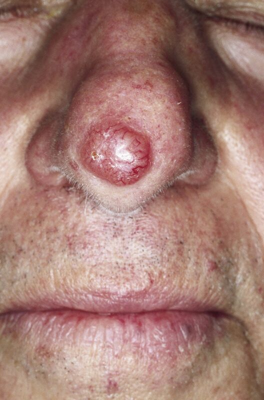
Nodular BCC often has a translucent or pearly surface, fine visible vessels called telangiectasias, and a raised border. Ulceration can develop. These observations suggest BCC but are not proof. Superficial BCC may be a thin red scaly patch on the trunk, pigmented BCC may resemble a melanocytic lesion, and morpheaform BCC may resemble a firm pale scar with an indistinct edge.
Typical histology shows nests of basaloid cells with peripheral palisading, meaning nuclei align along the nest's edge. Retraction spaces may separate nests from surrounding stroma. Morpheaform tumors instead show narrow infiltrating strands in fibrous stroma. Palisading is a microscopic pattern that supports diagnosis in context. It is not an established optical explanation for a pearly surface.
BCC rarely metastasizes, but untreated local growth can destroy tissue and compromise the eyelid, nose or other structures. A slowly enlarging tumor is therefore not harmless. Infiltrating strands can extend beyond the clinically visible boundary, making margin assessment especially important for recurrent or poorly defined tumors.
Surgery is the mainstay for localized disease. Standard excision suits many lower-risk tumors. Mohs micrographic surgery uses mapped examination of peripheral and deep surgical margins to direct additional excision while conserving uninvolved tissue. Recurrence, infiltrative histology, location, size and functional consequences guide its selection. A facial location alone is not a complete treatment plan. Selected superficial low-risk lesions may receive nonsurgical treatment when appropriate, with different clearance expectations. AAD BCC guidance
Hedgehog signaling is frequently activated in BCC. PTCH1 normally restrains Smoothened signaling; loss of that restraint can support tumor growth. Vismodegib directly inhibits Smoothened. Its US label covers adults with metastatic BCC, or locally advanced BCC that has recurred after surgery or is not suitable for surgery and is not suitable for radiation. Official vismodegib label This pathway association does not replace histologic diagnosis or justify systemic treatment for an easily resectable lesion. NCI BCC treatment
Consider a recurrent nasal scar with thin tumor strands extending through fibrous tissue. The problem is not merely the scar's color. The combination predicts that surface inspection may underestimate the tumor boundary.
What feature of margin-controlled surgery addresses that problem?
Mapped margin examination identifies residual tumor beyond the visible edge while limiting unnecessary tissue loss.
Nodular basal cell carcinoma from a published review of nasal lesions. Notice the rounded shiny surface and small vessels. Image: Not every basal cell carcinoma shares this appearance. M. Sand, D. Sand, C. Thrandorf, V. Paech, P. Altmeyer and F. G. Bechara; CC BY 2.0. Existing reduced-size copy, compared visually with the published original. No new alteration.
Case 1
Show answer and explanations for case 1
A. Destruction of the visible surface with cryotherapy (Why this does not fit)
Thin dermal strands may extend beyond the visible surface.
What does recurrent infiltrative growth require for reliable clearance?
A recurrent infiltrative tumor needs reliable margin assessment.
Read the complete reasoning
It can treat selected superficial skin cancers. Thin dermal strands may extend beyond the visible surface. A recurrent infiltrative tumor needs reliable margin assessment.
B. Mapped peripheral and deep margin assessment during excision (Best answer)
What do indistinct borders suggest about visible tumor size?
Visible boundaries may underestimate tumor extent.
How does mapped histology address that uncertainty?
It identifies involved peripheral and deep margins.
Visible boundaries may underestimate tumor extent. It identifies involved peripheral and deep margins. Directed additional excision conserves uninvolved tissue.
C. Topical treatment of the surrounding sun-damaged field (Why this does not fit)
What problem does field treatment address?
It treats selected superficial lesions across damaged epidermis.
Where is this tumor growing?
The biopsy shows infiltrating strands in dermal stroma.
What distinction determines treatment?
Field treatment does not substitute for clearance of an infiltrative recurrence.
Read the complete reasoning
It treats selected superficial lesions across damaged epidermis. The biopsy shows infiltrating strands in dermal stroma. Field treatment does not substitute for clearance of an infiltrative recurrence.
D. Excision limited to the visibly pale tissue (Why this does not fit)
Why might the pale area guide an initial surgical estimate?
It identifies the clinically apparent lesion.
What makes that estimate incomplete?
Morpheaform growth may extend beyond clinically apparent borders.
What must determine clearance?
Histologic margins matter more than visible color boundaries.
Read the complete reasoning
It identifies the clinically apparent lesion. Morpheaform growth may extend beyond clinically apparent borders. Histologic margins matter more than visible color boundaries.
Takeaway: Recurrence plus infiltrative growth favors careful margin control and tissue conservation.
Squamous atypia and dermal invasion answer different questions
Actinic keratosis (AK) is a UV-associated keratinocytic lesion with potential to progress to cSCC. It often feels rough on sun-damaged skin. Many AKs never become invasive cancer, and a universal per-lesion lifetime percentage is misleading. A tender, thickening or persistently ulcerated lesion deserves reassessment rather than automatic repeated field treatment. cSCC can resemble an AK clinically. Isolated AKs may receive lesion-directed treatment such as cryotherapy; broader fields may receive topical or photodynamic treatment, selected after assessment. NCI actinic keratosis overview
In SCC in situ, also called Bowen disease, malignant keratinocytes remain within the epidermis. Full-thickness atypia may be present, but the basement membrane has not been breached by invasive tumor. Erythroplasia of Queyrat is a historical clinical term for an in situ squamous lesion of the glans. Invasive cSCC extends into dermis. Well-differentiated tumors may form concentric keratin pearls or show intercellular bridges; poorly differentiated tumors may produce little obvious keratin.
Compare two tissue samples with extensive epidermal atypia. In the first, an adequately sampled dermis contains no tumor. In the second, atypical keratinocyte nests extend below the epidermal boundary. The second is invasive even if its surface looks less dramatic. If the first sample contains no meaningful dermis, it cannot provide the same reassurance.
UV-associated cSCC frequently contains alterations in tumor suppressors such as TP53, whose p53 protein helps coordinate cellular responses to DNA damage. Multiple other drivers occur. These are pathway associations, not exclusive genetic labels for each cancer. Primary cSCC genomic study
High-risk features include immunosuppression, recurrence, relevant sites such as the ear or lip, substantial depth, poor differentiation and perineural invasion. Perineural invasion means tumor involves a nerve and may help explain pain, numbness or other neurologic symptoms. cSCC can spread to regional nodes and distant sites. Risk assessment uses the combined findings rather than a single universal metastasis percentage. AAD full cSCC guideline
A new ulcer in a longstanding burn scar raises concern for scar-associated SCC, often termed a Marjolin ulcer. A rapidly enlarging nodule with a central keratin-filled crater raises a keratoacanthoma-type squamous differential. Clinical resemblance or possible regression does not safely exclude invasive cSCC. Obtain adequate tissue rather than watching for spontaneous resolution. A chronic inflammatory scar is itself a high-risk cSCC setting, even when a tumor is small and well differentiated. AAD cSCC risk framework
Surgery generally offers the most effective treatment for localized invasive disease. High-risk tumors often warrant margin-controlled treatment and tailored regional assessment. Sentinel lymph node biopsy has uncertain value in cSCC and is not automatically required by a high-risk label. Topical treatment for an epidermal lesion should not be transferred to an invasive tumor. If a new firm focus contains malignant keratinocyte nests in dermis, reassess the original in situ treatment plan. Surface clearing cannot establish clearance of that deeper component. AAD cSCC treatment principlesAAD staging and biopsy recommendations
Why can a superficial sample underestimate an ulcerated squamous lesion?
The sample may identify epidermal atypia without reaching the deeper invasive component.
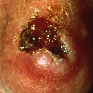
Cutaneous squamous cell carcinoma in a published National Cancer Institute image crop. Ulceration and crusting warrant assessment but are not exclusive to this diagnosis. The historical image is shown at its available resolution. National Cancer Institute, USA; published crop by Mikael Häggström; CC0 1.0. Existing published crop retained. No new alteration.
Case 5
Show answer and explanations for case 5
A. Repeat field treatment for all patches without further sampling (Why this does not fit)
Why is field treatment plausible for the surrounding patches?
Multiple AKs may be managed with field therapy.
What makes this lesion different?
It has new tenderness, induration and persistent ulceration.
Should new induration and ulceration be treated as unchanged AK?
A changing lesion needs reassessment before repeating its prior treatment.
Read the complete reasoning
Multiple AKs may be managed with field therapy. It has new tenderness, induration and persistent ulceration. A changing lesion needs reassessment before repeating its prior treatment.
B. Obtain an adequately deep specimen from the changing lesion (Best answer)
What is the clinically important question raised by induration?
An invasive component must be considered.
Why did the first sample fail to answer it?
No evaluable dermis was included.
What would improve the evidence?
Adequate deeper sampling can assess invasion.
Read the complete reasoning
An invasive component must be considered. No evaluable dermis was included. Adequate deeper sampling can assess invasion.
C. Assign an in situ stage from the absence of dermal tumor in this specimen (Why this does not fit)
Why might absent dermal tumor seem reassuring?
An adequate specimen without invasion could support a noninvasive interpretation.
What limits this specimen?
Dermis was not available for evaluation.
Can unsampled dermis be interpreted as free of tumor?
Unexamined tissue cannot be treated as tumor-free tissue.
Read the complete reasoning
An adequate specimen without invasion could support a noninvasive interpretation. Dermis was not available for evaluation. Unexamined tissue cannot be treated as tumor-free tissue.
D. Repeat superficial sampling from an adjacent flat scalp patch (Why this does not fit)
What could an adjacent patch sample establish?
It could characterize another lesion in the sun-damaged field.
Which lesion carries the unresolved concern for invasion?
The tender ulcerated patch has developed a firm base.
Where must the next specimen reach?
Sample that changing lesion deeply enough to evaluate its dermis.
Read the complete reasoning
It could characterize another lesion in the sun-damaged field. The tender ulcerated patch has developed a firm base. Sample that changing lesion deeply enough to evaluate its dermis.
Takeaway: A specimen without dermis cannot exclude dermal invasion in a changing keratotic lesion.
Melanoma recognition must include atypical presentations
Melanoma may be pigmented, variably colored or amelanotic, meaning it lacks obvious pigment. Nodular tumors may grow predominantly downward while remaining relatively symmetric. Superficial spreading melanoma commonly has an epidermal spreading component. These growth patterns help explain presentation, but none supplies a prognosis without staging.
Lentigo maligna is melanoma in situ on chronically sun-damaged skin, often presenting as a slowly enlarging facial patch. Once dermal invasion is present, it is invasive melanoma. A newly raised focus within a longstanding patch deserves particular attention because partial sampling of only a flat area can miss invasion. A long history is not proof that the entire lesion remains in situ.
Acral melanoma involves palms, soles or the nail apparatus and occurs in all skin tones. It represents different proportions of melanoma across populations; this does not mean people with darker skin have a greater absolute melanoma incidence. A widening irregular nail band, nail destruction or pigment extending onto adjacent nail-fold skin warrants assessment. Nail-fold pigment supports concern but is not independently diagnostic. A longitudinal band can originate in the nail matrix, the tissue that produces the nail plate. A clipping of the pigmented plate alone does not adequately assess that source. A clinician skilled in nail-unit biopsy selects the appropriate underlying tissue sample. AAD nail-apparatus samplingNCI recognition across skin tones
Many melanomas arise without a recognized pre-existing nevus. Dysplastic nevi are risk markers, not obligatory intermediate lesions. Histology evaluates atypical melanocytes, architectural disorder and invasion. S100, HMB-45 and Melan-A are immunohistochemical markers that can support melanocytic differentiation. Their results require a panel and morphologic context; one positive stain does not establish malignancy. AAD melanoma diagnosis guideline
Breslow thickness measures the vertical distance from the top of the granular layer, or the ulcer base when ulcerated, to the deepest invasive melanoma cell. Ulceration is loss of the overlying epidermis related to the tumor. Prognosis integrates thickness, ulceration, regional disease and distant metastases. Clark level describes anatomic invasion levels but does not replace modern thickness-based staging. NCI melanoma staging
For an ulcerated tumor, measure from the ulcer base rather than adding the thickness of crust or reconstructing the absent epidermis from adjacent skin. Compare a thin nonulcerated melanoma with a melanoma of the same thickness that is ulcerated. The depth is identical, but their primary-tumor categories may differ. Compare two tumors of the same subtype with different nodal findings, and subtype again cannot settle prognosis.
Does a small surface diameter establish a thin melanoma?
No. Surface diameter does not measure the deepest invasive cell.
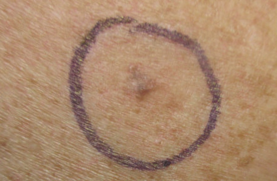
This small shoulder lesion is melanoma. The purple circle is an examination mark, not the lesion border. Image: A subtle appearance cannot establish benignity or provide tumour thickness. Dermanonymous; CC BY-SA 4.0. The examination mark is present in the published original. No new alteration.
Choose a sample that can measure what matters
For suspected melanoma, a narrow complete biopsy with adequate depth is preferred when feasible. It captures the full visible lesion and extends beneath its anticipated base. An elliptical excision, punch excision encompassing a small lesion, or sufficiently deep saucerization can accomplish this. Deep saucerization is not the same as a superficial shave.
A narrow diagnostic margin, commonly 1 to 3 mm, supports diagnosis and microstaging. It is different from the definitive wide-excision margin chosen after pathology. Taking a large definitive margin before knowing thickness can complicate later planning. Selected large lesions or constrained sites may require a carefully directed partial biopsy, with the sampling limitation communicated to pathology. AAD diagnostic biopsy recommendations
Imagine a specimen showing invasive melanoma to a depth of 0.7 mm, with tumor touching the cut base. The report may establish a thickness of at least 0.7 mm. It cannot establish that 0.7 mm is the true maximum. Deeper remaining tumor could alter staging and the discussion of nodal assessment. A reassuring-looking number does not repair an incomplete sample. If a raised residual component can be completely sampled, a narrow complete biopsy can clarify microstaging before final treatment planning. AAD incomplete microstaging guidance
Complete deep saucerization with a clear base can already supply adequate microstaging; its technique name alone is not a reason to repeat an ellipse. AAD complete biopsy techniques
The same geometry matters in a keratotic plaque. An epidermal fragment can demonstrate atypia, yet fail to show whether tumor enters dermis. The desired specimen depends on the diagnostic concern, lesion characteristics and site. Repeat or additional sampling may be necessary when the first specimen cannot answer the clinical question. AAD cSCC biopsy adequacy
After adequately staged melanoma, definitive clinical margins generally increase with thickness. In situ disease commonly starts with 0.5 to 1 cm, invasive disease up to 1 mm with 1 cm, disease greater than 1 to 2 mm with 1 to 2 cm, and thicker disease with 2 cm. Lentigo maligna and constrained sites may need specialized margin assessment. These AAD planning ranges apply to the US-clinic examples below. A clear 1 to 3 mm diagnostic margin does not replace the definitive margin. Local margins follow primary thickness; regional nodal involvement changes overall stage and may change adjuvant treatment discussions. A microscopic positive node prompts a stage-specific assessment, not an automatic medication decision. NCI stage-specific treatmentAAD excision guidance
If sampling ends above the tumor's deepest cell, what remains unknown?
The maximum invasive thickness remains unknown, even when the sampled tissue already proves melanoma.
Biopsy Depth Lab
Upper region: epidermis. Lower region: dermis. Lateral extent stays fixed and sufficient in this model. Shading is sampled tissue; the dashed line is the cut. This is qualitative tissue geometry, not procedural guidance or millimetre thresholds. Full-depth narrow complete sampling can use several methods, including deep saucerization.
Shallow sample. The sampled region misses the deepest invasive cells, so the specimen cannot establish the full depth.Transecting sample. The cut line crosses the fixed lesion. Deeper cells remain below the sample, limiting depth assessment.Adequate-depth sample. The sampled region extends beneath the deepest illustrated cell. The lesion base is represented.
Worked comparisons
Shallow sample: The sampled region misses the deepest invasive cells, so the specimen cannot establish the full depth.
Transecting sample: The cut line crosses the fixed lesion. Deeper cells remain below the sample, limiting depth assessment.
Adequate-depth sample: The sampled region extends beneath the deepest illustrated cell. The lesion base is represented.
Distinguish occult-node staging from an already suspicious node. A palpable abnormal regional node needs directed diagnostic assessment, often ultrasound-guided fine-needle aspiration or another suitable tissue sample. Imaging suspicion does not itself prove metastasis. This decision is separate from the primary tumor T category. AAD clinical nodal assessment
Sentinel lymph node biopsy (SLNB) samples a first draining node to detect clinically occult regional melanoma spread. Discussion commonly applies to T1b tumors, defined as 0.8 to 1.0 mm with or without ulceration, or less than 0.8 mm with ulceration. In the AAD framework, discuss and consider SLNB for T1b through an individual benefits-and-risks decision. Routine SLNB is not recommended for a nonulcerated tumor below 0.8 mm without additional adverse features. Thicker tumors commonly warrant recommendation or consideration according to stage and patient factors. AAD nodal guidance Mitotic activity remains informative but a mitotic rate of one is no longer the old automatic T1 threshold. NCI nodal assessment
A positive sentinel node does not automatically require completion lymph node dissection. In MSLT-II, immediate completion dissection improved regional control without improving melanoma-specific survival compared with structured observation and ultrasound; lymphedema was more frequent after dissection. Suitable patients can undergo specialist-directed nodal ultrasound surveillance. Reliable follow-up matters. This is not equivalent to ignoring a positive node. Original MSLT-II report
BRAF V600 alterations occur in a subset of melanomas. Testing can inform selected advanced or adjuvant therapy, not establish the diagnosis. BRAF plus mitogen-activated protein kinase kinase (MEK) inhibition targets successive signaling proteins; immune checkpoint therapy follows a different treatment logic. Neither a subtype name nor BRAF positivity automatically selects vemurafenib alone. Stage, resectability and patient factors guide specialist treatment. A BRAF result obtained elsewhere does not create a systemic-treatment indication for a definitively excised low-stage melanoma. This example does not recommend routine mutation testing of thin melanoma. NCI systemic treatment
Inherited patterns also guide care. Multiple early BCCs with jaw keratocysts and falx calcification suggest basal cell nevus syndrome, often associated with PTCH1. Medulloblastoma risk requires syndrome-specific counseling. NCI basal nevus syndrome
Xeroderma pigmentosum usually involves nucleotide excision repair defects affecting UV damage; the POLH variant instead affects damage bypass. Xeroderma pigmentosum is generally autosomal recessive, so an affected child can have unaffected parents. Its cancer susceptibility includes melanoma as well as keratinocyte cancers; the tumors already observed do not define the whole future risk spectrum. NCI UV-repair disorders
CDKN2A encodes cell-cycle regulatory proteins including p16, which restrains cyclin-dependent kinase activity. CDKN2A-associated familial melanoma can include pancreatic cancer risk and warrants genetic counseling. A concordantly benign biopsy answers a lesion-level question; it does not cancel the inherited predisposition. Monitor stable nevi, sample concerning changes, and continue whole-skin surveillance. Genetics-guided pancreatic surveillance planning addresses risk before symptoms, with timing chosen from the variant and family history. NCI skin cancer genetics
Keep other lineages in view. Merkel cell carcinoma is an aggressive cutaneous neuroendocrine cancer that can present as a rapidly growing firm red-purple nodule. Cytokeratin 20 and neuroendocrine markers such as synaptophysin support a tissue diagnosis in a panel; Merkel cell polyomavirus is associated with many cases, not all. Regional staging matters. NCI Merkel cell guidance
Let Merkel disease extent guide treatment
When the regional examination is negative, sentinel-node assessment can detect occult spread. A palpable suspicious node poses a different question. Sample that node directly, usually by fine-needle aspiration or core biopsy with an appropriate immunopanel, to establish involvement. The skin panel alone does not prove a nodal metastasis. NCCN abnormal-node workupESMO-EURACAN staging guidance
Localized Merkel carcinoma is treated with specialist-planned excision and nodal assessment. Radiation is selected according to recurrence risk and local-control needs. Proven regional disease may need nodal surgery and/or radiation through multidisciplinary planning. For metastatic disease, immune checkpoint therapy is a major systemic option, chosen with patient-specific risks in mind. The localized and metastatic plans are not an automatic three-treatment package for every patient. NCI stage-specific Merkel treatmentESMO-EURACAN treatment recommendations
Use Kaposi extent to choose the tumor treatment
Kaposi sarcoma is associated with human herpesvirus 8 and can involve skin and oral mucosa, especially with immune dysfunction. Its biopsy can show spindle cells and slit-like vascular spaces. Human immunodeficiency virus (HIV)-associated disease requires effective antiretroviral therapy (ART). Limited good-risk disease may regress with ART alone. Tumor-associated edema, ulceration, extensive oral disease or visceral involvement indicate greater tumor burden; symptomatic poor-risk disease generally needs systemic chemotherapy alongside ART. Local therapy can help selected lesions but cannot replace a plan for extensive disease. Lack of respiratory or gastrointestinal symptoms does not by itself exclude visceral involvement. NCI Kaposi staging and treatment
A stable dimpling fibrous papule may be dermatofibroma, a benign fibrous histiocytoma. Dermal tethering can cause dimpling under lateral pressure; dimpling alone cannot clear an evolving lesion. OHSU dermatofibroma teaching Dermatofibrosarcoma protuberans (DFSP) is a locally infiltrative dermal tumor often associated with COL1A1-PDGFB fusion. New growth or discordant pathology warrants reassessment rather than a permanent benign label. Dermatofibroma clinicopathologic studyMedlinePlus tumor genetics
Resectable DFSP needs definitive margin control. Excision must address peripheral and deep extent because infiltrating tumor can extend beyond a visible nodule. A positive deep edge after limited sampling does not establish that unassessed peripheral tissue is clear. Mohs or another appropriately planned excision with histologic margin assessment can provide local control. AAD DFSP treatment Imatinib is a systemic option in selected settings; its US label includes adult unresectable, recurrent and/or metastatic DFSP. A fusion alone does not make systemic therapy preferable for an easily resectable localized tumor. Official imatinib label
After BCC or cSCC, arrange at least annual skin assessment, adjusted upward for individual risk. Melanoma follow-up depends on stage and recurrence risk. Sun protection, avoiding tanning devices and noticing new or changing lesions remain useful after treatment; clear margins do not prevent an unrelated new primary cancer. AAD BCC follow-upAAD cSCC follow-upAAD melanoma follow-up
Why does clear local excision not end skin surveillance?
It treats that tumor, but the patient may still develop another primary skin cancer.
Case 13
Show answer and explanations for case 13
A. A routine complete regional node dissection should replace sentinel sampling (Why this does not fit)
Why is regional evaluation relevant?
Microscopic nodal disease can be clinically occult.
What procedure is under discussion for clinically negative nodes?
Is complete basin dissection the routine first procedure for clinically negative nodes?
Complete basin dissection is not the routine initial substitute.
Read the complete reasoning
Microscopic nodal disease can be clinically occult. Sentinel-node biopsy provides selective pathologic staging. Complete basin dissection is not the routine initial substitute.
B. No nodal discussion is appropriate unless mitotic rate reaches one (Why this does not fit)
Why might mitotic rate enter this reasoning?
Older thin-melanoma criteria used mitotic activity differently.
What defines this tumor as T1b now?
A thickness of 0.9 mm lies within the 0.8 to 1.0 mm range.
Do absent mitoses negate the T1b sentinel-node discussion?
Absent mitoses do not remove the reason for a sentinel-node discussion.
Read the complete reasoning
Older thin-melanoma criteria used mitotic activity differently. A thickness of 0.9 mm lies within the 0.8 to 1.0 mm range. Absent mitoses do not remove the reason for a sentinel-node discussion.
C. Discuss sentinel-node biopsy because the tumor falls within T1b (Best answer)
Which thickness range contains 0.9 mm?
It is within 0.8 to 1.0 mm.
What T1 category applies despite absent ulceration?
That thickness range qualifies as T1b.
What does the stated AAD guidance support for T1b disease?
Discuss and consider sentinel-node biopsy through an individualized benefits-and-risks decision, rather than mandating the procedure.
Read the complete reasoning
It is within 0.8 to 1.0 mm. That thickness range qualifies as T1b. Discuss and consider sentinel-node biopsy through an individualized benefits-and-risks decision, rather than mandating the procedure.
D. A normal node examination establishes pathologically negative nodes (Why this does not fit)
What does palpation detect?
It can detect clinically enlarged nodes.
What disease can still be present?
Microscopic nodal metastasis may remain undetected.
What is the distinction?
Clinically negative nodes are not the same as pathologically negative nodes.
Read the complete reasoning
It can detect clinically enlarged nodes. Microscopic nodal metastasis may remain undetected. Clinically negative nodes are not the same as pathologically negative nodes.
Takeaway: In the stated AAD framework, T1b includes 0.8 to 1.0 mm melanoma with or without ulceration and supports an individualized sentinel-node discussion, not mandatory biopsy.
A. Chronic eczematous dermatitis (Why this does not fit)
Why is dermatitis a reasonable clinical consideration?
A persistent red scaly patch can resemble eczema.
What does the tissue show instead?
The sample contains epidermis-attached basaloid buds with peripheral palisading.
Which finding takes priority?
The neoplastic architecture outweighs the nonspecific surface scale.
Read the complete reasoning
A persistent red scaly patch can resemble eczema. The sample contains epidermis-attached basaloid buds with peripheral palisading. The neoplastic architecture outweighs the nonspecific surface scale.
B. Squamous cell carcinoma in situ (Why this does not fit)
Why might SCC in situ resemble this lesion?
An epidermal squamous malignancy can form a persistent scaly plaque.
Which cells make up the sampled proliferation?
They are palisading basaloid buds, not the described pattern of full-thickness keratinocyte atypia.
What separates these overlapping plaques?
Use the cellular architecture rather than scale alone to distinguish basal and squamous tumors.
Read the complete reasoning
An epidermal squamous malignancy can form a persistent scaly plaque. They are palisading basaloid buds, not the described pattern of full-thickness keratinocyte atypia. Use the cellular architecture rather than scale alone to distinguish basal and squamous tumors.
C. Superficial basal cell carcinoma (Best answer)
Which architecture identifies the tumor lineage?
Basaloid buds with peripheral palisading support basal cell carcinoma.
How does their distribution fit the clinical lesion?
Their attachment beneath the epidermis fits a superficial BCC presenting as a thin scaly patch.
What is the useful clinical rule?
BCC need not form a raised pearly nodule; a superficial lesion can be plaque-like.
Read the complete reasoning
Basaloid buds with peripheral palisading support basal cell carcinoma. Their attachment beneath the epidermis fits a superficial BCC presenting as a thin scaly patch. BCC need not form a raised pearly nodule; a superficial lesion can be plaque-like.
D. Actinic keratosis (Why this does not fit)
Why is actinic keratosis initially plausible?
A rough scaly epidermal lesion can suggest actinic keratosis.
Does the sampled architecture support that explanation?
The biopsy describes palisading basaloid buds rather than a keratinocyte dysplasia pattern.
What determines the interpretation?
Positive architecture identifies this basal tumor; absence of one squamous pattern alone would not diagnose AK.
Read the complete reasoning
A rough scaly epidermal lesion can suggest actinic keratosis. The biopsy describes palisading basaloid buds rather than a keratinocyte dysplasia pattern. Positive architecture identifies this basal tumor; absence of one squamous pattern alone would not diagnose AK.
Takeaway: Superficial BCC can look like a scaly plaque; use its basaloid architecture to resolve the clinical overlap.
A. Excise A locally; consider Smoothened inhibition for B. (Best answer)
What signaling restraint is lost in these tumors?
PTCH1 loss permits Smoothened-mediated Hedgehog signaling.
Does the shared alteration displace feasible local treatment for A?
The small resectable primary remains suitable for local excision.
Which supplied circumstances make targeted systemic treatment relevant to B?
The recurrence is locally advanced and unsuitable for further local treatment.
Which inhibitor directly addresses that pathway in this setting?
Vismodegib inhibits Smoothened in eligible advanced BCC.
Read the complete reasoning
PTCH1 loss permits Smoothened-mediated Hedgehog signaling. The small resectable primary remains suitable for local excision. The recurrence is locally advanced and unsuitable for further local treatment. Vismodegib inhibits Smoothened in eligible advanced BCC.
B. Consider Smoothened inhibition for A as well as B. (Why this does not fit)
Why would the same drug appear relevant to both tumors?
Both tumors have the same Hedgehog-pathway alteration.
What treatment opportunity remains for A?
A can undergo complete local excision.
What must accompany a targetable alteration before systemic treatment is selected?
Disease setting must justify systemic therapy.
Read the complete reasoning
Both tumors have the same Hedgehog-pathway alteration. A can undergo complete local excision. Disease setting must justify systemic therapy.
C. Excise A locally; consider EGFR inhibition for B. (Why this does not fit)
Why might EGFR inhibition enter a skin-cancer treatment discussion?
EGFR-directed treatment has been used in selected squamous carcinomas.
Which pathway is altered in B?
PTCH1 loss affects Hedgehog signaling.
Which distinction excludes this as the requested direct treatment?
EGFR inhibition does not directly inhibit Smoothened.
Read the complete reasoning
EGFR-directed treatment has been used in selected squamous carcinomas. PTCH1 loss affects Hedgehog signaling. EGFR inhibition does not directly inhibit Smoothened.
D. Excise A locally; consider PD-1 blockade for B. (Why this does not fit)
Why is immune therapy a credible advanced-cancer alternative?
Checkpoint blockade can support antitumor immune activity.
What specific treatment property does this question request?
The requested drug directly inhibits the pathway disinhibited by PTCH1 loss.
Why does PD-1 blockade not supply that mechanism?
An immune checkpoint target is distinct from Smoothened.
Read the complete reasoning
Checkpoint blockade can support antitumor immune activity. The requested drug directly inhibits the pathway disinhibited by PTCH1 loss. An immune checkpoint target is distinct from Smoothened.
Takeaway: A targetable pathway and an appropriate disease setting are separate requirements for systemic treatment.
A. Treat with topical fluorouracil because the surface diameter is small (Why this does not fit)
Why might the small diameter seem reassuring?
Small size can be one favorable risk feature.
What supplied pathology defeats a superficial treatment plan?
The tumor is invasive with perineural involvement.
What determines treatment depth?
Invasive high-risk disease needs treatment beyond the epidermal surface.
Read the complete reasoning
Small size can be one favorable risk feature. The tumor is invasive with perineural involvement. Invasive high-risk disease needs treatment beyond the epidermal surface.
B. Classify as low risk because regional nodes are not palpable (Why this does not fit)
What does a negative node examination establish?
No regional adenopathy is clinically detected.
Does that erase the primary tumor risk factors?
Poor differentiation and perineural invasion still indicate increased risk.
What must remain part of assessment?
Tumor pathology and host immune status remain relevant despite nonpalpable nodes.
Read the complete reasoning
No regional adenopathy is clinically detected. Poor differentiation and perineural invasion still indicate increased risk. Tumor pathology and host immune status remain relevant despite nonpalpable nodes.
C. Require sentinel-node biopsy solely because the tumor is high risk (Why this does not fit)
Why consider regional staging here?
The combined findings increase concern for regional spread.
What is uncertain about sentinel-node biopsy in cSCC?
Its clinical value is not established for routine use.
Does a high-risk cSCC label establish a mandatory sentinel procedure?
High-risk cSCC prompts individualized assessment rather than an automatic sentinel procedure.
Read the complete reasoning
The combined findings increase concern for regional spread. Its clinical value is not established for routine use. High-risk cSCC prompts individualized assessment rather than an automatic sentinel procedure.
D. Arrange margin-controlled treatment and individualized regional assessment (Best answer)
Perineural invasion can extend tumor along a nerve.
What plan integrates these findings?
Margin control and tailored regional assessment address different components of risk.
Read the complete reasoning
Transplant-related immunosuppression increases cSCC risk. Perineural invasion can extend tumor along a nerve. Margin control and tailored regional assessment address different components of risk.
Takeaway: Primary-tumor risk is not erased by small size or a normal node examination.
A. Complete the topical course, using surface clearance to assess the firm focus. (Why this does not fit)
Why was topical treatment initially considered?
The initial sample supported an epidermal in situ lesion.
What does the second specimen establish?
Malignant keratinocytes now extend into the dermis.
Can surface clearance establish control of that component?
Resolution of scale cannot establish clearance of invasive cSCC.
Read the complete reasoning
The initial sample supported an epidermal in situ lesion. Malignant keratinocytes now extend into the dermis. Resolution of scale cannot establish clearance of invasive cSCC.
B. Plan excision, with a routine sentinel-node biopsy because invasion is present. (Why this does not fit)
Which part of this plan responds to dermal tumor?
Excision addresses the invasive primary lesion.
Does any dermal invasion establish a routine sentinel-node indication in cSCC?
The clinical value of routine cSCC sentinel-node biopsy remains uncertain.
What should guide regional assessment instead?
Regional assessment should follow the combined tumor and patient risk.
Read the complete reasoning
Excision addresses the invasive primary lesion. The clinical value of routine cSCC sentinel-node biopsy remains uncertain. Regional assessment should follow the combined tumor and patient risk.
C. Plan surgical clearance, using the invasive tumor risk assessment to select the approach. (Best answer)
How does the dermal focus change the original classification?
The new specimen demonstrates invasive cSCC.
Why must the planned epidermal treatment be reconsidered?
Topical fluorouracil does not provide definitive treatment for this invasive component.
What now determines the surgical approach?
Invasive-tumor risk features guide the choice of excision and margin assessment.
Read the complete reasoning
The new specimen demonstrates invasive cSCC. Topical fluorouracil does not provide definitive treatment for this invasive component. Invasive-tumor risk features guide the choice of excision and margin assessment.
D. Use cryotherapy for the firm focus and topical treatment for the remaining plaque. (Why this does not fit)
Why might treating the two visible areas separately seem reasonable?
The plaque contains clinically different components.
What has been demonstrated beneath the firm surface?
The focus contains invasive dermal carcinoma.
Why favor surgery in this fit patient?
Surgical clearance provides treatment with histologic margin assessment.
Read the complete reasoning
The plaque contains clinically different components. The focus contains invasive dermal carcinoma. Surgical clearance provides treatment with histologic margin assessment.
Takeaway: A new invasive component changes the treatment question, even when an earlier sample was in situ.
A. Use high-risk local treatment with tailored regional assessment, accounting for the chronic-scar origin. (Best answer)
What unfavorable context is not captured by diameter or differentiation?
The carcinoma arose in a chronic inflammatory scar.
How does that change the primary-tumor plan?
The scar origin supports high-risk local-control planning.
What does the normal node examination permit us to conclude?
It establishes no palpable adenopathy, not absence of occult disease.
How should regional evaluation be chosen?
Regional evaluation is individualized rather than automatically requiring SLNB.
Read the complete reasoning
The carcinoma arose in a chronic inflammatory scar. The scar origin supports high-risk local-control planning. It establishes no palpable adenopathy, not absence of occult disease. Regional evaluation is individualized rather than automatically requiring SLNB.
B. Use low-risk local excision with routine skin follow-up, prioritizing size and differentiation. (Why this does not fit)
Why is a low-risk approach tempting?
Small size and good differentiation are favorable findings.
Which supplied feature prevents them from settling the classification?
The tumor arose in a longstanding burn scar.
How should competing risk findings be integrated?
Favorable features do not cancel a high-risk chronic-inflammatory setting.
Read the complete reasoning
Small size and good differentiation are favorable findings. The tumor arose in a longstanding burn scar. Favorable features do not cancel a high-risk chronic-inflammatory setting.
C. Use high-risk local treatment with sentinel-node biopsy as the standard regional procedure. (Why this does not fit)
Why does regional assessment deserve consideration?
Scar-associated cSCC can behave aggressively.
What evidence limitation applies to routine sentinel sampling?
A clinical benefit from routine SLNB in cSCC is not established.
What distinction should guide planning?
High primary-tumor risk does not dictate a universal nodal procedure.
Read the complete reasoning
Scar-associated cSCC can behave aggressively. A clinical benefit from routine SLNB in cSCC is not established. High primary-tumor risk does not dictate a universal nodal procedure.
D. Use low-risk local excision now, escalating the risk pathway if regional nodes enlarge. (Why this does not fit)
Why could the node examination feel reassuring?
No regional adenopathy is palpable.
Which primary-tumor risk exists before nodes enlarge?
The chronic-scar origin is already a high-risk feature.
When should that feature affect local treatment?
Primary-tumor risk should guide initial clearance planning.
Read the complete reasoning
No regional adenopathy is palpable. The chronic-scar origin is already a high-risk feature. Primary-tumor risk should guide initial clearance planning.
Takeaway: Chronic-scar origin can outweigh favorable size and differentiation without making sentinel-node biopsy automatic.
A. Observe for spontaneous regression before obtaining more tissue (Why this does not fit)
Why might observation seem reasonable?
Some keratoacanthoma-type lesions can regress.
What uncertainty remains in this patient?
The small superficial specimen did not assess the base or full architecture of the persisting squamous lesion.
Why not rely on regression here?
A possible future change does not resolve the current uncertainty about invasive carcinoma.
Read the complete reasoning
Some keratoacanthoma-type lesions can regress. The small superficial specimen did not assess the base or full architecture of the persisting squamous lesion. A possible future change does not resolve the current uncertainty about invasive carcinoma.
B. Repeat a superficial sample of the central keratin plug (Why this does not fit)
What could another central sample show?
It could provide more keratin or superficial squamous cells.
Which information is missing now?
The lesion base and architectural relationships were not included.
Would repeating the same depth solve that problem?
A second superficial central sample may repeat the same limitation rather than assess invasion.
Read the complete reasoning
It could provide more keratin or superficial squamous cells. The lesion base and architectural relationships were not included. A second superficial central sample may repeat the same limitation rather than assess invasion.
C. Request deeper sections of the existing superficial specimen instead of obtaining additional lesion tissue (Why this does not fit)
What can deeper laboratory sections examine?
They can examine additional levels within tissue already submitted.
Was the missing lesion base included in that tissue?
No. The stem states that the original superficial specimen lacks the base and full architecture.
Can deeper cuts recover tissue never sampled?
No. Additional lesion sampling is needed to address a missing base; recutting the same superficial specimen cannot supply that tissue.
Read the complete reasoning
They can examine additional levels within tissue already submitted. No. The stem states that the original superficial specimen lacks the base and full architecture. No. Additional lesion sampling is needed to address a missing base; recutting the same superficial specimen cannot supply that tissue.
D. Obtain additional tissue that includes the lesion base and architecture (Best answer)
Which differential matters for the persisting nodule?
A keratoacanthoma-type squamous proliferation can overlap invasive cSCC.
What prevented an adequate distinction?
The first sample lacked the lesion base and full architecture.
What should additional sampling accomplish?
Obtain adequate lesion tissue, by an appropriate biopsy or excision, to assess architecture and invasion.
Read the complete reasoning
A keratoacanthoma-type squamous proliferation can overlap invasive cSCC. The first sample lacked the lesion base and full architecture. Obtain adequate lesion tissue, by an appropriate biopsy or excision, to assess architecture and invasion.
Takeaway: A persisting crateriform squamous lesion needs adequate architectural and depth assessment when the first sample is insufficient.
A. Schedule routine review because it is smaller than 6 mm (Why this does not fit)
Why might the diameter seem reassuring?
The ABCDE aid includes diameter greater than 6 mm.
What does the documented evolution change?
Growth and bleeding warrant assessment even in a smaller lesion.
Can diameter below 6 mm safely exclude melanoma?
The diameter aid is not a rule-out threshold.
Read the complete reasoning
The ABCDE aid includes diameter greater than 6 mm. Growth and bleeding warrant assessment even in a smaller lesion. The diameter aid is not a rule-out threshold.
B. Use dermoscopic photographs to reassess the growth in six months (Why this does not fit)
Why might photographic monitoring be considered?
It can document change in selected lesions.
What change is already documented here?
The lesion is rapidly growing and bleeding.
Should prolonged monitoring precede tissue assessment of this unresolved growing lesion?
Monitoring should not delay tissue diagnosis of an unresolved concerning lesion.
Read the complete reasoning
It can document change in selected lesions. The lesion is rapidly growing and bleeding. Monitoring should not delay tissue diagnosis of an unresolved concerning lesion.
C. Obtain an appropriate tissue biopsy despite the atypical appearance (Best answer)
What makes a stable benign interpretation uncertain?
The lesion is newly growing and bleeding.
Can absent pigment exclude melanoma?
Amelanotic melanoma may have no obvious brown pigment.
What resolves the uncertain lineage?
An appropriately planned biopsy supplies diagnostic tissue.
Read the complete reasoning
The lesion is newly growing and bleeding. Amelanotic melanoma may have no obvious brown pigment. An appropriately planned biopsy supplies diagnostic tissue.
D. Cauterize it as a presumed pyogenic granuloma without obtaining tissue (Why this does not fit)
Why might a vascular benign mimic be considered?
A bleeding pink papule can resemble pyogenic granuloma.
What prevents accepting that explanation here?
The clinician has not established a benign diagnosis for the growing lesion.
What would destruction without a specimen leave unresolved?
The lesion would remain histologically uncharacterized.
Read the complete reasoning
A bleeding pink papule can resemble pyogenic granuloma. The clinician has not established a benign diagnosis for the growing lesion. The lesion would remain histologically uncharacterized.
Takeaway: Small, symmetric or nonpigmented lesions can still require biopsy when their evolution is concerning.
A. Repeat diagnostic sampling by ellipse before using the reported thickness for planning. (Why this does not fit)
Why might the word saucerization raise concern?
A superficial shave can transect an invasive melanoma.
What does pathology establish about this specimen instead?
This deep saucerization captured the entire melanoma with a clear base.
What determines whether another diagnostic biopsy is needed?
Diagnostic adequacy depends on the specimen, not a blanket ban on saucerization.
Read the complete reasoning
A superficial shave can transect an invasive melanoma. This deep saucerization captured the entire melanoma with a clear base. Diagnostic adequacy depends on the specimen, not a blanket ban on saucerization.
B. Accept microstaging, plan a 1 cm definitive clinical margin, and discuss SLNB. (Best answer)
Can this diagnostic specimen support microstaging?
Complete sampling with a clear base supports use of the reported thickness.
What local treatment follows a 0.9 mm arm melanoma?
A 1 cm definitive clinical excision margin is recommended in the stated AAD framework.
What nodal decision follows its T1b thickness?
SLNB warrants an individualized benefits-and-risks discussion.
Read the complete reasoning
Complete sampling with a clear base supports use of the reported thickness. A 1 cm definitive clinical excision margin is recommended in the stated AAD framework. SLNB warrants an individualized benefits-and-risks discussion.
C. Accept microstaging and enter surveillance because the 2 mm diagnostic margins are clear. (Why this does not fit)
Why could clear edges suggest the procedure is finished?
No tumor reaches the diagnostic specimen edges.
What was the purpose of its narrow 2 mm margin?
The initial procedure was diagnostic sampling.
What additional local treatment remains?
Clear diagnostic edges do not replace the recommended definitive excision margin.
Read the complete reasoning
No tumor reaches the diagnostic specimen edges. The initial procedure was diagnostic sampling. Clear diagnostic edges do not replace the recommended definitive excision margin.
D. Accept microstaging, plan a 2 cm definitive margin, and omit SLNB discussion below 1 mm. (Why this does not fit)
Why might a wider margin seem more protective?
Definitive margins increase for thicker primary melanomas.
Which margin applies to this 0.9 mm primary?
The AAD recommendation for this T1 arm melanoma is 1 cm.
Why is the nodal threshold also misapplied?
A 0.9 mm melanoma supports T1b SLNB discussion despite being below 1 mm.
Read the complete reasoning
Definitive margins increase for thicker primary melanomas. The AAD recommendation for this T1 arm melanoma is 1 cm. A 0.9 mm melanoma supports T1b SLNB discussion despite being below 1 mm.
Takeaway: Adequate deep saucerization can complete diagnosis; definitive excision and nodal discussion remain separate decisions.
A. Use 0.7 mm as the maximum and proceed with a thin-melanoma excision plan. (Why this does not fit)
Why does 0.7 mm initially suggest a thin-tumor plan?
It is below the 0.8 mm T1b thickness threshold.
What makes this number an unreliable maximum?
Melanoma reaches the cut base beneath a residual raised component.
What should not be finalized from a truncated sample?
Final thickness-dependent decisions require adequate microstaging.
Read the complete reasoning
It is below the 0.8 mm T1b thickness threshold. Melanoma reaches the cut base beneath a residual raised component. Final thickness-dependent decisions require adequate microstaging.
B. Repeat a shallow peripheral biopsy to confirm the diagnosis before definitive excision. (Why this does not fit)
What diagnostic information could another peripheral sample reproduce?
It could again demonstrate melanocytic malignancy.
What question is already answered?
The existing biopsy has established melanoma.
Which unanswered question must the next specimen address?
The remaining deepest extent must be assessed.
Read the complete reasoning
It could again demonstrate melanocytic malignancy. The existing biopsy has established melanoma. The remaining deepest extent must be assessed.
C. Use a 2 cm definitive margin now, treating the positive base as evidence of thick melanoma. (Why this does not fit)
Why might a positive base prompt concern about a larger margin?
Additional invasive melanoma may lie below the specimen.
Does that demonstrate a thickness greater than 2 mm?
A positive base supplies no measured maximum thickness.
What prevents choosing a definitive range from that uncertainty alone?
Incomplete sampling is not a measured thick-tumor category.
Read the complete reasoning
Additional invasive melanoma may lie below the specimen. A positive base supplies no measured maximum thickness. Incomplete sampling is not a measured thick-tumor category.
D. Completely sample the remaining raised lesion narrowly before final thickness-based planning. (Best answer)
What does the cut-through measurement establish?
The melanoma is at least 0.7 mm thick.
Why could additional depth matter for the team?
A greater maximum thickness could alter the staging discussion.
Which feasible sampling plan answers that question?
Narrow complete sampling of the residual raised component can clarify microstaging.
Read the complete reasoning
The melanoma is at least 0.7 mm thick. A greater maximum thickness could alter the staging discussion. Narrow complete sampling of the residual raised component can clarify microstaging.
Takeaway: When residual tumor prevents reliable microstaging, select additional diagnostic sampling that can change the treatment plan.
A. Record 0.6 mm; discuss and consider SLNB using the ulcerated T1b classification. (Best answer)
Which upper reference applies after tumor-related ulceration?
Breslow thickness starts at the ulcer base.
Which supplied distance should be recorded?
The correct thickness is 0.6 mm.
How does ulceration change the category at that depth?
Ulceration makes this sub-0.8 mm melanoma T1b.
What does that category support under AAD guidance?
SLNB should be discussed and considered individually.
Read the complete reasoning
Breslow thickness starts at the ulcer base. The correct thickness is 0.6 mm. Ulceration makes this sub-0.8 mm melanoma T1b. SLNB should be discussed and considered individually.
B. Record 0.6 mm; omit routine SLNB discussion using the thin T1a classification. (Why this does not fit)
Which part of this choice uses the correct tissue reference?
The ulcer-based measurement is 0.6 mm.
What prevents T1a classification?
Confirmed tumor-related ulceration is present.
Which rule applies below 0.8 mm?
Thin ulcerated melanoma is T1b and supports a nodal discussion.
Read the complete reasoning
The ulcer-based measurement is 0.6 mm. Confirmed tumor-related ulceration is present. Thin ulcerated melanoma is T1b and supports a nodal discussion.
C. Record 0.9 mm; discuss and consider SLNB using the thickness-based T1b classification. (Why this does not fit)
Why might the neighboring granular layer seem useful?
It retains the normal upper reference for nonulcerated tumors.
Why is that reference wrong here?
The tumor has lost its overlying epidermis through ulceration.
What must remain accurate even if the nodal discussion is similar?
Record the actual ulcer-based thickness rather than substituting adjacent skin.
Read the complete reasoning
It retains the normal upper reference for nonulcerated tumors. The tumor has lost its overlying epidermis through ulceration. Record the actual ulcer-based thickness rather than substituting adjacent skin.
D. Record 0.8 mm; discuss and consider SLNB after including the overlying crust. (Why this does not fit)
How does this choice arrive at 0.8 mm?
It adds 0.2 mm of crust to the 0.6 mm tissue measurement.
Does crust form part of Breslow thickness?
Crust is not included in the ulcer-based measurement.
What finding supports discussion without that addition?
Tumor-related ulceration already makes the 0.6 mm lesion T1b.
Read the complete reasoning
It adds 0.2 mm of crust to the 0.6 mm tissue measurement. Crust is not included in the ulcer-based measurement. Tumor-related ulceration already makes the 0.6 mm lesion T1b.
Takeaway: Choose the correct Breslow reference first, then apply ulceration to the nodal discussion.
A. T1a; ultrasound-guided sampling of the abnormal node (Why this does not fit)
Why might the primary be mistaken for T1a?
Its measured thickness is below 0.8 mm.
Which supplied feature changes the subclassification?
Tumor-related ulceration makes this thin primary T1b.
Does the proposed node procedure fit the separate regional finding?
Direct sampling fits the suspicious node, but this option assigns the wrong primary T category.
Read the complete reasoning
Its measured thickness is below 0.8 mm. Tumor-related ulceration makes this thin primary T1b. Direct sampling fits the suspicious node, but this option assigns the wrong primary T category.
B. T1b; ultrasound-guided sampling of the abnormal node (Best answer)
How do thickness and ulceration combine?
A melanoma below 0.8 mm with tumor-related ulceration is T1b.
Is the regional question still an occult-disease screen?
No. A palpable node with suspicious ultrasound features is already present.
What should establish its nature?
Obtain directed sampling of that abnormal node; imaging suspicion alone does not establish metastasis.
Read the complete reasoning
A melanoma below 0.8 mm with tumor-related ulceration is T1b. No. A palpable node with suspicious ultrasound features is already present. Obtain directed sampling of that abnormal node; imaging suspicion alone does not establish metastasis.
C. T1b; sentinel lymph-node biopsy to look for occult disease (Why this does not fit)
Does this option classify the primary correctly?
Yes. Ulceration makes the 0.6 mm primary T1b.
What is the purpose of sentinel staging?
It investigates clinically occult regional nodal involvement in appropriately selected patients.
Which finding changes the immediate procedure here?
The palpable suspicious node needs direct evaluation rather than being treated as a clinically normal basin.
Read the complete reasoning
Yes. Ulceration makes the 0.6 mm primary T1b. It investigates clinically occult regional nodal involvement in appropriately selected patients. The palpable suspicious node needs direct evaluation rather than being treated as a clinically normal basin.
D. T1a; sentinel lymph-node biopsy to look for occult disease (Why this does not fit)
Why is the thin measurement not enough for T1a?
The primary is ulcerated, so its T1 subclassification is T1b.
Is the regional examination normal?
No. A palpable, ultrasound-suspicious inguinal node is described.
Which two distinctions are missed?
Classify the ulcerated primary correctly and directly sample the abnormal node instead of applying an occult-node pathway.
Read the complete reasoning
The primary is ulcerated, so its T1 subclassification is T1b. No. A palpable, ultrasound-suspicious inguinal node is described. Classify the ulcerated primary correctly and directly sample the abnormal node instead of applying an occult-node pathway.
Takeaway: Primary T category and regional-node evaluation are separate decisions; a suspicious palpable node is not an occult-node screening problem.
A. Completion dissection; the regional-control improvement demonstrates the survival gain she requires (Why this does not fit)
Which trial outcome favors completion dissection?
Regional nodal control was better with dissection.
Is regional control the same as melanoma-specific survival?
No. The supplied trial results did not demonstrate a melanoma-specific survival advantage.
Does the claimed benefit meet this patient's priority?
A regional-control gain cannot be relabeled as the demonstrated survival benefit she requires to accept the additional morbidity.
Read the complete reasoning
Regional nodal control was better with dissection. No. The supplied trial results did not demonstrate a melanoma-specific survival advantage. A regional-control gain cannot be relabeled as the demonstrated survival benefit she requires to accept the additional morbidity.
B. Ultrasound surveillance; the similar survival results establish equal regional control with less swelling (Why this does not fit)
Why can surveillance be discussed with this patient?
She has microscopic sentinel-node disease and reliable access to structured nodal monitoring.
Were the regional-control results equal?
No. The reported regional-control rate was lower with observation.
What must the discussion preserve?
Similar observed survival does not mean equal control in the nodal basin; surveillance requires acknowledging that tradeoff.
Read the complete reasoning
She has microscopic sentinel-node disease and reliable access to structured nodal monitoring. No. The reported regional-control rate was lower with observation. Similar observed survival does not mean equal control in the nodal basin; surveillance requires acknowledging that tradeoff.
C. Ultrasound surveillance; accept less regional control to reduce swelling without a demonstrated survival disadvantage (Best answer)
Which outcomes must be separated?
Dissection improved regional control and increased lymphedema, without a demonstrated melanoma-specific survival gain.
Can this patient undertake the alternative used in the trial?
She can attend serial high-quality nodal ultrasound and accepts scheduled monitoring.
Which plan fits her preference while retaining the trial limits?
Discuss structured surveillance to reduce morbidity while accepting less regional control; this is active follow-up, not proof of equivalence for every patient.
Read the complete reasoning
Dissection improved regional control and increased lymphedema, without a demonstrated melanoma-specific survival gain. She can attend serial high-quality nodal ultrasound and accepts scheduled monitoring. Discuss structured surveillance to reduce morbidity while accepting less regional control; this is active follow-up, not proof of equivalence for every patient.
D. Completion dissection; accept greater swelling for improved regional control despite no demonstrated survival gain (Why this does not fit)
Is this option describing the trial tradeoff accurately?
Yes. Dissection improves regional control at the cost of more lymphedema, without a demonstrated survival gain.
What matters to this patient when surveillance is feasible?
She prioritizes avoiding chronic swelling unless surgery offers a demonstrated survival benefit and accepts scheduled ultrasound monitoring.
Why is this accurate tradeoff not the best plan for her stated priorities?
Choosing extra regional control despite greater swelling conflicts with her preference when structured surveillance is available; the procedure remains a discussion option rather than a requirement.
Read the complete reasoning
Yes. Dissection improves regional control at the cost of more lymphedema, without a demonstrated survival gain. She prioritizes avoiding chronic swelling unless surgery offers a demonstrated survival benefit and accepts scheduled ultrasound monitoring. Choosing extra regional control despite greater swelling conflicts with her preference when structured surveillance is available; the procedure remains a discussion option rather than a requirement.
Takeaway: Separate survival, regional control and morbidity, then apply follow-up feasibility and patient priorities rather than assuming one outcome stands for all three.
A. Use the same 1 to 2 cm primary-margin range; B needs a stage III adjuvant-treatment assessment. (Best answer)
Which shared measurement guides the primary-site margin?
Both melanomas are 1.2 mm thick.
What local-margin range follows in the stated framework?
Both fit the AAD 1 to 2 cm range for T2 arm melanomas.
What additional implication follows from B's positive node?
B has regional stage III disease requiring a stage-specific adjuvant assessment.
Does the small deposit mandate adjuvant medication?
Treatment selection still depends on detailed risk and patient factors.
Read the complete reasoning
Both melanomas are 1.2 mm thick. Both fit the AAD 1 to 2 cm range for T2 arm melanomas. B has regional stage III disease requiring a stage-specific adjuvant assessment. Treatment selection still depends on detailed risk and patient factors.
B. Use a 1 cm margin for A and a 2 cm minimum for B; apply the same adjuvant assessment to both. (Why this does not fit)
Why might B seem to need a wider skin margin?
B has proven spread beyond the primary site.
Which measurement selects the primary-margin range?
Primary thickness guides that range.
Where should B's nodal finding affect care instead?
The positive node changes overall stage and the adjuvant assessment.
Read the complete reasoning
B has proven spread beyond the primary site. Primary thickness guides that range. The positive node changes overall stage and the adjuvant assessment.
C. Use the same 1 to 2 cm primary-margin range; apply the same adjuvant indications to both. (Why this does not fit)
Which part of this plan fits the equal primary measurements?
The local-margin range can be the same.
What staging information differs?
Only B has a pathologically positive regional node.
Can matching skin measurements establish matching adjuvant indications?
Adjuvant assessment incorporates overall stage rather than thickness alone.
Read the complete reasoning
The local-margin range can be the same. Only B has a pathologically positive regional node. Adjuvant assessment incorporates overall stage rather than thickness alone.
D. Use a 1 to 2 cm margin for A and a narrower skin margin for B; prioritize B's adjuvant assessment. (Why this does not fit)
Why should B receive additional oncologic assessment?
The regional metastasis changes B's stage.
Does regional spread reduce the primary clearance requirement?
The 1.2 mm primary still needs an appropriate definitive margin.
The regional metastasis changes B's stage. The 1.2 mm primary still needs an appropriate definitive margin. Stage-specific assessment complements adequate primary-site clearance.
Takeaway: Primary thickness guides the skin margin; regional involvement changes overall stage and adjuvant discussions.
A. Apply an in situ treatment plan to the whole lesion without additional assessment (Why this does not fit)
Why is an in situ plan initially plausible?
The sampled flat portion contains melanoma in situ.
What part of the lesion remains uncharacterized?
The newly raised firm focus was not sampled.
Can the flat sample classify a clinically different unsampled focus?
Partial pathology must not be assumed to represent a clinically different unsampled component.
Read the complete reasoning
The sampled flat portion contains melanoma in situ. The newly raised firm focus was not sampled. Partial pathology must not be assumed to represent a clinically different unsampled component.
B. Sample the raised focus adequately to assess for an invasive component (Best answer)
What does the new raised focus raise concern for?
It may contain an invasive component.
Why would that matter for definitive planning?
Invasive thickness can alter excision and staging discussions.
What resolves the uncertainty?
Adequate targeted sampling of that focus assesses invasion.
Read the complete reasoning
It may contain an invasive component. Invasive thickness can alter excision and staging discussions. Adequate targeted sampling of that focus assesses invasion.
C. Repeat a biopsy from another flat area to confirm the in situ finding (Why this does not fit)
What could another flat-area biopsy confirm?
It could reproduce the existing in situ result.
Which clinically different component would still be untested?
The newly raised firm focus would remain unsampled.
Which tissue should resolve the potential change in treatment?
The different raised component needs adequate assessment for invasion.
Read the complete reasoning
It could reproduce the existing in situ result. The newly raised firm focus would remain unsampled. The different raised component needs adequate assessment for invasion.
D. Assume the long history establishes a favorable prognosis (Why this does not fit)
Why might the long history feel reassuring?
Lentigo maligna can remain superficial for a prolonged interval.
What new finding changes that interpretation?
A newly raised firm focus represents recent evolution.
What should determine current risk?
Current tissue findings outweigh an assumed prognosis from duration.
Read the complete reasoning
Lentigo maligna can remain superficial for a prolonged interval. A newly raised firm focus represents recent evolution. Current tissue findings outweigh an assumed prognosis from duration.
Takeaway: A flat in situ sample cannot exclude invasion in a separate newly raised component.
A. Reassure because nail pigmentation is common in darker skin (Why this does not fit)
Why might benign pigmentation be considered?
Benign nail pigmentation occurs in people with darker skin.
Which findings make simple reassurance inadequate?
One band is progressively widening with new nail destruction.
Can common benign pigmentation explain away this individual lesion evolution?
A population tendency does not neutralize concerning individual evolution.
Read the complete reasoning
Benign nail pigmentation occurs in people with darker skin. One band is progressively widening with new nail destruction. A population tendency does not neutralize concerning individual evolution.
B. Diagnose a subungual hematoma and wait for the entire nail to regrow (Why this does not fit)
Why could blood enter the nail differential?
Subungual hemorrhage can produce dark discoloration.
What supplied pattern needs assessment rather than that assumption?
Persistent widening with nail-fold extension and splitting is concerning.
Can this progressive lesion be assigned a traumatic cause without evaluation?
Do not assign a traumatic explanation without resolving a progressive nail lesion.
Read the complete reasoning
Subungual hemorrhage can produce dark discoloration. Persistent widening with nail-fold extension and splitting is concerning. Do not assign a traumatic explanation without resolving a progressive nail lesion.
C. Arrange specialist nail-unit assessment with appropriately planned biopsy (Best answer)
What does this evolution suggest?
A nail-unit malignancy must be considered.
Does nail-fold pigment prove melanoma?
It supports concern but is not independently diagnostic.
What supplies the missing evidence?
Specialist assessment and appropriately planned tissue sampling establish diagnosis.
Read the complete reasoning
A nail-unit malignancy must be considered. It supports concern but is not independently diagnostic. Specialist assessment and appropriately planned tissue sampling establish diagnosis.
D. Send a nail clipping for histology as the sole diagnostic specimen (Why this does not fit)
Why might a clipping seem to target the abnormality?
The pigment is visible in the nail plate.
Where can the underlying melanocytic lesion originate?
A longitudinal pigmented band can arise from the nail matrix.
What tissue limitation makes the clipping insufficient?
A plate clipping does not adequately sample the matrix lesion.
Read the complete reasoning
The pigment is visible in the nail plate. A longitudinal pigmented band can arise from the nail matrix. A plate clipping does not adequately sample the matrix lesion.
Takeaway: Acral melanoma occurs in all skin tones; progressive single-nail changes require assessment, not demographic reassurance.
A. Offer BRAF plus MEK treatment to both, since the shared alteration provides a target. (Why this does not fit)
Why would both results appear actionable?
Both tumors carry a BRAF V600E alteration.
What clinical indication is missing for A?
A has definitively treated low-stage disease without evidence of spread.
What must accompany an actionable alteration?
A systemic-treatment indication must come from the disease setting.
Read the complete reasoning
Both tumors carry a BRAF V600E alteration. A has definitively treated low-stage disease without evidence of spread. A systemic-treatment indication must come from the disease setting.
B. Continue surveillance for A; prefer BRAF monotherapy for B on the basis of genotype alone. (Why this does not fit)
Why is BRAF monotherapy a credible alternative?
It can inhibit the demonstrated BRAF target.
What additional targeted strategy matters for B?
BRAF plus MEK combinations have stronger advanced-disease evidence than BRAF monotherapy.
What selection error remains?
Genotype alone does not justify defaulting to single-agent treatment.
Read the complete reasoning
It can inhibit the demonstrated BRAF target. BRAF plus MEK combinations have stronger advanced-disease evidence than BRAF monotherapy. Genotype alone does not justify defaulting to single-agent treatment.
C. Continue surveillance for A; evaluate B for targeted treatment but exclude checkpoint therapy. (Why this does not fit)
Which part of the proposal fits B's genotype?
BRAF V600E creates a potential targeted-treatment option.
Does it negate B's supplied immune-treatment eligibility?
Checkpoint therapy remains an option in BRAF-mutant melanoma.
How should the two treatment classes be considered?
Specialist selection should integrate disease and patient factors.
Read the complete reasoning
BRAF V600E creates a potential targeted-treatment option. Checkpoint therapy remains an option in BRAF-mutant melanoma. Specialist selection should integrate disease and patient factors.
D. Continue surveillance for A; assess B for BRAF plus MEK therapy or checkpoint therapy. (Best answer)
Does A's external result create an adjuvant indication?
The result does not create a systemic-treatment indication for this treated low-stage tumor.
What makes B's setting different?
B has unresectable metastatic disease requiring systemic-treatment assessment.
How does BRAF V600E refine that assessment?
It permits consideration of combined BRAF and MEK inhibition.
What separate option remains available?
Immune checkpoint treatment remains a stage-appropriate alternative.
Read the complete reasoning
The result does not create a systemic-treatment indication for this treated low-stage tumor. B has unresectable metastatic disease requiring systemic-treatment assessment. It permits consideration of combined BRAF and MEK inhibition. Immune checkpoint treatment remains a stage-appropriate alternative.
Takeaway: Stage establishes the systemic-treatment setting; BRAF refines options within that setting. These external results do not endorse routine testing of thin melanoma.
A. Loss of PTCH1-mediated restraint of Hedgehog signaling (Best answer)
Which combination suggests basal cell nevus syndrome?
Early multiple BCCs with jaw keratocysts and falx calcification support that syndrome.
Which gene commonly explains this pattern?
PTCH1 is a major associated predisposition gene.
Why does the family pattern matter?
It supports counseling for an inherited, often autosomal dominant predisposition.
Read the complete reasoning
Early multiple BCCs with jaw keratocysts and falx calcification support that syndrome. PTCH1 is a major associated predisposition gene. It supports counseling for an inherited, often autosomal dominant predisposition.
B. Loss of CDKN2A-mediated cell-cycle restraint (Why this does not fit)
Why might CDKN2A be considered?
It is associated with familial melanoma predisposition.
Which supplied features favor a different syndrome?
Jaw keratocysts and falx calcification accompany early basal tumors here.
Which associated findings should guide selection of the inherited syndrome?
Match the extracutaneous pattern to the cancer-predisposition syndrome.
Read the complete reasoning
It is associated with familial melanoma predisposition. Jaw keratocysts and falx calcification accompany early basal tumors here. Match the extracutaneous pattern to the cancer-predisposition syndrome.
C. Defective nucleotide excision repair of UV photoproducts (Why this does not fit)
Why might UV repair failure be considered?
It can cause multiple skin cancers early in life.
Which findings more specifically support the proposed basal syndrome?
The jaw and falx findings support basal cell nevus syndrome.
Which is more discriminating here, early onset alone or the combined syndromic findings?
Early skin cancer alone is less specific than the combined syndromic pattern.
Read the complete reasoning
It can cause multiple skin cancers early in life. The jaw and falx findings support basal cell nevus syndrome. Early skin cancer alone is less specific than the combined syndromic pattern.
D. Impaired mismatch repair during DNA replication (Why this does not fit)
Why consider a DNA repair syndrome?
Inherited repair defects can increase cancer susceptibility.
What does the jaw and basal-tumor combination favor?
It favors Hedgehog-pathway predisposition.
What should guide counseling?
The clinical syndrome should direct genetic evaluation rather than a generic cancer-gene label.
Read the complete reasoning
Inherited repair defects can increase cancer susceptibility. It favors Hedgehog-pathway predisposition. The clinical syndrome should direct genetic evaluation rather than a generic cancer-gene label.
Takeaway: Early BCCs plus jaw keratocysts and falx calcification support PTCH1-associated basal cell nevus syndrome.
A. Additional keratinocyte cancers are expected, but melanoma risk remains at baseline. (Why this does not fit)
Why might concern initially focus on keratinocytes?
The child already has two keratinocyte cancers.
What does the functional assay identify?
The assay demonstrates deficient nucleotide excision repair of UV photoproducts.
What is the relevant cancer-spectrum rule?
This XP form also increases melanoma susceptibility.
Read the complete reasoning
The child already has two keratinocyte cancers. The assay demonstrates deficient nucleotide excision repair of UV photoproducts. This XP form also increases melanoma susceptibility.
B. Melanoma risk is increased in addition to the keratinocyte-cancer risk. (Best answer)
Which repair process removes bulky UV photoproducts?
Nucleotide excision repair removes these lesions.
Which disorder fits the assay together with early photosensitivity and cancers?
The findings support a nucleotide-excision-repair form of xeroderma pigmentosum.
What further lineage needs attention?
Melanoma risk is increased as well as keratinocyte-cancer risk.
Read the complete reasoning
Nucleotide excision repair removes these lesions. The findings support a nucleotide-excision-repair form of xeroderma pigmentosum. Melanoma risk is increased as well as keratinocyte-cancer risk.
C. Melanoma risk should be revised upward if an affected first-degree relative is identified. (Why this does not fit)
Why is family history usually relevant to inherited cancer risk?
Affected relatives can support a hereditary predisposition.
Must a parent show this recessive disorder?
Parents of a child with recessive XP can be unaffected carriers.
What supplied evidence already supports broader risk?
The child's phenotype and repair defect establish concern without an affected relative.
Read the complete reasoning
Affected relatives can support a hereditary predisposition. Parents of a child with recessive XP can be unaffected carriers. The child's phenotype and repair defect establish concern without an affected relative.
D. Melanoma risk should be revised upward if BRAF V600 is detected in a sampled lesion. (Why this does not fit)
Why might a BRAF result enter a melanoma discussion?
It can identify an actionable alteration in an established melanoma.
What different evidence is already supplied here?
The child has a functional inherited UV-damage repair defect.
Must a particular tumor driver be found before recognizing susceptibility?
Inherited melanoma risk does not depend on demonstrating BRAF V600.
Read the complete reasoning
It can identify an actionable alteration in an established melanoma. The child has a functional inherited UV-damage repair defect. Inherited melanoma risk does not depend on demonstrating BRAF V600.
Takeaway: Nucleotide-excision-repair forms of xeroderma pigmentosum increase risk across several skin cancer lineages.
Biopsy clinically concerning lesions rather than prophylactically excising stable nevi.
Read the complete reasoning
The patient has a known melanoma predisposition. The remaining nevi are stable on serial images. Biopsy clinically concerning lesions rather than prophylactically excising stable nevi.
C. Monitor stable nevi with whole-skin surveillance and arrange pancreatic-risk surveillance planning. (Best answer)
What action do the concordant benign biopsy and stable images support for these nevi?
The supplied lesion findings support monitoring rather than further preventive excisions.
Why does whole-skin surveillance continue?
CDKN2A predisposition includes future primary melanoma risk.
What additional organ-risk plan remains appropriate?
The supplied lesion findings support monitoring rather than further preventive excisions. CDKN2A predisposition includes future primary melanoma risk. Genetics-guided pancreatic surveillance planning remains indicated.
D. Monitor stable nevi with whole-skin surveillance and assess pancreatic risk if symptoms arise. (Why this does not fit)
Which part of this plan addresses future melanoma?
Whole-skin surveillance addresses new cutaneous primaries.
What supplied inherited finding also matters beyond the skin?
CDKN2A predisposition includes pancreatic cancer risk.
Why should planning precede symptoms?
Inherited-risk pancreatic surveillance is intended for assessment before symptomatic disease.
Read the complete reasoning
Whole-skin surveillance addresses new cutaneous primaries. CDKN2A predisposition includes pancreatic cancer risk. Inherited-risk pancreatic surveillance is intended for assessment before symptomatic disease.
Takeaway: A benign lesion result guides that lesion; it does not cancel an inherited surveillance plan.
A. Sentinel-node mapping with epithelial and neuroendocrine staining of the sentinel specimen. (Why this does not fit)
Why is sentinel sampling relevant to this skin tumor?
Merkel carcinoma can have occult regional metastases.
What makes this regional presentation different?
The patient already has a clinically suspicious node.
Which diagnostic route answers the current question?
Direct sampling should establish whether the abnormal node contains tumor.
Read the complete reasoning
Merkel carcinoma can have occult regional metastases. The patient already has a clinically suspicious node. Direct sampling should establish whether the abnormal node contains tumor.
B. Core biopsy of the abnormal node with a melanocytic panel as the principal lineage workup. (Why this does not fit)
Why is direct node sampling appropriate?
The abnormal palpable node needs tissue assessment.
What does the skin marker panel favor over melanoma?
The epithelial neuroendocrine pattern supports Merkel carcinoma.
How should the node workup relate to that evidence?
Use a panel appropriate to the suspected primary rather than a melanoma-centered workup.
Read the complete reasoning
The abnormal palpable node needs tissue assessment. The epithelial neuroendocrine pattern supports Merkel carcinoma. Use a panel appropriate to the suspected primary rather than a melanoma-centered workup.
C. Open nodal-basin dissection using the skin marker panel as sufficient evidence of regional spread. (Why this does not fit)
Why is regional spread a serious possibility?
Merkel carcinoma can involve regional lymph nodes.
What has the skin panel established about this node?
The skin panel does not establish the node's histology.
What diagnostic step should precede therapeutic basin planning?
Confirm suspicious nodal involvement with directed tissue sampling.
Read the complete reasoning
Merkel carcinoma can involve regional lymph nodes. The skin panel does not establish the node's histology. Confirm suspicious nodal involvement with directed tissue sampling.
D. Needle biopsy of the abnormal node with an epithelial and neuroendocrine immunopanel. (Best answer)
Which lineage is supported by the skin morphology and stains?
The findings support Merkel cell carcinoma.
What nodal question differs from occult sentinel staging?
A clinically abnormal node requires direct confirmation of involvement.
How can the suspected lineage be evaluated in that sample?
Use needle-biopsy tissue with an appropriate epithelial and neuroendocrine panel.
Read the complete reasoning
The findings support Merkel cell carcinoma. A clinically abnormal node requires direct confirmation of involvement. Use needle-biopsy tissue with an appropriate epithelial and neuroendocrine panel.
A. Initiate ART and arrange systemic chemotherapy for the symptomatic extensive tumor. (Best answer)
Which tumor fits the vascular spindle-cell and viral findings?
The findings support HIV-associated Kaposi sarcoma.
Which extent findings make ART alone an inadequate planned approach?
Tumor-associated edema and extensive oral disease indicate poor-risk tumor burden.
What host-directed treatment is essential?
Effective antiretroviral therapy treats the underlying HIV.
What tumor-directed addition follows from this burden?
Systemic chemotherapy is generally needed alongside ART for this poor-risk setting.
Read the complete reasoning
The findings support HIV-associated Kaposi sarcoma. Tumor-associated edema and extensive oral disease indicate poor-risk tumor burden. Effective antiretroviral therapy treats the underlying HIV. Systemic chemotherapy is generally needed alongside ART for this poor-risk setting.
B. Initiate ART and initially observe the tumors for immune-restoration-related regression. (Why this does not fit)
When can an ART-first tumor observation approach be reasonable?
Limited good-risk Kaposi disease can regress with ART alone.
Which supplied finding argues against that limited-disease approach?
Extensive oral disease is already interfering with eating.
What treatment escalation does the tumor burden support?
Limited good-risk Kaposi disease can regress with ART alone. Extensive oral disease is already interfering with eating. Arrange systemic oncologic treatment alongside ART.
C. Initiate ART and use local radiation to the oral lesions as the sole tumor-directed therapy. (Why this does not fit)
What benefit could local radiation provide?
Radiation can palliate selected symptomatic Kaposi lesions.
What important burden lies outside the proposed field?
Painful tumor-associated leg edema accompanies multifocal disease.
What should the extent assessment change?
A local-only tumor plan is insufficient for this symptomatic extensive presentation.
Read the complete reasoning
Radiation can palliate selected symptomatic Kaposi lesions. Painful tumor-associated leg edema accompanies multifocal disease. A local-only tumor plan is insufficient for this symptomatic extensive presentation.
D. Begin systemic chemotherapy and defer ART until the oral disease has responded. (Why this does not fit)
Why is systemic chemotherapy relevant here?
The extent and symptoms support systemic tumor treatment.
What untreated host problem remains?
The patient has untreated HIV.
What is the integrated treatment principle?
Antitumor therapy should accompany effective ART rather than substitute for it.
Read the complete reasoning
The extent and symptoms support systemic tumor treatment. The patient has untreated HIV. Antitumor therapy should accompany effective ART rather than substitute for it.
Takeaway: Limited Kaposi disease may regress with ART; symptomatic extensive disease generally requires ART plus systemic tumor treatment.
A. Excise only the residual central deep tissue, leaving the unassessed peripheral scar edge. (Why this does not fit)
Why does the deep tissue need attention?
Tumor reaches the diagnostic specimen's deep edge.
Which additional boundary remains uncertain?
Peripheral margins were not comprehensively assessed.
What does DFSP's infiltrative growth require?
Definitive clearance must address peripheral as well as deep extent.
Read the complete reasoning
Tumor reaches the diagnostic specimen's deep edge. Peripheral margins were not comprehensively assessed. Definitive clearance must address peripheral as well as deep extent.
B. Plan definitive excision with peripheral and deep margin assessment. (Best answer)
What diagnosis supersedes the earlier benign impression?
The infiltrative fusion-positive tumor supports DFSP.
What does the limited specimen leave unresolved?
Histologic clearance has not been established.
What does feasible excision permit?
Definitive surgery can target clearance of the residual tumor.
Which boundaries require assessment?
Both peripheral and deep margins need evaluation.
Read the complete reasoning
The infiltrative fusion-positive tumor supports DFSP. Histologic clearance has not been established. Definitive surgery can target clearance of the residual tumor. Both peripheral and deep margins need evaluation.
C. Repeat limited excision to the visible nodule edge, then monitor the flat scar clinically. (Why this does not fit)
Why could the remaining nodules guide a limited excision?
They show the clinically visible residual tumor.
What tissue behavior makes the flat scar unreliable as a boundary?
DFSP infiltrates surrounding tissue beyond a simple visible nodule outline.
What establishes local clearance more reliably?
Histologic margin assessment should guide definitive clearance.
Read the complete reasoning
They show the clinically visible residual tumor. DFSP infiltrates surrounding tissue beyond a simple visible nodule outline. Histologic margin assessment should guide definitive clearance.
D. Use imatinib as definitive treatment now, reserving surgery for drug-resistant growth. (Why this does not fit)
Why is imatinib relevant to this diagnosis?
The fusion can provide a target for imatinib in selected DFSP settings.
What supplied feature favors definitive local treatment here?
The residual tumor is resectable with acceptable morbidity.
What should the molecular result not replace?
A targetable fusion alone does not displace feasible margin-controlled clearance.
Read the complete reasoning
The fusion can provide a target for imatinib in selected DFSP settings. The residual tumor is resectable with acceptable morbidity. A targetable fusion alone does not displace feasible margin-controlled clearance.
Takeaway: Resectable DFSP needs definitive clearance of its infiltrative extent; molecular actionability alone does not select systemic treatment.