Adrenal masses: hormones, feedback, and the incidentaloma fork
Separate aldosterone, cortisol, and catecholamine excess, then use modern hormone testing and CT features to evaluate an adrenal incidentaloma.
An adrenal mass is not one diagnosis. Start with hormone output, then ask whether the imaging phenotype is convincingly benign. The same small nodule can be clinically quiet, aldosterone-producing, cortisol-producing, or much less commonly a catecholamine tumor, so hormone physiology and computed tomography (CT) interpretation have to be read together.
Start by locating the hormone source
Which layer produces each signal? The adrenal gland is three endocrine problems packed into one small organ. The outer cortex contains the zona glomerulosa, which makes aldosterone, and the zona fasciculata, which makes cortisol. The medulla contains chromaffin cells that produce catecholamines. A mass therefore becomes easier when the stem is sorted into salt and potassium physiology, cortisol physiology, catecholamine physiology, or a nonfunctional imaging problem.
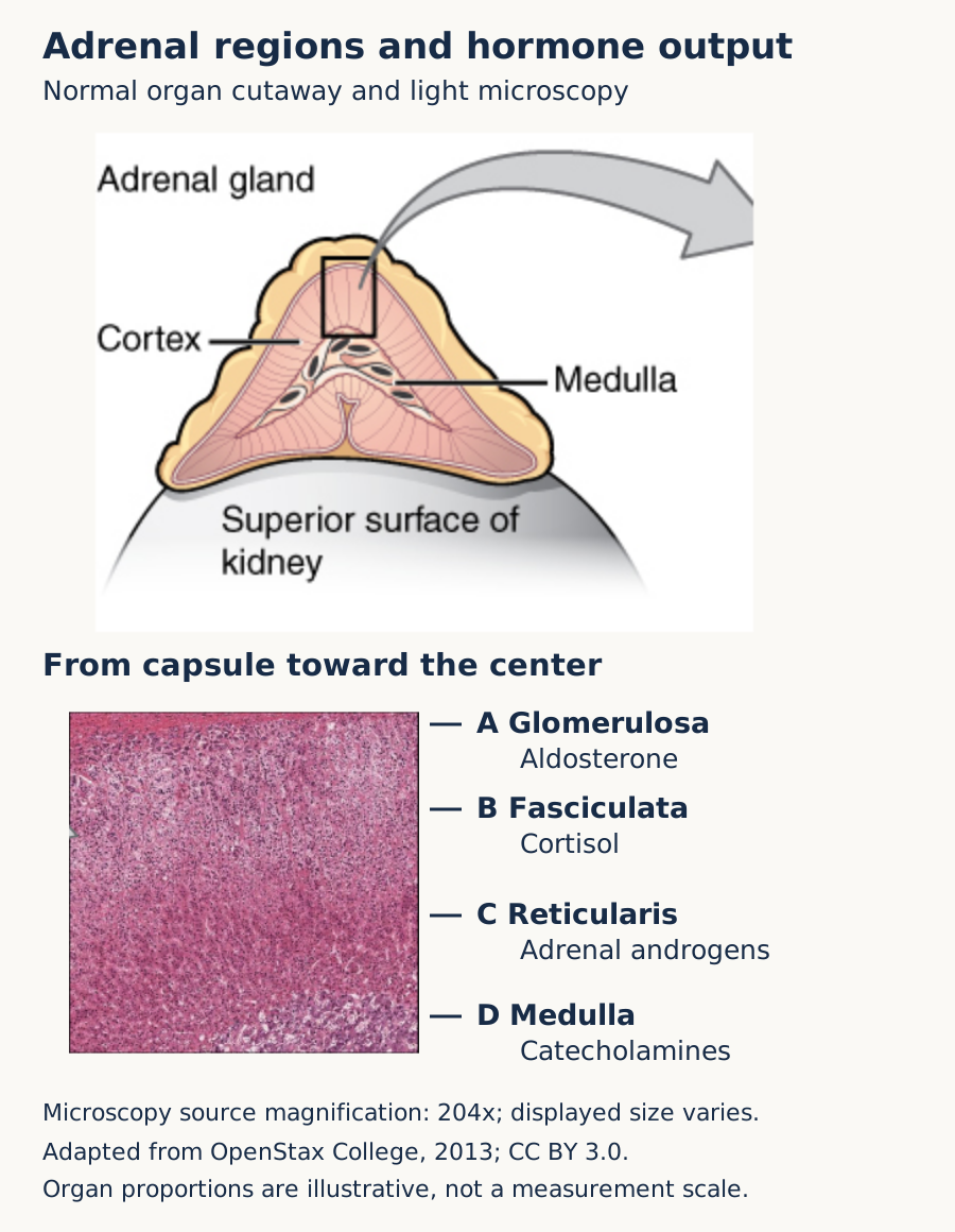
Inspect the normal organ overview, then follow the normal microscopy from capsule toward center. Source microscopy is 204x; screen size is not a calibrated scale. A later letter-only tissue view supports retrieval. [1][2]
First inspect the labeled anatomical reference: identify the adrenal gland above the kidney, the outer cortex, and the central medulla. In the enlarged histology, follow the outer-to-inner bands: glomerulosa, fasciculata, reticularis, then medulla. The boundary between cortex and medulla matters because a catecholamine tumor is not a cortical adenoma. The later label-hidden histology view lets you retrieve the same relationship without answer cues. [8]
Now predict where a hormone-producing lesion belongs before you look at a scan. Hypertension with potassium loss suggests a glomerulosa hormone; progressive bruising and proximal weakness suggest a fasciculata hormone; paroxysmal sweating and palpitations suggest a medullary source. These findings guide testing but cannot by themselves establish a lesion's function. Compare your predictions with the labeled histology; then transfer the localization to a patient with an adrenal nodule and normal glucose but an abnormal cortisol suppression test.
The exam transfer is simple: localize the hormone before deciding what the picture means. A CT nodule may be the source of the hormone, an unrelated adenoma, or a lesion that still needs characterization. Imaging by itself does not prove that a visible adrenal nodule is the gland making excess aldosterone.
Read aldosterone by reading renin first
Aldosterone increases distal sodium reabsorption and increases potassium and hydrogen secretion. Sustained excess therefore tends to produce hypertension, potassium loss, and metabolic alkalosis. The volume expansion feeds back on the kidney and suppresses renin and angiotensin II, so primary aldosteronism is fundamentally an inappropriate aldosterone signal occurring while its upstream renin signal is low. Hypokalemia is a severity finding, not a requirement; many affected patients have potassium in the reference range. [2]
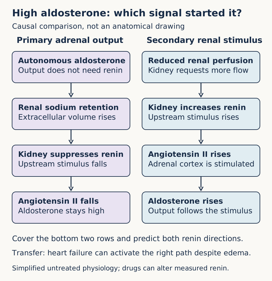
Cover the last two rows and predict renin and angiotensin II. Follow the arrows before comparing primary adrenal output with secondary renal stimulation. Simplified untreated physiology; medications can alter measured renin. [2]
The topic-specific method is a renin-first signal trace. On the renin diagram, predict renin, angiotensin II, and aldosterone for renal artery stenosis and autonomous adrenal secretion. Commit both patterns before opening the answer. Cover the diagnosis and ask whether the kidney is requesting aldosterone. If renin is low while aldosterone remains inappropriately high, the adrenal output is autonomous.
If renin is high and aldosterone is also high, the adrenal gland is responding to an upstream stimulus such as renal artery stenosis, reduced effective arterial volume in heart failure, or advanced cirrhosis. The same high aldosterone value has a completely different meaning depending on renin.
The 2025 Endocrine Society guideline conditionally suggests screening all people with hypertension when the health system can support it. Screening uses aldosterone plus renin and an aldosterone-to-renin ratio (ARR), with potassium measured so the aldosterone value can be interpreted correctly. Testing is preferably done in the morning with the patient seated and without recent sodium restriction. Low potassium can suppress aldosterone enough to create a false-negative screen, while beta blockers can suppress renin and create a false-positive ratio when aldosterone is only modestly increased. Assay-specific cutoffs matter, so a memorized ratio should never be treated as a universal law. [2]
Check the feedback trace
Renal artery stenosis raises renin, angiotensin II, and aldosterone. Autonomous adrenal aldosterone expands volume, suppressing renin and angiotensin II. Transfer to heart failure: reduced effective arterial perfusion raises all three despite edema.
Confirmation testing is also more selective than the old rule of sending every positive ratio to salt loading. Aldosterone-suppression testing can help when screening suggests an intermediate probability of lateralizing disease and surgery is being considered. It is not required before treatment in a patient with resistant hypertension or hypertension with hypokalemia plus plasma renin activity below 0.2 ng/mL/h and aldosterone above 20 ng/dL by immunoassay or above 15 ng/dL by liquid chromatography-tandem mass spectrometry, and it can be skipped when the patient will not pursue adrenal venous sampling and surgery. [2]
Once primary aldosteronism is established, the next question is unilateral versus bilateral secretion. CT describes anatomy and excludes a large concerning mass, but adrenal venous sampling is recommended for most surgical candidates because a visible nodule can be nonfunctioning while the opposite gland supplies excess aldosterone. A possible exception is a patient younger than 35 years with marked primary aldosteronism, hypokalemia, and a unilateral adenoma larger than 1 cm, when unilateral disease is especially likely.
Lateralizing disease in a surgical candidate is treated with unilateral adrenalectomy; bilateral or nonlateralized disease is usually treated with a mineralocorticoid receptor antagonist. Spironolactone is commonly favored for availability and cost, with blood pressure, potassium, renal function, and renin followed during titration. [2]
Type 4 renal tubular acidosis (RTA) is the physiologic mirror image worth keeping nearby. Too little effective aldosterone signaling reduces distal potassium and hydrogen secretion, producing hyperkalemic metabolic acidosis. Primary aldosteronism pushes in the opposite direction: potassium falls and bicarbonate tends to rise. That acid-base contrast can identify the direction of the aldosterone problem before a hormone result is shown. [7]
Renal sodium uptake by principal cells promotes potassium secretion through the distal electrical gradient; alpha-intercalated cells secrete hydrogen by a separate pathway. Aldosterone alters both, but this is not a single literal sodium-for-hydrogen exchange. In type 4 renal tubular acidosis, hyperkalemia also suppresses kidney ammonia generation, limiting urinary acid disposal. [7]
Try it here · Checkpoint 1 of 3
Make your prediction before reading the choices. A first attempt is just a starting point.
Case 6
Show answer and explanations for case 6
A. High-dose dexamethasone testing (Why this does not fit)
High-dose dexamethasone interrogates a cortisol pathway, not an aldosterone secretion source. It cannot substitute for localization in this patient.
Reasoning steps for option A
Can high-dose dexamethasone localize aldosterone 29 with renin 0.1?
High-dose dexamethasone investigates cortisol rather than aldosterone 29 with renin 0.1.
Which test would be used instead if cortisol co-secretion were suspected?
If cortisol co-secretion is suspected, use a 1-mg overnight test rather than a high-dose study.
B. Adrenalectomy without source localization (Why this does not fit)
Removing a gland before CT and functional localization risks operating on an incidental lesion or bilateral disease. A positive screen does not establish the operative side.
Reasoning steps for option B
Can these hormones alone identify which adrenal gland to remove?
Biochemistry cannot identify the operative gland without anatomy and often AVS.
Why could a single CT nodule still mislead the surgeon?
A unilateral CT nodule can still be nonfunctional; most surgical candidates require AVS later.
C. Salt-loading suppression testing (Why this does not fit)
Salt loading can clarify less convincing biochemical findings, but marked aldosterone elevation with suppressed renin and spontaneous hypokalemia is overt evidence. Requiring suppression before the planned surgical evaluation adds little here.
Reasoning steps for option C
Is salt loading compulsory with potassium 2.6 and overt renin suppression?
Potassium 2.6 with overt aldosterone elevation and suppressed renin need not undergo obligatory salt loading.
For which less convincing pattern would suppression testing add information?
Suppression testing has greater value for intermediate-probability results when lateralizing surgery is planned.
D. Adrenal CT (Best answer)
Marked aldosterone elevation despite suppressed renin and spontaneous hypokalemia supports primary aldosteronism without a mandatory salt load. CT is the appropriate next anatomical evaluation in this surgical candidate; later adrenal venous sampling will usually establish sidedness.
Reasoning steps for option D
What anatomy has not yet been assessed in this surgical candidate?
CT is the next anatomic evaluation because she is considering surgery and has no prior adrenal imaging.
Does CT later replace functional lateralization in most 44-year-olds?
CT evaluates lesions and concerning features; AVS subsequently determines functional laterality in most adults.
Takeaway: Modern primary aldosteronism evaluation does not force confirmatory suppression testing when renin-independent aldosterone production is already overt.
Can normal glucose exclude adrenal cortisol excess? Normal cortisol secretion is restrained by hypothalamic and pituitary feedback and follows a daily rhythm. Dexamethasone supplies an external glucocorticoid signal that should suppress pituitary adrenocorticotropic hormone (ACTH) and therefore cortisol. Failure to suppress after a low dose is an abnormal screening result, not proof of an autonomous adrenal source. Check drug interference and whether dexamethasone was taken; confirm persistent abnormality when appropriate and establish ACTH independence before assigning an adrenal source. For adrenal incidentalomas, the modern starting test is the 1-mg overnight dexamethasone suppression test. [1][5]
Before opening the answer, classify a post-dexamethasone cortisol of 3.1 micrograms/dL in a patient with normal glucose; predict the ACTH direction and a non-glucose consequence. In a patient without overt Cushing features, post-dexamethasone cortisol above 1.8 micrograms/dL is classified by the 2023 European Society of Endocrinology (ESE) guideline as mild autonomous cortisol secretion (MACS). Bone fragility, including vertebral fracture, can accompany cortisol autonomy despite normal glucose.
The result should be interpreted as a continuum rather than as a magic dividing line. ACTH independence should be demonstrated, and repeating the dexamethasone test is recommended when it will affect management. The important clinical follow-through is to look for cortisol-related comorbidities, especially hypertension and type 2 diabetes, rather than to demand a dramatic textbook appearance. [1]
Check the cortisol prediction
Cortisol of 3.1 micrograms/dL exceeds 1.8 and suggests mild autonomous cortisol secretion without overt features. Confirm ACTH independence and consider repeating the test. Normal glucose does not negate this result; in a patient with vertebral fracture, assess bone health too. [1]
Overt Cushing syndrome is a different clinical state. When the phenotype is convincing, validated initial tests include urine free cortisol, late-night salivary cortisol, or low-dose dexamethasone suppression testing. A high-dose dexamethasone test is not an initial test for whether Cushing syndrome exists, and it is not a shortcut that proves an adrenal mass is the source. Once hypercortisolism is established, ACTH helps separate ACTH-dependent from ACTH-independent disease. [5]
Very high cortisol can also create a mineralocorticoid-like renal pattern because renal protection from cortisol can be overwhelmed. Severe cortisol excess can therefore produce hypertension, hypokalemia, and alkalosis even when aldosterone is not the driver. When those findings coexist with unmistakable cortisol features, do not force every low-potassium hypertensive patient into the aldosterone pathway.
Under the 2023 European Society of Endocrinology guideline, for a unilateral adrenal mass with MACS and clinically important cortisol-related comorbidities, surgery is not automatic. Age, general health, degree and persistence of cortisol nonsuppression, comorbidities, and patient preference belong in an individualized multidisciplinary decision. The 2022 American Association of Endocrine Surgeons guidance is more intervention-oriented for unilateral MACS; these recommendations should not be presented as identical. [1][3]
After cortisol-producing adrenal surgery, chronic cortisol autonomy suppresses ACTH and the remaining adrenal cortex. Removing a unilateral source can therefore cause transient adrenal insufficiency. For adrenal surgery with post-1-mg dexamethasone cortisol above 1.8 micrograms/dL, give perioperative glucocorticoid coverage at surgical-stress doses, then individualize replacement and taper with endocrine follow-up until recovery of the hypothalamic-pituitary-adrenal axis is documented. Normal preoperative cortisol suppression does not itself trigger this cortisol-autonomy protocol, and unilateral surgery does not automatically mean lifelong replacement. [1]
Try it here · Checkpoint 2 of 3
Make your prediction before reading the choices. A first attempt is just a starting point.
Case 15
Show answer and explanations for case 15
A. Physiologic cortisol suppression (Why this does not fit)
A post-dexamethasone cortisol above 1.8 micrograms/dL does not meet the European Society of Endocrinology (ESE) criterion for normal suppression.
Reasoning steps for option A
Does 3.4 micrograms/dL after dexamethasone represent normal suppression?
No; it exceeds the 1.8 micrograms/dL incidentaloma cutoff.
Would the repeat 3.2 micrograms/dL rescue a physiologic-suppression interpretation?
No; a second valid elevated result with low ACTH makes a transient testing artifact less likely.
B. Mild autonomous cortisol secretion (Best answer)
Two valid post-dexamethasone cortisol results above 1.8 micrograms/dL without overt Cushing features support mild autonomous cortisol secretion; low adrenocorticotropic hormone (ACTH) further supports adrenal autonomy.
Reasoning steps for option B
What do two valid post-dexamethasone values of 3.4 and 3.2 micrograms/dL establish?
Persistent nonsuppression compatible with mild autonomous cortisol secretion in this patient without overt features.
Why does low ACTH sharpen that diagnosis?
Feedback suppression of pituitary ACTH supports an ACTH-independent adrenal source rather than pituitary drive.
C. Nonfunctioning adrenal cortical adenoma (Why this does not fit)
Low adrenocorticotropic hormone (ACTH) supports an adrenal autonomous cortisol source rather than a nonfunctioning lesion.
Reasoning steps for option C
Can a 2.5 cm mass be called nonfunctioning from its appearance alone?
No; the repeatedly abnormal suppression tests demonstrate a biochemical cortisol signal.
What different result would support a nonfunctioning designation?
A valid post-1-mg dexamethasone cortisol at or below 1.8 micrograms/dL, alongside otherwise negative indicated hormone testing.
D. Overt Cushing syndrome (Why this does not fit)
The biochemical result shows cortisol autonomy, but overt Cushing syndrome requires the appropriate clinical syndrome rather than one suppression value alone.
Reasoning steps for option D
Does cortisol of 3.2 micrograms/dL alone establish overt Cushing syndrome?
No; she has no overt Cushing features and the pattern fits mild autonomous cortisol secretion.
What presentation would shift this classification toward overt disease?
Progressive proximal weakness, wide purple striae and bruising with confirmed hypercortisolism would warrant an overt Cushing evaluation.
Takeaway: mild autonomous cortisol secretion (MACS) is defined by abnormal low-dose dexamethasone suppression in a patient without overt Cushing features, with adrenocorticotropic hormone (ACTH) independence established.
For catecholamines, establish biochemistry before provoking the tumor
Does a quiet symptom history exclude a catecholamine tumor? Pheochromocytoma arises from adrenal chromaffin tissue. The familiar episodic pattern is headache, perspiration, palpitations, and hypertension, but a catecholamine-producing tumor can present less theatrically. When biochemical testing is indicated, plasma free or urinary fractionated metanephrines are used because their production is more continuous than brief catecholamine surges. [6]
Incidentaloma imaging now changes who needs this biochemical test. In the 2023 ESE pathway, a homogeneous adrenal lesion with unenhanced attenuation at or below 10 Hounsfield units (HU) is so unlikely to be pheochromocytoma that routine metanephrine testing can be omitted. If the lesion is above 10 HU, indeterminate, or no unenhanced CT is available, pheochromocytoma should be excluded even if the patient denies the classic symptom triad. [1]
For a patient with high metanephrines headed to surgery, write which receptor to block first and predict the effect of giving only a beta blocker. Open the answer after committing; then transfer the sequence to persistent tachycardia after initial preparation.
Check the receptor sequence
Giving a beta blocker before adequate alpha receptor blockade can leave catecholamine-driven vasoconstriction unopposed. When beta blockade is needed for tachycardia, alpha blockade comes first. [4]
For a catecholamine-producing pheochromocytoma headed to surgery, perioperative preparation is designed to control blood pressure and restore intravascular volume before tumor manipulation. Alpha receptor blockade has been the standard first pharmacologic step; a beta blocker may be added later for persistent tachycardia after adequate alpha blockade. This order is the testable physiologic principle. [4]
The old fixed percentage rule for malignant pheochromocytoma is not a useful modern model. Current classification recognizes that all pheochromocytomas and paragangliomas have some metastatic potential, and metastatic disease is defined by tumor in sites where chromaffin tissue is not normally present. Genetics, location, size, biochemical phenotype, and other features influence risk, so follow-up cannot be compressed into a single percentage. [6]
Preparation continues beyond a prescription: specialist alpha blockade is titrated over approximately 7 to 14 days before elective surgery while seated and standing blood pressure and heart rate are assessed. Restore salt and fluid as tolerated to correct catecholamine-related contraction, but individualize fluid loading with heart failure or renal impairment; do not manipulate a suspected tumor before adequate preparation.
After confirmed pheochromocytoma or paraganglioma, offer genetic counseling and consideration of genetic testing, measure metanephrines 2 to 6 weeks after surgery to establish biochemical remission, and plan annual biochemical monitoring with risk-based surveillance, lifelong for high-risk groups. Imaging frequency is individualized by inherited risk, tumor phenotype and postoperative results. [4]
The incidentaloma fork: function and imaging are separate questions
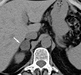
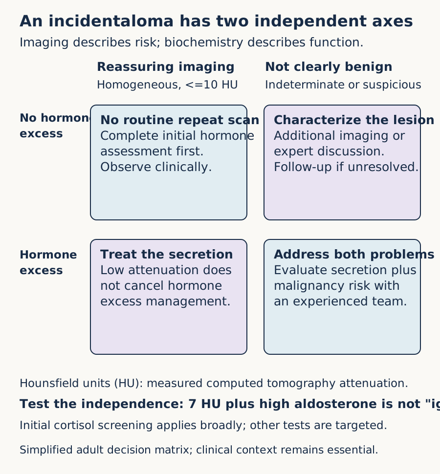
Can benign-looking anatomy establish normal hormone function? An adrenal incidentaloma should be evaluated on two axes at the same time. The hormone axis asks whether the lesion is producing cortisol, aldosterone, catecholamines, or another steroid. The imaging axis asks whether the morphology is convincingly benign. The annotated axial adrenal image illustrates the location of a real incidental lesion; the photograph alone cannot supply a measured attenuation or prove hormone function. [8] The first imaging number to find is the unenhanced CT attenuation, because intracellular lipid lowers the Hounsfield unit measurement of many cortical adenomas. [1][3]
Unenhanced axial abdominal computed tomography: the original arrow identifies a small right adrenal lesion between the liver and upper right kidney, on the viewer's left. Use this as guided orientation, then state which data are still needed: measured attenuation and biochemical results. This image alone establishes neither hormonal function nor a diagnostic attenuation value. No calibrated measurement scale is displayed; do not estimate lesion size from screen pixels. [8][8]Assign one imaging category and one hormone category. Apply the matrix to a 7-Hounsfield-unit nodule with high aldosterone: benign imaging does not establish normal function. Simplified adult framework, not a clinical scan. [1][2][3]
On the two-axis diagram, commit separate imaging and hormone decisions for a hypertensive patient with a homogeneous 2.4 cm mass at 7 HU before opening the answer. A homogeneous mass with unenhanced attenuation at or below 10 HU has a benign imaging phenotype in a patient without a known extra-adrenal cancer. The 2023 ESE guideline removed the old 4 cm size restriction from this rule: if the mass is homogeneous and at or below 10 HU, no further adrenal imaging is required. That replaces the older habit of automatically ordering serial CT scans for every small incidentaloma. [1]
When the attenuation is above 10 HU, the problem becomes one of characterization rather than immediate labeling. A homogeneous mass from 11 to 20 HU and smaller than 4 cm with a negative hormonal evaluation can undergo one additional imaging method, such as magnetic resonance imaging (MRI) chemical shift or an adrenal protocol CT, to establish benignity and avoid serial surveillance; interval noncontrast CT or MRI at about 12 months is an alternative.
A lesion at least 4 cm that is heterogeneous or has unenhanced attenuation above 20 HU deserves multidisciplinary review because malignancy risk is higher. Older teaching used absolute contrast washout above 60 percent to favor an adenoma. Washout has limited specificity and cannot alone exclude pheochromocytoma or prove benignity. A mass with both androgen and cortisol excess raises concern for adrenocortical carcinoma.
Size contributes to risk, but size alone is not an automatic surgery rule. [1][3]
Check both incidentaloma axes
A 2.4 cm homogeneous adrenal mass measuring 7 HU on unenhanced CT does not need scheduled adrenal imaging follow-up, but hypertension still warrants aldosterone and renin assessment and most patients need cortisol screening under the 2023 ESE recommendation. A 3.2 cm heterogeneous mass measuring 28 HU belongs in an indeterminate or suspicious pathway instead.
The biochemical screen is equally deliberate. Most patients with an incidentaloma receive a 1-mg overnight dexamethasone test. Metanephrines are added when the imaging is not typical of a benign adenoma, particularly when unenhanced HU is above 10 or the unenhanced value is unavailable. Aldosterone and renin are assessed when hypertension or unexplained hypokalemia is present; because the 2025 primary aldosteronism guideline conditionally recommends screening all patients with hypertension, an incidentaloma plus hypertension clearly qualifies. [1][2]
If the initial hormonal evaluation is normal, routine repeated hormone testing is not recommended unless new endocrine signs appear or related comorbidities such as hypertension or diabetes meaningfully worsen. If the imaging is clearly benign, repeated imaging is also unnecessary. This is a useful antidote to the idea that an incidental adrenal nodule automatically creates years of testing. [1]
Adrenal biopsy is not the first test for an indeterminate mass. Before biopsy is even considered, pheochromocytoma must be excluded and the result must be capable of changing management. Imaging, hormonal status, cancer history, and multidisciplinary judgment come first. [1]
Indeterminate does not mean discharged: if imaging remains indeterminate after workup and surgery is not selected, repeat noncontrast CT or MRI in 6 to 12 months; growth greater than 20% and at least 5 mm in maximum diameter prompts reconsideration of resection. A patient with mild autonomous cortisol secretion who does not undergo surgery instead needs annual reassessment of cortisol-related comorbidities, with endocrine reassessment if they newly appear or worsen, not automatic annual repeat dexamethasone testing.
Routine biopsy is discouraged: it is mainly considered in selected patients with an extra-adrenal malignancy only when the mass is hormonally inactive, imaging has not shown a benign lesion, and histology would change management. Exclude pheochromocytoma before invasive sampling. Biopsy poorly distinguishes primary adrenocortical adenoma from carcinoma and is generally avoided for a potentially resectable cortical cancer. [1]
Try it here · Checkpoint 3 of 3
Make your prediction before reading the choices. A first attempt is just a starting point.
Case 26
Show answer and explanations for case 26
A. Plasma free metanephrines (Best answer)
A 16-HU adrenal mass does not have the classic lipid-rich imaging phenotype despite the absence of spells. Plasma free metanephrines screen for catecholamine excess; imaging characterization is a separate next decision.
Reasoning steps for option A
Why measure plasma free metanephrines despite no spells?
The 16-HU lesion does not meet the homogeneous at-or-below-10-HU exemption from pheochromocytoma testing.
Does cortisol 1.0 micrograms/dL settle the catecholamine question?
No; normal cortisol suppression assesses a separate cortical axis, not medullary catecholamine secretion.
B. Adrenal venous sampling (Why this does not fit)
Adrenal venous sampling is reserved chiefly for aldosterone lateralization when surgery is considered. This normotensive patient instead requires metanephrine assessment for an indeterminate lesion.
Reasoning steps for option B
Does adrenal venous sampling address a 16-HU lesion without aldosterone data or hypertension?
No; sampling mainly localizes proven primary aldosteronism before selected surgery.
If she instead had hypertension and suppressed renin with high aldosterone, when could sampling matter?
It could distinguish unilateral from bilateral aldosterone secretion if adrenalectomy were being considered.
C. No additional biochemical evaluation (Why this does not fit)
A homogeneous lesion at 16 HU is not within the benign lipid-rich 10-HU group. A normal cortisol test does not eliminate the need for catecholamine assessment in this imaging category.
Reasoning steps for option C
Why not stop biochemical evaluation after a normal dexamethasone test?
A homogeneous 2.6 cm mass at 16 HU is not the classic lipid-rich phenotype, so asymptomatic pheochromocytoma remains to be excluded.
Is normal potassium enough to avoid every other hormone test?
No; potassium is not a catecholamine screen and normal levels also do not universally exclude primary aldosteronism in hypertensive patients.
D. Repeat overnight dexamethasone suppression testing (Why this does not fit)
Valid cortisol suppression at 1.0 micrograms/dL makes immediate repeat dexamethasone testing unnecessary. It gives no information about possible catecholamine secretion.
Reasoning steps for option D
Must the 1-mg overnight test be repeated when a valid result is 1.0 micrograms/dL?
No; that value is clearly suppressed below 1.8 micrograms/dL without a stated new cortisol concern.
What separate imaging choice remains after biochemical screening?
A homogeneous 11-20-HU lesion smaller than 4 cm may receive additional characterization or interval imaging depending on the pathway.
Takeaway: A homogeneous 11 to 20 Hounsfield units (HU) mass below 4 cm is indeterminate but often can be resolved with one additional dedicated imaging study rather than serial computed tomography (CT) by default.
Best first biochemical directionAldosterone plus renin, interpreted with potassium
What the imaging can and cannot doCT shows anatomy; most surgical candidates still need adrenal venous sampling (AVS) for lateralization
PatternSecondary hyperaldosteronism
Renin or ACTHRenin increased
Best first biochemical directionIdentify the upstream cause of renin activation
What the imaging can and cannot doAn adrenal nodule may be incidental
PatternMACS or adrenal cortisol excess
Renin or ACTHACTH low or suppressed when adrenal autonomous
Best first biochemical direction1-mg overnight dexamethasone test, then appropriate confirmation
What the imaging can and cannot doCT characterizes the mass but does not replace hormone testing
PatternPheochromocytoma
Renin or ACTHNot a renin or ACTH diagnosis
Best first biochemical directionPlasma free or urinary fractionated metanephrines when indicated
What the imaging can and cannot doHU at or below 10 in a homogeneous adenoma makes pheochromocytoma very unlikely
PatternType 4 RTA
Renin or ACTHLow effective aldosterone signaling
Best first biochemical directionHyperkalemic metabolic acidosis guides the physiology
What the imaging can and cannot doNo adrenal nodule is required
Before opening the final answer, decide whether suppressed renin and a left CT nodule prove left-sided secretion. What if venous sampling lateralizes right? The final exam habit is to make two passes. First, identify the feedback direction: low renin versus high renin, suppressed ACTH versus ACTH-dependent disease, or a catecholamine biochemical pattern. Second, interpret the mass: homogeneous and lipid-rich versus indeterminate or suspicious. A hormone result localizes physiology; a CT finding describes anatomy. They agree often enough to tempt shortcuts and disagree often enough that shortcuts fail.
Check physiology against anatomy
A 55-year-old patient has primary aldosteronism and a 1.8 cm left adrenal nodule, but surgery is desired. The hormone pattern proves autonomous aldosterone production; the CT does not prove the left nodule is the source. If sampling instead lateralizes right, reassess the left nodule as an incidental finding. In most such patients, AVS is used before deciding which gland to remove. [2]
For independent practice, the cases below deliberately remove the nearby teaching support. They require the same physiology, but the correct option may depend on a medication effect, a CT attenuation value, a feedback hormone, a comorbidity pattern, or the distinction between an anatomic nodule and a functional source.
Retrieve the tissue map without labels
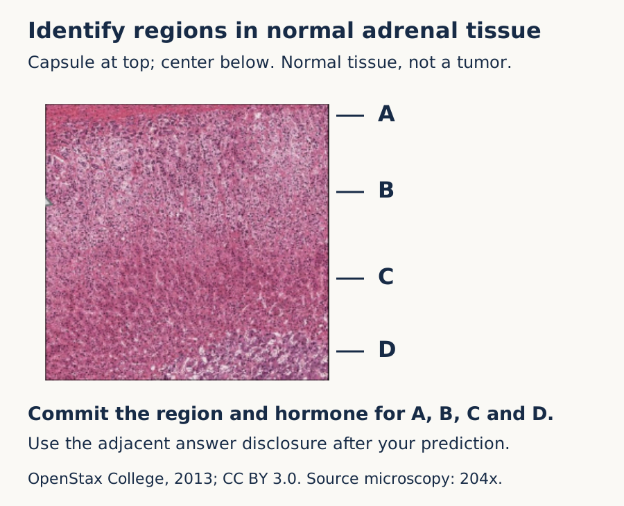
Can you identify which region makes each hormone without reading the anatomical reference? Inspect the label-hidden histology and locate regions A through D from the outer capsule toward the center. Name the tissue and its product for each position before opening the answer. This is retrieval from a real spatial view, not an invitation to infer hormone function from the scan alone.
Before opening the adjacent answer disclosure, commit a region and hormone for A through D. This is normal tissue, not tumor histology. Source magnification is 204x; displayed size varies. [1][2]Check tissue positions and hormones
A is the outer zona glomerulosa, which makes aldosterone. B is the zona fasciculata, which makes cortisol. C is the zona reticularis, which makes adrenal androgens. D is the medulla, which makes catecholamines. The three cortical layers surround the medulla; a medullary pheochromocytoma is not an adrenal cortical adenoma.
Apply the spatial map to a new situation: an adrenal lesion accompanies suppressed renin and inappropriately high aldosterone. The source pathway points toward the outer cortex, but which gland is secreting still requires functional localization if surgery is contemplated. If the same lesion accompanies markedly elevated metanephrines, investigate a medullary source and prepare safely before surgery. [2][4]
Independent practice
Case 1
Show answer and explanations for case 1
A. Adrenal CT alone (Why this does not fit)
CT identifies the left lesion but cannot prove that it produces the measured aldosterone. Functional sampling is needed before directing removal in this adult surgical candidate.
Reasoning steps for option A
Does a left 1.5-cm CT nodule prove left-sided hormone output?
CT identifies a 1.5-cm left nodule but cannot prove left-sided secretion.
What if adrenal venous sampling instead localizes secretion to the right?
If AVS instead lateralizes right, the left nodule may be incidental.
B. High-dose dexamethasone suppression (Why this does not fit)
High-dose dexamethasone concerns cortisol feedback rather than the source of aldosterone. It cannot show whether the left gland produces more aldosterone than the right.
Reasoning steps for option B
Can high-dose dexamethasone compare left and right aldosterone secretion?
High-dose dexamethasone probes cortisol feedback, not aldosterone laterality.
Which measurement actually establishes functional laterality?
Even an abnormal high-dose test would not establish aldosterone sidedness; AVS compares cortisol-corrected adrenal vein samples.
C. Adrenal venous sampling (Best answer)
Severe hypokalemia, aldosterone 25 ng/dL and renin activity 0.1 ng/mL/h after correction establish a high-probability autonomous pattern. The nodule does not show functional laterality; adrenal venous sampling is the next discriminator for most adult surgical candidates.
Reasoning steps for option C
What does aldosterone 25 with renin 0.1 after correction indicate?
Aldosterone 25 with renin 0.1 after correction establishes strongly renin-independent output.
Does this 36-year-old qualify for the narrow young-patient exception to AVS?
At age 36 she does not meet the narrow younger-than-35 exception; AVS usually guides surgical side.
D. Renal artery angiography (Why this does not fit)
Renal artery disease could explain hypertension but would generally raise renin. Her renin stays suppressed even after potassium and medication review, so renal angiography does not establish the secreting adrenal side.
Reasoning steps for option D
What renin response would renal artery stenosis generally produce?
Renal artery stenosis typically raises renin rather than suppressing it.
Would angiography establish which adrenal gland should be removed?
Even if stenosis were found, angiography could not identify the aldosterone-secreting adrenal.
Takeaway: High aldosterone with suppressed renin identifies renin-independent aldosterone production; hypokalemic alkalosis supports a more severe phenotype.
B. Loss of mineralocorticoid receptor signaling (Why this does not fit)
Reduced mineralocorticoid action can raise renin but would not explain a clinically coherent secondary aldosterone response to heart failure physiology as well.
Reasoning steps for option B
Does loss of mineralocorticoid signaling fit pulmonary congestion?
Mineralocorticoid resistance tends toward salt wasting rather than congestive underfilling.
What phenotype would point toward mineralocorticoid resistance instead?
Receptor resistance would favor renal sodium loss, hyperkalemia and often low pressure, unlike this congested heart-failure presentation.
C. Isolated adrenal cortisol secretion (Why this does not fit)
Cortisol autonomy does not produce the paired high-renin, high-aldosterone pattern expected from reduced renal perfusion.
Reasoning steps for option C
Could autonomous cortisol account for both elevated renin and aldosterone?
Isolated cortisol secretion does not explain the paired high renin and aldosterone.
What happens to the renin-aldosterone axis if cortisol strongly mimics mineralocorticoids?
If severe cortisol mimicked mineralocorticoid action, volume feedback would tend to lower both renin and aldosterone.
D. Autonomous adrenal aldosterone secretion (Why this does not fit)
Autonomous primary aldosteronism expands volume and suppresses renin; this patient has activated renin instead.
Reasoning steps for option D
Would autonomous aldosterone normally raise renin in heart failure?
Autonomous aldosterone expands effective volume and suppresses renin.
Would discovering an adrenal nodule outweigh the high-renin pattern?
A coincidental adrenal nodule would not override the high-renin heart-failure physiology.
Takeaway: High renin plus high aldosterone is a secondary response; heart failure can activate renin-angiotensin-aldosterone system (RAAS) because effective arterial volume is low despite edema.
A. Late-night salivary cortisol (Why this does not fit)
Late-night salivary cortisol screens for cortisol excess, not renin-independent aldosterone secretion. The hypertension and family history warrant aldosterone and renin assessment despite normal potassium.
Reasoning steps for option A
Does late-night salivary cortisol investigate the familial hypertension signal?
No; it screens cortisol excess, whereas pressure 152/94 and his sister’s adrenal surgery raise the need to assess aldosterone and renin.
If bruising and proximal weakness developed, what would change?
Those new cortisol features could warrant validated hypercortisolism testing; the present normokalemic hypertensive pattern still warrants an aldosterone-renin screen.
B. Urinary metanephrines (Why this does not fit)
Metanephrines evaluate catecholamine excess, not primary aldosteronism, and are not a general hypertension screening test.
Reasoning steps for option B
Do metanephrines screen for autonomous aldosterone?
Metanephrines investigate catecholamines, not routine primary aldosteronism screening.
Which newly appearing spells would change the catecholamine-testing decision?
New headache, sweating and palpitations would instead justify catecholamine evaluation.
C. Serum potassium alone (Why this does not fit)
Potassium helps interpret aldosterone but cannot screen for primary aldosteronism alone. A value of 4.1 mEq/L does not rule out autonomous aldosterone production.
Reasoning steps for option C
Could serum potassium 4.1 alone exclude primary aldosteronism?
No; many patients with renin-independent aldosterone secretion are normokalemic.
What information does potassium add to paired aldosterone and renin?
Potassium contextualizes the hormone result because hypokalemia can suppress aldosterone and falsely reassure; it cannot replace either hormone measurement.
D. Morning aldosterone and renin (Best answer)
The 2025 guideline conditionally suggests screening all people with hypertension using aldosterone and renin, with potassium measured for interpretation.
Reasoning steps for option D
Which paired hormones test this normokalemic patient with pressure 152/94?
A. Adrenal venous sampling (Why this does not fit)
Adrenal venous sampling (AVS) is a subtype test used after primary aldosteronism is established in patients pursuing surgery, not a rescue for an unreliable first screen.
Reasoning steps for option A
Is AVS appropriate before resolving aldosterone 8 during potassium depletion?
AVS localizes established primary aldosteronism, not an unreliable negative screen.
When could AVS become useful if the repeat screen establishes disease?
If corrected repeat screening establishes primary aldosteronism and the patient seeks surgery, adrenal imaging and usually venous sampling become relevant.
B. Correct potassium; repeat testing (Best answer)
Hypokalemia can falsely lower aldosterone; current guidance recommends correcting potassium and repeating the screen when suspicion remains high.
Reasoning steps for option B
How can potassium 2.7 distort aldosterone 8?
Potassium 2.7 can depress aldosterone to a falsely reassuring 8.
What must happen before repeating the paired hormone measurements?
Replete potassium and review medicines before repeating aldosterone with renin.
C. Conclude that primary aldosteronism has been excluded (Why this does not fit)
A single negative screen is unreliable when a factor known to lower aldosterone is present and the clinical probability remains high.
Reasoning steps for option C
Is one negative screen reliable in resistant hypertension with potassium 2.7?
One negative screen during severe hypokalemia does not exclude disease.
When would a negative aldosterone result be more persuasive?
A negative repeat after potassium normalization is more informative than this original sample.
D. High-dose dexamethasone testing (Why this does not fit)
Dexamethasone evaluates cortisol physiology and does not correct the interpretation problem in an aldosterone screen.
Reasoning steps for option D
Could high-dose dexamethasone fix a potassium-confounded aldosterone result?
High-dose dexamethasone cannot repair potassium-related distortion of aldosterone.
What separate clinical context would instead warrant cortisol screening?
An incidentaloma or convincing cortisol phenotype would call for appropriate low-dose screening, not high-dose rescue of this assay.
Takeaway: Low potassium can hide primary aldosteronism by lowering aldosterone secretion, so correct hypokalemia before accepting a negative result.
A. Unilateral adrenalectomy (Why this does not fit)
Neither a potentially distorted screen nor the ratio itself identifies which adrenal gland, if either, should be removed.
Reasoning steps for option A
Does an ARR of 60 on metoprolol identify a surgical gland?
Ratio 60 on metoprolol cannot justify gland removal.
What is the risk of surgery before verifying the hormone result?
Premature adrenalectomy could remove a healthy gland without proof of disease or laterality.
B. Repeat screening after medication review (Best answer)
Metoprolol can suppress renin and spuriously raise the ratio with aldosterone only 12 ng/dL; review medications and repeat screening after a safe adjustment, without stopping beta blockade abruptly.
Reasoning steps for option B
How does metoprolol affect renin 0.2 and the ARR?
Metoprolol suppresses renin to 0.2, potentially inflating the ratio despite aldosterone 12.
How can a repeat screen be obtained without abruptly stopping beta blockade?
Repeat paired hormones after a safe medication review, without abruptly stopping needed beta blockade.
C. Begin potassium supplementation without repeat testing (Why this does not fit)
Potassium is 4.0 mEq/L, so supplementation has no demonstrated need and will not resolve metoprolol-related renin suppression. Review interference and repeat aldosterone-renin screening safely instead.
Reasoning steps for option C
Does potassium 4.0 require potassium replacement?
Potassium 4.0 does not call for supplementation or rule out disease.
What diagnostic uncertainty remains after empiric supplements?
The unresolved issue is whether a valid aldosterone-renin pair demonstrates autonomy despite normal potassium.
D. Adrenal venous sampling (Why this does not fit)
A screening ratio does not establish laterality, and medication effects can distort the ratio before subtype testing is even considered.
Reasoning steps for option D
Should AVS follow an unverified beta-blocker-confounded ratio?
AVS is premature while the biochemical screen may be beta-blocker-confounded.
If biochemical disease is confirmed, what further evaluation precedes surgery?
If repeat testing confirms disease, CT and usually AVS are considered only if surgery is pursued.
Takeaway: Interpret the aldosterone-renin ratio in context: renin-suppressing drugs can inflate the ratio, especially when aldosterone is modest.
Preoperative autonomous cortisol suppressed ACTH and the contralateral cortex, so unilateral removal can precipitate adrenal crisis. Give stress-dose intravenous hydrocortisone immediately with fluid resuscitation, then plan endocrine-guided taper and recovery assessment.
Reasoning steps for option A
Why can removal of a cortisol-producing gland cause shock the next day?
After two valid post-dexamethasone cortisol values of 4.2 and low ACTH, source removal can cause cortisol deficiency.
What should accompany hydrocortisone at blood pressure 84/52?
Give immediate intravenous hydrocortisone and isotonic fluids for shock, then assess hypothalamic-pituitary-adrenal (HPA) axis recovery rather than assume permanent replacement.
B. A mineralocorticoid antagonist (Why this does not fit)
Mineralocorticoid blockade treats aldosterone excess but would worsen hypotension in this setting. Her suppressed ACTH and recent cortisol-source resection instead support acute glucocorticoid deficiency.
Reasoning steps for option B
Would blocking mineralocorticoid receptors improve pressure 84/52?
Mineralocorticoid blockade would aggravate current pressure 84/52.
Why should the earlier potassium 3.1 not distract from the postoperative collapse?
The preoperative potassium 3.1 is less urgent than postoperative hypotension with sodium 128.
C. Fludrocortisone alone (Why this does not fit)
Unilateral cortical surgery usually preserves mineralocorticoid production from the other adrenal gland. Glucocorticoid deficiency, not isolated aldosterone deficiency, explains the immediate postoperative collapse.
Reasoning steps for option C
Can fludrocortisone alone provide a surgical-stress cortisol response?
Fludrocortisone alone cannot supply stress-dose glucocorticoids after cortisol-source removal.
Does unilateral adrenalectomy necessarily remove all aldosterone production?
The remaining gland usually retains aldosterone secretion even while cortisol output is suppressed.
D. Isotonic saline alone (Why this does not fit)
Hypovolemia merits prompt intravenous fluid, but saline alone does not replace suppressed endogenous glucocorticoid production. Recent resection of an autonomous cortisol source and hypotension warrant immediate steroid coverage.
Reasoning steps for option D
Will isotonic saline address her intravascular deficit?
Saline treats hypovolemia but cannot replace the suppressed cortisol response.
What essential treatment does saline fail to provide after suppressed ACTH?
Fluid resuscitation must accompany, not replace, immediate stress-dose hydrocortisone.
Takeaway: Unilateral resection of a cortisol-autonomous gland can precipitate adrenal insufficiency: give stress-dose glucocorticoids perioperatively when post-dexamethasone cortisol exceeds 1.8 micrograms/dL and assess recovery. [1]
A. Remove the left adrenal nodule surgically (Why this does not fit)
Bilateral secretion means removing one gland is unlikely to correct the autonomous aldosterone source.
Reasoning steps for option A
Do AVS values 4.1 left and 3.9 right support removing the visible left nodule?
AVS ratios 4.1 versus 3.9 do not support left adrenalectomy despite the nodule.
What endocrine problem might persist after left-sided surgery?
Removing only the left gland risks leaving contralateral aldosterone excess untreated.
B. Beta-adrenergic blockade (Why this does not fit)
Beta blockers lower renin but do not directly block mineralocorticoid receptor effects and can complicate biochemical interpretation.
Reasoning steps for option B
Does beta blockade directly oppose excess aldosterone at the kidney?
Beta blockade does not block renal mineralocorticoid receptor effects.
Why may beta blockers distort a future aldosterone-renin ratio?
Beta blockers can further suppress renin, confounding later ratio interpretation.
C. Spironolactone therapy (Best answer)
Bilateral or nonlateralized primary aldosteronism is generally treated medically with a mineralocorticoid receptor antagonist (MRA) such as spironolactone, with potassium, renal function, blood pressure, and renin followed.
Reasoning steps for option C
What medication fits an AVS lateralization index of only 1.05?
Spironolactone treats nonlateralized aldosterone excess with adequate renal function.
Which safety and efficacy measures matter during spironolactone titration?
Monitor pressure, potassium, renal function and renin during antagonist titration.
D. Interval adrenal CT (Why this does not fit)
Growth does not establish which tissue is driving bilateral aldosterone secretion and does not treat the hormone excess.
Reasoning steps for option D
Could interval CT establish which side secreted aldosterone?
Potassium replacement does not block the cardiovascular and renal consequences of autonomous aldosterone secretion.
Reasoning steps for option A
Will supplements for potassium 3.0 treat aldosterone-driven sodium retention?
Potassium supplements correct 3.0 but not aldosterone-driven tissue injury.
What medical therapy remains possible if surgery is declined?
If surgery were declined, mineralocorticoid receptor blockade treats the mechanism better than supplements alone.
B. Bilateral adrenalectomy (Why this does not fit)
The disease is unilateral by adrenal venous sampling (AVS), so removing both glands would add unnecessary permanent adrenal insufficiency risk.
Reasoning steps for option B
Do cortisol-corrected ratios 12 left and 2 right warrant bilateral removal?
AVS index 12/2 = 6 identifies left rather than bilateral secretion.
What unnecessary long-term consequence follows bilateral adrenalectomy?
Removing both glands would unnecessarily cause permanent adrenal insufficiency.
C. Interval adrenal CT (Why this does not fit)
A lateralization index of 12/2 = 6 supports a left-sided functional source after successful bilateral sampling; observation alone ignores a treatable biochemical disease.
Reasoning steps for option C
Does interval CT add more lateralization than a successful AVS index of 6?
Interval CT cannot substitute for already-established functional localization.
Why is simple observation insufficient for a willing surgical candidate?
A lateralization index of 6 after successful bilateral sampling is already actionable.
D. Left unilateral adrenalectomy (Best answer)
Biochemical disease plus adrenal venous sampling (AVS)-proven left lateralization identifies a surgically targetable unilateral source.
Reasoning steps for option D
Which side dominates after cortisol correction, 12 or 2?
The left value is six times the right, giving a functionally dominant left source. This comparison supports source-directed surgery rather than selecting a side from CT appearance.
Why still follow pressure and potassium after unilateral surgery?
Postoperative pressure and potassium still need reassessment; cure of hypertension is not guaranteed.
Takeaway: When primary aldosteronism (PA) clearly lateralizes and the patient wants surgery, unilateral adrenalectomy targets the source rather than treating only downstream consequences.
A. Adrenal venous sampling (Why this does not fit)
Successful bilateral sampling has already demonstrated left-sided aldosterone lateralization. Repeating adrenal venous sampling would not evaluate the separate possibility of cortisol co-secretion raised by worsening diabetes.
Reasoning steps for option A
What did the already-completed venous ratios of 9.6 left and 2.0 right establish?
Their ratio is 4.8, supporting a left-sided aldosterone source after selective bilateral sampling; repeating localization will not assess another hormone.
Which unresolved secretory pathway could contribute to the worsening diabetes before left adrenalectomy?
Cortisol co-secretion remains possible despite established aldosterone lateralization. A validated low-dose suppression screen addresses that separate perioperative risk.
B. 1-mg overnight dexamethasone suppression test (Best answer)
The overnight low-dose test assesses whether cortisol suppresses normally despite the concurrent aldosterone pattern. Diabetes progression makes possible co-secretion relevant before adrenal surgery.
Reasoning steps for option B
What test screens a 2.1-cm adrenal mass for cortisol autonomy?
A 1-mg overnight dexamethasone test assesses cortisol autonomy with worsening diabetes.
How would cortisol nonsuppression change perioperative planning?
If post-test cortisol exceeds 1.8, check validity and ACTH independence and plan perioperative glucocorticoids if surgery proceeds.
C. Random afternoon serum cortisol measurement (Why this does not fit)
A random afternoon cortisol may fall within its broad daily range despite mild autonomous secretion. It is not the recommended initial test for an adrenal incidentaloma.
Reasoning steps for option C
Does a random afternoon cortisol reliably detect mild autonomous secretion?
Random afternoon cortisol cannot sensitively assess mild nonsuppression.
What feedback response does overnight dexamethasone interrogate?
Dexamethasone tests feedback suppression rather than relying on a circadian snapshot.
D. High-dose dexamethasone suppression test (Why this does not fit)
High-dose dexamethasone is not the initial screen for cortisol secretion in an adrenal nodule. Worsening diabetes calls for a validated screening test before etiologic localization.
Reasoning steps for option D
Is high-dose dexamethasone the first screen for this incidentaloma?
High-dose dexamethasone is not the initial incidentaloma cortisol screen.
Could a high-dose result alone identify the cortisol-producing gland?
A high-dose result cannot by itself prove adrenal source; low-dose nonsuppression requires contextual confirmation.
Takeaway: An adrenal adenoma in primary aldosteronism can co-secrete cortisol, so a 1-mg overnight dexamethasone test is part of current evaluation.
A. Cortisol-mediated mineralocorticoid activation (Best answer)
Marked ACTH-independent cortisol excess suggests adrenal production. At high levels cortisol can evade renal inactivation and stimulate mineralocorticoid receptors, leading to potassium and hydrogen loss even while aldosterone is low.
Reasoning steps for option A
How can high cortisol cause potassium 2.5 despite suppressed aldosterone?
Marked cortisol excess can overwhelm renal 11-beta-hydroxysteroid dehydrogenase type 2 (11-beta-HSD2) and activate mineralocorticoid receptors.
Why are both renin and aldosterone suppressed with bicarbonate 34?
Suppressed ACTH supports adrenal cortisol production, while renin and aldosterone fall from volume feedback.
B. Renin-dependent aldosterone secretion (Why this does not fit)
Renin and aldosterone are both suppressed here, unlike renin-dependent secondary aldosteronism. It cannot explain the substantial potassium loss in the setting of marked cortisol excess.
Reasoning steps for option B
Does a renin-dependent mechanism fit two suppressed hormones?
Secondary aldosterone requires elevated renin, unlike both suppressed measurements.
What renin pattern would renal artery stenosis cause instead?
Renal artery stenosis would increase renin, not produce this paired suppression.
C. Primary aldosterone secretion (Why this does not fit)
Aldosterone is suppressed, so independent aldosterone production does not match the measurements. It also would not account for high urine cortisol with suppressed ACTH.
Reasoning steps for option C
Can primary aldosterone excess coexist with suppressed aldosterone as its explanation?
Primary aldosteronism requires inappropriate aldosterone, not suppressed aldosterone.
Which measured steroid instead accounts for the renal effects?
Elevated urine free cortisol and low ACTH identify the competing steroid source.
D. Reduced distal tubular response to aldosterone (Why this does not fit)
Reduced mineralocorticoid response impairs potassium and acid excretion, favoring hyperkalemic normal-anion-gap acidosis. Her potassium depletion and alkalosis instead indicate excess mineralocorticoid-like action.
Reasoning steps for option D
Would weak distal aldosterone signaling produce potassium 2.5 and alkalosis?
Reduced aldosterone response favors hyperkalemic acidosis, not potassium 2.5 and bicarbonate 34.
What potassium and bicarbonate pattern would point to type 4 renal tubular acidosis?
A type 4 renal tubular acidosis counterfactual would feature high potassium and low bicarbonate.
Takeaway: Severe cortisol excess can mimic mineralocorticoid action, so hypokalemic alkalosis is not exclusive to primary aldosteronism.
A homogeneous unenhanced attenuation of 7 Hounsfield units (HU) already establishes a reassuring lipid-rich phenotype. A contrast washout study would add an unnecessary characterization test rather than improve indicated follow-up.
Reasoning steps for option A
Does a homogeneous 7-HU nodule require immediate washout CT?
A homogeneous 7-HU mass already meets benign unenhanced CT criteria.
When might additional imaging characterization instead be appropriate?
An 11-20 HU lesion could require additional characterization, unlike this 7-HU mass.
B. Repeat overnight dexamethasone testing every year (Why this does not fit)
Normal initial hormonal evaluation does not require yearly dexamethasone retesting without emerging symptoms or worsening relevant comorbidities. The reassuring image requires no routine surveillance either.
Reasoning steps for option B
Should valid post-dexamethasone cortisol 1.1 be retested yearly?
A valid cortisol of 1.1 after dexamethasone does not warrant routine annual retesting.
Which later clinical changes could justify reconsidering endocrine tests?
New endocrine signs or materially worse hypertension or diabetes would justify reassessment.
C. No scheduled adrenal-specific follow-up (Best answer)
The homogeneous lesion at 7 HU is benign-appearing regardless of its 2.3-cm size. With normal initial endocrine findings, neither scheduled imaging nor routine hormone retesting is indicated unless the clinical picture changes.
Reasoning steps for option C
What do unenhanced 7 HU and cortisol suppression to 1.1 jointly establish?
The combination supports discharge from routine adrenal-specific surveillance, not a promise that the patient will never need general medical follow-up.
Would new hypertension create a separate biochemical question?
Yes; new hypertension would warrant aldosterone-renin screening even if potassium stayed normal, whereas neither indication is present now.
D. Repeat adrenal CT in six months (Why this does not fit)
The homogeneous 7-HU phenotype is convincingly benign, and the valid cortisol test suppresses. Repeated CT every six months adds little absent new concerning findings.
Reasoning steps for option D
Does a homogeneous 2.3-cm nodule at 7 HU need six-month CT?
Six-month CT adds no benefit for a homogeneous lipid-rich 2.3-cm lesion.
What is the downside of imaging without an unresolved risk question?
Repeated CT adds radiation and incidental findings without a question to resolve.
Takeaway: Homogeneous unenhanced computed tomography (CT) attenuation at or below 10 Hounsfield units (HU) is the modern imaging stop point for a benign adrenal incidentaloma.
Most incidentalomas receive cortisol screening, while a homogeneous lesion at or below 10 Hounsfield units (HU) can omit routine metanephrines and a normotensive normokalemic patient does not require aldosterone-to-renin ratio (ARR) testing under the incidentaloma guideline.
Reasoning steps for option A
Does homogeneous 6 HU establish that cortisol production is normal?
A benign 6-HU appearance does not rule out mild cortisol secretion.
Which post-dexamethasone result would prompt further cortisol workup?
Post-dexamethasone cortisol above 1.8 without overt signs would prompt verification and ACTH assessment.
B. Aldosterone-to-renin ratio (Why this does not fit)
An aldosterone-to-renin ratio (ARR) is indicated for hypertension or unexplained hypokalemia in an incidentaloma evaluation. Neither is present, whereas normal-appearing imaging does not exclude cortisol autonomy.
Reasoning steps for option B
Is aldosterone screening indicated at pressure 116/72 and potassium 4.2?
Pressure 116/72 and potassium 4.2 do not prompt aldosterone screening here.
What if hypertension developed while potassium remained normal?
If hypertension developed, paired aldosterone-renin testing becomes indicated even with normal potassium.
C. Plasma free metanephrines and urinary metanephrines (Why this does not fit)
A homogeneous 6-Hounsfield-unit (HU) lesion can omit routine metanephrine testing under the 2023 incidentaloma guideline. When testing is indicated, plasma free or urinary fractionated metanephrines are alternative initial approaches, not a requirement to order both.
Reasoning steps for option C
Are both plasma and urine metanephrines necessary at homogeneous 6 HU?
Homogeneous unenhanced 6 HU allows routine metanephrines to be omitted.
What if the same lesion measured 26 HU without adrenergic spells?
If unenhanced attenuation instead measured 26 HU, check plasma free or urinary fractionated metanephrines even without spells.
D. Adrenal venous sampling (Why this does not fit)
Adrenal venous sampling (AVS) compares gland-specific aldosterone secretion after a biochemical diagnosis in selected surgical candidates. It is not an initial hormone screen for a normotensive, normokalemic incidentaloma.
Reasoning steps for option D
Can AVS function as the first hormone test for an incidentaloma?
AVS is not an initial screen for a normotensive incidentaloma.
What diagnosis and treatment decision must precede AVS?
A future surgical AVS decision requires established primary aldosteronism, not simply an adrenal nodule.
Takeaway: A benign computed tomography (CT) phenotype narrows the hormone workup but does not remove the need for cortisol screening in most patients.
A. Percutaneous adrenal biopsy (Why this does not fit)
Biopsy before biochemical evaluation can provoke a catecholamine-producing tumor. In an adrenal mass without known extra-adrenal cancer, biopsy also has limited value for distinguishing a primary cortical cancer.
Reasoning steps for option A
What danger arises from biopsying an unevaluated 27-HU adrenal mass?
Biopsy before excluding pheochromocytoma can trigger catecholamine crisis.
Even after excluding pheochromocytoma, why might biopsy be unhelpful?
Even after biochemical exclusion, biopsy poorly distinguishes cortical carcinoma from adenoma without known extra-adrenal cancer.
B. Observation without hormonal assessment (Why this does not fit)
Size below 4 cm does not erase a heterogeneous 27-HU imaging phenotype. Hormonal evaluation and multidisciplinary characterization are needed before declaring it benign.
Reasoning steps for option B
Does measuring under 4 cm erase heterogeneous 27-HU morphology?
A 3.8-cm heterogeneous mass at 27 HU is concerning despite size below 4 cm.
What separate hormone and imaging questions remain unanswered?
Check cortisol and metanephrines and characterize imaging with multidisciplinary input before choosing surveillance or resection.
C. Annual serum potassium measurement (Why this does not fit)
Normal potassium does not exclude cortisol or catecholamine secretion. A year without characterization would leave this suspicious imaging finding unresolved.
Reasoning steps for option C
Does normal potassium rule out cortisol or catecholamine secretion?
Normal potassium does not exclude cortisol or catecholamine secretion.
What could be missed by checking potassium alone next year?
Waiting a year to measure only potassium would leave a potentially dangerous lesion unexplored.
D. Expert endocrine and imaging review (Best answer)
Both the 27-HU heterogeneity and unknown hormonal status need investigation. Endocrine testing and specialist imaging review address those distinct concerns without premature invasive sampling.
Reasoning steps for option D
Why refer a heterogeneous 3.8-cm mass for specialist review?
Expert endocrine and imaging review addresses separate hormone and malignancy questions.
Which specific biochemical and imaging assessments should follow?
Coordinate 1-mg dexamethasone testing, plasma or urine metanephrines, and specialist imaging review.
Takeaway: High Hounsfield units (HU) and heterogeneity require a structured endocrine and imaging evaluation even when the lesion is smaller than 4 cm and symptoms are absent.
A. Annual clinical comorbidity reassessment (Why this does not fit)
Annual assessment of hypertension, diabetes and other cortisol-related comorbidities is useful if surgery is deferred. Here hypertension and diabetes remain difficult to control, so surveillance alone without a specialist treatment discussion is insufficient.
Reasoning steps for option A
When is annual comorbidity review appropriate for mild autonomous cortisol secretion?
It is appropriate when adrenalectomy is deferred, to monitor hypertension, diabetes and other cortisol-related disease.
Why is review alone insufficient now?
At 59, with two values of 3.1 and 3.3 micrograms/dL and difficult-to-control hypertension and diabetes, she already has a consequential treatment decision to discuss.
B. Schedule unilateral adrenalectomy (Why this does not fit)
Persistently abnormal tests raise concern but do not alone establish the best surgery decision. Comorbidity severity, surgical risk and patient preference must be assessed together.
Reasoning steps for option B
Do suppressed ACTH and a unilateral 3.1 cm mass mandate immediate removal?
No; persistent cortisol autonomy does not make surgery automatic in mild autonomous cortisol secretion.
Which considerations could change the balance?
Operative risk, severity and persistence of hypertension and diabetes, age and her preferences belong in a shared multidisciplinary decision.
C. Discuss adrenalectomy with an expert team (Best answer)
Persistent ACTH-independent cortisol autonomy with difficult hypertension and diabetes warrants an individualized multidisciplinary discussion of adrenalectomy. Surgery is not automatic; weigh comorbidity impact, operative risk and patient preference.
Reasoning steps for option C
What supports discussing adrenalectomy here?
Repeated cortisol nonsuppression, suppressed ACTH and clinically important hypertension and diabetes make a unilateral adrenal source potentially actionable.
What must the expert discussion establish beyond biochemical diagnosis?
Whether expected improvement in comorbidities outweighs surgical and postoperative adrenal-insufficiency risks for this particular patient.
D. Escalate diabetes therapy without adrenal review (Why this does not fit)
Diabetes treatment remains necessary, but difficult glycemic and blood pressure control alongside persistent cortisol autonomy and a unilateral mass warrants adrenal management review. Neither treatment should be assumed to replace the other.
Reasoning steps for option D
Would intensifying diabetes medication alone address the entire problem?
No; it treats one consequence while leaving possible unilateral cortisol autonomy and difficult hypertension unreviewed.
If surgery is ultimately declined, what remains necessary?
Continue targeted treatment and reassess cortisol-related comorbidities annually rather than ignoring their progression.
A. Morning adrenocorticotropic hormone (Why this does not fit)
ACTH can inform evaluation after an abnormal cortisol test, but this cortisol suppressed normally. The hypertensive hypokalemia points instead toward evaluating the aldosterone pathway.
Reasoning steps for option A
Does cortisol 1.2 micrograms/dL after dexamethasone require ACTH source localization?
No; it is below the 1.8 micrograms/dL nonsuppression cutoff.
What finding instead directs hormone testing?
Blood pressure 158/96 mmHg on two drugs with potassium 3.1 mEq/L calls for aldosterone and renin assessment.
B. High-dose dexamethasone suppression (Why this does not fit)
The cortisol result is below the 1.8-microgram/dL nonsuppression threshold. High-dose dexamethasone is not the next investigation for this patient’s separate electrolyte pattern.
Reasoning steps for option B
Would high-dose dexamethasone explain this patient's potassium loss?
No; high-dose testing addresses selected established hypercortisolism questions, not this suppressed cortisol result.
Which renal effect better links hypertension to potassium 3.1 mEq/L?
Excess mineralocorticoid activity promotes distal sodium uptake and potassium secretion.
C. Plasma aldosterone and renin (Best answer)
The 1.2-microgram/dL cortisol after dexamethasone is suppressed, but hypertension with potassium 3.1 warrants separate aldosterone and renin screening. Cortisol and aldosterone require independent interpretation.
Reasoning steps for option C
Which paired measurements test for primary aldosteronism?
Plasma aldosterone and renin, with potassium checked for interpretation.
What pattern would distinguish an autonomous source from renal artery stenosis?
Inappropriately high aldosterone with suppressed renin favors primary disease; elevated renin with elevated aldosterone favors upstream stimulation.
D. Repeat late-night salivary cortisol (Why this does not fit)
Normal cortisol suppression closes only the cortisol branch. Persistent hypertension and potassium loss remain unexplained without aldosterone and renin measurements.
Reasoning steps for option D
Why not repeat late-night salivary cortisol first?
The valid post-dexamethasone cortisol of 1.2 micrograms/dL already shows suppression, without a stated overt Cushing phenotype.
Could low potassium obscure the preferred screen?
Yes; potassium depletion may depress aldosterone and yield a false-negative aldosterone-renin screen, so correct it when feasible.
Takeaway: Normal dexamethasone suppression does not exclude an independent aldosterone pathway in hypertensive hypokalemia.
Multiple abnormal validated cortisol tests establish hypercortisolism, and low ACTH places its source outside pituitary stimulation. The unilateral adrenal lesion then supports an adrenal source.
Reasoning steps for option A
What do repeated high salivary and urine cortisol tests establish in this symptomatic 46-year-old?
They support genuine hypercortisolism accompanying weakness, bruising, plethora and purple striae.
How do suppressed ACTH and a unilateral adrenal mass localize it?
Low pituitary drive favors ACTH-independent adrenal cortisol production, with the mass a plausible source.
B. Ectopic ACTH production (Why this does not fit)
Ectopic ACTH production would ordinarily elevate or inappropriately maintain ACTH. This patient’s suppressed ACTH contradicts an ACTH-dependent source.
Reasoning steps for option B
What would ectopic ACTH secretion usually do to ACTH?
It would raise or maintain inappropriate ACTH rather than suppress it.
Could an adrenal mass coexist incidentally with ectopic disease?
Yes, but that counterfactual would require an ACTH-dependent biochemical pattern, which this patient lacks.
C. Exogenous glucocorticoid exposure (Why this does not fit)
Exogenous steroids can cause Cushing features but usually suppress endogenous urine and salivary cortisol; both are repeatedly elevated here. Medication exposure still merits history, not selection as this pattern’s most likely source.
Reasoning steps for option C
Why is exogenous glucocorticoid use an incomplete explanation for these tests?
External steroids can suppress ACTH but ordinarily suppress endogenous cortisol rather than cause repeatedly high endogenous urine and salivary cortisol.
What history would make this alternative important?
Documented steroid prescriptions, injections or covert use would require checking assay cross-reactivity and interpreting cortisol measurements accordingly.
D. Pituitary adrenocorticotropic hormone secretion (Why this does not fit)
Pituitary production would sustain ACTH rather than suppress it. The low ACTH plus proven cortisol excess supports an adrenal source, although the mass alone would not establish function.
Reasoning steps for option D
Would a pituitary ACTH-producing tumor fit suppressed ACTH?
If ACTH had instead been elevated, what would the adrenal CT finding mean?
The unilateral nodule might be incidental; ACTH-dependent localization would then be required rather than attributing cortisol to the scan.
Takeaway: Once hypercortisolism is established, suppressed adrenocorticotropic hormone (ACTH) identifies ACTH-independent physiology; a concordant adrenal mass then becomes clinically meaningful.
A. Adrenal venous sampling of the remaining gland (Why this does not fit)
Venous sampling evaluates aldosterone lateralization, not inherited chromaffin tumor risk. Normal metanephrines after surgery do not justify an invasive lateralization procedure.
Reasoning steps for option A
Does adrenal venous sampling assess familial pheochromocytoma risk?
No; it lateralizes aldosterone secretion in selected primary aldosteronism surgical candidates.
What does normal metanephrine after removal actually show?
It supports postoperative biochemical remission now, not absence of future inherited chromaffin tumors.
B. Annual adrenal CT without biochemical assessment (Why this does not fit)
Repeated CT alone misses biochemical recurrence before it is radiologically apparent and adds imaging burden. Hereditary risk requires genetic counseling and a risk-adapted longitudinal surveillance plan.
Reasoning steps for option B
Could annual CT alone detect all relevant recurrence early?
No; a new catecholamine-secreting lesion may declare itself biochemically before a CT finding.
Why does his mother's paraganglioma at 39 matter for imaging strategy?
It raises inherited-risk concern, so genetics and risk-tailored long-term surveillance should guide whether and when imaging is used.
C. Discharge after one normal metanephrine result (Why this does not fit)
Normal postoperative biochemistry supports remission but does not exclude hereditary disease or later recurrence. A first-degree relative with a paraganglioma at a young age raises particular concern.
Reasoning steps for option C
Does one normal metanephrine result permit discharge at age 41?
No; normalization after a 3-cm pheochromocytoma confirms remission at that time only.
What counterfactual would make ongoing surveillance especially critical?
A pathogenic susceptibility variant or later abnormal metanephrines would heighten concern for recurrent or new disease.
D. Genetic counseling and ongoing biochemical surveillance (Best answer)
Confirmed pheochromocytoma warrants consideration of genetic testing, especially with a first-degree relative affected at a young age. Postoperative biochemical remission is reassuring but not permanent clearance; arrange genetic counseling and ongoing risk-based surveillance.
Reasoning steps for option D
What follow-up addresses his family history?
Offer genetic counseling and consideration of testing, with ongoing biochemical surveillance after documented remission.
How would identifying a familial variant add to his own follow-up?
It can refine lifelong risk-based surveillance and enable appropriate counseling and testing of relatives, including assessment of his mother's tumor history.
Takeaway: Confirmed pheochromocytoma prompts hereditary-risk assessment and biochemical confirmation of remission, followed by risk-based surveillance; a family history heightens concern. [4]
Propranolol blocks beta receptors without first protecting against catecholamine-driven alpha vasoconstriction. Starting it before adequate alpha blockade risks severe hypertension, even though tachycardia is present.
Reasoning steps for option A
Why is propranolol first dangerous with metanephrines four times normal?
Beta blockade before alpha blockade can leave catecholamine-mediated vasoconstriction unopposed, worsening pressure already 176/100 mmHg.
When could propranolol help her pulse of 112/min?
After adequate alpha blockade, persistent tachycardia may justify adding a beta blocker.
B. Doxazosin (Best answer)
Doxazosin blocks α₁ receptors and is an accepted first agent for specialist preoperative preparation of a catecholamine-producing tumor. Titrate blockade and restore volume as tolerated before surgery; add beta blockade only later if tachycardia persists.
Reasoning steps for option B
Why is doxazosin appropriate before planned pheochromocytoma surgery?
Alpha-1 blockade limits catecholamine-driven vasoconstriction before manipulation of the 3-cm tumor.
What preparation accompanies titration over roughly 7-14 days?
Monitor seated and standing pressure and pulse and restore salt and fluid as tolerated to counter volume contraction.
C. Amlodipine (Why this does not fit)
A calcium channel blocker can be an adjunct or selected alternative when alpha blockers cannot be tolerated. In this untreated surgical candidate, standard preparation begins with alpha blockade rather than amlodipine alone.
Reasoning steps for option C
Can amlodipine alone replace standard initial preparation here?
Not as the best choice for markedly elevated metanephrines and pressure 176/100 mmHg; specialist alpha blockade is the standard first step.
When might amlodipine nevertheless be useful?
It can be an adjunct for residual blood pressure elevation or an individualized alternative when alpha blockade is not tolerated.
D. Spironolactone (Why this does not fit)
Spironolactone antagonizes mineralocorticoid receptors and treats aldosterone-mediated hypertension. It does not protect against the catecholamine vasoconstriction associated with markedly elevated metanephrines and tumor manipulation.
Reasoning steps for option D
Would spironolactone block the main source of vasoconstriction?
No; mineralocorticoid receptor antagonism treats aldosterone effects, not the catecholamine-driven alpha-receptor response of pheochromocytoma.
What biochemical counterfactual would favor spironolactone?
Hypertension with inappropriately high aldosterone and suppressed renin, rather than metanephrines four times normal, would favor primary aldosteronism treatment.
Takeaway: For pheochromocytoma surgery, alpha blockade precedes beta blockade because beta-first treatment can intensify unopposed alpha vasoconstriction.
A. Omit routine metanephrine testing (Best answer)
A homogeneous adrenal mass at or below 10 Hounsfield units (HU) on unenhanced computed tomography (CT) is so unlikely to represent pheochromocytoma that routine metanephrine testing can be omitted in the European Society of Endocrinology (ESE) pathway.
Reasoning steps for option A
What does homogeneous 6 HU unenhanced attenuation imply for pheochromocytoma?
A lipid-rich benign phenotype makes pheochromocytoma sufficiently unlikely to omit routine metanephrines under the ESE pathway.
Would the decision change if the same 2.4 cm lesion measured 26 HU?
Yes; despite absent spells, an indeterminate high-attenuation lesion would merit metanephrine testing.
B. Proceed to adrenal biopsy (Why this does not fit)
Biopsy is not a substitute for biochemical evaluation and should not be considered until pheochromocytoma has been excluded when relevant.
Reasoning steps for option B
Does biopsy improve evaluation of a homogeneous 6-HU mass?
No; a benign imaging phenotype and no cancer history offer no useful biopsy indication.
What safety check is needed if biopsy of a different indeterminate mass is contemplated?
Exclude pheochromocytoma first and establish that histology could alter management.
C. Obtain both plasma and urine metanephrines (Why this does not fit)
A homogeneous lesion at 6 Hounsfield units (HU) is a typical lipid-rich adenoma phenotype, so routine catecholamine testing can be omitted. If such testing were indicated, one validated plasma or urine approach usually suffices initially.
Reasoning steps for option C
Is obtaining both plasma and urine metanephrines necessary in this asymptomatic patient?
No; even a single routine metanephrine screen can be omitted for homogeneous unenhanced attenuation at or below 10 HU.
What biochemical screen may still apply despite omitting both catecholamine tests?
Most incidentalomas still warrant a 1-mg dexamethasone suppression test; her normal pressure does not itself create an aldosterone-screen indication.
D. Obtain plasma free metanephrines (Why this does not fit)
The current guideline uses imaging phenotype to avoid routine metanephrine testing in clearly lipid-rich benign lesions.
Reasoning steps for option D
Would plasma free metanephrines be obligatory solely because an adrenal mass exists?
No; CT at 6 HU with homogeneous texture meets the ESE benign-imaging exception.
If no unenhanced CT value were available, would the exception apply?
No; without proof of the lipid-rich phenotype, biochemical exclusion of pheochromocytoma is appropriate.
Takeaway: In the European Society of Endocrinology (ESE) incidentaloma pathway, a homogeneous unenhanced computed tomography (CT) attenuation at or below 10 Hounsfield units (HU) can remove the need for routine pheochromocytoma biochemistry.
Renal artery narrowing lowers kidney perfusion and typically increases renin, driving secondary aldosterone release. Both hormones are low here, and the hyperkalemic acidosis is the opposite of an aldosterone-driven potassium-loss pattern.
Reasoning steps for option A
What renin direction is expected with renal artery stenosis?
Reduced renal perfusion ordinarily increases renin and secondarily aldosterone, opposite to both low values here.
Would excess aldosterone produce bicarbonate 17 mEq/L with potassium 6.0 mEq/L?
Usually not; it favors potassium and hydrogen secretion, yielding hypokalemia and alkalosis.
B. Diabetic ketoacidosis (Why this does not fit)
Diabetic ketoacidosis produces a raised anion gap from ketones, while this patient has a normal gap and low aldosterone. The combination favors impaired distal acid and potassium handling rather than ketosis.
Reasoning steps for option B
Does diabetic ketoacidosis typically have a normal anion gap?
No; retained ketoacids generally produce a high-anion-gap metabolic acidosis.
What connects his diabetic kidney disease and ACE inhibitor more directly to these electrolytes?
The diabetic low-renin state can reduce aldosterone, while angiotensin-converting enzyme inhibition further reduces angiotensin II and aldosterone. Together they impair potassium handling, not generate a ketone-related anion gap.
C. Distal type 1 renal tubular acidosis (Why this does not fit)
Classic distal type 1 tubular acidosis commonly has potassium depletion and impaired urinary acidification. This patient instead has hyperkalemia with low renin and aldosterone, favoring type 4 physiology.
Reasoning steps for option C
Does classic distal type 1 renal tubular acidosis explain potassium 6.0 mEq/L?
It more commonly causes hypokalemia from impaired distal acid secretion, unlike this hyperkalemic presentation.
What specifically points toward type 4 rather than type 1 physiology?
Measured low renin and aldosterone plus diabetes implicate reduced mineralocorticoid signaling and reduced ammonium excretion.
D. Hyporeninemic hypoaldosteronism (Best answer)
Diabetic kidney disease can cause hyporeninemic hypoaldosteronism. An angiotensin-converting enzyme (ACE) inhibitor normally increases renin but reduces angiotensin II and aldosterone, aggravating potassium retention; it does not itself explain the low renin. This patient has hyperkalemic normal-anion-gap acidosis from impaired effective aldosterone action.
Reasoning steps for option D
What mechanism links low renin and low aldosterone to his potassium 6.0?
Why is bicarbonate 17 with chloride 112 also expected?
Hyperkalemia limits renal ammoniagenesis and low aldosterone impairs acid elimination, generating hyperchloremic normal-gap acidosis; ACE inhibition lowers angiotensin II and aldosterone and may aggravate this diabetic low-renin state, although ACE inhibitors ordinarily raise renin.
Takeaway: Type 4 renal tubular acidosis (RTA) is the physiologic opposite of aldosterone excess: potassium rises and bicarbonate falls when distal potassium and acid secretion are reduced.
A. Perform adrenal biopsy before treatment (Why this does not fit)
Markedly elevated plasma metanephrines and adrenergic episodes make a catecholamine-producing lesion likely. Biopsy before appropriate preparation risks a hemodynamic crisis.
Reasoning steps for option A
Why is biopsy unsafe with markedly elevated metanephrines and adrenergic spells?
Needle manipulation of a likely pheochromocytoma can trigger abrupt catecholamine release and hemodynamic crisis.
Would a biopsy become routine simply after alpha blockade?
No; a biochemically established potentially resectable pheochromocytoma generally needs specialist definitive management, not diagnostic percutaneous sampling.
B. Repeat CT in six months (Why this does not fit)
This indeterminate 28-HU lesion is already accompanied by strong biochemical evidence of catecholamine excess. Delaying management for another CT ignores the immediate procedural risk.
Reasoning steps for option B
Why is six-month CT observation inadequate for this 28-HU mass?
The 3.0 cm lesion already has strong biochemical evidence of catecholamine secretion and symptomatic hypertension risk.
What would make interval imaging more reasonable?
A hormonally inactive lesion remaining indeterminate after expert characterization when surgery is not chosen may receive interval CT or MRI.
C. Preoperative alpha blockade (Best answer)
Metanephrines are markedly elevated in a patient with adrenergic spells and an adrenal mass. Defer the proposed biopsy and prepare for surgery with alpha blockade rather than manipulate the lesion unprotected.
Reasoning steps for option C
What is the immediate elective preoperative priority?
Begin specialist alpha blockade for this likely pheochromocytoma before tumor manipulation.
What should follow if tachycardia persists once alpha blockade is adequate?
A beta blocker can then be added, alongside blood pressure monitoring and individualized salt and fluid repletion.
D. Adrenal venous sampling (Why this does not fit)
Venous sampling distinguishes sides in primary aldosteronism, not catecholamine-secreting tumors. It does not address the risk of needle manipulation in this patient.
Reasoning steps for option D
Does adrenal venous sampling localize a metanephrine-producing tumor?
No; it is designed mainly for aldosterone lateralization, whereas this patient has a visible mass and a catecholamine biochemical phenotype.
What would change the role of venous sampling?
A separate diagnosis of primary aldosteronism with a surgical lateralization decision could justify sampling after the catecholamine tumor is safely addressed.
Takeaway: Biopsy of a suspected pheochromocytoma can provoke a catecholamine crisis; defer manipulation and arrange specialist alpha blockade and surgical preparation. [1] [4]
A. Reduced hepatic clearance of aldosterone (Why this does not fit)
Cirrhosis can impair aldosterone metabolism, but reduced clearance alone does not explain the accompanying renin increase. Arterial underfilling activates the upstream renal signal and better explains both measurements.
Reasoning steps for option A
Can impaired hepatic aldosterone clearance alone account for both hormones rising?
No; slower hepatic clearance could increase aldosterone concentration but does not explain the elevated renal renin signal.
B. Reduced effective arterial volume (Best answer)
Splanchnic vasodilation and reduced effective arterial filling in cirrhosis activate renal renin release, causing secondary hyperaldosteronism even while total-body fluid is increased.
Reasoning steps for option B
How can tense ascites coexist with renal perception of underfilling?
Fluid accumulates outside effective arterial circulation while vasodilation decreases perfusion sensed by the kidney.
What sequence accounts for high renin and aldosterone?
Renin raises angiotensin II, which stimulates secondary aldosterone secretion and contributes to sodium retention.
C. A unilateral aldosterone-producing adrenal adenoma (Why this does not fit)
An aldosterone-producing adrenal adenoma generally suppresses renin through volume expansion. Here renin is increased alongside aldosterone despite edema and ascites.
Reasoning steps for option C
What would renin look like in a unilateral autonomous aldosterone-producing adenoma?
Volume expansion would typically suppress renin, unlike his high renin.
Would finding a small unilateral nodule overturn the cirrhosis mechanism?
No; CT anatomy cannot supersede the high-renin feedback pattern and may merely reveal an incidental lesion.
D. Mineralocorticoid receptor resistance (Why this does not fit)
Receptor resistance can produce increased renin and aldosterone with renal salt wasting and hyperkalemia. The clinical setting of decompensated cirrhosis instead supports secondary activation from reduced effective arterial filling.
Reasoning steps for option D
Would mineralocorticoid receptor resistance typically raise aldosterone?
Yes, inadequate receptor-mediated sodium retention can activate renin and aldosterone, but it does not explain the evident cirrhotic splanchnic vasodilation and ascites as directly.
What accompanying pattern would make receptor resistance more plausible?
Persistent renal salt wasting with hyperkalemia despite high aldosterone, rather than edema and tense ascites from decompensated cirrhosis, would suggest ineffective mineralocorticoid signaling.
Takeaway: Secondary hyperaldosteronism follows perceived renal underfilling, so edema and renin-angiotensin-aldosterone system (RAAS) activation can coexist in cirrhosis.
A. Adrenal biopsy before biochemical evaluation (Why this does not fit)
Rapid virilization plus a large irregular high-attenuation lesion suggests potentially resectable cortical cancer. Biopsy is generally avoided because it poorly distinguishes adenoma from carcinoma and can complicate definitive surgery; hormonal evaluation and expert staging precede treatment.
Reasoning steps for option A
Why avoid biopsy first in this 6.2 cm irregular lesion?
Potentially resectable adrenocortical carcinoma is poorly distinguished from adenoma by needle sampling; invasive testing also requires excluding pheochromocytoma.
What evaluation should precede definitive treatment?
Expert staging and assessment of hormone output, including the markedly elevated dehydroepiandrosterone sulfate-associated androgen phenotype, guide an oncologic plan.
B. Surveillance CT to assess for interval growth (Why this does not fit)
The combination of rapid hormone effects and suspicious imaging features is not a benign incidentaloma phenotype.
Reasoning steps for option B
Is watchful CT surveillance suitable for rapid voice deepening and hirsutism?
No; virilization plus a heterogeneous irregular 32-HU mass is a concerning functional and imaging combination.
Would a homogeneous 6-HU lesion without androgen excess justify the same urgency?
No; that alternative would have a reassuring benign imaging phenotype and a different hormonal assessment pathway.
C. Aldosterone-renin screening alone (Why this does not fit)
The dominant phenotype is androgen excess with a suspicious mass; an isolated aldosterone screen does not address the malignancy concern or steroid profile.
Reasoning steps for option C
Would an aldosterone-renin ratio alone evaluate this presentation?
No; hypertension may warrant it, but the dominant signal is rapid androgen excess and suspected cortical malignancy.
Which additional secretory pattern would further raise cancer concern?
Coexisting cortisol and androgen secretion would strengthen suspicion for adrenocortical carcinoma.
D. Expert adrenal cancer assessment (Best answer)
Rapid virilization plus a large heterogeneous high-attenuation adrenal mass and androgen excess is concerning for adrenocortical carcinoma and warrants comprehensive endocrine and oncologic evaluation.
Reasoning steps for option D
Why refer for expert adrenal cancer assessment?
The 6.2 cm heterogeneous, irregular, 32-HU tumor with rapidly progressive virilization warrants multidisciplinary staging and surgical planning.
How does this differ from automatically operating on size alone?
Urgency comes from convergent malignant imaging and hormone features, not simply crossing a 4-cm threshold.
Takeaway: A heterogeneous high-attenuation adrenal mass with rapid virilization is a suspicious functional lesion, not a routine incidental adenoma.
A. Discontinue spironolactone (Why this does not fit)
Potassium normalization is a safety and response measure but does not mean the aldosterone pathway is adequately blocked. Persistent hypertension with suppressed renin calls for dose reassessment, not discontinuation.
Reasoning steps for option A
Does potassium improving from 3.0 to 4.3 mEq/L mean spironolactone should stop?
No; it shows a response, but persistent hypertension and suppressed renin signal residual mineralocorticoid effect.
What might happen to potassium if receptor blockade is withdrawn?
Aldosterone-driven distal potassium loss could recur, alongside worsening blood pressure.
B. Increase spironolactone (Best answer)
With potassium 4.3 mEq/L and stable kidney function, persistent hypertension and suppressed renin support cautious spironolactone titration. Monitor potassium and renal function closely as the dose changes.
Pressure remains elevated and renin suppressed after initial treatment, suggesting incomplete receptor blockade in nonlateralizing disease.
What measurements protect against overtreatment during titration?
Track potassium, renal function, blood pressure and renin; stable kidney function and potassium 4.3 permit consideration of a cautious increase.
C. Add potassium supplementation (Why this does not fit)
Potassium has risen from 3.0 to 4.3 mEq/L, so supplementation could produce hyperkalemia. It does not correct persistent mineralocorticoid receptor signaling.
Reasoning steps for option C
Would potassium supplements address the current deficit?
No; potassium is already 4.3 mEq/L on spironolactone.
What risk would supplements create if the antagonist dose increases?
Excess potassium could produce hyperkalemia, especially if kidney function later declines.
D. Substitute a beta blocker for spironolactone (Why this does not fit)
A beta blocker can reduce blood pressure but does not target the mineralocorticoid receptor. It may further suppress renin without correcting the aldosterone-related pathology.
Reasoning steps for option D
Can a beta blocker replace blockade of aldosterone receptors?
No; it can lower pressure but does not directly counter aldosterone-mediated sodium retention and potassium wasting.
How might beta blockade complicate a future aldosterone-renin interpretation?
It suppresses renin independently and can distort ratio-based screening, though treatment monitoring is interpreted in context.
Takeaway: During medical treatment of primary aldosteronism, persistent renin suppression with uncontrolled blood pressure can signal insufficient mineralocorticoid receptor blockade.
Repeat CT might show anatomic growth but would not establish whether the left, right, or both glands produce aldosterone. Static nodule size cannot reliably direct surgery.
Reasoning steps for option A
Would another CT identify which 1.4 cm adrenal nodule secretes aldosterone?
No; repeat anatomy cannot reliably lateralize function when nodules occur on both sides.
What if the larger-looking nodule grows?
Growth changes an imaging assessment, but still does not by itself prove that gland produces the aldosterone.
B. Dexamethasone test (Why this does not fit)
Dexamethasone addresses cortisol suppression rather than the source of aldosterone. The bilateral nodules need functional, not cortisol-feedback, localization.
Reasoning steps for option B
Would dexamethasone identify the aldosterone-producing side?
No; it tests cortisol suppression, not aldosterone lateralization.
Could cortisol screening still matter separately in an incidentaloma?
Yes; evaluating possible cortisol secretion is a separate hormone-axis decision and does not replace functional aldosterone localization.
C. Adrenal venous sampling (Best answer)
Hypertension, hypokalemia, high aldosterone and suppressed renin support primary aldosteronism. Bilateral CT nodules do not establish laterality; selective adrenal venous sampling compares gland-specific secretion before surgery.
Reasoning steps for option C
What does aldosterone 25 ng/dL with renin activity 0.1 ng/mL/h suggest?
Inappropriately high aldosterone with strongly suppressed renin, hypertension and potassium 3.0 mEq/L strongly supports primary aldosteronism.
Which test resolves her surgery-only-if-unilateral condition?
Adrenal venous sampling compares cortisol-corrected aldosterone output from each gland despite bilateral 1.4 cm nodules.
D. Salt suppression test (Why this does not fit)
Salt loading examines whether aldosterone suppresses but does not identify which of the two visible glands is secreting. Biochemical autonomy and sidedness are separate questions.
Reasoning steps for option D
Would salt suppression answer which adrenal gland to remove?
No; it assesses whether aldosterone suppresses, not whether secretion is left-sided, right-sided or bilateral.
Why might confirmation be unnecessary before localization here?
Hypertension with hypokalemia, overt aldosterone elevation and clearly suppressed renin is a high-probability pattern in which suppression testing may be skipped when surgery is contemplated.
Takeaway: Bilateral nodules make the difference between anatomy and function especially obvious: adrenal venous sampling (AVS), not nodule size, guides surgical laterality.