Cortisol physiology: timing, feedback, and stress adaptation
Trace cortisol through the HPA axis, connect its metabolic, vascular, and immune effects, and interpret excess, deficiency, and suppression patterns.
Cortisol is not the body's instant alarm siren. The fastest seconds-to-minutes stress responses are largely catecholamine driven; cortisol is the slower steroid signal that helps preserve fuel availability, vascular responsiveness, and control of inflammation. The central question is simple: where is the HPA signal coming from, and what should happen upstream and downstream when that signal changes? By the end, you should be able to trace CRH, ACTH, and cortisol; predict the metabolic and immune consequences of cortisol excess or deficiency; and interpret common suppression and localization patterns. [1]
Trace the HPA axis before memorizing a disorder
The hypothalamus integrates circadian timing, stress, and metabolic signals and releases CRH into hypophyseal portal blood. CRH stimulates anterior pituitary corticotrophs to release ACTH. ACTH then stimulates the adrenal cortex, especially the zona fasciculata, to synthesize cortisol. The familiar adrenal cortex order from outside inward remains glomerulosa, fasciculata, reticularis: aldosterone, cortisol, and adrenal androgens. [1]
Cortisol closes the loop by suppressing hypothalamic CRH and pituitary ACTH. That feedback gives you a reliable prediction rule. If cortisol is supplied from outside the body for long enough, endogenous CRH and ACTH fall. If the adrenal cortex fails and cortisol falls, negative feedback is lost and ACTH rises when the pituitary is intact. [1][6]
Cortisol secretion is pulsatile and circadian rather than flat. In a typical sleep-wake schedule, secretion rises before waking, is highest around the early morning, and falls toward a late-evening or sleep-period nadir. Acute stress can increase secretion on top of that background. A single random cortisol value therefore answers much less than a time-appropriate or dynamic test. [1]
Read forward for stimulation, then read backward for negative feedback. Cortisol then feeds back to suppress CRH and ACTH, and sustained external glucocorticoid strengthens that feedback.
Prediction: after sustained exogenous glucocorticoid exposure, expect low endogenous ACTH and reduced adrenal stimulation. The adrenal gland did not suddenly become the original problem; the upstream drive was suppressed. [3]
Predict before opening: primary adrenal destruction
Low cortisol removes negative feedback, so ACTH rises if the pituitary is intact. In primary adrenal insufficiency, high ACTH can contribute to hyperpigmentation because ACTH and melanocortin peptides share the POMC precursor. Aldosterone deficiency may add hyperkalemia and volume depletion. [6]
Try it here · Checkpoint 1 of 3
Make your prediction before reading the choices. A first attempt is just a starting point.
Case 3
Show answer and explanations for case 3
A. High CRH and high ACTH (Why this does not fit)
Sustained glucocorticoid receptor activation suppresses rather than stimulates both hypothalamic CRH and pituitary ACTH.
Reasoning steps for option A
Why might high CRH and high ACTH seem to fit a low morning endogenous cortisol?
In primary adrenal failure, low cortisol removes feedback and both upstream signals rise.
What does sustained dexamethasone do at hypothalamic and pituitary glucocorticoid receptors?
It keeps them activated and strengthens feedback, so both signals are suppressed; the low assayed cortisol is a consequence, not the feedback signal.
B. High CRH and low ACTH (Why this does not fit)
Glucocorticoid feedback acts at both hypothalamus and pituitary, so isolated pituitary suppression is incomplete.
Reasoning steps for option B
What is the partial truth in suppressing only ACTH?
The pituitary corticotroph is a major site of glucocorticoid feedback, so ACTH does fall.
Why would CRH not stay high during weeks of dexamethasone?
Glucocorticoid feedback also acts on hypothalamic CRH neurons, so CRH falls as well.
C. Low CRH and low ACTH (Best answer)
Exogenous glucocorticoid activates negative feedback at the hypothalamus and pituitary, reducing CRH and ACTH and therefore reducing endogenous adrenal stimulation.
Reasoning steps for option C
What signal are the hypothalamus and pituitary sensing during dexamethasone treatment?
Total glucocorticoid receptor activation, which dexamethasone keeps high even though endogenous cortisol measures low.
How do CRH and ACTH respond to that sustained receptor activation?
Negative feedback suppresses both, so CRH and ACTH fall together.
Why is his morning endogenous cortisol low when the adrenal glands are healthy?
With ACTH suppressed, the adrenal cortex loses its trophic drive and makes less cortisol.
D. Low CRH and high ACTH (Why this does not fit)
High ACTH would conflict with intact glucocorticoid negative feedback from sustained dexamethasone exposure.
Reasoning steps for option D
What might make a low-CRH, high-ACTH pairing seem reasonable?
Low endogenous cortisol usually prompts the pituitary to raise ACTH, as in primary adrenal failure.
What prevents ACTH from rising here even though endogenous cortisol is low?
Dexamethasone acts directly on corticotrophs, and with CRH also suppressed there is no drive to raise ACTH.
Takeaway: Exogenous glucocorticoid drives CRH and ACTH down; prolonged loss of ACTH stimulation can reduce adrenal cortisol output.
Cortisol is lipophilic. It crosses the cell membrane and binds the intracellular glucocorticoid receptor. The activated receptor complex can translocate to the nucleus, bind glucocorticoid-response elements, and alter transcription; it also modifies the activity of other transcription factors. These genomic actions explain why the core receptor classification is an intracellular steroid receptor, not cAMP or a receptor tyrosine kinase. Faster non-genomic effects also exist, but they do not change that classification. [1]
One clinically important effect is permissive rather than a simple direct pressor action. Physiologic cortisol helps the vasculature maintain responsiveness to catecholamines. When cortisol is profoundly deficient, vasopressors may work poorly until glucocorticoid deficiency is treated. The useful relationship is cortisol supports adrenergic vascular responsiveness; it is not accurate to reduce every blood-pressure effect to direct vasoconstriction. [1][6]
Peptide signal such as ACTH Cell-surface receptor, second-messenger signaling, rapid stimulation of adrenal steroidogenesis.
Steroid signal such as cortisol Crosses the membrane, binds an intracellular receptor, then changes gene regulation in target cells.
Predict before opening: cortisol falls during septic shock
If true cortisol deficiency is contributing to shock, loss of permissive vascular support makes catecholamine-mediated vasoconstriction less effective. Restoring glucocorticoid activity can improve vascular responsiveness while the underlying illness still requires treatment. [6]
During stress, cortisol shifts fuel toward availability
Cortisol supports hepatic glucose production by increasing expression of gluconeogenic enzymes and increasing availability of substrates. It also opposes insulin action in peripheral tissues. The combination explains why sustained glucocorticoid excess can produce hyperglycemia and insulin resistance, rather than a one-pathway story in which gluconeogenesis acts alone. [1]
Protein catabolism supplies amino acids for hepatic metabolism. With chronic excess, the cost becomes visible: proximal muscle weakness, thin extremities, skin atrophy, bruising, impaired collagen formation, and poor wound repair. A normal creatine kinase with symmetric proximal weakness in a patient taking long-term glucocorticoids fits a catabolic myopathy better than inflammatory muscle destruction. [1]
Cortisol also promotes lipolytic substrate availability, yet chronic hypercortisolism is associated with central and visceral fat accumulation. That redistribution is multifactorial, involving glucocorticoid effects interacting with insulin, adipocyte biology, appetite, and tissue-specific steroid metabolism. It should not be taught as a single proven difference in receptor number between trunk and limbs. [1]
Bone loss under chronic glucocorticoid exposure reflects impaired bone formation, altered osteoblast and osteocyte function, changes in calcium handling, muscle weakness, and effects on bone resorption over time. The practical pattern is fracture risk despite a normal serum calcium value. [1]
Liver
More gluconeogenic capacity and substrate use.
Muscle and connective tissue
Protein catabolism contributes amino acids but weakens tissue with chronic excess.
Adipose tissue
Fatty-acid and glycerol availability rises while chronic excess can redistribute fat centrally.
Follow carbon toward circulating fuel, then ask what repeated exposure costs the tissue donating that substrate.Predict before opening: months of high-dose prednisone
Expect the pattern to combine higher glucose, proximal weakness, thin skin or bruising, and bone risk. One metabolic pathway does not explain all of those findings; glucocorticoid excess changes several tissues at once. [1]
Anti-inflammatory effects begin with gene regulation
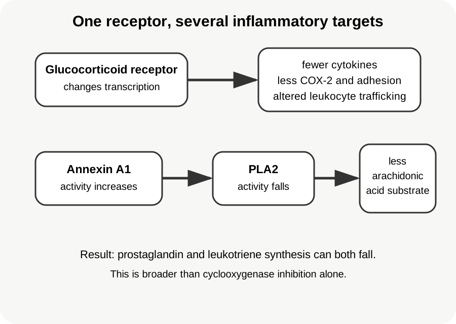
The older shortcut that glucocorticoids simply "stabilize membranes" is too narrow. Activated glucocorticoid receptors alter transcription of many inflammatory genes and interact with transcription factors such as NF-κB and AP-1. Downstream effects include reduced production of inflammatory cytokines, adhesion molecules, cyclooxygenase-2, and other mediators. [2]
Glucocorticoids also increase anti-inflammatory proteins including annexin A1, which can reduce cytosolic phospholipase A2 activity and therefore reduce arachidonic-acid availability for both prostaglandin and leukotriene synthesis. This helps explain why a glucocorticoid can suppress a broader inflammatory program than a drug that inhibits only cyclooxygenase. [2]
Glucocorticoid anti-inflammatory action is broader than a single membrane effect or COX inhibition. [2]
Leukocyte counts require interpretation rather than reflex. Glucocorticoids can cause neutrophilia through redistribution and reduced tissue egress while reducing circulating lymphocytes, eosinophils, and monocytes. A high neutrophil count after steroid exposure therefore does not by itself prove bacterial infection. Clinical context still decides. [2]
Membrane phospholipid PLA2 can release arachidonic acid
Arachidonic acid COX and lipoxygenase pathways diverge
Inflammatory mediators Prostaglandins and leukotrienes are reduced when upstream substrate release is suppressed
NSAIDs mainly inhibit COX. Glucocorticoids act at multiple transcriptional and upstream inflammatory targets, so the comparison is broader than one enzyme.Predict before opening: steroid versus NSAID
If phospholipase A2 activity and inflammatory gene expression are reduced, both prostaglandin and leukotriene production can fall. COX inhibition alone primarily reduces prostaglandin and thromboxane synthesis while leaving leukotriene synthesis available. [2]
Try it here · Checkpoint 2 of 3
Make your prediction before reading the choices. A first attempt is just a starting point.
Case 17
Show answer and explanations for case 17
A. Upstream glucocorticoid regulation versus NSAID cyclooxygenase inhibition (Best answer)
Glucocorticoids act through receptor-mediated regulation at several inflammatory targets, including upstream phospholipase A2 pathways; NSAIDs primarily inhibit COX enzymes.
Reasoning steps for option A
Which enzyme does ibuprofen inhibit, and which products does that spare?
Cyclooxygenase. Prostaglandins fall, but the lipoxygenase route to leukotrienes stays open.
How can a glucocorticoid reach the leukotriene branch?
By reducing phospholipase A2-driven arachidonic-acid release and suppressing inflammatory genes, it limits substrate for both branches.
B. Glucocorticoids selectively inhibit lipoxygenase, whereas NSAIDs selectively inhibit phospholipase A2 (Why this does not fit)
This reverses the relevant pathway logic. Standard NSAIDs inhibit COX, and glucocorticoids are not simply selective lipoxygenase inhibitors.
Reasoning steps for option B
What part of this pairing sounds right?
It correctly implies that the two drug classes act at different steps of eicosanoid synthesis.
Which enzyme does ibuprofen actually inhibit?
Cyclooxygenase, not phospholipase A2, and glucocorticoids act upstream rather than as selective lipoxygenase blockers.
C. Glucocorticoids and NSAIDs both act only at cyclooxygenase, but glucocorticoids bind longer (Why this does not fit)
Glucocorticoids have broad genomic effects and can reduce upstream arachidonic-acid release; they do not act only as longer-lasting COX inhibitors.
Reasoning steps for option C
Why might both drugs seem to share a COX mechanism?
Glucocorticoids do reduce COX-2 expression, so prostaglandin output falls with both.
Why does a COX-only mechanism fail for glucocorticoids?
It could not explain leukotriene reduction; glucocorticoids also lower arachidonic-acid supply and cytokine gene expression.
D. NSAIDs reduce both prostaglandins and leukotrienes, whereas glucocorticoids reduce only cytokines (Why this does not fit)
COX inhibition does not directly suppress the lipoxygenase leukotriene branch, while glucocorticoids can affect both eicosanoid and cytokine pathways.
Reasoning steps for option D
What is attractive about crediting NSAIDs with broad eicosanoid control?
NSAIDs are effective anti-inflammatories, and cytokine suppression is a real glucocorticoid effect.
What does the stem itself say about ibuprofen and leukotrienes?
Ibuprofen does not directly block leukotriene synthesis, which contradicts NSAIDs lowering both eicosanoid classes.
Takeaway: Glucocorticoids regulate inflammation at several levels, while NSAIDs chiefly inhibit the COX branch of arachidonic-acid metabolism.
Use physiology first, then choose the diagnostic test
Suspected endogenous Cushing syndrome is screened with validated tests that exploit either excess integrated cortisol production or loss of normal feedback and circadian regulation. Recommended first-line options include at least two 24-hour urinary free cortisol collections, two late-night salivary cortisol measurements, a 1-mg overnight dexamethasone suppression test, or the longer low-dose dexamethasone test. Random cortisol and random ACTH are not screening tests for establishing Cushing syndrome. [4]
After hypercortisolism is established, plasma ACTH helps localize the physiology. Suppressed ACTH supports an ACTH-independent adrenal process. Measurable or high ACTH supports an ACTH-dependent process. Pituitary MRI is then interpreted with biochemical evidence; when the source remains uncertain, specialist dynamic testing and bilateral inferior petrosal sinus sampling may be needed. A high-dose dexamethasone response alone should not be treated as a perfect pituitary-versus-ectopic separator. [5]
For primary adrenal insufficiency, a low cortisol with ACTH more than twice the upper reference limit supports adrenal failure when cortisol deficiency is confirmed. The standard 250-microgram cosyntropin test measures adrenal cortisol reserve. The historical peak threshold near 18 micrograms/dL came from older assays; newer specific immunoassays and LC-MS/MS can use lower cutoffs around 14 to 15 micrograms/dL. Use the assay-specific reference rather than memorizing one universal number. [6][7]
Exogenous glucocorticoids can suppress CRH and ACTH and eventually reduce adrenal responsiveness. The 2024 joint guideline emphasizes exposure: dose, duration, potency, route, and individual susceptibility all matter. Courses shorter than about 3 to 4 weeks generally do not require tapering solely to prevent HPA suppression, while longer therapy is reduced toward physiologic dosing before slower taper or recovery assessment. For patients tapering toward discontinuation, morning cortisol is often the first recovery test; values above 10 micrograms/dL suggest recovery, while lower values require context and follow-up. [3]
Patients with current or recent glucocorticoid exposure who have not demonstrated HPA recovery may need stress-dose coverage during illness or procedures. Oral dosing may suffice for minor stress when absorption and hemodynamics are intact; parenteral glucocorticoids are used for major stress, persistent vomiting, or hemodynamic instability. In suspected adrenal crisis, treatment should not be delayed for laboratory perfection. [3][6]
Pregnancy changes the reference frame. Estrogen increases cortisol-binding globulin and total cortisol, and physiologic free cortisol also rises as pregnancy progresses. The circadian rhythm is altered but not simply abolished. Nonpregnant reference ranges and routine dexamethasone assumptions can therefore mislead. [8]
Classic salt-wasting 21-hydroxylase deficiency reduces cortisol and aldosterone synthesis and increases ACTH drive toward adrenal androgen production. Treatment replaces glucocorticoid, and mineralocorticoid plus salt replacement is used when mineralocorticoid deficiency is present. The useful link is not "every congenital adrenal hyperplasia patient gets the same two drugs"; it is replace the hormones the enzyme block makes deficient. [9]
Start with cortisol and ACTH direction
Pattern
Physiologic interpretation
Next question
PatternLow cortisol, high ACTH
Physiologic interpretationPrimary adrenal failure
Next questionIs mineralocorticoid function also impaired (potassium, volume status, pigmentation)?
PatternLow cortisol, low or inappropriately normal ACTH
Physiologic interpretationCentral or exogenous suppression
Next questionIs there pituitary disease or glucocorticoid exposure?
Do not localize a suspected Cushing syndrome until hypercortisolism has been established with an appropriate screening strategy.
Predict before opening: ACTH-dependent hypercortisolism with equivocal pituitary MRI
Do not let a tiny incidental pituitary focus settle the case. When biochemical and imaging evidence do not confidently identify a pituitary source, specialist testing such as bilateral inferior petrosal sinus sampling can be required. [5]
Try it here · Checkpoint 3 of 3
Make your prediction before reading the choices. A first attempt is just a starting point.
Case 23
Show answer and explanations for case 23
A. Adrenal CT because every high-cortisol state begins in the adrenal gland (Why this does not fit)
High ACTH indicates an ACTH-dependent process, so adrenal autonomy is not the leading localization.
Reasoning steps for option A
Why might adrenal imaging seem a reasonable step in any hypercortisolism?
Adrenal tumors are a common source of cortisol excess.
What does her high ACTH say about an adrenal source?
High ACTH means the adrenal glands are being driven, so the search belongs upstream in the pituitary or an ectopic tumor.
B. Random midnight serum ACTH as the sole localization test (Why this does not fit)
A random ACTH value can establish ACTH dependence but cannot reliably distinguish pituitary from ectopic secretion by itself.
Reasoning steps for option B
What useful information does her ACTH already provide?
A measurable or high ACTH establishes that the hypercortisolism is ACTH dependent.
Why can a single ACTH value not settle the source?
Pituitary and ectopic ACTH values overlap widely, so the level does not identify where ACTH is made.
C. Bilateral inferior petrosal sinus sampling (Best answer)
When ACTH-dependent Cushing syndrome is confirmed but pituitary imaging is not convincingly diagnostic, bilateral inferior petrosal sinus sampling can distinguish a central pituitary ACTH gradient from an ectopic source.
Reasoning steps for option C
What has been established so far?
ACTH-dependent hypercortisolism, plus a 3-mm pituitary focus that could be incidental.
Why is a tiny indeterminate pituitary finding not enough?
Small incidental pituitary lesions are common in the general population, so imaging alone cannot prove the source.
How does petrosal sampling decide the source?
A central-to-peripheral ACTH gradient, especially after CRH or desmopressin stimulation, indicates a pituitary source; no gradient suggests ectopic ACTH.
D. High-dose dexamethasone suppression as a definitive source test (Why this does not fit)
Suppression patterns can support the evaluation but are not sufficiently definitive to replace source-localization strategies when imaging and biochemistry remain uncertain.
Reasoning steps for option D
Why might dexamethasone suppression seem to separate the sources?
Many pituitary corticotroph tumors partly suppress with high-dose dexamethasone, while many ectopic sources do not.
Why can it not replace direct sampling here?
Overlap between pituitary and ectopic responses is too large for a definitive answer when imaging is equivocal.
Takeaway: With confirmed ACTH-dependent hypercortisolism and equivocal pituitary imaging, inferior petrosal sinus sampling may be needed for source localization.
A. Suppression of pituitary ACTH by high cortisol (Why this does not fit)
High cortisol would suppress ACTH and would not fit the low cortisol, hypotension, and hyperkalemia.
Reasoning steps for option A
Why could an ACTH-suppression mechanism seem connected to a pigment change?
Skin pigmentation tracks ACTH levels, so a change in ACTH is the right place to look.
Which findings rule out high cortisol in this woman?
Her morning cortisol is low, with hypotension and hyperkalemia; suppressed ACTH would also remove the pigment drive rather than create it.
B. Loss of aldosterone causing direct melanin synthesis (Why this does not fit)
Mineralocorticoid deficiency contributes to salt wasting and hyperkalemia but does not directly drive melanocyte pigmentation.
Reasoning steps for option B
Which findings point to aldosterone deficiency in this woman?
Salt craving, sodium 127 mEq/L, potassium 5.8 mEq/L and hypotension all indicate mineralocorticoid loss.
Does mineralocorticoid deficiency act on melanocytes?
No. Aldosterone loss explains salt wasting and hyperkalemia but has no direct pigmentary action.
C. Pituitary failure causing low ACTH (Why this does not fit)
Low ACTH can cause central adrenal insufficiency, but it does not explain diffuse hyperpigmentation and usually preserves aldosterone better.
Reasoning steps for option C
What makes pituitary failure a plausible cause of her low morning cortisol?
Loss of ACTH causes central adrenal insufficiency with fatigue and low cortisol.
Which two findings argue against a central cause?
Diffuse hyperpigmentation needs high ACTH, and hyperkalemia shows aldosterone loss, which is usually preserved when only ACTH is missing.
D. High ACTH after loss of cortisol feedback (Best answer)
Primary adrenal failure removes cortisol feedback, so ACTH rises. ACTH derives from POMC, which is linked to melanocortin signaling and hyperpigmentation.
Reasoning steps for option D
What do low cortisol, hyperkalemia and hypotension together say about the level of the lesion?
Loss of both cortisol and aldosterone points to primary adrenal failure.
How does primary adrenal failure change pituitary ACTH output?
Without cortisol feedback, the intact pituitary increases POMC processing and ACTH secretion.
How does the raised ACTH darken the skin?
ACTH and melanocyte-stimulating peptides both come from POMC, and ACTH can itself activate melanocortin 1 receptors on melanocytes.
Takeaway: Low cortisol plus high ACTH physiology identifies primary adrenal failure and explains hyperpigmentation through POMC-derived melanocortin signaling.
A. Predominant renin-angiotensin and potassium control of aldosterone (Best answer)
Central ACTH deficiency lowers cortisol but usually leaves zona glomerulosa mineralocorticoid regulation largely intact because aldosterone is governed mainly by angiotensin II and potassium.
Reasoning steps for option A
What do low cortisol and low ACTH after pituitary surgery indicate?
Central adrenal insufficiency from loss of corticotroph ACTH.
Which regulators keep aldosterone secretion going when ACTH is lost?
Angiotensin II and serum potassium drive the zona glomerulosa, so aldosterone and renal potassium excretion are largely preserved.
Why is sodium still mildly low at 130 mEq/L?
Cortisol deficiency impairs free-water excretion and increases vasopressin release, lowering sodium without raising potassium.
B. ACTH directly stimulates renal potassium excretion (Why this does not fit)
ACTH is not the principal renal controller of potassium excretion; aldosterone is.
Reasoning steps for option B
Why might ACTH seem linked to potassium balance in this patient?
ACTH is the missing hormone, and it can acutely stimulate some aldosterone release.
Which hormone actually controls collecting-duct potassium secretion?
Aldosterone. ACTH has no major direct renal action, and if it did, losing it would predict hyperkalemia rather than normal potassium.
C. Cortisol normally blocks potassium secretion in the collecting duct (Why this does not fit)
Cortisol is not the principal physiologic brake on collecting-duct potassium secretion.
Reasoning steps for option C
Why could a cortisol effect on potassium seem worth considering?
Cortisol can occupy mineralocorticoid receptors when renal 11β-HSD2 is overwhelmed, so it has some renal salt and potassium effects.
Why does that idea fail to explain her normal potassium?
If cortisol normally braked potassium secretion, losing it would lower potassium; the collecting duct is governed chiefly by aldosterone, which is still present.
D. Pituitary surgery causes renal potassium retention to offset adrenal loss (Why this does not fit)
Pituitary surgery does not create a compensatory renal process that replaces aldosterone.
Reasoning steps for option D
Why might the recent surgery itself seem responsible for her electrolytes?
Transsphenoidal surgery can disturb water balance through vasopressin changes, so a surgical renal effect sounds possible.
Is there a compensatory renal pathway that stands in for aldosterone?
No. Potassium is normal because aldosterone itself is still made under angiotensin II and potassium control.
Takeaway: Central adrenal insufficiency lowers ACTH and cortisol while mineralocorticoid function is usually preserved.
A. Autoimmune destruction of the adrenal cortex (Why this does not fit)
Primary adrenal destruction can cause hypotension and low cortisol but more often raises ACTH and can impair aldosterone enough to cause hyperkalemia.
Reasoning steps for option A
Which features overlap with autoimmune adrenalitis?
Hypotension, hyponatremia, nausea and a low morning cortisol all occur in primary adrenal failure.
What in the history and potassium points away from primary adrenal destruction?
Eight months of prednisone stopped three days ago explains central suppression, and potassium 4.4 mEq/L suggests preserved aldosterone.
B. Chronic suppression of CRH and ACTH (Best answer)
Long-term supraphysiologic glucocorticoid exposure suppresses hypothalamic CRH and pituitary ACTH. Abrupt discontinuation can reveal inadequate endogenous cortisol production before the axis recovers.
Reasoning steps for option B
What did prednisone 20 mg daily for 8 months do to the HPA axis?
Supraphysiologic exposure suppressed CRH and ACTH for months, leaving the adrenal cortex understimulated.
Why did symptoms appear three days after the prescription ran out?
The exogenous glucocorticoid disappeared before the suppressed axis could recover, so endogenous cortisol was inadequate.
Why is potassium normal despite hypotension?
Aldosterone is regulated mainly by renin-angiotensin and potassium, so it is largely preserved in this central form.
C. Prednisone-induced aldosterone receptor destruction (Why this does not fit)
Prednisone does not cause adrenal crisis by destroying mineralocorticoid receptors.
Reasoning steps for option C
Why might a mineralocorticoid problem seem to fit his hypotension and hyponatremia?
Volume loss and low sodium can both result from failed mineralocorticoid action.
What argues against a receptor-level mineralocorticoid failure?
Potassium is normal, prednisone does not destroy mineralocorticoid receptors, and the timing points to withdrawal.
D. Rebound ACTH secretion causing adrenal hemorrhage (Why this does not fit)
Withdrawal does not normally produce adrenal hemorrhage through an ACTH surge.
Reasoning steps for option D
Why could adrenal hemorrhage come to mind with sudden hypotension?
Bilateral adrenal hemorrhage does cause abrupt adrenal crisis.
What would ACTH be doing three days after stopping chronic prednisone?
It remains suppressed rather than surging, because corticotroph recovery takes weeks to months.
Takeaway: After long-term supraphysiologic glucocorticoids, endogenous cortisol production may lag because CRH and ACTH have been suppressed.
The adrenal medulla produces catecholamines and does not serve as the central circadian pacemaker for cortisol secretion.
Reasoning steps for option A
Why might the adrenal medulla seem connected to a daily hormone rhythm?
Catecholamine output also varies with sleep and waking.
Can the medulla set the cortisol rhythm?
No. It secretes catecholamines, and the cortisol pattern depends on central timing of CRH and ACTH.
B. Zona fasciculata (Why this does not fit)
The zona fasciculata makes cortisol in response to ACTH but is not the principal central clock setting the daily HPA pattern.
Reasoning steps for option B
What makes the fasciculata tempting as the timing source?
It is where the measured cortisol is made, and adrenal cells carry their own peripheral clock genes.
What sets the phase of the daily HPA pattern?
A central pacemaker. The fasciculata follows ACTH input and adjusts its sensitivity but is not the principal clock.
C. Hypothalamic suprachiasmatic nucleus (Best answer)
The suprachiasmatic nucleus coordinates circadian timing and influences hypothalamic HPA output, producing a daily rhythm on which ultradian pulses are superimposed.
Reasoning steps for option C
Which features of the curve point to a clock rather than a stress response?
Cortisol rises before waking and falls to a nadir near usual sleep on a stable schedule, a predictable 24-hour cycle.
Which structure acts as the master circadian pacemaker?
The suprachiasmatic nucleus of the hypothalamus, entrained by light through the retina.
How does that timing signal reach the adrenal gland?
It shapes paraventricular CRH output, which drives pulsatile ACTH and cortisol secretion across the day.
D. Posterior pituitary (Why this does not fit)
The posterior pituitary releases vasopressin and oxytocin but is not the central circadian pacemaker for cortisol secretion.
Reasoning steps for option D
Why might the posterior pituitary seem involved in HPA timing?
It is an extension of the hypothalamus, and vasopressin can potentiate CRH action on ACTH release.
Why is the posterior pituitary not the circadian pacemaker?
It releases vasopressin and oxytocin from nerve terminals; the circadian signal originates in the suprachiasmatic nucleus.
Takeaway: Cortisol has a circadian pattern coordinated by the central clock, with pulsatile secretion superimposed.
A. The result proves endogenous Cushing syndrome because total cortisol should not change in pregnancy (Why this does not fit)
Pregnancy changes cortisol physiology and commonly raises total cortisol, so a nonpregnant total-cortisol comparison is not diagnostic.
Reasoning steps for option A
Why might a doubled total cortisol raise concern for Cushing syndrome?
A value twice the prepregnancy level looks like hypercortisolism by nonpregnant standards.
What argues against endogenous Cushing syndrome in this woman?
She lacks striae, proximal weakness and new hypertension, and pregnancy itself raises total cortisol through higher cortisol-binding globulin.
B. The high total cortisol is entirely an assay error caused by estrogen (Why this does not fit)
Estrogen changes physiology by increasing cortisol-binding globulin; it does not simply create a laboratory artifact.
Reasoning steps for option B
Why could an estrogen effect on the result look like a laboratory problem?
Estrogen does shift the total cortisol value without any Cushing features.
Is the rise an artifact or real physiology?
Real physiology. Estrogen increases hepatic cortisol-binding globulin synthesis, and the assay accurately measures the extra bound cortisol.
C. Only cortisol-binding globulin rises; free cortisol must remain identical to the nonpregnant state (Why this does not fit)
Free cortisol also rises physiologically as pregnancy progresses, so the change cannot be reduced to binding protein alone.
Reasoning steps for option C
What is correct about attributing the rise to binding globulin?
High cortisol-binding globulin does explain much of the rise in total cortisol.
What happens to free cortisol as pregnancy advances?
It also rises, partly because placental CRH stimulates the axis, so binding protein alone does not explain the change.
D. Pregnancy raises cortisol-binding globulin and physiologic cortisol exposure, so pregnancy-specific interpretation is required (Best answer)
Estrogen increases cortisol-binding globulin and total cortisol, while physiologic free cortisol also rises during pregnancy. Nonpregnant reference assumptions can therefore mislead.
Reasoning steps for option D
Why does total cortisol rise in the third trimester?
Estrogen raises cortisol-binding globulin, increasing the bound fraction.
Besides binding protein, what else changes in pregnancy?
Free cortisol also rises physiologically as pregnancy progresses.
How should cortisol tests be interpreted during pregnancy?
Against pregnancy-specific expectations, because nonpregnant thresholds and dexamethasone responses can mislead.
Takeaway: Pregnancy increases cortisol-binding globulin and physiologic cortisol exposure, so nonpregnant cortisol ranges cannot be applied mechanically.
A. Intracellular glucocorticoid receptor (Best answer)
Cortisol is a steroid hormone that crosses the plasma membrane and binds the intracellular glucocorticoid receptor, which regulates gene transcription.
Reasoning steps for option A
Where does cortisol bind its receptor in this description?
In the cytoplasm, after crossing the plasma membrane as a lipophilic steroid.
What does the activated complex do next?
It moves into the nucleus and changes transcription at glucocorticoid-response elements, the hallmark of an intracellular steroid receptor.
B. Gs-coupled cell-surface receptor (Why this does not fit)
Gs-coupled receptors activate second messengers at the plasma membrane and describe ACTH signaling better than cortisol signaling.
Reasoning steps for option B
Which adrenal-axis hormone does use a Gs-coupled receptor?
ACTH, which acts through the melanocortin 2 receptor and raises cAMP in adrenal cells.
Why does a Gs-coupled receptor not fit the pathway in the stem?
Gs-coupled receptors sit in the plasma membrane, while the described receptor binds hormone in the cytoplasm and moves to the nucleus.
C. Receptor tyrosine kinase (Why this does not fit)
Receptor tyrosine kinases are membrane proteins used by signals such as insulin, not the classic receptor for cortisol.
Reasoning steps for option C
Why might a receptor tyrosine kinase seem relevant in muscle?
Insulin, whose action cortisol opposes in muscle, signals through a receptor tyrosine kinase.
What separates a tyrosine kinase receptor from the pathway described?
It is a membrane protein that signals by phosphorylation, not a cytoplasmic receptor that travels to the nucleus.
D. Ligand-gated ion channel (Why this does not fit)
Ligand-gated ion channels alter membrane conductance rather than mediating the genomic steroid response described.
Reasoning steps for option D
Why could an ion channel seem plausible for a hormone acting on muscle?
Muscle function depends heavily on ligand-gated channels at the neuromuscular junction.
What in the stem excludes a channel mechanism?
The response is nuclear accumulation and altered transcription, not a change in membrane ion conductance.
Takeaway: Cortisol is a steroid hormone whose classic signaling mechanism uses an intracellular glucocorticoid receptor.
A. Direct activation of α₁ receptors by hydrocortisone (Why this does not fit)
Hydrocortisone does not act as an α₁ agonist.
Reasoning steps for option A
Why might direct α₁ activation seem to explain the falling norepinephrine requirement?
The improvement involves adrenergic vascular tone, and α₁ receptors mediate norepinephrine vasoconstriction.
Is hydrocortisone an α₁ agonist?
No. It does not bind α₁ receptors; it restores the vessel's response to norepinephrine over hours.
B. Permissive support of vascular responsiveness to catecholamines (Best answer)
Physiologic glucocorticoid activity helps maintain vascular responsiveness to catecholamines, so severe cortisol deficiency can contribute to vasopressor-resistant hypotension.
Reasoning steps for option B
Why does norepinephrine work poorly in this patient before hydrocortisone?
Known adrenal insufficiency plus pneumonia leaves cortisol severely deficient, and vessels respond poorly to catecholamines without it.
What does the improvement over several hours after hydrocortisone indicate?
Restored glucocorticoid activity lets the vasculature respond to catecholamines again, a permissive effect rather than direct vasoconstriction.
C. Immediate replacement of aldosterone as the only pressor mechanism (Why this does not fit)
Hydrocortisone can have mineralocorticoid activity at stress doses, but the rapid improvement in catecholamine responsiveness is not explained solely by aldosterone replacement.
Reasoning steps for option C
What makes aldosterone replacement tempting in primary adrenal insufficiency?
Primary adrenal failure usually includes aldosterone deficiency, and stress-dose hydrocortisone has mineralocorticoid activity.
Why does aldosterone replacement not explain this response?
Mineralocorticoid effects restore volume gradually; the specific change here is better catecholamine responsiveness, a glucocorticoid action.
D. Suppression of sympathetic catecholamine release (Why this does not fit)
Suppressing catecholamines would be expected to reduce rather than restore vascular support in shock.
Reasoning steps for option D
Why might cortisol seem to restrain sympathetic output?
Cortisol takes part in several negative-feedback loops.
What would lowering catecholamine release do to this patient?
Worsen the shock; the improvement required a stronger catecholamine effect, not less catecholamine.
Takeaway: Cortisol permits normal catecholamine-mediated vascular responsiveness; severe deficiency can contribute to refractory hypotension.
A. Immune-mediated myocyte necrosis with enzyme leakage (Why this does not fit)
Inflammatory muscle destruction usually produces myocyte injury and often a high creatine kinase, unlike this painless normal-CK pattern.
Reasoning steps for option A
Why is an inflammatory myopathy a reasonable concern with symmetric proximal weakness?
Polymyositis and related myopathies present with exactly that distribution.
Which laboratory value argues against myocyte necrosis?
Creatine kinase of 58 U/L is normal, whereas inflammatory necrosis usually leaks CK into blood.
B. Peripheral motor-nerve demyelination (Why this does not fit)
A demyelinating neuropathy would be expected to produce neurologic findings beyond isolated symmetric proximal weakness.
Reasoning steps for option B
Why might a nerve process seem possible when she cannot rise from a chair?
Weakness of many kinds can arise from peripheral nerve disease.
What in the examination argues against demyelination?
The weakness is purely proximal and symmetric without sensory loss, unlike the distal and sensory pattern of most neuropathies.
C. Neuromuscular-junction failure caused by acetylcholine blockade (Why this does not fit)
Glucocorticoids do not cause this chronic proximal pattern by blocking acetylcholine receptors.
Reasoning steps for option C
What makes a neuromuscular junction disorder tempting?
Myasthenia gravis also causes proximal weakness with a normal CK.
Why does acetylcholine blockade not fit long-term prednisone use?
Glucocorticoids do not block acetylcholine receptors, and her weakness is steady rather than fatigable.
D. Glucocorticoid-driven protein catabolism with muscle atrophy (Best answer)
Chronic glucocorticoid excess promotes protein catabolism and can cause painless proximal muscle atrophy with a normal or only mildly changed CK.
Reasoning steps for option D
What do 18 months of prednisone do to skeletal-muscle protein?
Chronic glucocorticoid exposure increases protein breakdown and reduces synthesis, especially in fast-twitch fibers, causing atrophy.
Why is CK normal despite clear weakness?
Atrophy from catabolism does not rupture myocytes, so little enzyme leaks into blood.
Takeaway: Chronic glucocorticoid excess can cause proximal myopathy through protein catabolism without the enzyme pattern of inflammatory muscle necrosis.
A. Reduced fibroblast collagen synthesis (Best answer)
Chronic glucocorticoid excess impairs fibroblast proliferation and collagen production, weakening dermis and slowing structural repair.
Reasoning steps for option A
What does the biopsy show about the dermis?
Reduced collagen and fewer active fibroblasts, the cells that build dermal matrix.
How does chronic glucocorticoid excess link that biopsy to his skin signs?
It suppresses fibroblast proliferation and collagen synthesis, so the thin dermis tears into wide striae, bruises easily and repairs slowly.
B. Activation of mast-cell degranulation (Why this does not fit)
Glucocorticoids generally suppress inflammatory mediator release rather than promote mast-cell degranulation.
Reasoning steps for option B
Why could mast cells seem relevant to bruising and skin change?
Mast-cell mediators affect vessels and inflammation in the skin.
What do glucocorticoids actually do to mast-cell mediator release?
They generally reduce it, and the biopsy shows matrix loss rather than inflammation.
C. Increased keratinocyte mitosis with dermal thickening (Why this does not fit)
This predicts thicker skin, the opposite of the observed atrophy and bruising.
Reasoning steps for option C
Why might epidermal cell turnover seem connected to abnormal skin?
Many skin diseases do reflect altered keratinocyte proliferation.
What does the biopsy show that contradicts thickening?
Reduced dermal collagen and fewer fibroblasts mean atrophy, the opposite of a thicker dermis.
D. Increased collagen cross-linking from isolated hyperglycemia (Why this does not fit)
Hyperglycemia can impair wound healing, but reduced fibroblast activity and reduced collagen identify a direct glucocorticoid connective-tissue effect more specifically.
Reasoning steps for option D
Why is hyperglycemia a reasonable suspect for slow healing?
Hypercortisolism raises glucose, and hyperglycemia impairs wound repair.
Which biopsy finding points to a direct glucocorticoid effect instead?
Fewer active fibroblasts with reduced collagen indicates suppressed matrix synthesis, not extra cross-linking of normal collagen.
Takeaway: Skin thinning and poor wound repair in cortisol excess reflect impaired fibroblast function and collagen production.
A. Severe vitamin D deficiency causing isolated osteomalacia (Why this does not fit)
Her vitamin D and mineral values do not support isolated osteomalacia, and chronic glucocorticoids harm bone through several direct and indirect pathways.
Reasoning steps for option A
Why is vitamin D deficiency a reasonable thought with low bone density in an older woman?
Osteomalacia also lowers measured bone density and raises fracture risk.
Which laboratory values rule out osteomalacia here?
Her 25-hydroxyvitamin D, calcium and phosphate are all within reference ranges.
B. Reduced osteoblast and osteocyte function (Best answer)
Chronic glucocorticoids suppress bone formation and impair osteoblast and osteocyte function, with additional effects on resorption, calcium handling, and muscle.
Reasoning steps for option B
What does a fracture with normal calcium, phosphate and vitamin D suggest?
A problem in bone-cell function rather than in mineral supply.
Which bone cells does long-term prednisone impair most?
Osteoblasts and osteocytes; glucocorticoids suppress bone formation and increase their apoptosis, with an early rise in resorption.
C. Excess calcitonin secretion causing diffuse bone resorption (Why this does not fit)
Calcitonin excess is not the mechanism of glucocorticoid-induced osteoporosis.
Reasoning steps for option C
Why might a calcium-regulating hormone come to mind with bone loss?
Hormones that regulate calcium also act on bone, so they are an intuitive target.
What does calcitonin actually do to bone?
It inhibits osteoclast resorption, so excess would not cause bone loss, and it has no role in glucocorticoid osteoporosis.
D. Replacement of endogenous cortisol with a bone-protective synthetic steroid (Why this does not fit)
Prednisone is not bone protective; prolonged systemic glucocorticoid exposure increases fracture risk.
Reasoning steps for option D
Why might a synthetic steroid be assumed to behave differently from cortisol?
Prednisone is a synthetic analogue, so its bone effects might seem different.
What does years of prednisone do to fracture risk?
It raises it, because prednisone acts through the same glucocorticoid receptor and suppresses bone formation.
Takeaway: Glucocorticoid osteoporosis is driven strongly by impaired bone formation and altered bone-cell function, not by a required fall in serum calcium.
A. Inhibition of hormone-sensitive lipase with triglyceride storage (Why this does not fit)
Inhibiting lipolysis would lower rather than raise circulating glycerol and free fatty acids.
Reasoning steps for option A
Why is hormone-sensitive lipase a sensible enzyme to think about here?
It is a key regulated step in adipose triglyceride breakdown, so it governs the measured fuels.
Which measurement shows the lipase is active rather than inhibited?
Free fatty acids and glycerol are higher, which happens only when triglyceride breakdown increases.
B. Conversion of fatty acids directly into glucose in the liver (Why this does not fit)
Humans cannot convert even-carbon fatty-acid carbon into net glucose, although glycerol can enter gluconeogenesis.
Reasoning steps for option B
Why might fatty acids seem to feed hepatic glucose output?
Fatty acids rose at the same time that hepatic glucose production increased.
Can even-chain fatty-acid carbon become net glucose in humans?
No. Acetyl-CoA cannot be converted to pyruvate, so only the glycerol backbone is a gluconeogenic substrate, while fatty-acid oxidation supplies energy.
C. Lipolysis releasing glycerol for gluconeogenesis (Best answer)
Cortisol supports lipolytic substrate availability. Glycerol can enter hepatic gluconeogenesis, while fatty acids provide energy for other tissues and hepatic metabolism.
Reasoning steps for option C
What do rising glycerol and free fatty acids indicate about adipose tissue?
Triglycerides are being broken down, a process cortisol supports alongside catecholamines.
Which lipolysis product can the liver turn into glucose?
Glycerol, which enters gluconeogenesis through glycerol-3-phosphate, while fatty acids fuel other tissues and hepatic energy needs.
D. Blockade of adipose triglyceride breakdown with increased ketone clearance (Why this does not fit)
This predicts reduced substrate release from adipose tissue, the opposite of the measurements.
Reasoning steps for option D
What makes a ketone-focused explanation tempting during fasting stress?
Ketone production and clearance do change during fasting.
What do the measurements show about adipose triglyceride breakdown?
Higher glycerol and fatty acids prove that breakdown increased rather than being blocked.
Takeaway: Cortisol can support fuel availability by promoting lipolytic substrate release; glycerol can feed gluconeogenesis.
A. Direct irreversible inhibition of cyclooxygenase-1 only (Why this does not fit)
Selective COX-1 inhibition would not explain the broad reduction in cytokine signaling and leukotriene-related pathways.
Reasoning steps for option A
Why does cyclooxygenase inhibition seem to fit falling eicosanoid production?
Cyclooxygenase makes prostaglandins, so blocking it lowers some eicosanoids.
What would COX-1 inhibition leave untouched in this flare?
Cytokine signaling and the lipoxygenase leukotriene branch, both of which matter in asthma.
B. Activation of complement proteins that consume inflammatory mediators (Why this does not fit)
Glucocorticoids do not suppress inflammation by activating the complement cascade.
Reasoning steps for option B
Why might complement seem connected to inflammatory mediators?
Complement activation is a major inflammatory pathway.
What would activating complement do to inflammation?
It would generate anaphylatoxins and amplify inflammation; glucocorticoids do not work this way.
C. Neutralization of circulating cytokines by direct steroid binding (Why this does not fit)
Glucocorticoids do not work by physically binding and neutralizing cytokines in plasma.
Reasoning steps for option C
Why might direct binding seem to explain the fall in cytokine signaling?
Cytokine-neutralizing antibodies do work that way in other therapies.
Where does a glucocorticoid act to lower cytokines?
Inside cells through its receptor, reducing cytokine gene transcription over hours rather than binding cytokines in plasma.
D. Glucocorticoid-receptor regulation of inflammatory genes and PLA2 pathways (Best answer)
Activated glucocorticoid receptors alter inflammatory transcription and can increase annexin A1, reducing cytosolic phospholipase A2 activity and arachidonic-acid availability while also suppressing cytokine pathways.
Reasoning steps for option D
What does a response over several hours suggest about the mechanism?
A transcriptional effect, consistent with the glucocorticoid receptor changing gene expression.
How does receptor activation lower both cytokines and eicosanoids?
It represses inflammatory genes by interfering with NF-κB and AP-1 and induces annexin A1, which restrains phospholipase A2 and arachidonic-acid release.
Takeaway: Glucocorticoids suppress inflammation through broad receptor-mediated gene regulation, including effects on annexin A1, phospholipase A2, cytokines, and COX-2.
A. Bone-marrow bacterial infection causing accelerated band release (Why this does not fit)
A bacterial infection can cause neutrophilia, but the patient is afebrile, clinically improved, and lacks a left shift immediately after glucocorticoid exposure.
Reasoning steps for option A
Why is infection a reasonable concern with a WBC count of 15,800/µL?
Bacterial infection is a common cause of neutrophilia in a patient receiving immunosuppression.
Which findings make infection unlikely here?
She is afebrile and improving, there is no left shift, and the rise followed high-dose prednisone within days.
B. Glucocorticoid-induced neutrophil demargination and altered trafficking (Best answer)
Glucocorticoids increase circulating neutrophils through demargination and changes in tissue trafficking while often reducing circulating lymphocytes and eosinophils.
Reasoning steps for option B
What changed three days before the leukocytosis?
She started high-dose prednisone, which predictably alters neutrophil trafficking.
How do glucocorticoids raise the neutrophil count without infection?
They mobilize marginated neutrophils, speed marrow release and slow exit into tissues, while lowering lymphocytes and eosinophils.
C. Direct conversion of lymphocytes into neutrophils (Why this does not fit)
Mature lymphocytes do not convert into neutrophils in response to glucocorticoids.
Reasoning steps for option C
Why might lymphocytes seem linked to the neutrophil rise?
Glucocorticoids lower circulating lymphocytes while neutrophils rise, so a shift between them looks plausible.
Can lymphocytes turn into neutrophils?
No. They belong to separate lineages, and lymphocytes fall because they redistribute to lymphoid tissue.
D. Complement activation causing neutrophil proliferation in blood (Why this does not fit)
Glucocorticoids suppress many inflammatory pathways and do not produce this pattern through complement-driven proliferation.
Reasoning steps for option D
Why might complement seem able to raise neutrophils?
Complement fragments such as C5a attract and activate neutrophils.
What does prednisone do to that inflammatory pathway?
It suppresses inflammation, and neutrophils do not proliferate in circulating blood.
Takeaway: Glucocorticoids can raise the circulating neutrophil count through demargination and altered trafficking, so neutrophilia alone does not diagnose infection.
Mineralocorticoid replacement addresses salt wasting but does not replace deficient cortisol or suppress excess ACTH-driven androgen production.
Reasoning steps for option A
Which findings make mineralocorticoid replacement essential?
Sodium 121 mEq/L, potassium 6.6 mEq/L and dehydration show aldosterone deficiency with salt wasting.
Which findings remain untreated by fludrocortisone alone?
Hypoglycemia and virilization reflect cortisol deficiency and ACTH-driven androgen excess, which need glucocorticoid.
B. Hydrocortisone alone without mineralocorticoid or salt replacement (Why this does not fit)
Glucocorticoid replacement is necessary but does not fully address the mineralocorticoid deficiency of a classic salt-wasting phenotype.
Reasoning steps for option B
Why is hydrocortisone clearly needed in this infant?
Glucose 49 mg/dL and a markedly high 17-hydroxyprogesterone show cortisol deficiency with ACTH drive toward androgens.
Why is hydrocortisone alone insufficient here?
Hyponatremia and hyperkalemia show aldosterone deficiency, and replacement hydrocortisone doses give too little mineralocorticoid effect to correct it.
C. Hydrocortisone plus fludrocortisone with salt replacement (Best answer)
Classic salt-wasting 21-hydroxylase deficiency causes cortisol and aldosterone deficiency. Hydrocortisone replaces glucocorticoid and suppresses ACTH drive, while fludrocortisone and salt address mineralocorticoid deficiency.
Reasoning steps for option C
What do markedly high 17-hydroxyprogesterone and virilization indicate?
21-hydroxylase deficiency, which blocks both cortisol and aldosterone synthesis and diverts precursors toward androgens.
How does hydrocortisone address two problems at once?
It replaces cortisol and, through feedback, lowers ACTH-driven androgen overproduction.
Why add fludrocortisone and sodium chloride in a salt-wasting infant?
The electrolytes show aldosterone deficiency, and infant feeds supply little sodium, so mineralocorticoid plus salt supplementation is recommended.
D. Dexamethasone plus spironolactone (Why this does not fit)
Spironolactone blocks mineralocorticoid action and would worsen a salt-wasting state; dexamethasone is not the usual replacement choice for an infant.
Reasoning steps for option D
What makes dexamethasone seem a reasonable glucocorticoid choice?
It strongly suppresses ACTH and adrenal androgen production.
Why would this combination harm the infant?
Spironolactone blocks mineralocorticoid receptors and would worsen salt loss and hyperkalemia, and dexamethasone is avoided in growing children because it suppresses growth.
Takeaway: Classic salt-wasting 21-hydroxylase deficiency requires glucocorticoid replacement and mineralocorticoid plus salt replacement.
A. Loss of the normal late-night cortisol nadir (Best answer)
Normal cortisol secretion falls toward a late-night or sleep-period nadir. Repeated high late-night salivary cortisol suggests loss of that circadian suppression.
Reasoning steps for option A
Why is her high-normal 8 AM serum cortisol not reassuring?
Morning cortisol is normally near its daily peak, so early hypercortisolism can overlap normal values at that hour.
What do two elevated late-night salivary values reveal?
Cortisol is not falling to its usual sleep-period nadir, a loss of circadian suppression typical of Cushing syndrome.
B. Failure of the adrenal medulla to release epinephrine (Why this does not fit)
Late-night salivary cortisol measures free cortisol physiology, not catecholamine output from the adrenal medulla.
Reasoning steps for option B
Why could adrenal medullary function seem relevant to a nighttime test?
Both cortical and medullary hormone output vary over the day.
What does a salivary cortisol sample measure?
Free cortisol from the adrenal cortex, not epinephrine from the medulla.
C. Loss of aldosterone regulation by renin (Why this does not fit)
Aldosterone regulation is not what late-night salivary cortisol testing evaluates.
Reasoning steps for option C
Why might aldosterone come up in a patient with bruising and weakness?
Mineralocorticoid excess can cause weakness through hypokalemia.
Does late-night salivary testing assess the renin-aldosterone system?
No. It measures cortisol at its expected nadir, while aldosterone regulation needs renin and aldosterone measurement.
D. Excess cortisol-binding globulin production (Why this does not fit)
Salivary cortisol reflects free hormone and is not intended as a measure of cortisol-binding globulin.
Reasoning steps for option D
Why might binding-globulin excess seem to explain a high cortisol value?
High cortisol-binding globulin does raise total serum cortisol.
Why would binding globulin not raise this result?
Saliva contains free cortisol, so a rise in binding globulin would not increase salivary values.
Takeaway: Late-night salivary cortisol tests whether the normal sleep-period cortisol nadir has been lost.
A. Adrenal insufficiency is proven because every assay requires at least 18 micrograms/dL (Why this does not fit)
The historical 18-microgram/dL cutoff was derived from older assays and can overdiagnose adrenal insufficiency when applied to newer specific assays.
Reasoning steps for option A
Why does 18 micrograms/dL feel like the decisive threshold?
It is the classic historical cutoff for a normal cosyntropin response.
Why does that number not apply to this laboratory?
It came from older polyclonal immunoassays; newer specific assays read lower and use validated cutoffs near 14 to 15 micrograms/dL.
B. The test is uninterpretable because only a 60-minute value can be used (Why this does not fit)
Validated interpretation can use assay-specific 30-minute or 60-minute values depending on the protocol and laboratory method.
Reasoning steps for option B
Why might the timing of the stimulated sample seem to matter?
Protocols differ, and some collect both 30-minute and 60-minute samples.
Is his 30-minute value interpretable?
Yes. The laboratory's cutoff is validated for its protocol, so the 30-minute value can be read directly.
C. The baseline value alone proves normal adrenal reserve (Why this does not fit)
A low baseline cortisol does not prove adequate reserve; the stimulated response is the relevant dynamic information here.
Reasoning steps for option C
What information does the baseline cortisol provide?
It is the starting point, and a very low value can raise suspicion of insufficiency.
Why can a baseline of 3.2 micrograms/dL not show normal reserve?
A low baseline does not demonstrate reserve; the stimulated peak shows how much cortisol the adrenal can produce.
D. Adequate response by this assay's validated cutoff (Best answer)
Modern specific cortisol assays can use stimulated cutoffs around 14 to 15 micrograms/dL. A value of 15.2 exceeds the laboratory's validated 14.6 cutoff.
Reasoning steps for option D
Which cutoff should be applied to his result?
The laboratory's validated stimulated cutoff of 14.6 micrograms/dL for its modern assay.
How does his 30-minute value compare?
At 15.2 micrograms/dL it exceeds 14.6, indicating an adequate adrenal response by this method.
Takeaway: Cosyntropin thresholds are assay dependent; use the validated cutoff for the laboratory method rather than a universal historical number.
A. Morning cortisol supports HPA recovery and stopping physiologic glucocorticoid replacement (Best answer)
For patients tapering toward discontinuation, a morning cortisol above 10 micrograms/dL is a practical threshold suggesting HPA-axis recovery in the guideline.
Reasoning steps for option A
Which recovery test does the 2024 guideline suggest once the dose is near physiologic replacement?
A morning serum cortisol, drawn with enough time after the last glucocorticoid dose to reflect endogenous production.
How should her value of 11.2 micrograms/dL be interpreted?
It is above the 10 micrograms/dL level that suggests recovery, so glucocorticoid can be stopped.
B. She must remain on prednisone until a cosyntropin test exceeds 18 micrograms/dL (Why this does not fit)
Routine dynamic testing is not required for every patient tapering glucocorticoids, and a universal 18-microgram/dL stimulated cutoff is not assay independent.
Reasoning steps for option B
Why could dynamic testing seem a safer requirement?
Cosyntropin testing gives dynamic information about adrenal reserve.
What two problems make this requirement wrong?
Routine dynamic testing is not needed when morning cortisol already suggests recovery, and 18 micrograms/dL is not a universal cutoff.
C. The value proves primary adrenal destruction and requires fludrocortisone (Why this does not fit)
Glucocorticoid-induced adrenal insufficiency is central suppression, not primary destruction, and fludrocortisone is not routinely indicated.
Reasoning steps for option C
Why might any adrenal insufficiency seem to call for fludrocortisone?
Fludrocortisone is standard replacement in primary adrenal insufficiency.
What kind of adrenal suppression follows long-term prednisone?
Central suppression with intact aldosterone, so fludrocortisone is not routinely needed, and 11.2 micrograms/dL does not suggest adrenal destruction.
D. Morning cortisol cannot be used during glucocorticoid tapering under any circumstance (Why this does not fit)
Morning cortisol is specifically recommended as a first recovery test when a patient has reached physiologic dosing and aims to stop therapy.
Reasoning steps for option D
Why might ongoing glucocorticoid therapy seem to make cortisol measurement useless?
Some glucocorticoids, such as prednisolone, cross-react with cortisol immunoassays.
How does the guideline handle that problem?
It recommends morning cortisol once physiologic dosing is reached, timed after the last dose so the result reflects her own production.
Takeaway: During tapering near physiologic dosing, morning cortisol can guide HPA recovery; a value above 10 micrograms/dL supports recovery in the 2024 guideline.
A. Every systemic glucocorticoid course must be tapered regardless of duration (Why this does not fit)
The risk of clinically important HPA suppression depends strongly on exposure, and short courses generally do not require a taper solely for adrenal protection.
Reasoning steps for option A
Why might tapering every course seem cautious?
Longer glucocorticoid therapy can suppress the HPA axis, and abrupt stopping can then cause crisis.
What does 5 days of prednisone mean for HPA risk?
Exposure is short, and courses under about 3 to 4 weeks generally need no taper for adrenal protection.
B. No HPA-focused taper is needed after this 5-day course (Best answer)
The 2024 joint guideline indicates that systemic glucocorticoid courses shorter than about 3 to 4 weeks generally do not need tapering solely to prevent adrenal insufficiency.
Reasoning steps for option B
What glucocorticoid exposure did this patient have?
Prednisone 40 mg daily for only 5 days, with no other glucocorticoid use.
How does the 2024 guideline treat courses this short?
Courses shorter than about 3 to 4 weeks can generally be stopped without a taper solely for HPA protection.
C. He should receive fludrocortisone for one week after stopping prednisone (Why this does not fit)
Glucocorticoid-induced adrenal suppression does not routinely require mineralocorticoid replacement, and a brief course does not create this indication.
Reasoning steps for option C
Why might mineralocorticoid cover seem protective after stopping a steroid?
Fludrocortisone is part of replacement in primary adrenal insufficiency.
Why is fludrocortisone unnecessary here?
Glucocorticoid-induced suppression spares aldosterone, and a 5-day course creates no replacement need.
D. He must have a cosyntropin test before the final prednisone dose (Why this does not fit)
Routine adrenal testing is not recommended after a short systemic course when there is no other reason to suspect adrenal insufficiency.
Reasoning steps for option D
Why could testing before stopping seem careful?
A cosyntropin test does assess adrenal reserve.
What makes testing unnecessary after 5 days?
Routine testing is not advised after short courses when nothing else suggests adrenal insufficiency.
Takeaway: Short systemic glucocorticoid courses under about 3 to 4 weeks generally do not need tapering solely to prevent HPA suppression.
A. Wait for a morning cortisol result before giving glucocorticoid (Why this does not fit)
In hemodynamic instability with possible adrenal crisis, treatment should not be delayed for laboratory confirmation.
Reasoning steps for option A
Why is it tempting to confirm with a cortisol level first?
A laboratory result would document adrenal insufficiency before treatment.
Why is waiting dangerous here?
Hypotension, confusion and vomiting after recent withdrawal suggest adrenal crisis, which can be fatal if treatment is delayed.
B. Give only oral prednisone because all stress dosing should use the outpatient route (Why this does not fit)
Repeated vomiting makes oral absorption unreliable, and major stress with hemodynamic instability calls for parenteral coverage.
Reasoning steps for option B
Why might oral dosing seem enough?
Oral stress dosing works for minor illness when absorption is reliable.
Which findings make oral therapy unsuitable?
Repeated vomiting prevents absorption, and blood pressure 80/46 mmHg with confusion signals major stress.
C. Give parenteral stress-dose glucocorticoid and resuscitative care promptly (Best answer)
Current or recent glucocorticoid users without documented HPA recovery should receive stress coverage, and parenteral glucocorticoid is appropriate with vomiting or hemodynamic instability.
Reasoning steps for option C
Why is this patient at risk of adrenal crisis?
She stopped long-term dexamethasone two weeks ago without evidence of HPA recovery, so her axis is likely still suppressed.
What route and approach fit vomiting with hypotension?
Parenteral stress-dose glucocorticoid, usually hydrocortisone, with fluid resuscitation and treatment of the gastroenteritis.
D. Give fludrocortisone alone and avoid glucocorticoid until ACTH is measured (Why this does not fit)
Glucocorticoid-induced adrenal insufficiency is primarily a cortisol problem from central suppression, and fludrocortisone alone does not treat adrenal crisis.
Reasoning steps for option D
Why might a mineralocorticoid seem to help her hypotension?
Fludrocortisone promotes sodium retention and volume expansion.
Which hormone deficit drives this crisis?
Cortisol, from central suppression with aldosterone largely intact, so fludrocortisone alone would not treat it.
Takeaway: Recent glucocorticoid exposure without documented HPA recovery warrants stress coverage; major illness, vomiting, or shock favors parenteral glucocorticoid.
A. Weak glucocorticoid activity with strong mineralocorticoid action (Why this does not fit)
Dexamethasone is a potent glucocorticoid with minimal mineralocorticoid activity, so this description is reversed.
Reasoning steps for option A
Why does steroid potency matter for this test?
The test relies on a steroid that signals strongly through glucocorticoid receptors.
What is dexamethasone's actual receptor profile?
A potent glucocorticoid with negligible mineralocorticoid activity, the reverse of this description.
B. Direct inhibition of adrenal CYP11B1 with no action at the pituitary (Why this does not fit)
Dexamethasone suppresses the HPA axis through glucocorticoid receptors at hypothalamic and pituitary levels; it is not a CYP11B1 inhibitor.
Reasoning steps for option B
Why might enzyme inhibition seem to explain a low morning cortisol?
CYP11B1 inhibitors such as metyrapone do lower cortisol synthesis.
Where does dexamethasone act instead?
At glucocorticoid receptors in the hypothalamus and pituitary, suppressing CRH and ACTH rather than blocking an adrenal enzyme.
C. Displacement of cortisol from binding globulin with rapid urinary clearance (Why this does not fit)
Dexamethasone does not test HPA feedback by displacing cortisol from cortisol-binding globulin.
Reasoning steps for option C
Why might binding-globulin effects seem relevant to cortisol measurement?
Changes in cortisol-binding globulin alter total cortisol values.
Is displacement how the test works?
No. Dexamethasone binds cortisol-binding globulin poorly, and the test measures feedback suppression, not cortisol clearance.
D. Potent long-acting glucocorticoid feedback with little interference in standard cortisol measurement (Best answer)
Dexamethasone provides strong, sustained glucocorticoid feedback and generally has little cross-reactivity in standard cortisol assays, allowing the morning value to reflect endogenous cortisol suppression.
Reasoning steps for option D
What must the suppressing steroid do in a 1-mg overnight test?
Suppress ACTH strongly for many hours, which dexamethasone's potency and long action allow.
Why can the morning cortisol still be read as endogenous?
Dexamethasone shows little cross-reactivity in standard cortisol assays, so the measured value reflects the patient's own suppressed output.
Takeaway: Dexamethasone is useful for suppression testing because it strongly activates glucocorticoid feedback while generally contributing little signal to standard cortisol assays.