Glucagonoma Syndrome: NME, Catabolism, and Pancreatic NET Reasoning
Recognize glucagonoma syndrome, connect glucagon excess to NME and catabolism, interpret fasting glucagon, localize disease, and choose management.
A glucagonoma question is rarely solved by one isolated laboratory value. The useful task is to recognize a catabolic syndrome, decide whether the skin finding really fits necrolytic migratory erythema, and connect that pattern to a glucagon-secreting pancreatic neuroendocrine tumor. By the end, you should be able to explain the metabolic pattern, interpret fasting glucagon in context, choose localization or staging tests, and separate symptom control from tumor-directed care.
Start with the syndrome, not the tumor name
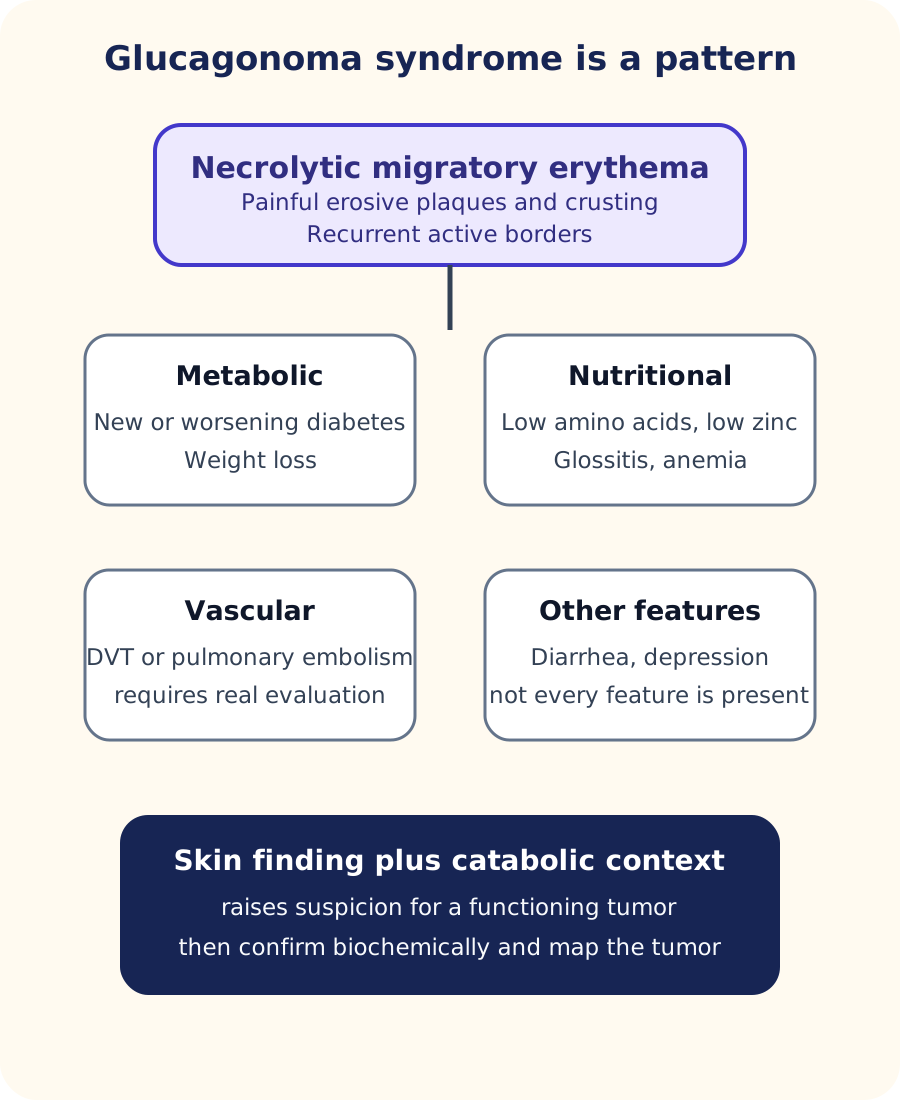
A patient with a painful recurrent eruption in the groin, perineum, buttocks, or legs may have necrolytic migratory erythema, often shortened to NME. Lesions can begin as erythematous plaques, become eroded or vesicular, and develop crusted or healing centers while active borders appear elsewhere. In glucagonoma syndrome, NME often travels with new or worsening diabetes, weight loss, glossitis or cheilitis, anemia, low amino acids, low zinc, venous thromboembolism, diarrhea, or depression. Not every patient has every feature. [1]
Use the whole pattern rather than treating the rash as proof of a tumor. NME becomes more informative when diabetes, weight loss, nutrient depletion, oral findings, or venous thrombosis fit the same syndrome. [1]
The old shortcut that the rash by itself proves glucagonoma is unsafe. Similar-appearing NME can occur without a glucagon-secreting tumor in severe nutritional deficiency, malabsorption, liver disease, and other states often grouped under pseudoglucagonoma. The rash therefore raises the probability; the surrounding metabolic pattern and biochemical evaluation determine whether a functioning pancreatic tumor is the explanation. [1]
Concrete example: Compare two patients with the same crusted intertriginous eruption. One has fasting glucose 286 mg/dL, marked weight loss, low amino acids, and a pancreatic mass. The other has chronic malabsorption, normal glucose, and no pancreatic lesion. The skin morphology points both toward NME, but only the first pattern strongly supports glucagonoma syndrome.
Predict before revealing: which added finding most changes the interpretation of an NME-like rash?
A concordant metabolic pattern, such as new diabetes plus weight loss and hypoaminoacidemia, makes a glucagon-secreting tumor much more coherent than an isolated rash. The next application is biochemical confirmation and tumor localization, not declaring a tumor from appearance alone. [1]
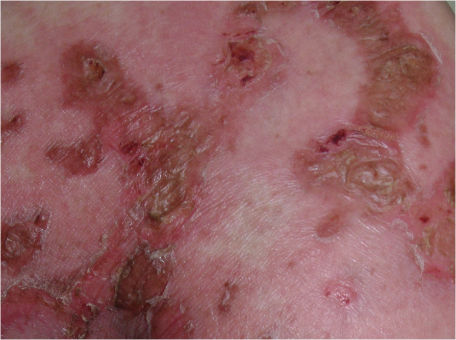
For visual recognition, the clinical photograph shows erythematous, eroded, crusted gluteal plaques from a published glucagonoma case. A single photograph cannot demonstrate migration over time, and it cannot establish the tumor by itself. That limitation is part of the interpretation.
Erythematous, eroded, crusted plaques in a published glucagonoma case. This static photograph supports morphology recognition but cannot show migration over time or prove the tumor by itself. Image: Sheng-li Wu, Ji-gang Bai, Jun Xu, Qing-yong Ma and Zheng Wu; original source; CC BY 4.0.
Try it here · Checkpoint 1 of 3
Make your prediction before reading the choices. A first attempt is just a starting point.
Case 1
Show answer and explanations for case 1
A. Glucagonoma syndrome (Best answer)
The NME pattern, new diabetes, weight loss, hypoaminoacidemia, and pancreatic lesion form a coherent glucagonoma syndrome.
Reasoning steps for option A
Which skin features in this woman fit necrolytic migratory erythema?
Painful recurrent groin and buttock plaques with eroded, crusted centers and newer active borders match the NME pattern.
What turns an NME-like rash into a unifying tumor diagnosis here?
New fasting hyperglycemia of 278 mg/dL, 8-kg weight loss, low amino acids and a 4.8 cm pancreatic lesion complete the glucagonoma syndrome.
B. Bullous pemphigoid (Why this does not fit)
Bullous pemphigoid can blister in an older adult, but it does not account for the catabolic metabolic pattern and pancreatic lesion.
Reasoning steps for option B
Why might pemphigoid come to mind for a 61-year-old with eroded plaques?
Bullous pemphigoid is common in older adults and can leave erosions after blisters rupture.
Which findings does pemphigoid leave unexplained in this woman?
It cannot account for the new diabetes, weight loss, hypoaminoacidemia or the pancreatic lesion.
C. Dermatitis herpetiformis (Why this does not fit)
Dermatitis herpetiformis is linked to gluten-sensitive enteropathy and usually favors extensor surfaces rather than this NME pattern with diabetes and marked catabolism.
Reasoning steps for option C
What could make dermatitis herpetiformis tempting in a patient with a chronic eroded rash?
It is an itchy, recurrent eruption linked to an enteropathy, which could seem to fit weight loss.
How do the distribution and metabolic findings argue against dermatitis herpetiformis?
It favors extensor surfaces, and it does not produce new diabetes, low amino acids or a pancreatic mass.
D. Contact dermatitis (Why this does not fit)
Contact dermatitis follows an exposure distribution and does not explain weight loss, hypoaminoacidemia, hyperglycemia, and a pancreatic tumor.
Reasoning steps for option D
Why could contact dermatitis seem reasonable for a groin and buttock eruption?
Intertriginous skin is often exposed to irritants and allergens, so a localized dermatitis is a common first thought.
What part of this presentation can an exposure rash never explain?
Contact dermatitis stays in the skin; it does not cause 8-kg weight loss, hyperglycemia, low amino acids or a pancreatic tumor.
Takeaway: An NME pattern becomes far more specific when paired with the catabolic and endocrine features of glucagonoma syndrome.
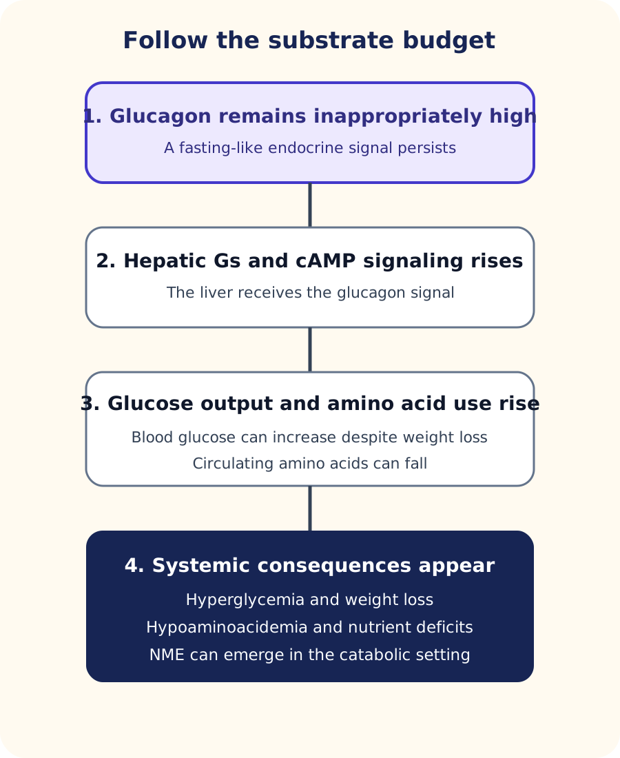
The central relationship is hepatic. Glucagon binds a G protein-coupled receptor that activates adenylyl cyclase and cAMP, shifting the liver toward glucose production and amino acid use. Chronic glucagon excess is therefore not simply high blood sugar. It can create a persistent fasting-like signal in a person who is eating, with hepatic glucose output remaining inappropriately high and circulating amino acids being consumed. [1][7]
This corrects a common overextension of basic physiology. Human skeletal muscle is not an established direct glucagon target in the way the liver is, and the syndrome should not be explained as glucagon directly commanding every tissue to break itself down. The more defensible picture is hepatic glucose and amino acid metabolism driven by excess glucagon, with systemic catabolic consequences that include weight loss and hypoaminoacidemia. [7]
Substrate budget: persistent glucagon signal → hepatic cAMP signaling → increased hepatic glucose production and amino acid turnover → hyperglycemia plus depletion of circulating building blocks → weight loss and impaired epithelial maintenance. The diagram below shows this as four steps.
The sequence keeps the primary human glucagon signal in the liver and links it to hyperglycemia, low amino acids, weight loss, and the nutritional setting in which NME can occur. [1][7]Run the substrate budget: what changes if dietary intake is adequate but glucagon remains very high?
Calories entering the gut do not automatically cancel the tumor signal. Hyperglycemia can coexist with weight loss because the liver continues to receive a fasting-like hormonal instruction. If amino acids and zinc are low, nutritional replacement treats a real consequence, while control of glucagon secretion addresses the continuing driver. [1]
Normal glucagon physiology still matters for examination questions: pancreatic alpha cells make glucagon, and insulin from beta cells has opposing effects on glucose handling. But the familiar drawing of alpha cells as a neat outer ring around a beta-cell core is mainly a rodent pattern. Human islet cells are more intermingled, so that cartoon should not be used to predict where a human glucagonoma must arise. [6]
Separate biochemical diagnosis from tumor mapping
When the syndrome fits, obtain a fasting plasma glucagon. Endotext describes fasting levels above 500 pg/mL in the proper clinical setting as diagnostic of glucagonoma syndrome, while also warning that lesser increases can occur with renal failure, liver disease, pancreatitis, sepsis, diabetes, diabetic ketoacidosis, stress, and other conditions. A number is therefore interpreted with the phenotype, not in isolation. [1]
After biochemical evidence supports a functioning tumor, cross-sectional imaging answers a different question: where is the lesion and how far has disease spread? Multiphasic CT or MRI evaluates the pancreas and liver. Because many glucagonomas express somatostatin receptors, somatostatin receptor PET can add whole-body localization and staging information and can later help determine whether receptor-targeted therapy is relevant. Most glucagonomas are already larger than 4 to 5 cm at diagnosis, and many have spread to regional lymph nodes, liver, or bone. [1][3]
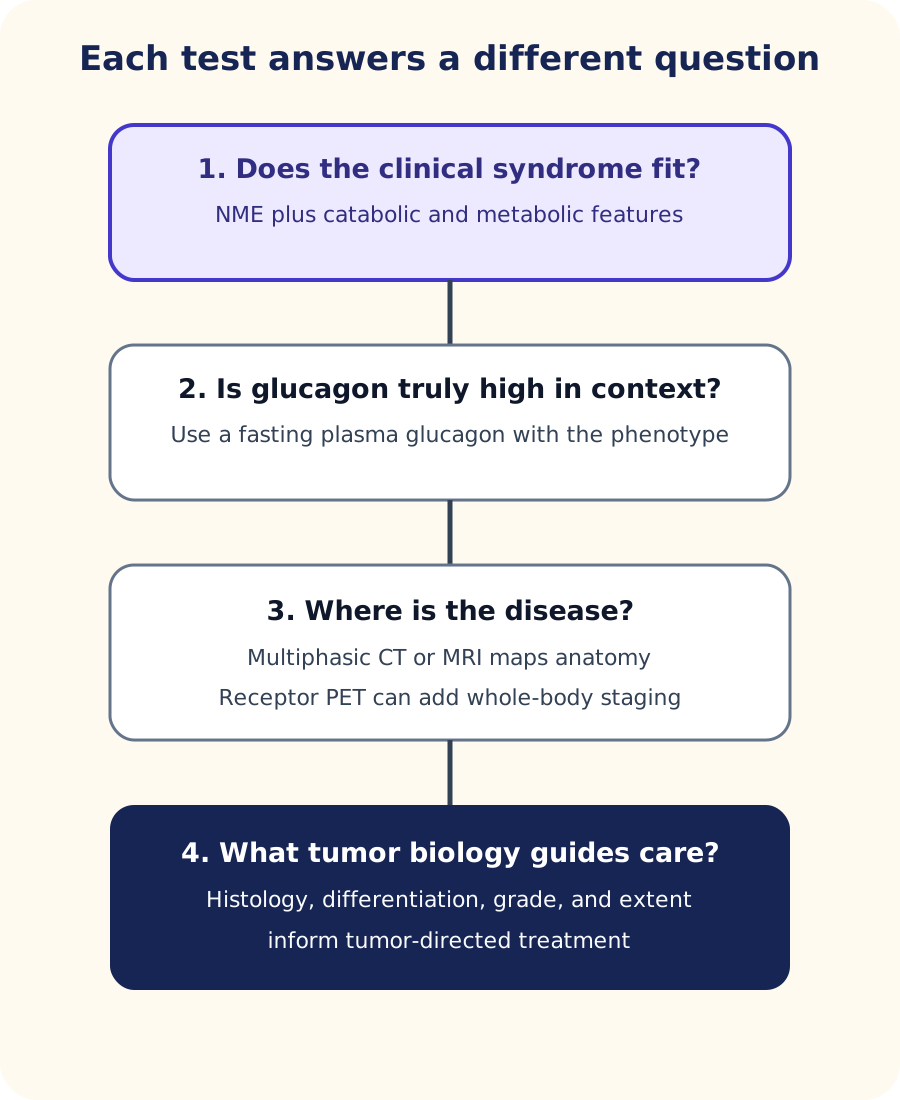
Do not ask one test to do every job. Clinical pattern and fasting glucagon establish the functional syndrome; imaging maps disease; pathology supplies tumor biology. [1][3]
Histology supplies tumor classification and grade. A pancreatic neuroendocrine tumor can contain glucagon without producing the full clinical syndrome, so tissue hormone staining does not replace the clinical-biochemical diagnosis of a functioning glucagonoma. Grade, differentiation, extent of disease, symptoms, and resectability all matter to treatment planning. [1][3]
Choose the next question: a patient has NME, diabetes, weight loss, and fasting glucagon 1,100 pg/mL. What does CT add?
The biochemical and clinical findings establish a strong functional diagnosis; CT now answers location and extent. It is not being asked to prove that the hormone level is high. The diagnostic-route diagram keeps biochemical confirmation, anatomic mapping, receptor imaging, and pathology in separate lanes. [1]
Try it here · Checkpoint 2 of 3
Make your prediction before reading the choices. A first attempt is just a starting point.
Case 3
Show answer and explanations for case 3
A. Repeat fasting glucose after an overnight fast (Why this does not fit)
Glucose documents a consequence of the syndrome but does not stage the neuroendocrine tumor or characterize receptor-positive disease.
Reasoning steps for option A
Why might repeating fasting glucose seem useful in a patient with new diabetes?
Glucose tracks one consequence of glucagon excess and is easy to repeat.
What question about the liver lesions can glucose never answer?
Glucose does not show where tumor is or whether it expresses somatostatin receptors, which is the staging question asked.
B. Endoscopic retrograde cholangiopancreatography (Why this does not fit)
ERCP evaluates pancreaticobiliary ducts and is not a whole-body staging study for glucagonoma.
Reasoning steps for option B
What might make ERCP attractive for a 5.2 cm pancreatic body mass?
It directly images the pancreatic duct and can sample some pancreatic lesions.
Why does ERCP fail the whole-body staging requirement?
It examines only the biliary and pancreatic ducts and cannot map receptor-positive disease in the liver or elsewhere.
C. Somatostatin receptor PET (Best answer)
Glucagonomas commonly express somatostatin receptors, so receptor PET can identify additional receptor-positive disease and inform later treatment selection.
Reasoning steps for option C
What property of glucagonomas makes receptor imaging informative?
Most glucagonomas strongly express somatostatin receptors, so receptor PET highlights tumor throughout the body.
How does receptor PET help with the indeterminate liver lesions?
Uptake can show whether those lesions are receptor-positive metastases and whether receptor-directed treatment could later apply.
D. Plain abdominal radiography (Why this does not fit)
A plain film has no useful sensitivity for mapping pancreatic neuroendocrine tumor burden or receptor expression.
Reasoning steps for option D
Why might a plain film be ordered in a patient with abdominal disease?
It is quick and inexpensive and can show gross calcification or obstruction.
What limitation disqualifies plain radiography for this staging question?
It cannot detect pancreatic or liver neuroendocrine deposits or show receptor expression.
Takeaway: After the functional diagnosis is established, somatostatin receptor imaging can add disease-extent and receptor-status information.
Pancreatic neuroendocrine syndromes overlap in nonspecific findings such as weight loss or diarrhea, so the discriminating physiology matters. Insulinoma produces hypoglycemia. Gastrinoma produces acid hypersecretion and refractory or recurrent peptic ulcer disease. VIPoma produces prominent secretory watery diarrhea with electrolyte loss. Somatostatinoma can produce diabetes, steatorrhea, and gallbladder disease. Glucagonoma is the one that brings the catabolic pattern together with NME. [2][8]
MEN1 is relevant, but another old shortcut needs refinement. MEN1 includes parathyroid, pituitary, and gastroenteropancreatic neuroendocrine tumors. GeneReviews lists nonfunctioning gastroenteropancreatic tumors more often than any single functioning syndrome, while gastrinoma is common among functioning MEN1 tumors and glucagonoma is rare. In a patient with MEN1, the presenting physiology still decides which functioning syndrome is active. [5]
Apply the contrast: MEN1 plus refractory duodenal ulcers favors gastrinoma. MEN1 plus recurrent hypoglycemia favors insulinoma. MEN1 plus NME, diabetes, weight loss, and a markedly high fasting glucagon supports glucagonoma. The inherited syndrome narrows the tumor family; it does not replace the hormone phenotype.
Test the contrast: why is a pancreatic mass plus diabetes not enough to call glucagonoma?
Diabetes is common and pancreatic neuroendocrine tumors can be nonfunctioning. A convincing diagnosis needs hormone-specific evidence and a compatible syndrome. This same rule prevents an incidental glucagon-positive tumor from being confused with glucagonoma syndrome. [1][5]
Treat the hormone syndrome and the tumor as related but separate problems
For localized resectable glucagonoma, surgical resection offers the only curative approach. Before an operation, severe catabolism, hyperglycemia, amino acid deficiency, zinc deficiency, and other nutritional problems may need correction. Somatostatin analog therapy can suppress glucagon secretion and improve the functional syndrome, while perioperative planning must address the important risk of venous thromboembolism. [1][2][8]
A somatostatin analog such as octreotide or lanreotide is best understood as hormone control, not as a guarantee that a large tumor will shrink. Glucose therapy and nutritional repletion treat downstream consequences. Anticoagulation is used when venous thrombosis is diagnosed, and prophylaxis is considered according to surgical and patient risk. These interventions solve different parts of the same syndrome. [1]
Metastatic or unresectable disease is not governed by a single fixed sequence. Management may include somatostatin analogs, surgery or cytoreduction in selected patients, liver-directed therapy for appropriate liver-dominant disease, targeted agents, cytotoxic therapy, and receptor-targeted radionuclide therapy in selected somatostatin receptor positive disease. Tumor burden, symptoms, grade, receptor status, distribution, and prior therapy determine the plan. [1][3][4][8]
Apply the split: a patient has liver metastases, severe NME, and high glucagon. What are the two parallel goals?
First, reduce the functional syndrome with hormone suppression and metabolic support. Second, choose tumor-directed therapy from the actual stage, grade, receptor status, disease distribution, and resectability. One goal can improve before the other, which is why response should not be judged by skin findings alone. [1][3]
Try it here · Checkpoint 3 of 3
Make your prediction before reading the choices. A first attempt is just a starting point.
Case 10
Show answer and explanations for case 10
A. Proceed without metabolic preparation because the tumor is resectable (Why this does not fit)
Resectability does not erase severe catabolism, hyperglycemia, and nutritional deficits that can increase perioperative risk.
Reasoning steps for option A
Why might immediate surgery seem appropriate for a resectable 4 cm tumor?
Resection is the only curative treatment and removes the source of glucagon.
Which findings make unprepared surgery risky in this woman?
Glucose of 326 mg/dL, albumin of 2.0 g/dL, low zinc and marked hypoaminoacidemia raise wound, infection and thrombotic risk if uncorrected.
B. Metabolic optimization, hormone suppression as appropriate, then surgery (Best answer)
Preoperative care targets the catabolic state and hormone excess while surgery remains the definitive treatment for localized resectable disease.
Reasoning steps for option B
Which problems in this woman need correction before the operation?
Severe hyperglycemia, protein-calorie depletion, low zinc and low amino acids, along with high venous thromboembolism risk.
How does a somatostatin analog fit into the preoperative plan?
It suppresses glucagon secretion, helping reverse catabolism and the rash before resection, which remains the definitive treatment.
C. Use only topical corticosteroids until the rash clears completely (Why this does not fit)
Topical therapy does not correct glucagon excess, amino acid depletion, or hyperglycemia, and complete rash resolution is not the sole operative criterion.
Reasoning steps for option C
Why might topical steroids be offered for severe NME?
The rash is the most visible and painful problem, and topical steroids treat many dermatoses.
What does topical therapy leave untouched before surgery?
It does not lower glucagon, correct amino acid and zinc deficits or control glucose, and rash clearance is not the goal of preparation.
D. Give exogenous glucagon to restore feedback inhibition before surgery (Why this does not fit)
Adding glucagon worsens the hormonal excess and does not provide a useful feedback treatment for an autonomous tumor.
Reasoning steps for option D
What misconception makes exogenous glucagon sound like feedback therapy?
Some hormone excess states can be suppressed by negative feedback from the hormone itself.
Why would added glucagon harm this patient?
The tumor secretes autonomously, so extra glucagon would only worsen hyperglycemia and catabolism.
Takeaway: Localized glucagonoma care combines definitive resection with correction of severe metabolic and thrombotic risks around the operation.
When a vignette gives a recurrent intertriginous or acral erosive rash, ask first whether the morphology and time course fit NME. Then search for the catabolic partners: diabetes, weight loss, hypoaminoacidemia, anemia, oral inflammation, low zinc, or venous thrombosis. A coherent syndrome justifies fasting glucagon testing; marked glucagon excess in that context justifies anatomic localization and staging. [1]
The most useful error check is to ask what your chosen diagnosis fails to explain. Pemphigoid can explain blisters but not the metabolic syndrome. Ordinary type 2 diabetes can explain hyperglycemia but not NME and severe catabolism. A nonfunctioning pancreatic tumor can explain an incidental mass but not a hormone-specific syndrome. A nutritional disorder can explain NME-like skin disease but needs its own evidence if glucagon is not convincingly high. [1]
Final transfer: if the rash improves after somatostatin analog therapy but the liver lesions enlarge, has the cancer been controlled?
No. Better rash and glucose can show hormone suppression while imaging shows tumor progression. Functional response and tumor response are related measurements, not synonyms. Reassess tumor-directed treatment using stage, grade, receptor expression, and the patient's overall condition. [1][4]
Independent practice
Case 2
Show answer and explanations for case 2
A. The glucagon value alone establishes glucagonoma (Why this does not fit)
A modest glucagon increase without the clinical syndrome is not enough; renal failure is among conditions that can increase glucagon.
Reasoning steps for option A
Why might a fasting glucagon of 260 pg/mL raise concern for a tumor?
It is above the usual fasting reference range, and glucagonoma is defined by glucagon excess.
What does this patient lack that a glucagonoma diagnosis needs?
He has no NME, no weight loss, near-normal glucose and no pancreatic lesion, and his level is well below the 500 pg/mL range typical of the syndrome.
B. Nonspecific glucagon increase in chronic kidney disease (Best answer)
Renal failure can produce a glucagon increase, and this patient lacks the characteristic syndrome or a pancreatic lesion.
Reasoning steps for option B
Which background condition can raise glucagon without a tumor in this man?
Stage 4 chronic kidney disease reduces glucagon clearance and is a recognized cause of modest hyperglucagonemia.
Why does the rest of the evaluation support a nonspecific explanation?
Stable weight, normal skin, glucose of 116 mg/dL and a normal pancreatic MRI show no functioning syndrome to explain.
C. The patient has an occult insulinoma (Why this does not fit)
Insulinoma produces inappropriate insulin with hypoglycemia, neither of which is present.
Reasoning steps for option C
Why might a pancreatic hormone abnormality bring insulinoma to mind?
Insulinoma is the most familiar functioning pancreatic neuroendocrine tumor.
Which laboratory finding rules against insulinoma in this patient?
His glucose is 116 mg/dL; insulinoma requires documented hypoglycemia with inappropriate insulin, which is absent.
D. The patient has MEN1 until genetic testing proves otherwise (Why this does not fit)
MEN1 is not diagnosed from a modest isolated glucagon value and no MEN1 tumor pattern is supplied.
Reasoning steps for option D
Why could MEN1 enter the discussion after an abnormal islet hormone value?
MEN1 predisposes to pancreatic neuroendocrine tumors, so any islet hormone abnormality can bring it to mind.
What would be needed before MEN1 becomes a reasonable concern here?
A MEN1 pattern such as hyperparathyroidism, pituitary disease or a pancreatic tumor; none is present, and a modest isolated glucagon value is not enough.
Takeaway: Interpret glucagon with the phenotype; modest increases can occur in illnesses that are not glucagonoma.
A. Persistent hepatic glucose production and amino acid use (Best answer)
Chronic glucagon excess sustains a fasting-like hepatic program with excessive glucose output and amino acid utilization, producing hyperglycemia, low amino acids, and weight loss.
Reasoning steps for option A
Why does weight loss continue even though this woman eats normally?
Glucagon near 1,000 pg/mL keeps the liver in a fasting-like mode regardless of intake.
How does one hepatic process explain both hyperglycemia and low amino acids?
Sustained hepatic glucose production raises glucose while increased hepatic amino acid uptake and use drain the circulating pool.
B. Direct destruction of pancreatic beta cells by glucagon (Why this does not fit)
Glucagonoma does not require beta-cell destruction; the dominant disturbance is inappropriate hepatic substrate handling driven by glucagon.
Reasoning steps for option B
Why might beta-cell destruction seem to fit a glucose of 265 mg/dL?
Loss of insulin secretion is the familiar cause of severe hyperglycemia.
What does beta-cell loss fail to explain in this patient?
Glucagonoma hyperglycemia comes from hepatic glucagon action, and beta-cell loss would not explain the markedly low amino acids.
C. Primary intestinal failure to absorb all macronutrients (Why this does not fit)
Global malabsorption could cause weight loss, but it does not explain the marked glucagon excess and hyperglycemia as a single process.
Reasoning steps for option C
What makes malabsorption a reasonable consideration for weight loss despite eating?
Failure to absorb nutrients can cause weight loss even with normal intake.
Which findings point away from primary intestinal failure?
Malabsorption would not produce glucagon of 1,020 pg/mL or hyperglycemia; one hormone-driven process explains all three findings.
D. Direct glucagon signaling in skeletal muscle as the main human target (Why this does not fit)
Human skeletal muscle is not an established primary glucagon receptor target; the liver is central to the syndrome's glucose and amino acid effects.
Reasoning steps for option D
Why might skeletal muscle seem a likely source of the lost weight?
Muscle is the body's largest protein store and breaks down in many catabolic states.
What is wrong with making muscle the main glucagon target?
Human skeletal muscle is not an established direct glucagon target; the liver drives the glucose output and amino acid consumption.
Takeaway: Think of glucagonoma as an inappropriate hepatic fasting signal that can consume amino acid resources even when food intake is adequate.
A. The human specimen excludes normal islet tissue (Why this does not fit)
Intermixed endocrine-cell distribution is compatible with human islet architecture and does not by itself indicate disease.
Reasoning steps for option A
Why might the intermixed pattern look abnormal to this trainee?
It differs from a beta-cell core surrounded by alpha cells.
Why does intermixing not indicate disease in a human specimen?
Human islets normally show alpha and beta cells distributed throughout, so this pattern is compatible with normal tissue.
B. Rodent islet architecture does not rigidly describe human islets (Best answer)
Rodent islets often show a beta-cell core with alpha and δ cells toward the periphery, while human islet architecture is more intermingled and variable.
Reasoning steps for option B
Where does the core-and-rim islet drawing come from?
It reflects the typical rodent islet, where beta cells form a core and alpha and δ cells lie toward the periphery.
What does the human specimen show about applying that drawing?
Human islets are more intermingled and variable, so the rodent pattern does not rigidly describe them.
C. Alpha cells are absent from normal human pancreas (Why this does not fit)
Human alpha cells are normal glucagon-producing islet cells; the issue is their distribution, not their existence.
Reasoning steps for option C
Why might a trainee doubt that alpha cells are present in this specimen?
If alpha cells are expected only at the rim, not seeing a rim can look like absence.
What confirms that alpha cells are part of normal human pancreas?
Alpha cells are normal glucagon-producing islet cells; the specimen shows them intermixed with beta cells, not missing.
D. A glucagonoma must arise in the pancreatic tail because alpha cells occupy the islet rim (Why this does not fit)
Human islet cell arrangement does not establish a rule that predicts tumor location from an outer-ring alpha-cell diagram. Glucagonomas arise most often in the pancreatic tail, but nonfunctioning, insulin-secreting and other pancreatic neuroendocrine tumors also occur there, so tail location alone never establishes glucagon secretion.
Reasoning steps for option D
What real observation lies behind linking glucagonoma to the pancreatic tail?
Glucagonomas arise most often in the pancreatic tail.
Why can neither islet architecture nor location decide a tumor's type?
Human alpha cells are not confined to an islet rim, and other pancreatic neuroendocrine tumors also arise in the tail, so location is a tendency, not a rule.
Takeaway: Human islets are not faithfully represented by the simple rodent core-and-mantle cartoon.
MEN1 can include glucagonoma, but this patient lacks NME, hyperglycemia, weight loss, or high glucagon and instead has an acid-hypersecretion syndrome.
Reasoning steps for option A
Why might glucagonoma be considered in a woman with a MEN1 variant?
MEN1 predisposes to several pancreatic neuroendocrine tumors, and glucagonoma is one of them.
Which normal findings argue against glucagonoma in this woman?
Her glucose is 88 mg/dL, her skin is normal and her fasting glucagon is normal, so no glucagon syndrome is present.
B. Insulinoma (Why this does not fit)
Insulinoma causes inappropriate hypoglycemia rather than refractory acid-related ulcer disease.
Reasoning steps for option B
What makes insulinoma a realistic MEN1 consideration?
Insulinoma is among the functioning pancreatic tumors seen in MEN1.
Why does insulinoma not explain her current symptoms?
Insulinoma causes fasting hypoglycemia, whereas her problem is refractory duodenal ulcers with a normal glucose.
C. Gastrinoma (Best answer)
MEN1 plus recurrent refractory duodenal ulcer disease is strongly compatible with gastrinoma and Zollinger-Ellison syndrome.
Reasoning steps for option C
Which symptom pattern points to acid hypersecretion in this woman?
Recurrent duodenal ulcers that persist despite high-dose acid suppression suggest a gastrin-driven state.
How does the MEN1 background strengthen that conclusion?
Gastrinoma is the most common functioning gastroenteropancreatic tumor in MEN1, and hyperparathyroidism confirms the syndrome context.
D. VIPoma (Why this does not fit)
VIPoma is defined by prominent secretory watery diarrhea and electrolyte loss, not refractory peptic ulcer disease.
Reasoning steps for option D
Why might VIPoma be considered in a patient with a MEN1 pancreatic tumor syndrome?
VIPoma is another rare functioning pancreatic tumor that can occur in MEN1.
What clinical pattern would VIPoma require that she lacks?
VIPoma causes high-volume watery diarrhea with hypokalemia and reduced acid, not refractory ulcers from excess acid.
Takeaway: In MEN1, identify the active functioning tumor from its hormone syndrome rather than from the inherited syndrome alone.
A. MEN1 excludes glucagonoma because only gastrinomas occur in the pancreas (Why this does not fit)
MEN1 includes several gastroenteropancreatic neuroendocrine tumors, including rare glucagonomas.
Reasoning steps for option A
Why might a learner associate MEN1 pancreatic disease only with gastrinoma?
Gastrinoma is the functioning tumor most often taught with MEN1.
What evidence in this man shows that MEN1 does not exclude glucagonoma?
He has NME-like plaques, hyperglycemia, weight loss and glucagon of 940 pg/mL with a pancreatic tumor, a full glucagonoma syndrome.
B. The skin finding is unrelated because MEN1 tumors never produce dermatologic syndromes (Why this does not fit)
A MEN1-associated glucagonoma can produce the same functional glucagonoma syndrome as a sporadic tumor.
Reasoning steps for option B
Why might the groin rash be dismissed as unrelated to MEN1?
MEN1 is usually remembered for endocrine tumors, not skin disease.
How does the glucagon result link the rash to his tumor?
A MEN1-associated glucagonoma produces the same NME and catabolic syndrome as a sporadic tumor, and his glucagon is markedly high.
C. Glucagonoma is the most frequent gastroenteropancreatic tumor in MEN1 (Why this does not fit)
GeneReviews lists nonfunctioning tumors more often overall and gastrinoma more often among major functioning syndromes; glucagonoma is rare in MEN1.
Reasoning steps for option C
Why might a learner overestimate how often glucagonoma occurs in MEN1?
A vivid case like this one can make glucagonoma seem like a typical MEN1 tumor.
What is the actual frequency order of pancreatic tumors in MEN1?
Nonfunctioning tumors are most frequent and gastrinoma leads the functioning syndromes, while glucagonoma is rare.
D. MEN1 permits glucagonoma, but phenotype identifies the functioning syndrome (Best answer)
The MEN1 background raises the possibility of pancreatic neuroendocrine tumors, while NME, hyperglycemia, weight loss, and high fasting glucagon identify the active glucagonoma syndrome.
Reasoning steps for option D
What does the MEN1 background contribute to this diagnosis?
It explains why he is at risk for pancreatic neuroendocrine tumors of several types.
Which findings identify which functioning tumor is active?
NME, hyperglycemia, weight loss and fasting glucagon of 940 pg/mL specify glucagonoma syndrome within that MEN1 risk.
Takeaway: MEN1 is a predisposition context; the hormone phenotype determines which functioning syndrome is present.
A. Compression ultrasonography of the symptomatic leg (Best answer)
Glucagonoma is associated with venous thromboembolism, and unilateral swelling with deep venous tenderness warrants direct evaluation for DVT with compression ultrasonography.
Reasoning steps for option A
Which features of this man's leg suggest deep vein thrombosis?
Sudden unilateral calf pain, a 4 cm larger calf and tenderness along the deep veins are typical of DVT.
Why is compression ultrasonography the right next step?
Glucagonoma carries a high venous thromboembolism risk, and compression ultrasound directly confirms or excludes DVT in a stable patient.
B. Skin biopsy of the calf (Why this does not fit)
The acute unilateral swelling is a vascular presentation, not a question about NME morphology.
Reasoning steps for option B
Why might a skin biopsy seem relevant in a patient with glucagonoma?
NME often involves the legs, so any new leg problem could seem like another skin lesion.
What makes this presentation vascular rather than cutaneous?
Acute unilateral swelling and deep venous tenderness point to thrombosis, which a skin biopsy cannot diagnose.
C. Repeat fasting glucagon before evaluating the leg (Why this does not fit)
The glucagonoma is already known; delaying DVT evaluation for another hormone value does not address the acute vascular problem.
Reasoning steps for option C
Why might rechecking glucagon feel like a logical step in known glucagonoma?
Glucagon tracks disease activity, and new symptoms can lead to reassessment of the tumor.
Why should the hormone level not come before the leg evaluation?
The diagnosis is already known, and delaying DVT imaging for a hormone value risks missing a thrombus that needs treatment.
D. No testing because thrombosis is not part of glucagonoma syndrome (Why this does not fit)
DVT and pulmonary embolism are recognized complications of glucagonoma syndrome and require ordinary evidence-based evaluation.
Reasoning steps for option D
Why might thrombosis be overlooked as part of this syndrome?
The skin and glucose findings usually dominate teaching about glucagonoma.
What makes skipping testing unsafe in this man?
DVT and pulmonary embolism are recognized glucagonoma complications, and his leg findings warrant standard DVT evaluation.
Takeaway: Treat new unilateral leg findings in glucagonoma as a real thromboembolism problem, not as another skin manifestation.
A. It is primarily used to replace deficient endogenous glucagon (Why this does not fit)
The syndrome is caused by glucagon excess, so replacement would oppose the therapeutic goal.
Reasoning steps for option A
Why might a learner think glucagon needs replacing in advanced disease?
Somatostatin analogs suppress several hormones, so replacement can sound like a necessary add-on.
What is the actual hormonal problem in this man?
He has glucagon excess from metastatic tumor, so replacing glucagon would oppose the goal of treatment.
B. It proves that all metastatic lesions will regress if the rash improves (Why this does not fit)
Symptom improvement can reflect hormone suppression without establishing radiographic tumor regression.
Reasoning steps for option B
Why might rash improvement be read as tumor shrinkage?
Both the rash and the tumor are caused by the same neoplasm, so improvement in one seems to imply the other.
Why can the skin response not predict regression of metastases?
Somatostatin analogs can reduce hormone secretion without shrinking tumor, so imaging must assess tumor burden separately.
C. It suppresses glucagon and improves the functional syndrome (Best answer)
Somatostatin analogs can reduce hormone secretion and improve symptoms; control of the functional syndrome and control of tumor burden are related but distinct goals.
Reasoning steps for option C
What does a strongly positive receptor scan predict about somatostatin analog therapy?
Receptor-expressing tumor is likely to respond to the analog with reduced hormone secretion.
How should the analog's benefit be framed in this man?
It can lower glucagon and improve the rash and hyperglycemia, while tumor-directed treatment is judged on its own terms.
D. It is contraindicated once liver metastases are present (Why this does not fit)
Metastatic disease does not by itself preclude somatostatin analog therapy, particularly for symptom control in receptor-expressing disease.
Reasoning steps for option D
Why might liver metastases seem to rule out somatostatin analog therapy?
Advanced spread can make any single drug seem futile or unsafe.
Why is analog therapy appropriate despite metastases?
Metastatic, receptor-positive disease is a main setting for analog therapy, particularly to control hormone symptoms.
Takeaway: Somatostatin analog therapy can control hormone excess even when separate tumor-directed treatment is still needed.
Each positive site is a visible target, and radiation treats localized tumor.
Why does receptor positivity not create an indication to irradiate every site?
Receptor uptake identifies a systemic target for receptor-directed therapy, not a reason for external-beam treatment of all lesions.
D. 177Lu-DOTATATE peptide receptor radionuclide therapy (Best answer)
Progressive somatostatin receptor-positive gastroenteropancreatic neuroendocrine tumor can be considered for 177Lu-DOTATATE in an appropriate clinical setting.
Reasoning steps for option D
What does intense receptor uptake in all major lesions make possible?
It shows that a radiolabeled somatostatin analog can reach essentially all of the known tumor.
Why is this woman an appropriate candidate for 177Lu-DOTATATE?
She has progressive, well-differentiated, receptor-positive disease after a somatostatin analog, the setting where this therapy is considered.
Takeaway: Strong somatostatin receptor expression can identify a therapeutic target for receptor-directed radionuclide therapy.
A. Continue syndrome control and discuss tumor-directed options (Best answer)
Liver-dominant progression requires a tumor-directed decision based on distribution, grade, receptor status, prior therapy, and patient factors, while hormone control remains important.
Reasoning steps for option A
What does improvement of NME and glucose on the analog tell you?
Hormone secretion is being suppressed, so the functional syndrome is controlled for now.
Which finding requires a separate tumor-directed decision?
Growing liver-dominant tumor burden calls for choosing among liver-directed, systemic or receptor-based options while continuing hormone control.
B. Skin improvement proves the tumor burden is controlled (Why this does not fit)
A functional response can occur while radiographic disease progresses, so skin response cannot substitute for tumor assessment.
Reasoning steps for option B
Why might skin improvement be taken as proof of disease control?
The rash was the most visible sign of the tumor, so its clearance feels like tumor control.
Which finding directly contradicts that interpretation?
Liver tumor burden is increasing, so the tumor is progressing despite the functional response.
C. Stop all tumor-directed therapy because metastatic glucagonoma has no additional options (Why this does not fit)
Advanced glucagonoma can be managed with several systemic and liver-directed strategies selected by disease features.
Reasoning steps for option C
Why might a clinician think no further tumor treatment exists?
Metastatic glucagonoma is rare and often presented as incurable.
What options remain for liver-dominant progression?
Liver-directed therapy, targeted agents, chemotherapy and receptor-directed radionuclide therapy can be selected by tumor features.
D. Use only insulin because hyperglycemia is the sole clinically relevant problem (Why this does not fit)
Insulin may help glucose control but does not address progressive tumor burden, NME, nutritional deficits, or other consequences of glucagon excess.
Reasoning steps for option D
Why might insulin seem to address the main problem?
Hyperglycemia is a measurable, treatable consequence of glucagon excess.
What does insulin fail to treat in this man?
It does not slow tumor growth or address nutritional deficits, thrombosis risk or other effects of glucagon excess.
Takeaway: Functional improvement and tumor control must be assessed separately in advanced glucagonoma.
Glucagonoma can include diarrhea, but the defining pattern here is massive watery diarrhea with hypokalemia and reduced acid secretion, without NME or hyperglycemia.
Reasoning steps for option A
Why might glucagonoma be considered in a woman with diarrhea and weight loss?
Diarrhea and weight loss are both recognized features of glucagonoma syndrome.
Which missing and present findings argue against glucagonoma?
She has no rash and a normal glucose, and her 4 liters of daily diarrhea with hypokalemia and low acid output fit a different tumor.
B. Gastrinoma (Why this does not fit)
Gastrinoma causes acid hypersecretion and recurrent ulcers, the opposite gastric-acid pattern from this vignette.
Reasoning steps for option B
Why might gastrinoma be considered for severe diarrhea?
Gastrinoma is a classic functioning pancreatic cause of chronic diarrhea.
Which gastric finding points in the opposite direction from gastrinoma?
Gastrinoma causes acid hypersecretion, while this woman has reduced gastric acid secretion.
C. Insulinoma (Why this does not fit)
Insulinoma causes inappropriate hypoglycemia rather than a severe secretory diarrhea and electrolyte-loss syndrome.
Reasoning steps for option C
Why might insulinoma be listed among pancreatic hormone syndromes here?
It is the most common functioning pancreatic neuroendocrine tumor.
What does insulinoma fail to explain in this woman?
It causes hypoglycemia, not high-volume secretory diarrhea with potassium and bicarbonate loss.
D. VIPoma (Best answer)
Very high-volume watery diarrhea, hypokalemia, and reduced gastric acid secretion form the classic VIPoma physiology.
Reasoning steps for option D
Which findings describe a secretory diarrhea syndrome?
Four liters of watery stool daily with potassium of 2.7 mEq/L and bicarbonate of 17 mEq/L show massive intestinal fluid and electrolyte loss.
What additional finding completes the classic VIPoma pattern?
Reduced gastric acid secretion completes the watery diarrhea, hypokalemia and hypochlorhydria triad of VIPoma.
Takeaway: When diarrhea dominates, use its volume, electrolytes, acid physiology, and accompanying findings to distinguish VIPoma from glucagonoma.
Documented fasting hypoglycemia with inappropriate endogenous insulin and C-peptide supports insulinoma, while the weight gain and absence of NME oppose glucagonoma.
Reasoning steps for option A
What pattern do this man's fasting symptoms and laboratory values show?
Confusion and sweating with glucose of 39 mg/dL, while insulin and C-peptide stay high, indicate endogenous hyperinsulinemic hypoglycemia.
How do his weight and skin findings fit an insulin-secreting tumor?
Weight gain from frequent eating to avoid symptoms and the absence of NME are consistent with insulinoma.
B. Glucagonoma (Why this does not fit)
Glucagonoma more often produces hyperglycemia, weight loss, and a catabolic syndrome rather than endogenous hyperinsulinemic hypoglycemia.
Reasoning steps for option B
Why might glucagonoma appear on a differential for a pancreatic tumor with glucose symptoms?
Both glucagonoma and insulinoma are islet tumors that disturb glucose.
Why does the direction of the glucose change exclude glucagonoma?
Glucagonoma raises glucose and causes weight loss, whereas this man has hypoglycemia with high insulin and weight gain.
C. VIPoma (Why this does not fit)
VIPoma causes secretory diarrhea and electrolyte loss, not fasting endogenous hyperinsulinemic hypoglycemia.
Reasoning steps for option C
Why might VIPoma be considered among functioning pancreatic tumors here?
It is one of the classic rare islet tumor syndromes.
What does VIPoma lack to explain this presentation?
VIPoma causes secretory diarrhea and electrolyte loss, not fasting hypoglycemia with high insulin and C-peptide.
D. Somatostatinoma (Why this does not fit)
Somatostatinoma can cause diabetes and malabsorption-related features, not the demonstrated high-insulin fasting hypoglycemia.
Reasoning steps for option D
Why could somatostatinoma seem related to a glucose disturbance?
Somatostatin inhibits insulin, so a somatostatin-secreting tumor affects glucose control.
Why does the glucose direction rule out somatostatinoma?
Somatostatinoma suppresses insulin and causes diabetes, the opposite of this high-insulin hypoglycemia.
Takeaway: Glucose direction is a powerful discriminator: insulinoma produces endogenous hyperinsulinemic hypoglycemia, while glucagonoma commonly produces hyperglycemia.
Gastrinoma is centered on acid hypersecretion and refractory ulcer disease rather than the diabetes, steatorrhea, and gallstone combination.
Reasoning steps for option A
Why might gastrinoma be considered for a pancreatic tumor with digestive symptoms?
Gastrinoma is a common functioning pancreatic tumor with gastrointestinal effects.
What core gastrinoma feature is missing in this woman?
There is no acid hypersecretion or refractory ulcer disease, and gastrinoma does not explain diabetes with gallstones.
B. Somatostatinoma (Best answer)
Somatostatin excess can inhibit insulin and gastrointestinal hormones, producing diabetes, steatorrhea, and gallbladder disease.
Reasoning steps for option B
Which three findings together suggest somatostatin excess?
Diabetes, steatorrhea and gallstones with a pancreatic neuroendocrine tumor form the classic inhibitory syndrome.
How does somatostatin produce each of those findings?
It inhibits insulin release, pancreatic enzyme secretion and cholecystokinin-driven gallbladder emptying.
C. Glucagonoma (Why this does not fit)
Diabetes overlaps, but the absence of NME and glucagon excess plus steatorrhea and gallstones favors somatostatinoma.
Reasoning steps for option C
Why might glucagonoma be considered in a patient with diabetes and a pancreatic tumor?
New diabetes with a pancreatic neuroendocrine tumor is a common glucagonoma presentation.
Which findings in this woman argue against glucagonoma?
She has no NME and a normal fasting glucagon, while steatorrhea and gallstones point to somatostatin excess.
D. Insulinoma (Why this does not fit)
Insulinoma produces hypoglycemia, not diabetes with steatorrhea and gallstones.
Reasoning steps for option D
Why might insulinoma be considered for any functioning pancreatic tumor?
It is the most frequent functioning pancreatic neuroendocrine tumor.
What makes insulinoma incompatible with this presentation?
Excess insulin would push glucose down and cause fasting hypoglycemia, the opposite of her diabetes, and it would not impair fat absorption or gallbladder emptying.
Takeaway: Shared diabetes does not identify the tumor; the accompanying gastrointestinal and skin pattern provides the discrimination.
A. The positive stain alone proves glucagonoma syndrome (Why this does not fit)
Tumor glucagon immunoreactivity does not prove a clinically functioning syndrome when circulating glucagon and the phenotype are absent.
Reasoning steps for option A
Why might a positive glucagon stain seem to settle the diagnosis?
It shows that the tumor cells contain glucagon, the hormone that defines glucagonoma.
What does the stain not show in this woman?
It does not show a functioning syndrome; her fasting glucagon, glucose and amino acids are normal and she has no NME.
B. The patient must have occult NME that has not yet been examined (Why this does not fit)
No evidence supports hidden NME, and the absence of the broader hormone phenotype matters.
Reasoning steps for option B
Why might a hidden rash be assumed in a glucagon-positive tumor?
NME is the most characteristic sign of glucagonoma, so a clinician might look for it after a positive stain.
What evidence argues against an unrecognized NME here?
Nothing supports hidden skin disease, and her normal glucagon, glucose and amino acids show no hormone effect.
C. The tumor should be classified as insulinoma because glucose is normal (Why this does not fit)
Normal glucose does not identify insulinoma; insulinoma requires inappropriate insulin-mediated hypoglycemia.
Reasoning steps for option C
Why might a normal glucose suggest another hormone type?
Without hyperglycemia, a learner might look for a different glucose-related tumor.
What would insulinoma require that this woman does not have?
Insulinoma needs documented hypoglycemia with inappropriate insulin; a normal glucose does not suggest it.
D. This tumor expresses glucagon but does not produce a clinical glucagonoma syndrome (Best answer)
Pancreatic neuroendocrine tumors can contain or secrete peptides without causing a functional syndrome; clinical and biochemical evidence define glucagonoma syndrome.
Reasoning steps for option D
What does the tumor's glucagon staining establish?
The tumor cells can make or contain glucagon.
Why does that not amount to glucagonoma syndrome here?
A functioning syndrome requires clinical and biochemical glucagon effects, and her glucagon, glucose, amino acids and skin are all normal.
Takeaway: Functioning tumor syndromes are defined by hormone-related clinical and biochemical effects, not immunostaining alone.
A. Multiphasic contrast-enhanced CT or MRI of the abdomen (Best answer)
After the functional syndrome is established, multiphasic CT or MRI is appropriate for pancreatic localization and assessment of liver and regional disease.
Reasoning steps for option A
What diagnostic step is already complete in this woman?
Recurrent NME, glucose of 238 mg/dL and fasting glucagon of 1,060 pg/mL establish the functional syndrome.
Why is multiphasic CT or MRI the first anatomic study?
It localizes the pancreatic tumor and shows liver and regional disease, the anatomy needed for staging and surgery.
B. Somatostatin receptor PET as the only study needed for surgical anatomy (Why this does not fit)
Receptor PET is valuable for whole-body staging, but cross-sectional CT or MRI remains important for detailed anatomy and operative planning.
Reasoning steps for option B
Why might receptor PET seem sufficient on its own?
Glucagonomas usually express somatostatin receptors, and receptor PET is very sensitive for tumor deposits.
What does receptor PET not provide for surgical planning?
It lacks the detailed pancreatic and vascular anatomy of cross-sectional CT or MRI, which remain essential.
C. Plain abdominal radiograph (Why this does not fit)
A plain radiograph cannot adequately localize a small pancreatic neuroendocrine tumor or stage liver disease.
Reasoning steps for option C
Why might a plain radiograph be ordered first for abdominal symptoms?
It is fast and widely available.
What can a plain radiograph not show in this patient?
It cannot localize a pancreatic tumor or detect liver metastases.
D. Barium swallow (Why this does not fit)
A barium swallow evaluates the esophagus and does not answer pancreatic localization or metastatic-staging questions.
Reasoning steps for option D
Why could a barium study seem relevant to a gastrointestinal tumor?
Contrast studies outline parts of the digestive tract.
Why does a barium swallow miss the question entirely?
It examines the esophagus and does not image the pancreas or liver.
Takeaway: Biochemical diagnosis and anatomic localization are separate steps; multiphasic CT or MRI provides the first detailed anatomic map.
They could explain bullous pemphigoid but would not unify the oral inflammation, anemia, weight loss, and new hyperglycemia.
Reasoning steps for option A
Why might anti-BP180 antibodies be considered for a crusted eruption?
They identify bullous pemphigoid, a common eroded or blistering rash in older adults.
What would anti-BP180 antibodies leave unexplained?
They would not account for glossitis, anemia, weight loss, new hyperglycemia or depression.
B. Low morning cortisol (Why this does not fit)
Adrenal insufficiency can cause weight loss and fatigue but does not fit the NME-like eruption plus hyperglycemia.
Reasoning steps for option B
Why might low cortisol be tested in a woman with fatigue and weight loss?
Adrenal insufficiency causes fatigue, weight loss and mood change.
Which findings conflict with adrenal insufficiency?
It tends to lower glucose rather than cause new hyperglycemia, and it does not produce an NME-like eruption.
C. High serum gastrin with gastric acid hypersecretion (Why this does not fit)
Gastrinoma can cause ulcer disease and diarrhea, but it does not explain the characteristic catabolic skin and oral pattern.
Reasoning steps for option C
Why might gastrinoma be considered for a pancreatic hormone cause of weight loss?
Gastrinoma is a well-known functioning tumor that can cause diarrhea and weight loss.
What does gastrinoma fail to explain in this woman?
It does not cause glossitis, NME-like skin disease, anemia or new hyperglycemia.
D. High glucagon plus pancreatic tumor (Best answer)
Glucagonoma syndrome can include NME, glossitis, anemia, weight loss, diabetes, and depression, bringing the findings into one endocrine syndrome.
Reasoning steps for option D
Which findings in this woman form a recognizable syndrome?
Painful glossitis, normocytic anemia, a recurrent crusted groin eruption, weight loss, new hyperglycemia and depression.
Which result would tie all of them to one cause?
High fasting glucagon with a pancreatic tumor would unify them as glucagonoma syndrome.
Takeaway: Glucagonoma may present as a cluster of dermatologic, metabolic, hematologic, oral, thrombotic, and neuropsychiatric findings rather than one complete textbook set.
A. Location cannot establish glucagonoma; require a hormone phenotype (Best answer)
Human alpha cells are not restricted to a peripheral ring, and a pancreatic tumor's location does not substitute for clinical and biochemical evidence of glucagonoma syndrome.
Reasoning steps for option A
What does this man's tumor lack for a glucagonoma diagnosis?
He has no rash, weight loss or diabetes and no glucagon increase, so no functioning syndrome is present.
Why is the trainee's alpha-cell argument unsound?
Human alpha cells are not restricted to an islet rim, and tumor location cannot substitute for clinical and biochemical evidence.
B. The trainee is correct because all tail neuroendocrine tumors arise from alpha cells (Why this does not fit)
Glucagonomas arise most often in the pancreatic tail, but nonfunctioning, insulin-secreting and other pancreatic neuroendocrine tumors also occur there, so tail location alone never establishes glucagon secretion.
Reasoning steps for option B
What true observation lies behind the trainee's claim about tail tumors?
Glucagonomas are found most often in the pancreatic tail, so the association itself is real.
Why does a tail location still fail to identify tumor type?
This man's tail tumor has no glucagon rise and no syndrome at all, which shows that a tail tumor can come from another cell type or secrete nothing.
C. The absence of rash is irrelevant because glucagonoma never affects glucose or weight (Why this does not fit)
Hyperglycemia and weight loss are common components of glucagonoma syndrome, so their absence matters when glucagon is also normal.
Reasoning steps for option C
Why might the absence of a rash seem unimportant in a pancreatic tumor?
NME is absent in some glucagonoma patients, so its absence alone is not decisive.
Why does the absence of metabolic effects matter here?
Hyperglycemia and weight loss are common in glucagonoma, and their absence alongside normal glucagon argues against it.
D. The tumor is necessarily gastrinoma because glucagon is normal (Why this does not fit)
Normal glucagon only argues against glucagonoma syndrome; it does not identify gastrinoma without an acid-hypersecretion phenotype.
Reasoning steps for option D
Why might a normal glucagon suggest that the tumor must secrete a different hormone?
Excluding one functioning syndrome can seem to point to another common one.
What would gastrinoma require that this man lacks?
Gastrinoma requires acid hypersecretion such as refractory ulcers, which is not described; the tumor may be nonfunctioning.
Takeaway: Do not derive a human tumor diagnosis from a rodent islet cartoon or from pancreatic location alone.