Trace sellar anatomy, prolactin and cortisol feedback, GH excess, and renal water handling to distinguish pituitary tumors, SIAD and diabetes insipidus.
A sellar mass can disturb vision, hormone signaling, or water balance. Start by separating local compression from hormone excess or deficiency. Then ask whether the kidney is conserving water when it should excrete it, or losing water when it should conserve it.
What does the location explain?
Does a large pituitary lesion necessarily secrete excess hormone? No. A nonfunctioning adenoma, often of gonadotroph lineage, may present through incidental imaging, compression, or hormone deficiency without a recognizable hormone-excess syndrome. A microadenoma measures less than 10 mm; a macroadenoma measures at least 10 mm. Size describes anatomy, not endocrine activity. [4][10]
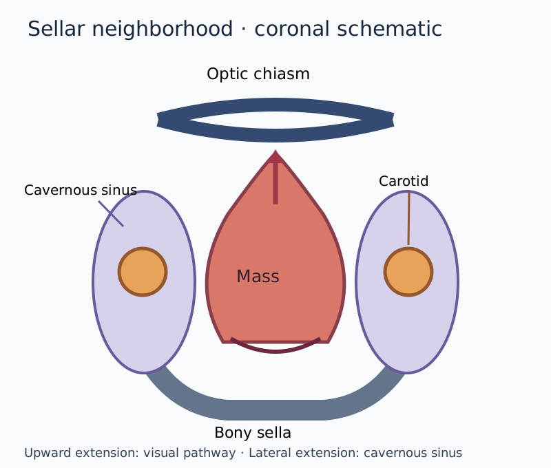
Use the sellar diagram to predict what upward versus lateral extension would affect. The optic chiasm lies above the sella: compression of crossing nasal retinal fibers causes temporal visual-field loss in both eyes. The lateral cavernous sinus contains cranial nerves III, IV, V1, V2 and VI, so extension can produce diplopia or facial sensory loss. A third-nerve palsy can include ptosis, a dilated pupil and a down-and-out eye. Headache can accompany dural stretch but does not measure tumor size reliably. [10]
Localize visual loss and lateral cranial nerve findings to the nearby structures. [10]
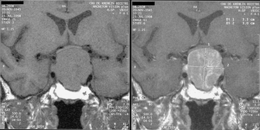
A patient has difficulty seeing cars approaching from either side, but preserved eye movements. Before reading further, place the abnormality above or beside the sella. Bitemporal field loss favors superior extension toward the chiasm, not isolated cavernous-sinus involvement. Formal visual-field testing matters when a lesion abuts the chiasm even if the patient reports normal vision. In the clinical MRI below, locate the sellar lesion and compare its extent with the schematic; imaging alone cannot establish which hormone it secretes. [4][10][14]
Pituitary macroadenoma with suprasellar extension compressing the optic chiasm. Compare its real imaging appearance with the schematic; the image alone does not establish hormonal function. Image: Philippe Chanson and Sylvie Salenave, Acromegaly, Orphanet J Rare Dis 2008;3:17; original source; CC BY 2.0. [14].
Loss of gonadal or GH function may appear early with compression, but there is no dependable sequence that permits you to ignore ACTH deficiency. Assess appropriate pituitary axes, including free T4 with TSH rather than TSH alone. Low free T4 with an inappropriately low or normal TSH can indicate central hypothyroidism. Assess or cover cortisol deficiency before levothyroxine, because thyroid replacement can precipitate adrenal crisis in an untreated cortisol-deficient patient. [8]
Now change the example: sudden severe headache, visual deterioration and hypotension. Suspect pituitary apoplexy, hemorrhage or infarction in the gland or a tumor, and arrange emergency endocrine and neurosurgical assessment; suspected adrenal insufficiency requires prompt glucocorticoids, not a wait for routine outpatient testing. Chronic stable lesions follow a different path: visual compromise, neurological compression, growth and endocrine activity guide surgery, whereas selected nonfunctioning lesions, including some macroadenomas, can be monitored. There is no routine reliably tumor-shrinking drug for all nonfunctioning adenomas. [8][10]
Why can excess prolactin suppress fertility?
Galactorrhea suggests prolactin activity, but does an elevated result prove a prolactinoma? Prolactinomas are the most common functioning pituitary adenomas, yet first consider pregnancy, lactation, stress, nipple stimulation, renal disease, primary hypothyroidism and drugs such as antipsychotics or metoclopramide. In primary hypothyroidism, increased TRH can stimulate prolactin secretion. Dopamine normally inhibits lactotrophs through D2 receptors. A stalk lesion can interrupt that inhibitory signal and raise prolactin without being a prolactin-secreting tumor. There is no universal prolactin cutoff that perfectly separates stalk effect from a prolactinoma. [1]
Consider amenorrhea, infertility and a persistent prolactin elevation after pregnancy, thyroid, renal and medication causes have been assessed. Trace the sequence aloud: prolactin excess suppresses hypothalamic reproductive signaling, including kisspeptin/GnRH activity; LH and FSH signaling falls; ovarian or testicular steroid production and fertility decline. Patients may have amenorrhea, galactorrhea, reduced libido, erectile dysfunction or occasionally gynecomastia. Men often present with larger lesions because the reproductive symptoms may be recognized later. [1]
Cabergoline activates inhibitory D2 receptors on lactotrophs, reducing secretion and commonly shrinking the tumor. Recovery of GnRH and gonadal function follows removal of prolactin-mediated suppression; the drug is not a direct GnRH agonist. Bromocriptine is an alternative dopamine agonist. Monitor prolactin, tumor response and adverse effects such as nausea, orthostasis and impulse-control problems; valve assessment is individualized to exposure and clinical findings. [1]
Predict the result of effective treatment in the infertility example before revealing the comparison. A falling prolactin level can restore ovulation as well as reduce lesion size, so contraception or pregnancy planning needs discussion. Dopamine agonists remain usual initial therapy for many patients, but expert surgery is a legitimate option for selected circumscribed tumors, patient preference, intolerance or resistance. An acute threat to vision requires individualized urgent management, not an absolute rule that prolactinomas never need surgery. [1]
Compare two apparently mild prolactin elevations
A very large sellar lesion with unexpectedly modest prolactin calls for assay dilution to check for a high-dose hook effect: extreme antigen concentration can produce a falsely low immunoassay result. A persistent elevation with few compatible symptoms instead raises the possibility of macroprolactin, a less bioactive circulating form detected by some assays. These are different laboratory problems, not interchangeable explanations. [1]
Transfer the distinction to a patient taking a dopamine-blocking antipsychotic: confirm the relationship and coordinate any medication change with the treating clinician rather than stopping essential therapy abruptly. Conversely, a giant lesion with a surprisingly low result needs the hook effect assessed before it is confidently labeled nonfunctioning. The result, the phenotype and the image must agree. [1]
Try it here · Checkpoint 1 of 3
Make your prediction before reading the choices. A first attempt is just a starting point.
Case 1
Show answer and explanations for case 1
A. Ovarian estrogen receptors; reduced pituitary prolactin secretion (Why this does not fit)
Estrogen signaling can affect reproductive tissues, but direct ovarian treatment does not explain the concurrent fall in prolactin and tumor shrinkage.
Reasoning steps for option A
What makes an ovarian estrogen-receptor drug tempting in a woman with amenorrhea and infertility?
Her complaints are reproductive, so a drug acting on the ovary seems to match the symptom that brought her in.
Which part of the response cannot come from an ovarian target?
Prolactin normalized and the sellar lesion shrank. An ovarian drug has no way to lower pituitary prolactin or shrink a lactotroph tumor.
B. Hypothalamic GnRH receptors; direct ovarian stimulation (Why this does not fit)
Restored reproductive signaling is downstream of reduced prolactin, not the direct target responsible for shrinking a lactotroph tumor.
Reasoning steps for option B
Why might a hypothalamic GnRH target seem to explain the return of ovulation?
Ovulation depends on pulsatile GnRH, and GnRH signaling does recover in this patient.
Why is GnRH recovery a consequence here rather than the drug's target?
GnRH signaling returns only after prolactin falls. Acting on GnRH receptors would neither normalize prolactin nor shrink the 9-mm lesion.
C. Lactotroph D2 receptors; recovery of GnRH signaling (Best answer)
The baseline pattern supports a prolactinoma. D2 agonism lowers prolactin and shrinks the lesion, releasing hypothalamic reproductive signaling from inhibition and allowing ovulation.
Reasoning steps for option C
What does prolactin 165 ng/mL with a sellar lesion suggest once pregnancy, thyroid and renal causes are excluded?
A prolactin-secreting adenoma is the leading explanation for the amenorrhea and infertility.
Which oral drug target lowers prolactin and shrinks a lactotroph tumor?
Dopamine agonists such as cabergoline activate inhibitory D2 receptors on lactotrophs, reducing prolactin secretion and tumor size.
How does lowering prolactin lead to ovulation?
Prolactin excess suppresses hypothalamic kisspeptin and GnRH output. Once prolactin normalizes, gonadotropin pulses and ovulation can recover.
D. Pituitary somatostatin receptors; reduced IGF-I production (Why this does not fit)
Somatostatin receptor ligands principally treat GH excess; neither IGF-I excess nor an acral phenotype explains this prolactin-linked reproductive and imaging response.
Reasoning steps for option D
Why could a somatostatin receptor ligand look reasonable for a pituitary adenoma?
Octreotide and lanreotide act on pituitary somatostatin receptors and can shrink some pituitary tumors.
What is missing from this vignette for a GH-directed drug to fit?
There is no acral growth or IGF-I excess. The findings are prolactin-driven, and IGF-I reduction would not explain restored ovulation.
Takeaway: Match both tumor response and restored reproductive feedback to the drug target.
Can one hormone value or a small MRI lesion establish the diagnosis? GH secretion is pulsatile, and small pituitary lesions can be incidental. Start with the phenotype and an appropriate biochemical test. GH stimulates hepatic and tissue IGF-I production, which mediates much of the somatic growth response. GH excess before epiphyseal closure causes excessive linear growth, or gigantism; after closure, acromegaly causes progressive acral, facial and soft-tissue enlargement rather than simply greater height. [2][9]
A patient needs larger rings and shoes and has prognathism, frontal bossing, macroglossia and hand paresthesias from carpal tunnel involvement. Choose between a random GH and an age-adjusted IGF-I as the initial integrated marker. The latter is more useful. In a typical phenotype, IGF-I above 1.3 times the age-adjusted upper limit can confirm acromegaly under current consensus. Repeat equivocal IGF-I results using the same validated assay and account for confounders; an oral glucose test can assess failure of GH suppression when uncertainty remains. It is not mandatory in every unequivocal case. [2]
Now connect the phenotype to complications. GH excess promotes insulin resistance, while tissue growth contributes to obstructive sleep apnea, hypertension, cardiomyopathy and arthropathy. Colonic polyps and possible colorectal cancer risk warrant appropriate screening, with follow-up based on findings, risk and guidance rather than one universal interval. Contemporary causes of mortality vary with disease control and population; cardiomyopathy is not inevitably the leading cause of death. [2][13]
Transsphenoidal surgery is commonly the first treatment for a resectable GH-secreting tumor. Persistent or unresectable disease may require octreotide or lanreotide, somatostatin-receptor agonists that suppress GH secretion, or pegvisomant, a GH-receptor antagonist that reduces peripheral GH action. Radiation is reserved for selected situations and can cause delayed hypopituitarism. Predict the monitoring result with receptor blockade: IGF-I can normalize even if GH does not, so GH is not the biochemical efficacy marker for pegvisomant. Continue tumor surveillance and drug-specific safety monitoring. [2][13][14]
For cortisol, separate Cushing syndrome, any pathological glucocorticoid excess, from Cushing disease, a pituitary ACTH-secreting tumor. Ask about exogenous glucocorticoids first, including nonoral preparations. Proximal weakness, easy bruising, wide violaceous striae, hypertension, glucose intolerance and osteoporosis make sustained excess more concerning than isolated obesity. Pituitary ACTH stimulates both adrenal glands and can produce bilateral adrenal hyperplasia. [3]
Three different questions in a cortisol evaluation
Question
Useful evidence
What it does not establish
QuestionIs endogenous cortisol excess present?
Useful evidenceLate-night salivary cortisol, 24-hour urinary free cortisol, or low-dose dexamethasone testing selected for the patient, with repeat or complementary testing
What it does not establishA tumor location from one abnormal screen
QuestionIs secretion ACTH-dependent?
Useful evidenceACTH interpreted after cortisol excess is established
What it does not establishPituitary versus ectopic ACTH from a detectable ACTH alone
What it does not establishProof from a small incidental MRI lesion alone
After 1 mg overnight dexamethasone, cortisol below 1.8 micrograms/dL is a commonly used normal suppression threshold. An abnormal result is not sufficient by itself: medication interactions, adherence, estrogen-related binding changes, alcohol use, depression and other nonneoplastic hypercortisolism can complicate interpretation. Test selection and repeat measurements matter. Urinary free cortisol, late-night salivary cortisol and dexamethasone testing are alternatives with different limitations, not a compulsory three-step ladder. [3]
Try this localization problem: confirmed cortisol excess, suppressed ACTH and an adrenal lesion. The suppressed ACTH favors ACTH-independent adrenal secretion, even if a small pituitary incidentaloma is also present. With ACTH-dependent excess, consider pituitary disease and ectopic sources such as small-cell lung cancer or a bronchial neuroendocrine tumor. Rapid progression and severe hypokalemia raise concern for ectopic ACTH but do not prove it or exclude a recognizable cushingoid appearance. [3]
High-dose dexamethasone suppression may support pituitary disease, and CRH may provoke a rise in ACTH and cortisol from a corticotroph tumor. Neither response perfectly separates pituitary from ectopic disease. In a patient with established active ACTH-dependent hypercortisolism and a small or discordant MRI finding, expert inferior petrosal sinus sampling may be needed. It localizes ACTH secretion; it is not the test that establishes cortisol excess. [3]
Transsphenoidal surgery is usually initial treatment for Cushing disease. Persistent disease may require repeat surgery, cortisol-directed medication or radiation. Selected refractory cases undergo bilateral adrenalectomy, with lifelong glucocorticoid and mineralocorticoid replacement. Loss of cortisol feedback can permit corticotroph tumor progression, sometimes accompanied by very high ACTH and hyperpigmentation, historically called Nelson syndrome. Transfer the logic: adrenalectomy controls cortisol production but does not remove the pituitary tumor, so pituitary surveillance remains necessary. [3]
Follow water before naming the sodium disorder
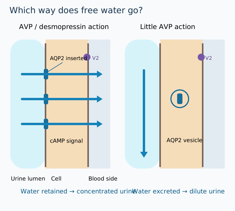
Does a low serum sodium always mean that the body lacks sodium? Serum sodium is a concentration reflecting exchangeable solute relative to water. Arginine vasopressin, AVP or ADH, is synthesized in hypothalamic supraoptic and paraventricular neurons, transported down their axons, and stored and released from the posterior pituitary. In the collecting duct, V2 receptor signaling increases apical aquaporin-2 availability, allowing water reabsorption. The posterior pituitary releases AVP; it does not synthesize it. [5][6]
Use the water-direction diagram as a tracing exercise. Follow AVP from secretion to V2 signaling to aquaporin-2, and predict urine volume and concentration before looking at the comparison below. More antidiuretic activity usually means less excreted water and more concentrated urine; insufficient secretion or renal resistance means more dilute urine. Plasma sodium also depends on intake and access to water, so these are directional predictions rather than inevitable sodium values. [5][6]
Contrast water retention in antidiuresis with excretion in AVP deficiency or resistance. [5][6]
Predict three collecting-duct states, assuming comparable solute excretion and water availability
StateAVP secretion is inadequate despite hypertonic plasma
Urine responseLarge volume of dilute urine
Plasma tendencySodium rises if drinking cannot replace losses
StateAVP is present but the kidney resists it
Urine responseLarge volume of dilute urine
Plasma tendencySodium rises if drinking cannot replace losses
Make the water cost concrete: assume the kidneys must excrete 600 mOsm of solute per day. Approximate obligatory urine volume equals daily solute excretion divided by urine osmolality, taking 1 kg of water as approximately 1 L. Calculate the volume at 100 mOsm/kg and again at 600 mOsm/kg before opening the comparison. This simplified steady-state exercise isolates concentration capacity; it is not a fluid prescription or a complete balance equation. [6][7]
Compare your calculation and change one condition
600 divided by 100 gives 6 L/day; 600 divided by 600 gives 1 L/day. With the same solute load, greater concentration capacity lowers the volume needed for excretion. If the patient with dilute polyuria cannot drink after surgery, hypernatremia can develop quickly. If a thirsty ambulatory patient replaces every lost liter, serum sodium can remain normal despite substantial AVP deficiency. [6][7]
The clinical consequence is visible without the disclosure: interpret paired plasma and urine osmolality, intake and urine volume together. A urine osmolality of 150 mOsm/kg is inappropriately concentrated for markedly hypotonic plasma, yet inappropriately dilute for hypertonic plasma. The same urine result can therefore support opposite physiological problems in different contexts. [5][7]
When is antidiuresis inappropriate, and when is treatment urgent?
A patient with pneumonia has sodium 122 mmol/L, plasma osmolality 260 mOsm/kg and urine osmolality 520 mOsm/kg. Why has the kidney not produced maximally dilute urine? Persistent antidiuresis is plausible, but SIAD, syndrome of inappropriate antidiuresis, is a diagnosis of exclusion. The traditional name SIADH emphasizes ADH secretion; SIAD also accommodates inappropriate antidiuresis without a documented high circulating AVP measurement. [5]
Typical supporting findings are hypotonic plasma, usually below 275 mOsm/kg, urine osmolality above 100 mOsm/kg, apparent euvolemia and urine sodium often above 30 mmol/L with adequate salt intake. Exclude adrenal insufficiency, severe hypothyroidism and relevant renal dysfunction; review diuretics and other drugs. A high urine sodium by itself does not prove SIAD, and clinical volume assessment is imperfect. Hyperglycemic hyponatremia and pseudohyponatremia require different interpretation because the plasma is not simply hypotonic in the same way. [5]
Predict whether the pneumonia patient should have edema. In SIAD, initial water retention prompts compensatory natriuresis and limits sustained extracellular expansion, often yielding an apparently euvolemic examination without edema. This does not mean total-body sodium is invariably unchanged. Contrast a patient with vomiting and orthostatic hypotension: antidiuresis may appropriately defend perfusion despite low sodium. The same concentrated urine cannot distinguish the two without the clinical setting. [5]
Search for the stimulus: CNS disease such as hemorrhage, stroke, infection, trauma or tumors; pulmonary disease such as pneumonia, tuberculosis or abscess; positive-pressure ventilation; malignancy, especially small-cell lung cancer; or medications. SSRIs, carbamazepine or oxcarbazepine, cyclophosphamide, NSAIDs and exogenous desmopressin can contribute by differing pathways. Chlorpropamide is a historical example. Pulmonary and CNS associations are multifactorial, not all explained by one pressure or venous-return mechanism. Treat the cause rather than stopping at the syndrome name. [5]
For chronic, nonsevere SIAD, fluid restriction is often first-line, individualized to urine concentration, intake and feasibility; less than about 1 L/day is a common starting approach, not an automatic prescription for every patient. [5] Very concentrated urine can make restriction ineffective. It does not instantly turn off AVP or guarantee dilute urine. In selected persistent cases, specialist options include oral urea or a vasopressin antagonist.
Tolvaptan blocks V2; conivaptan blocks V1a and V2. Their aquaresis can overcorrect sodium, and they are not rescue treatment for severe neurological symptoms. Demeclocycline induces renal AVP resistance but is generally avoided because of toxicity and limited evidence. [5]
Change the patient: sodium 112 mmol/L with a seizure. The immediate problem is cerebral edema, not choosing a long-term restriction plan. Use monitored 3% hypertonic saline boluses according to the emergency protocol; one accepted regimen is 100 mL over about 10 minutes, repeated if required after reassessment. Other protocols use 150 mL boluses. Seek an initial rise of roughly 4 to 6 mmol/L to improve severe symptoms, not immediate normalization. Frequent sodium measurements and urine-output surveillance are essential. [5]
Do not wait for a seizure to assess urgency. New confusion, headache, vomiting or worsening alertness in a hyponatremic patient warrants prompt clinical reassessment for symptomatic hyponatremia and alternative causes. The need for monitored hypertonic treatment depends on symptom severity and trajectory, not one sodium threshold alone. [5]
For chronic or unknown-duration hyponatremia, distinguish the initial treatment target from the correction ceiling. A conservative ceiling is no more than 8 mmol/L in any 24 hours, especially with very low starting sodium, malnutrition, alcohol use disorder, hypokalemia or advanced liver disease; many high-risk patients should have a goal nearer 4 to 6 mmol/L. Some guidelines permit 10 mmol/L in the first day for lower-risk patients and 8 subsequently. These are limits, not required achievements. Potassium replacement and a sudden water diuresis also contribute to the measured sodium rise. [5]
During prolonged hypotonicity, brain cells lose osmolytes to limit swelling. A rapid increase in extracellular tonicity can then dehydrate vulnerable cells and cause osmotic demyelination syndrome, involving pontine or extrapontine regions. Delayed dysarthria, dysphagia, behavioral change or spastic weakness may appear days after apparent initial recovery. Outcome varies; injury is not invariably irreversible. ODS can occur despite nominal adherence to limits in highly susceptible patients. [5]
Transfer the warning to a patient whose sodium rises from 108 to 116 mmol/L in 8 hours as urine output suddenly increases. The daily ceiling has already been reached; continuing correction is unsafe. Stop the cause of excessive correction and obtain urgent expert management, which may include desmopressin and electrolyte-free water such as D5W to control water losses and re-lower sodium. Do not wait for neurological symptoms or assume the prescribed saline dose alone predicts the final change. [5]
Try it here · Checkpoint 2 of 3
Make your prediction before reading the choices. A first attempt is just a starting point.
Case 15
Show answer and explanations for case 15
A. Isotonic saline to raise sodium by 4 to 6 mmol/L (Why this does not fit)
The target is modest, but severe symptomatic hypotonic hyponatremia calls for monitored hypertonic saline rather than relying on isotonic saline of uncertain effect.
Reasoning steps for option A
Which part of the isotonic saline option is reasonable?
An initial rise of 4 to 6 mmol/L is an appropriate first target for severe symptoms.
Why is isotonic saline the wrong fluid for a seizure at sodium 110?
Its effect is small and unreliable, especially with inappropriate antidiuresis. Seizures need prompt monitored 3% hypertonic saline boluses.
B. Fluid restriction until sodium reaches 120 mmol/L (Why this does not fit)
Restriction is too slow to address a seizure attributed to severe hypotonicity. It also substitutes an arbitrary final concentration for a monitored initial change.
Reasoning steps for option B
When does fluid restriction play a role in hyponatremia?
It is often first-line for chronic, nonsevere SIAD.
Why is it wrong for a patient who has just seized?
It raises sodium slowly over days and does not treat cerebral edema. Aiming for 120 mmol/L also sets a final target instead of a monitored initial rise.
C. Hypertonic saline until sodium reaches 135 mmol/L (Why this does not fit)
Hypertonic saline addresses cerebral edema, but immediate normalization risks ODS when chronic adaptation may already be present.
Reasoning steps for option C
What is correct about choosing hypertonic saline?
Hypertonic saline is the right treatment for severe symptoms from cerebral edema.
Why is correcting to 135 mmol/L dangerous?
The hyponatremia may be chronic, so the brain may have adapted. A rise of about 25 mmol/L far exceeds safe daily limits and risks osmotic demyelination.
D. Hypertonic boluses for an initial 4 to 6 mmol/L rise (Best answer)
Seizure requires rapid partial relief of cerebral edema. Unknown duration requires protection against chronic-adaptation injury, so give protocol-based boluses with frequent reassessment and stop short of immediate normalization.
Reasoning steps for option D
What does the seizure require immediately?
Rapid partial reduction of cerebral edema with monitored 3% saline boluses, aiming for an initial 4 to 6 mmol/L rise.
How does the uncertain duration shape the rest of the plan?
Because chronic adaptation may be present, sodium is checked frequently and correction stops short of normalization within the daily ceiling.
Takeaway: Severe symptoms justify prompt partial correction; uncertain duration argues against rapid normalization.
Is water loss due to deficient AVP or renal resistance?
Polyuria is a measured volume, not simply urinary frequency. In an adult, more than approximately 3 L/day or 50 mL/kg/day prompts evaluation. Confirm hypotonic urine, commonly below 300 mOsm/kg and sometimes with specific gravity below 1.005, and exclude osmotic diuresis from glucose, mannitol or other solutes. AVP deficiency, traditionally central diabetes insipidus, and AVP resistance, traditionally nephrogenic diabetes insipidus, both produce inappropriately dilute urine. Normal sodium does not exclude either when thirst and water access are intact. [6][7]
Central causes include pituitary surgery, head trauma, craniopharyngioma, infiltrative disease such as sarcoidosis or Langerhans-cell histiocytosis, granulomatosis with polyangiitis, inflammatory hypophysitis and cases without an identified cause. Do not assign one fixed percentage to idiopathic disease. A previously untreated sellar lesion with AVP deficiency should broaden the differential beyond an ordinary pituitary adenoma toward stalk, hypothalamic, infiltrative or metastatic disease. [6]
Renal resistance can result from lithium, hypercalcemia, hypokalemia, chronic kidney disease, or inherited defects in AVPR2 or AQP2. AVPR2 disease is usually X-linked; AQP2 disorders can be recessive or dominant. Lithium enters collecting-duct cells through ENaC and disrupts concentrating mechanisms including aquaporin-2 regulation; it is not simply a competitive V2-receptor antagonist. Hypercalcemia and hypokalemia impair concentrating capacity through functional transport changes, not just calcium deposition. [7][11][12]
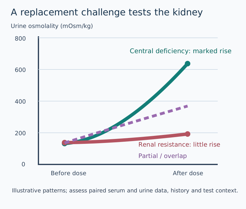
Use the desmopressin diagram to compare two patients with plasma osmolality 310 mOsm/kg and urine osmolality near 100 mOsm/kg. After supervised desmopressin, one reaches 650 and the other remains near 110. First identify the shared failure to concentrate despite hypertonic plasma; then locate the defect from the response. A large increase, often more than 50% in complete deficiency, supports intact renal response to missing AVP. A minimal response suggests complete renal resistance. Partial disorders and chronic primary polydipsia overlap, so these examples are not universal diagnostic cutoffs. [6][7]
Compare paired urine concentrations before and after replacement without treating cartoon values as cutoffs. [6][7]
The preceding deprivation phase tests endogenous AVP function: rising plasma tonicity should stimulate AVP and concentrate urine. Complete AVP deficiency or resistance fails to produce an adequate concentrating response. Primary polydipsia generally preserves that capacity, but prolonged excess drinking can reduce the medullary concentration gradient and blur the distinction from partial disease. The subsequent desmopressin phase asks whether supplying hormone can correct the defect. [6][7]
A formal water-deprivation test is reserved for selected stable indeterminate presentations under supervision, with weight, sodium, osmolality and stopping criteria monitored. It is not an instruction to withhold water from a hypernatremic postoperative patient already losing dilute urine. Specialized stimulated copeptin testing can improve distinction among AVP deficiency, resistance and primary polydipsia where available; protocols also require expert monitoring. Urine sodium is not a reliable central-versus-nephrogenic classifier. [6][7]
Treat central deficiency with access to water and individualized desmopressin, available by oral, sublingual, nasal or parenteral routes. Avoid excessive fluid intake while antidiuresis is fixed; treatment can cause hyponatremia. Planned periods of aquaresis, where appropriate and supervised, and sodium monitoring reduce that risk. In a patient unable to drink, replacement of water losses and monitoring are urgent. Glucocorticoid replacement can unmask previously obscured AVP deficiency by restoring water excretion. [6][8]
After sellar surgery, the trajectory may change: early dilute polyuria can be followed by delayed antidiuresis from release of stored AVP and, in some patients, recurrent permanent deficiency. Predict what happens if the initial desmopressin schedule continues unchanged during the antidiuretic interval: hyponatremia becomes more likely. The possible triphasic response requires reassessment of sodium, intake and output rather than assuming that the first postoperative pattern will persist. [6]
For renal resistance, address the cause and correct calcium or potassium abnormalities. A lower-sodium diet and appropriate reduction of excessive protein-derived solute can reduce obligatory water loss without compromising nutrition. Thiazides paradoxically reduce urine volume through mild volume contraction, increased proximal reabsorption and lower distal delivery. Amiloride is particularly useful in lithium-associated disease because ENaC blockade limits lithium entry; coordinate any lithium change with the prescribing clinician and monitor renal function and potassium. NSAIDs can reduce urine output in selected patients but carry renal and gastrointestinal risks and are not a routine harmless solution. [11][12][15]
Transfer the distinction to a person with polydipsia, low-normal plasma sodium and dilute urine. Excess drinking can suppress AVP normally; do not prescribe desmopressin solely because the urine is dilute. Conversely, hypernatremia with very dilute urine is an inappropriate renal response requiring prompt evaluation. Always return to plasma context, urine concentration and volume before attaching a diagnosis. [6][7]
Try it here · Checkpoint 3 of 3
Make your prediction before reading the choices. A first attempt is just a starting point.
Case 20
Show answer and explanations for case 20
A. Renal vasopressin resistance (Why this does not fit)
A rise from 85 to 560 after desmopressin demonstrates intact renal response, not resistance.
Reasoning steps for option A
Why might renal resistance be considered in postoperative dilute polyuria?
Both central AVP deficiency and renal resistance produce large volumes of dilute urine with rising sodium.
What does the desmopressin response show?
Urine osmolality rose from 85 to 560, so the collecting duct responds normally to an AVP analog, which rules out renal resistance.
B. Central vasopressin deficiency (Best answer)
Hypertonic plasma with dilute polyuria indicates ineffective antidiuresis; a large desmopressin response establishes preserved renal response.
Reasoning steps for option B
What do sodium 151, plasma osmolality 313 and urine osmolality 85 show after pituitary surgery?
Hypertonic plasma should stimulate maximal AVP, yet the urine is very dilute, so antidiuresis is failing.
What does the rise to 560 after desmopressin localize?
The kidneys respond to replacement, so the defect is missing AVP release, consistent with surgical injury to the posterior pituitary or stalk.
C. Glucose-mediated solute diuresis (Why this does not fit)
Glucosuria causes osmotically concentrated urine rather than a urine osmolality of 85 with sodium 151.
Reasoning steps for option C
Why could hyperglycemia cause polyuria after surgery?
Perioperative steroids or stress can raise glucose, and glucosuria causes an osmotic diuresis.
Why does the urine exclude a solute diuresis?
An osmotic diuresis produces urine with relatively high osmolality from solute. Urine at 85 mOsm/kg is water diuresis.
D. Primary polydipsia from excess oral intake (Why this does not fit)
Excess water intake generally lowers plasma tonicity rather than causing sodium 151.
Reasoning steps for option D
Why might excess drinking be considered when urine is very dilute?
Primary polydipsia also produces large volumes of dilute urine.
Which finding separates this patient from primary polydipsia?
Polydipsia lowers plasma sodium and osmolality. Sodium 151 with osmolality 313 means water is being lost, not taken in excess.
Takeaway: Hypertonic plasma with dilute polyuria indicates ineffective antidiuresis; a large desmopressin response establishes preserved renal response.
For each case, state the finding that separates the two strongest alternatives before choosing. Then use the all-option explanations to test your reasoning. Distinguish diagnosis from localization and an urgent physiological threat from the eventual long-term treatment.
Case 2
Show answer and explanations for case 2
A. Stalk effect; prompt surgical assessment (Best answer)
A persistent modest prolactin elevation after dilution favors stalk-related loss of dopamine inhibition. Progressive visual loss requires surgical evaluation rather than observation alone.
Reasoning steps for option A
Why does prolactin 48 ng/mL that stays 49 after dilution point to stalk effect with a 25-mm mass?
A large prolactinoma usually produces much higher levels, and dilution has excluded a hook effect. A modest rise fits loss of dopamine inhibition through a compressed stalk.
Why does the plan need surgical assessment instead of observation or medication?
Bitemporal field loss is progressing from chiasmal compression, and dopamine agonists do not reliably shrink a nonfunctioning adenoma. Prompt neurosurgical decompression is warranted.
B. Prolactinoma; dopamine agonist treatment (Why this does not fit)
A dopamine agonist is often appropriate for a prolactinoma, but modest prolactin that does not increase on dilution is less consistent with this large mass being a highly secreting tumor. The visual deficit also needs prompt specialist assessment.
Reasoning steps for option B
What makes a prolactinoma diagnosis tempting here?
Prolactin is above the upper limit and there is a pituitary macroadenoma, and dopamine agonists are the usual first treatment for prolactinomas.
How does the unchanged value after dilution argue against a prolactinoma of this size?
A 25-mm prolactinoma typically raises prolactin far higher. A modest level that does not rise on dilution fits stalk compression, so relying on a dopamine agonist could delay decompression of the chiasm.
C. Nonfunctioning lesion; imaging surveillance (Why this does not fit)
A nonfunctioning lesion is plausible, but progressive chiasmal visual impairment makes surveillance alone inadequate.
Reasoning steps for option C
Which findings make a nonfunctioning adenoma plausible?
There is no GH or cortisol excess, and the modest prolactin rise can come from stalk compression by a nonfunctioning mass.
Why is imaging surveillance the wrong plan for this lesion?
Surveillance suits selected asymptomatic lesions. Progressive bitemporal loss with a mass contacting the chiasm is an indication for surgical evaluation.
D. Drug-induced elevation; medication withdrawal (Why this does not fit)
Dopamine antagonists can cause modest prolactin elevation, but none is used and that explanation would not address the mass-associated visual loss.
Reasoning steps for option D
Why would a clinician first ask about medications with a modest prolactin elevation?
Dopamine-blocking drugs are a common cause of mild hyperprolactinemia and are reviewed before attributing the value to a tumor.
What in the vignette removes medication as the explanation?
No dopamine antagonist is used, and a drug effect would not explain a 25-mm mass causing progressive field loss.
Takeaway: Separate a modest stalk-related hormone rise from the independent danger of chiasmal compression.
A. Repeat the undiluted prolactin assay (Why this does not fit)
Repetition can help with transient physiological elevations, but repeating the same assay conditions may reproduce an antigen-excess artifact.
Reasoning steps for option A
Why might repeating the prolactin assay seem sensible?
Repeat testing helps when stress, venipuncture or other transient factors could have raised a single value.
Why would an undiluted repeat not resolve this discrepancy?
If antigen excess is saturating the two-site assay, the same undiluted conditions will reproduce the falsely modest result.
B. Measure macroprolactin as the first priority (Why this does not fit)
Macroprolactin helps evaluate an elevation discordant with biological symptoms. Here an invasive mass with a surprisingly modest result raises the more consequential possibility of severe underestimation.
Reasoning steps for option B
When is macroprolactin the right laboratory question?
Macroprolactin is checked when an elevated prolactin seems out of keeping with few or no symptoms.
Why is it not the first priority for a 30-mm invasive mass?
The concern here is the opposite error: a very high true prolactin being underreported, which would change the tumor's classification and treatment.
C. Measure GH suppression after oral glucose (Why this does not fit)
GH testing answers a different secretion question. It does not resolve the discrepancy between the large mass and unexpectedly modest prolactin result.
Reasoning steps for option C
What could draw attention to GH testing in a large sellar mass?
Macroadenomas can secrete other hormones, and a full evaluation of pituitary excess can include GH.
Why does a glucose suppression test not answer the question asked?
The stem asks how to interpret prolactin 42 with a 30-mm invasive mass. GH testing leaves that possible assay artifact unresolved.
D. Measure prolactin after serial dilution (Best answer)
A large prolactin-secreting tumor can saturate a two-site assay and yield a falsely modest value. Dilution addresses the hook effect before the result is used to label the tumor nonfunctioning.
Reasoning steps for option D
What is surprising about prolactin 42 ng/mL with a 30-mm invasive sellar mass?
A large prolactinoma often produces much higher prolactin, so the modest value is out of proportion to the tumor size.
How can a very large prolactinoma produce a falsely modest result?
Extreme prolactin concentrations can saturate both antibodies of a two-site immunoassay, the high-dose hook effect.
Why does serial dilution matter before tumor-directed therapy is chosen?
If diluted samples reveal very high prolactin, the tumor is a prolactinoma usually treated first with a dopamine agonist rather than labeled nonfunctioning.
Takeaway: An apparent hormone-size mismatch can be an assay problem that changes tumor classification.
A. Bitemporal visual field loss (Why this does not fit)
This reflects superior chiasmal compression rather than additional lateral cavernous involvement.
Reasoning steps for option A
Why might bitemporal field loss come to mind with a sellar lesion and new eye symptoms?
It is the classic visual finding of pituitary macroadenomas that extend upward.
Why would bitemporal loss not support the lateral extension described?
It reflects superior compression of the optic chiasm, and the stem states that temporal fields remain intact.
B. Right ptosis with impaired adduction (Best answer)
Cavernous sinus extension can also affect ipsilateral CN III along with VI and V1.
Reasoning steps for option B
Which structures does the combination of right abduction weakness and right corneal sensory loss localize to?
Horizontal diplopia on right gaze and reduced right corneal sensation suggest right CN VI and V1 involvement, both of which travel through the right cavernous sinus.
Which other nerve in that sinus would give ptosis and impaired adduction?
CN III runs in the lateral wall of the cavernous sinus. Its palsy produces ipsilateral ptosis and weak adduction, extending the same localization.
C. Left afferent pupillary defect (Why this does not fit)
This favors a left optic nerve pathway lesion rather than right cavernous extension.
Reasoning steps for option C
Why might a pupillary abnormality seem relevant to a sellar mass?
Sellar lesions can compress anterior visual pathways, and pupillary testing is part of that examination.
Why does a left afferent defect not support right cavernous extension?
An afferent pupillary defect localizes to the left retina or optic nerve, a different pathway on the opposite side from the right CN VI and V1 findings.
D. Bilateral papilledema and headache (Why this does not fit)
Raised intracranial pressure does not specifically corroborate right VI and V1 involvement.
Reasoning steps for option D
What makes headache with papilledema seem related to an enlarging mass?
Large intracranial masses can raise intracranial pressure and cause headache and papilledema.
Why is this finding not specific support for the same extension?
Papilledema reflects diffuse pressure. It does not localize to the right cavernous sinus the way another ipsilateral cranial nerve deficit would.
Takeaway: Cavernous sinus extension can also affect ipsilateral CN III along with VI and V1.
A. Glucocorticoid replacement for central adrenal deficiency (Best answer)
Morning cortisol 3 with inappropriately low ACTH and hypotension supports central adrenal insufficiency; glucocorticoids should precede levothyroxine to prevent adrenal crisis.
Reasoning steps for option A
What do morning cortisol 3 mcg/dL and ACTH 7 pg/mL indicate in a man with a macroadenoma?
Low cortisol with a low ACTH means the pituitary is not driving the adrenals: central adrenal insufficiency, and his hypotension may reflect it.
Why must glucocorticoid replacement come before levothyroxine?
Thyroid hormone increases cortisol clearance and metabolic demand. Starting it first in untreated cortisol deficiency can precipitate adrenal crisis.
B. Levothyroxine replacement for central hypothyroidism (Why this does not fit)
Low free T4 needs replacement, but levothyroxine before securing cortisol replacement can precipitate adrenal crisis in this hypotensive patient.
Reasoning steps for option B
Why is levothyroxine an understandable choice?
Free T4 is low with an inappropriately normal TSH, so he does have central hypothyroidism needing replacement.
What makes levothyroxine the wrong first replacement in this hypotensive man?
His cortisol deficiency is untreated. Thyroid hormone raises cortisol requirements and clearance and could turn his hypotension into adrenal crisis.
C. Testosterone replacement for gonadotropin deficiency (Why this does not fit)
Gonadal evaluation matters later, but hypotension with cortisol 3 demands immediate adrenal evaluation.
Reasoning steps for option C
Why could testosterone replacement seem relevant here?
Low libido in a man with a macroadenoma suggests gonadotropin deficiency, which is often affected early.
Why is it not the replacement that must come before thyroid hormone?
Hypogonadism is not immediately dangerous. The hypotension with cortisol 3 mcg/dL makes glucocorticoid replacement the urgent priority.
D. Growth hormone replacement for somatotroph deficiency (Why this does not fit)
GH replacement does not treat a low ACTH-low cortisol emergency.
Reasoning steps for option D
Why might GH deficiency be considered in this patient?
GH secretion is often lost early when a macroadenoma compresses the gland, and fatigue can reflect GH deficiency.
Why is GH replacement irrelevant to the thyroid sequencing question?
GH replacement is elective and does not address cortisol deficiency. It is never given ahead of glucocorticoids in a hypotensive patient.
Takeaway: Low cortisol with non-elevated ACTH and hypotension suggests central adrenal insufficiency; address glucocorticoid deficiency before thyroid replacement.
A. Prolactin may rise while psychosis improves (Why this does not fit)
Cabergoline stimulates D2 receptors and tends to lower prolactin. It does not reinforce the antipsychotic D2 blockade responsible for psychiatric benefit.
Reasoning steps for option A
What link between dopamine and prolactin makes this option tempting?
Dopamine-receptor drugs change prolactin, and cabergoline is used for hyperprolactinemia.
Why does this option reverse cabergoline's effects?
Cabergoline is a D2 agonist, so it lowers prolactin. It does not strengthen the antipsychotic D2 blockade that controls psychosis.
B. Prolactin may fall while relapse risk disappears (Why this does not fit)
Lower prolactin is plausible, but restoration of central dopamine signaling can destabilize psychiatric disease rather than eliminate relapse risk.
Reasoning steps for option B
Why is a fall in prolactin a reasonable prediction with cabergoline?
Stimulating lactotroph D2 receptors suppresses prolactin secretion even during antipsychotic therapy.
What does her earlier hospitalization after unplanned withdrawal show about relapse risk?
Her psychiatric illness is sensitive to changes in dopamine blockade. A D2 agonist can oppose risperidone centrally, so relapse risk rises rather than disappears.
C. Prolactin may fall while psychosis worsens (Best answer)
The timing and absent lesion favor medication-related D2 blockade. Cabergoline restores D2-mediated inhibition of prolactin secretion while potentially opposing central antipsychotic blockade, so coordinated management is needed.
Reasoning steps for option C
What do the timing after a dose increase and the normal MRI suggest?
Risperidone's D2 blockade is raising prolactin; no pituitary tumor is present.
What are the two opposite effects of adding cabergoline?
It restores D2 inhibition of lactotrophs, lowering prolactin, but it can also counter central antipsychotic blockade and worsen psychosis.
D. Prolactin stays high while psychosis is prevented (Why this does not fit)
D2 agonism is expected to reduce lactotroph prolactin secretion and is not a means of preserving the antipsychotic effect.
Reasoning steps for option D
Why might someone hope prolactin stays high while psychosis stays controlled?
Leaving antipsychotic effect intact is the priority given her earlier hospitalization.
Why is this outcome inconsistent with cabergoline's action?
A D2 agonist lowers prolactin rather than leaving it high, and it opposes rather than preserves central D2 blockade.
Takeaway: A receptor action helpful for one endocrine effect can oppose treatment of another illness.
A. Normal GH suppression; investigate isolated sleep apnea (Why this does not fit)
The nadir remains above the laboratory suppression threshold. Sleep apnea alone does not account for the repeated IGF-I abnormality and progressive acral changes.
Reasoning steps for option A
Why does a GH fall from 2.1 to 1.7 ng/mL look like possible suppression?
GH did decline after glucose, and IGF-I is only 1.15 times the upper limit, so the values can look near normal.
Why is the result still abnormal, and why is sleep apnea alone not enough?
The nadir of 1.7 stays above this laboratory's 1.0 ng/mL threshold. Sleep apnea cannot explain repeated high IGF-I with progressive ring enlargement.
B. Supported GH excess; obtain dedicated pituitary MRI (Best answer)
The equivocal IGF-I requires context. Failed glucose suppression plus progressive phenotype supports GH excess, after which pituitary imaging evaluates its usual source.
Reasoning steps for option B
Why is an IGF-I of 1.15 times the upper limit not diagnostic by itself?
Current consensus treats values below about 1.3 times the upper limit as equivocal, so dynamic testing is needed in a suggestive phenotype.
What does failure to suppress below 1.0 ng/mL add?
It confirms autonomous GH secretion that glucose cannot switch off, completing the biochemical diagnosis of acromegaly.
Why is pituitary MRI the next investigation?
Once GH excess is established biochemically, imaging looks for its usual source, a somatotroph pituitary adenoma.
C. Autonomous adrenal secretion; obtain adrenal imaging (Why this does not fit)
Dexamethasone and cortisol assess adrenal autonomy. These data instead concern IGF-I and failure of glucose to suppress GH.
Reasoning steps for option C
Why might adrenal autonomy come up in a patient with sleep apnea and weight-related features?
Cortisol excess can also cause metabolic and soft-tissue changes, and adrenal incidentalomas are common.
Which data in the stem are unrelated to adrenal function?
All the testing concerns IGF-I and glucose suppression of GH. No cortisol or dexamethasone result points toward the adrenals.
D. Assay-normal growth signaling; repeat random GH alone (Why this does not fit)
A modest IGF-I elevation is not automatically normal, and random GH is pulsatile. The supplied supervised suppression result is more informative than another isolated random value.
Reasoning steps for option D
Why might a clinician want another GH value?
The IGF-I elevation is modest, and a repeat measurement feels like a cautious way to confirm or dismiss it.
Why is a random GH less useful than the test already done?
GH secretion is pulsatile, so a random value is hard to interpret. The supervised glucose suppression test already answers the question.
Takeaway: Interpret assay-specific dynamic results before imaging the likely hormonal source.
A. Somatostatin receptor activation; titrate solely to GH (Why this does not fit)
Secretion-suppressing therapy should lower GH as well as its downstream effects. A GH-only plan disregards the distinctive dissociation between controlled IGF-I and persistent GH here.
Reasoning steps for option A
Why could a somatostatin receptor ligand have been the added medication?
Octreotide and lanreotide are standard treatments for residual acromegaly after surgery, and IGF-I did normalize.
Which result argues against it, and why is a GH-only plan wrong?
A drug suppressing secretion should lower GH, yet GH remains high while IGF-I is normal. Titrating to GH would ignore the result that best measures control.
B. Dopamine receptor activation; replace IGF-I with prolactin monitoring (Why this does not fit)
A dopamine agonist can help selected mild acromegaly, but prolactin does not measure control of peripheral GH action. The given response does not justify abandoning IGF-I.
Reasoning steps for option B
Why might a dopamine agonist be considered after acromegaly surgery?
Cabergoline can help selected patients with mild residual GH excess.
Why does prolactin monitoring not fit this situation?
Prolactin does not measure GH action, and the high GH with normal IGF-I fits peripheral receptor blockade rather than a dopamine agonist.
C. Peripheral GH receptor blockade; follow age-adjusted IGF-I (Best answer)
Controlled IGF-I despite persistent GH and unchanged tumor size favors receptor blockade by pegvisomant. IGF-I measures its biochemical efficacy; GH does not, and tumor surveillance continues.
Reasoning steps for option C
What does a normal IGF-I with persistently high GH and an unchanged tumor suggest about the drug?
GH is still being secreted but can no longer act on its receptors, the effect of pegvisomant.
Which marker measures the efficacy of receptor blockade?
Age-adjusted IGF-I reflects peripheral GH action, so it guides dosing; GH does not fall with this drug.
What monitoring besides IGF-I remains necessary?
Pegvisomant does not treat the tumor, so the residual cavernous-sinus tumor still needs MRI surveillance, along with liver tests.
D. GH receptor blockade; escalate until GH normalizes (Why this does not fit)
The mechanism fits, but the monitoring plan does not. Pegvisomant does not reliably suppress GH secretion, so a high GH alone is not an indication to escalate treatment when IGF-I is controlled.
Reasoning steps for option D
Why does GH receptor blockade fit the pattern?
Normal IGF-I with persistently high GH is exactly what pegvisomant produces.
Why is it wrong to escalate until GH normalizes?
Pegvisomant does not suppress GH secretion, so GH may stay high indefinitely. Escalating for GH alone would overtreat a patient whose IGF-I is controlled.
Takeaway: Infer the treatment target from dissociated hormone results, then choose the matching efficacy marker.
A. Autonomous adrenal cortisol production (Best answer)
Confirmed cortisol excess suppresses ACTH if adrenal autonomous; ACTH-producing sources would not have undetectable ACTH.
Reasoning steps for option A
What does ACTH below the detection limit mean once endogenous cortisol excess is confirmed?
High cortisol has suppressed pituitary ACTH through negative feedback, so cortisol production is ACTH-independent.
Where does ACTH-independent cortisol excess come from?
It usually comes from an autonomous adrenal adenoma, less often carcinoma or bilateral adrenal disease, so adrenal imaging is next.
B. Pituitary ACTH-producing adenoma (Why this does not fit)
An ACTH-producing pituitary lesion would not produce undetectable ACTH in endogenous hypercortisolism.
Reasoning steps for option B
Why might a pituitary source seem likely in Cushing syndrome?
Cushing disease is the most common endogenous cause of cortisol excess.
Why does undetectable ACTH rule out a corticotroph adenoma here?
A pituitary tumor drives cortisol by secreting ACTH, so ACTH would be normal or high rather than undetectable.
C. Ectopic ACTH-producing tumor (Why this does not fit)
Ectopic ACTH should remain measurable or elevated rather than suppressed.
Reasoning steps for option C
What features could suggest ectopic ACTH?
Ectopic ACTH can cause marked cortisol excess with weakness and bruising, as seen here.
Why does the ACTH result exclude an ectopic source?
Ectopic tumors produce high ACTH, and an undetectable level is the opposite of what they cause.
D. Medication-induced cortisol excess (Why this does not fit)
Exogenous glucocorticoids can suppress ACTH, but exposure is explicitly absent. The phenotype and endogenous cortisol excess with suppressed ACTH therefore prompt adrenal assessment.
Reasoning steps for option D
Why would exogenous glucocorticoids otherwise explain suppressed ACTH?
Prescribed or hidden glucocorticoids produce a cushingoid phenotype and suppress ACTH.
What in the stem shows this is endogenous?
No glucocorticoids of any kind are used, so the high cortisol with suppressed ACTH comes from the patient's own adrenal glands.
Takeaway: Confirmed cortisol excess suppresses ACTH if adrenal autonomous; ACTH-producing sources would not have undetectable ACTH.
A. Repeat dedicated pituitary MRI (Why this does not fit)
Better imaging can characterize the small focus but cannot prove it secretes ACTH; incidental pituitary lesions occur.
Reasoning steps for option A
Why might repeating a dedicated pituitary MRI seem attractive?
Better imaging could define the 4-mm focus more clearly.
Why can MRI not settle the source here?
Small pituitary incidentalomas are common, so seeing the focus again does not prove it secretes ACTH while a pulmonary nodule remains a competing source.
B. Bilateral inferior petrosal sinus ACTH sampling (Best answer)
Established ACTH-dependent excess and competing small lesions require functional localization. Central-to-peripheral ACTH comparison can identify a pituitary source when performed and interpreted by an expert team during active disease.
Reasoning steps for option B
What is established and what remains unresolved in this patient?
ACTH-dependent cortisol excess is confirmed, but both a 4-mm pituitary focus and a pulmonary nodule could be the source.
How does inferior petrosal sinus sampling distinguish the two sources?
It compares ACTH draining from the pituitary with peripheral ACTH, usually after CRH or desmopressin stimulation. A high central-to-peripheral gradient indicates a pituitary source.
C. Repeat high-dose dexamethasone testing (Why this does not fit)
Some pituitary tumors suppress and some ectopic sources also show responses. Overlap prevents this test from definitively resolving the supplied ambiguity.
Reasoning steps for option C
Why has high-dose dexamethasone been used to localize ACTH-dependent disease?
Many corticotroph adenomas partially suppress, while many ectopic tumors do not.
Why will repeating it not resolve the ambiguity?
Responses overlap: some ectopic tumors suppress and some pituitary tumors do not. It cannot separate the two with confidence.
D. Repeat late-night salivary cortisol testing (Why this does not fit)
This measures cortisol excess, which is already established. It does not identify the anatomical ACTH source.
Reasoning steps for option D
What does late-night salivary cortisol measure?
It tests loss of the normal nighttime cortisol nadir, a screen for cortisol excess.
Why does it not help with localization?
Cortisol excess is already confirmed. The remaining question is where ACTH comes from, which salivary cortisol cannot answer.
Takeaway: Once active ACTH-dependent excess is confirmed, select a localization test rather than another test of cortisol excess.
A. Adrenal regrowth causes the mass; taper glucocorticoids (Why this does not fit)
Autonomous adrenal regrowth would tend to suppress ACTH, not explain a growing sellar lesion with progressive pigmentation. The absent adrenal reserve cannot be presumed restored.
Reasoning steps for option A
Why might adrenal regrowth come up after adrenalectomy?
Residual adrenal tissue can occasionally regrow and resume cortisol production.
Why does regrowth not fit the findings?
Autonomous adrenal tissue would suppress ACTH, and it cannot explain an enlarging sellar lesion. Tapering glucocorticoids would risk crisis in a patient without adrenal glands.
B. Excess replacement causes the ACTH rise; stop both steroids (Why this does not fit)
Excess glucocorticoid feedback would suppress ACTH. Stopping essential replacement after bilateral adrenalectomy risks adrenal crisis.
Reasoning steps for option B
Why might the patient suspect the steroid replacement?
The replacement steroids are the only hormones being given, so they seem a natural suspect for the rising ACTH.
Why is stopping them both wrong and dangerous?
Excess glucocorticoid would suppress ACTH, not raise it. After bilateral adrenalectomy, stopping replacement causes adrenal crisis.
C. A new somatotroph tumor causes pigmentation; retain only mineralocorticoid (Why this does not fit)
GH excess causes an acral phenotype rather than ACTH-associated pigmentation. Bilateral adrenalectomy also leaves a continuing glucocorticoid requirement.
Reasoning steps for option C
Why might a new pituitary tumor be considered with an enlarging sellar lesion?
Any pituitary tumor can present as a growing sellar mass on MRI.
Why does a somatotroph tumor not explain the pigmentation or justify stopping glucocorticoids?
Pigmentation reflects very high ACTH, not GH. Without adrenal glands the patient needs both glucocorticoid and mineralocorticoid replacement.
D. Corticotroph tumor progression is likely; maintain replacement (Best answer)
Rising ACTH, pigmentation and sellar growth after adrenalectomy support corticotroph progression. High ACTH cannot restore missing adrenal glands, so replacement remains essential while the tumor is evaluated.
Reasoning steps for option D
What do rising ACTH, darker skin and an enlarging sellar lesion after adrenalectomy indicate?
Loss of cortisol feedback has allowed the corticotroph tumor to grow, the pattern historically called Nelson syndrome.
Why can high ACTH not replace the steroid therapy?
ACTH needs adrenal glands to act on. After bilateral adrenalectomy, replacement must continue while the tumor is evaluated and treated.
Takeaway: Pituitary tumor progression after adrenalectomy does not restore adrenal hormone production.
A. Inappropriate antidiuresis with water retention (Best answer)
Hypotonic plasma paired with concentrated urine and urinary sodium despite no volume depletion favors SIAD.
Reasoning steps for option A
What does urine osmolality 510 mOsm/kg mean when plasma osmolality is 260?
Hypotonic plasma should suppress AVP and produce maximally dilute urine, so concentrated urine shows persistent, inappropriate antidiuresis.
Which findings exclude the main alternatives to SIAD?
Euvolemia without edema or orthostasis, no diuretics, and normal renal, thyroid and adrenal function meet the exclusions, and small-cell lung cancer is a classic cause.
B. Low effective circulating volume with renal sodium retention (Why this does not fit)
Low effective volume usually yields renal sodium conservation; this patient has urine sodium 52 without orthostasis or edema.
Reasoning steps for option B
Why can low effective circulating volume produce hyponatremia with concentrated urine?
Baroreceptor-driven AVP release in hypovolemia or heart failure also retains water.
Which findings argue against low effective volume here?
Urine sodium is 52 mEq/L rather than conserved, and there is no orthostasis or edema.
C. Primary polydipsia with suppressed ADH (Why this does not fit)
Urine osmolality 510 is inconsistent with appropriately suppressed antidiuresis.
Reasoning steps for option C
Why is primary polydipsia a cause of hypotonic hyponatremia to consider?
Very high water intake can overwhelm excretion and dilute plasma sodium.
Why does the urine result exclude it?
With AVP suppressed, urine should be maximally dilute, usually below 100 mOsm/kg, not 510.
D. Cortisol deficiency (Why this does not fit)
Adrenal function has already been documented normal, removing this important SIAD mimic.
Reasoning steps for option D
Why is cortisol deficiency an important mimic of SIAD?
It can cause hyponatremia with concentrated urine through nonosmotic AVP release.
Why is it not the explanation in this patient?
Adrenal function has already been documented as normal, so this mimic has been excluded.
Takeaway: Hypotonic plasma paired with concentrated urine and urinary sodium despite no volume depletion favors SIAD.
A. Sodium rises because infused sodium exceeds plasma sodium (Why this does not fit)
Comparing infusate with plasma alone ignores renal handling. Persistent urinary sodium plus potassium above the infusate concentration can permit salt excretion with relative water retention.
Reasoning steps for option A
Why does it seem that saline with 154 mmol/L sodium must raise plasma sodium 129?
The infusion has a higher sodium concentration than the plasma, so adding it looks as if it should raise the concentration.
What does that comparison leave out?
Renal handling. With urine sodium plus potassium of 175 mmol/L, the kidney can excrete the infused salt in less water than was given.
B. Sodium falls because urinary water loss exceeds cation loss (Why this does not fit)
Disproportionate electrolyte-free water loss would raise serum sodium. Here the concentrated urinary electrolytes favor the opposite balance under the stated assumptions.
Reasoning steps for option B
Why might the urine be expected to lose a lot of water?
Saline increases urine flow, and more urine is easily equated with water loss.
What would dilute urine losses actually do, and does this urine fit?
Losing water in excess of cations would raise sodium, not lower it. Here the urine is concentrated, with sodium plus potassium above the infusate.
C. Sodium falls as infused salt is excreted in less water (Best answer)
Urine sodium plus potassium is 175 mmol/L, above 154 in the infusion. In persistent SIAD, infused electrolyte can be excreted in a smaller urine volume, leaving water behind and worsening hyponatremia.
Reasoning steps for option C
How do urine sodium and potassium together compare with the infusate?
Urine sodium 95 plus potassium 80 equals 175 mmol/L, which is higher than the 154 mmol/L sodium in 0.9% saline.
What happens to the water from the infusion when the urine is this concentrated?
The infused salt leaves in a smaller urine volume and part of the infused water is retained, so plasma sodium can fall further in persistent SIAD.
D. Sodium rises because urine must become maximally dilute (Why this does not fit)
The urine is concentrated despite hypotonic plasma. Isotonic saline does not necessarily switch off inappropriate antidiuresis, so dilution cannot be assumed.
Reasoning steps for option D
Why might saline be expected to make the urine dilute?
If hyponatremia came from volume depletion, restoring volume would remove the AVP stimulus and produce dilute urine.
Why can that not be assumed here?
This patient is euvolemic with inappropriate antidiuresis, and isotonic saline does not switch off the drive causing concentrated urine.
Takeaway: Compare urinary sodium plus potassium with infused sodium, not just saline with plasma.
A. Tolvaptan to hasten electrolyte-free water excretion (Why this does not fit)
The sodium rise is already excessive in a high-risk patient. Increasing aquaresis would worsen rather than correct the hazard.
Reasoning steps for option A
Why might tolvaptan seem like a way to fix the sodium problem?
It increases electrolyte-free water excretion and raises sodium in SIAD.
Why would it be harmful now?
Sodium has already risen 10 mmol/L in 12 hours in a malnourished patient. Adding an aquaretic would push correction even further past the safe limit.
B. Desmopressin plus D5W for controlled relowering (Best answer)
The 10 mmol/L rise is already excessive and brisk aquaresis can continue after saline stops. Expert desmopressin to control losses plus electrolyte-free water can halt and partially reverse overcorrection.
Reasoning steps for option B
Why is the patient still at risk after saline is stopped?
Sodium rose 10 mmol/L in 12 hours, above the ceiling for a high-risk malnourished patient, and urine output of 450 mL/hour shows an ongoing water diuresis.
How do desmopressin and D5W address that risk?
Desmopressin stops the free-water loss and D5W supplies electrolyte-free water. Together, under expert monitoring, they halt correction and can lower sodium again.
C. Isotonic saline to match the urine volume (Why this does not fit)
Replacing dilute urinary losses with a sodium-containing solution can permit further sodium rise. The problem is excessive tonicity correction, not simply the volume of urine.
Reasoning steps for option C
Why could matching urine output with isotonic saline sound protective?
Replacing a large urine volume seems to prevent dehydration.
Why would it worsen the actual problem?
The urine is dilute. Replacing it with 154 mmol/L saline adds sodium and further raises plasma sodium, which is already overcorrected.
D. Fluid restriction with serial sodium measurements (Why this does not fit)
Restriction is useful in selected persistent SIAD but does not counteract ongoing water loss during aquaresis. Monitoring alone without an active response may allow further harmful correction.
Reasoning steps for option D
Why do fluid restriction and serial sodium checks feel cautious?
Monitoring is essential, and restriction is part of SIAD treatment.
Why is this inadequate now?
The urgent problem is ongoing water loss driving sodium up. Restricting intake would add to the rise, and monitoring alone does not stop it.
Takeaway: Stopping saline may not stop correction when the kidney has begun a brisk water diuresis.
A. Loss of intracellular osmolytes during prolonged hypotonicity (Best answer)
The delayed bulbar and motor syndrome after a large sodium rise supports ODS. Prior osmolyte loss limits swelling in chronic hyponatremia but leaves cells vulnerable when extracellular tonicity rises too quickly.
Reasoning steps for option A
What do delayed dysarthria, dysphagia and spastic weakness with symmetric pontine lesions indicate?
Osmotic demyelination syndrome, appearing days after sodium rose 20 mmol/L in 18 hours.
What adaptation during prolonged hyponatremia made the brain vulnerable?
Brain cells had shed intracellular osmolytes to limit swelling. When extracellular tonicity rose rapidly, they lost water and myelin was injured before osmolytes could be restored.
B. Accumulation of intracellular osmolytes during prolonged hypotonicity (Why this does not fit)
That direction of adaptation would worsen initial swelling. The relevant chronic response is osmolyte loss, which makes subsequent rapid correction hazardous.
Reasoning steps for option B
Why might osmolyte accumulation sound like the adaptive change?
Cells do accumulate osmolytes when they adapt to hypertonic states such as chronic hypernatremia.
Why is that the wrong direction for hyponatremia?
In hypotonic plasma, accumulating osmolytes would draw in more water and worsen swelling. The adaptation to hyponatremia is osmolyte loss.
C. Rapid influx of water after sodium increased (Why this does not fit)
Increasing extracellular tonicity tends to draw water out of adapted cells. Water influx instead explains edema during acute hypotonicity, not this delayed postcorrection pattern.
Reasoning steps for option C
Why could water movement seem responsible for brain injury?
Cerebral edema from water entering cells is the danger of acute hyponatremia.
Which way does water move after rapid correction?
Water leaves adapted brain cells as extracellular tonicity rises. Water influx explains acute hyponatremic edema, not delayed postcorrection injury.
D. Compensatory myelin synthesis during hypertonic treatment (Why this does not fit)
New myelin production does not explain the characteristic injury pattern or timing. The risk follows osmotic stress on chronically adapted brain tissue.
Reasoning steps for option D
Why might myelin synthesis be linked to these symptoms?
The syndrome is named for demyelination, so a change in myelin turnover seems relevant.
Why is compensatory myelin synthesis not the answer?
The injury comes from osmotic stress on adapted cells, especially oligodendrocytes, not from new myelin produced during treatment.
Takeaway: Connect the delayed injury pattern to the direction of chronic brain osmotic adaptation.
A. Restrict oral free water while monitoring sodium (Why this does not fit)
Restriction can aggravate the documented negative balance and orthostatic hypovolemia.
Reasoning steps for option A
Why is fluid restriction the team's reflex?
Hyponatremia after brain injury with high urine sodium and concentrated urine resembles SIAD, where restriction is standard.
What does the volume assessment show instead?
Weight has fallen 3 kg, balance is negative and the patient is orthostatic with tachycardia. Restricting fluid would worsen true hypovolemia and cerebral perfusion.
B. Give a V2 antagonist to increase water clearance (Why this does not fit)
Aquaretic treatment can worsen true volume depletion after cerebral injury.
Reasoning steps for option B
Why might a V2 antagonist appeal for hyponatremia after brain injury?
Vaptans raise sodium by increasing water excretion in SIAD.
Why is this dangerous in the patient described?
The patient is volume depleted, and an aquaretic would increase water loss and could worsen hypotension after subarachnoid hemorrhage.
C. Treat with desmopressin to stop salt loss (Why this does not fit)
Desmopressin retains water but does not replace the depleted sodium and circulating volume.
Reasoning steps for option C
What logic makes desmopressin seem helpful?
The patient is losing urine, and desmopressin reduces urine output.
Why does desmopressin not treat this problem?
It retains water, not sodium, so it would worsen hyponatremia without replacing the sodium and circulating volume the patient has lost.
D. Replace volume and sodium; investigate renal salt loss (Best answer)
High urine sodium overlaps SIAD, but documented hypovolemia argues against simple SIAD and makes restriction hazardous.
Reasoning steps for option D
Why does high urine sodium not settle the diagnosis after subarachnoid hemorrhage?
Both SIAD and renal salt loss produce high urine sodium, so volume status is what separates them.
What do the weight loss, negative balance and orthostasis indicate?
True volume depletion with ongoing renal salt loss, consistent with cerebral salt wasting.
How does that change management?
Sodium and volume are replaced, often with isotonic or hypertonic saline, while the cause is investigated, and fluid restriction is avoided.
Takeaway: High urine sodium overlaps SIAD, but documented hypovolemia argues against simple SIAD and makes restriction hazardous.
A. Persistent maximal antidiuresis from cortisol replacement (Why this does not fit)
Restored glucocorticoids generally reduce the inappropriate vasopressin drive of cortisol deficiency.
Reasoning steps for option A
Why might glucocorticoids be thought to sustain antidiuresis?
Glucocorticoids are part of the stress response, which is linked to AVP release.
What actually happens to AVP after cortisol replacement?
Cortisol deficiency removes normal inhibition of AVP. Replacement restores that inhibition, so antidiuresis lessens rather than persists.
B. Further sodium dilution from obligatory water retention (Why this does not fit)
Removal of cortisol deficiency often increases rather than reduces free-water clearance.
Reasoning steps for option B
Why could further dilution be expected with continued fluid intake?
Concentrated urine of 380 suggests ongoing water retention, which would keep diluting sodium.
How does replacement change that prediction?
Once cortisol is replaced, AVP falls and free-water excretion increases, so sodium tends to rise rather than fall further.
C. Dilute diuresis with rapid sodium correction (Best answer)
Correcting central cortisol deficiency removes a stimulus to vasopressin, which can cause brisk water excretion and fast sodium correction.
Reasoning steps for option C
What do cortisol 2 mcg/dL and ACTH 5 pg/mL indicate as the cause of hyponatremia?
Central adrenal insufficiency, where cortisol deficiency drives inappropriate AVP release and water retention.
What happens when glucocorticoid is replaced?
AVP is suppressed and a brisk dilute diuresis can follow, raising sodium quickly.
What does this mean for monitoring?
Urine output and sodium need frequent checks so correction stays within safe limits. Desmopressin or free water may be needed if sodium rises too fast.
D. No change because ACTH remains low (Why this does not fit)
Glucocorticoid replacement can alter vasopressin and water clearance even when ACTH is suppressed.
Reasoning steps for option D
Why might a low ACTH seem to mean replacement changes nothing?
ACTH stays low in central disease, so it could seem that the hormonal situation is unchanged.
Why does water handling change anyway?
The missing hormone is cortisol, and giving it directly removes the AVP stimulus whatever ACTH does.
Takeaway: Correcting central cortisol deficiency removes a stimulus to vasopressin, which can cause brisk water excretion and fast sodium correction.
A. X-linked AVPR2 receptor dysfunction (Best answer)
Maternal-line affected males and desmopressin resistance point to X-linked renal V2 receptor dysfunction.
Reasoning steps for option A
What does the pedigree show about inheritance?
Only males are affected, linked through unaffected mothers, with no father-to-son transmission: an X-linked recessive pattern.
How does the desmopressin result localize the defect?
A negligible response means renal resistance. AVPR2 loss-of-function on the X chromosome explains both the pedigree and the renal defect.
B. Autosomal dominant central AVP deficiency (Why this does not fit)
Central AVP deficiency should produce appreciable renal concentration with desmopressin.
Reasoning steps for option B
Why might an inherited central form be considered?
Familial central AVP deficiency exists and causes lifelong dilute polyuria.
Which findings argue against it?
Central deficiency responds well to desmopressin and is usually autosomal dominant, so it matches neither the test result nor the pedigree.
C. Autosomal AQP2-mediated renal resistance (Why this does not fit)
AQP2 defects cause renal resistance but usually lack this classic maternal-uncle X-linked pattern.
Reasoning steps for option C
Why is an AQP2 defect a reasonable consideration?
AQP2 mutations cause congenital renal AVP resistance with a poor desmopressin response.
Why does the family pattern not fit?
AQP2 disorders are autosomal recessive or dominant. Affected males linked only through unaffected mothers point instead to an X-linked gene.
D. X-linked activating AVPR2 mutation (Why this does not fit)
Constitutive V2 activation promotes water retention and can cause hyponatremia. It does not explain lifelong dilute polyuria, hypernatremia and resistance to replacement.
Reasoning steps for option D
Why could an AVPR2 variant with an X-linked pattern seem right?
The gene and inheritance pattern match the pedigree.
Why does an activating mutation give the opposite phenotype?
Constitutive V2 activation causes water retention and hyponatremia, not dilute polyuria, hypernatremia and poor response to desmopressin.
Takeaway: Maternal-line affected males and desmopressin resistance point to X-linked renal V2 receptor dysfunction.
A. Start desmopressin and follow symptom relief (Why this does not fit)
Low plasma tonicity with dilute urine may reflect an appropriate response to excess drinking. Desmopressin could retain ingested water and worsen hyponatremia.
Reasoning steps for option A
Why might desmopressin be tempting for frequent urination?
It reduces urine output and often relieves symptoms quickly in AVP deficiency.
What could desmopressin do in this patient?
Sodium 134 and plasma osmolality 272 with dilute urine suggest excess drinking suppressing AVP. Desmopressin while intake continues could cause dangerous hyponatremia.
B. Record intake, 24-hour urine volume and sodium (Best answer)
Document true polyuria rather than frequency and assess intake-output relationships. The low tonicity favors water excess; supervised dynamic testing is reserved for persisting uncertainty after baseline evaluation.
Reasoning steps for option B
What does low-normal sodium with plasma osmolality 272 and urine osmolality 90 suggest?
The kidney is appropriately excreting dilute urine in response to high intake, as in primary polydipsia.
Why is measuring intake and 24-hour urine volume the first step?
It confirms true polyuria above about 3 L/day rather than frequency and shows how output relates to intake.
When would dynamic testing follow?
Only if uncertainty persists after baseline evaluation, and then as a supervised water-deprivation or copeptin test.
C. Arrange a home overnight deprivation test (Why this does not fit)
Baseline volume confirmation is incomplete. Unsupervised deprivation is unsafe if an AVP disorder is present; any later dynamic evaluation needs appropriate supervision.
Reasoning steps for option C
Why might a deprivation test seem like the standard next step?
Water deprivation is the classic way to distinguish AVP deficiency from primary polydipsia.
Why is a home overnight test inappropriate?
True polyuria has not been confirmed, and unsupervised deprivation can cause dangerous dehydration if an AVP disorder is present. Dynamic tests need supervision.
D. Start a thiazide and reassess urinary frequency (Why this does not fit)
A thiazide can reduce output in renal AVP resistance, but that diagnosis is not established and baseline hyponatremia creates an additional treatment hazard.
Reasoning steps for option D
Why might a thiazide be considered in someone with polyuria?
Thiazides reduce urine volume in renal AVP resistance.
Why is starting one premature and potentially harmful?
Renal resistance has not been shown, and thiazides can worsen hyponatremia in someone with sodium 134 who is drinking heavily.
Takeaway: Confirm the volume disorder and plasma context before using a treatment or dynamic test as a diagnostic shortcut.
A. Persistent urine concentration because native AVP secretion is intact (Why this does not fit)
The original hypertonic dilute urine followed by a marked desmopressin response supports deficient endogenous AVP, not an intact compensatory response.
Reasoning steps for option A
Why might the concentrated urine suggest recovery of AVP secretion?
Urine osmolality 650 during hyponatremia could look like native AVP secretion has returned.
Why is desmopressin the better explanation?
The patient took a scheduled dose. The earlier hypertonic dilute urine that concentrated with desmopressin showed missing endogenous AVP, so the concentration is drug effect.
B. Stable sodium because reduced drinking offsets all renal losses (Why this does not fit)
Fluid intake alone does not ensure balance when antidiuretic medication wears off. A brisk water diuresis can exceed intake and rapidly raise sodium.
Reasoning steps for option B
Why might cutting back on drinking seem enough to keep sodium stable?
The hyponatremia came from drinking several liters under desmopressin, so reducing intake addresses the cause.
What happens to urine output when the dose wears off?
With deficient AVP, a large dilute diuresis resumes and can exceed intake, raising sodium quickly from 126.
C. Brisk dilute diuresis with an excessively rapid sodium rise (Best answer)
The original response localizes missing AVP with responsive kidneys. Current water retention occurred under desmopressin; withdrawal can expose the underlying deficiency and cause rapid aquaresis, requiring close monitored management.
Reasoning steps for option C
What did the earlier desmopressin test establish?
Urine concentrated from 85 to 560, so AVP is missing but the kidneys respond normally.
Why did sodium fall to 126?
The patient drank several liters while desmopressin fixed antidiuresis, so the water could not be excreted.
What happens when the next dose is withheld?
Antidiuresis wears off and brisk aquaresis can raise sodium too quickly, so sodium and urine output must be monitored closely.
D. New solute diuresis because desmopressin increases glucosuria (Why this does not fit)
Desmopressin controls water permeability rather than glucose excretion. There is no supplied solute load to explain this predicted transition.
Reasoning steps for option D
Why might a solute diuresis be considered after desmopressin is stopped?
Large urine volumes can come from glucose or other solutes.
Why does this explanation not fit?
Desmopressin affects water permeability, not glucose excretion, and no solute load is described. The expected diuresis is dilute water loss.
Takeaway: Use the earlier replacement response to predict what can happen when antidiuresis wears off during hyponatremia.