Connect liver lesion architecture with contrast behavior, distinguish hemangioma, FNH, and adenoma, and choose reassurance, surveillance, or treatment.
An incidental liver mass raises two separate questions: what is it, and what danger does it create? A bright arterial-phase lesion is not automatically cancer, and a benign label does not automatically mean observation. Learn to connect tissue architecture with contrast behavior, then decide when reassurance, surveillance, tissue assessment, or treatment is appropriate.
Does a liver mass need a needle first?
Characterization usually precedes sampling. Start with the patient as well as the mass. Ask about chronic hepatitis, cirrhosis, known cancer, constitutional symptoms, estrogen-containing contraception, anabolic-androgenic steroids, obesity, metabolic disease, pregnancy plans, and prior imaging. Examine the background liver and compare lesion size over time. Liver tests and selected tumor markers contribute to the assessment, but normal results do not establish benignity. [1]
A single portal venous CT image may detect a lesion without identifying it. For an uncertain solid lesion, use dedicated multiphasic contrast-enhanced MRI or CT, including arterial, portal venous, and delayed imaging. MRI additionally characterizes fat and blood products. Hepatobiliary-specific contrast is particularly helpful when the distinction is focal nodular hyperplasia versus adenoma. Contrast-enhanced ultrasound is another useful modality where expertise is available. [1][2]
Consider a 2.5 cm echogenic focus in two patients: one otherwise healthy, the other undergoing cancer staging. Predict whether the ultrasound finding alone deserves identical reassurance. It does not. The cancer history requires contrast characterization rather than a low-risk shortcut. In a healthy liver, a small lesion meeting a complete characteristic hemangioma ultrasound pattern can sometimes be diagnosed without further imaging; ACG uses a less-than-2-cm criterion for that shortcut. Echogenicity alone is insufficient. [1]
Biopsy of a suspected hemangioma is avoided when possible because sampling a vascular lesion creates bleeding risk. Typical focal nodular hyperplasia also needs no biopsy. Neither principle means that all liver biopsy is forbidden: an atypical lesion, suspected transformation, or unresolved management-changing uncertainty warrants multidisciplinary review and sometimes planned tissue assessment. Sampling error and overlap with well-differentiated carcinoma limit what a small specimen can settle. [1][2]
Apply this distinction: a lesion remains indeterminate after high-quality MRI. The next decision is not automatically reassurance or automatic biopsy. Identify what remains uncertain, whether tissue would change care, and the safest route to that answer with a liver tumor team.
Three masses, three different biological problems
Does benign mean biologically identical? Hemangioma, focal nodular hyperplasia, and hepatocellular adenoma can all be incidental findings, often in women, but their tissue origins explain different risks. Demographics adjust probability; they do not replace characterization. [1]
Three tissue patterns, not three diagnostic scans. Hemangioma is vascular; FNH is a hyperplastic response; adenoma is a hepatocellular neoplasm. Original Bone Wizardry construction. [1][3] Open the image for a larger view.
Hemangioma is the most common benign solid hepatic lesion. It consists of blood-filled spaces lined by endothelium and separated by fibrous tissue. Most are asymptomatic. Once the diagnosis is secure in a low-risk liver, reassurance is usually sufficient. A large diameter alone is not an indication for preventive surgery. Rare attributable complications, including compression, hemorrhage, or consumptive coagulopathy, change that assessment. [1]
Focal nodular hyperplasia, or FNH, is a polyclonal hyperplastic response associated with abnormal local arterial supply, not the same process as a clonal adenoma. It occurs most often in young to middle-aged women. A central scar and radiating fibrous septa may be present. Securely diagnosed FNH has no meaningful malignant potential and usually needs neither treatment nor routine follow-up. Underlying vascular liver disease or atypical imaging requires a more individualized assessment. [1][2][4]
Hepatocellular adenoma, or HCA, is a clonal hepatocyte neoplasm. Estrogen exposure, anabolic-androgenic steroids, obesity and metabolic dysfunction, and some inherited disorders such as glycogen storage disease are relevant associations. Pregnancy can stimulate growth. Hemorrhage and malignant transformation, not simply the word benign, determine management. [1][4]
Match each tissue drawing to the question it creates: Can imaging establish a vascular pattern? Is this secure hyperplasia? Could this neoplasm bleed or transform? The consequence is different care: typical hemangioma and FNH usually need reassurance; adenoma needs risk stratification. Transfer this to a man with a small liver lesion: male sex changes adenoma management, but does not by itself create a resection indication for FNH.
What does the contrast actually measure?
Arterial enhancement describes delivery of contrast, not proof of a particular cell type. Later hepatobiliary-phase MRI asks a different question: can the lesion's hepatocytes take up the contrast through membrane transporters? Keep blood-pool persistence separate from hepatocyte-mediated uptake. [1][3]
Architecture explains common imaging behavior
Structure
Expected consequence
StructureCavernous vascular spaces
Expected consequenceVery high T2 signal and progressive contrast filling that follows the blood pool.
StructureFNH hepatocytes, abnormal arteries, scar, and ductular reaction
Expected consequenceBrisk arterial enhancement; often hepatobiliary uptake. The scar may enhance on delayed extracellular imaging.
StructureAdenoma without normal portal tracts and bile ducts
Expected consequencePredominantly arterial supply; variable fat, hemorrhage, or necrosis; usually low hepatobiliary-phase signal.
Predict what happens if microscopic fat is mixed with water inside an adenoma. On opposed-phase compared with in-phase MRI, the signals can cancel, producing signal loss. This supports intracellular fat, often prominent in HNF1A-inactivated adenoma. Blood products can also make a lesion bright before contrast, so apparent brightness after injection must be interpreted against the precontrast images. [1][3]
Now change the example: a lesion retains hepatobiliary contrast but has irregular, heterogeneous architecture. Does uptake prove FNH? No. Some inflammatory and beta-catenin-activated adenomas also retain contrast. Conversely, uncommon FNH lesions lack expected uptake. Interpret the full sequence and clinical context rather than one bright phase. [1]
Trace the contrast rather than naming one still image
Several lesions enhance in the arterial phase. The discriminating relationship is how enhancement begins, changes, and compares with the background liver or blood pool. A typical hemangioma begins with discontinuous peripheral nodules that fill inward. FNH usually enhances more uniformly, then becomes similar to liver; a central scar may enhance later. Adenoma is variable, often with arterial enhancement and heterogeneous fat or blood products, and usually appears dark on hepatobiliary imaging. [1]
Read downward through the same conceptual lesion. Peripheral nodules expand inward while following the blood pool. Larger hemangiomas may not fill completely; small flash-filling lesions can enhance rapidly. This is a pattern model, not a clock or scan. Original Bone Wizardry construction. [1] Open the image for a larger view.
Contrast tracing exercise
Start with separated peripheral enhancing nodules. Predict one spatial change before opening each optional comparison. Each comparison stays open until you close it, so you can inspect the accumulated states. Closing all three comparisons resets this local exercise. The complete reference drawing above remains visible.
Where is contrast first visible?
It first appears in discontinuous peripheral nodules. A continuous enhancing rim would not be this classic hemangioma pattern.
Which region should enhance next?
Enhancement extends inward from the peripheral spaces. Progressive filling, rather than an isolated bright arterial image, supports the vascular-space explanation.
What should the later center do?
The center fills progressively while enhancement follows blood pool. Not every hemangioma fills completely; this simplified example illustrates the direction, not a required endpoint.
The visible result is inward contrast filling. If the later images instead show a different pattern, do not force the mass into this model. A continuous rim, new washout, or other atypical feature needs its own differential and expert assessment.
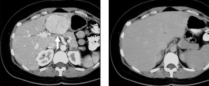
Compare the same liver region across phases: the FNH stands out on the left late-arterial image but approaches background enhancement on the right portal-venous image. These CT phases do not assess hepatobiliary uptake. Image: Kristie Guite, Louis Hinshaw and Fred Lee; source; CC BY 3.0. [5]
For transfer, consider a scar-containing lesion that is heterogeneous and lacks hepatobiliary uptake. A scar alone cannot distinguish FNH from an atypical adenoma or fibrolamellar carcinoma. When the complete pattern is not characteristic, preserve diagnostic uncertainty rather than treating a familiar feature as a final diagnosis. [1]
How much can an adenoma subtype tell you?
Subtype is a risk modifier, not a label that can always be read from MRI. Imaging can strongly support HNF1A-inactivated or inflammatory adenoma, but it cannot reliably establish a specific beta-catenin mutation or exclude well-differentiated carcinoma. Sex and exposure increase suspicion without proving a genotype. [1][2]
HNF1A-inactivated: often diffuse microscopic fat, with loss of liver fatty acid-binding protein in tumor tissue. Malignant potential is comparatively low, not a promise of zero risk.
Inflammatory: associated with obesity and metabolic dysfunction; sinusoidal dilatation can produce a T2-bright peripheral rim, the atoll sign. Enhancement may persist. Tumor serum amyloid A and C-reactive protein staining support the phenotype. Hemorrhage remains possible, and a beta-catenin mutation can coexist.
Beta-catenin activated: exon 3 CTNNB1 mutations are especially associated with malignant transformation. Exon 7 or 8 mutations do not carry the same established malignant risk. Male sex and androgen exposure are important associations, not mutation assays.
Sonic hedgehog and unclassified: sonic hedgehog adenoma has a bleeding association. An unclassified lesion is not automatically low risk; uncertainty and clinical behavior still matter.
Compare two tissue results in a 3.6 cm adenoma: isolated HNF1A inactivation versus proven CTNNB1 exon 3 activation. Predict which result strengthens the indication for resection independent of size. The exon 3 result does, because its malignant potential differs. The low-risk result does not erase other reasons for treatment, such as growth, hemorrhage, or male sex.
Apply that result to a different patient: an inflammatory-appearing adenoma in a man cannot be declared low risk simply because the atoll sign is present. Mixed subtypes exist, and the recommendation for adenoma resection in men does not require MRI proof of beta-catenin activation. Routine biopsy solely to subtype every small adenoma in a woman is not recommended; reserve tissue assessment for questions that affect care. [1]
Which risk changes the plan now?
First separate an emergency from an elective decision. Sudden pain, hypotension, anemia, and hemoperitoneum in a patient with an adenoma indicate possible rupture. Resuscitate and control bleeding; when arterial bleeding persists and expertise is available, transarterial embolization is an important immediate treatment. Surgery remains necessary in selected situations. Definitive tumor assessment follows stabilization rather than delaying it. [1][2]
Position explains the consequence of rupture. Larger size, growth, and exophytic location increase concern; internal lesions can also bleed. This schematic is not a quantitative risk calculator. Original Bone Wizardry construction. [1] Open the image for a larger view.
Trace the surface lesion into the surrounding cavity in the drawing. Predict the physiologic change if enough blood escapes: circulating volume falls and shock can develop. A new situation then follows successful embolization: the patient is stable, but follow-up imaging shows viable residual adenoma. Hemostasis is not eradication; resection is generally indicated in an operative candidate with residual viable lesion after bleeding. [1][2]
For a stable patient, use identity, trajectory, and context
In a woman with an uncomplicated adenoma below 5 cm and no high-risk features, stop estrogen-containing contraception, review other hormonal exposures, discontinue anabolic steroids, and support weight loss when appropriate. Arrange alternative contraception and exposure decisions with the treating team. ACG advises contrast-enhanced imaging every six months for two years, then annually. A small lesion still needs follow-up; there is no established universal stopping point. [1]
For a larger adenoma in a woman, a period of risk-factor modification and reassessment is reasonable if there is no bleeding or concern for malignancy. EASL specifies six months; ACG allows an observation interval of six to twelve months in appropriate patients. A lesion that remains at least 5 cm, continues to grow, or develops suspicious features warrants treatment assessment, usually resection when feasible. Suspicion of malignant transformation should not wait through a routine observation interval. [1][2]
Adenomas in men are generally resected regardless of size. Proven high-risk beta-catenin activation, especially exon 3, also favors resection independent of size. Withdrawal of the growth stimulus remains appropriate but does not replace these recommendations. Embolization or ablation can be considered when treatment is indicated but surgery is unsafe. Multiple adenomas are assessed by the dominant or otherwise high-risk lesions rather than their number alone. [1][2]
Compare a woman's lesion shrinking from 6.3 to 5.6 cm over twelve months with a man's lesion shrinking from 2.7 to 2.3 cm. Both shrink, but neither trend automatically ends the treatment discussion: the first remains above the size threshold, and the second retains the male-sex indication. The same measurements in a secure hemangioma would not justify importing adenoma rules.
Discuss pregnancy before conception. A persistent adenoma at least 5 cm generally merits treatment beforehand. Smaller stable nonexophytic adenomas can often be monitored during pregnancy with coordinated hepatology and obstetric care; ACG describes ultrasound every six to twelve weeks as one strategy. Growth, bleeding, or high-risk morphology changes the plan. These are individualized decisions, not a universal prohibition on pregnancy. [1]
Finally, return to the two common conservative diagnoses. Typical FNH generally requires neither routine follow-up nor routine contraceptive withdrawal; pain may have another cause and may not improve with resection. A secure asymptomatic hemangioma in a low-risk liver also generally needs no surveillance, even when large. Cirrhosis, hepatitis B, unresolved imaging uncertainty, or attributable complications require a different assessment. Five centimeters is an adenoma decision threshold, not a universal liver-mass rule.[1]
Apply the reasoning to new patients
Case 1
Show answer and explanations for case 1
A. Obtain a core biopsy to exclude adenoma (Why this does not fit)
Discontinuous peripheral nodules that fill inward and match the blood pool, together with very high T2 signal, establish a characteristic hemangioma pattern. There is no unresolved imaging question requiring tissue here; biopsy would expose blood-filled vascular spaces to avoidable bleeding risk.
Reasoning steps for option A
Which enhancement feature distinguishes this 2.8 cm mass from an adenoma before biopsy?
Discontinuous peripheral nodules that fill inward and match the blood pool, together with very high T2 signal, establish a characteristic hemangioma pattern.
What diagnostic benefit would a core biopsy add to this securely characterized vascular lesion?
There is no unresolved imaging question requiring tissue here; biopsy would expose blood-filled vascular spaces to avoidable bleeding risk.
B. Arrange resection because the lesion is hypervascular (Why this does not fit)
Arterial brightness describes vascularity; the subsequent inward blood-pool filling identifies a typical hemangioma rather than a surgical indication. She has no liver-related symptoms or reported complication, so hypervascularity alone does not justify resection.
Reasoning steps for option B
Does the arterial brightness of this asymptomatic mass establish a need for resection?
Arterial brightness describes vascularity; the subsequent inward blood-pool filling identifies a typical hemangioma rather than a surgical indication.
Which feature of this patient argues against operating on the characterized hemangioma?
She has no liver-related symptoms or reported complication, so hypervascularity alone does not justify resection.
C. Reassure without routine lesion surveillance (Best answer)
Cavernous hemangioma accounts for the combination of very high T2 signal and progressive enhancement that follows the blood pool. A secure diagnosis in this low-risk, asymptomatic patient permits reassurance without routine lesion surveillance or intervention.
Reasoning steps for option C
What lesion accounts for the very high T2 signal and inward expansion of peripheral enhancing nodules?
Cavernous hemangioma accounts for the combination of very high T2 signal and progressive enhancement that follows the blood pool.
How do the absent cancer history, absent chronic liver disease, and lack of symptoms affect follow-up of this hemangioma?
A secure diagnosis in this low-risk, asymptomatic patient permits reassurance without routine lesion surveillance or intervention.
D. Stop estrogen and repeat MRI in six months (Why this does not fit)
Estrogen withdrawal and scheduled reassessment address the growth risk of an appropriately selected hepatocellular adenoma. The peripheral-to-central vascular filling identifies hemangioma, for which routine estrogen cessation and six-month MRI are not required after secure diagnosis in this setting.
Reasoning steps for option D
For which diagnosis would estrogen withdrawal with six-month MRI fit this proposed management plan?
Estrogen withdrawal and scheduled reassessment address the growth risk of an appropriately selected hepatocellular adenoma.
Why does the blood-pool filling in this case argue against that adenoma strategy?
The peripheral-to-central vascular filling identifies hemangioma, for which routine estrogen cessation and six-month MRI are not required after secure diagnosis in this setting.
Takeaway: A secure vascular pattern can prevent an unnecessary biopsy.
A. Continue appropriate contraception and end routine lesion-specific imaging follow-up (Best answer)
Uniform arterial enhancement, delayed scar enhancement, and homogeneous hepatobiliary uptake support the expert-confirmed FNH diagnosis without a need for biopsy. Secure asymptomatic FNH without vascular liver disease requires neither routine lesion surveillance nor routine contraceptive withdrawal; other contraceptive contraindications still matter.
Reasoning steps for option A
How does the combined arterial, scar, and hepatobiliary pattern establish the basis for ending lesion follow-up?
Uniform arterial enhancement, delayed scar enhancement, and homogeneous hepatobiliary uptake support the expert-confirmed FNH diagnosis without a need for biopsy.
Does this asymptomatic 4.3 cm FNH require stopping an otherwise appropriate contraceptive?
Secure asymptomatic FNH without vascular liver disease requires neither routine lesion surveillance nor routine contraceptive withdrawal; other contraceptive contraindications still matter.
B. Discontinue contraception and obtain MRI every six months (Why this does not fit)
That combination is used for many hepatocellular adenomas because hormonal exposure can promote growth and imaging measures the response. Contraceptive use does not override the complete, expert-confirmed FNH pattern, so the adenoma withdrawal-and-surveillance plan should not be applied here.
Reasoning steps for option B
What diagnosis would justify combining hormone withdrawal with regular MRI in a contraceptive user?
That combination is used for many hepatocellular adenomas because hormonal exposure can promote growth and imaging measures the response.
Does taking a combined contraceptive change the established identity of this 4.3 cm lesion?
Contraceptive use does not override the complete, expert-confirmed FNH pattern, so the adenoma withdrawal-and-surveillance plan should not be applied here.
C. Resect the lesion before it reaches 5 cm (Why this does not fit)
The 5 cm threshold applies to selected adenomas, not to every benign liver lesion. This lesion is asymptomatic FNH, so its 4.3 cm diameter does not create an indication to operate before it reaches an adenoma threshold.
Reasoning steps for option C
Does the proposed 5 cm resection threshold belong to FNH or to selected adenomas?
The 5 cm threshold applies to selected adenomas, not to every benign liver lesion.
Does the confirmed FNH diagnosis justify preventive resection as this lesion approaches 5 cm?
This lesion is asymptomatic FNH, so its 4.3 cm diameter does not create an indication to operate before it reaches an adenoma threshold.
D. Obtain core biopsy before allowing continued contraception (Why this does not fit)
Biopsy can be useful when imaging leaves a management-changing diagnosis uncertain, but expert review has already established typical FNH here. Contraception does not create a biopsy indication in securely diagnosed FNH; routine tissue confirmation is unnecessary before continuing an otherwise appropriate method.
Reasoning steps for option D
What uncertainty would core biopsy need to resolve before changing management of this scar-containing lesion?
Biopsy can be useful when imaging leaves a management-changing diagnosis uncertain, but expert review has already established typical FNH here.
Does continued contraceptive use create a separate tissue-sampling requirement for confirmed FNH?
Contraception does not create a biopsy indication in securely diagnosed FNH; routine tissue confirmation is unnecessary before continuing an otherwise appropriate method.
Takeaway: Management follows lesion identity, not just size or contraceptive use.
A. Estrogen withdrawal followed by elective surgical resection (Why this does not fit)
The lesion is below 5 cm and has no demonstrated growth, hemorrhage, or suspicious internal nodule, supporting an initial conservative approach. Weight-loss support and scheduled imaging can assess the response to risk-factor modification; resection is reserved for treatment indications rather than automatically following withdrawal.
Reasoning steps for option A
Which findings make immediate elective resection unnecessary for this woman with a 4.1 cm adenoma?
The lesion is below 5 cm and has no demonstrated growth, hemorrhage, or suspicious internal nodule, supporting an initial conservative approach.
What should follow estrogen withdrawal before surgery is selected for this uncomplicated lesion?
Weight-loss support and scheduled imaging can assess the response to risk-factor modification; resection is reserved for treatment indications rather than automatically following withdrawal.
B. Continued estrogen-containing contraception with six-month MRI surveillance (Why this does not fit)
MRI can detect growth or regression, but surveillance does not remove the estrogen growth stimulus. Her contraceptive contains estrogen, a modifiable driver of adenoma growth, so imaging should accompany withdrawal rather than substitute for it.
Reasoning steps for option B
What does six-month MRI measure in this estrogen-exposed adenoma, and what does it leave unchanged?
MRI can detect growth or regression, but surveillance does not remove the estrogen growth stimulus.
Why is continuing her combined contraceptive an incomplete plan despite arranging surveillance?
Her contraceptive contains estrogen, a modifiable driver of adenoma growth, so imaging should accompany withdrawal rather than substitute for it.
C. Core biopsy followed by a subtype-specific management plan (Why this does not fit)
Tissue testing is useful for selected unresolved or higher-risk lesions, but MRI has confidently characterized this stable adenoma without suspicious features. Routine biopsy solely for subtype selection is not recommended for this otherwise low-risk presentation; exposure modification and surveillance can begin without it.
Reasoning steps for option C
What management-changing uncertainty would justify subtype biopsy in this 4.1 cm adenoma?
Tissue testing is useful for selected unresolved or higher-risk lesions, but MRI has confidently characterized this stable adenoma without suspicious features.
Must every small adenoma in a woman be assigned a tissue subtype before conservative management?
Routine biopsy solely for subtype selection is not recommended for this otherwise low-risk presentation; exposure modification and surveillance can begin without it.
D. Stop estrogen exposure, provide weight-loss support, and repeat liver MRI in six months (Best answer)
Her estrogen-containing contraceptive and obesity identify targets for estrogen withdrawal and supported weight loss. Regression is not guaranteed after risk-factor modification, so six-month MRI checks the response while preserving surveillance for a lesion initially suitable for observation.
Reasoning steps for option D
Which two modifiable growth drivers are present in this woman with an uncomplicated adenoma?
Her estrogen-containing contraceptive and obesity identify targets for estrogen withdrawal and supported weight loss.
Why is MRI in six months needed after those changes for a stable 4.1 cm lesion?
Regression is not guaranteed after risk-factor modification, so six-month MRI checks the response while preserving surveillance for a lesion initially suitable for observation.
Takeaway: Risk-factor modification is followed by measurement, not an assumption of cure.
A. Continue surveillance after androgen withdrawal (Why this does not fit)
The decrease is an encouraging response after removal of an androgen exposure, but it does not establish that the residual adenoma is risk-free. Adenomas in men warrant resection regardless of size or apparent regression; androgen withdrawal does not remove that treatment indication.
Reasoning steps for option A
What does shrinkage from 2.7 to 2.3 cm after steroid withdrawal show about this adenoma?
The decrease is an encouraging response after removal of an androgen exposure, but it does not establish that the residual adenoma is risk-free.
Why does regression not justify surveillance alone for this male operative candidate?
Adenomas in men warrant resection regardless of size or apparent regression; androgen withdrawal does not remove that treatment indication.
B. Refer for surgical resection (Best answer)
Male sex retains the indication for resection even though the lesion has decreased to 2.3 cm. He can undergo the preferred definitive treatment, surgical resection; continued avoidance of anabolic steroids remains appropriate but does not replace surgery.
Reasoning steps for option B
Which patient characteristic outweighs the favorable size trend when choosing treatment for this adenoma?
Male sex retains the indication for resection even though the lesion has decreased to 2.3 cm.
How do normal hepatic reserve and operative fitness affect the choice after androgen withdrawal?
He can undergo the preferred definitive treatment, surgical resection; continued avoidance of anabolic steroids remains appropriate but does not replace surgery.
C. Biopsy and observe if a fatty subtype is found (Why this does not fit)
A fatty HNF1A-inactivated phenotype can indicate comparatively lower biological risk, but it does not negate the recommendation to resect adenomas in men. Subtype-based observation would still leave the male-sex indication unaddressed; routine preoperative biopsy is unnecessary when proceeding to resection.
Reasoning steps for option C
What reassurance could a fatty adenoma subtype provide, and why would it be incomplete in this man?
A fatty HNF1A-inactivated phenotype can indicate comparatively lower biological risk, but it does not negate the recommendation to resect adenomas in men.
Would a low-risk subtype result justify observing this shrinking lesion instead of resection?
Subtype-based observation would still leave the male-sex indication unaddressed; routine preoperative biopsy is unnecessary when proceeding to resection.
D. Choose ablation instead of resection (Why this does not fit)
Ablation can be an alternative when surgery is unsafe or unsuitable, rather than the preferred replacement for feasible resection. Normal hepatic reserve and operative fitness support resection, so the case supplies no surgical limitation that favors choosing ablation instead.
Reasoning steps for option D
What surgical limitation would favor ablation for this man's adenoma?
Ablation can be an alternative when surgery is unsafe or unsuitable, rather than the preferred replacement for feasible resection.
Which details make that alternative less appropriate for this man with a 2.3 cm adenoma?
Normal hepatic reserve and operative fitness support resection, so the case supplies no surgical limitation that favors choosing ablation instead.
Takeaway: Small size and regression do not cancel the adenoma recommendation for men.
A. Urgent selective transarterial embolization (Best answer)
These findings identify hemorrhagic shock from active bleeding after adenoma rupture, not an elective characterization problem. Urgent selective transarterial embolization can control the bleeding while resuscitation continues, allowing definitive tumor management to be planned after stabilization.
Reasoning steps for option A
What do blood pressure of 82/48 mmHg, hemoperitoneum, and arterial extravasation establish?
These findings identify hemorrhagic shock from active bleeding after adenoma rupture, not an elective characterization problem.
How can immediately available interventional radiology address the demonstrated arterial bleeding?
Urgent selective transarterial embolization can control the bleeding while resuscitation continues, allowing definitive tumor management to be planned after stabilization.
B. MRI with hepatobiliary contrast before hemostasis (Why this does not fit)
MRI could refine tumor characterization, but CT has already identified active arterial bleeding into the peritoneal cavity. Further diagnostic imaging would delay control of the hemorrhage causing shock; stabilization and bleeding control take priority over subtype assessment.
Reasoning steps for option B
What could hepatobiliary MRI clarify that the emergency CT has not already answered?
MRI could refine tumor characterization, but CT has already identified active arterial bleeding into the peritoneal cavity.
Why should that characterization not precede hemostasis in a patient with syncope and severe hypotension?
Further diagnostic imaging would delay control of the hemorrhage causing shock; stabilization and bleeding control take priority over subtype assessment.
C. Core biopsy to exclude carcinoma before treatment (Why this does not fit)
A biopsy could investigate a later concern for carcinoma, but it would not stop the arterial hemorrhage producing shock. Sampling adds procedural and bleeding risk during an ongoing hemorrhagic emergency, so it should not delay resuscitation and hemostasis.
Reasoning steps for option C
Would core biopsy address the immediate cause of hypotension in this ruptured adenoma?
A biopsy could investigate a later concern for carcinoma, but it would not stop the arterial hemorrhage producing shock.
How does active bleeding change the risk-benefit balance of sampling this lesion before treatment?
Sampling adds procedural and bleeding risk during an ongoing hemorrhagic emergency, so it should not delay resuscitation and hemostasis.
D. Repeat CT after a six-hour observation period (Why this does not fit)
Observation can be considered in selected patients who are stable after bleeding has stopped, not in a patient with shock and demonstrated active leakage. Active arterial extravasation and blood pressure of 82/48 mmHg show a need for immediate hemostatic treatment rather than delayed repeat CT.
Reasoning steps for option D
What would need to change before observation could be considered for this hypotensive patient with active adenoma bleeding?
Observation can be considered in selected patients who are stable after bleeding has stopped, not in a patient with shock and demonstrated active leakage.
Which CT and clinical findings make a six-hour delay unsafe here?
Active arterial extravasation and blood pressure of 82/48 mmHg show a need for immediate hemostatic treatment rather than delayed repeat CT.
Takeaway: Control rupture-related bleeding before refining tumor classification.
A. Continue annual surveillance after partial regression (Why this does not fit)
The lesion has partially regressed but remains above 5 cm after twelve months of estrogen withdrawal and weight management. Persistent size above the threshold after adequate observation still warrants definitive treatment assessment; shrinkage alone does not resolve the large-lesion risk.
Reasoning steps for option A
Does the change from 6.3 to 5.8 to 5.6 cm establish regression below the treatment threshold?
The lesion has partially regressed but remains above 5 cm after twelve months of estrogen withdrawal and weight management.
Why is annual surveillance alone insufficient despite the favorable direction of change?
Persistent size above the threshold after adequate observation still warrants definitive treatment assessment; shrinkage alone does not resolve the large-lesion risk.
B. Biopsy and observe if an HNF1A subtype is found (Why this does not fit)
HNF1A inactivation is associated with comparatively lower malignant potential, so it can modify the assessment of transformation risk. A lower-risk subtype would not eliminate size-associated hemorrhage concerns or the indication to treat a persistent large adenoma after observation.
Reasoning steps for option B
Which risk of this 5.6 cm adenoma could an HNF1A-inactivated result make less concerning?
HNF1A inactivation is associated with comparatively lower malignant potential, so it can modify the assessment of transformation risk.
Would that result remove the treatment indication for a lesion still measuring 5.6 cm after a year?
A lower-risk subtype would not eliminate size-associated hemorrhage concerns or the indication to treat a persistent large adenoma after observation.
C. Offer elective surgical resection (Best answer)
The final diameter is 5.6 cm, so the adenoma remains at least 5 cm despite a substantial observation period and partial regression. Persistent size above the threshold supports elective surgical resection in this operative candidate, even without hemorrhage or a suspicious internal nodule.
Reasoning steps for option C
How should the twelve-month diameter be compared with the 5 cm threshold after risk-factor modification?
The final diameter is 5.6 cm, so the adenoma remains at least 5 cm despite a substantial observation period and partial regression.
Why does operative fitness favor elective resection rather than another routine surveillance interval?
Persistent size above the threshold supports elective surgical resection in this operative candidate, even without hemorrhage or a suspicious internal nodule.
D. Substitute serial AFP testing for imaging (Why this does not fit)
AFP can contribute to a malignancy evaluation, but it does not measure the adenoma diameter or its response to risk-factor modification. AFP cannot assess size-associated hemorrhage risk, so serial marker testing is not a substitute for imaging and management of this persistent large adenoma.
Reasoning steps for option D
What does AFP testing assess that differs from the serial MRI measurements in this woman?
AFP can contribute to a malignancy evaluation, but it does not measure the adenoma diameter or its response to risk-factor modification.
Can AFP replace imaging or treatment assessment for the persistent 5.6 cm lesion?
AFP cannot assess size-associated hemorrhage risk, so serial marker testing is not a substitute for imaging and management of this persistent large adenoma.
Takeaway: Partial regression does not satisfy a below-5-cm management target.
A. Continue the same surveillance plan until 5 cm (Why this does not fit)
Adenomas below 5 cm may be observed when stable or regressing, but this lesion has increased from 3.0 to 4.1 cm despite estrogen withdrawal. The increase is real rather than a measurement-plane artifact, so continued substantial growth independently warrants treatment reassessment instead of waiting for a size threshold.
Reasoning steps for option A
Does the current 4.1 cm diameter alone make continued observation appropriate?
Adenomas below 5 cm may be observed when stable or regressing, but this lesion has increased from 3.0 to 4.1 cm despite estrogen withdrawal.
Why does confirmed growth on comparable imaging matter before the lesion reaches 5 cm?
The increase is real rather than a measurement-plane artifact, so continued substantial growth independently warrants treatment reassessment instead of waiting for a size threshold.
B. Refer for consideration of definitive treatment (Best answer)
The confirmed increase from 3.0 to 4.1 cm shows continued growth despite removal of a recognized growth stimulus. Specialist assessment for definitive treatment is appropriate on the basis of growth, even below 5 cm; the absence of bleeding permits an elective rather than emergency assessment.
Reasoning steps for option B
What does growth eight months after estrogen withdrawal indicate about the response to conservative care?
The confirmed increase from 3.0 to 4.1 cm shows continued growth despite removal of a recognized growth stimulus.
How should that trajectory change the plan while the patient has no acute bleeding?
Specialist assessment for definitive treatment is appropriate on the basis of growth, even below 5 cm; the absence of bleeding permits an elective rather than emergency assessment.
C. Arrange urgent transarterial embolization (Why this does not fit)
Urgent embolization can control ongoing arterial hemorrhage, whereas growth alone does not establish an emergency hemostatic indication. The case explicitly states that there is no acute bleeding, so the next step is elective treatment reassessment rather than urgent arterial hemostasis.
Reasoning steps for option C
Which acute complication would make embolization urgent in a growing adenoma?
Urgent embolization can control ongoing arterial hemorrhage, whereas growth alone does not establish an emergency hemostatic indication.
Does the increase to 4.1 cm supply evidence of the hemorrhage needed for that urgent action?
The case explicitly states that there is no acute bleeding, so the next step is elective treatment reassessment rather than urgent arterial hemostasis.
D. Obtain routine subtype biopsy before any treatment referral (Why this does not fit)
Confirmed enlargement despite eight months without estrogen is a treatment-assessment indication even before a molecular subtype is known. Biopsy can address a specific management-changing uncertainty, but routine subtype sampling is not a prerequisite to referral and should not delay that assessment.
Reasoning steps for option D
Has the growth already supplied a reason for referral without a tissue subtype?
Confirmed enlargement despite eight months without estrogen is a treatment-assessment indication even before a molecular subtype is known.
What role could biopsy have after specialist review of this growing lesion?
Biopsy can address a specific management-changing uncertainty, but routine subtype sampling is not a prerequisite to referral and should not delay that assessment.
Takeaway: Trajectory matters independently of the current diameter.
A. Follow with annual ultrasound until 5 cm (Why this does not fit)
Small size can support surveillance of a lower-risk adenoma, but this biopsy proves a CTNNB1 exon 3 activating mutation. The mutation confers important malignant-transformation risk independent of size, making annual ultrasound until 5 cm an insufficient plan.
Reasoning steps for option A
Why might the 3.6 cm diameter initially favor observation, and what tissue result changes that assessment?
Small size can support surveillance of a lower-risk adenoma, but this biopsy proves a CTNNB1 exon 3 activating mutation.
Does remaining below 5 cm neutralize the risk carried by proven exon 3 activation?
The mutation confers important malignant-transformation risk independent of size, making annual ultrasound until 5 cm an insufficient plan.
B. Use embolization as first-line definitive therapy (Why this does not fit)
Embolization can be considered when indicated surgery is not feasible, rather than replacing preferred resection in an operative candidate. She is fit for surgery, so resection is preferred for her high-risk subtype instead of first-line definitive embolization.
Reasoning steps for option B
What would favor embolization over resection for this woman's biopsy-proven high-risk adenoma?
Embolization can be considered when indicated surgery is not feasible, rather than replacing preferred resection in an operative candidate.
Does this woman with proven exon 3 activation have the surgical limitation that would favor embolization first?
She is fit for surgery, so resection is preferred for her high-risk subtype instead of first-line definitive embolization.
C. Withdraw hormonal exposure and repeat MRI in six months (Why this does not fit)
Stopping estrogen would remove a possible growth stimulus, but hormonal exposure is not stated and withdrawal would not erase the proven molecular risk. CTNNB1 exon 3 activation supports definitive treatment regardless of small size, so exposure modification and delayed reassessment alone do not address the important long-term risk.
Reasoning steps for option C
What could hormonal withdrawal address in this biopsy-proven adenoma if exposure is present?
Stopping estrogen would remove a possible growth stimulus, but hormonal exposure is not stated and withdrawal would not erase the proven molecular risk.
Why is waiting six months for MRI inadequate as the sole response to the exon 3 mutation?
CTNNB1 exon 3 activation supports definitive treatment regardless of small size, so exposure modification and delayed reassessment alone do not address the important long-term risk.
D. Offer elective surgical resection (Best answer)
The biopsy-proven CTNNB1 exon 3 activating mutation identifies a high-risk adenoma subtype that supports resection regardless of size. The indication addresses malignant potential, not proof of existing carcinoma; operative fitness makes elective surgical resection the preferred plan.
Reasoning steps for option D
Which feature establishes a treatment indication despite the 3.6 cm diameter?
The biopsy-proven CTNNB1 exon 3 activating mutation identifies a high-risk adenoma subtype that supports resection regardless of size.
Does offering resection mean that the biopsy has already demonstrated carcinoma?
The indication addresses malignant potential, not proof of existing carcinoma; operative fitness makes elective surgical resection the preferred plan.
Takeaway: A proven high-risk subtype can outweigh an otherwise favorable size.
A. Diffuse serum amyloid A expression with preserved liver fatty acid-binding protein (Why this does not fit)
That tissue combination supports an inflammatory adenoma rather than the HNF1A-inactivated phenotype. Diffuse opposed-phase signal loss indicates microscopic fat and favors HNF1A inactivation; a T2-bright rim with persistent enhancement is not the dominant pattern supplied.
Reasoning steps for option A
Which subtype would the proposed serum amyloid A expression with preserved liver fatty acid-binding protein identify in this lesion?
That tissue combination supports an inflammatory adenoma rather than the HNF1A-inactivated phenotype.
Which supplied MRI clue makes the inflammatory result less likely than loss of liver fatty acid-binding protein?
Diffuse opposed-phase signal loss indicates microscopic fat and favors HNF1A inactivation; a T2-bright rim with persistent enhancement is not the dominant pattern supplied.
B. Nuclear beta-catenin staining with an exon 3 mutation (Why this does not fit)
Chemical-shift signal loss indicates microscopic fat, not a mutation assay, so it cannot establish exon 3 activation. The diffuse fatty pattern favors HNF1A-inactivated adenoma; nuclear beta-catenin with exon 3 activation would identify a different, higher-risk subtype not established by these images.
Reasoning steps for option B
Would diffuse opposed-phase signal loss establish a CTNNB1 exon 3 mutation?
Chemical-shift signal loss indicates microscopic fat, not a mutation assay, so it cannot establish exon 3 activation.
Why is nuclear beta-catenin with an exon 3 mutation less likely than the HNF1A-associated tissue result here?
The diffuse fatty pattern favors HNF1A-inactivated adenoma; nuclear beta-catenin with exon 3 activation would identify a different, higher-risk subtype not established by these images.
C. Loss of liver fatty acid-binding protein in tumor hepatocytes (Best answer)
Microscopic intracellular fat accounts for the diffuse signal loss when fat and water signals cancel on opposed-phase imaging. The pattern favors HNF1A-inactivated adenoma, whose tumor hepatocytes characteristically lose liver fatty acid-binding protein expression.
Reasoning steps for option C
What tissue component causes this lesion to lose signal on opposed-phase compared with in-phase imaging?
Microscopic intracellular fat accounts for the diffuse signal loss when fat and water signals cancel on opposed-phase imaging.
Which protein loss is expected if tissue confirms the adenoma subtype favored by that fatty pattern?
The pattern favors HNF1A-inactivated adenoma, whose tumor hepatocytes characteristically lose liver fatty acid-binding protein expression.
D. Endothelial-lined vascular cavities without hepatocellular proliferation (Why this does not fit)
Cavernous hemangioma has that vascular architecture and typically enhances by peripheral nodular filling that follows the blood pool. This lesion lacks the characteristic hemangioma filling pattern and instead contains diffuse microscopic fat, supporting a hepatocellular adenoma rather than a vascular-space lesion.
Reasoning steps for option D
Would endothelial-lined cavities without hepatocellular proliferation identify the same lesion as the supplied fatty MRI pattern?
Cavernous hemangioma has that vascular architecture and typically enhances by peripheral nodular filling that follows the blood pool.
How do the absent nodular filling pattern and diffuse chemical-shift signal loss weigh against those vascular cavities?
This lesion lacks the characteristic hemangioma filling pattern and instead contains diffuse microscopic fat, supporting a hepatocellular adenoma rather than a vascular-space lesion.
Takeaway: Chemical-shift imaging can predict a metabolic tumor phenotype without proving every molecular detail.
A. A coexisting CTNNB1 exon 3 activating mutation (Best answer)
Together these imaging and tissue findings establish an inflammatory adenoma phenotype. Exon 3 beta-catenin activation can coexist with inflammatory features and add substantial malignant-transformation risk; the inflammatory phenotype does not exclude a mixed high-risk subtype.
Reasoning steps for option A
What phenotype is established by the T2-bright rim, persistent enhancement, sinusoidal dilatation, and serum amyloid A staining?
Together these imaging and tissue findings establish an inflammatory adenoma phenotype.
Why would an additional CTNNB1 exon 3 activating mutation change the malignant-risk assessment of that inflammatory lesion?
Exon 3 beta-catenin activation can coexist with inflammatory features and add substantial malignant-transformation risk; the inflammatory phenotype does not exclude a mixed high-risk subtype.
B. Persistence of enhancement into the portal venous phase (Why this does not fit)
It reinforces the inflammatory phenotype already supported by the T2-bright rim, sinusoidal dilatation, and serum amyloid A staining. Persistent enhancement does not establish CTNNB1 exon 3 activation, so it does not add the mutation-specific malignant risk being tested.
Reasoning steps for option B
What does portal venous enhancement persistence add to the already described inflammatory pattern?
It reinforces the inflammatory phenotype already supported by the T2-bright rim, sinusoidal dilatation, and serum amyloid A staining.
Does another demonstration of persistent enhancement prove a high-risk molecular component?
Persistent enhancement does not establish CTNNB1 exon 3 activation, so it does not add the mutation-specific malignant risk being tested.
C. Metabolic dysfunction-associated steatotic liver disease (Why this does not fit)
It would fit the metabolic background commonly associated with inflammatory adenoma rather than identify a new tumor genotype. Background steatotic disease is not equivalent to a CTNNB1 exon 3 mutation in the tumor and does not establish the mixed high-risk subtype.
Reasoning steps for option C
How would metabolic dysfunction-associated steatotic liver disease fit this woman with obesity and an inflammatory adenoma?
It would fit the metabolic background commonly associated with inflammatory adenoma rather than identify a new tumor genotype.
Would background steatosis demonstrate the same malignant-risk change as exon 3 activation within the adenoma?
Background steatotic disease is not equivalent to a CTNNB1 exon 3 mutation in the tumor and does not establish the mixed high-risk subtype.
D. A repeat MRI diameter of 4.3 cm (Why this does not fit)
That small decrease is compatible with stability or slight regression, rather than evidence of continued enlargement. A diameter measurement does not demonstrate a high-risk mutation, so this small decrease does not supply the malignant-risk discriminator requested.
Reasoning steps for option D
How should a repeat diameter of 4.3 cm be interpreted relative to the original 4.4 cm measurement?
That small decrease is compatible with stability or slight regression, rather than evidence of continued enlargement.
Can that size change identify the molecular finding that would most increase transformation concern?
A diameter measurement does not demonstrate a high-risk mutation, so this small decrease does not supply the malignant-risk discriminator requested.
Takeaway: Inflammatory imaging or staining does not exclude a mixed high-risk subtype.
A. Accept a diagnosis of focal nodular hyperplasia (Why this does not fit)
FNH often retains hepatobiliary contrast, but some inflammatory and beta-catenin-activated adenomas also retain it. Growth, heterogeneous arterial enhancement, and lack of a complete typical FNH pattern require further assessment rather than a benign label based on one phase.
Reasoning steps for option A
Is hepatobiliary contrast retention specific enough to establish FNH in this growing heterogeneous lesion?
FNH often retains hepatobiliary contrast, but some inflammatory and beta-catenin-activated adenomas also retain it.
Which features prevent accepting the report based only on late uptake in this man?
Growth, heterogeneous arterial enhancement, and lack of a complete typical FNH pattern require further assessment rather than a benign label based on one phase.
B. Seek multidisciplinary review of the complete liver imaging findings (Best answer)
These findings increase concern for adenoma in a lesion with atypical morphology, while hepatobiliary uptake alone cannot settle its identity. The team should integrate the full imaging pattern and clinical context, then consider planned tissue assessment or definitive treatment if management-changing uncertainty remains.
Reasoning steps for option B
Why do growth and anabolic steroid exposure matter when late contrast uptake suggests FNH?
These findings increase concern for adenoma in a lesion with atypical morphology, while hepatobiliary uptake alone cannot settle its identity.
What should a multidisciplinary review resolve before this 3.8 cm lesion is labeled harmless?
The team should integrate the full imaging pattern and clinical context, then consider planned tissue assessment or definitive treatment if management-changing uncertainty remains.
C. Favor a vascular-space lesion with delayed filling (Why this does not fit)
Hemangioma enhancement reflects contrast in vascular spaces; hepatobiliary uptake reflects hepatocyte-mediated contrast handling rather than delayed filling of those spaces. No characteristic peripheral nodular filling is supplied, so late uptake in this atypical hepatocellular lesion does not justify favoring a hemangioma.
Reasoning steps for option C
How does blood-pool persistence in a hemangioma differ from hepatobiliary uptake in this lesion?
Hemangioma enhancement reflects contrast in vascular spaces; hepatobiliary uptake reflects hepatocyte-mediated contrast handling rather than delayed filling of those spaces.
Does the described enhancement sequence support calling this mass a vascular-space lesion?
No characteristic peripheral nodular filling is supplied, so late uptake in this atypical hepatocellular lesion does not justify favoring a hemangioma.
D. Assign a beta-catenin adenoma subtype from MRI (Why this does not fit)
Male sex, androgen exposure, growth, and possible hepatobiliary retention make that subtype a relevant consideration, not an established diagnosis. MRI cannot reliably provide a mutation-specific diagnosis or exclude all well-differentiated carcinoma, so subtype assignment from these images alone exceeds the evidence.
Reasoning steps for option D
Why is beta-catenin-activated adenoma a possibility in this steroid-exposed man?
Male sex, androgen exposure, growth, and possible hepatobiliary retention make that subtype a relevant consideration, not an established diagnosis.
Can the MRI assign a beta-catenin mutation or exclude well-differentiated carcinoma?
MRI cannot reliably provide a mutation-specific diagnosis or exclude all well-differentiated carcinoma, so subtype assignment from these images alone exceeds the evidence.
Takeaway: Hepatobiliary uptake is evidence about transport, not a certificate of FNH.
A. Necrosis has eliminated perfusion within a solid tumor (Why this does not fit)
Absent Doppler signal alone does not establish tissue necrosis; the sharply bordered echogenic focus with posterior enhancement supports a small hemangioma. The supplied low-risk context and characteristic ultrasound pattern favor hemangioma with flow below Doppler detection, without evidence that a solid tumor has become necrotic.
Reasoning steps for option A
Does absent internal color Doppler flow establish necrosis in this 1.8 cm echogenic focus?
Absent Doppler signal alone does not establish tissue necrosis; the sharply bordered echogenic focus with posterior enhancement supports a small hemangioma.
Why is slow vascular flow a better explanation than loss of perfusion from tumor necrosis?
The supplied low-risk context and characteristic ultrasound pattern favor hemangioma with flow below Doppler detection, without evidence that a solid tumor has become necrotic.
B. Dense fibrous tissue has replaced the vascular spaces (Why this does not fit)
Extensive sclerosis can alter a hemangioma and its imaging appearance, so fibrous replacement is different from ordinary slow flow in cavernous spaces. The described posterior enhancement and typical small hemangioma pattern do not supply evidence of sclerosis; absent color flow alone cannot establish fibrous replacement.
Reasoning steps for option B
What structural change would fibrous replacement propose for this apparently avascular echogenic lesion?
Extensive sclerosis can alter a hemangioma and its imaging appearance, so fibrous replacement is different from ordinary slow flow in cavernous spaces.
Does this small, sharply bordered echogenic lesion provide evidence for that replacement?
The described posterior enhancement and typical small hemangioma pattern do not supply evidence of sclerosis; absent color flow alone cannot establish fibrous replacement.
C. Intracellular lipid has suppressed the Doppler signal (Why this does not fit)
Fat can contribute to echogenicity, but echogenicity and Doppler detection of blood motion measure different properties. Intracellular lipid does not itself suppress the Doppler flow signal; slow flow within cavernous vascular spaces better reconciles the vascular lesion with apparent avascularity.
Reasoning steps for option C
What part of the ultrasound appearance could intracellular lipid influence?
Fat can contribute to echogenicity, but echogenicity and Doppler detection of blood motion measure different properties.
Why does lipid not explain the absent color signal in this otherwise typical hemangioma?
Intracellular lipid does not itself suppress the Doppler flow signal; slow flow within cavernous vascular spaces better reconciles the vascular lesion with apparent avascularity.
D. Slow flow remains below color Doppler detection (Best answer)
Those features support a small cavernous hemangioma in a patient without liver disease or a malignancy history. Blood can move too slowly within its cavernous spaces to produce a detectable color Doppler signal, so apparent avascularity does not mean that blood flow is absent.
Reasoning steps for option D
Which diagnosis fits the 1.8 cm echogenic focus with sharp borders and posterior enhancement in a low-risk liver?
Those features support a small cavernous hemangioma in a patient without liver disease or a malignancy history.
How can that vascular lesion lack detectable internal color Doppler flow?
Blood can move too slowly within its cavernous spaces to produce a detectable color Doppler signal, so apparent avascularity does not mean that blood flow is absent.
Takeaway: Absent Doppler signal does not necessarily mean absent blood flow.
A. Reassure after the ultrasound examination (Why this does not fit)
Echogenicity can occur in hemangioma, but it is not a definitive diagnosis in this oncology context. There is no prior imaging or complete contrast characterization to distinguish a benign lesion from metastasis, so the ultrasound finding cannot end the workup.
Reasoning steps for option A
Does echogenicity establish hemangioma in a patient with newly diagnosed colon cancer?
Echogenicity can occur in hemangioma, but it is not a definitive diagnosis in this oncology context.
What missing information prevents reassurance about this solitary 2.5 cm focus?
There is no prior imaging or complete contrast characterization to distinguish a benign lesion from metastasis, so the ultrasound finding cannot end the workup.
B. Obtain core biopsy before dedicated liver imaging (Why this does not fit)
Dedicated contrast-enhanced liver imaging has not yet characterized the lesion and may establish its identity without tissue sampling. Tissue can be considered if high-quality imaging leaves management-changing uncertainty, but biopsy should not automatically precede dedicated liver imaging.
Reasoning steps for option B
What diagnostic step has not yet been performed before the proposed core biopsy?
Dedicated contrast-enhanced liver imaging has not yet characterized the lesion and may establish its identity without tissue sampling.
When could biopsy become useful for this unresolved colon-cancer staging finding?
Tissue can be considered if high-quality imaging leaves management-changing uncertainty, but biopsy should not automatically precede dedicated liver imaging.
C. Obtain dedicated contrast-enhanced liver MRI (Best answer)
Distinguishing a benign lesion from a liver metastasis could change cancer staging, even though the patient has no liver-related symptoms. MRI can characterize the 2.5 cm focus and assess the remaining liver, providing a timely basis for distinguishing benign disease from metastatic involvement.
Reasoning steps for option C
How does the colon cancer history change the importance of characterizing this echogenic lesion?
Distinguishing a benign lesion from a liver metastasis could change cancer staging, even though the patient has no liver-related symptoms.
What can dedicated contrast-enhanced MRI contribute before a staging conclusion is made?
MRI can characterize the 2.5 cm focus and assess the remaining liver, providing a timely basis for distinguishing benign disease from metastatic involvement.
D. Repeat noncontrast ultrasound in six months (Why this does not fit)
Interval ultrasound could assess change over time, but it would postpone definitive characterization of an unresolved staging lesion. The lesion may affect current cancer staging and needs timely contrast characterization rather than six months of observation based on echogenicity alone.
Reasoning steps for option D
What would a six-month noncontrast ultrasound mainly establish about this currently uncharacterized focus?
Interval ultrasound could assess change over time, but it would postpone definitive characterization of an unresolved staging lesion.
Why is that delay inappropriate in newly diagnosed colon cancer?
The lesion may affect current cancer staging and needs timely contrast characterization rather than six months of observation based on echogenicity alone.
Takeaway: The same echogenic focus requires a different workup in an oncology patient.
A. Repeat lesion imaging in three to six months (Best answer)
His background liver meets a high-risk context for HCC surveillance, while the 1.4 cm lesion has not been definitively established as benign. ACG recommends lesion imaging every three to six months for at least one year, while ongoing indicated HCC surveillance also continues.
Reasoning steps for option A
Why does the term suspected hemangioma matter in a man with hepatitis B cirrhosis?
His background liver meets a high-risk context for HCC surveillance, while the 1.4 cm lesion has not been definitively established as benign.
What interval does ACG recommend for following a suspected hemangioma in that surveillance-eligible setting?
ACG recommends lesion imaging every three to six months for at least one year, while ongoing indicated HCC surveillance also continues.
B. Return only to routine surveillance without lesion-specific follow-up (Why this does not fit)
Background HCC surveillance remains necessary because the surrounding liver retains cancer risk. Its uncertain identity warrants documented short-interval lesion follow-up; returning only to routine surveillance without tracking this focal finding would leave that uncertainty unaddressed.
Reasoning steps for option B
What ongoing surveillance does hepatitis B cirrhosis require even if the focal lesion appears benign?
Background HCC surveillance remains necessary because the surrounding liver retains cancer risk.
Why should the suspected 1.4 cm hemangioma not simply disappear from the follow-up plan?
Its uncertain identity warrants documented short-interval lesion follow-up; returning only to routine surveillance without tracking this focal finding would leave that uncertainty unaddressed.
C. Refer for resection based on the cirrhotic background (Why this does not fit)
Cirrhosis raises HCC risk but does not prove malignancy in a lesion that expert contrast imaging favors as hemangioma. No definite malignant feature or confirmed carcinoma is supplied, so the cirrhotic background alone does not justify resection instead of interval lesion assessment.
Reasoning steps for option C
Does hepatitis B cirrhosis establish that this 1.4 cm lesion is carcinoma?
Cirrhosis raises HCC risk but does not prove malignancy in a lesion that expert contrast imaging favors as hemangioma.
What finding needed to justify immediate cancer-directed resection is not established here?
No definite malignant feature or confirmed carcinoma is supplied, so the cirrhotic background alone does not justify resection instead of interval lesion assessment.
D. Arrange immediate biopsy of the suspected vascular lesion (Why this does not fit)
Sampling a suspected hemangioma creates avoidable bleeding risk when imaging-based follow-up is the appropriate next strategy. The case has no definite malignant feature, so short-interval imaging is preferred here; biopsy remains an option for selected unresolved questions rather than an automatic next step.
Reasoning steps for option D
What concern accompanies immediate biopsy of a lesion still thought to be vascular?
Sampling a suspected hemangioma creates avoidable bleeding risk when imaging-based follow-up is the appropriate next strategy.
Does the expert-reviewed 1.4 cm lesion require routine immediate tissue sampling because the liver is cirrhotic?
The case has no definite malignant feature, so short-interval imaging is preferred here; biopsy remains an option for selected unresolved questions rather than an automatic next step.
Takeaway: A benign-appearing lesion does not cancel surveillance required by the surrounding liver.
A. Manage as FNH and end routine surveillance (Why this does not fit)
A central scar can occur in FNH but cannot settle the diagnosis when the remaining imaging pattern is atypical. Heterogeneous enhancement and a hepatobiliary-phase defect leave adenoma versus fibrolamellar carcinoma unresolved, so benign FNH management is not justified.
Reasoning steps for option A
Can the central scar alone establish FNH in this 7 cm mass?
A central scar can occur in FNH but cannot settle the diagnosis when the remaining imaging pattern is atypical.
Which accompanying MRI findings prevent ending surveillance as though this were secure FNH?
Heterogeneous enhancement and a hepatobiliary-phase defect leave adenoma versus fibrolamellar carcinoma unresolved, so benign FNH management is not justified.
B. Multidisciplinary review and focused tissue evaluation (Best answer)
The radiologist cannot distinguish adenoma from fibrolamellar carcinoma despite characterization, leaving a consequential possibility of malignancy. Multidisciplinary review with planned, targeted tissue evaluation is appropriate to resolve the uncertainty and guide treatment rather than assume a harmless diagnosis.
Reasoning steps for option B
What management-changing differential remains after MRI of this scar-containing mass?
The radiologist cannot distinguish adenoma from fibrolamellar carcinoma despite characterization, leaving a consequential possibility of malignancy.
How should the stable patient proceed when imaging cannot resolve that differential?
Multidisciplinary review with planned, targeted tissue evaluation is appropriate to resolve the uncertainty and guide treatment rather than assume a harmless diagnosis.
C. Withdraw estrogen exposure and reassess in one year (Why this does not fit)
Exposure withdrawal can be part of care for an established adenoma, but this 7 cm lesion still has an unresolved malignant differential. Presumed adenoma surveillance would delay evaluation of possible fibrolamellar carcinoma; the large indeterminate mass needs prompt specialist assessment instead.
Reasoning steps for option C
What diagnosis would need to be supported before a hormone-withdrawal observation strategy could address this mass?
Exposure withdrawal can be part of care for an established adenoma, but this 7 cm lesion still has an unresolved malignant differential.
Why is reassessment in one year unsafe as the only response to that uncertainty?
Presumed adenoma surveillance would delay evaluation of possible fibrolamellar carcinoma; the large indeterminate mass needs prompt specialist assessment instead.
D. Treat as a hemangioma without further characterization (Why this does not fit)
A hemangioma can appear dark on hepatobiliary imaging, but a defect is nonspecific and does not establish a vascular-space lesion. No peripheral nodular enhancement with progressive blood-pool filling is supplied, so the defect cannot justify treating the lesion as a hemangioma without further evaluation.
Reasoning steps for option D
Does a hepatobiliary-phase defect identify this 7 cm mass as hemangioma?
A hemangioma can appear dark on hepatobiliary imaging, but a defect is nonspecific and does not establish a vascular-space lesion.
Which characteristic contrast behavior is missing from the evidence for calling this mass hemangioma?
No peripheral nodular enhancement with progressive blood-pool filling is supplied, so the defect cannot justify treating the lesion as a hemangioma without further evaluation.
Takeaway: A central scar is not sufficient to establish a harmless diagnosis.
A. Use normal AFP to continue the prior surveillance plan (Why this does not fit)
AFP can remain normal in hepatocellular malignancy, so a normal result does not exclude transformation. Growth from 3.5 to 4.6 cm and a new nodule with washout and restricted diffusion require prompt reassessment rather than continuation of the prior plan.
Reasoning steps for option A
Can an AFP result within the reference range exclude malignancy in this changing hepatocellular lesion?
AFP can remain normal in hepatocellular malignancy, so a normal result does not exclude transformation.
Which interval findings outweigh that reassuring blood test when deciding whether to continue surveillance unchanged?
Growth from 3.5 to 4.6 cm and a new nodule with washout and restricted diffusion require prompt reassessment rather than continuation of the prior plan.
B. Defer treatment assessment until the lesion reaches 5 cm (Why this does not fit)
The 5 cm threshold is only one treatment consideration and does not override new suspicious internal architecture. The lesion has grown and developed a nodule with washout and restricted diffusion, which warrants evaluation now rather than waiting to reach 5 cm.
Reasoning steps for option B
Does remaining below 5 cm make the new internal nodule safe to observe until further enlargement?
The 5 cm threshold is only one treatment consideration and does not override new suspicious internal architecture.
What has changed independently of the current 4.6 cm diameter?
The lesion has grown and developed a nodule with washout and restricted diffusion, which warrants evaluation now rather than waiting to reach 5 cm.
C. Assign a beta-catenin genotype from the new MRI findings (Why this does not fit)
Those findings, especially with growth, raise concern for a higher-risk hepatocellular lesion or malignant transformation. They do not establish a CTNNB1 mutation, so assigning a beta-catenin genotype from MRI alone would confuse suspicious behavior with molecular proof.
Reasoning steps for option C
What do washout and restricted diffusion in the new nodule suggest about biological concern?
Those findings, especially with growth, raise concern for a higher-risk hepatocellular lesion or malignant transformation.
Do those MRI findings reveal a specific beta-catenin genotype?
They do not establish a CTNNB1 mutation, so assigning a beta-catenin genotype from MRI alone would confuse suspicious behavior with molecular proof.
D. Promptly assess for malignant transformation (Best answer)
Interval enlargement together with a new internal nodule showing washout and restricted diffusion raises concern for malignant transformation. Normal AFP does not dismiss the imaging concern; prompt specialist assessment, often including tissue evaluation and treatment planning, is warranted without assuming carcinoma is already proven.
Reasoning steps for option D
Which combination raises concern for transformation rather than uncomplicated adenoma growth?
Interval enlargement together with a new internal nodule showing washout and restricted diffusion raises concern for malignant transformation.
How should the normal AFP influence the urgency of assessing those new findings?
Normal AFP does not dismiss the imaging concern; prompt specialist assessment, often including tissue evaluation and treatment planning, is warranted without assuming carcinoma is already proven.
Takeaway: A normal tumor marker cannot overrule new suspicious architecture.
A. Proceed with conception using symptom-triggered imaging (Why this does not fit)
The lesion remains above 5 cm after twelve months without estrogen, so absence of pain does not remove the preconception treatment concern. Pain can signal a complication after it has occurred; a persistent large adenoma should undergo treatment assessment before conception rather than rely on symptomatic rescue.
Reasoning steps for option A
Would absence of symptoms make this persistent 6.1 cm adenoma suitable for symptom-triggered monitoring after conception?
The lesion remains above 5 cm after twelve months without estrogen, so absence of pain does not remove the preconception treatment concern.
What is the problem with waiting for symptoms during a planned pregnancy?
Pain can signal a complication after it has occurred; a persistent large adenoma should undergo treatment assessment before conception rather than rely on symptomatic rescue.
B. Treat the adenoma before planned conception (Best answer)
It remains above 5 cm after an adequate observation period without estrogen, supporting definitive treatment in an operative candidate. Addressing the persistent large lesion beforehand reduces the likelihood of needing intervention during pregnancy, when hormonal stimulation can promote adenoma growth.
Reasoning steps for option B
Has this 6.1 cm adenoma met a treatment criterion before pregnancy begins?
It remains above 5 cm after an adequate observation period without estrogen, supporting definitive treatment in an operative candidate.
Why is treatment before conception preferable to waiting for pregnancy-related intervention?
Addressing the persistent large lesion beforehand reduces the likelihood of needing intervention during pregnancy, when hormonal stimulation can promote adenoma growth.
C. Delay conception until a subtype biopsy supports observation (Why this does not fit)
A low-risk subtype would not eliminate the size-associated hemorrhage concern in this persistent 6.1 cm adenoma. The lesion already has size and pregnancy-related reasons for preconception treatment assessment, so subtyping is not a way to bypass those indications.
Reasoning steps for option C
What concern would remain even if a biopsy identified a comparatively low-risk subtype?
A low-risk subtype would not eliminate the size-associated hemorrhage concern in this persistent 6.1 cm adenoma.
Would a reassuring subtype result justify delaying conception only long enough to establish an observation plan?
The lesion already has size and pregnancy-related reasons for preconception treatment assessment, so subtyping is not a way to bypass those indications.
D. Defer treatment and use ultrasound during pregnancy (Why this does not fit)
Selected smaller, stable adenomas can be monitored during pregnancy, with management adjusted for growth and other risk features. Her adenoma is persistently 6.1 cm after twelve months of observation and she is fit for surgery, favoring treatment before conception rather than deferral to pregnancy surveillance.
Reasoning steps for option D
Is pregnancy ultrasound surveillance primarily suited to persistent large adenomas like this 6.1 cm lesion or to selected smaller stable lesions?
Selected smaller, stable adenomas can be monitored during pregnancy, with management adjusted for growth and other risk features.
Why is that strategy not the preferred substitute for treatment in this woman planning conception?
Her adenoma is persistently 6.1 cm after twelve months of observation and she is fit for surgery, favoring treatment before conception rather than deferral to pregnancy surveillance.
Takeaway: Pregnancy planning is an opportunity to address a high-risk lesion before hormonal stimulation.
A. Resect during the current trimester (Why this does not fit)
The adenoma is 3.1 cm, unchanged, nonexophytic, and without hemorrhage, supporting a monitored pregnancy rather than automatic intervention. Pregnancy requires continued assessment, but these lower-risk findings do not establish a need for preemptive resection; growth, bleeding, or high-risk morphology would change the decision.
Reasoning steps for option A
Which supplied lesion features lower the need for immediate resection at ten weeks of pregnancy?
The adenoma is 3.1 cm, unchanged, nonexophytic, and without hemorrhage, supporting a monitored pregnancy rather than automatic intervention.
Does pregnancy alone create a reason to resect this stable lesion in the current trimester?
Pregnancy requires continued assessment, but these lower-risk findings do not establish a need for preemptive resection; growth, bleeding, or high-risk morphology would change the decision.
B. Wait until postpartum before repeat imaging (Why this does not fit)
A small stable adenoma can still enlarge during pregnancy, so the current scan is not a guarantee of an unchanged course. Interval growth or another evolving lesion risk could go undetected during pregnancy, making scheduled monitoring preferable to postponing all imaging until after delivery.
Reasoning steps for option B
Does the current stability of a 3.1 cm adenoma exclude growth later in pregnancy?
A small stable adenoma can still enlarge during pregnancy, so the current scan is not a guarantee of an unchanged course.
What change could be missed by waiting until postpartum for the next image?
Interval growth or another evolving lesion risk could go undetected during pregnancy, making scheduled monitoring preferable to postponing all imaging until after delivery.
C. Coordinate care with scheduled ultrasound (Best answer)
A stable 3.1 cm nonexophytic adenoma without hemorrhage fits a lower-risk pattern that can be followed during pregnancy instead of automatically treated. Scheduled ultrasound every six to twelve weeks is one described strategy, individualized through coordinated hepatology and obstetric care.
Reasoning steps for option C
Why do the small diameter, internal position, and lack of interval growth support ultrasound monitoring?
A stable 3.1 cm nonexophytic adenoma without hemorrhage fits a lower-risk pattern that can be followed during pregnancy instead of automatically treated.
What monitoring approach does ACG describe for a pregnancy with those lesion features?
Scheduled ultrasound every six to twelve weeks is one described strategy, individualized through coordinated hepatology and obstetric care.
D. Begin routine contrast-enhanced CT surveillance (Why this does not fit)
The lesion can be followed by ultrasound, so serial contrast-enhanced CT is unnecessary for the stated surveillance task. Ultrasound avoids repeated ionizing radiation while monitoring growth; this preference for routine surveillance does not prohibit CT when a separate clinical need justifies it.
Reasoning steps for option D
Does following this sonographically visible, stable adenoma require routine contrast-enhanced CT?
The lesion can be followed by ultrasound, so serial contrast-enhanced CT is unnecessary for the stated surveillance task.
What exposure does ultrasound avoid while still checking the adenoma during pregnancy?
Ultrasound avoids repeated ionizing radiation while monitoring growth; this preference for routine surveillance does not prohibit CT when a separate clinical need justifies it.
Takeaway: Small stable adenomas in pregnancy need a monitoring plan, not automatic surgery or dismissal.
A. Offer resection of the viable residual lesion (Best answer)
Viable adenoma remains despite successful control of the earlier rupture-related hemorrhage. Resection of the viable residual adenoma is appropriate in this operative candidate to address the remaining lesion and recurrent complication risk.
Reasoning steps for option A
What does enhancing tissue within the 4.2 cm residual mass show three months after embolization?
Viable adenoma remains despite successful control of the earlier rupture-related hemorrhage.
How do residual viability and operative fitness determine the next treatment after bleeding has stopped?
Resection of the viable residual adenoma is appropriate in this operative candidate to address the remaining lesion and recurrent complication risk.
B. End lesion follow-up after successful hemostasis (Why this does not fit)
Embolization controlled the bleeding, but enhancement in the residual mass shows that complete tumor eradication has not occurred. Absence of current symptoms does not remove the enhancing viable adenoma, so successful hemostasis is not a reason to end lesion care.
Reasoning steps for option B
Which endpoint did the earlier embolization achieve, and which endpoint has follow-up MRI not shown?
Embolization controlled the bleeding, but enhancement in the residual mass shows that complete tumor eradication has not occurred.
Can follow-up end because the patient is now asymptomatic after hemostasis?
Absence of current symptoms does not remove the enhancing viable adenoma, so successful hemostasis is not a reason to end lesion care.
C. Continue surveillance until the residual mass exceeds 5 cm (Why this does not fit)
Size-based observation of a small uncomplicated adenoma does not fully apply to a lesion that has already ruptured. Prior hemorrhage and persistent viable tissue support definitive treatment now in an operative candidate, even though the residual diameter is below 5 cm.
Reasoning steps for option C
Why might the 4.2 cm size favor observation in an uncomplicated adenoma but not in this case?
Size-based observation of a small uncomplicated adenoma does not fully apply to a lesion that has already ruptured.
Which two facts argue against waiting for the residual mass to exceed 5 cm?
Prior hemorrhage and persistent viable tissue support definitive treatment now in an operative candidate, even though the residual diameter is below 5 cm.
D. Use serial AFP to decide whether viable tissue remains (Why this does not fit)
AFP is not a measure of tumor perfusion or residual viability after embolization. Enhancement within the residual 4.2 cm mass demonstrates viable tissue, so AFP should not replace the MRI finding in treatment planning.
Reasoning steps for option D
Can AFP testing establish whether this embolized mass still contains perfused viable tissue?
AFP is not a measure of tumor perfusion or residual viability after embolization.
Which follow-up finding already provides evidence relevant to that viability question?
Enhancement within the residual 4.2 cm mass demonstrates viable tissue, so AFP should not replace the MRI finding in treatment planning.
Takeaway: Successful hemostasis and definitive tumor treatment are different endpoints.
A. Refer for transplantation because of lesion count (Why this does not fit)
Lesion count alone is not an indication for transplantation, which is reserved for highly selected unresectable or complicated presentations. Hepatic function is normal, no suspicious malignant feature is supplied, and one persistent large lesion drives the risk while the other eleven remain small.
Reasoning steps for option A
Does the presence of twelve bilobar adenomas alone establish a liver-transplant indication?
Lesion count alone is not an indication for transplantation, which is reserved for highly selected unresectable or complicated presentations.
What features favor risk-directed treatment rather than automatic transplantation in this patient?
Hepatic function is normal, no suspicious malignant feature is supplied, and one persistent large lesion drives the risk while the other eleven remain small.
B. Continue surveillance because most lesions are small (Why this does not fit)
Their small size supports their own surveillance, but it does not reduce the risk from a different lesion that remains 6.4 cm. The dominant lesion remains above 5 cm after observation, so it still warrants treatment assessment despite the reassuring majority.
Reasoning steps for option B
Do the eleven stable 1 to 2 cm lesions determine the risk of the remaining adenoma?
Their small size supports their own surveillance, but it does not reduce the risk from a different lesion that remains 6.4 cm.
Why is surveillance of the entire group insufficient after twelve months of hormone withdrawal?
The dominant lesion remains above 5 cm after observation, so it still warrants treatment assessment despite the reassuring majority.
C. Biopsy every lesion before any treatment planning (Why this does not fit)
Adenoma subtypes can differ, but routine sampling of every small stable lesion is not required solely because several lesions coexist. The largest adenoma remains 6.4 cm after twelve months of hormone withdrawal, providing a clear dominant-lesion reason for treatment planning.
Reasoning steps for option C
Does the possibility of different subtypes among twelve adenomas require biopsy of every lesion?
Adenoma subtypes can differ, but routine sampling of every small stable lesion is not required solely because several lesions coexist.
What treatment-relevant finding is already established without universal sampling?
The largest adenoma remains 6.4 cm after twelve months of hormone withdrawal, providing a clear dominant-lesion reason for treatment planning.
D. Treat the dominant high-risk lesion while surveilling the remaining adenomas (Best answer)
The persistent 6.4 cm adenoma is the dominant risk-bearing lesion, unlike the eleven stable lesions measuring 1 to 2 cm. Targeting the dominant lesion while continuing surveillance of the smaller adenomas directs treatment toward the largest or otherwise high-risk lesions rather than the number alone.
Reasoning steps for option D
Which of the twelve lesions should drive intervention after the year of hormone withdrawal?
The persistent 6.4 cm adenoma is the dominant risk-bearing lesion, unlike the eleven stable lesions measuring 1 to 2 cm.
How can the plan address that unequal risk without treating lesion count as the indication?
Targeting the dominant lesion while continuing surveillance of the smaller adenomas directs treatment toward the largest or otherwise high-risk lesions rather than the number alone.
Takeaway: Multiplicity does not replace assessment of the most dangerous lesion.
A. Continue annual imaging as the only plan (Why this does not fit)
Severe pulmonary disease changes treatment feasibility, but it does not remove the concern from an adenoma remaining above 5 cm after observation. The lesion still warrants treatment assessment, so inability to resect should prompt consideration of nonsurgical options instead of treating routine surveillance as the only plan.
Reasoning steps for option A
Does prohibitive pulmonary surgical risk make a persistent 6.2 cm adenoma low risk?
Severe pulmonary disease changes treatment feasibility, but it does not remove the concern from an adenoma remaining above 5 cm after observation.
Why should the team discuss more than annual imaging alone for this lesion?
The lesion still warrants treatment assessment, so inability to resect should prompt consideration of nonsurgical options instead of treating routine surveillance as the only plan.
B. Discuss embolization or ablation with specialists (Best answer)
Embolization or ablation can provide alternatives for a patient who cannot undergo indicated surgical resection. A multidisciplinary team should consider size, anatomy, available expertise, and the possibility of residual viable tissue when selecting and following a nonsurgical treatment.
Reasoning steps for option B
What treatment options remain when this large adenoma warrants therapy but resection is unsafe?
Embolization or ablation can provide alternatives for a patient who cannot undergo indicated surgical resection.
What needs to be assessed before choosing between those options for the 6.2 cm lesion?
A multidisciplinary team should consider size, anatomy, available expertise, and the possibility of residual viable tissue when selecting and following a nonsurgical treatment.
C. Refer for transplantation as the routine alternative (Why this does not fit)
Transplantation is reserved for selected unresectable complicated cases rather than serving as the routine substitute for resection. Locoregional options such as embolization or ablation may address the lesion, so the supplied surgical contraindication does not establish a routine transplant indication.
Reasoning steps for option C
Does inability to resect a solitary adenoma make transplantation the routine next option?
Transplantation is reserved for selected unresectable complicated cases rather than serving as the routine substitute for resection.
What should be considered before transplant referral for this lesion without imaging evidence of carcinoma?
Locoregional options such as embolization or ablation may address the lesion, so the supplied surgical contraindication does not establish a routine transplant indication.
D. Obtain repeated biopsies in place of lesion treatment (Why this does not fit)
Biopsy samples tissue for diagnosis; it does not treat the adenoma or protect against size-associated hemorrhage. The persistent large lesion would remain untreated despite its management indication, so diagnostic sampling cannot substitute for discussing feasible nonsurgical therapy.
Reasoning steps for option D
Would repeated biopsy reduce the hemorrhage concern from this persistent large adenoma?
Biopsy samples tissue for diagnosis; it does not treat the adenoma or protect against size-associated hemorrhage.
What problem remains if repeated sampling replaces treatment planning for the 6.2 cm mass?
The persistent large lesion would remain untreated despite its management indication, so diagnostic sampling cannot substitute for discussing feasible nonsurgical therapy.
Takeaway: A nonsurgical candidate still needs risk-directed lesion care.
A. Observe without preventive intervention (Best answer)
Expert MRI establishes a cavernous hemangioma, and large size alone does not require preventive intervention for that diagnosis. She has no pain, early satiety, hemorrhage, or coagulopathy, so no attributable symptom or complication is supplied to justify treatment.
Reasoning steps for option A
Does the 10.8 cm diameter change the secure blood-pool diagnosis into an automatic treatment indication?
Expert MRI establishes a cavernous hemangioma, and large size alone does not require preventive intervention for that diagnosis.
Which absent clinical problems support observation of this large hemangioma?
She has no pain, early satiety, hemorrhage, or coagulopathy, so no attributable symptom or complication is supplied to justify treatment.
B. Resect because the lesion exceeds 5 cm (Why this does not fit)
The threshold guides treatment of selected hepatocellular adenomas and is not a universal rule for liver masses. This is a securely characterized, asymptomatic hemangioma, so importing an adenoma size criterion would create an unsupported surgical indication.
Reasoning steps for option B
To which lesion does the 5 cm treatment threshold belong rather than to this hemangioma?
The threshold guides treatment of selected hepatocellular adenomas and is not a universal rule for liver masses.
Why does exceeding that threshold not justify resection of this 10.8 cm lesion?
This is a securely characterized, asymptomatic hemangioma, so importing an adenoma size criterion would create an unsupported surgical indication.
C. Embolize prophylactically to reduce rupture risk (Why this does not fit)
Hemangioma rupture is rare, and diameter alone is not an indication for prophylactic embolization. There is no hemorrhage, pain, compression symptom, or coagulopathy, so the case provides no attributable complication that warrants intervention.
Reasoning steps for option C
Does a large hemangioma routinely need embolization to prevent a future rupture?
Hemangioma rupture is rare, and diameter alone is not an indication for prophylactic embolization.
What in this patient argues against preventive embolization despite the 10.8 cm measurement?
There is no hemorrhage, pain, compression symptom, or coagulopathy, so the case provides no attributable complication that warrants intervention.
D. Perform core biopsy before choosing observation (Why this does not fit)
Biopsy can help selected indeterminate lesions, but expert MRI has already established the classic blood-pool pattern of hemangioma. It adds procedural bleeding risk without resolving a stated diagnostic question, so tissue is unnecessary before choosing observation here.
Reasoning steps for option D
What uncertainty would a core biopsy need to resolve before observation of this large lesion?
Biopsy can help selected indeterminate lesions, but expert MRI has already established the classic blood-pool pattern of hemangioma.
What does sampling add when both the vascular identity and absence of complications are secure?
It adds procedural bleeding risk without resolving a stated diagnostic question, so tissue is unnecessary before choosing observation here.
Takeaway: A size threshold belongs to a diagnosis, not to all liver masses.
A. Recommend resection because the lesion exceeds 4 cm (Why this does not fit)
FNH can occasionally cause symptoms, but size above 4 cm does not by itself demonstrate that this lesion is producing the pain. Gallstones provide a competing explanation while the FNH has no mass effect or hemorrhage, so size alone cannot justify resection or guarantee pain relief.
Reasoning steps for option A
Does a 4.6 cm FNH without mass effect establish the cause of right upper quadrant discomfort?
FNH can occasionally cause symptoms, but size above 4 cm does not by itself demonstrate that this lesion is producing the pain.
How do symptomatic gallstones change the argument for resection based on FNH size?
Gallstones provide a competing explanation while the FNH has no mass effect or hemorrhage, so size alone cannot justify resection or guarantee pain relief.
B. Recommend resection to prevent malignant transformation (Why this does not fit)
Secure FNH has no meaningful malignant potential that warrants preventive resection; adenoma risk should not be transferred to it. It would invoke an unsupported cancer-prevention indication and would not establish that removing the FNH will relieve pain attributable to another condition.
Reasoning steps for option B
Does securely diagnosed FNH have the malignant potential that drives preventive resection of some adenomas?
Secure FNH has no meaningful malignant potential that warrants preventive resection; adenoma risk should not be transferred to it.
Would proposing cancer-preventive FNH surgery address this patient's question about pain?
It would invoke an unsupported cancer-prevention indication and would not establish that removing the FNH will relieve pain attributable to another condition.
C. Evaluate whether the pain is attributable to FNH before considering lesion-directed treatment (Best answer)
Symptomatic gallstones are present, while the securely diagnosed FNH has neither hemorrhage nor mass effect. The cause of pain must be assessed because the FNH may be incidental; liver resection may not relieve symptoms arising from gallstones or another source.
Reasoning steps for option C
What competing diagnosis must be considered before assigning the discomfort to FNH?
Symptomatic gallstones are present, while the securely diagnosed FNH has neither hemorrhage nor mass effect.
What should the patient understand before liver resection is considered for symptom relief?
The cause of pain must be assessed because the FNH may be incidental; liver resection may not relieve symptoms arising from gallstones or another source.
D. Try contraceptive withdrawal as the primary pain treatment (Why this does not fit)
Routine contraceptive cessation is not recommended for FNH, unlike exposure modification used in adenoma care. It does not address the supplied symptomatic gallstones or establish FNH as the pain source, so it should not replace assessment of symptom attribution.
Reasoning steps for option D
Does the contraceptive-withdrawal strategy for adenoma apply to this confirmed FNH?
Routine contraceptive cessation is not recommended for FNH, unlike exposure modification used in adenoma care.
Why is hormonal withdrawal not a supported primary treatment for this woman's discomfort?
It does not address the supplied symptomatic gallstones or establish FNH as the pain source, so it should not replace assessment of symptom attribution.
Takeaway: A visible liver lesion is not necessarily the cause of abdominal symptoms.
A. Cavernous endothelial spaces with no hepatocytes (Why this does not fit)
Cavernous vascular spaces explain blood-pool enhancement in a hemangioma rather than the cellular architecture of FNH. They lack the functioning hepatocytes responsible for hepatobiliary contrast handling, so they do not account for the expert-confirmed FNH pattern.
Reasoning steps for option A
Which component of the contrast pattern could cavernous endothelial spaces explain?
Cavernous vascular spaces explain blood-pool enhancement in a hemangioma rather than the cellular architecture of FNH.
Why would spaces containing no hepatocytes fail to explain this lesion's hepatobiliary uptake?
They lack the functioning hepatocytes responsible for hepatobiliary contrast handling, so they do not account for the expert-confirmed FNH pattern.
Hyperplastic hepatocytes associated with abnormal arterial supply, fibrous septa, and ductular reaction form the characteristic FNH architecture underlying brisk arterial enhancement. The functioning hyperplastic hepatocytes take up hepatobiliary contrast through their transport machinery, linking cellular uptake to the same lesion whose abnormal arterial supply produces early enhancement.
Reasoning steps for option B
What FNH architecture explains the uniform arterial enhancement of this 3.0 cm lesion?
Hyperplastic hepatocytes associated with abnormal arterial supply, fibrous septa, and ductular reaction form the characteristic FNH architecture underlying brisk arterial enhancement.
Which part of that architecture explains uptake during the hepatobiliary phase?
The functioning hyperplastic hepatocytes take up hepatobiliary contrast through their transport machinery, linking cellular uptake to the same lesion whose abnormal arterial supply produces early enhancement.
C. Clonal hepatocytes lacking normal portal tracts and bile ducts with diffuse fat (Why this does not fit)
That architecture describes a hepatocellular adenoma pattern, particularly a fatty phenotype, rather than hyperplastic FNH. Some adenomas retain hepatobiliary contrast, but this case establishes typical FNH by expert integrated MRI review, so uptake alone should not replace that diagnosis with adenoma.
Reasoning steps for option C
What diagnosis is suggested by clonal fatty hepatocytes without normal portal tracts or bile ducts?
That architecture describes a hepatocellular adenoma pattern, particularly a fatty phenotype, rather than hyperplastic FNH.
Can occasional hepatobiliary uptake in adenomas override the complete FNH diagnosis stated here?
Some adenomas retain hepatobiliary contrast, but this case establishes typical FNH by expert integrated MRI review, so uptake alone should not replace that diagnosis with adenoma.
D. Malignant glands embedded in desmoplastic stroma (Why this does not fit)
That architecture suggests a biliary or metastatic malignancy rather than a benign hyperplastic hepatocellular lesion. Malignant glands and desmoplastic stroma are not the hyperplastic hepatocytes with altered arterial supply that account for the characteristic FNH pattern in this case.
Reasoning steps for option D
What type of lesion would malignant glands in desmoplastic stroma suggest instead of FNH?
That architecture suggests a biliary or metastatic malignancy rather than a benign hyperplastic hepatocellular lesion.
Why does it fail to explain the expert-confirmed combination of uniform arterial enhancement and hepatobiliary uptake?
Malignant glands and desmoplastic stroma are not the hyperplastic hepatocytes with altered arterial supply that account for the characteristic FNH pattern in this case.
Takeaway: Connect enhancement to both vascular delivery and the cells handling the contrast.
A. Biliary obstruction from compression at the hepatic hilum (Why this does not fit)
Compression at the hepatic hilum could obstruct bile ducts, but the distinguishing location supplied here is projection beyond the liver capsule. A growing 5.8 cm exophytic adenoma raises concern for rupture, while no hilar compression is supplied to support biliary obstruction as the explanation.
Reasoning steps for option A
Does the stated exophytic location supply the hilar compression required for the proposed biliary obstruction?
Compression at the hepatic hilum could obstruct bile ducts, but the distinguishing location supplied here is projection beyond the liver capsule.
Why do the second lesion's size and growth favor a different complication than hilar obstruction?
A growing 5.8 cm exophytic adenoma raises concern for rupture, while no hilar compression is supplied to support biliary obstruction as the explanation.
B. Consumptive coagulopathy within cavernous vascular spaces (Why this does not fit)
Consumptive coagulopathy can complicate a large cavernous hemangioma, whose blood-filled spaces differ from adenoma tissue. Both lesions are adenomas; the larger lesion's growth and exophytic position increase concern for rupture rather than consumption within hemangioma-type vascular spaces.
Reasoning steps for option B
Does consumption within cavernous vascular spaces describe an adenoma complication or a complication of a different lesion?
Consumptive coagulopathy can complicate a large cavernous hemangioma, whose blood-filled spaces differ from adenoma tissue.
Why is that mechanism a poor match for the growing surface lesion in this comparison?
Both lesions are adenomas; the larger lesion's growth and exophytic position increase concern for rupture rather than consumption within hemangioma-type vascular spaces.
C. Portal hypertension from obstruction of the main portal vein (Why this does not fit)
Obstruction of major portal inflow, such as the main portal vein, could produce portal hypertension. No main portal vein involvement is supplied; the stated surface position, growth, and large size instead support concern for hemorrhagic rupture.
Reasoning steps for option C
What portal venous involvement would be required for the proposed complication of this exophytic adenoma?
Obstruction of major portal inflow, such as the main portal vein, could produce portal hypertension.
Does projection beyond the capsule establish that mechanism in the second adenoma?
No main portal vein involvement is supplied; the stated surface position, growth, and large size instead support concern for hemorrhagic rupture.
D. Rupture with hemorrhage into the peritoneal cavity (Best answer)
The second adenoma is larger, has enlarged over six months, and projects beyond the capsule, combining features that increase hemorrhage and rupture concern. A surface-projecting lesion can spill blood into the peritoneal cavity, explaining the greater urgency of addressing the larger growing adenoma.
Reasoning steps for option D
Which differences make the 5.8 cm lesion more concerning than the stable 3.9 cm intraparenchymal lesion?
The second adenoma is larger, has enlarged over six months, and projects beyond the capsule, combining features that increase hemorrhage and rupture concern.
Where can blood escape when an exophytic adenoma ruptures?
A surface-projecting lesion can spill blood into the peritoneal cavity, explaining the greater urgency of addressing the larger growing adenoma.
Takeaway: Size, trajectory, and surface position together identify a hemorrhagic hazard.