Separate bile salts from bilirubin, follow pigment through blood and bowel, and explain how ileal disease changes fat absorption and stone risk.
Bile salts help you absorb fat. Bilirubin is waste pigment. They share a delivery fluid, but a problem with one does not automatically explain the other.
Start with the two different jobs Bile is a fluid made by the liver. It carries bile salts, bilirubin, cholesterol, phospholipids, water, and dissolved ions toward the intestine. A hepatocyte is a liver cell that makes and secretes components of this fluid. A cholangiocyte is a cell lining a bile duct that modifies the fluid as it passes. The gallbladder stores and concentrates bile; it does not replace the liver as its source. [2] [4]
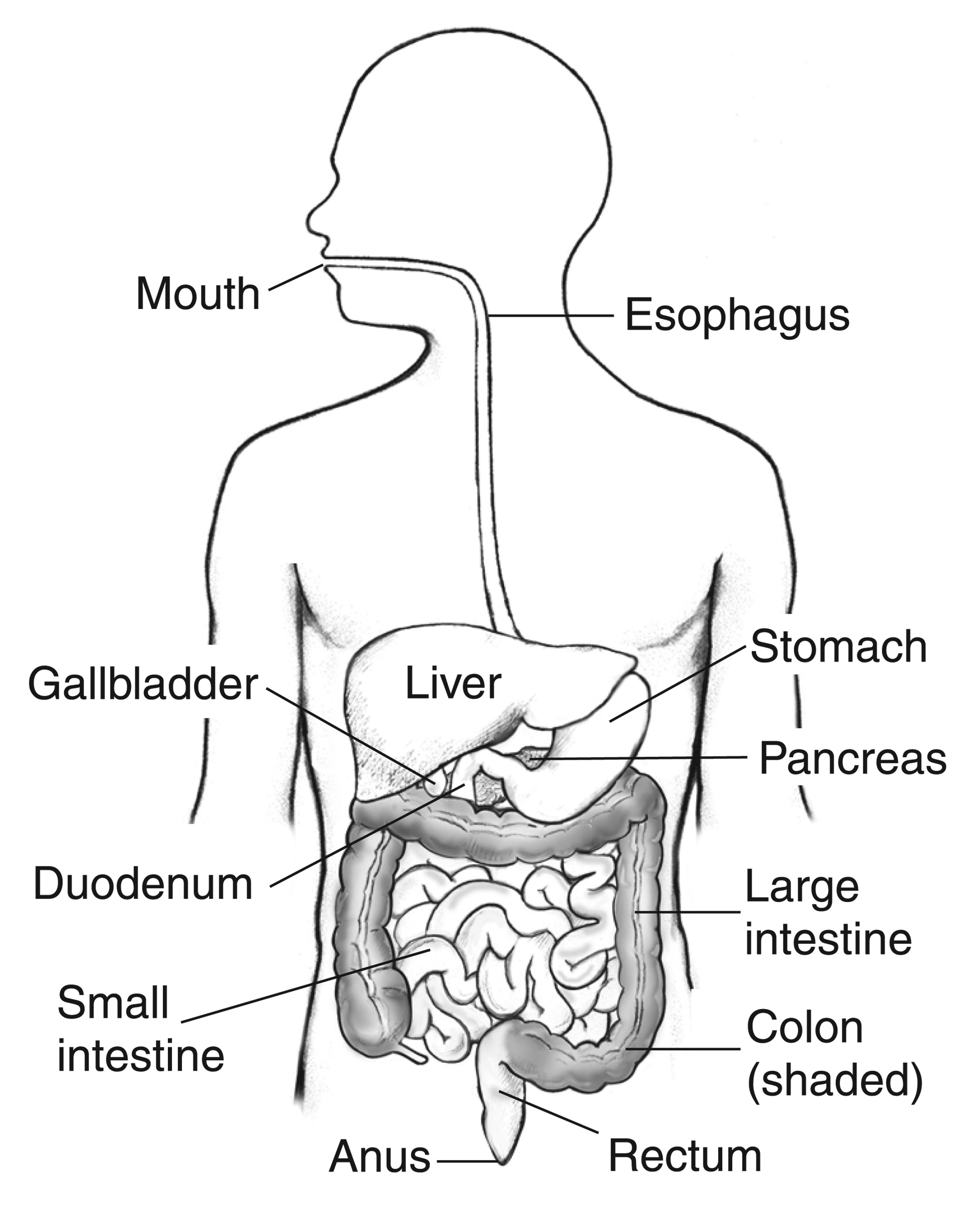
Enlarge the whole image The liver makes bile; the gallbladder stores it. Their position beside the duodenum helps connect bile delivery with intestinal fat absorption.
National Institute of Diabetes and Digestive and Kidney Diseases, NIH. Public domain . Source and provenance
Whole teaching image Close Show full-size image The liver makes bile; the gallbladder stores it. Their position beside the duodenum helps connect bile delivery with intestinal fat absorption.
National Institute of Diabetes and Digestive and Kidney Diseases, NIH. Public domain . Source and provenance
Separate pigment from digestive help Bile carries bilirubin and bile salts. Which one helps fat absorption?
Check this thought Bile salts help bring lipid digestion products toward the intestinal lining.
Then what is bilirubin doing in the same fluid?
Check this thought It is heme-breakdown waste being carried toward excretion.
A duct is completely blocked. Why can stools become both pale and greasy?
Check this thought Less pigment reaches the gut, and less bile-salt assistance is available for fat absorption.
One blocked delivery route can interrupt two different jobs. [1] [2] [4]
Try this reasoning again Read every step
Start with the two different jobs Pair digestive assistance, pigment disposal, and shared duct delivery with their distinct functions.
Digestive assistance Waste disposal Shared delivery
A duct problem can interrupt both jobs without making them the same substance. Bilirubin passes through liver processing and bile before intestinal pigment formation. Bile salts help lipid digestion products reach the intestinal surface.
Try again Read the worked solution Digestive assistance Bile salts help lipid digestion products reach the intestinal surface. [2] [4]
Waste disposal Bilirubin passes through liver processing and bile before intestinal pigment formation. [1]
Shared delivery A duct problem can interrupt both jobs without making them the same substance. [1] [4]
Bile salts assist fat handling. Their water-interacting and fat-interacting surfaces help disperse fat and keep digestion products available near the intestinal lining. Bilirubin has a different role. It is a pigment produced when heme, the iron-containing component of hemoglobin and other proteins, is broken down. The body must process and eliminate it. [1] [4]
Keep these jobs separate when a patient has jaundice and greasy stools. Jaundice means bilirubin has accumulated enough to yellow skin or eyes. Fat in stool means absorption has failed. An obstructed duct can connect these findings because it interrupts delivery of both pigment and bile salts. The two findings still arise through different mechanisms. Bile provides a route for eliminating excess cholesterol as well as bilirubin and other excreted substances. [1] [2] [4]
Digestive assistance Bile salts help lipid digestion products reach the intestinal surface. [2] [4]
Waste disposal Bilirubin passes through liver processing and bile before intestinal pigment formation. [1]
Shared delivery A duct problem can interrupt both jobs without making them the same substance. [1] [4]
Try it here · Checkpoint 1 of 3
Make your prediction before reading the choices. A first attempt is just a starting point.
Case 1
Show answer and explanations for case 1
A. Bile salts (Best answer)
Work through this option Which task is impaired: pigment delivery or lipid delivery?
Check this thought Reduced micellar lipid delivery is the fat-absorption task of bile salts.
How does this option fit the finding?
Check this thought Bile salts maintain lipid digestion products in mixed micelles; their reduced intestinal delivery impairs fat absorption.
What rule transfers to a new case?
Check this thought Shared bile delivery does not make bile salts and bilirubin interchangeable.
Shared bile delivery does not make bile salts and bilirubin interchangeable. [1] [4]
Try this reasoning again Read every step
Bile salts maintain lipid digestion products in mixed micelles; their reduced intestinal delivery impairs fat absorption.
B. Bilirubin (Why this does not fit)
Work through this option Which task is impaired: pigment delivery or lipid delivery?
Check this thought Reduced micellar lipid delivery is the fat-absorption task of bile salts.
How does this option fit the finding?
Check this thought Reduced bilirubin delivery explains pale stool, but bilirubin is waste pigment rather than the lipid-delivery component.
Shared bile delivery does not make bile salts and bilirubin interchangeable. [1] [4]
Try this reasoning again Read every step
Reduced bilirubin delivery explains pale stool, but bilirubin is waste pigment rather than the lipid-delivery component.
C. Albumin (Why this does not fit)
Work through this option Which task is impaired: pigment delivery or lipid delivery?
Check this thought Reduced micellar lipid delivery is the fat-absorption task of bile salts.
How does this option fit the finding?
Check this thought Albumin carries unconjugated bilirubin in plasma; it is not the intestinal micelle-forming substance.
Shared bile delivery does not make bile salts and bilirubin interchangeable. [1] [4]
Try this reasoning again Read every step
Albumin carries unconjugated bilirubin in plasma; it is not the intestinal micelle-forming substance.
D. Urobilinogen (Why this does not fit)
Work through this option Which task is impaired: pigment delivery or lipid delivery?
Check this thought Reduced micellar lipid delivery is the fat-absorption task of bile salts.
How does this option fit the finding?
Check this thought Urobilinogen is a downstream pigment product, not a facilitator of intestinal fat absorption.
Shared bile delivery does not make bile salts and bilirubin interchangeable. [1] [4]
Try this reasoning again Read every step
Urobilinogen is a downstream pigment product, not a facilitator of intestinal fat absorption.
Takeaway: Shared bile delivery does not make bile salts and bilirubin interchangeable.
Case sources: [1] [4]
Save this spot
Try a question on this topic A 67-year-old woman comes to the clinic because of 8 months of bulky, greasy, foul-smelling stools, abdominal bloating, and a 6-kg weight loss. She also reports tingling in both feet for 2 months. A contrast study 2 years ago showed multiple large diverticula of the jejunum. She takes no medications and drinks no alcohol. Temperature is 36.8°C (98.2°F), pulse is 84/min, and blood pressure is 118/74 mm Hg. She appears thin. The abdomen is distended and tympanitic without tenderness. Vibration sense is decreased at both great toes. Stool testing shows increased fecal fat. Tissue transglutaminase IgA antibodies are not detected, and fecal elastase is normal. After a 2-week course of an oral nonabsorbable antibiotic, her stools become formed and the bloating resolves. Laboratory studies before treatment show:
Laboratory studies Test Result Hemoglobin 10.4 g/dL Mean corpuscular volume 108 fL Serum cobalamin 140 pg/mL Serum folate 24 ng/mL Albumin 3.2 g/dL
Which of the following best explains her fat malabsorption?
A. Destruction of villous enterocytes by gluten-reactive T cells B. Deficient pancreatic lipase secretion from acinar cell loss C. Loss of ileal bile acid reabsorption from transmural inflammation D. Bacterial deconjugation of bile acids impairing micelle formation E. Blockade of lymphatic chylomicron transport by lamina propria macrophages
Choose an answer before revealing the reasoning.
Get one of these every morning · Practice more in the QBank
Follow pigment from an old red cell to bile Macrophages are cells that digest old cells and other material. In the spleen, liver, and bone marrow, they process aging red blood cells. Heme oxygenase changes heme into biliverdin. Biliverdin reductase then changes biliverdin into bilirubin. At this point bilirubin is unconjugated, meaning glucuronic acid has not yet been attached. Other heme-containing proteins also contribute to bilirubin production. [1]
Follow the unprocessed pigment Heme has become biliverdin. What reaction comes next?
Check this thought Biliverdin reductase produces unconjugated bilirubin.
That pigment travels in watery blood. What carries it?
Check this thought Albumin binds the poorly water-soluble unconjugated pigment.
Inside the liver cell, what must happen before usual biliary export?
Check this thought UGT1A1 adds glucuronic acid. MRP2 then exports the conjugated pigment toward bile.
Carrying, conjugating, and exporting are three different jobs. [1] [3]
Try this reasoning again Read every step
Follow pigment from an old red cell to bile Reconstruct the main pigment route from heme processing through albumin transport, chemical conjugation, and canalicular export.
Export Chemical change Transport Production
Try again Read the worked solution Production Heme becomes biliverdin, then unconjugated bilirubin. [1]
Transport Albumin carries unconjugated bilirubin through blood. [1]
Chemical change UGT1A1 adds glucuronic acid inside hepatocytes. [1] [3]
Export MRP2 moves conjugated bilirubin toward canaliculi and bile ducts. [1]
Unconjugated bilirubin dissolves poorly in water. It travels through plasma bound tightly to albumin, a blood protein. Albumin is the carrier, not the enzyme that makes bilirubin. The albumin-bound pigment is not freely filtered by the kidney. Increased unconjugated bilirubin therefore does not by itself produce bilirubin in urine. [1]
After hepatic uptake, UGT1A1 attaches glucuronic acid to bilirubin inside the hepatocyte. This conjugation makes the pigment more water soluble. A separate transport step is still needed. MRP2, encoded by ABCC2, exports conjugated bilirubin across the canalicular membrane toward bile. Canaliculi are tiny channels between neighboring hepatocytes that drain into ducts. More water solubility does not remove the need for an export protein. [1] [3]
Reduced UGT1A1 activity leaves an upstream accumulation of unconjugated bilirubin. Impaired export or bile flow can instead increase conjugated bilirubin in blood. Some conjugated pigment returns to blood through other transport routes and is normally taken up again by liver cells. Thus a blood result reflects several handling steps, not just one chemical reaction. [1] [3]
Production Heme becomes biliverdin, then unconjugated bilirubin. [1] Transport Albumin carries unconjugated bilirubin through blood. [1] Chemical change UGT1A1 adds glucuronic acid inside hepatocytes. [1] [3] Export MRP2 moves conjugated bilirubin toward canaliculi and bile ducts. [1] Try it here · Checkpoint 2 of 3
Make your prediction before reading the choices. A first attempt is just a starting point.
Case 4
Show answer and explanations for case 4
A. The kidney converts all bilirubin into bile salts (Why this does not fit)
Work through this option Which bilirubin fraction is increased in this patient?
Check this thought Only the indirect, largely unconjugated fraction is increased.
How does this option fit the finding?
Check this thought Bile acids are synthesized from cholesterol in the liver, not from bilirubin in the kidney.
An isolated indirect increase does not produce bilirubinuria. [1] [8]
Try this reasoning again Read every step
Bile acids are synthesized from cholesterol in the liver, not from bilirubin in the kidney.
B. Albumin-bound unconjugated bilirubin is not freely filtered (Best answer)
Work through this option Which bilirubin fraction is increased in this patient?
Check this thought Only the indirect, largely unconjugated fraction is increased.
How does this option fit the finding?
Check this thought The circulating excess is poorly water soluble and bound to albumin, preventing free glomerular filtration.
What rule transfers to a new case?
Check this thought An isolated indirect increase does not produce bilirubinuria.
An isolated indirect increase does not produce bilirubinuria. [1] [8]
Try this reasoning again Read every step
The circulating excess is poorly water soluble and bound to albumin, preventing free glomerular filtration.
C. Hemolysis stops all hepatic bile secretion (Why this does not fit)
Work through this option Which bilirubin fraction is increased in this patient?
Check this thought Only the indirect, largely unconjugated fraction is increased.
How does this option fit the finding?
Check this thought The liver can continue processing and secreting bilirubin during hemolysis.
An isolated indirect increase does not produce bilirubinuria. [1] [8]
Try this reasoning again Read every step
The liver can continue processing and secreting bilirubin during hemolysis.
D. Urobilinogen prevents the bilirubin dipstick reaction (Why this does not fit)
Work through this option Which bilirubin fraction is increased in this patient?
Check this thought Only the indirect, largely unconjugated fraction is increased.
How does this option fit the finding?
Check this thought The two urine tests measure different substances; urobilinogen does not explain the filtration barrier.
An isolated indirect increase does not produce bilirubinuria. [1] [8]
Try this reasoning again Read every step
The two urine tests measure different substances; urobilinogen does not explain the filtration barrier.
Takeaway: An isolated indirect increase does not produce bilirubinuria.
Case sources: [1] [8]
Save this spot
Read blood fractions and urine tests together In the intestine, bacteria modify delivered bilirubin into urobilinogen and related products. Urobilinogen is colorless and water soluble. Downstream stercobilin contributes to brown stool. Some intestinal urobilinogen is absorbed and returned to the liver; a small amount reaches the kidneys. Its oxidation product, urobilin, contributes to yellow urine. Urine bilirubin and urine urobilinogen tests therefore measure different substances. [1] [4] [8]
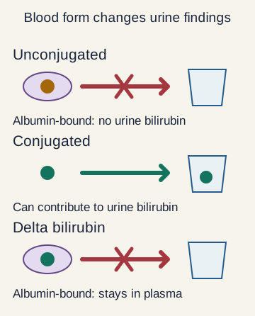
Enlarge the whole image Albumin-bound forms differ from the filterable conjugated fraction. Direct bilirubin is an assay result, not a promise that every measured molecule can enter urine.
Bone Wizardry. Original educational schematic. Source and provenance
Whole teaching image Close Show full-size image Albumin-bound forms differ from the filterable conjugated fraction. Direct bilirubin is an assay result, not a promise that every measured molecule can enter urine.
Bone Wizardry. Original educational schematic. Source and provenance
Predict the urine before naming a disease Only unconjugated bilirubin is increased. Is urine bilirubin expected?
Check this thought No. The poorly water-soluble, albumin-bound fraction is not normally filtered into urine.
Now conjugated bilirubin builds up in blood. What becomes possible?
Check this thought Some can enter urine, so the bilirubin test may become positive.
Does a high direct result always mean freely filterable conjugated bilirubin?
Check this thought No. The direct measurement can include delta bilirubin, which is bound to albumin.
Use the measured fraction and the carrier, not urine color alone. [1] [8]
Try this reasoning again Read every step
Direct and indirect are laboratory terms, not perfect chemical synonyms. The direct result mainly reflects conjugated bilirubin. Indirect bilirubin is usually calculated by subtracting direct from total. For example, a total of 4.0 mg/dL and direct of 0.5 mg/dL give an indirect value of 3.5 mg/dL. This fictional example establishes an indirect-predominant pattern, not its cause or treatment. [1]
Delta bilirubin is conjugated bilirubin firmly bound to albumin. It contributes to the direct measurement but is not freely filtered into urine. It can remain elevated after prolonged obstruction improves because it clears with albumin. A lingering direct result therefore does not always mean that a duct remains blocked. [1]
Hemolysis increases heme breakdown and can produce indirect hyperbilirubinemia without bilirubinuria. Urine urobilinogen may increase. Complete obstruction reduces intestinal pigment delivery, so stools may become pale and urobilinogen low while filterable conjugated bilirubin darkens urine. Partial obstruction is less predictable. Hepatocyte injury can affect multiple steps and produce mixed findings. Dark urine alone is not a bilirubin assay; concentration or blood pigments can also change its color. [1] [8]
Indirect predominance Consider increased production or impaired uptake or conjugation; check the clinical context. [1]
Conjugated accumulation Filterable conjugated bilirubin can enter urine; reduced gut delivery can pale stool. [1] [8]
Delta bilirubin Direct-reacting but albumin-bound pigment explains one important blood-versus-urine exception. [1]
Save this spot
Separate bile assistance from enzyme digestion Hepatocytes make the primary bile acids cholic acid and chenodeoxycholic acid from cholesterol. Cholesterol 7-alpha-hydroxylase, also called CYP7A1, controls the rate-limiting step of the classical synthesis pathway. The liver then attaches glycine or taurine to bile acids. This conjugation helps them remain soluble and ionized in the intestine, where they are commonly called bile salts. [4]
Do not give bile the lipase job A fat droplet is large. How do bile salts help?
Check this thought They aid emulsification and form mixed micelles with lipid digestion products.
Which molecule performs triglyceride breakdown?
Check this thought Pancreatic lipase performs the enzymatic digestion. Bile salts do not replace it.
Lipid leaves a micelle near an enterocyte. Must the bile salt enter with it?
Check this thought No. Most bile salts remain in the lumen until later recovery, mainly in the terminal ileum.
Micelles help deliver digestion products; they are not the digestive enzyme. [2] [4]
Try this reasoning again Read every step
Separate bile assistance from enzyme digestion Connect bile-acid formation, enzyme digestion, micellar delivery, and later bile-salt recovery without treating them as one reaction.
Make and modify Digest Deliver Recover later
Lipid uptake and bile-salt recovery are different processes at different intestinal sites. Bile salts support dispersion and mixed micellar delivery to the intestinal surface. Pancreatic lipase breaks fat into smaller molecules. Cholesterol becomes primary bile acids; glycine or taurine is then attached.
Try again Read the worked solution Make and modify Cholesterol becomes primary bile acids; glycine or taurine is then attached. [4]
Digest Pancreatic lipase breaks fat into smaller molecules. [2]
Deliver Bile salts support dispersion and mixed micellar delivery to the intestinal surface. [4]
Recover later Lipid uptake and bile-salt recovery are different processes at different intestinal sites. [4]
The word conjugation appears twice in this lesson, but the attached molecules differ. Bile-acid conjugation adds glycine or taurine. Bilirubin conjugation adds glucuronic acid. Do not use a bilirubin enzyme defect to explain every problem involving a conjugated bile acid. [1] [3] [4]
Bile salts are amphipathic, with regions that interact with water and others with fat. They assist emulsification, the dispersion of larger fat droplets into smaller droplets. Pancreatic lipase performs chemical digestion of fat. Bile is not a substitute for that digestive enzyme. Mixed micelles then help deliver lipid digestion products through the watery intestinal environment toward the absorbing surface. [2] [4]
Micelles deliver lipid molecules; the whole micelle is not absorbed intact. Most bile salts remain in the intestinal contents until later recovery. Phospholipids such as lecithin help organize bile lipids and maintain cholesterol in a dispersed state. When cholesterol exceeds the capacity of bile salts and phospholipids to keep it dispersed, crystals can form. Impaired emptying and other factors then influence whether crystals become stones. [4]
Make and modify Cholesterol becomes primary bile acids; glycine or taurine is then attached. [4] Digest Pancreatic lipase breaks fat into smaller molecules. [2] Deliver Bile salts support dispersion and mixed micellar delivery to the intestinal surface. [4] Recover later Lipid uptake and bile-salt recovery are different processes at different intestinal sites. [4] Save this spot
Follow storage, release, and recycling Between meals, the gallbladder concentrates bile by absorbing water and electrolytes. When fat and protein enter the duodenum, the first part of the small intestine, intestinal cells release cholecystokinin, or CCK. CCK helps coordinate gallbladder contraction and relaxation of the sphincter of Oddi, the muscle controlling the duct outlet. These actions deliver stored bile into the intestine. [2] [4]
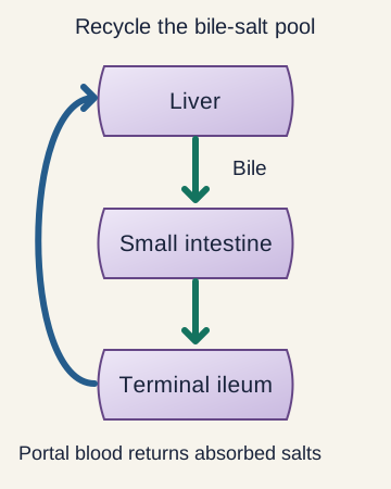
Enlarge the whole image Follow one recycling trip. The terminal ileum recovers bile salts; portal blood carries them back to the liver. This is a route diagram, not a map of organ position.
Bone Wizardry. Original educational schematic. Source and provenance
Whole teaching image Close Show full-size image Follow one recycling trip. The terminal ileum recovers bile salts; portal blood carries them back to the liver. This is a route diagram, not a map of organ position.
Bone Wizardry. Original educational schematic. Source and provenance
Keep the reservoir separate from the factory The gallbladder is removed. Is primary bile-acid production removed too?
Check this thought No. The liver still makes primary bile acids from cholesterol.
After a meal, what coordinates release from a gallbladder that is present?
Check this thought CCK promotes gallbladder contraction and relaxation of the sphincter of Oddi.
Where are most bile salts recovered for the return trip?
Check this thought They are recovered in the terminal ileum and return to the liver through portal blood.
The liver makes bile; the gallbladder stores it; the ileum recycles bile salts. [2] [4]
Try this reasoning again Read every step
Follow storage, release, and recycling Rebuild one meal-associated cycle, starting with liver secretion and ending with portal return for reuse.
Portal blood Terminal ileum Gallbladder and outlet Liver
Try again Read the worked solution Liver Produces and resecretes bile components. [2] [4]
Gallbladder and outlet Store and release bile in coordination with meal signals. [4]
Terminal ileum Actively recovers most bile salts after their digestive work. [4]
Portal blood Returns recovered bile salts to the liver for reuse. [4]
Secretin is a different intestinal hormone. It is released in response to acid and promotes bicarbonate-rich secretion from bile-duct cells. Bicarbonate helps neutralize acidic intestinal contents. CCK chiefly coordinates delivery of stored bile, whereas secretin increases alkaline fluid secretion. Their actions cooperate, but one hormone should not be substituted for the other in an explanation. [4]
After assisting lipid absorption, most bile salts are actively recovered in the terminal ileum, the last part of the small intestine. They return through portal blood to the liver and are secreted again. This intestine-to-liver return is called enterohepatic circulation. Repeated recycling means the liver normally replaces losses rather than synthesizing a completely new pool after each meal. [4]
Intestinal bacteria modify some bile acids into secondary bile acids. Some of this material can also be reabsorbed, while the remainder leaves in stool. A patient can still make bile after gallbladder removal because hepatocytes remain present. Removing a storage reservoir changes delivery, not the identity of the organ that synthesizes primary bile acids. [2] [4]
Liver Produces and resecretes bile components. [2] [4] Gallbladder and outlet Store and release bile in coordination with meal signals. [4] Terminal ileum Actively recovers most bile salts after their digestive work. [4] Portal blood Returns recovered bile salts to the liver for reuse. [4] Save this spot
Distinguish excess colonic bile acids from too few intestinal bile salts Crohn disease can injure the terminal ileum, and surgery may remove part of it. Either can reduce bile-salt recovery. The consequence depends on both where the unrecovered bile acids go and whether enough remain available for digestion. These are two separate questions, so watery diarrhea and fat malabsorption should not be treated as mandatory stages of one fixed sequence. [5] [6]
Which side of the balance is failing? Bile acids escape the ileum but the pool remains adequate. Where can they cause trouble?
Check this thought Excess bile acids reaching the colon can drive watery diarrhea.
Loss becomes greater than hepatic replacement. What happens to the pool?
Check this thought The available bile-salt pool falls.
Why can the stool become greasy now?
Check this thought Too few bile salts are available for normal micellar fat absorption.
Colonic excess causes one problem; depletion of the recycling pool causes another. [5] [6]
Try this reasoning again Read every step
Distinguish excess colonic bile acids from too few intestinal bile salts Match colonic bile-acid excess, inadequate intestinal supply, and overlapping disease with their different consequences.
Adequate pool, excess in colon Loss exceeds replacement Overlapping consequences
Assess bowel continuity, nutrition, and other causes rather than forcing one label. Insufficient intestinal bile salts impair lipid delivery and can produce steatorrhea. Bile acids can produce watery diarrhea despite sufficient supply for fat absorption.
Try again Read the worked solution Adequate pool, excess in colon Bile acids can produce watery diarrhea despite sufficient supply for fat absorption. [6]
Loss exceeds replacement Insufficient intestinal bile salts impair lipid delivery and can produce steatorrhea. [5]
Overlapping consequences Assess bowel continuity, nutrition, and other causes rather than forcing one label. [5] [6]
When excess bile acids reach a connected colon, they can increase secretion and alter intestinal movement, producing watery diarrhea. A patient may have this problem while the remaining bile-salt supply is still adequate for fat absorption. Bile-acid diarrhea is one possibility after ileal disease, not the only explanation for every postoperative loose stool. [5] [6]
With sufficiently extensive loss, fecal bile-acid loss may exceed hepatic replacement. The available bile-salt pool then becomes too small for effective micellar delivery. Fat remains unabsorbed and can appear as bulky, greasy stools. This is steatorrhea. Vitamins A, D, E, and K depend on fat absorption, so ongoing malabsorption creates nutritional risk. [4] [5]
These mechanisms may overlap in the same patient. Remaining small-bowel function, the connected colon, food intake, and hepatic replacement all matter. A treatment that binds bile acids in the colon can help selected bile-acid diarrhea, but further reducing an already depleted bile-salt supply may worsen fat malabsorption. Clinical management therefore needs the actual bowel anatomy and nutritional pattern, not a reflex based solely on the word diarrhea. [5] [6]
Adequate pool, excess in colon Bile acids can produce watery diarrhea despite sufficient supply for fat absorption. [6]
Loss exceeds replacement Insufficient intestinal bile salts impair lipid delivery and can produce steatorrhea. [5]
Overlapping consequences Assess bowel continuity, nutrition, and other causes rather than forcing one label. [5] [6]
Try it here · Checkpoint 3 of 3
Make your prediction before reading the choices. A first attempt is just a starting point.
Case 22
Show answer and explanations for case 22
A. Increased colonic water secretion alone creates stool fat (Why this does not fit)
Work through this option Is the liver replacing all the lost bile acids?
Check this thought No. Loss exceeds replacement, so the circulating bile-salt pool is depleted.
How does this option fit the finding?
Check this thought Water secretion can cause diarrhea but does not by itself explain unabsorbed dietary fat.
A depleted recycling pool can turn ileal loss into fat malabsorption. [4] [5]
Try this reasoning again Read every step
Water secretion can cause diarrhea but does not by itself explain unabsorbed dietary fat.
B. Excess conjugated bilirubin acts as pancreatic lipase (Why this does not fit)
Work through this option Is the liver replacing all the lost bile acids?
Check this thought No. Loss exceeds replacement, so the circulating bile-salt pool is depleted.
How does this option fit the finding?
Check this thought Bilirubin is pigment, not the missing digestive or micellar function.
A depleted recycling pool can turn ileal loss into fat malabsorption. [4] [5]
Try this reasoning again Read every step
Bilirubin is pigment, not the missing digestive or micellar function.
C. The kidney stops filtering albumin-bound bilirubin (Why this does not fit)
Work through this option Is the liver replacing all the lost bile acids?
Check this thought No. Loss exceeds replacement, so the circulating bile-salt pool is depleted.
How does this option fit the finding?
Check this thought That plasma filtration property does not explain intestinal fat malabsorption.
A depleted recycling pool can turn ileal loss into fat malabsorption. [4] [5]
Try this reasoning again Read every step
That plasma filtration property does not explain intestinal fat malabsorption.
D. The depleted bile-salt pool impairs micellar lipid delivery (Best answer)
Work through this option Is the liver replacing all the lost bile acids?
Check this thought No. Loss exceeds replacement, so the circulating bile-salt pool is depleted.
How does this option fit the finding?
Check this thought Loss beyond replacement leaves inadequate bile salts for efficient fat absorption.
What rule transfers to a new case?
Check this thought A depleted recycling pool can turn ileal loss into fat malabsorption.
A depleted recycling pool can turn ileal loss into fat malabsorption. [4] [5]
Try this reasoning again Read every step
Loss beyond replacement leaves inadequate bile salts for efficient fat absorption.
Takeaway: A depleted recycling pool can turn ileal loss into fat malabsorption.
Case sources: [4] [5]
Save this spot
Explain the kidney stone without confusing it with a gallstone Normally, calcium in the intestine can bind dietary oxalate. The bound material is poorly absorbed and leaves in stool. With fat malabsorption, unabsorbed fatty acids instead bind some of that calcium. Less calcium remains available to bind oxalate, so more oxalate stays soluble and available for absorption. [5]
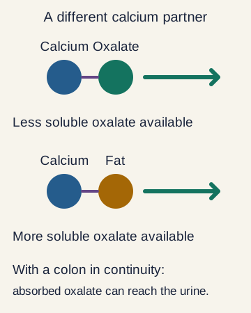
Enlarge the whole image Unabsorbed fatty acids can bind calcium, leaving more soluble oxalate for absorption through a connected colon. No quantities or individual stone risk are predicted.
Bone Wizardry. Original educational schematic. Source and provenance
Whole teaching image Close Show full-size image Unabsorbed fatty acids can bind calcium, leaving more soluble oxalate for absorption through a connected colon. No quantities or individual stone risk are predicted.
Bone Wizardry. Original educational schematic. Source and provenance
Follow the calcium partner Fat is not absorbed. What can those fatty acids bind in the gut?
Check this thought They bind calcium that might otherwise bind oxalate.
Less calcium is available for oxalate. Which fraction of oxalate rises?
Check this thought More oxalate remains soluble and available for absorption.
With a colon in continuity, why can this become a kidney-stone problem?
Check this thought More absorbed oxalate can reach the urine and contribute to calcium-oxalate stones.
Unabsorbed fat can free oxalate for absorption; a gallstone is a different problem. [5] [7]
Try this reasoning again Read every step
Explain the kidney stone without confusing it with a gallstone Assume fat malabsorption with a connected colon. As more unabsorbed fatty acids bind intestinal calcium, predict the direction of each listed change.
Calcium available to bind oxalate
Decreases No change Increases Soluble oxalate available for absorption
Decreases No change Increases Urinary oxalate load
Decreases No change Increases
Try again Read the worked solution Calcium available to bind oxalate Decreases. Unabsorbed fatty acids bind calcium, leaving less available for oxalate binding. [5]
Soluble oxalate available for absorption Increases. Less binding leaves more soluble oxalate available for absorption through the connected colon. [5]
Urinary oxalate load Increases. Increased intestinal oxalate absorption can increase the amount subsequently excreted in urine. [5]
A connected colon is important in the classic enteric hyperoxaluria mechanism. Soluble oxalate is absorbed through the colon and later excreted in urine. Hyperoxaluria means increased urinary oxalate. It can promote calcium-oxalate crystals and kidney stones. Diarrheal fluid losses may add a separate problem by reducing urine volume and concentrating urinary solutes. [5]
Notice what this explanation does not require. The patient does not have to eat more oxalate or develop high blood calcium. The changed intestinal binding partners alter how much existing dietary oxalate is absorbed. Ileal surgery alone does not establish this mechanism; the teaching example also requires fat malabsorption and a colon receiving intestinal contents. [5]
Gallstones form in bile, not urine. Reduced bile salts relative to cholesterol can favor cholesterol crystallization, but ileal disease does not guarantee cholesterol stones. Changes in intestinal bilirubin cycling and gallbladder emptying can contribute to pigment stones as well. The observed bowel history raises risk; actual stone composition and location still require their own evidence. [4] [5] [7]
Fatty acids bind calcium Less intestinal calcium is available to bind oxalate. [5] Soluble oxalate reaches the colon More can be absorbed when the colon remains in continuity. [5] Kidneys excrete the absorbed oxalate Urinary oxalate and low urine volume can promote calcium-oxalate stones. [5] Biliary stones need a separate explanation Cholesterol solubility and bilirubin handling concern bile, not urinary oxalate.
[4] [7] Save this spot
Apply the lesson to clinical cases You can pause here and return to any case. Choose an answer when ready, then compare the explanations.
Case 2
Show answer and explanations for case 2
A. UGT1A1 attaches glucuronic acid (Why this does not fit)
Work through this option The substrate is biliverdin. Which step are we still in?
Check this thought We are still producing bilirubin, before hepatic conjugation or export.
How does this option fit the finding?
Check this thought UGT1A1 acts on bilirubin after hepatic uptake, not on biliverdin to create bilirubin.
Separate production of bilirubin from its later conjugation and excretion. [1]
Try this reasoning again Read every step
UGT1A1 acts on bilirubin after hepatic uptake, not on biliverdin to create bilirubin.
B. MRP2 exports pigment into bile (Why this does not fit)
Work through this option The substrate is biliverdin. Which step are we still in?
Check this thought We are still producing bilirubin, before hepatic conjugation or export.
How does this option fit the finding?
Check this thought Export occurs after bilirubin production and conjugation within the hepatocyte.
Separate production of bilirubin from its later conjugation and excretion. [1]
Try this reasoning again Read every step
Export occurs after bilirubin production and conjugation within the hepatocyte.
C. Intestinal bacteria form urobilinogen (Why this does not fit)
Work through this option The substrate is biliverdin. Which step are we still in?
Check this thought We are still producing bilirubin, before hepatic conjugation or export.
How does this option fit the finding?
Check this thought Bacterial processing occurs downstream after pigment reaches the intestine.
Separate production of bilirubin from its later conjugation and excretion. [1]
Try this reasoning again Read every step
Bacterial processing occurs downstream after pigment reaches the intestine.
D. Biliverdin reductase acts on biliverdin (Best answer)
Work through this option The substrate is biliverdin. Which step are we still in?
Check this thought We are still producing bilirubin, before hepatic conjugation or export.
How does this option fit the finding?
Check this thought This reaction produces unconjugated bilirubin before hepatic conjugation.
What rule transfers to a new case?
Check this thought Separate production of bilirubin from its later conjugation and excretion.
Separate production of bilirubin from its later conjugation and excretion. [1]
Try this reasoning again Read every step
This reaction produces unconjugated bilirubin before hepatic conjugation.
Takeaway: Separate production of bilirubin from its later conjugation and excretion.
Case sources: [1]
Case 3
Show answer and explanations for case 3
A. Conversion to stercobilin in blood (Why this does not fit)
Work through this option Has glucuronic acid been attached yet?
Check this thought No. The unconjugated molecule needs its usual albumin carrier.
How does this option fit the finding?
Check this thought Stercobilin is a downstream intestinal pigment; it is not the plasma carrier.
Albumin transports unconjugated bilirubin; it does not chemically conjugate it. [1]
Try this reasoning again Read every step
Stercobilin is a downstream intestinal pigment; it is not the plasma carrier.
B. Binding to a canalicular export pump (Why this does not fit)
Work through this option Has glucuronic acid been attached yet?
Check this thought No. The unconjugated molecule needs its usual albumin carrier.
How does this option fit the finding?
Check this thought MRP2 is a membrane transporter at the hepatocyte bile-facing surface, not a circulating carrier.
Albumin transports unconjugated bilirubin; it does not chemically conjugate it. [1]
Try this reasoning again Read every step
MRP2 is a membrane transporter at the hepatocyte bile-facing surface, not a circulating carrier.
C. Binding to albumin (Best answer)
Work through this option Has glucuronic acid been attached yet?
Check this thought No. The unconjugated molecule needs its usual albumin carrier.
How does this option fit the finding?
Check this thought Poorly water-soluble unconjugated bilirubin circulates tightly bound to albumin.
What rule transfers to a new case?
Check this thought Albumin transports unconjugated bilirubin; it does not chemically conjugate it.
Albumin transports unconjugated bilirubin; it does not chemically conjugate it. [1]
Try this reasoning again Read every step
Poorly water-soluble unconjugated bilirubin circulates tightly bound to albumin.
D. Entry into an intestinal mixed micelle (Why this does not fit)
Work through this option Has glucuronic acid been attached yet?
Check this thought No. The unconjugated molecule needs its usual albumin carrier.
How does this option fit the finding?
Check this thought Mixed micelles assist lipid delivery in intestinal contents, not bilirubin transport through plasma.
Albumin transports unconjugated bilirubin; it does not chemically conjugate it. [1]
Try this reasoning again Read every step
Mixed micelles assist lipid delivery in intestinal contents, not bilirubin transport through plasma.
Takeaway: Albumin transports unconjugated bilirubin; it does not chemically conjugate it.
Case sources: [1]
Case 5
Show answer and explanations for case 5
A. Predominantly indirect bilirubin elevation without bilirubinuria (Best answer)
Work through this option Does UGT1A1 perform a chemical reaction or membrane export?
Check this thought It performs the conjugating reaction inside the liver cell.
How does this option fit the finding?
Check this thought Less conjugation leaves more albumin-bound unconjugated bilirubin in plasma.
What rule transfers to a new case?
Check this thought Identify the chemical step before predicting the laboratory fraction.
Identify the chemical step before predicting the laboratory fraction. [1] [3]
Try this reasoning again Read every step
Less conjugation leaves more albumin-bound unconjugated bilirubin in plasma.
B. Predominantly direct bilirubin elevation with pale stools (Why this does not fit)
Work through this option Does UGT1A1 perform a chemical reaction or membrane export?
Check this thought It performs the conjugating reaction inside the liver cell.
How does this option fit the finding?
Check this thought That pattern suggests impaired export or bile flow rather than an isolated conjugation defect.
Identify the chemical step before predicting the laboratory fraction. [1] [3]
Try this reasoning again Read every step
That pattern suggests impaired export or bile flow rather than an isolated conjugation defect.
C. Increased urinary bile salts with a normal bilirubin concentration (Why this does not fit)
Work through this option Does UGT1A1 perform a chemical reaction or membrane export?
Check this thought It performs the conjugating reaction inside the liver cell.
How does this option fit the finding?
Check this thought UGT1A1 glucuronidates bilirubin; this is not a primary bile-salt synthesis disorder.
Identify the chemical step before predicting the laboratory fraction. [1] [3]
Try this reasoning again Read every step
UGT1A1 glucuronidates bilirubin; this is not a primary bile-salt synthesis disorder.
D. Persistent delta bilirubin as the principal initial product (Why this does not fit)
Work through this option Does UGT1A1 perform a chemical reaction or membrane export?
Check this thought It performs the conjugating reaction inside the liver cell.
How does this option fit the finding?
Check this thought Delta bilirubin derives from conjugated pigment bound to albumin, not newly unprocessed unconjugated pigment.
Identify the chemical step before predicting the laboratory fraction. [1] [3]
Try this reasoning again Read every step
Delta bilirubin derives from conjugated pigment bound to albumin, not newly unprocessed unconjugated pigment.
Takeaway: Identify the chemical step before predicting the laboratory fraction.
Case sources: [1] [3]
Case 6
Show answer and explanations for case 6
A. Attachment of glucuronic acid (Why this does not fit)
Work through this option The cell formed a conjugate normally. Which stage remains?
Check this thought It still needs export across the bile-facing membrane.
How does this option fit the finding?
Check this thought The model states that conjugation is intact, and UGT1A1 performs that reaction.
Conjugation and canalicular export are separate steps. [1]
Try this reasoning again Read every step
The model states that conjugation is intact, and UGT1A1 performs that reaction.
B. Conversion of heme to biliverdin (Why this does not fit)
Work through this option The cell formed a conjugate normally. Which stage remains?
Check this thought It still needs export across the bile-facing membrane.
How does this option fit the finding?
Check this thought This upstream macrophage reaction is catalyzed by heme oxygenase.
Conjugation and canalicular export are separate steps. [1]
Try this reasoning again Read every step
This upstream macrophage reaction is catalyzed by heme oxygenase.
C. Albumin synthesis as the bilirubin carrier (Why this does not fit)
Work through this option The cell formed a conjugate normally. Which stage remains?
Check this thought It still needs export across the bile-facing membrane.
How does this option fit the finding?
Check this thought MRP2 dysfunction concerns membrane export, not albumin production.
Conjugation and canalicular export are separate steps. [1]
Try this reasoning again Read every step
MRP2 dysfunction concerns membrane export, not albumin production.
D. Transport of conjugated bilirubin toward bile (Best answer)
Work through this option The cell formed a conjugate normally. Which stage remains?
Check this thought It still needs export across the bile-facing membrane.
How does this option fit the finding?
Check this thought MRP2 is the bile-facing export transporter; solubility alone cannot replace its function.
What rule transfers to a new case?
Check this thought Conjugation and canalicular export are separate steps.
Conjugation and canalicular export are separate steps. [1]
Try this reasoning again Read every step
MRP2 is the bile-facing export transporter; solubility alone cannot replace its function.
Takeaway: Conjugation and canalicular export are separate steps.
Case sources: [1]
Case 7
Show answer and explanations for case 7
A. Formation of intestinal micelles within renal tubules (Why this does not fit)
Work through this option Which fraction accumulated upstream of the complete duct blockage?
Check this thought Conjugated bilirubin accumulated in blood and can contribute to urine bilirubin.
How does this option fit the finding?
Check this thought Micelles explain intestinal lipid delivery, not renal filtration of bilirubin.
Filterable conjugated pigment can enter urine when its blood concentration rises. [1]
Try this reasoning again Read every step
Micelles explain intestinal lipid delivery, not renal filtration of bilirubin.
B. Conversion of albumin-bound bilirubin into red blood cells (Why this does not fit)
Work through this option Which fraction accumulated upstream of the complete duct blockage?
Check this thought Conjugated bilirubin accumulated in blood and can contribute to urine bilirubin.
How does this option fit the finding?
Check this thought Red-cell production is unrelated to the filtration mechanism in obstruction.
Filterable conjugated pigment can enter urine when its blood concentration rises. [1]
Try this reasoning again Read every step
Red-cell production is unrelated to the filtration mechanism in obstruction.
C. Water solubility of conjugated bilirubin available for filtration (Best answer)
Work through this option Which fraction accumulated upstream of the complete duct blockage?
Check this thought Conjugated bilirubin accumulated in blood and can contribute to urine bilirubin.
How does this option fit the finding?
Check this thought Conjugated pigment can be filtered when it is not bound in a nonfilterable albumin complex.
What rule transfers to a new case?
Check this thought Filterable conjugated pigment can enter urine when its blood concentration rises.
Filterable conjugated pigment can enter urine when its blood concentration rises. [1]
Try this reasoning again Read every step
Conjugated pigment can be filtered when it is not bound in a nonfilterable albumin complex.
D. Increased lipid solubility after glucuronidation (Why this does not fit)
Work through this option Which fraction accumulated upstream of the complete duct blockage?
Check this thought Conjugated bilirubin accumulated in blood and can contribute to urine bilirubin.
How does this option fit the finding?
Check this thought Glucuronidation increases water solubility rather than lipid solubility.
Filterable conjugated pigment can enter urine when its blood concentration rises. [1]
Try this reasoning again Read every step
Glucuronidation increases water solubility rather than lipid solubility.
Takeaway: Filterable conjugated pigment can enter urine when its blood concentration rises.
Case sources: [1]
Case 8
Show answer and explanations for case 8
A. Increased stool pigment and no change in bacterial substrate (Why this does not fit)
Work through this option Does bilirubin reach the intestine in this complete obstruction?
Check this thought Delivery is markedly reduced, leaving less substrate for downstream intestinal pigments.
How does this option fit the finding?
Check this thought Complete obstruction reduces the bilirubin substrate reaching intestinal bacteria.
The gut cannot form its usual downstream pigments from bilirubin that never arrives. [1] [4] [8]
Try this reasoning again Read every step
Complete obstruction reduces the bilirubin substrate reaching intestinal bacteria.
B. Pale stool and reduced urine urobilinogen (Best answer)
Work through this option Does bilirubin reach the intestine in this complete obstruction?
Check this thought Delivery is markedly reduced, leaving less substrate for downstream intestinal pigments.
How does this option fit the finding?
Check this thought Less intestinal pigment delivery reduces downstream stool pigments and urobilinogen formation.
What rule transfers to a new case?
Check this thought The gut cannot form its usual downstream pigments from bilirubin that never arrives.
The gut cannot form its usual downstream pigments from bilirubin that never arrives. [1] [4] [8]
Try this reasoning again Read every step
Less intestinal pigment delivery reduces downstream stool pigments and urobilinogen formation.
C. Dark stool and increased urine urobilinogen (Why this does not fit)
Work through this option Does bilirubin reach the intestine in this complete obstruction?
Check this thought Delivery is markedly reduced, leaving less substrate for downstream intestinal pigments.
How does this option fit the finding?
Check this thought These changes require increased or preserved downstream pigment availability rather than absent delivery.
The gut cannot form its usual downstream pigments from bilirubin that never arrives. [1] [4] [8]
Try this reasoning again Read every step
These changes require increased or preserved downstream pigment availability rather than absent delivery.
D. Pale stool and increased conjugation by intestinal UGT1A1 (Why this does not fit)
Work through this option Does bilirubin reach the intestine in this complete obstruction?
Check this thought Delivery is markedly reduced, leaving less substrate for downstream intestinal pigments.
How does this option fit the finding?
Check this thought Hepatic UGT1A1 conjugation does not explain pale stool or replace bile delivery.
The gut cannot form its usual downstream pigments from bilirubin that never arrives. [1] [4] [8]
Try this reasoning again Read every step
Hepatic UGT1A1 conjugation does not explain pale stool or replace bile delivery.
Takeaway: The gut cannot form its usual downstream pigments from bilirubin that never arrives.
Case sources: [1] [4] [8]
Case 9
Show answer and explanations for case 9
A. They can coexist because the assays measure different substances (Best answer)
Work through this option Are bilirubin and urobilinogen the same substance?
Check this thought No. Urobilinogen is formed downstream of bilirubin in the intestinal pathway.
How does this option fit the finding?
Check this thought Some intestinal urobilinogen normally reaches urine even though bilirubin is directed into bile.
What rule transfers to a new case?
Check this thought Bilirubin and urobilinogen are different measurements at different points in the pathway.
Bilirubin and urobilinogen are different measurements at different points in the pathway. [1] [8]
Try this reasoning again Read every step
Some intestinal urobilinogen normally reaches urine even though bilirubin is directed into bile.
B. The urobilinogen result proves the bilirubin result is false (Why this does not fit)
Work through this option Are bilirubin and urobilinogen the same substance?
Check this thought No. Urobilinogen is formed downstream of bilirubin in the intestinal pathway.
How does this option fit the finding?
Check this thought Normal downstream urobilinogen does not require a positive urine bilirubin result.
Bilirubin and urobilinogen are different measurements at different points in the pathway. [1] [8]
Try this reasoning again Read every step
Normal downstream urobilinogen does not require a positive urine bilirubin result.
C. They establish complete extrahepatic obstruction (Why this does not fit)
Work through this option Are bilirubin and urobilinogen the same substance?
Check this thought No. Urobilinogen is formed downstream of bilirubin in the intestinal pathway.
How does this option fit the finding?
Check this thought Complete obstruction usually reduces intestinal urobilinogen formation rather than explaining this normal pattern.
Bilirubin and urobilinogen are different measurements at different points in the pathway. [1] [8]
Try this reasoning again Read every step
Complete obstruction usually reduces intestinal urobilinogen formation rather than explaining this normal pattern.
D. They establish an isolated UGT1A1 defect (Why this does not fit)
Work through this option Are bilirubin and urobilinogen the same substance?
Check this thought No. Urobilinogen is formed downstream of bilirubin in the intestinal pathway.
How does this option fit the finding?
Check this thought These routine urine findings alone do not diagnose an inherited conjugation disorder.
Bilirubin and urobilinogen are different measurements at different points in the pathway. [1] [8]
Try this reasoning again Read every step
These routine urine findings alone do not diagnose an inherited conjugation disorder.
Takeaway: Bilirubin and urobilinogen are different measurements at different points in the pathway.
Case sources: [1] [8]
Case 10
Show answer and explanations for case 10
A. 7.6 mg/dL (Why this does not fit)
Work through this option What calculation keeps the original units?
Check this thought Subtract direct bilirubin from total bilirubin: 6.2 minus 1.4 equals 4.8 mg/dL.
How does this option fit the finding?
Check this thought Adding the two values double-counts the direct fraction already contained within total.
Indirect bilirubin is usually total minus direct, with units unchanged. [1]
Try this reasoning again Read every step
Adding the two values double-counts the direct fraction already contained within total.
B. 1.4 mg/dL (Why this does not fit)
Work through this option What calculation keeps the original units?
Check this thought Subtract direct bilirubin from total bilirubin: 6.2 minus 1.4 equals 4.8 mg/dL.
How does this option fit the finding?
Check this thought This is the reported direct fraction, not the calculated indirect fraction.
Indirect bilirubin is usually total minus direct, with units unchanged. [1]
Try this reasoning again Read every step
This is the reported direct fraction, not the calculated indirect fraction.
C. 6.2 mg/dL (Why this does not fit)
Work through this option What calculation keeps the original units?
Check this thought Subtract direct bilirubin from total bilirubin: 6.2 minus 1.4 equals 4.8 mg/dL.
How does this option fit the finding?
Check this thought This is the total; the direct component must be subtracted to estimate indirect bilirubin.
Indirect bilirubin is usually total minus direct, with units unchanged. [1]
Try this reasoning again Read every step
This is the total; the direct component must be subtracted to estimate indirect bilirubin.
D. 4.8 mg/dL (Best answer)
Work through this option What calculation keeps the original units?
Check this thought Subtract direct bilirubin from total bilirubin: 6.2 minus 1.4 equals 4.8 mg/dL.
How does this option fit the finding?
Check this thought Subtract direct from total: 6.2 minus 1.4 equals 4.8 mg/dL.
What rule transfers to a new case?
Check this thought Indirect bilirubin is usually total minus direct, with units unchanged.
Indirect bilirubin is usually total minus direct, with units unchanged. [1]
Try this reasoning again Read every step
Subtract direct from total: 6.2 minus 1.4 equals 4.8 mg/dL.
Takeaway: Indirect bilirubin is usually total minus direct, with units unchanged.
Case sources: [1]
Case 11
Show answer and explanations for case 11
A. Stercobilin retained in plasma albumin (Why this does not fit)
Work through this option Has the patient been shown to have persistent obstruction?
Check this thought No. Recovery is documented, so a slowly clearing pigment fraction deserves consideration.
How does this option fit the finding?
Check this thought Stercobilin is a downstream stool pigment, not the albumin-bound direct fraction described.
A direct result includes an albumin-bound component that may outlast the obstruction. [1]
Try this reasoning again Read every step
Stercobilin is a downstream stool pigment, not the albumin-bound direct fraction described.
B. Urobilinogen trapped in hepatocyte nuclei (Why this does not fit)
Work through this option Has the patient been shown to have persistent obstruction?
Check this thought No. Recovery is documented, so a slowly clearing pigment fraction deserves consideration.
How does this option fit the finding?
Check this thought This does not explain persistent direct-reacting albumin-bound bilirubin.
A direct result includes an albumin-bound component that may outlast the obstruction. [1]
Try this reasoning again Read every step
This does not explain persistent direct-reacting albumin-bound bilirubin.
C. Delta bilirubin bound firmly to albumin (Best answer)
Work through this option Has the patient been shown to have persistent obstruction?
Check this thought No. Recovery is documented, so a slowly clearing pigment fraction deserves consideration.
How does this option fit the finding?
Check this thought It reacts in the direct assay and clears with albumin rather than being freely filtered.
What rule transfers to a new case?
Check this thought A direct result includes an albumin-bound component that may outlast the obstruction.
A direct result includes an albumin-bound component that may outlast the obstruction. [1]
Try this reasoning again Read every step
It reacts in the direct assay and clears with albumin rather than being freely filtered.
D. Unconjugated bilirubin newly converted into a bile acid (Why this does not fit)
Work through this option Has the patient been shown to have persistent obstruction?
Check this thought No. Recovery is documented, so a slowly clearing pigment fraction deserves consideration.
How does this option fit the finding?
Check this thought Bilirubin does not become a bile acid; bile acids derive from cholesterol.
A direct result includes an albumin-bound component that may outlast the obstruction. [1]
Try this reasoning again Read every step
Bilirubin does not become a bile acid; bile acids derive from cholesterol.
Takeaway: A direct result includes an albumin-bound component that may outlast the obstruction.
Case sources: [1]
Case 12
Show answer and explanations for case 12
A. Increased urine urobilinogen must be the cause (Why this does not fit)
Work through this option Did the assay show urine bilirubin?
Check this thought No. Dark color alone does not establish bilirubinuria.
How does this option fit the finding?
Check this thought Urobilinogen must be measured; it cannot be inferred solely from dark urine.
Use the actual assays instead of treating a color description as a laboratory diagnosis. [1] [8]
Try this reasoning again Read every step
Urobilinogen must be measured; it cannot be inferred solely from dark urine.
B. Urine color alone does not establish conjugated hyperbilirubinemia (Best answer)
Work through this option Did the assay show urine bilirubin?
Check this thought No. Dark color alone does not establish bilirubinuria.
How does this option fit the finding?
Check this thought Concentration and other pigments can darken urine without bilirubinuria.
What rule transfers to a new case?
Check this thought Use the actual assays instead of treating a color description as a laboratory diagnosis.
Use the actual assays instead of treating a color description as a laboratory diagnosis. [1] [8]
Try this reasoning again Read every step
Concentration and other pigments can darken urine without bilirubinuria.
C. Complete bile-duct obstruction is proven (Why this does not fit)
Work through this option Did the assay show urine bilirubin?
Check this thought No. Dark color alone does not establish bilirubinuria.
How does this option fit the finding?
Check this thought Color without corroborating bilirubin, stool, enzyme, and imaging findings does not establish obstruction.
Use the actual assays instead of treating a color description as a laboratory diagnosis. [1] [8]
Try this reasoning again Read every step
Color without corroborating bilirubin, stool, enzyme, and imaging findings does not establish obstruction.
D. An isolated indirect bilirubin elevation must be present (Why this does not fit)
Work through this option Did the assay show urine bilirubin?
Check this thought No. Dark color alone does not establish bilirubinuria.
How does this option fit the finding?
Check this thought A negative urine bilirubin test does not identify the serum fraction or prove hyperbilirubinemia.
Use the actual assays instead of treating a color description as a laboratory diagnosis. [1] [8]
Try this reasoning again Read every step
A negative urine bilirubin test does not identify the serum fraction or prove hyperbilirubinemia.
Takeaway: Use the actual assays instead of treating a color description as a laboratory diagnosis.
Case sources: [1] [8]
Case 13
Show answer and explanations for case 13
A. The liver synthesizes primary bile acids; the gallbladder stores bile (Best answer)
Work through this option Which organ makes primary bile acids?
Check this thought The liver makes them; the gallbladder is a storage reservoir.
How does this option fit the finding?
Check this thought Hepatic synthesis persists because cholecystectomy removes a reservoir rather than hepatocytes.
What rule transfers to a new case?
Check this thought Separate the liver factory from the gallbladder reservoir.
Separate the liver factory from the gallbladder reservoir. [2] [4]
Try this reasoning again Read every step
Hepatic synthesis persists because cholecystectomy removes a reservoir rather than hepatocytes.
B. The gallbladder synthesizes bile acids from bilirubin (Why this does not fit)
Work through this option Which organ makes primary bile acids?
Check this thought The liver makes them; the gallbladder is a storage reservoir.
How does this option fit the finding?
Check this thought Primary bile acids are made from cholesterol in hepatocytes, not from bilirubin in the gallbladder.
Separate the liver factory from the gallbladder reservoir. [2] [4]
Try this reasoning again Read every step
Primary bile acids are made from cholesterol in hepatocytes, not from bilirubin in the gallbladder.
C. The pancreas takes over all bile-acid synthesis after surgery (Why this does not fit)
Work through this option Which organ makes primary bile acids?
Check this thought The liver makes them; the gallbladder is a storage reservoir.
How does this option fit the finding?
Check this thought The pancreas supplies digestive enzymes; it does not normally replace hepatic primary bile-acid synthesis.
Separate the liver factory from the gallbladder reservoir. [2] [4]
Try this reasoning again Read every step
The pancreas supplies digestive enzymes; it does not normally replace hepatic primary bile-acid synthesis.
D. The terminal ileum becomes the source of newly synthesized bile (Why this does not fit)
Work through this option Which organ makes primary bile acids?
Check this thought The liver makes them; the gallbladder is a storage reservoir.
How does this option fit the finding?
Check this thought The ileum recovers bile salts; recovery is not de novo hepatic synthesis.
Separate the liver factory from the gallbladder reservoir. [2] [4]
Try this reasoning again Read every step
The ileum recovers bile salts; recovery is not de novo hepatic synthesis.
Takeaway: Separate the liver factory from the gallbladder reservoir.
Case sources: [2] [4]
Case 14
Show answer and explanations for case 14
A. Glycine attaches to bilirubin; glucuronic acid attaches to all bile acids (Why this does not fit)
Work through this option Are the two conjugation reactions modifying the same substrate?
Check this thought No. Bilirubin and bile acids are different substrates with different attached molecules.
How does this option fit the finding?
Check this thought This reverses the principal conjugation processes taught here.
Name the substrate and attached molecule when using the word conjugation. [1] [3] [4]
Try this reasoning again Read every step
This reverses the principal conjugation processes taught here.
B. Albumin covalently conjugates both substances before secretion (Why this does not fit)
Work through this option Are the two conjugation reactions modifying the same substrate?
Check this thought No. Bilirubin and bile acids are different substrates with different attached molecules.
How does this option fit the finding?
Check this thought Albumin is a plasma carrier and does not perform these hepatic conjugation reactions.
Name the substrate and attached molecule when using the word conjugation. [1] [3] [4]
Try this reasoning again Read every step
Albumin is a plasma carrier and does not perform these hepatic conjugation reactions.
C. UGT1A1 converts cholesterol directly into cholic acid (Why this does not fit)
Work through this option Are the two conjugation reactions modifying the same substrate?
Check this thought No. Bilirubin and bile acids are different substrates with different attached molecules.
How does this option fit the finding?
Check this thought UGT1A1 processes bilirubin; the classical bile-acid pathway begins with cholesterol hydroxylation.
Name the substrate and attached molecule when using the word conjugation. [1] [3] [4]
Try this reasoning again Read every step
UGT1A1 processes bilirubin; the classical bile-acid pathway begins with cholesterol hydroxylation.
D. Glucuronic acid attaches to bilirubin; glycine or taurine attaches to bile acids (Best answer)
Work through this option Are the two conjugation reactions modifying the same substrate?
Check this thought No. Bilirubin and bile acids are different substrates with different attached molecules.
How does this option fit the finding?
Check this thought The two processes share the term conjugation but use different attached molecules.
What rule transfers to a new case?
Check this thought Name the substrate and attached molecule when using the word conjugation.
Name the substrate and attached molecule when using the word conjugation. [1] [3] [4]
Try this reasoning again Read every step
The two processes share the term conjugation but use different attached molecules.
Takeaway: Name the substrate and attached molecule when using the word conjugation.
Case sources: [1] [3] [4]
Case 15
Show answer and explanations for case 15
A. Attachment of glucuronic acid to bilirubin (Why this does not fit)
Work through this option Which pathway includes CYP7A1?
Check this thought CYP7A1 participates in the classical conversion of cholesterol toward primary bile acids.
How does this option fit the finding?
Check this thought That is the UGT1A1 reaction, not CYP7A1 activity.
CYP7A1 belongs to bile-acid synthesis, not bilirubin conjugation. [4]
Try this reasoning again Read every step
That is the UGT1A1 reaction, not CYP7A1 activity.
B. Active recovery of bile salts in the terminal ileum (Why this does not fit)
Work through this option Which pathway includes CYP7A1?
Check this thought CYP7A1 participates in the classical conversion of cholesterol toward primary bile acids.
How does this option fit the finding?
Check this thought Ileal transport is a separate process from hepatic synthesis.
CYP7A1 belongs to bile-acid synthesis, not bilirubin conjugation. [4]
Try this reasoning again Read every step
Ileal transport is a separate process from hepatic synthesis.
C. The classical pathway of bile-acid synthesis from cholesterol (Best answer)
Work through this option Which pathway includes CYP7A1?
Check this thought CYP7A1 participates in the classical conversion of cholesterol toward primary bile acids.
How does this option fit the finding?
Check this thought CYP7A1 is cholesterol 7-alpha-hydroxylase, the rate-limiting enzyme of that pathway.
What rule transfers to a new case?
Check this thought CYP7A1 belongs to bile-acid synthesis, not bilirubin conjugation.
CYP7A1 belongs to bile-acid synthesis, not bilirubin conjugation. [4]
Try this reasoning again Read every step
CYP7A1 is cholesterol 7-alpha-hydroxylase, the rate-limiting enzyme of that pathway.
D. Conversion of biliverdin to bilirubin (Why this does not fit)
Work through this option Which pathway includes CYP7A1?
Check this thought CYP7A1 participates in the classical conversion of cholesterol toward primary bile acids.
How does this option fit the finding?
Check this thought Biliverdin reductase performs that pigment-production step.
CYP7A1 belongs to bile-acid synthesis, not bilirubin conjugation. [4]
Try this reasoning again Read every step
Biliverdin reductase performs that pigment-production step.
Takeaway: CYP7A1 belongs to bile-acid synthesis, not bilirubin conjugation.
Case sources: [4]
Case 16
Show answer and explanations for case 16
A. Albumin-bound bilirubin must enter intestinal micelles first (Why this does not fit)
Work through this option Is bile delivery missing in this scenario?
Check this thought No. Bile arrives, but pancreatic enzymatic digestion is impaired.
How does this option fit the finding?
Check this thought Plasma bilirubin transport is unrelated to the missing pancreatic enzyme reaction.
Bile assists fat handling; pancreatic lipase performs enzymatic breakdown. [2] [4]
Try this reasoning again Read every step
Plasma bilirubin transport is unrelated to the missing pancreatic enzyme reaction.
B. Bile salts assist dispersion and delivery but do not replace pancreatic lipase (Best answer)
Work through this option Is bile delivery missing in this scenario?
Check this thought No. Bile arrives, but pancreatic enzymatic digestion is impaired.
How does this option fit the finding?
Check this thought Lipase performs chemical fat digestion; adequate bile delivery alone does not supply that enzyme.
What rule transfers to a new case?
Check this thought Bile assists fat handling; pancreatic lipase performs enzymatic breakdown.
Bile assists fat handling; pancreatic lipase performs enzymatic breakdown. [2] [4]
Try this reasoning again Read every step
Lipase performs chemical fat digestion; adequate bile delivery alone does not supply that enzyme.
C. Bilirubin normally serves as the principal triglyceride enzyme (Why this does not fit)
Work through this option Is bile delivery missing in this scenario?
Check this thought No. Bile arrives, but pancreatic enzymatic digestion is impaired.
How does this option fit the finding?
Check this thought Bilirubin is waste pigment rather than a digestive enzyme.
Bile assists fat handling; pancreatic lipase performs enzymatic breakdown. [2] [4]
Try this reasoning again Read every step
Bilirubin is waste pigment rather than a digestive enzyme.
D. Bile salts must be converted into lipase before absorption (Why this does not fit)
Work through this option Is bile delivery missing in this scenario?
Check this thought No. Bile arrives, but pancreatic enzymatic digestion is impaired.
How does this option fit the finding?
Check this thought Bile salts and pancreatic lipase are different substances with complementary roles.
Bile assists fat handling; pancreatic lipase performs enzymatic breakdown. [2] [4]
Try this reasoning again Read every step
Bile salts and pancreatic lipase are different substances with complementary roles.
Takeaway: Bile assists fat handling; pancreatic lipase performs enzymatic breakdown.
Case sources: [2] [4]
Case 17
Show answer and explanations for case 17
A. They remain in intestinal contents for later recovery, mainly in the terminal ileum (Best answer)
Work through this option Which material leaves the micelle near the enterocyte?
Check this thought Lipid digestion products leave; most bile salts remain available for later recycling.
How does this option fit the finding?
Check this thought Micelles deliver lipids without being absorbed intact as one unit.
What rule transfers to a new case?
Check this thought Lipid absorption precedes the main site of bile-salt recovery.
Lipid absorption precedes the main site of bile-salt recovery. [4]
Try this reasoning again Read every step
Micelles deliver lipids without being absorbed intact as one unit.
B. They enter enterocytes only as an intact micelle containing all lipids (Why this does not fit)
Work through this option Which material leaves the micelle near the enterocyte?
Check this thought Lipid digestion products leave; most bile salts remain available for later recycling.
How does this option fit the finding?
Check this thought Intact micelle uptake is not the normal mechanism of lipid delivery.
Lipid absorption precedes the main site of bile-salt recovery. [4]
Try this reasoning again Read every step
Intact micelle uptake is not the normal mechanism of lipid delivery.
C. They become conjugated bilirubin inside the intestinal wall (Why this does not fit)
Work through this option Which material leaves the micelle near the enterocyte?
Check this thought Lipid digestion products leave; most bile salts remain available for later recycling.
How does this option fit the finding?
Check this thought Bile salts are not converted into bilirubin during absorption.
Lipid absorption precedes the main site of bile-salt recovery. [4]
Try this reasoning again Read every step
Bile salts are not converted into bilirubin during absorption.
D. They are all immediately lost in stool after one use (Why this does not fit)
Work through this option Which material leaves the micelle near the enterocyte?
Check this thought Lipid digestion products leave; most bile salts remain available for later recycling.
How does this option fit the finding?
Check this thought Enterohepatic circulation recovers most bile salts for reuse.
Lipid absorption precedes the main site of bile-salt recovery. [4]
Try this reasoning again Read every step
Enterohepatic circulation recovers most bile salts for reuse.
Takeaway: Lipid absorption precedes the main site of bile-salt recovery.
Case sources: [4]
Case 18
Show answer and explanations for case 18
A. Secretin converts unconjugated bilirubin into bilirubin glucuronides (Why this does not fit)
Work through this option Which meal signal coordinates stored bile release?
Check this thought Fat and protein stimulate CCK, which coordinates gallbladder contraction and sphincter relaxation.
How does this option fit the finding?
Check this thought Secretin stimulates ductal bicarbonate secretion; UGT1A1 conjugates bilirubin.
CCK coordinates delivery; it is not the conjugating enzyme. [4]
Try this reasoning again Read every step
Secretin stimulates ductal bicarbonate secretion; UGT1A1 conjugates bilirubin.
B. CYP7A1 contracts the cystic duct during each meal (Why this does not fit)
Work through this option Which meal signal coordinates stored bile release?
Check this thought Fat and protein stimulate CCK, which coordinates gallbladder contraction and sphincter relaxation.
How does this option fit the finding?
Check this thought CYP7A1 is a hepatic synthesis enzyme, not the hormone controlling motility.
CCK coordinates delivery; it is not the conjugating enzyme. [4]
Try this reasoning again Read every step
CYP7A1 is a hepatic synthesis enzyme, not the hormone controlling motility.
C. Albumin relaxes the sphincter while bile salts digest the duct wall (Why this does not fit)
Work through this option Which meal signal coordinates stored bile release?
Check this thought Fat and protein stimulate CCK, which coordinates gallbladder contraction and sphincter relaxation.
How does this option fit the finding?
Check this thought Albumin is a blood carrier, and bile salts do not normally digest the duct wall.
CCK coordinates delivery; it is not the conjugating enzyme. [4]
Try this reasoning again Read every step
Albumin is a blood carrier, and bile salts do not normally digest the duct wall.
D. CCK promotes gallbladder contraction and sphincter of Oddi relaxation (Best answer)
Work through this option Which meal signal coordinates stored bile release?
Check this thought Fat and protein stimulate CCK, which coordinates gallbladder contraction and sphincter relaxation.
How does this option fit the finding?
Check this thought These paired actions move stored bile toward the intestinal lumen.
What rule transfers to a new case?
Check this thought CCK coordinates delivery; it is not the conjugating enzyme.
CCK coordinates delivery; it is not the conjugating enzyme. [4]
Try this reasoning again Read every step
These paired actions move stored bile toward the intestinal lumen.
Takeaway: CCK coordinates delivery; it is not the conjugating enzyme.
Case sources: [4]
Case 19
Show answer and explanations for case 19
A. Heme oxygenase-mediated bile concentration (Why this does not fit)
Work through this option What reached the duodenum?
Check this thought Acid stimulates secretin and the alkaline ductal-fluid response.
How does this option fit the finding?
Check this thought Heme oxygenase processes heme; concentration occurs through gallbladder absorption of water and electrolytes.
Acid stimulates secretin; ductal bicarbonate helps neutralize it. [4]
Try this reasoning again Read every step
Heme oxygenase processes heme; concentration occurs through gallbladder absorption of water and electrolytes.
B. Ileal albumin secretion into the common bile duct (Why this does not fit)
Work through this option What reached the duodenum?
Check this thought Acid stimulates secretin and the alkaline ductal-fluid response.
How does this option fit the finding?
Check this thought This is not the mechanism for hormone-regulated ductal alkaline secretion.
Acid stimulates secretin; ductal bicarbonate helps neutralize it. [4]
Try this reasoning again Read every step
This is not the mechanism for hormone-regulated ductal alkaline secretion.
C. Secretin-stimulated bicarbonate secretion (Best answer)
Work through this option What reached the duodenum?
Check this thought Acid stimulates secretin and the alkaline ductal-fluid response.
How does this option fit the finding?
Check this thought Cholangiocytes respond to secretin by increasing bicarbonate-rich fluid secretion.
What rule transfers to a new case?
Check this thought Acid stimulates secretin; ductal bicarbonate helps neutralize it.
Acid stimulates secretin; ductal bicarbonate helps neutralize it. [4]
Try this reasoning again Read every step
Cholangiocytes respond to secretin by increasing bicarbonate-rich fluid secretion.
D. CCK-mediated bilirubin glucuronidation (Why this does not fit)
Work through this option What reached the duodenum?
Check this thought Acid stimulates secretin and the alkaline ductal-fluid response.
How does this option fit the finding?
Check this thought CCK regulates bile delivery rather than the UGT1A1 chemical reaction.
Acid stimulates secretin; ductal bicarbonate helps neutralize it. [4]
Try this reasoning again Read every step
CCK regulates bile delivery rather than the UGT1A1 chemical reaction.
Takeaway: Acid stimulates secretin; ductal bicarbonate helps neutralize it.
Case sources: [4]
Case 20
Show answer and explanations for case 20
A. Glucuronidation of bilirubin by UGT1A1 (Why this does not fit)
Work through this option Which part of the recycling route is injured?
Check this thought The terminal ileum, the major recovery site for bile salts, is injured.
How does this option fit the finding?
Check this thought The conjugation reaction occurs inside hepatocytes.
The terminal ileum recovers bile salts; it does not make or store bile. [4]
Try this reasoning again Read every step
The conjugation reaction occurs inside hepatocytes.
B. Active recovery of bile salts for portal return to the liver (Best answer)
Work through this option Which part of the recycling route is injured?
Check this thought The terminal ileum, the major recovery site for bile salts, is injured.
How does this option fit the finding?
Check this thought The terminal ileum is the main active recovery site in enterohepatic circulation.
What rule transfers to a new case?
Check this thought The terminal ileum recovers bile salts; it does not make or store bile.
The terminal ileum recovers bile salts; it does not make or store bile. [4]
Try this reasoning again Read every step
The terminal ileum is the main active recovery site in enterohepatic circulation.
C. Primary synthesis of bile acids from cholesterol (Why this does not fit)
Work through this option Which part of the recycling route is injured?
Check this thought The terminal ileum, the major recovery site for bile salts, is injured.
How does this option fit the finding?
Check this thought That function occurs in hepatocytes, which are intact in this example.
The terminal ileum recovers bile salts; it does not make or store bile. [4]
Try this reasoning again Read every step
That function occurs in hepatocytes, which are intact in this example.
D. Storage and concentration of bile between meals (Why this does not fit)
Work through this option Which part of the recycling route is injured?
Check this thought The terminal ileum, the major recovery site for bile salts, is injured.
How does this option fit the finding?
Check this thought That is primarily the gallbladder role rather than the ileal role.
The terminal ileum recovers bile salts; it does not make or store bile. [4]
Try this reasoning again Read every step
That is primarily the gallbladder role rather than the ileal role.
Takeaway: The terminal ileum recovers bile salts; it does not make or store bile.
Case sources: [4]
Case 21
Show answer and explanations for case 21
A. Bile acids stimulate colonic secretion and intestinal activity (Best answer)
Work through this option Is fat absorption still preserved?
Check this thought Yes. This pattern emphasizes excess bile acids reaching the colon, not a depleted digestive pool.
How does this option fit the finding?
Check this thought Colonic exposure can cause watery diarrhea while the remaining pool still supports lipid absorption.
What rule transfers to a new case?
Check this thought Excess bile acids in the colon and too few bile salts for fat absorption are distinct problems.
Excess bile acids in the colon and too few bile salts for fat absorption are distinct problems. [5] [6]
Try this reasoning again Read every step
Colonic exposure can cause watery diarrhea while the remaining pool still supports lipid absorption.
B. Complete exhaustion of the bile-salt pool is required (Why this does not fit)
Work through this option Is fat absorption still preserved?
Check this thought Yes. This pattern emphasizes excess bile acids reaching the colon, not a depleted digestive pool.
How does this option fit the finding?
Check this thought The stem specifies preserved fat absorption; watery bile-acid diarrhea does not require severe pool depletion.
Excess bile acids in the colon and too few bile salts for fat absorption are distinct problems. [5] [6]
Try this reasoning again Read every step
The stem specifies preserved fat absorption; watery bile-acid diarrhea does not require severe pool depletion.
C. Delta bilirubin directly prevents colonic water absorption (Why this does not fit)
Work through this option Is fat absorption still preserved?
Check this thought Yes. This pattern emphasizes excess bile acids reaching the colon, not a depleted digestive pool.
How does this option fit the finding?
Check this thought Delta bilirubin is an albumin-bound blood pigment, not the main colonic mechanism here.
Excess bile acids in the colon and too few bile salts for fat absorption are distinct problems. [5] [6]
Try this reasoning again Read every step
Delta bilirubin is an albumin-bound blood pigment, not the main colonic mechanism here.
D. Absorbed micelles injure the renal tubules (Why this does not fit)
Work through this option Is fat absorption still preserved?
Check this thought Yes. This pattern emphasizes excess bile acids reaching the colon, not a depleted digestive pool.
How does this option fit the finding?
Check this thought This confuses intestinal bile-acid effects with urinary complications.
Excess bile acids in the colon and too few bile salts for fat absorption are distinct problems. [5] [6]
Try this reasoning again Read every step
This confuses intestinal bile-acid effects with urinary complications.
Takeaway: Excess bile acids in the colon and too few bile salts for fat absorption are distinct problems.
Case sources: [5] [6]
Case 23
Show answer and explanations for case 23
A. Only vitamin B12 because all fat is absorbed in the terminal ileum (Why this does not fit)
Work through this option Which nutrient-handling pathway is impaired?
Check this thought Fat absorption is impaired, which places fat-soluble vitamins at particular risk.
How does this option fit the finding?
Check this thought Ileal disease can affect B12 separately, but the question asks about fat-dependent absorption and not all lipid is absorbed there.
Recognize the fat-soluble vitamin group without collapsing separate nutritional mechanisms. [4] [5]
Try this reasoning again Read every step
Ileal disease can affect B12 separately, but the question asks about fat-dependent absorption and not all lipid is absorbed there.
B. Only vitamin C because it forms mixed micelles (Why this does not fit)
Work through this option Which nutrient-handling pathway is impaired?
Check this thought Fat absorption is impaired, which places fat-soluble vitamins at particular risk.
How does this option fit the finding?
Check this thought Vitamin C is water soluble and is not the characteristic micelle-dependent vitamin in this setting.
Recognize the fat-soluble vitamin group without collapsing separate nutritional mechanisms. [4] [5]
Try this reasoning again Read every step
Vitamin C is water soluble and is not the characteristic micelle-dependent vitamin in this setting.
C. Vitamins A, D, E, and K (Best answer)
Work through this option Which nutrient-handling pathway is impaired?
Check this thought Fat absorption is impaired, which places fat-soluble vitamins at particular risk.
How does this option fit the finding?
Check this thought These fat-soluble vitamins are vulnerable when normal lipid absorption fails.
What rule transfers to a new case?
Check this thought Recognize the fat-soluble vitamin group without collapsing separate nutritional mechanisms.
Recognize the fat-soluble vitamin group without collapsing separate nutritional mechanisms. [4] [5]
Try this reasoning again Read every step
These fat-soluble vitamins are vulnerable when normal lipid absorption fails.
D. All water-soluble vitamins solely because they circulate with albumin (Why this does not fit)
Work through this option Which nutrient-handling pathway is impaired?
Check this thought Fat absorption is impaired, which places fat-soluble vitamins at particular risk.
How does this option fit the finding?
Check this thought The stated mechanism specifically concerns fat-soluble vitamins, not albumin carriage of every vitamin.
Recognize the fat-soluble vitamin group without collapsing separate nutritional mechanisms. [4] [5]
Try this reasoning again Read every step
The stated mechanism specifically concerns fat-soluble vitamins, not albumin carriage of every vitamin.
Takeaway: Recognize the fat-soluble vitamin group without collapsing separate nutritional mechanisms.
Case sources: [4] [5]
Case 24
Show answer and explanations for case 24
A. Reduced urine volume directly increases intestinal oxalate uptake (Why this does not fit)
Work through this option Is the colon still connected?
Check this thought Yes. With fat malabsorption, the colon can absorb additional soluble oxalate.
How does this option fit the finding?
Check this thought Low urine volume can increase urinary crystallization risk but is not the intestinal absorption mechanism.
Fatty acids take calcium away from oxalate, increasing the absorbable oxalate fraction. [5]
Try this reasoning again Read every step
Low urine volume can increase urinary crystallization risk but is not the intestinal absorption mechanism.
B. Fatty acids bind calcium, leaving more oxalate soluble (Best answer)
Work through this option Is the colon still connected?
Check this thought Yes. With fat malabsorption, the colon can absorb additional soluble oxalate.
How does this option fit the finding?
Check this thought Less calcium is available to bind dietary oxalate, which can then be absorbed through the colon.
What rule transfers to a new case?
Check this thought Fatty acids take calcium away from oxalate, increasing the absorbable oxalate fraction.
Fatty acids take calcium away from oxalate, increasing the absorbable oxalate fraction. [5]
Try this reasoning again Read every step
Less calcium is available to bind dietary oxalate, which can then be absorbed through the colon.
C. More calcium binds oxalate in intestinal contents (Why this does not fit)
Work through this option Is the colon still connected?
Check this thought Yes. With fat malabsorption, the colon can absorb additional soluble oxalate.
How does this option fit the finding?
Check this thought That would reduce soluble oxalate available for absorption rather than increase it.
Fatty acids take calcium away from oxalate, increasing the absorbable oxalate fraction. [5]
Try this reasoning again Read every step
That would reduce soluble oxalate available for absorption rather than increase it.
D. Bile salts are recovered more efficiently in the diseased ileum (Why this does not fit)
Work through this option Is the colon still connected?
Check this thought Yes. With fat malabsorption, the colon can absorb additional soluble oxalate.
How does this option fit the finding?
Check this thought Improved recovery would oppose pool depletion and fat malabsorption.
Fatty acids take calcium away from oxalate, increasing the absorbable oxalate fraction. [5]
Try this reasoning again Read every step
Improved recovery would oppose pool depletion and fat malabsorption.
Takeaway: Fatty acids take calcium away from oxalate, increasing the absorbable oxalate fraction.
Case sources: [5]
Case 25
Show answer and explanations for case 25
A. The history raises risk but does not establish stone composition (Best answer)
Work through this option Does the operation history identify the stone composition?
Check this thought No. It establishes a risk context, not direct evidence of the stone composition.
How does this option fit the finding?
Check this thought Reduced bile-salt recycling, altered bilirubin cycling, and emptying can contribute; pigment stones are also possible.
What rule transfers to a new case?
Check this thought Location and composition require evidence beyond a bowel-surgery history.
Location and composition require evidence beyond a bowel-surgery history. [4] [5] [7]
Try this reasoning again Read every step
Reduced bile-salt recycling, altered bilirubin cycling, and emptying can contribute; pigment stones are also possible.
B. All postoperative stones are urinary calcium-oxalate stones (Why this does not fit)
Work through this option Does the operation history identify the stone composition?
Check this thought No. It establishes a risk context, not direct evidence of the stone composition.
How does this option fit the finding?
Check this thought The identified stones are in the biliary system, which is distinct from the urinary tract.
Location and composition require evidence beyond a bowel-surgery history. [4] [5] [7]
Try this reasoning again Read every step
The identified stones are in the biliary system, which is distinct from the urinary tract.
C. Ileal resection excludes pigment stones because bilirubin never recycles (Why this does not fit)
Work through this option Does the operation history identify the stone composition?
Check this thought No. It establishes a risk context, not direct evidence of the stone composition.
How does this option fit the finding?
Check this thought Altered intestinal bilirubin handling can contribute to pigment-stone risk.
Location and composition require evidence beyond a bowel-surgery history. [4] [5] [7]
Try this reasoning again Read every step
Altered intestinal bilirubin handling can contribute to pigment-stone risk.
D. Normal hepatic bile synthesis rules out any gallstone (Why this does not fit)
Work through this option Does the operation history identify the stone composition?
Check this thought No. It establishes a risk context, not direct evidence of the stone composition.
How does this option fit the finding?
Check this thought Stones can form despite ongoing hepatic synthesis when composition and emptying favor crystallization.
Location and composition require evidence beyond a bowel-surgery history. [4] [5] [7]
Try this reasoning again Read every step
Stones can form despite ongoing hepatic synthesis when composition and emptying favor crystallization.
Takeaway: Location and composition require evidence beyond a bowel-surgery history.
Case sources: [4] [5] [7]