Distinguish failed canalicular export from failed hepatic reuptake, interpret urinary coproporphyrins, and recognize when benign jaundice is not enough.
Two people can have conjugated jaundice and healthy liver function for different reasons. Locate the transport problem before using pigment or a syndrome name.
First establish what is isolated Dubin-Johnson syndrome is an inherited defect of bilirubin transport. The liver can conjugate bilirubin, but its usual export into bile is impaired. Conjugated bilirubin consequently accumulates in blood. Rotor syndrome also produces predominantly conjugated hyperbilirubinemia, but its principal defect is hepatic reuptake from blood. Neither syndrome should be grouped with an isolated UGT1A1 conjugation defect. [1] [2] [3]
First decide whether the pattern is isolated Conjugated bilirubin is raised. Do the other liver tests still matter?
Check this thought Yes. Enzymes and synthetic function help distinguish an isolated transport pattern from liver injury.
The bilirubin is isolated and longstanding. Which pair becomes relevant?
Check this thought Dubin-Johnson and Rotor are inherited transport disorders that can fit this pattern.
Can urine bilirubin alone distinguish the two?
Check this thought No. Conjugated bilirubin can appear in urine in both conditions.
An isolated pattern opens the comparison; it does not complete the diagnosis. [1] [2] [4]
Try this reasoning again Read every step
First establish what is isolated Match bilirubin, other liver tests, and new clinical findings with the different questions each answers.
Bilirubin fraction Injury and function Changed clinical picture
Investigate new findings rather than assigning everything to the inherited diagnosis. Typically preserved in the uncomplicated adult phenotype. Predominantly conjugated in both transport disorders.
Try again Read the worked solution Bilirubin fraction Predominantly conjugated in both transport disorders. [2]
Injury and function Typically preserved in the uncomplicated adult phenotype. [2] [5]
Changed clinical picture Investigate new findings rather than assigning everything to the inherited diagnosis. [2]
The usual adult presentation is mild, sometimes intermittent jaundice with otherwise reassuring liver tests. AST and ALT are markers of hepatocyte injury. Alkaline phosphatase and GGT help identify a cholestatic injury pattern. Albumin and INR contribute information about synthetic function. Normal results outside bilirubin support a benign transport disorder; the yellow color itself does not establish liver failure. [2] [5]
Isolated is an important qualification, not a shortcut. Review previous results, symptoms, medicines, supplements, alcohol exposure, family history, and examination. New abdominal pain, fever, significant itching, pale stools, abnormal enzymes, or impaired clotting may indicate another process. A known inherited syndrome does not protect a person from hepatitis, obstruction, or a drug reaction. [2] [5]
Both disorders can cause bilirubin in urine because some circulating conjugated bilirubin is available for renal filtration. This differs from the usual absence of bilirubinuria in an isolated unconjugated increase. Urine findings help identify the fraction but cannot distinguish Dubin-Johnson from Rotor by themselves. The next useful comparison is where each transporter normally operates. [2] [4]
Bilirubin fraction Predominantly conjugated in both transport disorders. [2]
Injury and function Typically preserved in the uncomplicated adult phenotype. [2] [5]
Changed clinical picture Investigate new findings rather than assigning everything to the inherited diagnosis. [2]
Save this spot
Try a question on this topic A 22-year-old woman comes to the physician because her eyes turned yellow 3 weeks after she began taking an oral contraceptive. She feels well and has no itching, abdominal pain, or change in weight. Her brother has had intermittent mild yellowing of the eyes since adolescence. She drinks alcohol rarely and takes no other medications. Vital signs are within normal limits. Scleral icterus is present. The liver and spleen are not palpable. Ultrasonography shows a normal liver, normal bile ducts, and a normal gallbladder. Urine is dark and positive for bilirubin. During evaluation, a percutaneous liver biopsy shows normal lobular architecture without inflammation or fibrosis, but centrilobular hepatocytes contain abundant coarse dark brown pigment granules within lysosomes. The total urinary coproporphyrin level is normal, with more than 80% present as coproporphyrin I. Laboratory studies show:
Serum Test Result Total bilirubin 4.8 mg/dL Direct bilirubin 3.5 mg/dL Alkaline phosphatase 82 U/L Alanine aminotransferase 24 U/L Hemoglobin 13.6 g/dL
A defect in which of the following proteins is the most likely cause of these findings?
A. Canalicular conjugate export pump MRP2 B. Hepatocyte UDP-glucuronosyltransferase 1A1 C. Sinusoidal reuptake transporter OATP1B D. Canalicular bile salt export pump E. Canalicular phospholipid flippase MDR3
Choose an answer before revealing the reasoning.
Get one of these every morning · Practice more in the QBank
Find the liver cell surface that has failed A hepatocyte has a blood-facing sinusoidal surface and a bile-facing canalicular surface. Sinusoids are the small blood channels beside liver cells. Canaliculi are tiny channels between adjacent hepatocytes that collect bile. Thinking of these two sides prevents the common error of calling every bilirubin handling problem failed conjugation. [3] [4]
Name the membrane before the syndrome Which membrane faces bile?
Check this thought The canalicular membrane faces the bile channel and contains MRP2.
MRP2 is impaired. Is conjugation necessarily lost?
Check this thought No. Dubin-Johnson impairs export after conjugation.
Which different route is impaired in Rotor?
Check this thought Combined OATP1B1 and OATP1B3 loss impairs uptake and reuptake from blood.
Dubin-Johnson limits export; Rotor limits reuptake. [1] [2] [3]
Try this reasoning again Read every step
Find the liver cell surface that has failed Match the reaction or transporter with its direction at the hepatocyte. These are locations and branches, not a single sequential loop.
Inside the cell Toward bile Back through blood Into the cell again
OATP1B1 and OATP1B3 recover it; combined loss produces Rotor syndrome. MRP3 returns some conjugated pigment to sinusoidal blood. MRP2 exports conjugated pigment; this is the main Dubin-Johnson defect. UGT1A1 conjugates bilirubin.
Try again Read the worked solution Inside the cell UGT1A1 conjugates bilirubin. [3] [4]
Toward bile MRP2 exports conjugated pigment; this is the main Dubin-Johnson defect. [1] [4]
Back through blood MRP3 returns some conjugated pigment to sinusoidal blood. [4]
Into the cell again OATP1B1 and OATP1B3 recover it; combined loss produces Rotor syndrome. [2] [4]
UGT1A1 acts inside the hepatocyte and attaches glucuronic acid to bilirubin. The resulting conjugated pigment is more water soluble, but it still needs membrane transport. MRP2, encoded by ABCC2, exports conjugated bilirubin across the canalicular surface into bile. Dubin-Johnson syndrome results from reduced or absent function of this exporter. Its inheritance is autosomal recessive. [1] [3] [4]
Some conjugated bilirubin also returns to sinusoidal blood through MRP3. Hepatic OATP1B1 and OATP1B3 transporters can take it up again. They are encoded by SLCO1B1 and SLCO1B3, respectively. Rotor syndrome reflects combined loss of these reuptake functions. Its MRP2 exporter remains intact. This explains how an uptake disorder can still raise a pigment that has already been conjugated. [2] [3] [4]
The distinction is directional. Dubin-Johnson impairs the bile-facing exit. Rotor impairs the blood-facing return into the liver cell. Neither requires excessive destruction of red blood cells, and neither is equivalent to a stone blocking a large duct. A pathway diagram is useful only when every arrow has a named location and direction. [1] [2] [4]
Inside the cell UGT1A1 conjugates bilirubin.
[3] [4] Toward bile MRP2 exports conjugated pigment; this is the main Dubin-Johnson defect.
[1] [4] Back through blood MRP3 returns some conjugated pigment to sinusoidal blood.
[4] Into the cell again OATP1B1 and OATP1B3 recover it; combined loss produces Rotor syndrome.
[2] [4] Try it here · Checkpoint 1 of 3
Make your prediction before reading the choices. A first attempt is just a starting point.
Case 1
Show answer and explanations for case 1
A. Canalicular export of conjugated bilirubin by MRP2 (Best answer)
Work through this option What protein does ABCC2 encode?
Check this thought ABCC2 encodes MRP2, the canalicular export transporter.
How does this option fit the finding?
Check this thought ABCC2 encodes the transporter that moves conjugated bilirubin from the hepatocyte toward bile.
What rule transfers to a new case?
Check this thought Dubin-Johnson affects the bile-facing export step after conjugation.
Dubin-Johnson affects the bile-facing export step after conjugation. [1] [3] [4]
Try this reasoning again Read every step
ABCC2 encodes the transporter that moves conjugated bilirubin from the hepatocyte toward bile.
B. Conjugation of bilirubin by UGT1A1 (Why this does not fit)
Work through this option What protein does ABCC2 encode?
Check this thought ABCC2 encodes MRP2, the canalicular export transporter.
How does this option fit the finding?
Check this thought A conjugation defect primarily raises unconjugated bilirubin and does not match ABCC2.
Dubin-Johnson affects the bile-facing export step after conjugation. [1] [3] [4]
Try this reasoning again Read every step
A conjugation defect primarily raises unconjugated bilirubin and does not match ABCC2.
C. Sinusoidal reuptake by OATP1B1 and OATP1B3 (Why this does not fit)
Work through this option What protein does ABCC2 encode?
Check this thought ABCC2 encodes MRP2, the canalicular export transporter.
How does this option fit the finding?
Check this thought Combined failure of these transporters characterizes Rotor syndrome, not ABCC2-related disease.
Dubin-Johnson affects the bile-facing export step after conjugation. [1] [3] [4]
Try this reasoning again Read every step
Combined failure of these transporters characterizes Rotor syndrome, not ABCC2-related disease.
D. Production of biliverdin from heme (Why this does not fit)
Work through this option What protein does ABCC2 encode?
Check this thought ABCC2 encodes MRP2, the canalicular export transporter.
How does this option fit the finding?
Check this thought This upstream production step is separate from the documented canalicular transport defect.
Dubin-Johnson affects the bile-facing export step after conjugation. [1] [3] [4]
Try this reasoning again Read every step
This upstream production step is separate from the documented canalicular transport defect.
Takeaway: Dubin-Johnson affects the bile-facing export step after conjugation.
Case sources: [1] [3] [4]
Save this spot
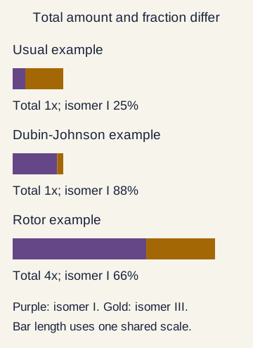
Read the total amount and the isomer pattern separately Coproporphyrins are byproducts related to heme synthesis. Their urinary distribution provides another view of hepatic organic-anion transport. Isomers I and III are different molecular arrangements. The useful test is not simply whether coproporphyrin is present. Ask how much is excreted in total and what proportion is isomer I. [3] [5]
Enlarge the whole image Read two measurements. The normal-total, 88-percent example differs from the fourfold-total, 66-percent example. Values illustrate the described patterns; they are not independent diagnostic thresholds.
Bone Wizardry. Original educational schematic. Source and provenance
Whole teaching image Close Show full-size image Read two measurements. The normal-total, 88-percent example differs from the fourfold-total, 66-percent example. Values illustrate the described patterns; they are not independent diagnostic thresholds.
Bone Wizardry. Original educational schematic. Source and provenance
Read the amount before the percentage Total output is normal but isomer I is 88 percent. Which pattern fits?
Check this thought That illustrative combination supports Dubin-Johnson in the appropriate clinical setting.
Now total output is four times normal and isomer I is 66 percent. What changes?
Check this thought The raised total together with this distribution supports Rotor.
Does increased total output alone establish Rotor?
Check this thought No. Other hepatobiliary disease can increase total coproporphyrins; the full context still matters.
A percentage describes a mixture, not the total amount excreted. [2] [3] [5]
Try this reasoning again Read every step
Read the total amount and the isomer pattern separately Match the reference, Dubin-Johnson, and Rotor patterns using both total amount and isomer distribution.
Reference pattern Dubin-Johnson pattern Rotor pattern
Increased total, commonly 2.5 to 5 times normal; isomer I about 65 percent. Usually normal total; isomer I greater than 80 percent. Total within range; isomer III predominates.
Try again Read the worked solution Reference pattern Total within range; isomer III predominates. [5]
Dubin-Johnson pattern Usually normal total; isomer I greater than 80 percent. [3] [5]
Rotor pattern Increased total, commonly 2.5 to 5 times normal; isomer I about 65 percent. [2] [3]
In the usual reference pattern, isomer III accounts for about three quarters of urinary coproporphyrin and isomer I for about one quarter. In Dubin-Johnson syndrome, total urinary coproporphyrin is usually normal, but isomer I accounts for more than 80 percent. The important abnormality is therefore the distribution, not a necessary increase in the total amount. [3] [5]
Rotor syndrome typically increases total urinary coproporphyrin to roughly 2.5 to 5 times normal. Isomer I accounts for approximately 65 percent in the classic comparison. These are characteristic patterns rather than mathematical diagnostic boundaries that override the rest of an assessment. Other liver diseases can increase urinary coproporphyrin, so a high total alone is not specific for Rotor. [2] [3]
For a worked comparison, consider two fictional patients with otherwise isolated conjugated jaundice. One has a normal total and 88 percent isomer I; that favors Dubin-Johnson. Another has four times the reference total and 66 percent isomer I; that favors Rotor. Looking only at the fact that isomer I predominates would miss the difference between them. [2] [3] [5]
Reference pattern Total within range; isomer III predominates. [5]
Dubin-Johnson pattern Usually normal total; isomer I greater than 80 percent. [3] [5]
Rotor pattern Increased total, commonly 2.5 to 5 times normal; isomer I about 65 percent. [2] [3]
Try it here · Checkpoint 2 of 3
Make your prediction before reading the choices. A first attempt is just a starting point.
Case 4
Show answer and explanations for case 4
A. Gilbert syndrome (Why this does not fit)
Work through this option What changed in both measurements?
Check this thought Total excretion is fourfold higher, with isomer I around two-thirds of the mixture.
How does this option fit the finding?
Check this thought Gilbert is a mild unconjugated disorder and does not account for this transport-associated urinary pattern.
Rotor changes both total urinary coproporphyrin and its isomer distribution. [2] [3]
Try this reasoning again Read every step
Gilbert is a mild unconjugated disorder and does not account for this transport-associated urinary pattern.
B. Rotor syndrome (Best answer)
Work through this option What changed in both measurements?
Check this thought Total excretion is fourfold higher, with isomer I around two-thirds of the mixture.
How does this option fit the finding?
Check this thought A several-fold increase in total output with isomer I near two thirds is the classic Rotor comparison.
What rule transfers to a new case?
Check this thought Rotor changes both total urinary coproporphyrin and its isomer distribution.
Rotor changes both total urinary coproporphyrin and its isomer distribution. [2] [3]
Try this reasoning again Read every step
A several-fold increase in total output with isomer I near two thirds is the classic Rotor comparison.
C. Dubin-Johnson syndrome (Why this does not fit)
Work through this option What changed in both measurements?
Check this thought Total excretion is fourfold higher, with isomer I around two-thirds of the mixture.
How does this option fit the finding?
Check this thought Dubin-Johnson usually has a normal total and a higher isomer I proportion, commonly above 80 percent.
Rotor changes both total urinary coproporphyrin and its isomer distribution. [2] [3]
Try this reasoning again Read every step
Dubin-Johnson usually has a normal total and a higher isomer I proportion, commonly above 80 percent.
D. Crigler-Najjar syndrome type I (Why this does not fit)
Work through this option What changed in both measurements?
Check this thought Total excretion is fourfold higher, with isomer I around two-thirds of the mixture.
How does this option fit the finding?
Check this thought This severe conjugation disorder produces predominantly unconjugated bilirubin, usually beginning in infancy.
Rotor changes both total urinary coproporphyrin and its isomer distribution. [2] [3]
Try this reasoning again Read every step
This severe conjugation disorder produces predominantly unconjugated bilirubin, usually beginning in infancy.
Takeaway: Rotor changes both total urinary coproporphyrin and its isomer distribution.
Case sources: [2] [3]
Save this spot
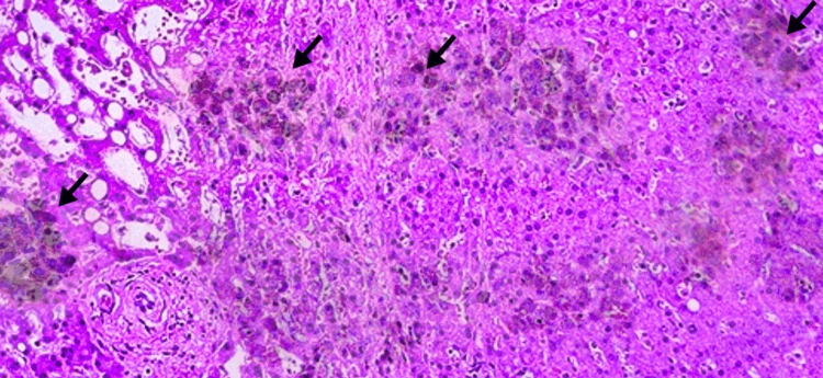
Use the black liver as a supporting finding, not a biopsy order Dubin-Johnson syndrome classically produces a dark or black liver at gross examination. Microscopy shows coarse pigment granules in centrilobular hepatocytes, often within lysosomes. Centrilobular means near the central vein of a liver lobule. The surrounding architecture is usually preserved. The pigment is not evidence that the whole organ has become cirrhotic. [1] [5]
Enlarge the whole image Published Dubin-Johnson histology example: coarse intracellular pigment. Original arrows are retained; no markers were added. This illustrates an association, not an indication for routine biopsy.
Siddiqui, Alsabe, Tehseen, Hatamleh, Taslim, Abdelrahman and Saleem (2023), Figure 1. CC BY 4.0 . Source and provenance
Whole teaching image Close Show full-size image Published Dubin-Johnson histology example: coarse intracellular pigment. Original arrows are retained; no markers were added. This illustrates an association, not an indication for routine biopsy.
Siddiqui, Alsabe, Tehseen, Hatamleh, Taslim, Abdelrahman and Saleem (2023), Figure 1. CC BY 4.0 . Source and provenance
Use the image without ordering an unnecessary biopsy A specimen contains coarse dark hepatocyte pigment with preserved architecture. Which association fits?
Check this thought That is compatible with the characteristic pigment described in Dubin-Johnson.
An ultrasound does not mention pigment. Does it exclude the association?
Check this thought No. Ultrasound is not a histological examination of intracellular granules.
A stable biochemical pattern is already convincing. Is routine biopsy needed?
Check this thought No. The pathology association does not itself create an indication for an invasive test.
Recognize the pigment without treating biopsy as a routine requirement. [1] [2] [5]
Try this reasoning again Read every step
Use the black liver as a supporting finding, not a biopsy order Match the tissue findings and testing decision without turning a remembered liver color into an automatic biopsy order.
Dubin-Johnson tissue Rotor tissue Practical decision
Do not obtain invasive tissue solely to demonstrate a memorized color. No characteristic dark hepatic pigment. Coarse centrilobular pigment with generally preserved architecture.
Try again Read the worked solution Dubin-Johnson tissue Coarse centrilobular pigment with generally preserved architecture. [1] [5]
Rotor tissue No characteristic dark hepatic pigment. [2]
Practical decision Do not obtain invasive tissue solely to demonstrate a memorized color. [2] [5]
The retained dark material is distinct from bilirubin itself and reflects the broader transport role of the affected protein. Rotor syndrome does not have this characteristic dark pigment accumulation and usually has normal histology. This is a useful comparison when tissue already exists for another indication. It is not a reason to biopsy every otherwise well patient with a fitting biochemical pattern. [1] [2] [5]
An absent mention of black pigment on an ultrasound report does not exclude Dubin-Johnson syndrome. Neither does an unexamined liver provide a meaningful negative histologic result. Start with fractionated bilirubin, the remainder of the liver panel, exclusion of competing causes, and targeted testing. Biopsy is not routinely needed merely to establish a benign hereditary jaundice diagnosis. [2] [5]
Historical dye-clearance tests and hepatobiliary scintigraphy illustrate the transport differences, but can also confuse interpretation. In Dubin-Johnson syndrome, liver uptake may be maintained while biliary excretion is delayed. In Rotor syndrome, hepatic uptake is markedly delayed. Such patterns must not automatically be interpreted as a gallstone requiring surgery. Modern specialist evaluation can use molecular testing when a clinical diagnosis remains uncertain. [2] [5]
Dubin-Johnson tissue Coarse centrilobular pigment with generally preserved architecture. [1] [5]
Rotor tissue No characteristic dark hepatic pigment. [2]
Practical decision Do not obtain invasive tissue solely to demonstrate a memorized color. [2] [5]
Save this spot
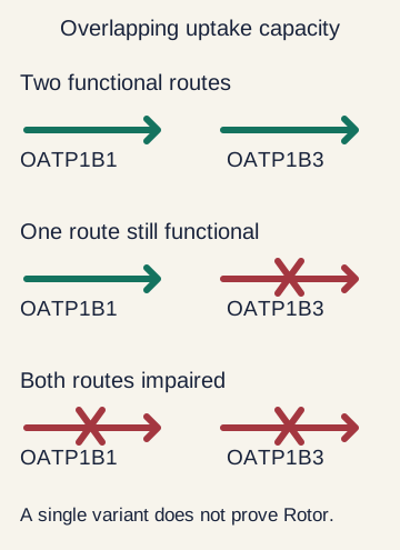
Match the genetic result to the transport model A confirmed pathogenic ABCC2 genotype can support Dubin-Johnson syndrome when the clinical phenotype fits. Rotor syndrome is different. Both SLCO1B1 and SLCO1B3 must have functionally important loss affecting both copies. The genes are close together, so the inherited pattern commonly resembles an ordinary recessive disorder even though two genes participate. [1] [2]
Enlarge the whole image The uptake systems overlap. A single reduced-function variant does not automatically establish the combined functional loss of classic Rotor syndrome.
Bone Wizardry. Original educational schematic. Source and provenance
Whole teaching image Close Show full-size image The uptake systems overlap. A single reduced-function variant does not automatically establish the combined functional loss of classic Rotor syndrome.
Bone Wizardry. Original educational schematic. Source and provenance
A variant is not the same as lost function ABCC2 encodes which transport system?
Check this thought It encodes MRP2, the bile-facing export transporter.
Rotor involves which combined transport loss?
Check this thought Loss of both OATP1B1 and OATP1B3, encoded by SLCO1B1 and SLCO1B3.
One reduced-function variant or an uncertain variant is reported. Is the syndrome proven?
Check this thought No. Interpret the actual combined functional effect, variant classification, and clinical pattern.
Name the genotype precisely before assigning a syndromic diagnosis. [1] [2]
Try this reasoning again Read every step
A single common reduced-function SLCO1B1 variant is not equivalent to Rotor syndrome. The two transporters have overlapping capacity. GeneReviews notes that at least one functional allele of either gene prevents classic Rotor-type hyperbilirubinemia. This matters when a pharmacogenetic test reports one transporter variant and a clinician is tempted to turn that result into a complete disease diagnosis. [2]
Variant interpretation also matters. A variant of uncertain significance is not a confirmed pathogenic result. An uncertain genetic report should be reconciled with the phenotype, laboratory pattern, family findings, and the limits of the assay. Genetic counseling can help a family understand which variants were identified and what those results do and do not establish. [2]
Most importantly, a genetic explanation does not make current symptoms irrelevant. A person with an inherited transport disorder may separately develop acquired disease. Preserve the documented baseline in the record, then investigate a meaningful change from it. This avoids both unnecessary testing of a stable pattern and premature reassurance about a new one. [2]
ABCC2 Fits a canalicular export disorder. [1]
Both SLCO1B1 and SLCO1B3 Combined biallelic loss fits Rotor syndrome. [2]
Uncertain or single-gene finding Requires careful interpretation rather than automatic syndrome labeling. [2]
Save this spot
Reassure the stable pattern and investigate departures from it Uncomplicated adult Dubin-Johnson and Rotor syndromes usually do not require bilirubin-lowering treatment. Once a sound diagnosis has excluded a more harmful cause, explain that intermittent visible jaundice can occur without progressive injury or liver failure. Repeated invasive testing simply because the same isolated bilirubin result persists adds burden without necessarily adding useful information. [1] [2] [5]
Reassurance has conditions A typical stable adult pattern remains isolated. Is bilirubin-lowering therapy routinely required?
Check this thought No. These inherited transport disorders generally need explanation rather than treatment of the bilirubin number.
New itching, pale stools, or abnormal liver enzymes appear. Does the old label settle this?
Check this thought No. Those changes need assessment for an additional hepatobiliary problem.
A young infant has pale stools and conjugated jaundice. Is family history reassuring enough?
Check this thought No. Infant cholestasis needs urgent age-appropriate evaluation rather than borrowing reassurance from an adult relative.
A benign baseline does not explain away a new pattern. [1] [2] [4] [5]
Try this reasoning again Read every step
Reassure the stable pattern and investigate departures from it Match a stable adult pattern, a medicine question, and a changed presentation with their appropriate assessment goals.
Stable isolated bilirubin Medication decision New symptoms or infant cholestasis
Reopen the differential rather than borrowing reassurance from a typical adult case. Review the relevant transporter and actual prescribing information. Explain the benign pattern and avoid unnecessary treatment.
Try again Read the worked solution Stable isolated bilirubin Explain the benign pattern and avoid unnecessary treatment. [1] [2] [5]
Medication decision Review the relevant transporter and actual prescribing information. [2]
New symptoms or infant cholestasis Reopen the differential rather than borrowing reassurance from a typical adult case. [1] [2]
Illness, pregnancy, and estrogen exposure can make Dubin-Johnson jaundice more noticeable. That history can explain a fluctuation, but should not become a blanket dismissal. New itching during pregnancy, abnormal liver enzymes, impaired coagulation, fever, or persistent abdominal pain still deserves assessment for pregnancy-related or other hepatobiliary disease. [2] [5]
Transporters also handle medicines and their metabolites. Tell the prescribing team about the diagnosis and review relevant medications rather than treating every drug as forbidden. GeneReviews warns that absent OATP1B function could affect hepatic uptake and toxicity, while also noting that documented adverse drug effects specifically in Rotor syndrome are limited. A theoretical transport concern is not itself a dose instruction. [2]
Rare neonatal presentations exist, but an infant with conjugated jaundice must not be reassured by the benign adult description. Infant cholestasis needs timely evaluation for obstruction and treatable medical disease. The final practical rule is to pair the inherited diagnosis with its expected phenotype, then identify any finding that falls outside that phenotype. [1] [2]
Stable isolated bilirubin Explain the benign pattern and avoid unnecessary treatment. [1] [2] [5]
Medication decision Review the relevant transporter and actual prescribing information. [2]
New symptoms or infant cholestasis Reopen the differential rather than borrowing reassurance from a typical adult case. [1] [2]
Try it here · Checkpoint 3 of 3
Make your prediction before reading the choices. A first attempt is just a starting point.
Case 13
Show answer and explanations for case 13
A. Assess for an additional cholestatic or obstructive disorder (Best answer)
Work through this option Does the new episode remain isolated?
Check this thought No. Itching, pale stools and elevated ALP are a changed pattern.
How does this option fit the finding?
Check this thought These are departures from the expected uncomplicated isolated bilirubin pattern.
What rule transfers to a new case?
Check this thought A benign diagnosis explains its expected phenotype, not every future liver-related symptom.
A benign diagnosis explains its expected phenotype, not every future liver-related symptom. [2] [5]
Try this reasoning again Read every step
These are departures from the expected uncomplicated isolated bilirubin pattern.
B. Attribute all findings to Dubin-Johnson without further evaluation (Why this does not fit)
Work through this option Does the new episode remain isolated?
Check this thought No. Itching, pale stools and elevated ALP are a changed pattern.
How does this option fit the finding?
Check this thought The new symptoms and enzyme change should prevent anchoring on the old diagnosis.
A benign diagnosis explains its expected phenotype, not every future liver-related symptom. [2] [5]
Try this reasoning again Read every step
The new symptoms and enzyme change should prevent anchoring on the old diagnosis.
C. Switch the diagnosis to Gilbert syndrome solely because itching is present (Why this does not fit)
Work through this option Does the new episode remain isolated?
Check this thought No. Itching, pale stools and elevated ALP are a changed pattern.
How does this option fit the finding?
Check this thought Gilbert is an unconjugated disorder and does not explain the new cholestatic findings.
A benign diagnosis explains its expected phenotype, not every future liver-related symptom. [2] [5]
Try this reasoning again Read every step
Gilbert is an unconjugated disorder and does not explain the new cholestatic findings.
D. Treat the ALP rise as proof of cirrhosis without assessing its cause (Why this does not fit)
Work through this option Does the new episode remain isolated?
Check this thought No. Itching, pale stools and elevated ALP are a changed pattern.
How does this option fit the finding?
Check this thought The abnormality requires evaluation but does not alone establish cirrhosis or its mechanism.
A benign diagnosis explains its expected phenotype, not every future liver-related symptom. [2] [5]
Try this reasoning again Read every step
The abnormality requires evaluation but does not alone establish cirrhosis or its mechanism.
Takeaway: A benign diagnosis explains its expected phenotype, not every future liver-related symptom.
Case sources: [2] [5]
Save this spot
Apply the lesson to clinical cases You can pause here and return to any case. Choose an answer when ready, then compare the explanations.
Case 2
Show answer and explanations for case 2
A. Dubin-Johnson syndrome (Why this does not fit)
Work through this option Which transporters lost function while MRP2 was preserved?
Check this thought Both OATP1B1 and OATP1B3 lost function, pointing to the blood-facing recovery route.
How does this option fit the finding?
Check this thought Dubin-Johnson primarily affects MRP2-mediated canalicular export rather than the two reuptake transporters.
A conjugated increase can reflect failed reuptake, not only failed biliary export. [2] [4]
Try this reasoning again Read every step
Dubin-Johnson primarily affects MRP2-mediated canalicular export rather than the two reuptake transporters.
B. Gilbert syndrome (Why this does not fit)
Work through this option Which transporters lost function while MRP2 was preserved?
Check this thought Both OATP1B1 and OATP1B3 lost function, pointing to the blood-facing recovery route.
How does this option fit the finding?
Check this thought Gilbert is a UGT1A1-related unconjugated pattern, not combined OATP deficiency.
A conjugated increase can reflect failed reuptake, not only failed biliary export. [2] [4]
Try this reasoning again Read every step
Gilbert is a UGT1A1-related unconjugated pattern, not combined OATP deficiency.
C. Crigler-Najjar syndrome type II (Why this does not fit)
Work through this option Which transporters lost function while MRP2 was preserved?
Check this thought Both OATP1B1 and OATP1B3 lost function, pointing to the blood-facing recovery route.
How does this option fit the finding?
Check this thought This also concerns impaired conjugation and does not explain the stated transporter findings.
A conjugated increase can reflect failed reuptake, not only failed biliary export. [2] [4]
Try this reasoning again Read every step
This also concerns impaired conjugation and does not explain the stated transporter findings.
D. Rotor syndrome (Best answer)
Work through this option Which transporters lost function while MRP2 was preserved?
Check this thought Both OATP1B1 and OATP1B3 lost function, pointing to the blood-facing recovery route.
How does this option fit the finding?
Check this thought Combined loss of hepatic sinusoidal reuptake transporters with intact MRP2 is the Rotor mechanism.
What rule transfers to a new case?
Check this thought A conjugated increase can reflect failed reuptake, not only failed biliary export.
A conjugated increase can reflect failed reuptake, not only failed biliary export. [2] [4]
Try this reasoning again Read every step
Combined loss of hepatic sinusoidal reuptake transporters with intact MRP2 is the Rotor mechanism.
Takeaway: A conjugated increase can reflect failed reuptake, not only failed biliary export.
Case sources: [2] [4]
Case 3
Show answer and explanations for case 3
A. Gilbert syndrome (Why this does not fit)
Work through this option Is the total output elevated or is the mixture shifted?
Check this thought The total is normal; the isomer-I fraction is markedly increased.
How does this option fit the finding?
Check this thought Gilbert raises mainly unconjugated bilirubin and does not produce this characteristic isomer pattern.
Read the total and the isomer fraction together. [2] [3] [5]
Try this reasoning again Read every step
Gilbert raises mainly unconjugated bilirubin and does not produce this characteristic isomer pattern.
B. Acute obstructive cholangitis (Why this does not fit)
Work through this option Is the total output elevated or is the mixture shifted?
Check this thought The total is normal; the isomer-I fraction is markedly increased.
How does this option fit the finding?
Check this thought That diagnosis requires evidence of infection and obstruction, not this isolated stable biochemical pattern.
Read the total and the isomer fraction together. [2] [3] [5]
Try this reasoning again Read every step
That diagnosis requires evidence of infection and obstruction, not this isolated stable biochemical pattern.
C. Dubin-Johnson syndrome (Best answer)
Work through this option Is the total output elevated or is the mixture shifted?
Check this thought The total is normal; the isomer-I fraction is markedly increased.
How does this option fit the finding?
Check this thought Normal total excretion with marked isomer I predominance is the characteristic Dubin-Johnson pattern.
What rule transfers to a new case?
Check this thought Read the total and the isomer fraction together.
Read the total and the isomer fraction together. [2] [3] [5]
Try this reasoning again Read every step
Normal total excretion with marked isomer I predominance is the characteristic Dubin-Johnson pattern.
D. Rotor syndrome (Why this does not fit)
Work through this option Is the total output elevated or is the mixture shifted?
Check this thought The total is normal; the isomer-I fraction is markedly increased.
How does this option fit the finding?
Check this thought Rotor more typically increases total coproporphyrin several-fold with isomer I around 65 percent.
Read the total and the isomer fraction together. [2] [3] [5]
Try this reasoning again Read every step
Rotor more typically increases total coproporphyrin several-fold with isomer I around 65 percent.
Takeaway: Read the total and the isomer fraction together.
Case sources: [2] [3] [5]
Case 5
Show answer and explanations for case 5
A. Coproporphyrinuria can occur in other liver diseases and must be interpreted with the full phenotype (Best answer)
Work through this option Is this an isolated bilirubin pattern?
Check this thought No. Elevated ALT and ALP require attention to other hepatobiliary disease.
How does this option fit the finding?
Check this thought The enzyme abnormalities are not the expected isolated benign pattern, and increased total output is not specific.
What rule transfers to a new case?
Check this thought A characteristic test pattern is useful only within a matching clinical context.
A characteristic test pattern is useful only within a matching clinical context. [2]
Try this reasoning again Read every step
The enzyme abnormalities are not the expected isolated benign pattern, and increased total output is not specific.
B. Any increased coproporphyrin total confirms Rotor regardless of other tests (Why this does not fit)
Work through this option Is this an isolated bilirubin pattern?
Check this thought No. Elevated ALT and ALP require attention to other hepatobiliary disease.
How does this option fit the finding?
Check this thought This ignores both the lack of specificity and the new evidence of liver injury.
A characteristic test pattern is useful only within a matching clinical context. [2]
Try this reasoning again Read every step
This ignores both the lack of specificity and the new evidence of liver injury.
C. The result instead proves Dubin-Johnson syndrome (Why this does not fit)
Work through this option Is this an isolated bilirubin pattern?
Check this thought No. Elevated ALT and ALP require attention to other hepatobiliary disease.
How does this option fit the finding?
Check this thought Dubin-Johnson typically has normal total output, and the result alone cannot establish that diagnosis either.
A characteristic test pattern is useful only within a matching clinical context. [2]
Try this reasoning again Read every step
Dubin-Johnson typically has normal total output, and the result alone cannot establish that diagnosis either.
D. Urinary coproporphyrins distinguish every obstructive from nonobstructive disorder (Why this does not fit)
Work through this option Is this an isolated bilirubin pattern?
Check this thought No. Elevated ALT and ALP require attention to other hepatobiliary disease.
How does this option fit the finding?
Check this thought They are a supporting transport test, not a universal anatomic or etiologic classifier.
A characteristic test pattern is useful only within a matching clinical context. [2]
Try this reasoning again Read every step
They are a supporting transport test, not a universal anatomic or etiologic classifier.
Takeaway: A characteristic test pattern is useful only within a matching clinical context.
Case sources: [2]
Case 6
Show answer and explanations for case 6
A. Black pigment alone establishes advanced cirrhosis (Why this does not fit)
Work through this option Is the dark pigment accompanied by destroyed architecture?
Check this thought No. The specimen describes coarse pigment with otherwise preserved architecture.
How does this option fit the finding?
Check this thought Fibrosis and architectural disruption, not gross color alone, establish cirrhosis.
Dark hepatic pigment and preserved architecture can coexist in a benign transport disorder. [1] [2] [5]
Try this reasoning again Read every step
Fibrosis and architectural disruption, not gross color alone, establish cirrhosis.
B. The finding is characteristic of Rotor syndrome (Why this does not fit)
Work through this option Is the dark pigment accompanied by destroyed architecture?
Check this thought No. The specimen describes coarse pigment with otherwise preserved architecture.
How does this option fit the finding?
Check this thought Rotor typically lacks the dark pigment described.
Dark hepatic pigment and preserved architecture can coexist in a benign transport disorder. [1] [2] [5]
Try this reasoning again Read every step
Rotor typically lacks the dark pigment described.
C. The granules show isolated UGT1A1 deficiency (Why this does not fit)
Work through this option Is the dark pigment accompanied by destroyed architecture?
Check this thought No. The specimen describes coarse pigment with otherwise preserved architecture.
How does this option fit the finding?
Check this thought The pigment supports a transport disorder rather than the usual conjugation-defect phenotype.
Dark hepatic pigment and preserved architecture can coexist in a benign transport disorder. [1] [2] [5]
Try this reasoning again Read every step
The pigment supports a transport disorder rather than the usual conjugation-defect phenotype.
D. The pigment supports Dubin-Johnson syndrome without itself indicating cirrhosis (Best answer)
Work through this option Is the dark pigment accompanied by destroyed architecture?
Check this thought No. The specimen describes coarse pigment with otherwise preserved architecture.
How does this option fit the finding?
Check this thought This is the classic distribution in an otherwise structurally preserved liver.
What rule transfers to a new case?
Check this thought Dark hepatic pigment and preserved architecture can coexist in a benign transport disorder.
Dark hepatic pigment and preserved architecture can coexist in a benign transport disorder. [1] [2] [5]
Try this reasoning again Read every step
This is the classic distribution in an otherwise structurally preserved liver.
Takeaway: Dark hepatic pigment and preserved architecture can coexist in a benign transport disorder.
Case sources: [1] [2] [5]
Case 7
Show answer and explanations for case 7
A. Perform ERCP to prove there is no stone despite no obstructive evidence (Why this does not fit)
Work through this option Is an invasive procedure needed merely to demonstrate an association?
Check this thought No. A convincing stable biochemical pattern does not automatically require biopsy.
How does this option fit the finding?
Check this thought An invasive duct procedure is not justified solely by a stable inherited transport pattern.
Do not turn a useful pathology association into an unnecessary procedure. [2] [5]
Try this reasoning again Read every step
An invasive duct procedure is not justified solely by a stable inherited transport pattern.
B. Begin chronic phenobarbital to test whether bilirubin can be lowered (Why this does not fit)
Work through this option Is an invasive procedure needed merely to demonstrate an association?
Check this thought No. A convincing stable biochemical pattern does not automatically require biopsy.
How does this option fit the finding?
Check this thought A drug challenge is not needed to establish this export disorder or treat a benign isolated result.
Do not turn a useful pathology association into an unnecessary procedure. [2] [5]
Try this reasoning again Read every step
A drug challenge is not needed to establish this export disorder or treat a benign isolated result.
C. Explain the diagnosis without routine liver biopsy solely to demonstrate pigment (Best answer)
Work through this option Is an invasive procedure needed merely to demonstrate an association?
Check this thought No. A convincing stable biochemical pattern does not automatically require biopsy.
How does this option fit the finding?
Check this thought Invasive tissue is usually unnecessary when the clinical and targeted laboratory findings fit.
What rule transfers to a new case?
Check this thought Do not turn a useful pathology association into an unnecessary procedure.
Do not turn a useful pathology association into an unnecessary procedure. [2] [5]
Try this reasoning again Read every step
Invasive tissue is usually unnecessary when the clinical and targeted laboratory findings fit.
D. Require biopsy before any reassurance because pigment is mandatory to see (Why this does not fit)
Work through this option Is an invasive procedure needed merely to demonstrate an association?
Check this thought No. A convincing stable biochemical pattern does not automatically require biopsy.
How does this option fit the finding?
Check this thought The characteristic pigment is not an instruction to obtain invasive tissue in every case.
Do not turn a useful pathology association into an unnecessary procedure. [2] [5]
Try this reasoning again Read every step
The characteristic pigment is not an instruction to obtain invasive tissue in every case.
Takeaway: Do not turn a useful pathology association into an unnecessary procedure.
Case sources: [2] [5]
Case 8
Show answer and explanations for case 8
A. The bilirubin must be predominantly unconjugated (Why this does not fit)
Work through this option Did ultrasound examine intracellular pigment granules?
Check this thought No. The report is not a histological assessment.
How does this option fit the finding?
Check this thought An imaging description cannot override the measured bilirubin fraction.
A finding that was not meaningfully assessed cannot be used as a decisive exclusion. [2] [5]
Try this reasoning again Read every step
An imaging description cannot override the measured bilirubin fraction.
B. The report does not provide a reliable negative histologic test for Dubin-Johnson syndrome (Best answer)
Work through this option Did ultrasound examine intracellular pigment granules?
Check this thought No. The report is not a histological assessment.
How does this option fit the finding?
Check this thought Failure to describe pigment on routine imaging does not exclude the transport disorder.
What rule transfers to a new case?
Check this thought A finding that was not meaningfully assessed cannot be used as a decisive exclusion.
A finding that was not meaningfully assessed cannot be used as a decisive exclusion. [2] [5]
Try this reasoning again Read every step
Failure to describe pigment on routine imaging does not exclude the transport disorder.
C. Dubin-Johnson syndrome has been definitively excluded (Why this does not fit)
Work through this option Did ultrasound examine intracellular pigment granules?
Check this thought No. The report is not a histological assessment.
How does this option fit the finding?
Check this thought The absence of an imaging description is not equivalent to a diagnostic tissue examination.
A finding that was not meaningfully assessed cannot be used as a decisive exclusion. [2] [5]
Try this reasoning again Read every step
The absence of an imaging description is not equivalent to a diagnostic tissue examination.
D. Rotor syndrome is confirmed without additional assessment (Why this does not fit)
Work through this option Did ultrasound examine intracellular pigment granules?
Check this thought No. The report is not a histological assessment.
How does this option fit the finding?
Check this thought A missing pigment description does not establish combined OATP deficiency.
A finding that was not meaningfully assessed cannot be used as a decisive exclusion. [2] [5]
Try this reasoning again Read every step
A missing pigment description does not establish combined OATP deficiency.
Takeaway: A finding that was not meaningfully assessed cannot be used as a decisive exclusion.
Case sources: [2] [5]
Case 9
Show answer and explanations for case 9
A. No; classic Rotor syndrome requires combined loss affecting both transporter genes (Best answer)
Work through this option Is the second uptake system still functional?
Check this thought Yes. The report describes intact SLCO1B3 function, not proven combined uptake loss.
How does this option fit the finding?
Check this thought Their overlapping function prevents equating a single reduced-function finding with the complete disease phenotype.
What rule transfers to a new case?
Check this thought A pharmacogenetic variant and a syndromic diagnosis are not interchangeable.
A pharmacogenetic variant and a syndromic diagnosis are not interchangeable. [2]
Try this reasoning again Read every step
Their overlapping function prevents equating a single reduced-function finding with the complete disease phenotype.
B. Yes; any SLCO1B1 variant is sufficient (Why this does not fit)
Work through this option Is the second uptake system still functional?
Check this thought Yes. The report describes intact SLCO1B3 function, not proven combined uptake loss.
How does this option fit the finding?
Check this thought GeneReviews requires a combined biallelic loss pattern rather than any isolated variant.
A pharmacogenetic variant and a syndromic diagnosis are not interchangeable. [2]
Try this reasoning again Read every step
GeneReviews requires a combined biallelic loss pattern rather than any isolated variant.
C. Yes; intact SLCO1B3 is the defining confirmation (Why this does not fit)
Work through this option Is the second uptake system still functional?
Check this thought Yes. The report describes intact SLCO1B3 function, not proven combined uptake loss.
How does this option fit the finding?
Check this thought Preserved transporter function argues against the complete combined deficiency.
A pharmacogenetic variant and a syndromic diagnosis are not interchangeable. [2]
Try this reasoning again Read every step
Preserved transporter function argues against the complete combined deficiency.
D. No; Rotor is caused exclusively by ABCC2 (Why this does not fit)
Work through this option Is the second uptake system still functional?
Check this thought Yes. The report describes intact SLCO1B3 function, not proven combined uptake loss.
How does this option fit the finding?
Check this thought ABCC2 causes Dubin-Johnson; rejecting the single-variant conclusion does not change the Rotor genes.
A pharmacogenetic variant and a syndromic diagnosis are not interchangeable. [2]
Try this reasoning again Read every step
ABCC2 causes Dubin-Johnson; rejecting the single-variant conclusion does not change the Rotor genes.
Takeaway: A pharmacogenetic variant and a syndromic diagnosis are not interchangeable.
Case sources: [2]
Case 10
Show answer and explanations for case 10
A. The variants establish the diagnosis because both gene names appear (Why this does not fit)
Work through this option Are uncertain variants established pathogenic findings?
Check this thought No. Their classification cannot be silently upgraded to diagnostic proof.
How does this option fit the finding?
Check this thought The names alone do not show that the variants abolish the required transporter functions.
Genetic certainty must match the actual classification of the variants. [2]
Try this reasoning again Read every step
The names alone do not show that the variants abolish the required transporter functions.
B. The variants prove there is no inherited disorder (Why this does not fit)
Work through this option Are uncertain variants established pathogenic findings?
Check this thought No. Their classification cannot be silently upgraded to diagnostic proof.
How does this option fit the finding?
Check this thought Uncertainty is not evidence that a variant is benign or that every inherited cause has been excluded.
Genetic certainty must match the actual classification of the variants. [2]
Try this reasoning again Read every step
Uncertainty is not evidence that a variant is benign or that every inherited cause has been excluded.
C. The result proves progressive liver failure (Why this does not fit)
Work through this option Are uncertain variants established pathogenic findings?
Check this thought No. Their classification cannot be silently upgraded to diagnostic proof.
How does this option fit the finding?
Check this thought Variant classification does not determine current hepatic synthetic function.
Genetic certainty must match the actual classification of the variants. [2]
Try this reasoning again Read every step
Variant classification does not determine current hepatic synthetic function.
D. The result does not by itself confirm or exclude Rotor syndrome (Best answer)
Work through this option Are uncertain variants established pathogenic findings?
Check this thought No. Their classification cannot be silently upgraded to diagnostic proof.
How does this option fit the finding?
Check this thought Uncertain variants require phenotype, family, and laboratory interpretation rather than being treated as established pathogenic alleles.
What rule transfers to a new case?
Check this thought Genetic certainty must match the actual classification of the variants.
Genetic certainty must match the actual classification of the variants. [2]
Try this reasoning again Read every step
Uncertain variants require phenotype, family, and laboratory interpretation rather than being treated as established pathogenic alleles.
Takeaway: Genetic certainty must match the actual classification of the variants.
Case sources: [2]
Case 11
Show answer and explanations for case 11
A. UGT1A1 normally reverses conjugation before bilirubin reaches blood (Why this does not fit)
Work through this option Can processed bilirubin return from liver cells to blood?
Check this thought Yes. Basolateral export provides a route back to blood before hepatic reuptake.
How does this option fit the finding?
Check this thought Reuptake does not require the proposed reversal of the hepatic reaction.
A pathway can return processed material to blood before final excretion. [3] [4]
Try this reasoning again Read every step
Reuptake does not require the proposed reversal of the hepatic reaction.
B. MRP2 and OATP1B1 are two names for the same transporter (Why this does not fit)
Work through this option Can processed bilirubin return from liver cells to blood?
Check this thought Yes. Basolateral export provides a route back to blood before hepatic reuptake.
How does this option fit the finding?
Check this thought They operate at different hepatocyte surfaces and perform different transport steps.
A pathway can return processed material to blood before final excretion. [3] [4]
Try this reasoning again Read every step
They operate at different hepatocyte surfaces and perform different transport steps.
C. MRP3 returns some conjugated pigment to blood, where OATP transporters normally recover it (Best answer)
Work through this option Can processed bilirubin return from liver cells to blood?
Check this thought Yes. Basolateral export provides a route back to blood before hepatic reuptake.
How does this option fit the finding?
Check this thought Rotor disrupts a recycling step after conjugation, allowing more pigment to remain in circulation.
What rule transfers to a new case?
Check this thought A pathway can return processed material to blood before final excretion.
A pathway can return processed material to blood before final excretion. [3] [4]
Try this reasoning again Read every step
Rotor disrupts a recycling step after conjugation, allowing more pigment to remain in circulation.
D. All circulating conjugated bilirubin must have been produced by kidney cells (Why this does not fit)
Work through this option Can processed bilirubin return from liver cells to blood?
Check this thought Yes. Basolateral export provides a route back to blood before hepatic reuptake.
How does this option fit the finding?
Check this thought Hepatocytes conjugate bilirubin; a return route through blood explains the finding without renal production.
A pathway can return processed material to blood before final excretion. [3] [4]
Try this reasoning again Read every step
Hepatocytes conjugate bilirubin; a return route through blood explains the finding without renal production.
Takeaway: A pathway can return processed material to blood before final excretion.
Case sources: [3] [4]
Case 12
Show answer and explanations for case 12
A. Promise that no liver disease can occur during this pregnancy (Why this does not fit)
Work through this option Does this episode still match the established isolated pattern?
Check this thought Yes. The scenario reports reassuring examination, enzymes and synthetic function without itching or pain.
How does this option fit the finding?
Check this thought The inherited diagnosis does not exclude a later independent pregnancy-related or acquired disorder.
Reassure a fitting baseline without promising immunity from new disease. [2] [5]
Try this reasoning again Read every step
The inherited diagnosis does not exclude a later independent pregnancy-related or acquired disorder.
B. Review the clinical context and explain that the inherited pattern can become more noticeable during pregnancy (Best answer)
Work through this option Does this episode still match the established isolated pattern?
Check this thought Yes. The scenario reports reassuring examination, enzymes and synthetic function without itching or pain.
How does this option fit the finding?
Check this thought A similar isolated fluctuation can occur without evidence of new liver injury; new symptoms would change the assessment.
What rule transfers to a new case?
Check this thought Reassure a fitting baseline without promising immunity from new disease.
Reassure a fitting baseline without promising immunity from new disease. [2] [5]
Try this reasoning again Read every step
A similar isolated fluctuation can occur without evidence of new liver injury; new symptoms would change the assessment.
C. Diagnose intrahepatic cholestasis of pregnancy solely from bilirubin (Why this does not fit)
Work through this option Does this episode still match the established isolated pattern?
Check this thought Yes. The scenario reports reassuring examination, enzymes and synthetic function without itching or pain.
How does this option fit the finding?
Check this thought That diagnosis needs the appropriate clinical and biochemical assessment rather than bilirubin alone.
Reassure a fitting baseline without promising immunity from new disease. [2] [5]
Try this reasoning again Read every step
That diagnosis needs the appropriate clinical and biochemical assessment rather than bilirubin alone.
D. Start an invasive obstruction workup automatically despite no new supporting findings (Why this does not fit)
Work through this option Does this episode still match the established isolated pattern?
Check this thought Yes. The scenario reports reassuring examination, enzymes and synthetic function without itching or pain.
How does this option fit the finding?
Check this thought A stable known pattern should guide proportional testing, not an automatic procedure.
Reassure a fitting baseline without promising immunity from new disease. [2] [5]
Try this reasoning again Read every step
A stable known pattern should guide proportional testing, not an automatic procedure.
Takeaway: Reassure a fitting baseline without promising immunity from new disease.
Case sources: [2] [5]
Case 14
Show answer and explanations for case 14
A. Declare every prescription medicine contraindicated (Why this does not fit)
Work through this option Does no bilirubin-specific treatment mean transport is irrelevant to drugs?
Check this thought No. Hepatic transport can still matter for individual medicines.
How does this option fit the finding?
Check this thought The risk is drug-specific; a blanket prohibition is unsupported.
No treatment for jaundice does not mean no relevance to medication review. [2]
Try this reasoning again Read every step
The risk is drug-specific; a blanket prohibition is unsupported.
B. Assume drug handling is normal because bilirubin needs no treatment (Why this does not fit)
Work through this option Does no bilirubin-specific treatment mean transport is irrelevant to drugs?
Check this thought No. Hepatic transport can still matter for individual medicines.
How does this option fit the finding?
Check this thought Benign jaundice does not mean the missing transporters have no other substrates.
No treatment for jaundice does not mean no relevance to medication review. [2]
Try this reasoning again Read every step
Benign jaundice does not mean the missing transporters have no other substrates.
C. Use the bilirubin concentration alone to calculate a replacement dose (Why this does not fit)
Work through this option Does no bilirubin-specific treatment mean transport is irrelevant to drugs?
Check this thought No. Hepatic transport can still matter for individual medicines.
How does this option fit the finding?
Check this thought A bilirubin value is not a validated universal dosing equation for transporter substrates.
No treatment for jaundice does not mean no relevance to medication review. [2]
Try this reasoning again Read every step
A bilirubin value is not a validated universal dosing equation for transporter substrates.
D. Review drug-specific uptake and prescribing information with the treating team (Best answer)
Work through this option Does no bilirubin-specific treatment mean transport is irrelevant to drugs?
Check this thought No. Hepatic transport can still matter for individual medicines.
How does this option fit the finding?
Check this thought Transport loss can alter exposure, but the diagnosis does not supply a universal contraindication or dose rule.
What rule transfers to a new case?
Check this thought No treatment for jaundice does not mean no relevance to medication review.
No treatment for jaundice does not mean no relevance to medication review. [2]
Try this reasoning again Read every step
Transport loss can alter exposure, but the diagnosis does not supply a universal contraindication or dose rule.
Takeaway: No treatment for jaundice does not mean no relevance to medication review.
Case sources: [2]
Case 15
Show answer and explanations for case 15
A. Use absent itching to exclude all dangerous causes (Why this does not fit)
Work through this option Is this an evaluated stable adult phenotype?
Check this thought No. This is an infant with conjugated jaundice and pale stools who needs prompt evaluation.
How does this option fit the finding?
Check this thought Infants may have serious cholestasis without an adult symptom pattern.
Do not transfer reassurance from a typical adult phenotype to an unevaluated infant. [1] [2]
Try this reasoning again Read every step
Infants may have serious cholestasis without an adult symptom pattern.
B. Diagnose physiologic jaundice because inherited disorders are always unconjugated (Why this does not fit)
Work through this option Is this an evaluated stable adult phenotype?
Check this thought No. This is an infant with conjugated jaundice and pale stools who needs prompt evaluation.
How does this option fit the finding?
Check this thought Dubin-Johnson itself is conjugated, and infant direct hyperbilirubinemia is not ordinary physiologic jaundice.
Do not transfer reassurance from a typical adult phenotype to an unevaluated infant. [1] [2]
Try this reasoning again Read every step
Dubin-Johnson itself is conjugated, and infant direct hyperbilirubinemia is not ordinary physiologic jaundice.
C. Arrange timely infant cholestasis evaluation rather than assuming the familial benign adult pattern (Best answer)
Work through this option Is this an evaluated stable adult phenotype?
Check this thought No. This is an infant with conjugated jaundice and pale stools who needs prompt evaluation.
How does this option fit the finding?
Check this thought Rare neonatal inherited disease is possible, but obstruction and treatable medical causes must be assessed promptly.
What rule transfers to a new case?
Check this thought Do not transfer reassurance from a typical adult phenotype to an unevaluated infant.
Do not transfer reassurance from a typical adult phenotype to an unevaluated infant. [1] [2]
Try this reasoning again Read every step
Rare neonatal inherited disease is possible, but obstruction and treatable medical causes must be assessed promptly.
D. Reassure solely from the relative diagnosis and wait for adolescence (Why this does not fit)
Work through this option Is this an evaluated stable adult phenotype?
Check this thought No. This is an infant with conjugated jaundice and pale stools who needs prompt evaluation.
How does this option fit the finding?
Check this thought Family history does not safely explain an infant cholestatic presentation without evaluation.
Do not transfer reassurance from a typical adult phenotype to an unevaluated infant. [1] [2]
Try this reasoning again Read every step
Family history does not safely explain an infant cholestatic presentation without evaluation.
Takeaway: Do not transfer reassurance from a typical adult phenotype to an unevaluated infant.
Case sources: [1] [2]