Follow gut signals from sensor to target, predict meal and fasting responses, and distinguish normal feedback from gastrinoma, VIP excess and drug effects.
A meal asks the stomach to make acid and the intestine to neutralize it. That is coordination, not contradiction. The useful question is: what reached which sensor, and what should its target do next? By the end, you should be able to predict acid, bicarbonate, enzyme, bile, insulin and motor responses, then distinguish an appropriate response from hormone excess.
Does the vagus send the same message to every gastric cell? No. Vagal pathways use acetylcholine to promote parietal-cell acid and chief-cell pepsinogen secretion, while gastrin-releasing peptide, or GRP, stimulates antral G cells. Gastrin enters blood and stimulates enterochromaffin-like, or ECL, cells to release histamine. Histamine acts locally on parietal H2 receptors. Gastrin also acts on parietal cells, but the ECL relay is a major amplifier. Neural, endocrine and local signals cooperate rather than substitute for one another. [1][10]
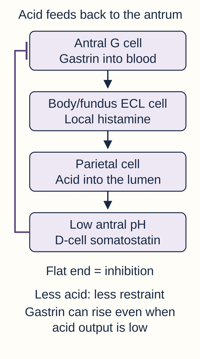
Gastric feedback. Arrows stimulate; a flat-ended line inhibits. The antral sensor and the acid-secreting body/fundus are different locations.
Peptides, amino acids and gastric distention increase gastrin during a meal. Gastrin supports acid secretion, gastric mucosal growth and gastric motility. Acetylcholine and gastrin use calcium-linked signaling in their principal gastric pathways; histamine increases parietal cAMP. These inputs converge on the acid-secreting machinery, so blocking one receptor need not abolish every stimulus. A proton pump inhibitor acts farther downstream at the H+/K+ ATPase. [10][4]
As buffering by food wanes and antral pH falls, D cells release somatostatin. Local somatostatin inhibits G cells and restrains histamine and acid secretion. This is negative feedback: the product, acid, eventually reduces further stimulation. D cells also occur elsewhere in the stomach and intestine; pancreatic delta cells use the same peptide to restrain islet hormone secretion.
Trace the response: Imagine gastric pH rising during acid suppression. Predict the direction of the gastrin response before checking.
Does gastrin rise or fall?
It rises because less acid-dependent inhibitory feedback reaches the G cell. A high gastrin concentration can therefore coexist with low acid output. Interpret the hormone together with its regulated variable.
What if antral acid increases instead?
D-cell somatostatin increases and restrains gastrin. The sign reverses because the sensor now detects more acid, not because the G cell has changed identity.
Apply the comparison to a patient with loss of acid-producing gastric glands. High gastrin is an appropriate attempt to restore acid, not by itself proof of a tumor. A gastrin-producing tumor instead drives secretion despite an already acidic stomach. Medication exposure and measured acidity are therefore essential context. [4]
The pancreas has two different outputs
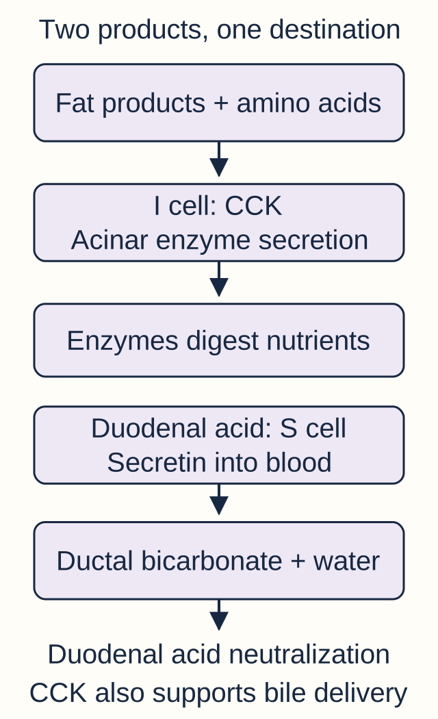
If pancreatic fluid increases, must enzyme output have increased too? Separate the compartments first. Acini supply digestive enzymes; ducts supply bicarbonate-rich fluid. Fat digestion products and amino acids stimulate I cells, concentrated in the duodenum and jejunum, to release cholecystokinin, or CCK. CCK promotes enzyme secretion through neural cholinergic pathways and acinar signaling; the relative direct contribution varies with species and experimental conditions. [1][3]
One organ, two products. Enzymes digest food; bicarbonate protects the environment in which they act. Line labels, not color alone, identify the products.
Acid entering the duodenum stimulates S cells to release secretin. S cells are concentrated proximally, with some extending into jejunum. Secretin activates cAMP-linked signaling in pancreatic ductal cells, including the small duct system beginning at centroacinar cells. CFTR and associated anion transporters support bicarbonate secretion; water follows the secreted electrolytes. Secretin also stimulates biliary duct bicarbonate and reduces further gastric acid delivery by restraining secretion and emptying. [2][9]
CCK contracts the gallbladder while coordinating sphincter of Oddi relaxation, so stored bile can reach the intestine. It also slows gastric emptying and contributes to satiation. Thus a signal can stimulate one target and inhibit another. Bile assists fat digestion and absorption; it is not a pancreatic enzyme. Secretin-induced bicarbonate-rich bile duct fluid is likewise different from CCK-induced emptying of stored bile. [1][3][9]
Trace a duodenal challenge
Use the two output routes above. For each input, name the output expected to increase most directly, then open its answer diagram. Keep both answers open to compare; close them to retry. This qualitative model assumes patent ducts and functioning target tissue, not measured secretion volumes.
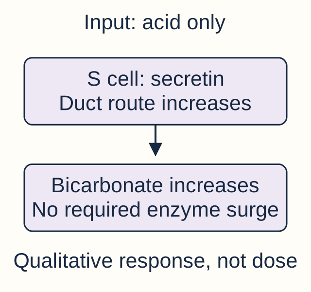
Acid without nutrients: which route responds?
Acid recruits S-cell secretin and ductal bicarbonate. More enzymes alone would not neutralize the acid.
Acid chiefly requests bicarbonate, not an enzyme-only response.
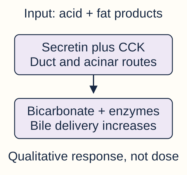
Add fat products: what additional route responds?
Fat products recruit CCK-supported enzymes and bile while the acid response continues.
The combined meal needs both neutralization and digestion. CCK and cholinergic input can potentiate secretin-driven bicarbonate secretion; the branches are useful distinctions, not isolated systems.
The complete worked comparison is visible without the activity: acid primarily increases ductal bicarbonate; fat products add CCK-supported enzyme secretion and bile delivery. A mixed meal recruits both. If a patient has damaged pancreatic ducts, a normal acid stimulus and normal secretin release do not guarantee normal bicarbonate output. Localize a failed response to sensor, messenger, target or drainage before assuming hormone deficiency. [2][9]
Nutrient location changes the insulin response
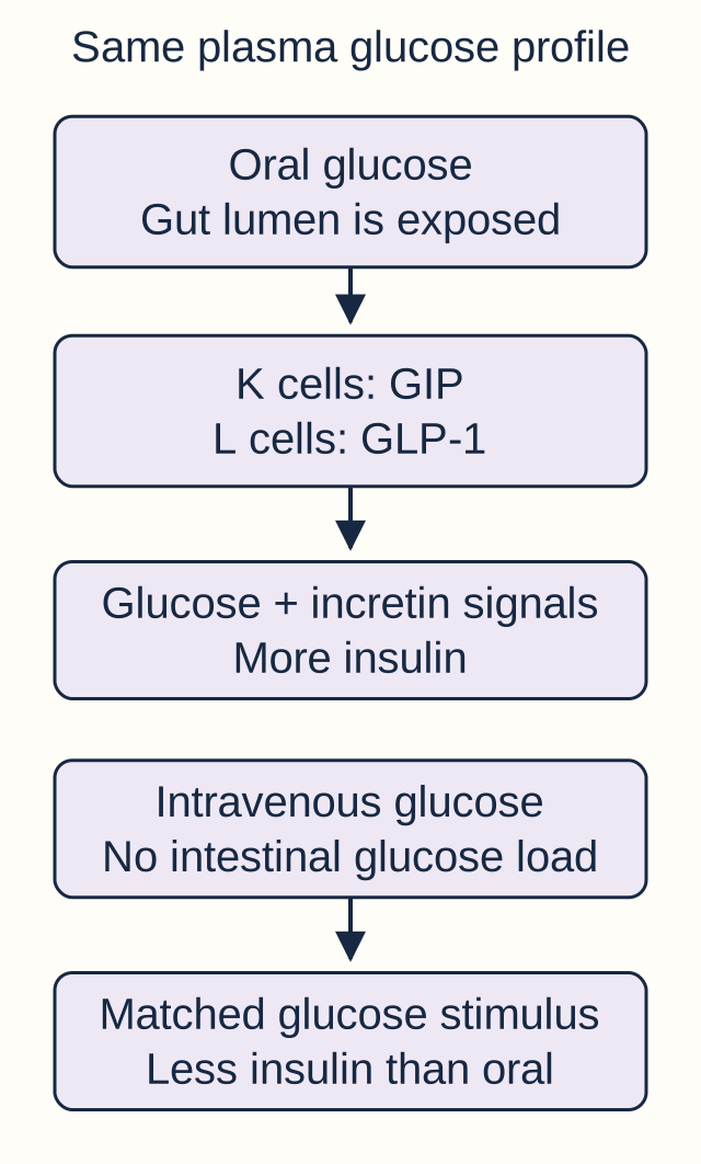
Can identical blood glucose profiles produce different insulin responses? Yes, because oral nutrients also signal from the gut. An isoglycemic comparison matches the plasma glucose profile after oral glucose with a variable intravenous infusion; it does not require equal administered doses. More insulin after oral delivery demonstrates the incretin effect. Both GIP and GLP-1 contribute. [1][3]
Match glucose, not the administered dose. The diagram separates direct glucose stimulation from added intestinal signaling; it does not quantify either hormone's share.
K cells in duodenum and jejunum release glucose-dependent insulinotropic polypeptide, or GIP, in response to nutrients including glucose and fat. Its older name, gastric inhibitory peptide, overemphasizes acid inhibition relative to its important human role in insulin secretion. L cells, more abundant distally in ileum and colon but also present proximally, release GLP-1. Both amplify glucose-dependent insulin secretion. GLP-1 also restrains glucagon during hyperglycemia, slows gastric emptying and promotes satiety. DPP-4 rapidly inactivates these incretin peptides. [1][3]
Cover the lower half of the figure. If the intravenous infusion is adjusted to reproduce the oral glucose profile, predict which delivery route still has the larger insulin response.
Check the comparison
Oral delivery still stimulates intestinal incretins. A glucose concentration difference cannot explain the result because that variable was matched. A controlled comparison identifies the extra gut signal, not one exclusive hormone.
Now transfer the relationship: a drug that prevents incretin breakdown preserves meal-linked insulin amplification rather than supplying bicarbonate or triggering fasting contractions. Its effects still depend on functioning beta cells and the glycemic context. This physiological distinction is not a complete prescribing comparison between diabetes medications.
G cells are concentrated in the antrum with a smaller proximal duodenal population; I, S and K cells survey the proximal small intestine for different inputs. The cell letters describe useful predominant sources, not rigid anatomical borders or a rule that each enteroendocrine cell makes only one peptide. Use the regional map to locate a response, then confirm the stimulus and target. During a mixed meal, proximal CCK, secretin and GIP signals overlap with GLP-1 signaling rather than arriving in a compulsory numbered order. [3]
Fasting contractions and sphincter relaxation are different jobs
Does every signal associated with fasting do the same thing? Motilin from upper-small-intestinal motilin-producing endocrine cells, often called Mo cells, organizes the fasting migrating motor complex, including periodic strong contractions that clear residual contents. Eating interrupts the fasting program. Ghrelin, produced mainly by gastric endocrine cells in the fundus, is associated with premeal hunger and stimulates appetite and growth hormone release. It is not interchangeable with motilin simply because both relate to fasting. [1][3]
Two concrete observations
Observation A: Overnight manometry shows periodic propulsive contractions; feeding replaces that pattern with fed activity.
Observation B: A person reports hunger before a meal, without any measurement of intestinal contractions.
Assign the more direct physiological association to each observation before reading on. A points toward the motilin-associated motor program; B fits an appetite signal such as ghrelin but does not establish a hormone concentration or diagnosis. Specify the measured target before naming a regulator.
Would erythromycin test the same relationship as an appetite rating?
No. Erythromycin activates motilin receptors and can accelerate gastric emptying. An appetite rating measures a different outcome. In gastroparesis, short-term prokinetic use is limited by tachyphylaxis, commonly within weeks, rather than offering durable correction of the underlying disorder. [6]
VIP and nitric oxide are important enteric neural signals, not simply additional meal hormones from lettered epithelial cells. VIP supports intestinal fluid secretion and relaxes smooth muscle. Nitric oxide mediates inhibitory smooth-muscle signaling, including sphincter relaxation. In achalasia, loss of inhibitory neurons reduces NO/VIP signaling, leaving impaired lower-esophageal-sphincter relaxation and disordered or absent peristalsis. An obstructed passage can therefore reflect too little inhibition, not too little contraction. [7]
Apply that distinction to dysphagia for liquids and solids with absent peristalsis and poor sphincter relaxation: increasing a fasting motor signal is not equivalent to restoring coordinated swallowing. Conversely, delayed gastric emptying without a mechanical obstruction supports a gastric motor disorder, not by itself achalasia. These are localization decisions, not a list of interchangeable prokinetic treatments. [6][7]
Interpret hormone excess through its target
A high hormone result does not identify its cause until you inspect the response. Compare two educational examples with fasting gastrin 1,500 pg/mL (laboratory upper limit 100): one has gastric pH 1.3 and recurrent distal ulcers; the other has pH 7 and loss of acid-producing glands. Circle the example in which gastrin remains high despite strong acid feedback.
The first pattern is inappropriate hypergastrinemia and strongly supports Zollinger-Ellison syndrome in the correct testing context. The second is an appropriate response to achlorhydria. Marked gastrin increases can occur with atrophic gastritis or PPI exposure, so the value alone cannot establish gastrinoma. Gastrinomas may be duodenal or pancreatic; consider MEN1 when associated parathyroid or pituitary disease is present. [4]
What does a smaller gastrin increase require?
When gastrin is increased but less than tenfold and gastric pH is at most 2, further biochemical assessment may include a specialist-supervised secretin stimulation test. A paradoxical gastrin increase supports gastrinoma in an appropriate testing context; achlorhydria and acid-suppressing drugs can produce misleading results. A provocative result needs interpretable baseline physiology.
Do not abruptly stop a PPI to investigate suspected gastrinoma. Loss of acid control can cause serious complications. Medication adjustment and acid testing require a supervised specialist plan. This lesson explains the physiology, not a medication-withdrawal protocol. [4]
Gastrinoma can produce recurrent ulcers, prominent gastric folds and diarrhea. Excess duodenal acid can impair digestion, including pancreatic lipase activity. VIPoma instead causes marked intestinal secretion, often with profuse watery diarrhea persisting during fasting, potassium depletion and reduced gastric acid. Hypochlorhydria or achlorhydria is characteristic but not a mandatory finding in every patient. The dangerous immediate consequence is volume and electrolyte depletion, not just the tumor label. [1][4][5]
Somatostatin broadly inhibits gastrointestinal hormones such as gastrin, CCK and secretin, along with acid, pancreatic and intestinal secretions, gallbladder contraction, insulin and glucagon. Octreotide mimics selected somatostatin effects and lasts longer than the native peptide; injectable short-acting and depot preparations are not the same formulation. It can reduce VIPoma-associated diarrhea, but broad suppression also explains gallbladder sludge or stones, fat malabsorption and either high or low glucose. A beneficial reduction in watery output does not mean every later loose stool is persistent hormone excess. [5]
A patient's watery diarrhea improves on octreotide, but new oily stools and weight loss develop. Predict a treatment-related explanation before checking.
Connect the changed stool pattern to a target
Suppression of pancreatic enzymes and bile delivery can impair fat absorption. Reassess for exocrine insufficiency rather than automatically escalating suppression for presumed recurrent VIP secretion. A different output pattern can indicate a different cause.
Octreotide is also a vasoactive option in suspected acute variceal bleeding because it reduces splanchnic inflow, not because it neutralizes acid. It accompanies resuscitation, antibiotic prophylaxis and urgent endoscopic care; it does not replace them. Baveno VII recommends starting vasoactive treatment promptly when variceal bleeding is suspected. [8]
For a new patient, return to four questions: What stimulated the signal? Where is its source? Which target changed? Is that change appropriate for the measured environment? Those questions distinguish feedback from autonomous secretion and useful treatment from predictable adverse effects.
Apply the relationships independently
Case 1
Show answer and explanations for case 1
A. Reduced gastrin with loss of direct cholinergic acid stimulation (Why this does not fit)
Gastrin should fall, but the experiment explicitly preserves parietal muscarinic signaling. Blocking GRP does not block every vagal secretory route.
Reasoning steps for option A
For the choice "Reduced gastrin with loss of direct cholinergic acid stimulation", how does the fact that gastrin should fall, but the experiment explicitly preserves parietal muscarinic signaling bear on the mechanism proposed here?
Gastrin should fall, but the experiment explicitly preserves parietal muscarinic signaling.
How should the observation that blocking GRP does not block every vagal secretory route change your assessment of "Reduced gastrin with loss of direct cholinergic acid stimulation"?
Blocking GRP does not block every vagal secretory route.
In this case, what follows from the fact that vagal GRP stimulates G cells, while parallel muscarinic input can still stimulate parietal cells for the option "Reduced gastrin with loss of direct cholinergic acid stimulation"?
Vagal GRP stimulates G cells, while parallel muscarinic input can still stimulate parietal cells.
B. Preserved gastrin with loss of direct cholinergic acid stimulation (Why this does not fit)
This would fit muscarinic blockade better than selective GRP blockade. The intervention targets G-cell stimulation rather than the retained direct parietal input.
Reasoning steps for option B
When evaluating "Preserved gastrin with loss of direct cholinergic acid stimulation", why is the case detail that this would fit muscarinic blockade better than selective GRP blockade a useful discriminator?
This would fit muscarinic blockade better than selective GRP blockade.
Which physiological link is being tested by the fact that the intervention targets G-cell stimulation rather than the retained direct parietal input when considering "Preserved gastrin with loss of direct cholinergic acid stimulation"?
The intervention targets G-cell stimulation rather than the retained direct parietal input.
How does the specific clue that vagal GRP stimulates G cells, while parallel muscarinic input can still stimulate parietal cells separate "Preserved gastrin with loss of direct cholinergic acid stimulation" from the competing choices?
Vagal GRP stimulates G cells, while parallel muscarinic input can still stimulate parietal cells.
C. Reduced gastrin with retained direct cholinergic acid stimulation (Best answer)
GRP blockade interrupts vagal G-cell stimulation, while the stipulated intact muscarinic input can still stimulate parietal cells. Separate vagal transmitters by their targets.
Reasoning steps for option C
What does the finding that gRP blockade interrupts vagal G-cell stimulation, while the stipulated intact muscarinic input can still stimulate parietal tell you about whether "Reduced gastrin with retained direct cholinergic acid stimulation" fits this case?
GRP blockade interrupts vagal G-cell stimulation, while the stipulated intact muscarinic input can still stimulate parietal cells.
Why does the case evidence that separate vagal transmitters by their targets support or weaken the choice "Reduced gastrin with retained direct cholinergic acid stimulation"?
Separate vagal transmitters by their targets.
What localization or feedback rule is tested by the observation that vagal GRP stimulates G cells, while parallel muscarinic input can still stimulate parietal cells for "Reduced gastrin with retained direct cholinergic acid stimulation"?
Vagal GRP stimulates G cells, while parallel muscarinic input can still stimulate parietal cells.
D. Preserved gastrin with retained direct cholinergic acid stimulation (Why this does not fit)
The direct parietal component remains, but G-cell stimulation is not preserved after its GRP input is blocked. A selective intervention changes its own target even when parallel pathways survive.
Reasoning steps for option D
How should the observation that the direct parietal component remains, but G-cell stimulation is not preserved after its GRP input is change your assessment of "Preserved gastrin with retained direct cholinergic acid stimulation"?
The direct parietal component remains, but G-cell stimulation is not preserved after its GRP input is blocked.
In this case, what follows from the fact that a selective intervention changes its own target even when parallel pathways survive for the option "Preserved gastrin with retained direct cholinergic acid stimulation"?
A selective intervention changes its own target even when parallel pathways survive.
Why is the detail that vagal GRP stimulates G cells, while parallel muscarinic input can still stimulate parietal cells decisive when you test the option "Preserved gastrin with retained direct cholinergic acid stimulation"?
Vagal GRP stimulates G cells, while parallel muscarinic input can still stimulate parietal cells.
Takeaway: Vagal GRP stimulates G cells, while parallel muscarinic input can still stimulate parietal cells.
A. Impaired gastrin-linked intermediary signaling; histamine-evoked acid falls (Best answer)
Preserved responses to histamine and muscarinic stimulation argue against shared parietal failure and favor impairment in gastrin-linked signaling upstream of the histamine response. H2 blockade then reduces that preserved downstream response. The experiment does not identify a unique molecular lesion.
Reasoning steps for option A
Which physiological link is being tested by the fact that preserved responses to histamine and muscarinic stimulation argue against shared parietal failure and favor impairment in when considering "Impaired gastrin-linked intermediary signaling; histamine-evoked acid falls"?
Preserved responses to histamine and muscarinic stimulation argue against shared parietal failure and favor impairment in gastrin-linked signaling upstream of the histamine response.
How does the specific clue that h2 blockade then reduces that preserved downstream response separate "Impaired gastrin-linked intermediary signaling; histamine-evoked acid falls" from the competing choices?
H2 blockade then reduces that preserved downstream response.
For the choice "Impaired gastrin-linked intermediary signaling; histamine-evoked acid falls", how does the fact that bypass responses distinguish a failed intermediary signal from an unresponsive final target bear on the mechanism proposed here?
Bypass responses distinguish a failed intermediary signal from an unresponsive final target.
B. Impaired final proton-pump function; histamine-evoked acid falls (Why this does not fit)
H2 blockade would reduce histamine signaling, but a shared pump failure would also impair the initially preserved histamine and muscarinic responses. Both the localization and the later antagonist effect must fit.
Reasoning steps for option B
Why does the case evidence that h2 blockade would reduce histamine signaling, but a shared pump failure would also impair the initially support or weaken the choice "Impaired final proton-pump function; histamine-evoked acid falls"?
H2 blockade would reduce histamine signaling, but a shared pump failure would also impair the initially preserved histamine and muscarinic responses.
What localization or feedback rule is tested by the observation that both the localization and the later antagonist effect must fit for "Impaired final proton-pump function; histamine-evoked acid falls"?
Both the localization and the later antagonist effect must fit.
When evaluating "Impaired final proton-pump function; histamine-evoked acid falls", why is the case detail that bypass responses distinguish a failed intermediary signal from an unresponsive final target a useful discriminator?
Bypass responses distinguish a failed intermediary signal from an unresponsive final target.
C. Impaired gastrin-linked intermediary signaling; histamine-evoked acid rises (Why this does not fit)
The initial localization fits the bypass results, but an H2 antagonist reduces rather than enhances the retained histamine response. Correctly locating a defect does not reverse antagonist pharmacology.
Reasoning steps for option C
In this case, what follows from the fact that the initial localization fits the bypass results, but an H2 antagonist reduces rather than enhances the for the option "Impaired gastrin-linked intermediary signaling; histamine-evoked acid rises"?
The initial localization fits the bypass results, but an H2 antagonist reduces rather than enhances the retained histamine response.
Why is the detail that correctly locating a defect does not reverse antagonist pharmacology decisive when you test the option "Impaired gastrin-linked intermediary signaling; histamine-evoked acid rises"?
Correctly locating a defect does not reverse antagonist pharmacology.
What does the finding that bypass responses distinguish a failed intermediary signal from an unresponsive final target tell you about whether "Impaired gastrin-linked intermediary signaling; histamine-evoked acid rises" fits this case?
Bypass responses distinguish a failed intermediary signal from an unresponsive final target.
D. Impaired final proton-pump function; histamine-evoked acid rises (Why this does not fit)
A final effector defect conflicts with the preserved acid responses to two stimuli, and H2 blockade does not enhance histamine-evoked secretion. Compare both the bypass and the proposed intervention.
Reasoning steps for option D
How does the specific clue that a final effector defect conflicts with the preserved acid responses to two stimuli, and H2 blockade separate "Impaired final proton-pump function; histamine-evoked acid rises" from the competing choices?
A final effector defect conflicts with the preserved acid responses to two stimuli, and H2 blockade does not enhance histamine-evoked secretion.
For the choice "Impaired final proton-pump function; histamine-evoked acid rises", how does the fact that compare both the bypass and the proposed intervention bear on the mechanism proposed here?
Compare both the bypass and the proposed intervention.
How should the observation that bypass responses distinguish a failed intermediary signal from an unresponsive final target change your assessment of "Impaired final proton-pump function; histamine-evoked acid rises"?
Bypass responses distinguish a failed intermediary signal from an unresponsive final target.
Takeaway: Bypass responses distinguish a failed intermediary signal from an unresponsive final target.
A. Reduced gastrin synthesis in antral G cells (Why this does not fit)
H2 antagonism is not a direct G-cell synthesis inhibitor. As acid falls, gastrin can increase through reduced feedback. Follow the location of the receptor, not just the final acid output.
Reasoning steps for option A
What localization or feedback rule is tested by the observation that h2 antagonism is not a direct G-cell synthesis inhibitor for "Reduced gastrin synthesis in antral G cells"?
H2 antagonism is not a direct G-cell synthesis inhibitor.
When evaluating "Reduced gastrin synthesis in antral G cells", why is the case detail that as acid falls, gastrin can increase through reduced feedback a useful discriminator?
As acid falls, gastrin can increase through reduced feedback.
Which physiological link is being tested by the fact that h2 antagonism reduces parietal histamine signaling rather than directly eliminating ECL cells or gastrin when considering "Reduced gastrin synthesis in antral G cells"?
H2 antagonism reduces parietal histamine signaling rather than directly eliminating ECL cells or gastrin.
B. Reduced acetylcholine binding to parietal M3 receptors (Why this does not fit)
Muscarinic inhibition can reduce acid but is not the target of an H2 antagonist. Different receptor pathways converge on acid secretion.
Reasoning steps for option B
Why is the detail that muscarinic inhibition can reduce acid but is not the target of an H2 antagonist decisive when you test the option "Reduced acetylcholine binding to parietal M3 receptors"?
Muscarinic inhibition can reduce acid but is not the target of an H2 antagonist.
What does the finding that different receptor pathways converge on acid secretion tell you about whether "Reduced acetylcholine binding to parietal M3 receptors" fits this case?
Different receptor pathways converge on acid secretion.
Why does the case evidence that h2 antagonism reduces parietal histamine signaling rather than directly eliminating ECL cells or gastrin support or weaken the choice "Reduced acetylcholine binding to parietal M3 receptors"?
H2 antagonism reduces parietal histamine signaling rather than directly eliminating ECL cells or gastrin.
C. Reduced histamine synthesis through selective ECL destruction (Why this does not fit)
The stem preserves ECL function, and receptor blockade does not require cell destruction. A reduced response does not establish loss of the signal-producing cell.
Reasoning steps for option C
For the choice "Reduced histamine synthesis through selective ECL destruction", how does the fact that the stem preserves ECL function, and receptor blockade does not require cell destruction bear on the mechanism proposed here?
The stem preserves ECL function, and receptor blockade does not require cell destruction.
How should the observation that a reduced response does not establish loss of the signal-producing cell change your assessment of "Reduced histamine synthesis through selective ECL destruction"?
A reduced response does not establish loss of the signal-producing cell.
In this case, what follows from the fact that h2 antagonism reduces parietal histamine signaling rather than directly eliminating ECL cells or gastrin for the option "Reduced histamine synthesis through selective ECL destruction"?
H2 antagonism reduces parietal histamine signaling rather than directly eliminating ECL cells or gastrin.
D. Reduced parietal cAMP response to released histamine (Best answer)
H2 blockade prevents histamine from effectively activating its parietal signaling route. ECL histamine release can continue despite a reduced target response. A receptor blocker acts after ligand release.
Reasoning steps for option D
When evaluating "Reduced parietal cAMP response to released histamine", why is the case detail that h2 blockade prevents histamine from effectively activating its parietal signaling route a useful discriminator?
H2 blockade prevents histamine from effectively activating its parietal signaling route.
Which physiological link is being tested by the fact that eCL histamine release can continue despite a reduced target response when considering "Reduced parietal cAMP response to released histamine"?
ECL histamine release can continue despite a reduced target response.
How does the specific clue that h2 antagonism reduces parietal histamine signaling rather than directly eliminating ECL cells or gastrin separate "Reduced parietal cAMP response to released histamine" from the competing choices?
H2 antagonism reduces parietal histamine signaling rather than directly eliminating ECL cells or gastrin.
Takeaway: H2 antagonism reduces parietal histamine signaling rather than directly eliminating ECL cells or gastrin.
A. Autonomous gastrin secretion from a neuroendocrine tumor (Why this does not fit)
A tumor can cause hypergastrinemia and a PPI can mask its acid output. However, the documented normal baseline and change accompanying acid suppression more directly support feedback; this result alone neither diagnoses nor excludes gastrinoma.
Reasoning steps for option A
What does the finding that a tumor can cause hypergastrinemia and a PPI can mask its acid output tell you about whether "Autonomous gastrin secretion from a neuroendocrine tumor" fits this case?
A tumor can cause hypergastrinemia and a PPI can mask its acid output.
Why does the case evidence that however, the documented normal baseline and change accompanying acid suppression more directly support feedback support or weaken the choice "Autonomous gastrin secretion from a neuroendocrine tumor"?
However, the documented normal baseline and change accompanying acid suppression more directly support feedback; this result alone neither diagnoses nor excludes gastrinoma.
What localization or feedback rule is tested by the observation that interpret a new gastrin increase with acid status, treatment timing and prior tissue findings for "Autonomous gastrin secretion from a neuroendocrine tumor"?
Interpret a new gastrin increase with acid status, treatment timing and prior tissue findings.
B. Reduced acid-dependent feedback during proton pump inhibition (Best answer)
The new high-pH state follows treatment that suppresses parietal acid output, allowing a compensatory rise in gastrin. The temporal baseline supports this mechanism but does not permanently exclude a coexisting disorder.
Reasoning steps for option B
How should the observation that the new high-pH state follows treatment that suppresses parietal acid output, allowing a compensatory rise in change your assessment of "Reduced acid-dependent feedback during proton pump inhibition"?
The new high-pH state follows treatment that suppresses parietal acid output, allowing a compensatory rise in gastrin.
In this case, what follows from the fact that the temporal baseline supports this mechanism but does not permanently exclude a coexisting disorder for the option "Reduced acid-dependent feedback during proton pump inhibition"?
The temporal baseline supports this mechanism but does not permanently exclude a coexisting disorder.
Why is the detail that interpret a new gastrin increase with acid status, treatment timing and prior tissue findings decisive when you test the option "Reduced acid-dependent feedback during proton pump inhibition"?
Interpret a new gastrin increase with acid status, treatment timing and prior tissue findings.
C. Loss of corpus glands from autoimmune gastritis (Why this does not fit)
Autoimmune atrophy can produce high pH and high gastrin. Recent normal corpus histology and the close treatment-associated change favor pharmacological suppression over newly established extensive gland loss.
Reasoning steps for option C
Which physiological link is being tested by the fact that autoimmune atrophy can produce high pH and high gastrin when considering "Loss of corpus glands from autoimmune gastritis"?
Autoimmune atrophy can produce high pH and high gastrin.
How does the specific clue that recent normal corpus histology and the close treatment-associated change favor pharmacological suppression over newly established extensive separate "Loss of corpus glands from autoimmune gastritis" from the competing choices?
Recent normal corpus histology and the close treatment-associated change favor pharmacological suppression over newly established extensive gland loss.
For the choice "Loss of corpus glands from autoimmune gastritis", how does the fact that interpret a new gastrin increase with acid status, treatment timing and prior tissue findings bear on the mechanism proposed here?
Interpret a new gastrin increase with acid status, treatment timing and prior tissue findings.
D. Antral inflammation reducing local somatostatin release (Why this does not fit)
Antral inflammation can impair inhibitory feedback and increase gastrin. Here the new high-pH state directly follows a known acid-blocking intervention, making medication-related feedback the better-supported explanation.
Reasoning steps for option D
Why does the case evidence that antral inflammation can impair inhibitory feedback and increase gastrin support or weaken the choice "Antral inflammation reducing local somatostatin release"?
Antral inflammation can impair inhibitory feedback and increase gastrin.
What localization or feedback rule is tested by the observation that here the new high-pH state directly follows a known acid-blocking intervention, making medication-related feedback the better-supported for "Antral inflammation reducing local somatostatin release"?
Here the new high-pH state directly follows a known acid-blocking intervention, making medication-related feedback the better-supported explanation.
When evaluating "Antral inflammation reducing local somatostatin release", why is the case detail that interpret a new gastrin increase with acid status, treatment timing and prior tissue findings a useful discriminator?
Interpret a new gastrin increase with acid status, treatment timing and prior tissue findings.
Takeaway: Interpret a new gastrin increase with acid status, treatment timing and prior tissue findings.
A. Low acid output and reduced gastrin (Why this does not fit)
Parietal loss supports low acid, but reduced luminal acidity removes rather than intensifies acid-mediated inhibition of remaining antral G cells.
Reasoning steps for option A
In this case, what follows from the fact that parietal loss supports low acid, but reduced luminal acidity removes rather than intensifies acid-mediated inhibition of for the option "Low acid output and reduced gastrin"?
Parietal loss supports low acid, but reduced luminal acidity removes rather than intensifies acid-mediated inhibition of remaining antral G cells.
Why is the detail that parietal loss supports low acid, but reduced luminal acidity removes rather than intensifies acid-mediated inhibition of decisive when you test the option "Low acid output and reduced gastrin"?
Parietal loss supports low acid, but reduced luminal acidity removes rather than intensifies acid-mediated inhibition of remaining antral G cells.
What does the finding that corpus oxyntic loss predicts diminished acid capacity and compensatory gastrin when antral G cells remain tell you about whether "Low acid output and reduced gastrin" fits this case?
Corpus oxyntic loss predicts diminished acid capacity and compensatory gastrin when antral G cells remain.
B. Low acid output and elevated gastrin (Best answer)
Loss of corpus parietal cells lowers acid secretion; reduced acid feedback permits increased antral gastrin release.
Reasoning steps for option B
How does the specific clue that loss of corpus parietal cells lowers acid secretion separate "Low acid output and elevated gastrin" from the competing choices?
Loss of corpus parietal cells lowers acid secretion; reduced acid feedback permits increased antral gastrin release.
For the choice "Low acid output and elevated gastrin", how does the fact that loss of corpus parietal cells lowers acid secretion bear on the mechanism proposed here?
Loss of corpus parietal cells lowers acid secretion; reduced acid feedback permits increased antral gastrin release.
How should the observation that corpus oxyntic loss predicts diminished acid capacity and compensatory gastrin when antral G cells remain change your assessment of "Low acid output and elevated gastrin"?
Corpus oxyntic loss predicts diminished acid capacity and compensatory gastrin when antral G cells remain.
C. High acid output and elevated gastrin (Why this does not fit)
Increased gastrin could occur, but extensive oxyntic parietal-cell loss limits the capacity to secrete acid.
Reasoning steps for option C
What localization or feedback rule is tested by the observation that increased gastrin could occur, but extensive oxyntic parietal-cell loss limits the capacity to secrete acid for "High acid output and elevated gastrin"?
Increased gastrin could occur, but extensive oxyntic parietal-cell loss limits the capacity to secrete acid.
When evaluating "High acid output and elevated gastrin", why is the case detail that increased gastrin could occur, but extensive oxyntic parietal-cell loss limits the capacity to secrete acid a useful discriminator?
Increased gastrin could occur, but extensive oxyntic parietal-cell loss limits the capacity to secrete acid.
Which physiological link is being tested by the fact that corpus oxyntic loss predicts diminished acid capacity and compensatory gastrin when antral G cells remain when considering "High acid output and elevated gastrin"?
Corpus oxyntic loss predicts diminished acid capacity and compensatory gastrin when antral G cells remain.
D. High acid output and reduced gastrin (Why this does not fit)
Acid could suppress gastrin in an intact stomach, but the corpus biopsy predicts diminished acid-secretory capacity.
Reasoning steps for option D
Why is the detail that acid could suppress gastrin in an intact stomach, but the corpus biopsy predicts diminished acid-secretory capacity decisive when you test the option "High acid output and reduced gastrin"?
Acid could suppress gastrin in an intact stomach, but the corpus biopsy predicts diminished acid-secretory capacity.
What does the finding that acid could suppress gastrin in an intact stomach, but the corpus biopsy predicts diminished acid-secretory capacity tell you about whether "High acid output and reduced gastrin" fits this case?
Acid could suppress gastrin in an intact stomach, but the corpus biopsy predicts diminished acid-secretory capacity.
Why does the case evidence that corpus oxyntic loss predicts diminished acid capacity and compensatory gastrin when antral G cells remain support or weaken the choice "High acid output and reduced gastrin"?
Corpus oxyntic loss predicts diminished acid capacity and compensatory gastrin when antral G cells remain.
Takeaway: Corpus oxyntic loss predicts diminished acid capacity and compensatory gastrin when antral G cells remain.
A. Stronger secretin-linked pancreatic bicarbonate response in B (Why this does not fit)
B's high gastrin does not restore lost oxyntic capacity. Less acid reaches the duodenal sensor, so B is not expected to generate the stronger secretin stimulus.
Reasoning steps for option A
For the choice "Stronger secretin-linked pancreatic bicarbonate response in B", how does the fact that b's high gastrin does not restore lost oxyntic capacity bear on the mechanism proposed here?
B's high gastrin does not restore lost oxyntic capacity.
How should the observation that less acid reaches the duodenal sensor, so B is not expected to generate the stronger secretin change your assessment of "Stronger secretin-linked pancreatic bicarbonate response in B"?
Less acid reaches the duodenal sensor, so B is not expected to generate the stronger secretin stimulus.
In this case, what follows from the fact that gastric target capacity determines the acid challenge that recruits downstream secretin for the option "Stronger secretin-linked pancreatic bicarbonate response in B"?
Gastric target capacity determines the acid challenge that recruits downstream secretin; gastrin concentration alone does not.
B. Stronger CCK-linked acinar enzyme response in A from gastrin alone (Why this does not fit)
The controlled protein exposure is matched, and circulating gastrin is similarly high. The differing gastric pathology more directly changes the acid-dependent duct response than it establishes a different CCK nutrient stimulus.
Reasoning steps for option B
When evaluating "Stronger CCK-linked acinar enzyme response in A from gastrin alone", why is the case detail that the controlled protein exposure is matched, and circulating gastrin is similarly high a useful discriminator?
The controlled protein exposure is matched, and circulating gastrin is similarly high.
Which physiological link is being tested by the fact that the differing gastric pathology more directly changes the acid-dependent duct response than it establishes a different when considering "Stronger CCK-linked acinar enzyme response in A from gastrin alone"?
The differing gastric pathology more directly changes the acid-dependent duct response than it establishes a different CCK nutrient stimulus.
How does the specific clue that gastric target capacity determines the acid challenge that recruits downstream secretin separate "Stronger CCK-linked acinar enzyme response in A from gastrin alone" from the competing choices?
Gastric target capacity determines the acid challenge that recruits downstream secretin; gastrin concentration alone does not.
C. Stronger CCK-linked acinar enzyme response in B from gland loss (Why this does not fit)
Loss of corpus glands does not directly generate the fat- or amino-acid stimulus that recruits CCK. With matched protein input, the better-supported difference is acid sensing and ductal bicarbonate rather than an enhanced acinar request in B.
Reasoning steps for option C
What does the finding that loss of corpus glands does not directly generate the fat- or amino-acid stimulus that recruits CCK tell you about whether "Stronger CCK-linked acinar enzyme response in B from gland loss" fits this case?
Loss of corpus glands does not directly generate the fat- or amino-acid stimulus that recruits CCK.
Why does the case evidence that with matched protein input, the better-supported difference is acid sensing and ductal bicarbonate rather than an support or weaken the choice "Stronger CCK-linked acinar enzyme response in B from gland loss"?
With matched protein input, the better-supported difference is acid sensing and ductal bicarbonate rather than an enhanced acinar request in B.
What localization or feedback rule is tested by the observation that gastric target capacity determines the acid challenge that recruits downstream secretin for "Stronger CCK-linked acinar enzyme response in B from gland loss"?
Gastric target capacity determines the acid challenge that recruits downstream secretin; gastrin concentration alone does not.
D. Stronger secretin-linked pancreatic bicarbonate response in A (Best answer)
A's ulcer pattern and preserved glands favor retained excessive acid output; B's oxyntic loss predicts achlorhydria. Greater acid delivery in A recruits S-cell secretin and ductal bicarbonate. Infer gastric capacity before predicting the downstream response.
Reasoning steps for option D
How should the observation that a's ulcer pattern and preserved glands favor retained excessive acid output change your assessment of "Stronger secretin-linked pancreatic bicarbonate response in A"?
A's ulcer pattern and preserved glands favor retained excessive acid output; B's oxyntic loss predicts achlorhydria.
In this case, what follows from the fact that greater acid delivery in A recruits S-cell secretin and ductal bicarbonate for the option "Stronger secretin-linked pancreatic bicarbonate response in A"?
Greater acid delivery in A recruits S-cell secretin and ductal bicarbonate.
Why is the detail that gastric target capacity determines the acid challenge that recruits downstream secretin decisive when you test the option "Stronger secretin-linked pancreatic bicarbonate response in A"?
Gastric target capacity determines the acid challenge that recruits downstream secretin; gastrin concentration alone does not.
Takeaway: Gastric target capacity determines the acid challenge that recruits downstream secretin; gastrin concentration alone does not.
A. Specialist assessment of acid status and a supervised medication plan before interpreting provocative testing (Best answer)
Recent bleeding makes unsupervised acid withdrawal hazardous, and acidity is needed to interpret high gastrin. A tailored specialist plan addresses both safety and the hormone-feedback context.
Reasoning steps for option A
Which physiological link is being tested by the fact that recent bleeding makes unsupervised acid withdrawal hazardous, and acidity is needed to interpret high gastrin when considering "Specialist assessment of acid status and a supervised medication plan before interpreting provocative testing"?
Recent bleeding makes unsupervised acid withdrawal hazardous, and acidity is needed to interpret high gastrin.
How does the specific clue that a tailored specialist plan addresses both safety and the hormone-feedback context separate "Specialist assessment of acid status and a supervised medication plan before interpreting provocative testing" from the competing choices?
A tailored specialist plan addresses both safety and the hormone-feedback context.
For the choice "Specialist assessment of acid status and a supervised medication plan before interpreting provocative testing", how does the fact that high-risk acid suppression should not be changed without a specialist plan for safety and interpretable testing bear on the mechanism proposed here?
High-risk acid suppression should not be changed without a specialist plan for safety and interpretable testing.
B. Continue the PPI and proceed directly to secretin testing before assessing acid status (Why this does not fit)
Maintaining protection is reasonable, but provocative testing without the acid context risks misinterpretation. PPI exposure or achlorhydria can alter the meaning of the response; testing conditions require specialist assessment.
Reasoning steps for option B
Why does the case evidence that maintaining protection is reasonable, but provocative testing without the acid context risks misinterpretation support or weaken the choice "Continue the PPI and proceed directly to secretin testing before assessing acid status"?
Maintaining protection is reasonable, but provocative testing without the acid context risks misinterpretation.
What localization or feedback rule is tested by the observation that pPI exposure or achlorhydria can alter the meaning of the response for "Continue the PPI and proceed directly to secretin testing before assessing acid status"?
PPI exposure or achlorhydria can alter the meaning of the response; testing conditions require specialist assessment.
When evaluating "Continue the PPI and proceed directly to secretin testing before assessing acid status", why is the case detail that high-risk acid suppression should not be changed without a specialist plan for safety and interpretable testing a useful discriminator?
High-risk acid suppression should not be changed without a specialist plan for safety and interpretable testing.
C. Continue the PPI and use somatostatin-receptor imaging as the initial confirmation of gastrinoma (Why this does not fit)
Imaging can help localize neuroendocrine disease, but uptake does not establish that hypergastrinemia is inappropriate. First distinguish medication-associated feedback from a functional acid-hypersecretory syndrome.
Reasoning steps for option C
In this case, what follows from the fact that imaging can help localize neuroendocrine disease, but uptake does not establish that hypergastrinemia is inappropriate for the option "Continue the PPI and use somatostatin-receptor imaging as the initial confirmation of gastrinoma"?
Imaging can help localize neuroendocrine disease, but uptake does not establish that hypergastrinemia is inappropriate.
Why is the detail that first distinguish medication-associated feedback from a functional acid-hypersecretory syndrome decisive when you test the option "Continue the PPI and use somatostatin-receptor imaging as the initial confirmation of gastrinoma"?
First distinguish medication-associated feedback from a functional acid-hypersecretory syndrome.
What does the finding that high-risk acid suppression should not be changed without a specialist plan for safety and interpretable testing tell you about whether "Continue the PPI and use somatostatin-receptor imaging as the initial confirmation of gastrinoma" fits this case?
High-risk acid suppression should not be changed without a specialist plan for safety and interpretable testing.
D. Replace the PPI with an H2 antagonist at home, then obtain fasting gastrin and secretin testing (Why this does not fit)
A carefully supervised H2-based strategy is sometimes used, but an unsupervised medication switch soon after bleeding does not address the risk of recurrent acid injury. Medication changes and timing need an individualized specialist plan.
Reasoning steps for option D
How does the specific clue that a carefully supervised H2-based strategy is sometimes used, but an unsupervised medication switch soon after bleeding separate "Replace the PPI with an H2 antagonist at home, then obtain fasting gastrin and secretin testing" from the competing choices?
A carefully supervised H2-based strategy is sometimes used, but an unsupervised medication switch soon after bleeding does not address the risk of recurrent acid injury.
For the choice "Replace the PPI with an H2 antagonist at home, then obtain fasting gastrin and secretin testing", how does the fact that medication changes and timing need an individualized specialist plan bear on the mechanism proposed here?
Medication changes and timing need an individualized specialist plan.
How should the observation that high-risk acid suppression should not be changed without a specialist plan for safety and interpretable testing change your assessment of "Replace the PPI with an H2 antagonist at home, then obtain fasting gastrin and secretin testing"?
High-risk acid suppression should not be changed without a specialist plan for safety and interpretable testing.
Takeaway: High-risk acid suppression should not be changed without a specialist plan for safety and interpretable testing.
A. The 190 pg/mL rise supports gastrinoma equally because both exceed the same threshold (Why this does not fit)
The arithmetic agrees, but the physiological contexts differ. A numerical challenge criterion does not remove the known interpretive limitation of achlorhydria.
Reasoning steps for option A
What localization or feedback rule is tested by the observation that the arithmetic agrees, but the physiological contexts differ for "The 190 pg/mL rise supports gastrinoma equally because both exceed the same threshold"?
The arithmetic agrees, but the physiological contexts differ.
When evaluating "The 190 pg/mL rise supports gastrinoma equally because both exceed the same threshold", why is the case detail that a numerical challenge criterion does not remove the known interpretive limitation of achlorhydria a useful discriminator?
A numerical challenge criterion does not remove the known interpretive limitation of achlorhydria.
Which physiological link is being tested by the fact that an identical provocative hormone increase can have different significance in acidic and achlorhydric states when considering "The 190 pg/mL rise supports gastrinoma equally because both exceed the same threshold"?
An identical provocative hormone increase can have different significance in acidic and achlorhydric states.
B. The 190 pg/mL rise supports gastrinoma more strongly in B because acid suppresses tumor responses in A (Why this does not fit)
B's gland loss supplies a non-tumor explanation for hypergastrinemia and limits the challenge interpretation. A's measured acidity makes inappropriate secretion more, not less, concerning.
Reasoning steps for option B
Why is the detail that b's gland loss supplies a non-tumor explanation for hypergastrinemia and limits the challenge interpretation decisive when you test the option "The 190 pg/mL rise supports gastrinoma more strongly in B because acid suppresses tumor responses in A"?
B's gland loss supplies a non-tumor explanation for hypergastrinemia and limits the challenge interpretation.
What does the finding that a's measured acidity makes inappropriate secretion more, not less, concerning tell you about whether "The 190 pg/mL rise supports gastrinoma more strongly in B because acid suppresses tumor responses in A" fits this case?
A's measured acidity makes inappropriate secretion more, not less, concerning.
Why does the case evidence that an identical provocative hormone increase can have different significance in acidic and achlorhydric states support or weaken the choice "The 190 pg/mL rise supports gastrinoma more strongly in B because acid suppresses tumor responses in A"?
An identical provocative hormone increase can have different significance in acidic and achlorhydric states.
C. The 190 pg/mL rise supports gastrinoma more strongly in A because B has achlorhydria (Best answer)
Both increases exceed the supplied criterion, but acidity remains essential. A has high gastrin despite acid; B has a feedback explanation for baseline hypergastrinemia and an increased risk of misleading provocative results.
Reasoning steps for option C
For the choice "The 190 pg/mL rise supports gastrinoma more strongly in A because B has achlorhydria", how does the fact that both increases exceed the supplied criterion, but acidity remains essential bear on the mechanism proposed here?
Both increases exceed the supplied criterion, but acidity remains essential.
How should the observation that a has high gastrin despite acid change your assessment of "The 190 pg/mL rise supports gastrinoma more strongly in A because B has achlorhydria"?
A has high gastrin despite acid; B has a feedback explanation for baseline hypergastrinemia and an increased risk of misleading provocative results.
In this case, what follows from the fact that an identical provocative hormone increase can have different significance in acidic and achlorhydric states for the option "The 190 pg/mL rise supports gastrinoma more strongly in A because B has achlorhydria"?
An identical provocative hormone increase can have different significance in acidic and achlorhydric states.
D. The 190 pg/mL rise favors normal feedback in A because strong acid ordinarily increases gastrin (Why this does not fit)
Ordinary acid feedback inhibits gastrin, so A's high gastrin in an acidic stomach is inappropriate. The same direction of feedback cannot explain A and achlorhydric B.
Reasoning steps for option D
When evaluating "The 190 pg/mL rise favors normal feedback in A because strong acid ordinarily increases gastrin", why is the case detail that ordinary acid feedback inhibits gastrin, so A's high gastrin in an acidic stomach is inappropriate a useful discriminator?
Ordinary acid feedback inhibits gastrin, so A's high gastrin in an acidic stomach is inappropriate.
Which physiological link is being tested by the fact that the same direction of feedback cannot explain A and achlorhydric B when considering "The 190 pg/mL rise favors normal feedback in A because strong acid ordinarily increases gastrin"?
The same direction of feedback cannot explain A and achlorhydric B.
How does the specific clue that an identical provocative hormone increase can have different significance in acidic and achlorhydric states separate "The 190 pg/mL rise favors normal feedback in A because strong acid ordinarily increases gastrin" from the competing choices?
An identical provocative hormone increase can have different significance in acidic and achlorhydric states.
Takeaway: An identical provocative hormone increase can have different significance in acidic and achlorhydric states.
A. Acid overload reduces effective pancreatic enzyme activity in the duodenum (Best answer)
The diarrhea tracks acid control in a gastrin-producing tumor, supporting acid-mediated digestive dysfunction. Lowering acid can restore a more suitable luminal environment without increasing enzyme synthesis.
Reasoning steps for option A
What does the finding that the diarrhea tracks acid control in a gastrin-producing tumor, supporting acid-mediated digestive dysfunction tell you about whether "Acid overload reduces effective pancreatic enzyme activity in the duodenum" fits this case?
The diarrhea tracks acid control in a gastrin-producing tumor, supporting acid-mediated digestive dysfunction.
Why does the case evidence that lowering acid can restore a more suitable luminal environment without increasing enzyme synthesis support or weaken the choice "Acid overload reduces effective pancreatic enzyme activity in the duodenum"?
Lowering acid can restore a more suitable luminal environment without increasing enzyme synthesis.
What localization or feedback rule is tested by the observation that excess luminal acid can impair fat digestion even when enzyme-producing tissue remains functional for "Acid overload reduces effective pancreatic enzyme activity in the duodenum"?
Excess luminal acid can impair fat digestion even when enzyme-producing tissue remains functional.
B. VIP-mediated intestinal secretion independent of luminal acidity (Why this does not fit)
VIP excess can cause fasting diarrhea, but an isolated VIP mechanism does not explain the close response to control of documented acid hypersecretion. Use the response to treatment to test the proposed mediator.
Reasoning steps for option B
How should the observation that vIP excess can cause fasting diarrhea, but an isolated VIP mechanism does not explain the close change your assessment of "VIP-mediated intestinal secretion independent of luminal acidity"?
VIP excess can cause fasting diarrhea, but an isolated VIP mechanism does not explain the close response to control of documented acid hypersecretion.
In this case, what follows from the fact that use the response to treatment to test the proposed mediator for the option "VIP-mediated intestinal secretion independent of luminal acidity"?
Use the response to treatment to test the proposed mediator.
Why is the detail that excess luminal acid can impair fat digestion even when enzyme-producing tissue remains functional decisive when you test the option "VIP-mediated intestinal secretion independent of luminal acidity"?
Excess luminal acid can impair fat digestion even when enzyme-producing tissue remains functional.
C. Loss of acinar tissue reduces pancreatic enzyme synthesis (Why this does not fit)
Acinar destruction can cause maldigestion, but acid suppression does not replace lost enzyme-producing tissue. The supplied reversibility favors enzyme dysfunction from the luminal environment.
Reasoning steps for option C
Which physiological link is being tested by the fact that acinar destruction can cause maldigestion, but acid suppression does not replace lost enzyme-producing tissue when considering "Loss of acinar tissue reduces pancreatic enzyme synthesis"?
Acinar destruction can cause maldigestion, but acid suppression does not replace lost enzyme-producing tissue.
How does the specific clue that the supplied reversibility favors enzyme dysfunction from the luminal environment separate "Loss of acinar tissue reduces pancreatic enzyme synthesis" from the competing choices?
The supplied reversibility favors enzyme dysfunction from the luminal environment.
For the choice "Loss of acinar tissue reduces pancreatic enzyme synthesis", how does the fact that excess luminal acid can impair fat digestion even when enzyme-producing tissue remains functional bear on the mechanism proposed here?
Excess luminal acid can impair fat digestion even when enzyme-producing tissue remains functional.
D. Loss of bile delivery from fixed extrahepatic obstruction (Why this does not fit)
Biliary obstruction can impair fat digestion, but acid suppression does not restore mechanically blocked bile flow. The changing acid burden is the discriminating intervention.
Reasoning steps for option D
Why does the case evidence that biliary obstruction can impair fat digestion, but acid suppression does not restore mechanically blocked bile flow support or weaken the choice "Loss of bile delivery from fixed extrahepatic obstruction"?
Biliary obstruction can impair fat digestion, but acid suppression does not restore mechanically blocked bile flow.
What localization or feedback rule is tested by the observation that the changing acid burden is the discriminating intervention for "Loss of bile delivery from fixed extrahepatic obstruction"?
The changing acid burden is the discriminating intervention.
When evaluating "Loss of bile delivery from fixed extrahepatic obstruction", why is the case detail that excess luminal acid can impair fat digestion even when enzyme-producing tissue remains functional a useful discriminator?
Excess luminal acid can impair fat digestion even when enzyme-producing tissue remains functional.
Takeaway: Excess luminal acid can impair fat digestion even when enzyme-producing tissue remains functional.
Gastrinoma can cause diarrhea, but it usually does so with acid hypersecretion and ulcer disease. The supplied low acid output favors VIP rather than gastrin excess.
Reasoning steps for option A
In this case, what follows from the fact that gastrinoma can cause diarrhea, but it usually does so with acid hypersecretion and ulcer disease for the option "Gastrin"?
Gastrinoma can cause diarrhea, but it usually does so with acid hypersecretion and ulcer disease.
Why is the detail that the supplied low acid output favors VIP rather than gastrin excess decisive when you test the option "Gastrin"?
The supplied low acid output favors VIP rather than gastrin excess.
What does the finding that fasting watery diarrhea, potassium loss and low acid suggest VIP excess, but biochemical confirmation is still tell you about whether "Gastrin" fits this case?
Fasting watery diarrhea, potassium loss and low acid suggest VIP excess, but biochemical confirmation is still required.
B. Somatostatin (Why this does not fit)
Somatostatin excess can impair digestion and cause diarrhea, but broad suppression more typically produces fat malabsorption rather than this pronounced watery secretory pattern. Distinguish impaired digestion from electrolyte-rich secretion.
Reasoning steps for option B
How does the specific clue that somatostatin excess can impair digestion and cause diarrhea, but broad suppression more typically produces fat malabsorption separate "Somatostatin" from the competing choices?
Somatostatin excess can impair digestion and cause diarrhea, but broad suppression more typically produces fat malabsorption rather than this pronounced watery secretory pattern.
For the choice "Somatostatin", how does the fact that distinguish impaired digestion from electrolyte-rich secretion bear on the mechanism proposed here?
Distinguish impaired digestion from electrolyte-rich secretion.
How should the observation that fasting watery diarrhea, potassium loss and low acid suggest VIP excess, but biochemical confirmation is still change your assessment of "Somatostatin"?
Fasting watery diarrhea, potassium loss and low acid suggest VIP excess, but biochemical confirmation is still required.
C. Serotonin (Why this does not fit)
Serotonin-secreting tumors can cause secretory diarrhea, making this a meaningful alternative. The low-acid pancreatic endocrine pattern more specifically favors VIP; biochemical confirmation is still needed.
Reasoning steps for option C
What localization or feedback rule is tested by the observation that serotonin-secreting tumors can cause secretory diarrhea, making this a meaningful alternative for "Serotonin"?
Serotonin-secreting tumors can cause secretory diarrhea, making this a meaningful alternative.
When evaluating "Serotonin", why is the case detail that the low-acid pancreatic endocrine pattern more specifically favors VIP a useful discriminator?
The low-acid pancreatic endocrine pattern more specifically favors VIP; biochemical confirmation is still needed.
Which physiological link is being tested by the fact that fasting watery diarrhea, potassium loss and low acid suggest VIP excess, but biochemical confirmation is still when considering "Serotonin"?
Fasting watery diarrhea, potassium loss and low acid suggest VIP excess, but biochemical confirmation is still required.
D. Vasoactive intestinal peptide (Best answer)
Fasting-persistent high-volume diarrhea, potassium loss and low acid fit VIP-mediated secretion. A pancreatic mass supports a tumor source but the physiological pattern identifies the likely mediator.
Reasoning steps for option D
Why is the detail that fasting-persistent high-volume diarrhea, potassium loss and low acid fit VIP-mediated secretion decisive when you test the option "Vasoactive intestinal peptide"?
Fasting-persistent high-volume diarrhea, potassium loss and low acid fit VIP-mediated secretion.
What does the finding that a pancreatic mass supports a tumor source but the physiological pattern identifies the likely mediator tell you about whether "Vasoactive intestinal peptide" fits this case?
A pancreatic mass supports a tumor source but the physiological pattern identifies the likely mediator.
Why does the case evidence that fasting watery diarrhea, potassium loss and low acid suggest VIP excess, but biochemical confirmation is still support or weaken the choice "Vasoactive intestinal peptide"?
Fasting watery diarrhea, potassium loss and low acid suggest VIP excess, but biochemical confirmation is still required.
Takeaway: Fasting watery diarrhea, potassium loss and low acid suggest VIP excess, but biochemical confirmation is still required.
A. Secretin-linked pancreatic and biliary duct signaling (Why this does not fit)
The preserved bicarbonate-rich response to acid argues against the ductal pathway as the selective deficit. Ductal fluid secretion and gallbladder emptying are different outputs.
Reasoning steps for option A
For the choice "Secretin-linked pancreatic and biliary duct signaling", how does the fact that the preserved bicarbonate-rich response to acid argues against the ductal pathway as the selective deficit bear on the mechanism proposed here?
The preserved bicarbonate-rich response to acid argues against the ductal pathway as the selective deficit.
How should the observation that ductal fluid secretion and gallbladder emptying are different outputs change your assessment of "Secretin-linked pancreatic and biliary duct signaling"?
Ductal fluid secretion and gallbladder emptying are different outputs.
In this case, what follows from the fact that a shared CCK-linked target pattern does not by itself prove deficient hormone synthesis for the option "Secretin-linked pancreatic and biliary duct signaling"?
A shared CCK-linked target pattern does not by itself prove deficient hormone synthesis.
B. Gastrin-linked ECL and parietal signaling (Why this does not fit)
Gastrin mainly recruits gastric acid, not the combined gallbladder and acinar response to fat described here. Use the affected targets to localize the pathway.
Reasoning steps for option B
When evaluating "Gastrin-linked ECL and parietal signaling", why is the case detail that gastrin mainly recruits gastric acid, not the combined gallbladder and acinar response to fat described here a useful discriminator?
Gastrin mainly recruits gastric acid, not the combined gallbladder and acinar response to fat described here.
Which physiological link is being tested by the fact that use the affected targets to localize the pathway when considering "Gastrin-linked ECL and parietal signaling"?
Use the affected targets to localize the pathway.
How does the specific clue that a shared CCK-linked target pattern does not by itself prove deficient hormone synthesis separate "Gastrin-linked ECL and parietal signaling" from the competing choices?
A shared CCK-linked target pattern does not by itself prove deficient hormone synthesis.
C. CCK-linked acinar and gallbladder signaling (Best answer)
The shared nutrient response is impaired at two CCK-linked targets while the acid-secretin duct response persists. This identifies a pathway, not necessarily a deficiency of hormone synthesis.
Reasoning steps for option C
What does the finding that the shared nutrient response is impaired at two CCK-linked targets while the acid-secretin duct response persists tell you about whether "CCK-linked acinar and gallbladder signaling" fits this case?
The shared nutrient response is impaired at two CCK-linked targets while the acid-secretin duct response persists.
Why does the case evidence that this identifies a pathway, not necessarily a deficiency of hormone synthesis support or weaken the choice "CCK-linked acinar and gallbladder signaling"?
This identifies a pathway, not necessarily a deficiency of hormone synthesis.
What localization or feedback rule is tested by the observation that a shared CCK-linked target pattern does not by itself prove deficient hormone synthesis for "CCK-linked acinar and gallbladder signaling"?
A shared CCK-linked target pattern does not by itself prove deficient hormone synthesis.
D. Motilin-linked interdigestive motor signaling (Why this does not fit)
Motilin organizes fasting motor activity, whereas both impaired outputs follow a fatty meal. Match the physiological state as well as the target.
Reasoning steps for option D
How should the observation that motilin organizes fasting motor activity, whereas both impaired outputs follow a fatty meal change your assessment of "Motilin-linked interdigestive motor signaling"?
Motilin organizes fasting motor activity, whereas both impaired outputs follow a fatty meal.
In this case, what follows from the fact that match the physiological state as well as the target for the option "Motilin-linked interdigestive motor signaling"?
Match the physiological state as well as the target.
Why is the detail that a shared CCK-linked target pattern does not by itself prove deficient hormone synthesis decisive when you test the option "Motilin-linked interdigestive motor signaling"?
A shared CCK-linked target pattern does not by itself prove deficient hormone synthesis.
Takeaway: A shared CCK-linked target pattern does not by itself prove deficient hormone synthesis.
A. Deficient duodenal secretin release (Why this does not fit)
Circulating secretin is normal and added secretin still fails in isolated ducts, so inadequate ligand alone cannot explain the deficit.
Reasoning steps for option A
Which physiological link is being tested by the fact that circulating secretin is normal and added secretin still fails in isolated ducts, so inadequate ligand alone when considering "Deficient duodenal secretin release"?
Circulating secretin is normal and added secretin still fails in isolated ducts, so inadequate ligand alone cannot explain the deficit.
How does the specific clue that circulating secretin is normal and added secretin still fails in isolated ducts, so inadequate ligand alone separate "Deficient duodenal secretin release" from the competing choices?
Circulating secretin is normal and added secretin still fails in isolated ducts, so inadequate ligand alone cannot explain the deficit.
For the choice "Deficient duodenal secretin release", how does the fact that normal ligand with failed ligand response and preserved downstream bypass responses localizes a target-signaling defect bear on the mechanism proposed here?
Normal ligand with failed ligand response and preserved downstream bypass responses localizes a target-signaling defect.
B. Defective coupling of the secretin receptor to adenylyl cyclase (Best answer)
Normal secretin exposure and failed added-secretin response put the defect in the responding duct. Both direct cyclase activation and cAMP bypass rescue output, supporting a lesion upstream of cyclase activity but downstream of ligand availability.
Reasoning steps for option B
Why does the case evidence that normal secretin exposure and failed added-secretin response put the defect in the responding duct support or weaken the choice "Defective coupling of the secretin receptor to adenylyl cyclase"?
Normal secretin exposure and failed added-secretin response put the defect in the responding duct.
What localization or feedback rule is tested by the observation that both direct cyclase activation and cAMP bypass rescue output, supporting a lesion upstream of cyclase activity for "Defective coupling of the secretin receptor to adenylyl cyclase"?
Both direct cyclase activation and cAMP bypass rescue output, supporting a lesion upstream of cyclase activity but downstream of ligand availability.
When evaluating "Defective coupling of the secretin receptor to adenylyl cyclase", why is the case detail that normal ligand with failed ligand response and preserved downstream bypass responses localizes a target-signaling defect a useful discriminator?
Normal ligand with failed ligand response and preserved downstream bypass responses localizes a target-signaling defect.
C. Loss of ductal bicarbonate transport downstream of cAMP (Why this does not fit)
Restored bicarbonate output with a cAMP analogue establishes that downstream transport can function in this model.
Reasoning steps for option C
In this case, what follows from the fact that restored bicarbonate output with a cAMP analogue establishes that downstream transport can function in this model for the option "Loss of ductal bicarbonate transport downstream of cAMP"?
Restored bicarbonate output with a cAMP analogue establishes that downstream transport can function in this model.
Why is the detail that restored bicarbonate output with a cAMP analogue establishes that downstream transport can function in this model decisive when you test the option "Loss of ductal bicarbonate transport downstream of cAMP"?
Restored bicarbonate output with a cAMP analogue establishes that downstream transport can function in this model.
What does the finding that normal ligand with failed ligand response and preserved downstream bypass responses localizes a target-signaling defect tell you about whether "Loss of ductal bicarbonate transport downstream of cAMP" fits this case?
Normal ligand with failed ligand response and preserved downstream bypass responses localizes a target-signaling defect.
D. Failure of adenylyl cyclase catalytic capacity (Why this does not fit)
A receptor-independent activator of adenylyl cyclase restores output, indicating retained cyclase capacity in the stipulated experiment.
Reasoning steps for option D
How does the specific clue that a receptor-independent activator of adenylyl cyclase restores output, indicating retained cyclase capacity in the stipulated experiment separate "Failure of adenylyl cyclase catalytic capacity" from the competing choices?
A receptor-independent activator of adenylyl cyclase restores output, indicating retained cyclase capacity in the stipulated experiment.
For the choice "Failure of adenylyl cyclase catalytic capacity", how does the fact that a receptor-independent activator of adenylyl cyclase restores output, indicating retained cyclase capacity in the stipulated experiment bear on the mechanism proposed here?
A receptor-independent activator of adenylyl cyclase restores output, indicating retained cyclase capacity in the stipulated experiment.
How should the observation that normal ligand with failed ligand response and preserved downstream bypass responses localizes a target-signaling defect change your assessment of "Failure of adenylyl cyclase catalytic capacity"?
Normal ligand with failed ligand response and preserved downstream bypass responses localizes a target-signaling defect.
Takeaway: Normal ligand with failed ligand response and preserved downstream bypass responses localizes a target-signaling defect.
A. Faster gastric emptying and less gallbladder contraction (Best answer)
Blocking CCK-A signaling reduces both the restraint on emptying and the stimulus for gallbladder contraction. The opposite directions reflect different target actions of the same signal.
Reasoning steps for option A
What localization or feedback rule is tested by the observation that blocking CCK-A signaling reduces both the restraint on emptying and the stimulus for gallbladder contraction for "Faster gastric emptying and less gallbladder contraction"?
Blocking CCK-A signaling reduces both the restraint on emptying and the stimulus for gallbladder contraction.
When evaluating "Faster gastric emptying and less gallbladder contraction", why is the case detail that the opposite directions reflect different target actions of the same signal a useful discriminator?
The opposite directions reflect different target actions of the same signal.
Which physiological link is being tested by the fact that cCK receptor blockade reduces gallbladder stimulation while releasing part of the restraint on gastric emptying when considering "Faster gastric emptying and less gallbladder contraction"?
CCK receptor blockade reduces gallbladder stimulation while releasing part of the restraint on gastric emptying.
B. Slower gastric emptying and less gallbladder contraction (Why this does not fit)
The gallbladder change fits, but loss of CCK-mediated restraint favors faster rather than slower emptying relative to control. Specify each target separately.
Reasoning steps for option B
Why is the detail that the gallbladder change fits, but loss of CCK-mediated restraint favors faster rather than slower emptying relative decisive when you test the option "Slower gastric emptying and less gallbladder contraction"?
The gallbladder change fits, but loss of CCK-mediated restraint favors faster rather than slower emptying relative to control.
What does the finding that specify each target separately tell you about whether "Slower gastric emptying and less gallbladder contraction" fits this case?
Specify each target separately.
Why does the case evidence that cCK receptor blockade reduces gallbladder stimulation while releasing part of the restraint on gastric emptying support or weaken the choice "Slower gastric emptying and less gallbladder contraction"?
CCK receptor blockade reduces gallbladder stimulation while releasing part of the restraint on gastric emptying.
C. Faster gastric emptying and more gallbladder contraction (Why this does not fit)
The emptying change fits, but the gallbladder loses a contractile stimulus under receptor blockade. Antagonism does not reproduce CCK agonism.
Reasoning steps for option C
For the choice "Faster gastric emptying and more gallbladder contraction", how does the fact that the emptying change fits, but the gallbladder loses a contractile stimulus under receptor blockade bear on the mechanism proposed here?
The emptying change fits, but the gallbladder loses a contractile stimulus under receptor blockade.
How should the observation that antagonism does not reproduce CCK agonism change your assessment of "Faster gastric emptying and more gallbladder contraction"?
Antagonism does not reproduce CCK agonism.
In this case, what follows from the fact that cCK receptor blockade reduces gallbladder stimulation while releasing part of the restraint on gastric emptying for the option "Faster gastric emptying and more gallbladder contraction"?
CCK receptor blockade reduces gallbladder stimulation while releasing part of the restraint on gastric emptying.
D. Slower gastric emptying and more gallbladder contraction (Why this does not fit)
Both changes resemble enhanced CCK action, not selective blockade. A receptor antagonist reverses the relevant agonist effects in this controlled comparison.
Reasoning steps for option D
When evaluating "Slower gastric emptying and more gallbladder contraction", why is the case detail that both changes resemble enhanced CCK action, not selective blockade a useful discriminator?
Both changes resemble enhanced CCK action, not selective blockade.
Which physiological link is being tested by the fact that a receptor antagonist reverses the relevant agonist effects in this controlled comparison when considering "Slower gastric emptying and more gallbladder contraction"?
A receptor antagonist reverses the relevant agonist effects in this controlled comparison.
How does the specific clue that cCK receptor blockade reduces gallbladder stimulation while releasing part of the restraint on gastric emptying separate "Slower gastric emptying and more gallbladder contraction" from the competing choices?
CCK receptor blockade reduces gallbladder stimulation while releasing part of the restraint on gastric emptying.
Takeaway: CCK receptor blockade reduces gallbladder stimulation while releasing part of the restraint on gastric emptying.
A. Secretin receptor-to-cyclase signaling is impaired; extra secretin provides little rescue (Why this does not fit)
A receptor-coupling defect could resist extra ligand, but direct intracellular cAMP elevation should bypass it if downstream transport is intact. Failure of that bypass argues for a more distal duct defect.
Reasoning steps for option A
What does the finding that a receptor-coupling defect could resist extra ligand, but direct intracellular cAMP elevation should bypass it if tell you about whether "Secretin receptor-to-cyclase signaling is impaired; extra secretin provides little rescue" fits this case?
A receptor-coupling defect could resist extra ligand, but direct intracellular cAMP elevation should bypass it if downstream transport is intact.
Why does the case evidence that failure of that bypass argues for a more distal duct defect support or weaken the choice "Secretin receptor-to-cyclase signaling is impaired; extra secretin provides little rescue"?
Failure of that bypass argues for a more distal duct defect.
What localization or feedback rule is tested by the observation that failure of a cAMP bypass distinguishes downstream ductal transport failure from defective upstream secretin signaling for "Secretin receptor-to-cyclase signaling is impaired; extra secretin provides little rescue"?
Failure of a cAMP bypass distinguishes downstream ductal transport failure from defective upstream secretin signaling.
B. Duodenal secretin release is deficient; extra secretin restores the duct response (Why this does not fit)
The circulating secretin response is normal, and a downstream cAMP challenge also fails. These observations are not explained by deficient hormone release alone.
Reasoning steps for option B
How should the observation that the circulating secretin response is normal, and a downstream cAMP challenge also fails change your assessment of "Duodenal secretin release is deficient; extra secretin restores the duct response"?
The circulating secretin response is normal, and a downstream cAMP challenge also fails.
In this case, what follows from the fact that these observations are not explained by deficient hormone release alone for the option "Duodenal secretin release is deficient; extra secretin restores the duct response"?
These observations are not explained by deficient hormone release alone.
Why is the detail that failure of a cAMP bypass distinguishes downstream ductal transport failure from defective upstream secretin signaling decisive when you test the option "Duodenal secretin release is deficient; extra secretin restores the duct response"?
Failure of a cAMP bypass distinguishes downstream ductal transport failure from defective upstream secretin signaling.
C. Ductal transport downstream of cAMP is impaired; extra secretin provides little rescue (Best answer)
Normal secretin with preserved enzyme secretion separates the duct output problem from ligand availability and acinar function. Failure of the intracellular cAMP bypass places the defect downstream of that signal, so adding an upstream hormone does not correct the limiting transport step.
Reasoning steps for option C
Which physiological link is being tested by the fact that normal secretin with preserved enzyme secretion separates the duct output problem from ligand availability and acinar when considering "Ductal transport downstream of cAMP is impaired; extra secretin provides little rescue"?
Normal secretin with preserved enzyme secretion separates the duct output problem from ligand availability and acinar function.
How does the specific clue that failure of the intracellular cAMP bypass places the defect downstream of that signal, so adding an separate "Ductal transport downstream of cAMP is impaired; extra secretin provides little rescue" from the competing choices?
Failure of the intracellular cAMP bypass places the defect downstream of that signal, so adding an upstream hormone does not correct the limiting transport step.
For the choice "Ductal transport downstream of cAMP is impaired; extra secretin provides little rescue", how does the fact that failure of a cAMP bypass distinguishes downstream ductal transport failure from defective upstream secretin signaling bear on the mechanism proposed here?
Failure of a cAMP bypass distinguishes downstream ductal transport failure from defective upstream secretin signaling.
D. Acinar enzyme synthesis is deficient; extra secretin restores enzyme output (Why this does not fit)
Fat still increases enzyme secretion, and secretin principally supports duct bicarbonate rather than replacing acinar enzyme synthesis. The measured deficient product identifies the relevant compartment.
Reasoning steps for option D
Why does the case evidence that fat still increases enzyme secretion, and secretin principally supports duct bicarbonate rather than replacing acinar enzyme support or weaken the choice "Acinar enzyme synthesis is deficient; extra secretin restores enzyme output"?
Fat still increases enzyme secretion, and secretin principally supports duct bicarbonate rather than replacing acinar enzyme synthesis.
What localization or feedback rule is tested by the observation that the measured deficient product identifies the relevant compartment for "Acinar enzyme synthesis is deficient; extra secretin restores enzyme output"?
The measured deficient product identifies the relevant compartment.
When evaluating "Acinar enzyme synthesis is deficient; extra secretin restores enzyme output", why is the case detail that failure of a cAMP bypass distinguishes downstream ductal transport failure from defective upstream secretin signaling a useful discriminator?
Failure of a cAMP bypass distinguishes downstream ductal transport failure from defective upstream secretin signaling.
Takeaway: Failure of a cAMP bypass distinguishes downstream ductal transport failure from defective upstream secretin signaling.
A. A smaller oral insulin advantage caused by loss of direct beta-cell glucose sensing (Why this does not fit)
GIP is an amplifier rather than the beta cell's direct glucose sensor. Reduced gut amplification does not imply that intravenous glucose can no longer stimulate insulin through intact glucose sensing.
Reasoning steps for option A
In this case, what follows from the fact that gIP is an amplifier rather than the beta cell's direct glucose sensor for the option "A smaller oral insulin advantage caused by loss of direct beta-cell glucose sensing"?
GIP is an amplifier rather than the beta cell's direct glucose sensor.
Why is the detail that reduced gut amplification does not imply that intravenous glucose can no longer stimulate insulin through intact decisive when you test the option "A smaller oral insulin advantage caused by loss of direct beta-cell glucose sensing"?
Reduced gut amplification does not imply that intravenous glucose can no longer stimulate insulin through intact glucose sensing.
What does the finding that matched glycemia isolates gut-mediated insulin amplification tell you about whether "A smaller oral insulin advantage caused by loss of direct beta-cell glucose sensing" fits this case?
Matched glycemia isolates gut-mediated insulin amplification; blocking GIP need not eliminate intact GLP-1 action.
B. A greater oral insulin advantage from enhanced GIP-receptor signaling (Why this does not fit)
This predicts increased action through the receptor that has been blocked. Preserved GLP-1 supports residual amplification, not enhancement of the disabled pathway.
Reasoning steps for option B
How does the specific clue that this predicts increased action through the receptor that has been blocked separate "A greater oral insulin advantage from enhanced GIP-receptor signaling" from the competing choices?
This predicts increased action through the receptor that has been blocked.
For the choice "A greater oral insulin advantage from enhanced GIP-receptor signaling", how does the fact that preserved GLP-1 supports residual amplification, not enhancement of the disabled pathway bear on the mechanism proposed here?
Preserved GLP-1 supports residual amplification, not enhancement of the disabled pathway.
How should the observation that matched glycemia isolates gut-mediated insulin amplification change your assessment of "A greater oral insulin advantage from enhanced GIP-receptor signaling"?
Matched glycemia isolates gut-mediated insulin amplification; blocking GIP need not eliminate intact GLP-1 action.
C. A greater intravenous insulin response from increased intestinal glucose exposure (Why this does not fit)
Intravenous glucose does not supply a direct intestinal glucose load. Glycemia is matched, so this explanation incorrectly assigns the oral sensor exposure to the intravenous arm.
Reasoning steps for option C
What localization or feedback rule is tested by the observation that intravenous glucose does not supply a direct intestinal glucose load for "A greater intravenous insulin response from increased intestinal glucose exposure"?
Intravenous glucose does not supply a direct intestinal glucose load.
When evaluating "A greater intravenous insulin response from increased intestinal glucose exposure", why is the case detail that glycemia is matched, so this explanation incorrectly assigns the oral sensor exposure to the intravenous arm a useful discriminator?
Glycemia is matched, so this explanation incorrectly assigns the oral sensor exposure to the intravenous arm.
Which physiological link is being tested by the fact that matched glycemia isolates gut-mediated insulin amplification when considering "A greater intravenous insulin response from increased intestinal glucose exposure"?
Matched glycemia isolates gut-mediated insulin amplification; blocking GIP need not eliminate intact GLP-1 action.
D. A smaller oral insulin advantage with a residual gut-mediated component (Best answer)
Blocking one incretin pathway reduces its contribution, while functional GLP-1 can preserve part of the gut-mediated amplification. Matched glycemia prevents a different glucose stimulus from explaining the comparison.
Reasoning steps for option D
Why is the detail that blocking one incretin pathway reduces its contribution, while functional GLP-1 can preserve part of the gut-mediated decisive when you test the option "A smaller oral insulin advantage with a residual gut-mediated component"?
Blocking one incretin pathway reduces its contribution, while functional GLP-1 can preserve part of the gut-mediated amplification.
What does the finding that matched glycemia prevents a different glucose stimulus from explaining the comparison tell you about whether "A smaller oral insulin advantage with a residual gut-mediated component" fits this case?
Matched glycemia prevents a different glucose stimulus from explaining the comparison.
Why does the case evidence that matched glycemia isolates gut-mediated insulin amplification support or weaken the choice "A smaller oral insulin advantage with a residual gut-mediated component"?
Matched glycemia isolates gut-mediated insulin amplification; blocking GIP need not eliminate intact GLP-1 action.
Takeaway: Matched glycemia isolates gut-mediated insulin amplification; blocking GIP need not eliminate intact GLP-1 action.
A. Increased glucagon release during hyperglycemia (Why this does not fit)
This does not fit the glucagon-restraint component of the GLP-1 pathway suggested by gastric slowing and glucose-dependent insulin amplification. The glycemic context matters when interpreting alpha-cell responses.
Reasoning steps for option A
For the choice "Increased glucagon release during hyperglycemia", how does the fact that this does not fit the glucagon-restraint component of the GLP-1 pathway suggested by gastric slowing and bear on the mechanism proposed here?
This does not fit the glucagon-restraint component of the GLP-1 pathway suggested by gastric slowing and glucose-dependent insulin amplification.
How should the observation that the glycemic context matters when interpreting alpha-cell responses change your assessment of "Increased glucagon release during hyperglycemia"?
The glycemic context matters when interpreting alpha-cell responses.
In this case, what follows from the fact that gastric slowing plus glucose-dependent insulin amplification points toward GLP-1, which also restrains glucagon during hyperglycemia for the option "Increased glucagon release during hyperglycemia"?
Gastric slowing plus glucose-dependent insulin amplification points toward GLP-1, which also restrains glucagon during hyperglycemia.
B. Reduced glucagon release during hyperglycemia (Best answer)
Glucose-dependent insulin amplification together with gastric slowing favors GLP-1 rather than CCK or predominantly GIP signaling. GLP-1 also restrains glucagon during hyperglycemia. Identify the pathway from the paired observations before predicting a third target effect.
Reasoning steps for option B
When evaluating "Reduced glucagon release during hyperglycemia", why is the case detail that glucose-dependent insulin amplification together with gastric slowing favors GLP-1 rather than CCK or predominantly GIP signaling a useful discriminator?
Glucose-dependent insulin amplification together with gastric slowing favors GLP-1 rather than CCK or predominantly GIP signaling.
Which physiological link is being tested by the fact that gLP-1 also restrains glucagon during hyperglycemia when considering "Reduced glucagon release during hyperglycemia"?
GLP-1 also restrains glucagon during hyperglycemia.
How does the specific clue that gastric slowing plus glucose-dependent insulin amplification points toward GLP-1, which also restrains glucagon during hyperglycemia separate "Reduced glucagon release during hyperglycemia" from the competing choices?
Gastric slowing plus glucose-dependent insulin amplification points toward GLP-1, which also restrains glucagon during hyperglycemia.
C. Increased bicarbonate secretion as the principal ductal effect (Why this does not fit)
That is the main secretin-associated duct response rather than the added target effect of the inferred incretin. A peptide that slows gastric delivery is not necessarily secretin; the insulin pattern narrows the choice.
Reasoning steps for option C
What does the finding that that is the main secretin-associated duct response rather than the added target effect of the inferred tell you about whether "Increased bicarbonate secretion as the principal ductal effect" fits this case?
That is the main secretin-associated duct response rather than the added target effect of the inferred incretin.
Why does the case evidence that a peptide that slows gastric delivery is not necessarily secretin support or weaken the choice "Increased bicarbonate secretion as the principal ductal effect"?
A peptide that slows gastric delivery is not necessarily secretin; the insulin pattern narrows the choice.
What localization or feedback rule is tested by the observation that gastric slowing plus glucose-dependent insulin amplification points toward GLP-1, which also restrains glucagon during hyperglycemia for "Increased bicarbonate secretion as the principal ductal effect"?
Gastric slowing plus glucose-dependent insulin amplification points toward GLP-1, which also restrains glucagon during hyperglycemia.
D. Increased gallbladder contraction as the principal biliary effect (Why this does not fit)
That is especially associated with CCK. CCK-linked satiation and gastric slowing alone would not explain the supplied glucose-dependent insulin amplification as directly as GLP-1.
Reasoning steps for option D
How should the observation that that is especially associated with CCK change your assessment of "Increased gallbladder contraction as the principal biliary effect"?
That is especially associated with CCK.
In this case, what follows from the fact that cCK-linked satiation and gastric slowing alone would not explain the supplied glucose-dependent insulin amplification as directly for the option "Increased gallbladder contraction as the principal biliary effect"?
CCK-linked satiation and gastric slowing alone would not explain the supplied glucose-dependent insulin amplification as directly as GLP-1.
Why is the detail that gastric slowing plus glucose-dependent insulin amplification points toward GLP-1, which also restrains glucagon during hyperglycemia decisive when you test the option "Increased gallbladder contraction as the principal biliary effect"?
Gastric slowing plus glucose-dependent insulin amplification points toward GLP-1, which also restrains glucagon during hyperglycemia.
Takeaway: Gastric slowing plus glucose-dependent insulin amplification points toward GLP-1, which also restrains glucagon during hyperglycemia.
A. Metoclopramide, with a risk of tardive dyskinesia during prolonged exposure (Why this does not fit)
This is a genuine prokinetic and adverse-effect pairing, but metoclopramide does not reproduce the described motilin receptor mechanism. Match the proposed physiological route before selecting among clinically relevant drugs.
Reasoning steps for option A
Which physiological link is being tested by the fact that this is a genuine prokinetic and adverse-effect pairing, but metoclopramide does not reproduce the described motilin when considering "Metoclopramide, with a risk of tardive dyskinesia during prolonged exposure"?
This is a genuine prokinetic and adverse-effect pairing, but metoclopramide does not reproduce the described motilin receptor mechanism.
How does the specific clue that match the proposed physiological route before selecting among clinically relevant drugs separate "Metoclopramide, with a risk of tardive dyskinesia during prolonged exposure" from the competing choices?
Match the proposed physiological route before selecting among clinically relevant drugs.
For the choice "Metoclopramide, with a risk of tardive dyskinesia during prolonged exposure", how does the fact that identify the fasting motor program before linking its receptor agonist to short-lived prokinetic benefit bear on the mechanism proposed here?
Identify the fasting motor program before linking its receptor agonist to short-lived prokinetic benefit.
B. Octreotide, with a risk of biliary sludge during continued exposure (Why this does not fit)
The adverse-effect association is real, but octreotide is a somatostatin analog rather than the proposed motilin receptor agonist. Broad endocrine suppression is not the same as mimicking the fasting peptide described.
Reasoning steps for option B
Why does the case evidence that the adverse-effect association is real, but octreotide is a somatostatin analog rather than the proposed motilin support or weaken the choice "Octreotide, with a risk of biliary sludge during continued exposure"?
The adverse-effect association is real, but octreotide is a somatostatin analog rather than the proposed motilin receptor agonist.
What localization or feedback rule is tested by the observation that broad endocrine suppression is not the same as mimicking the fasting peptide described for "Octreotide, with a risk of biliary sludge during continued exposure"?
Broad endocrine suppression is not the same as mimicking the fasting peptide described.
When evaluating "Octreotide, with a risk of biliary sludge during continued exposure", why is the case detail that identify the fasting motor program before linking its receptor agonist to short-lived prokinetic benefit a useful discriminator?
Identify the fasting motor program before linking its receptor agonist to short-lived prokinetic benefit.
C. Erythromycin, with loss of prokinetic benefit during continued exposure (Best answer)
The fasting, meal-interrupted motor program points to motilin. Erythromycin activates motilin receptors, but tachyphylaxis limits sustained benefit. Identifying the endogenous program links the proposed drug to its treatment limitation.
Reasoning steps for option C
In this case, what follows from the fact that the fasting, meal-interrupted motor program points to motilin for the option "Erythromycin, with loss of prokinetic benefit during continued exposure"?
The fasting, meal-interrupted motor program points to motilin.
Why is the detail that erythromycin activates motilin receptors, but tachyphylaxis limits sustained benefit decisive when you test the option "Erythromycin, with loss of prokinetic benefit during continued exposure"?
Erythromycin activates motilin receptors, but tachyphylaxis limits sustained benefit.
What does the finding that identify the fasting motor program before linking its receptor agonist to short-lived prokinetic benefit tell you about whether "Erythromycin, with loss of prokinetic benefit during continued exposure" fits this case?
Identify the fasting motor program before linking its receptor agonist to short-lived prokinetic benefit.
D. A GLP-1 receptor agonist, with slower gastric emptying during treatment (Why this does not fit)
GLP-1 signaling can slow gastric delivery, opposite to the proposed prokinetic purpose, and does not identify the fasting motor program. A gastrointestinal peptide target alone is not enough to match the intervention.
Reasoning steps for option D
How does the specific clue that gLP-1 signaling can slow gastric delivery, opposite to the proposed prokinetic purpose, and does not identify separate "A GLP-1 receptor agonist, with slower gastric emptying during treatment" from the competing choices?
GLP-1 signaling can slow gastric delivery, opposite to the proposed prokinetic purpose, and does not identify the fasting motor program.
For the choice "A GLP-1 receptor agonist, with slower gastric emptying during treatment", how does the fact that a gastrointestinal peptide target alone is not enough to match the intervention bear on the mechanism proposed here?
A gastrointestinal peptide target alone is not enough to match the intervention.
How should the observation that identify the fasting motor program before linking its receptor agonist to short-lived prokinetic benefit change your assessment of "A GLP-1 receptor agonist, with slower gastric emptying during treatment"?
Identify the fasting motor program before linking its receptor agonist to short-lived prokinetic benefit.
Takeaway: Identify the fasting motor program before linking its receptor agonist to short-lived prokinetic benefit.
A. Reversible adaptation of the motilin-responsive pathway (Best answer)
Loss during sustained exposure and later return of the response support reversible pathway adaptation. Preserved cholinergic contractions show that a parallel route and the muscle can still function. These stipulated study observations do not establish a clinical washout strategy.
Reasoning steps for option A
What localization or feedback rule is tested by the observation that loss during sustained exposure and later return of the response support reversible pathway adaptation for "Reversible adaptation of the motilin-responsive pathway"?
Loss during sustained exposure and later return of the response support reversible pathway adaptation.
When evaluating "Reversible adaptation of the motilin-responsive pathway", why is the case detail that preserved cholinergic contractions show that a parallel route and the muscle can still function a useful discriminator?
Preserved cholinergic contractions show that a parallel route and the muscle can still function.
Which physiological link is being tested by the fact that exposure-dependent loss and return of one response with an intact parallel response supports pathway-specific adaptation when considering "Reversible adaptation of the motilin-responsive pathway"?
Exposure-dependent loss and return of one response with an intact parallel response supports pathway-specific adaptation.
B. Reversible suppression of all gastric contractile machinery (Why this does not fit)
Reversibility fits recovery, but global contractile suppression would also weaken the preserved cholinergic response during the low-response phase. The independent stimulus narrows the functional localization.
Reasoning steps for option B
Why is the detail that reversibility fits recovery, but global contractile suppression would also weaken the preserved cholinergic response during the decisive when you test the option "Reversible suppression of all gastric contractile machinery"?
Reversibility fits recovery, but global contractile suppression would also weaken the preserved cholinergic response during the low-response phase.
What does the finding that the independent stimulus narrows the functional localization tell you about whether "Reversible suppression of all gastric contractile machinery" fits this case?
The independent stimulus narrows the functional localization.
Why does the case evidence that exposure-dependent loss and return of one response with an intact parallel response supports pathway-specific adaptation support or weaken the choice "Reversible suppression of all gastric contractile machinery"?
Exposure-dependent loss and return of one response with an intact parallel response supports pathway-specific adaptation.
C. Reduced endogenous release of the fasting peptide (Why this does not fit)
A change in endogenous release could alter spontaneous activity but does not explain reduced response to an unchanged exogenous receptor agonist. The experiment bypasses ligand supply.
Reasoning steps for option C
For the choice "Reduced endogenous release of the fasting peptide", how does the fact that a change in endogenous release could alter spontaneous activity but does not explain reduced response to bear on the mechanism proposed here?
A change in endogenous release could alter spontaneous activity but does not explain reduced response to an unchanged exogenous receptor agonist.
How should the observation that the experiment bypasses ligand supply change your assessment of "Reduced endogenous release of the fasting peptide"?
The experiment bypasses ligand supply.
In this case, what follows from the fact that exposure-dependent loss and return of one response with an intact parallel response supports pathway-specific adaptation for the option "Reduced endogenous release of the fasting peptide"?
Exposure-dependent loss and return of one response with an intact parallel response supports pathway-specific adaptation.
D. Progressive structural loss of gastric smooth muscle (Why this does not fit)
Structural loss could reduce contractions, but it does not readily explain maintained cholinergic responsiveness and later recovery of the same agonist response. The paired time course and bypass favor functional adaptation.
Reasoning steps for option D
When evaluating "Progressive structural loss of gastric smooth muscle", why is the case detail that structural loss could reduce contractions, but it does not readily explain maintained cholinergic responsiveness and later a useful discriminator?
Structural loss could reduce contractions, but it does not readily explain maintained cholinergic responsiveness and later recovery of the same agonist response.
Which physiological link is being tested by the fact that the paired time course and bypass favor functional adaptation when considering "Progressive structural loss of gastric smooth muscle"?
The paired time course and bypass favor functional adaptation.
How does the specific clue that exposure-dependent loss and return of one response with an intact parallel response supports pathway-specific adaptation separate "Progressive structural loss of gastric smooth muscle" from the competing choices?
Exposure-dependent loss and return of one response with an intact parallel response supports pathway-specific adaptation.
Takeaway: Exposure-dependent loss and return of one response with an intact parallel response supports pathway-specific adaptation.
A. Premeal CCK supports appetite; postmeal ghrelin supports satiation and bile delivery (Why this does not fit)
This reverses the major associations: CCK responds to intestinal nutrients and promotes satiation, while ghrelin is associated with premeal hunger. The gallbladder observation is especially discriminating.
Reasoning steps for option A
What does the finding that this reverses the major associations: CCK responds to intestinal nutrients and promotes satiation, while ghrelin is tell you about whether "Premeal CCK supports appetite; postmeal ghrelin supports satiation and bile delivery" fits this case?
This reverses the major associations: CCK responds to intestinal nutrients and promotes satiation, while ghrelin is associated with premeal hunger.
Why does the case evidence that the gallbladder observation is especially discriminating support or weaken the choice "Premeal CCK supports appetite; postmeal ghrelin supports satiation and bile delivery"?
The gallbladder observation is especially discriminating.
What localization or feedback rule is tested by the observation that ghrelin-associated hunger and CCK-associated satiation concern different physiological phases and targets for "Premeal CCK supports appetite; postmeal ghrelin supports satiation and bile delivery"?
Ghrelin-associated hunger and CCK-associated satiation concern different physiological phases and targets.
B. Premeal ghrelin supports appetite; postmeal CCK supports satiation and bile delivery (Best answer)
The gastric fasting signal and the fat-responsive gallbladder signal have different sources and targets. Match both timing and measured function rather than using fasting as a universal label.
Reasoning steps for option B
How should the observation that the gastric fasting signal and the fat-responsive gallbladder signal have different sources and targets change your assessment of "Premeal ghrelin supports appetite; postmeal CCK supports satiation and bile delivery"?
The gastric fasting signal and the fat-responsive gallbladder signal have different sources and targets.
In this case, what follows from the fact that match both timing and measured function rather than using fasting as a universal label for the option "Premeal ghrelin supports appetite; postmeal CCK supports satiation and bile delivery"?
Match both timing and measured function rather than using fasting as a universal label.
Why is the detail that ghrelin-associated hunger and CCK-associated satiation concern different physiological phases and targets decisive when you test the option "Premeal ghrelin supports appetite; postmeal CCK supports satiation and bile delivery"?
Ghrelin-associated hunger and CCK-associated satiation concern different physiological phases and targets.
C. Premeal motilin supports appetite; postmeal secretin contracts the gallbladder (Why this does not fit)
Motilin is most directly tied to fasting motor patterns, not the gastric-source hormone described. Secretin promotes ductal bicarbonate, not the dominant stored-bile contraction response.
Reasoning steps for option C
Which physiological link is being tested by the fact that motilin is most directly tied to fasting motor patterns, not the gastric-source hormone described when considering "Premeal motilin supports appetite; postmeal secretin contracts the gallbladder"?
Motilin is most directly tied to fasting motor patterns, not the gastric-source hormone described.
How does the specific clue that secretin promotes ductal bicarbonate, not the dominant stored-bile contraction response separate "Premeal motilin supports appetite; postmeal secretin contracts the gallbladder" from the competing choices?
Secretin promotes ductal bicarbonate, not the dominant stored-bile contraction response.
For the choice "Premeal motilin supports appetite; postmeal secretin contracts the gallbladder", how does the fact that ghrelin-associated hunger and CCK-associated satiation concern different physiological phases and targets bear on the mechanism proposed here?
Ghrelin-associated hunger and CCK-associated satiation concern different physiological phases and targets.
D. Premeal GIP supports appetite; postmeal gastrin coordinates bile delivery (Why this does not fit)
GIP is a nutrient-linked intestinal incretin rather than the gastric fasting signal. Gastrin principally recruits acid secretion, not the observed combination of fat-linked fullness and gallbladder contraction.
Reasoning steps for option D
Why does the case evidence that gIP is a nutrient-linked intestinal incretin rather than the gastric fasting signal support or weaken the choice "Premeal GIP supports appetite; postmeal gastrin coordinates bile delivery"?
GIP is a nutrient-linked intestinal incretin rather than the gastric fasting signal.
What localization or feedback rule is tested by the observation that gastrin principally recruits acid secretion, not the observed combination of fat-linked fullness and gallbladder contraction for "Premeal GIP supports appetite; postmeal gastrin coordinates bile delivery"?
Gastrin principally recruits acid secretion, not the observed combination of fat-linked fullness and gallbladder contraction.
When evaluating "Premeal GIP supports appetite; postmeal gastrin coordinates bile delivery", why is the case detail that ghrelin-associated hunger and CCK-associated satiation concern different physiological phases and targets a useful discriminator?
Ghrelin-associated hunger and CCK-associated satiation concern different physiological phases and targets.
Takeaway: Ghrelin-associated hunger and CCK-associated satiation concern different physiological phases and targets.
A. Progressive acinar destruction together with tumor-related bile duct obstruction (Why this does not fit)
These lesions could impair fat digestion, but the new sludge without duct dilation and the temporal relation to octreotide favor functional suppression. The stem does not establish progressive structural pancreatic or biliary damage.
Reasoning steps for option A
In this case, what follows from the fact that these lesions could impair fat digestion, but the new sludge without duct dilation and the temporal for the option "Progressive acinar destruction together with tumor-related bile duct obstruction"?
These lesions could impair fat digestion, but the new sludge without duct dilation and the temporal relation to octreotide favor functional suppression.
Why is the detail that the stem does not establish progressive structural pancreatic or biliary damage decisive when you test the option "Progressive acinar destruction together with tumor-related bile duct obstruction"?
The stem does not establish progressive structural pancreatic or biliary damage.
What does the finding that improved secretory diarrhea can coexist with new octreotide-related pancreatic and biliary maldigestion tell you about whether "Progressive acinar destruction together with tumor-related bile duct obstruction" fits this case?
Improved secretory diarrhea can coexist with new octreotide-related pancreatic and biliary maldigestion.
B. Persistent VIP-driven fluid secretion together with secondary biliary obstruction (Why this does not fit)
VIP recurrence could restore watery diarrhea, but that symptom improved and does not readily explain new oily stools with sludge and no duct dilation. A new adverse-effect pattern should not automatically be called treatment failure.
Reasoning steps for option B
How does the specific clue that vIP recurrence could restore watery diarrhea, but that symptom improved and does not readily explain new separate "Persistent VIP-driven fluid secretion together with secondary biliary obstruction" from the competing choices?
VIP recurrence could restore watery diarrhea, but that symptom improved and does not readily explain new oily stools with sludge and no duct dilation.
For the choice "Persistent VIP-driven fluid secretion together with secondary biliary obstruction", how does the fact that a new adverse-effect pattern should not automatically be called treatment failure bear on the mechanism proposed here?
A new adverse-effect pattern should not automatically be called treatment failure.
How should the observation that improved secretory diarrhea can coexist with new octreotide-related pancreatic and biliary maldigestion change your assessment of "Persistent VIP-driven fluid secretion together with secondary biliary obstruction"?
Improved secretory diarrhea can coexist with new octreotide-related pancreatic and biliary maldigestion.
C. Acid-mediated enzyme inactivation together with acid-triggered bile retention (Why this does not fit)
Acid excess can impair enzyme function, but no acidic ulcer syndrome is supplied, and acid does not explain the octreotide-associated reduction in gallbladder emptying as directly. The shared drug exposure links the two new outputs.
Reasoning steps for option C
What localization or feedback rule is tested by the observation that acid excess can impair enzyme function, but no acidic ulcer syndrome is supplied, and acid does for "Acid-mediated enzyme inactivation together with acid-triggered bile retention"?
Acid excess can impair enzyme function, but no acidic ulcer syndrome is supplied, and acid does not explain the octreotide-associated reduction in gallbladder emptying as directly.
When evaluating "Acid-mediated enzyme inactivation together with acid-triggered bile retention", why is the case detail that the shared drug exposure links the two new outputs a useful discriminator?
The shared drug exposure links the two new outputs.
Which physiological link is being tested by the fact that improved secretory diarrhea can coexist with new octreotide-related pancreatic and biliary maldigestion when considering "Acid-mediated enzyme inactivation together with acid-triggered bile retention"?
Improved secretory diarrhea can coexist with new octreotide-related pancreatic and biliary maldigestion.
D. Suppressed pancreatic secretion together with reduced gallbladder emptying (Best answer)
The new stool quality suggests fat malabsorption rather than simple recurrence of secretory diarrhea. Octreotide can inhibit both pancreatic output and gallbladder contraction, accounting for maldigestion and sludge during therapeutic benefit.
Reasoning steps for option D
Why is the detail that the new stool quality suggests fat malabsorption rather than simple recurrence of secretory diarrhea decisive when you test the option "Suppressed pancreatic secretion together with reduced gallbladder emptying"?
The new stool quality suggests fat malabsorption rather than simple recurrence of secretory diarrhea.
What does the finding that octreotide can inhibit both pancreatic output and gallbladder contraction, accounting for maldigestion and sludge during therapeutic tell you about whether "Suppressed pancreatic secretion together with reduced gallbladder emptying" fits this case?
Octreotide can inhibit both pancreatic output and gallbladder contraction, accounting for maldigestion and sludge during therapeutic benefit.
Why does the case evidence that improved secretory diarrhea can coexist with new octreotide-related pancreatic and biliary maldigestion support or weaken the choice "Suppressed pancreatic secretion together with reduced gallbladder emptying"?
Improved secretory diarrhea can coexist with new octreotide-related pancreatic and biliary maldigestion.
Takeaway: Improved secretory diarrhea can coexist with new octreotide-related pancreatic and biliary maldigestion.
A. Reduced insulin action can dominate the glucose balance; inhibited digestion can explain the new stool pattern (Best answer)
Lower insulin can outweigh the glucose-lowering effect of reduced glucagon, so net glucose need not follow either hormone alone. Fat malabsorption after improved watery secretion fits suppressed pancreatic and biliary function rather than automatically indicating recurrent VIP excess.
Reasoning steps for option A
For the choice "Reduced insulin action can dominate the glucose balance; inhibited digestion can explain the new stool pattern", how does the fact that lower insulin can outweigh the glucose-lowering effect of reduced glucagon, so net glucose need not follow bear on the mechanism proposed here?
Lower insulin can outweigh the glucose-lowering effect of reduced glucagon, so net glucose need not follow either hormone alone.
How should the observation that fat malabsorption after improved watery secretion fits suppressed pancreatic and biliary function rather than automatically indicating change your assessment of "Reduced insulin action can dominate the glucose balance; inhibited digestion can explain the new stool pattern"?
Fat malabsorption after improved watery secretion fits suppressed pancreatic and biliary function rather than automatically indicating recurrent VIP excess.
In this case, what follows from the fact that net glucose reflects competing islet effects for the option "Reduced insulin action can dominate the glucose balance; inhibited digestion can explain the new stool pattern"?
Net glucose reflects competing islet effects; a changed stool pattern can reveal suppression of normal digestive functions.
B. Reduced glucagon action is the main cause of higher glucose; inhibited digestion can explain the new stool pattern (Why this does not fit)
The digestive interpretation fits, but lower glucagon generally reduces rather than increases hepatic glucose output. The observed glucose rise requires considering the opposing reduction in insulin action.
Reasoning steps for option B
When evaluating "Reduced glucagon action is the main cause of higher glucose; inhibited digestion can explain the new stool pattern", why is the case detail that the digestive interpretation fits, but lower glucagon generally reduces rather than increases hepatic glucose output a useful discriminator?
The digestive interpretation fits, but lower glucagon generally reduces rather than increases hepatic glucose output.
Which physiological link is being tested by the fact that the observed glucose rise requires considering the opposing reduction in insulin action when considering "Reduced glucagon action is the main cause of higher glucose; inhibited digestion can explain the new stool pattern"?
The observed glucose rise requires considering the opposing reduction in insulin action.
How does the specific clue that net glucose reflects competing islet effects separate "Reduced glucagon action is the main cause of higher glucose; inhibited digestion can explain the new stool pattern" from the competing choices?
Net glucose reflects competing islet effects; a changed stool pattern can reveal suppression of normal digestive functions.
C. Reduced insulin action can dominate the glucose balance; recurrent VIP secretion best explains the new stool pattern (Why this does not fit)
The endocrine balance is plausible, but new fat-rich greasy stools differ from the improved watery secretory syndrome. The changed phenotype more directly supports suppression of normal digestive output.
Reasoning steps for option C
What does the finding that the endocrine balance is plausible, but new fat-rich greasy stools differ from the improved watery secretory tell you about whether "Reduced insulin action can dominate the glucose balance; recurrent VIP secretion best explains the new stool pattern" fits this case?
The endocrine balance is plausible, but new fat-rich greasy stools differ from the improved watery secretory syndrome.
Why does the case evidence that the changed phenotype more directly supports suppression of normal digestive output support or weaken the choice "Reduced insulin action can dominate the glucose balance; recurrent VIP secretion best explains the new stool pattern"?
The changed phenotype more directly supports suppression of normal digestive output.
What localization or feedback rule is tested by the observation that net glucose reflects competing islet effects for "Reduced insulin action can dominate the glucose balance; recurrent VIP secretion best explains the new stool pattern"?
Net glucose reflects competing islet effects; a changed stool pattern can reveal suppression of normal digestive functions.
D. Reduced glucagon action is the main cause of higher glucose; recurrent VIP secretion best explains the new stool pattern (Why this does not fit)
Reduced glucagon does not directly account for the upward glucose change, and the new stool quality is not the same as the original watery secretion. Both observations require interpretation rather than assigning all later symptoms to tumor activity.
Reasoning steps for option D
How should the observation that reduced glucagon does not directly account for the upward glucose change, and the new stool quality change your assessment of "Reduced glucagon action is the main cause of higher glucose; recurrent VIP secretion best explains the new stool pattern"?
Reduced glucagon does not directly account for the upward glucose change, and the new stool quality is not the same as the original watery secretion.
In this case, what follows from the fact that both observations require interpretation rather than assigning all later symptoms to tumor activity for the option "Reduced glucagon action is the main cause of higher glucose; recurrent VIP secretion best explains the new stool pattern"?
Both observations require interpretation rather than assigning all later symptoms to tumor activity.
Why is the detail that net glucose reflects competing islet effects decisive when you test the option "Reduced glucagon action is the main cause of higher glucose; recurrent VIP secretion best explains the new stool pattern"?
Net glucose reflects competing islet effects; a changed stool pattern can reveal suppression of normal digestive functions.
Takeaway: Net glucose reflects competing islet effects; a changed stool pattern can reveal suppression of normal digestive functions.
A. Neutralization of acid already present in the stomach (Why this does not fit)
Octreotide is not an antacid, and the suspected bleeding source is portal hypertensive rather than simply acid-peptic. Distinguish the vascular target from luminal pH.
Reasoning steps for option A
Which physiological link is being tested by the fact that octreotide is not an antacid, and the suspected bleeding source is portal hypertensive rather than simply when considering "Neutralization of acid already present in the stomach"?
Octreotide is not an antacid, and the suspected bleeding source is portal hypertensive rather than simply acid-peptic.
How does the specific clue that distinguish the vascular target from luminal pH separate "Neutralization of acid already present in the stomach" from the competing choices?
Distinguish the vascular target from luminal pH.
For the choice "Neutralization of acid already present in the stomach", how does the fact that vasoactive therapy reduces portal inflow as an adjunct to resuscitation, antibiotics and endoscopic care bear on the mechanism proposed here?
Vasoactive therapy reduces portal inflow as an adjunct to resuscitation, antibiotics and endoscopic care.
B. Immediate obliteration of the dilated esophageal veins (Why this does not fit)
Vasoactive treatment changes inflow but does not physically obliterate varices. Endoscopic therapy remains important even when bleeding slows.
Reasoning steps for option B
Why does the case evidence that vasoactive treatment changes inflow but does not physically obliterate varices support or weaken the choice "Immediate obliteration of the dilated esophageal veins"?
Vasoactive treatment changes inflow but does not physically obliterate varices.
What localization or feedback rule is tested by the observation that endoscopic therapy remains important even when bleeding slows for "Immediate obliteration of the dilated esophageal veins"?
Endoscopic therapy remains important even when bleeding slows.
When evaluating "Immediate obliteration of the dilated esophageal veins", why is the case detail that vasoactive therapy reduces portal inflow as an adjunct to resuscitation, antibiotics and endoscopic care a useful discriminator?
Vasoactive therapy reduces portal inflow as an adjunct to resuscitation, antibiotics and endoscopic care.
C. Reduced splanchnic inflow to the portal circulation (Best answer)
Suppressing splanchnic vasodilatory signaling reduces portal inflow during acute treatment. This is an adjunct to resuscitation and endoscopic hemostasis, not a replacement for either.
Reasoning steps for option C
In this case, what follows from the fact that suppressing splanchnic vasodilatory signaling reduces portal inflow during acute treatment for the option "Reduced splanchnic inflow to the portal circulation"?
Suppressing splanchnic vasodilatory signaling reduces portal inflow during acute treatment.
Why is the detail that this is an adjunct to resuscitation and endoscopic hemostasis, not a replacement for either decisive when you test the option "Reduced splanchnic inflow to the portal circulation"?
This is an adjunct to resuscitation and endoscopic hemostasis, not a replacement for either.
What does the finding that vasoactive therapy reduces portal inflow as an adjunct to resuscitation, antibiotics and endoscopic care tell you about whether "Reduced splanchnic inflow to the portal circulation" fits this case?
Vasoactive therapy reduces portal inflow as an adjunct to resuscitation, antibiotics and endoscopic care.
D. Restoration of hepatic protein synthesis and coagulation (Why this does not fit)
Octreotide does not rapidly repair hepatic synthetic failure. Its acute role is hemodynamic rather than replacement of deficient clotting-factor production.
Reasoning steps for option D
How does the specific clue that octreotide does not rapidly repair hepatic synthetic failure separate "Restoration of hepatic protein synthesis and coagulation" from the competing choices?
Octreotide does not rapidly repair hepatic synthetic failure.
For the choice "Restoration of hepatic protein synthesis and coagulation", how does the fact that its acute role is hemodynamic rather than replacement of deficient clotting-factor production bear on the mechanism proposed here?
Its acute role is hemodynamic rather than replacement of deficient clotting-factor production.
How should the observation that vasoactive therapy reduces portal inflow as an adjunct to resuscitation, antibiotics and endoscopic care change your assessment of "Restoration of hepatic protein synthesis and coagulation"?
Vasoactive therapy reduces portal inflow as an adjunct to resuscitation, antibiotics and endoscopic care.
Takeaway: Vasoactive therapy reduces portal inflow as an adjunct to resuscitation, antibiotics and endoscopic care.
A. Reduced excitatory myenteric acetylcholine signaling (Why this does not fit)
Reduced excitatory drive could weaken contraction, but it does not explain preserved high outflow resistance from failed inhibitory relaxation. Assess sphincter behavior together with peristalsis.
Reasoning steps for option A
What localization or feedback rule is tested by the observation that reduced excitatory drive could weaken contraction, but it does not explain preserved high outflow resistance from for "Reduced excitatory myenteric acetylcholine signaling"?
Reduced excitatory drive could weaken contraction, but it does not explain preserved high outflow resistance from failed inhibitory relaxation.
When evaluating "Reduced excitatory myenteric acetylcholine signaling", why is the case detail that assess sphincter behavior together with peristalsis a useful discriminator?
Assess sphincter behavior together with peristalsis.
Which physiological link is being tested by the fact that an achalasia pattern links poor LES relaxation and absent normal peristalsis to deficient inhibitory signaling when considering "Reduced excitatory myenteric acetylcholine signaling"?
An achalasia pattern links poor LES relaxation and absent normal peristalsis to deficient inhibitory signaling.
B. Reduced inhibitory myenteric NO/VIP signaling (Best answer)
Poor LES relaxation plus absent normal peristalsis fits loss of inhibitory myenteric neurons in achalasia. The pattern concerns coordination, not merely the force of excitatory contraction.
Reasoning steps for option B
Why is the detail that poor LES relaxation plus absent normal peristalsis fits loss of inhibitory myenteric neurons in achalasia decisive when you test the option "Reduced inhibitory myenteric NO/VIP signaling"?
Poor LES relaxation plus absent normal peristalsis fits loss of inhibitory myenteric neurons in achalasia.
What does the finding that the pattern concerns coordination, not merely the force of excitatory contraction tell you about whether "Reduced inhibitory myenteric NO/VIP signaling" fits this case?
The pattern concerns coordination, not merely the force of excitatory contraction.
Why does the case evidence that an achalasia pattern links poor LES relaxation and absent normal peristalsis to deficient inhibitory signaling support or weaken the choice "Reduced inhibitory myenteric NO/VIP signaling"?
An achalasia pattern links poor LES relaxation and absent normal peristalsis to deficient inhibitory signaling.
C. Increased inhibitory myenteric NO/VIP signaling (Why this does not fit)
More inhibitory signaling would favor relaxation, opposite to the supplied LES defect. Similar transmitter names can predict opposite physiology depending on the direction of change.
Reasoning steps for option C
For the choice "Increased inhibitory myenteric NO/VIP signaling", how does the fact that more inhibitory signaling would favor relaxation, opposite to the supplied LES defect bear on the mechanism proposed here?
More inhibitory signaling would favor relaxation, opposite to the supplied LES defect.
How should the observation that similar transmitter names can predict opposite physiology depending on the direction of change change your assessment of "Increased inhibitory myenteric NO/VIP signaling"?
Similar transmitter names can predict opposite physiology depending on the direction of change.
In this case, what follows from the fact that an achalasia pattern links poor LES relaxation and absent normal peristalsis to deficient inhibitory signaling for the option "Increased inhibitory myenteric NO/VIP signaling"?
An achalasia pattern links poor LES relaxation and absent normal peristalsis to deficient inhibitory signaling.
D. Reduced striated-muscle activation at the neuromuscular junction (Why this does not fit)
That can cause oropharyngeal weakness, but the findings localize to distal esophageal smooth-muscle coordination and LES relaxation. Localize before assigning the neuronal defect.
Reasoning steps for option D
When evaluating "Reduced striated-muscle activation at the neuromuscular junction", why is the case detail that that can cause oropharyngeal weakness, but the findings localize to distal esophageal smooth-muscle coordination and LES a useful discriminator?
That can cause oropharyngeal weakness, but the findings localize to distal esophageal smooth-muscle coordination and LES relaxation.
Which physiological link is being tested by the fact that localize before assigning the neuronal defect when considering "Reduced striated-muscle activation at the neuromuscular junction"?
Localize before assigning the neuronal defect.
How does the specific clue that an achalasia pattern links poor LES relaxation and absent normal peristalsis to deficient inhibitory signaling separate "Reduced striated-muscle activation at the neuromuscular junction" from the competing choices?
An achalasia pattern links poor LES relaxation and absent normal peristalsis to deficient inhibitory signaling.
Takeaway: An achalasia pattern links poor LES relaxation and absent normal peristalsis to deficient inhibitory signaling.
A. Deficient smooth-muscle response to delivered nitric oxide (Why this does not fit)
The preserved donor-induced relaxation argues against this localization. Endogenous signal failure and target-cell failure can produce similar intact-organ symptoms but differ when the signal is supplied externally.
Reasoning steps for option A
What does the finding that the preserved donor-induced relaxation argues against this localization tell you about whether "Deficient smooth-muscle response to delivered nitric oxide" fits this case?
The preserved donor-induced relaxation argues against this localization.
Why does the case evidence that endogenous signal failure and target-cell failure can produce similar intact-organ symptoms but differ when the signal support or weaken the choice "Deficient smooth-muscle response to delivered nitric oxide"?
Endogenous signal failure and target-cell failure can produce similar intact-organ symptoms but differ when the signal is supplied externally.
What localization or feedback rule is tested by the observation that an exogenous signal that restores the response localizes failure upstream of the responsive target for "Deficient smooth-muscle response to delivered nitric oxide"?
An exogenous signal that restores the response localizes failure upstream of the responsive target.
B. Excess inhibitory signal delivery to responsive muscle (Why this does not fit)
Extra inhibitory output would favor relaxation, whereas nerve stimulation produces little response. The direction of the measured response contradicts excess inhibitory drive.
Reasoning steps for option B
How should the observation that extra inhibitory output would favor relaxation, whereas nerve stimulation produces little response change your assessment of "Excess inhibitory signal delivery to responsive muscle"?
Extra inhibitory output would favor relaxation, whereas nerve stimulation produces little response.
In this case, what follows from the fact that the direction of the measured response contradicts excess inhibitory drive for the option "Excess inhibitory signal delivery to responsive muscle"?
The direction of the measured response contradicts excess inhibitory drive.
Why is the detail that an exogenous signal that restores the response localizes failure upstream of the responsive target decisive when you test the option "Excess inhibitory signal delivery to responsive muscle"?
An exogenous signal that restores the response localizes failure upstream of the responsive target.
C. Failure of all smooth-muscle relaxation mechanisms (Why this does not fit)
A global muscle relaxation defect would also impair the exogenous donor response. A retained bypass response establishes that at least this downstream relaxation route still functions.
Reasoning steps for option C
Which physiological link is being tested by the fact that a global muscle relaxation defect would also impair the exogenous donor response when considering "Failure of all smooth-muscle relaxation mechanisms"?
A global muscle relaxation defect would also impair the exogenous donor response.
How does the specific clue that a retained bypass response establishes that at least this downstream relaxation route still functions separate "Failure of all smooth-muscle relaxation mechanisms" from the competing choices?
A retained bypass response establishes that at least this downstream relaxation route still functions.
For the choice "Failure of all smooth-muscle relaxation mechanisms", how does the fact that an exogenous signal that restores the response localizes failure upstream of the responsive target bear on the mechanism proposed here?
An exogenous signal that restores the response localizes failure upstream of the responsive target.
D. Deficient neural signal delivery to responsive muscle (Best answer)
The muscle relaxes when an inhibitory signal is supplied externally but not when its nerves are stimulated. The bypass comparison localizes failure upstream of the muscle response, without proving a specific cause of neuronal dysfunction.
Reasoning steps for option D
Why does the case evidence that the muscle relaxes when an inhibitory signal is supplied externally but not when its nerves are support or weaken the choice "Deficient neural signal delivery to responsive muscle"?
The muscle relaxes when an inhibitory signal is supplied externally but not when its nerves are stimulated.
What localization or feedback rule is tested by the observation that the bypass comparison localizes failure upstream of the muscle response, without proving a specific cause of for "Deficient neural signal delivery to responsive muscle"?
The bypass comparison localizes failure upstream of the muscle response, without proving a specific cause of neuronal dysfunction.
When evaluating "Deficient neural signal delivery to responsive muscle", why is the case detail that an exogenous signal that restores the response localizes failure upstream of the responsive target a useful discriminator?
An exogenous signal that restores the response localizes failure upstream of the responsive target.
Takeaway: An exogenous signal that restores the response localizes failure upstream of the responsive target.
A. A larger reduction in diarrhea after a somatostatin analog suppresses intestinal secretion (Why this does not fit)
This strategy is especially relevant to patient A's probable VIP-mediated secretory disease, though it can affect other endocrine syndromes. It is less specific for patient B than correcting the documented acid burden.
Reasoning steps for option A
In this case, what follows from the fact that this strategy is especially relevant to patient A's probable VIP-mediated secretory disease, though it can affect for the option "A larger reduction in diarrhea after a somatostatin analog suppresses intestinal secretion"?
This strategy is especially relevant to patient A's probable VIP-mediated secretory disease, though it can affect other endocrine syndromes.
Why is the detail that it is less specific for patient B than correcting the documented acid burden decisive when you test the option "A larger reduction in diarrhea after a somatostatin analog suppresses intestinal secretion"?
It is less specific for patient B than correcting the documented acid burden.
What does the finding that distinguish acid-driven from secretory diarrhea before predicting which intervention best addresses the dominant mechanism tell you about whether "A larger reduction in diarrhea after a somatostatin analog suppresses intestinal secretion" fits this case?
Distinguish acid-driven from secretory diarrhea before predicting which intervention best addresses the dominant mechanism.
B. A larger reduction in diarrhea after pancreatic enzyme replacement alone (Why this does not fit)
Enzymes may help some forms of maldigestion, but untreated excess acid can impair their activity in patient B. The documented acid exposure is a more direct target than enzyme replacement without acid control.
Reasoning steps for option B
How does the specific clue that enzymes may help some forms of maldigestion, but untreated excess acid can impair their activity in separate "A larger reduction in diarrhea after pancreatic enzyme replacement alone" from the competing choices?
Enzymes may help some forms of maldigestion, but untreated excess acid can impair their activity in patient B.
For the choice "A larger reduction in diarrhea after pancreatic enzyme replacement alone", how does the fact that the documented acid exposure is a more direct target than enzyme replacement without acid control bear on the mechanism proposed here?
The documented acid exposure is a more direct target than enzyme replacement without acid control.
How should the observation that distinguish acid-driven from secretory diarrhea before predicting which intervention best addresses the dominant mechanism change your assessment of "A larger reduction in diarrhea after pancreatic enzyme replacement alone"?
Distinguish acid-driven from secretory diarrhea before predicting which intervention best addresses the dominant mechanism.
C. A larger reduction in diarrhea after effective gastric acid suppression (Best answer)
Patient B has evidence of inappropriate gastrin-driven acid excess, making acid control especially relevant to the diarrheal mechanism. Patient A has a low-acid secretory pattern compatible with VIP excess. Treatment responses are comparative, not perfectly disease-specific.
Reasoning steps for option C
What localization or feedback rule is tested by the observation that patient B has evidence of inappropriate gastrin-driven acid excess, making acid control especially relevant to the for "A larger reduction in diarrhea after effective gastric acid suppression"?
Patient B has evidence of inappropriate gastrin-driven acid excess, making acid control especially relevant to the diarrheal mechanism.
When evaluating "A larger reduction in diarrhea after effective gastric acid suppression", why is the case detail that patient A has a low-acid secretory pattern compatible with VIP excess a useful discriminator?
Patient A has a low-acid secretory pattern compatible with VIP excess.
Which physiological link is being tested by the fact that distinguish acid-driven from secretory diarrhea before predicting which intervention best addresses the dominant mechanism when considering "A larger reduction in diarrhea after effective gastric acid suppression"?
Distinguish acid-driven from secretory diarrhea before predicting which intervention best addresses the dominant mechanism.
D. A larger reduction in diarrhea after neutral dietary sugars are withdrawn (Why this does not fit)
This would favor a nutrient-dependent osmotic mechanism, which neither supplied endocrine pattern establishes. Fasting persistence in A and autonomous acid hypersecretion in B argue against relying on removal of a dietary osmotic load.
Reasoning steps for option D
Why is the detail that this would favor a nutrient-dependent osmotic mechanism, which neither supplied endocrine pattern establishes decisive when you test the option "A larger reduction in diarrhea after neutral dietary sugars are withdrawn"?
This would favor a nutrient-dependent osmotic mechanism, which neither supplied endocrine pattern establishes.
What does the finding that fasting persistence in A and autonomous acid hypersecretion in B argue against relying on removal of tell you about whether "A larger reduction in diarrhea after neutral dietary sugars are withdrawn" fits this case?
Fasting persistence in A and autonomous acid hypersecretion in B argue against relying on removal of a dietary osmotic load.
Why does the case evidence that distinguish acid-driven from secretory diarrhea before predicting which intervention best addresses the dominant mechanism support or weaken the choice "A larger reduction in diarrhea after neutral dietary sugars are withdrawn"?
Distinguish acid-driven from secretory diarrhea before predicting which intervention best addresses the dominant mechanism.
Takeaway: Distinguish acid-driven from secretory diarrhea before predicting which intervention best addresses the dominant mechanism.