Heartburn that obeys meal timing. Ulcers that obey food. Bacteria you can breath-test away. The clinical medicine love this cluster because one symptom points to reflux, another to cancer, another to a bleed. Learn the gates: ALARM features, pain timing, and when PPI is enough versus when the scope comes first.
Medically reviewed by Fatima Ali, DO & Kaitlyn Cocuzzo, MD✦elite
Before you scroll
A 44-year-old woman comes to the office because of 4 months of burning substernal chest discomfort and sour regurgitation that worsens after large meals and when she lies flat at night. She denies dysphagia, odynophagia, vomiting, or GI bleeding. Vital signs are within normal limits. Physical examination is unremarkable. Which of the following is the most appropriate initial management?
Does she have ALARM features?
No dysphagia, odynophagia, weight loss, anemia, bleeding, recurrent vomiting, or new onset over age 60. Classic reflux without red flags.
What matches typical GERD?
Heartburn and regurgitation worse after meals and supine. That pattern supports gastroesophageal reflux disease (GERD).
First move without ALARM?
An empiric proton pump inhibitor (PPI) trial is first line. Endoscopy waits for ALARM features or failure to respond. No ALARM equals PPI trial first.
Scroll ↓ learn the ALARM gate
The Safety Checkpoint
ALARM Features: Scope Now
Typical reflux gets a PPI trial. Any ALARM feature means endoscopy first because you are hunting Barrett esophagus, stricture, or cancer, not just treating acid.
Classic GERD pattern
Heartburn and regurgitation worse after meals and when supine.
Without ALARM features, start an empiric PPI. Lifestyle measures help: weight loss, head-of-bed elevation, avoid late meals and trigger foods.
Meals and lying flat make it worse. No red flags? PPI trial.
ALARM features (endoscopy now)
Dysphagia (trouble swallowing solids), odynophagia (painful swallowing), unintentional weight loss, anemia, GI bleeding, recurrent vomiting, and new reflux symptoms starting after age 60. Any one of these skips the PPI trial and goes straight to scope.
One ALARM feature equals endoscopy before empiric PPI.
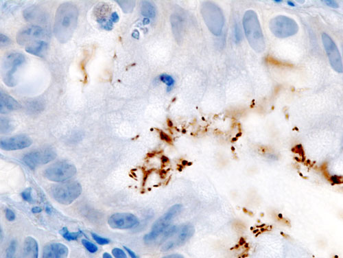
Barrett esophagus
Chronic reflux can metaplasia the distal esophagus to intestinal-type columnar epithelium (Barrett esophagus), raising adenocarcinoma risk. Endoscopy with biopsy confirms it; surveillance intervals follow pathology.
Chronic reflux can Barrett the esophagus. Surveillance catches dysplasia early.
Barrett esophagus
Tap each symptom. Build the case, then read the route.
Select symptoms to route PPI trial versus endoscopy now.
Pain Tells Location
Duodenal vs Gastric Ulcer
Meal timing is the board clue. Duodenal hurts when the stomach empties. Gastric hurts when food hits the ulcer. Toggle below; labels stay outside the figure on mobile.
Endoscopic finding
Pain vs meals
Improves with food because eating buffers acid in the duodenum. Wakes at night when the stomach empties and acid pours in. Strong association with H. pylori and NSAIDs.
Key step
Test and treat H. pylori if present. PPI heals the mucosa.
Peptic ulcerH. pylori on histology
Gastric ulcer extra steps
Gastric ulcers carry malignancy risk. Endoscopy with biopsy of the ulcer edge, then repeat endoscopy to document healing. Weight loss and pain worsened by food should raise your suspicion.
Gastric ulcer equals biopsy plus repeat scope to prove healing.
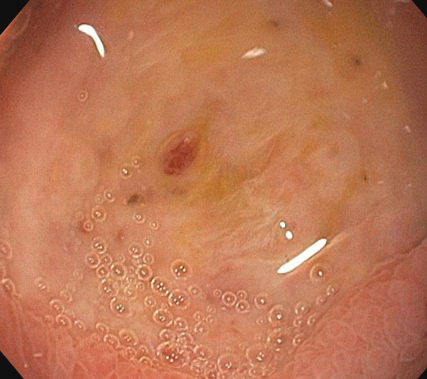
Esophageal Injury Spectrum
Red Mucosa Is Not Barrett
A smoker with reflux and a biopsy on the exhibit does not equal Barrett. The endoscopic color and the histology decide it. Climb the ladder: each rung is locked by one finding in plain language.
The trap a student walked into
A biopsy on the stem is not a diagnosis. You need goblet cells before you say Barrett.
Goblet cells are intestinal-type cells that do not belong in the esophagus. Erythematous, inflamed mucosa with a biopsy that shows inflammation and no goblet cells is reflux esophagitis, not Barrett. Tap each rung to reveal the locking finding.
Rung 1: red, inflamed, eroded mucosa · what is on biopsy?
Inflammation only: basal cell hyperplasia with scattered eosinophils and neutrophils, and no goblet cells. That is reflux (erosive) esophagitis, the most common esophagitis and a direct result of GERD. A smoker with heartburn and a red, inflamed esophagus whose biopsy shows inflammation and no goblet cells stops on this rung.
Rung 2: salmon-colored tongues above the junction · what is on biopsy?
Intestinal metaplasia, meaning goblet cells. Chronic acid reprograms the squamous lining into intestinal-type cells that grow goblet cells where none belong. Salmon-colored mucosa plus goblet cells is Barrett esophagus, not inflammation alone, and it raises adenocarcinoma risk.
Rung 3: Barrett that starts looking disorderly · what is on biopsy?
Goblet cells are still present, but now with crowded, enlarged, dark nuclei and loss of orderly maturation, and still no invasion. That is dysplasia. Low grade can regress; high grade is the final warning before cancer. Surveillance biopsies exist to catch exactly this change.
Rung 4: a mass, ulcer, or stricture in the distal esophagus · what is on biopsy?
Malignant glands invading past the basement membrane, often with high-grade dysplasia beside them. That is esophageal adenocarcinoma, the distal cancer Barrett raises the risk of. Progressively worsening trouble swallowing solids with weight loss is the clinical tell.
Goblet cells: intestinal metaplasia of Barrett esophagus
From the Attending
Do not let the word biopsy hypnotize you. A biopsy is a sample, not a diagnosis. Red, inflamed mucosa with inflammation and no goblet cells is reflux esophagitis. Salmon mucosa with goblet cells is Barrett. Crowded atypical nuclei on top of goblet cells is dysplasia. Glands punching through the basement membrane is adenocarcinoma. Find the goblet cells before you ever say Barrett.
Test, Treat, Confirm
H. pylori Eradication
Non-invasive testing first. Hold the PPI so you do not false-negative the breath or stool test. Treat, then confirm eradication only after antibiotics are done and PPI has been off long enough.
1. Suspect H. pylori (ulcer, dyspepsia, MALT lymphoma history)
2. Hold PPI ~2 weeks; test with urea breath or stool antigen
4. Confirm eradication: retest 4+ weeks after antibiotics, PPI held
Step 1: Indications include peptic ulcer disease, persistent dyspepsia, and gastric MALT lymphoma. If positive, eradication lowers recurrence and ulcer complications.
From the Attending
The exam loves the timing trap. You cannot confirm eradication while the patient is still on a PPI or immediately after finishing antibiotics. Wait at least four weeks off PPI, then urea breath or stool antigen again. Treat the bug, then prove it is gone.
When It Breaks Open
Bleed, Perforate, ZES
Upper GI bleeding, perforation, and gastrinoma each has a signature clue. Match the presentation to the move.
💧
Upper GI bleed
melena or hematemesis
▼
Tell
Hematemesis (vomiting blood) or melena (black, tarry stool). Lower GI bleed shows hematochezia (bright red blood per rectum).
First
Resuscitate: IV access, fluids, transfuse as needed. Start IV PPI, then urgent endoscopy for diagnosis and hemostasis.
⚠
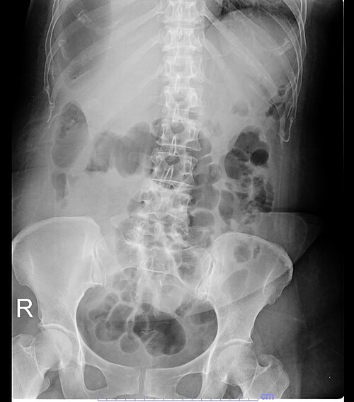
Perforated ulcer
sudden pain plus free air
▼
Tell
Sudden severe abdominal pain, board-like rigidity, pneumoperitoneum (free air under the diaphragm on upright film or CT).
Move
Surgery (or surgical consult). This is not an outpatient PPI problem.
📈
Zollinger-Ellison (ZES)
gastrinoma
▼
Tell
Multiple refractory peptic ulcers distal to the duodenal bulb, diarrhea, and elevated fasting gastrin. Standard PPI doses fail because acid is gastrin-driven.
Workup
Fasting gastrin, secretin stimulation test, imaging for gastrinoma (often in duodenum or pancreas). High-dose PPI plus surgical evaluation.
Pneumoperitoneum (free air)
Build the Plan
Walk a Dyspepsia Case
One patient pathway, one decision at a time. ALARM present? Scope. No ALARM? PPI. Ulcer found? Test H. pylori. This is the algorithm the exam encodes in vignettes.
From the Attending
Reflux without red flags is a PPI trial. One ALARM feature and you scope before you treat. Duodenal pain that feels better with food points down; gastric pain that worsens with food and weight loss points up and demands biopsy. Bleeding gets resuscitation, IV PPI, and endoscopy. Free air gets surgery. Know your clues.
The Osteopathic Lens
Upper GI Autonomics
Sympathetic and parasympathetic supply explain why rib raising and ganglion inhibition can adjunct functional dyspepsia. They never replace PPI or H. pylori eradication.
Autonomic supply
Sympathetics: upper GI viscera (stomach, proximal gut) via T5 to T9 greater splanchnic fibers. Chapman points for the stomach lie anteriorly along the rib cage. Parasympathetics: the vagus nerve drives acid secretion and gastric motility.
T5 to T9 splanchnics for sympathetics; vagus for acid and motility.
OMT as adjunct only
Rib raising, paraspinal inhibition, and ganglion release along the upper thoracic chain may ease functional dyspepsia by lowering sympathetic tone. This supports medical therapy; it does not replace PPI, endoscopy for ALARM features, or H. pylori triple or quadruple therapy.
Rib raising and ganglion inhibition adjunct dyspepsia; PPI and eradication still rule.
Rapid Fire
Five Quick Calls
Five questions from a pool of eight, reshuffled each visit. Cross out (right-click / long-press) and highlight (double-click / double-tap) as you read.
clinical Practice
Walk the Cases
Full vignettes, one at a time, shuffled order. Progress saves on this device. Cross out and highlight as you go.
Tip: kill wrong choices first, then read every explanation chain.
First Aid for the clinical medicine Step 1. Gastrointestinal: GERD, peptic ulcer disease, H. pylori, upper GI bleeding, Zollinger-Ellison syndrome.
Harrison's Principles of Internal Medicine. Peptic ulcer disease and Helicobacter pylori infection.
American College of Gastroenterology guidelines on GERD and H. pylori management.
Reviewed by Fatima Ali DO and Kaitlyn Cocuzzo MD. Vignettes are original clinical teaching cases. Confirm management against current guidelines at the point of care.
Bone Wizardry is an independent educational resource for visual learning in the medical sciences. It is not affiliated with, endorsed by, or sponsored by any licensing or examination board, contains no real or recalled examination questions, and does not guarantee any educational or examination outcome.
This is where Elite starts
The hook is free. The full board walkthroughs, elimination drills, and deeper mechanics are inside Elite.
Unlock every gated Bone Wizardry page
Keep the one-at-a-time walkthrough banks
Use the diagnostic games and answer logic without limits