Oral and salivary pathologies: location, time and tissue
Distinguish oral ulcers, salivary obstruction, infection and tumors using time course, duct anatomy, tissue patterns and safe diagnostic decisions.
A painful mouth lesion is not automatically an infection, and a painless salivary mass is not automatically benign. First locate the problem, then use its behavior over time to decide whether the patient needs symptom care, drainage assessment, or tissue diagnosis.
What is changing: the surface, the gland, or a nerve?
A patient points under the jaw and says, "It hurts when I eat." Another points to a sore on the tongue. Ask each person to indicate the exact site before assigning a diagnosis. A mucosal ulcer is a defect in the lining. A salivary swelling is a deeper enlargement that may occur with a normal mucosal surface. These are different starting problems, even when both make eating uncomfortable. [1][7]
Compare three observations. A shallow ulcer heals and later returns at another site. A gland repeatedly swells at the first bites of food and settles afterward. A focal mass steadily enlarges between meals. Classify them by recovery, stimulus dependence, and persistence before naming a disease. Recurrence with complete healing supports aphthae; stimulation-linked swelling suggests impaired salivary drainage; persistence requires evaluation for a structural lesion, including a tumor. Meal timing alone does not prove a stone because a stricture can also obstruct flow. [1][8][11]
Assess urgency before completing a routine workup. Inability to handle saliva, a rising tongue or tense floor-of-mouth swelling, breathing difficulty, or rapidly spreading infection requires emergency airway and specialist assessment. Do not delay stabilization to obtain a routine scan. A stable patient with an indurated oral lesion or a mass with facial weakness needs prompt cancer evaluation, not an indefinite antibiotic trial. [3][12][18]
Try the distinction: A previously meal-limited swelling becomes continuously painful and produces pus at a duct opening. State what has been added to the original problem.
Check that inference
Suppurative inflammation has developed on top of impaired drainage. A stone and infection can coexist; choosing one does not exclude the other. [9][11]
Apply it elsewhere: A mass remains after fever and tenderness resolve. Infection improvement does not establish that the remaining mass is benign. Re-examine it and complete the structural and tissue assessment appropriate to its location. [7][12]
Does an oral lesion heal completely?
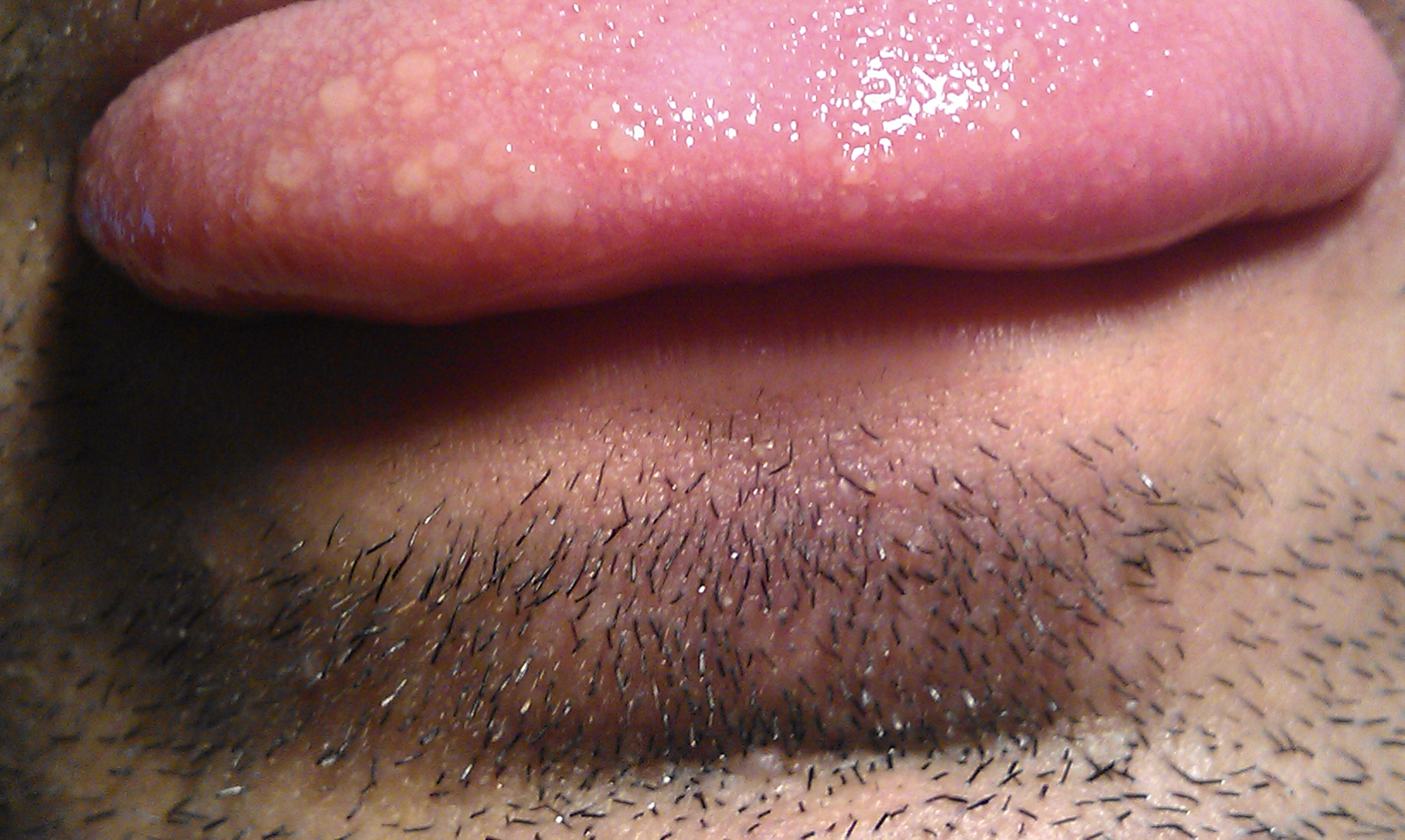
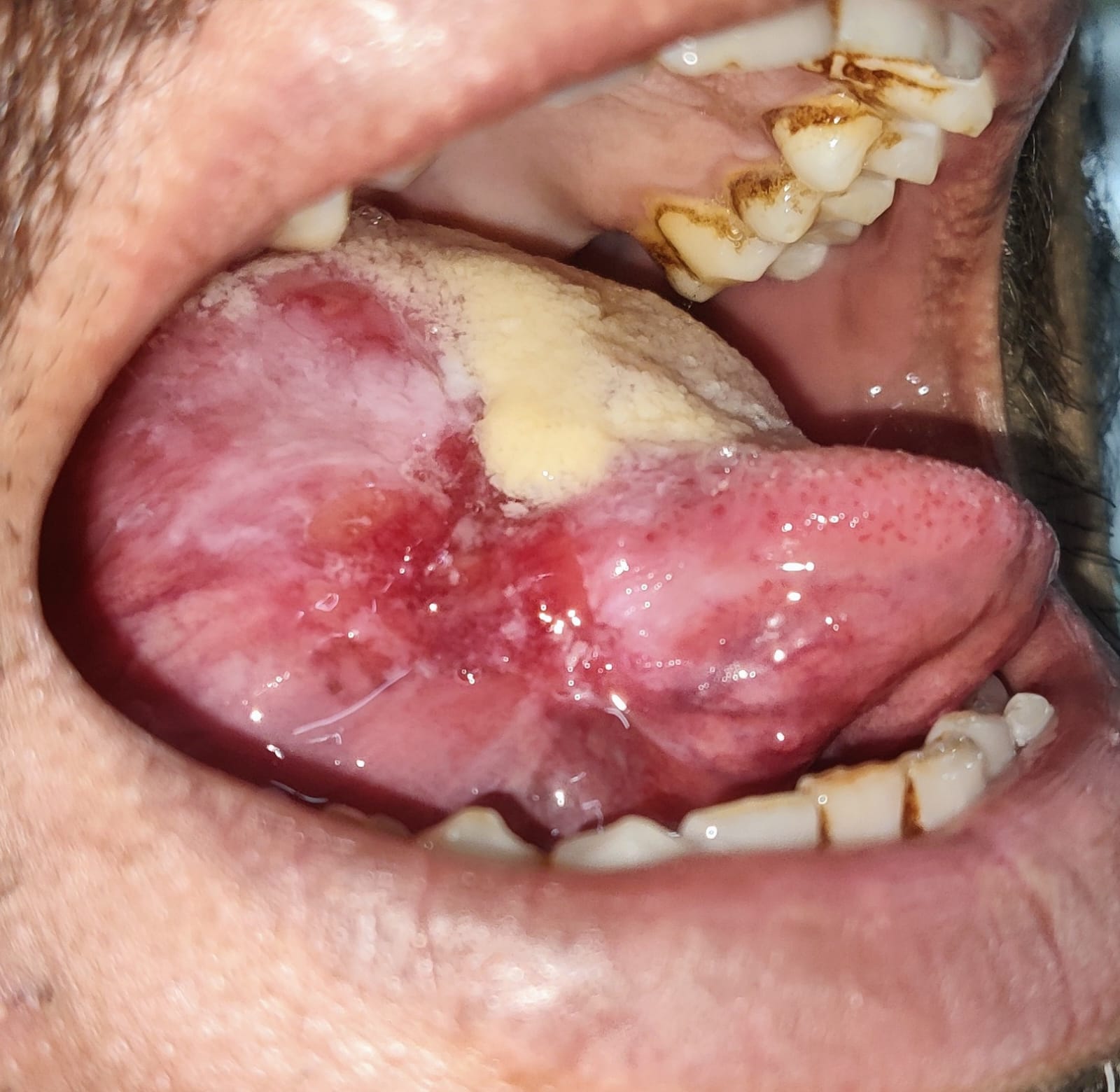
Does pain distinguish a harmless ulcer from cancer? No. Begin with the real photographs in this section. Describe the surface without assigning a diagnosis: a small pale-centered defect with a red rim in the aphthous example, and a much broader irregular red-and-white ulcerated area in the carcinoma example. A photograph cannot establish duration, induration, or histology. Add a history and palpation rather than diagnosing from color alone. [1][3]
The image shows this as an aphthous ulcer. Describe the pale center and surrounding redness, then add the healing history. The photograph cannot establish recurrence, duration, induration, or histology. Image: Prochoma; original source; CC BY-SA 3.0.
The source labels this tongue carcinoma. Compare its irregular surface with the small aphthous example, but do not infer a histologic subtype or grade from this photograph. Persistence, palpation and tissue assessment remain essential. Image: Dikkisherpa5; original source; CC0 1.0.
Minor aphthae are painful, round or oval, shallow ulcers on lining mucosa, such as the inner lip, cheek, or ventral tongue. A yellow-gray fibrinous surface is not the same as purulent salivary drainage. Individual minor ulcers generally heal in about one to two weeks without a scar. They are not contagious and are not caused by herpes simplex virus. Major aphthae are larger, deeper, persist longer, and can scar. Thus, "aphthae never scar" is an unsafe rule. [1][2]
Healing history matters, but it is not a permanent exemption from evaluation. A person with typical recurrent ulcers can develop an unrelated persistent lesion. Compare each new lesion with that person's prior pattern. A large or unfamiliar ulcer, a lesion that fails to resolve, or associated systemic illness deserves assessment. A clinically suspicious lesion requires prompt biopsy or specialist referral rather than waiting for an arbitrary duration. [2][3]
Try a time comparison: The inner-lip sore from last month disappeared completely. The current lateral-tongue lesion has remained in the same place, is firm at its base, and has an irregular raised edge. Decide which observation should control the evaluation.
Check the time comparison
The current lesion's persistence and induration control the decision. A history of healed ulcers does not explain a new persistent indurated lesion. [3]
Oral squamous-cell carcinoma can present as a persistent ulcer, a firm mass, unexplained pain, or a red, white, or mixed mucosal abnormality. Leukoplakia describes an otherwise unexplained white plaque; erythroplakia describes a concerning red patch. These clinical descriptions do not determine the histologic grade. Tobacco and alcohol increase oral cavity cancer risk, but their absence does not rule it out. Negative brush cytology must not override a persistent or progressing abnormality; tissue biopsy remains the diagnostic standard. [3]
Keep the anatomic cancer sites separate. The mobile anterior tongue belongs to the oral cavity. The tongue base and tonsillar region belong to the oropharynx, where HPV-associated squamous carcinoma is particularly important. Do not apply the HPV association indiscriminately to every oral ulcer. [3][4]
Apply it elsewhere: Recurrent healing ulcers accompanied by chronic diarrhea, weight loss, or iron deficiency warrant a systemic assessment. Celiac disease and inflammatory bowel disease belong in that assessment; ulcers alone do not distinguish them. Treating the oral discomfort without investigating the gastrointestinal findings misses the larger problem. [1]
Trace an outlet back to its gland
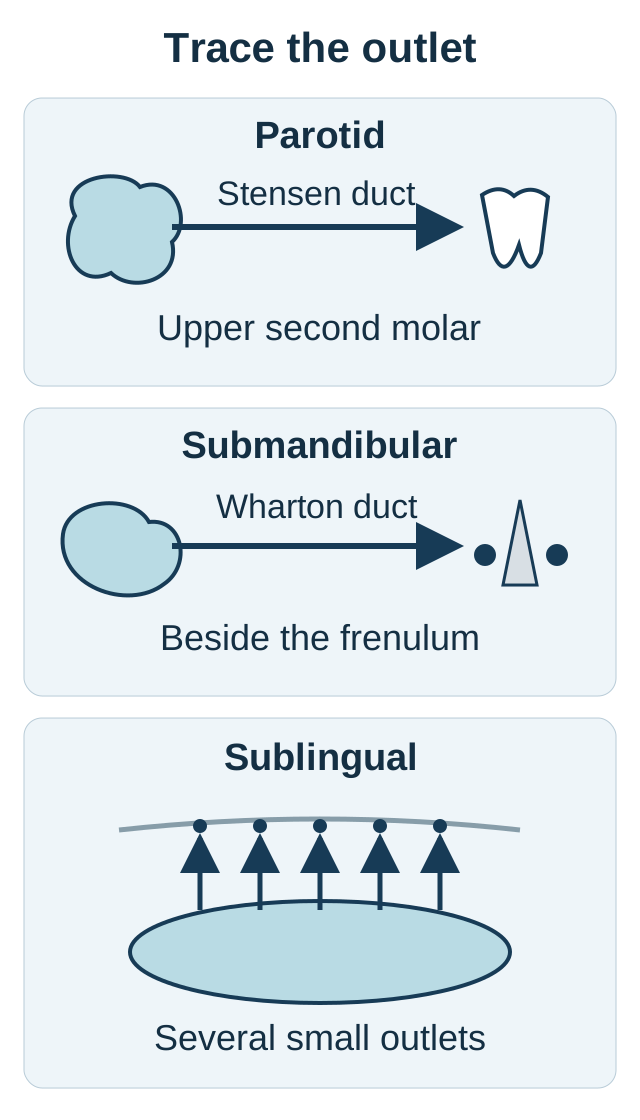
Two openings beneath the tongue do not mean that all saliva there comes from the sublingual glands. Use the outlet diagram to trace the route backward. The location of a duct opening can identify the affected gland even when surrounding swelling makes the gland difficult to feel. [5][6]
Trace each outlet backward before naming its gland. The arrows represent drainage relationships, not a scaled anatomic view; the full duct course and nerve relationships are explained in the text. [5][6][7][8]
The parotid lies in front of and below the ear and produces serous secretion. Its Stensen duct travels across the masseter, turns inward through the buccinator, and opens on the cheek mucosa opposite the upper second molar. Pus at this papilla localizes the involved duct to the parotid system. The facial nerve divides within the parotid and supplies facial expression; it does not provide that gland's secretomotor innervation. Postganglionic secretomotor fibers instead reach the parotid from the otic ganglion through the auriculotemporal nerve. A nerve passing through an organ does not necessarily control its secretion. [6]
The submandibular gland lies mainly below the mandible, with its deeper part curving around the posterior border of the mylohyoid. It produces mixed secretion. Wharton duct runs forward through the floor of the mouth to a sublingual caruncle beside the lingual frenulum. Most salivary stones occur in this system. A distal calculus may be palpable along the anterior floor of the mouth. The lingual nerve crosses in close relation to the duct, an important distinction from the hypoglossal nerve's tongue motor function during procedural assessment. [5][8]
The sublingual gland lies under the tongue above the mylohyoid and produces predominantly mucous secretion. Several small ducts open along the sublingual fold, rather than all emptying through a single Wharton outlet; a larger duct connection can vary. Minor glands are scattered in oral and upper aerodigestive mucosa and drain locally. Their small size does not protect a persistent submucosal mass from malignancy. [5][7][13]
Trace and predict: Locate the papilla beside the frenulum in the diagram. Follow its duct backward, then predict where a blocked gland would enlarge when saliva production increases. State the gland before checking.
Check the outlet trace
That papilla receives Wharton duct from the submandibular gland. Obstruction can produce swelling beneath the mandible, despite the outlet being underneath the tongue. [5][8]
Apply it elsewhere: After a parotid operation, facial asymmetry is a motor finding, not proof that the gland's secretomotor pathway was severed. After a floor-of-mouth duct procedure, altered tongue sensation and weak tongue protrusion suggest different nerves. Localize the function as well as the surgical site. [5][6]
Predict what happens on each side of an obstruction
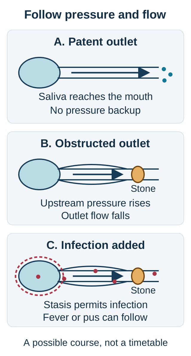
Must infection be present before a salivary gland hurts? Compare the three illustrated duct states: free drainage, an obstructed outlet during eating, and obstruction with infection. The point of the diagram is pressure and flow, not a literal measurement of duct size or a guarantee that every stone becomes infected. [8][11]
Predict upstream pressure and saliva arriving at the outlet when obstruction appears or is relieved. Infection is a possible added complication, not an inevitable final state; arrow size is not a quantitative flow measurement. [8][9][11]
Reduced flow, concentrated secretions, duct anatomy, and local injury can contribute to calculus formation. A stone contains organic material and deposited minerals. When anticipation of food or eating stimulates secretion against an obstructed outlet, saliva accumulates upstream. Pressure and gland swelling increase even though little saliva reaches the mouth. When the stimulus subsides, symptoms may ease. This explains intermittent meal-related swelling without requiring fever or bacterial invasion. [8]
Pressure-and-flow exercise: In state A the outlet is patent. In state B the gland produces more saliva but the outlet is blocked. Predict two separate outcomes in B: saliva arriving at the papilla, and pressure inside the upstream duct. Then ask what changes when patency is restored while the stimulus continues.
With obstruction, outlet flow is reduced while upstream pressure rises. Restoring drainage allows outlet flow to increase and upstream pressure to fall. More production and more delivered saliva are not interchangeable. A patient can feel a dry mouth while the obstructed gland is swollen. This is a directional model, not a quantitative formula. [8][11]
Check a new pressure prediction
If secretion falls while the obstruction remains, swelling may diminish temporarily. Symptom relief alone does not prove that the calculus has passed. [8]
Stasis can permit bacteria from the mouth to ascend into the duct. Fever, persistent tenderness, erythema, and purulent drainage support acute suppurative sialadenitis. Staphylococcus aureus and oral anaerobes are important considerations. Poor intake or medications that reduce salivary secretion can predispose to infection even when no stone is found. Repeated inflammation may damage secretory tissue and produce fibrosis, perpetuating poor function. This progression is possible, not inevitable. [9][11]
Management follows the added problem. Uncomplicated obstruction calls for hydration, appropriate analgesia, gentle supportive care, and salivary evaluation if symptoms persist or recur. A persistent calculus may require specialist treatment. Suppurative disease adds culture-guided antimicrobial treatment, selected for illness severity, allergies, local resistance, and likely organisms. An established abscess requires assessment for drainage; simply extending antibiotics may leave the collection untreated. Emergency airway concerns take priority over both. [8][9][10][18]
Apply it elsewhere: Months of dry eyes and dry mouth with bilateral gland enlargement and no purulence should broaden the differential toward autoimmune disease such as Sjogren syndrome. Viral parotitis is another alternative, particularly with an appropriate exposure history. Sialadenitis names inflammation, not a single bacterial cause. [7][9]
Separate how often a tumor occurs from how often it is malignant
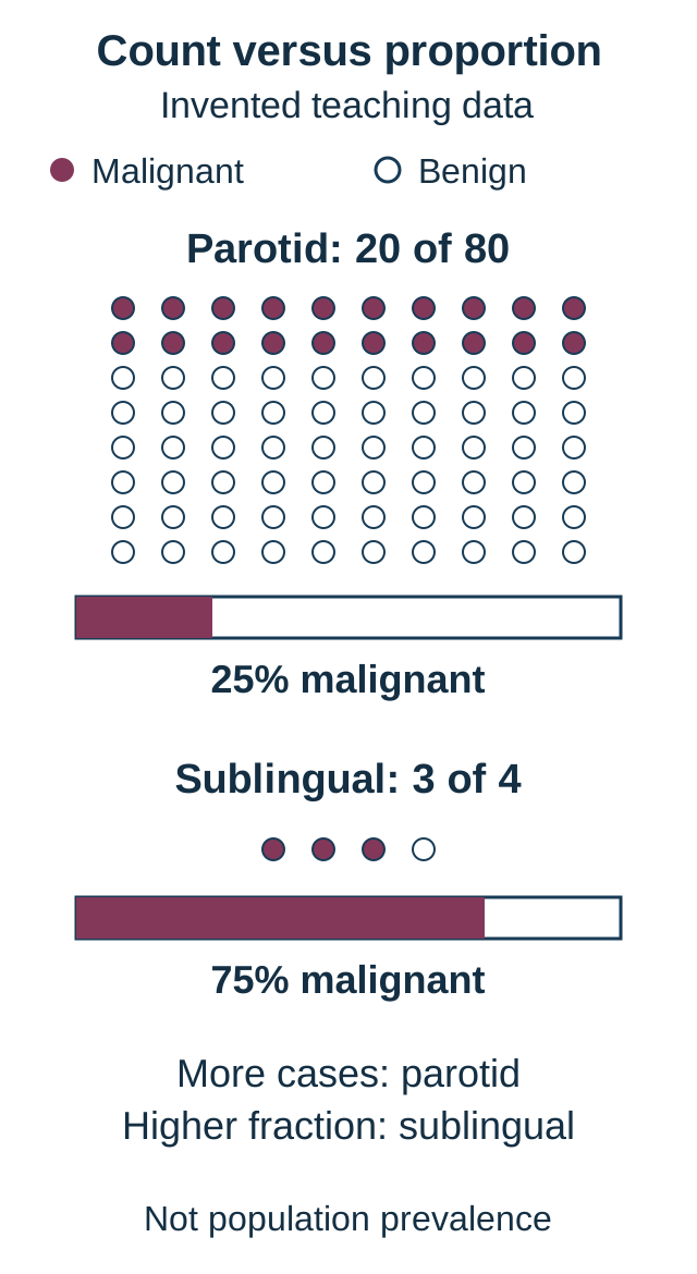
Does the site with the most tumors necessarily have the highest malignant fraction? Use the denominator illustration to answer that question before applying a location-based memory rule. Parotid tumors are common relative to tumors of other salivary glands, and most are benign. Submandibular, minor-gland, and especially sublingual neoplasms carry greater malignant concern. The proportions differ across series and minor-gland sites; physical gland size is not a diagnostic test. [13]
data, not population prevalence: 20 of 80 parotid neoplasms and 3 of 4 sublingual neoplasms are malignant. Compare the absolute count with the fraction among all tumors at that site. One circle represents one tumor. [13]
The illustration uses an dataset, not clinical prevalence: 20 malignant tumors among 80 parotid tumors, and 3 among 4 sublingual tumors. Count malignant tumors first, then divide by all tumors at the same site. The parotid contributes more malignant tumors by count, but the sublingual group has the larger malignant fraction: 25% versus 75%. Neither calculation tells you whether a particular patient's lesion is benign.
Choose the denominator: For a patient with a confirmed sublingual neoplasm, should the conditional estimate use all salivary tumors in the dataset or just the sublingual tumors?
Check the denominator
Use the tumors from the same site: 3 divided by 4. Dividing by all salivary tumors answers a different question.
Use location to set suspicion, then examine behavior. A mobile painless parotid mass still needs a diagnosis. A small painless palatal mass can arise from a minor gland and is not reassured by being hidden beneath intact mucosa. In a parotid-region mass, also consider a lymph node containing metastasis from a head or neck skin cancer rather than assuming every mass began in salivary epithelium. [12][13]
Apply it elsewhere: A submandibular mass has greater site-based malignant concern than a parotid neoplasm, not no change. Rapid growth, fixation, skin or mucosal ulceration, abnormal cervical nodes, or a new nerve deficit are additional reasons to expedite evaluation at either site. Absence of those findings does not exclude malignancy. [12][13]
Read the tissue pattern, then ask what it predicts
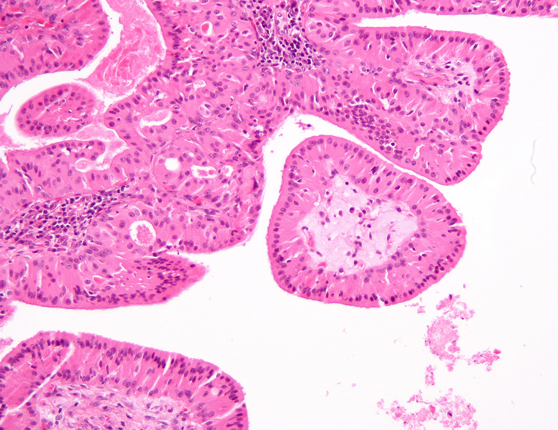
Does a cystic mass imply benign disease? No. Compare the tissue architecture, not just the presence of fluid. In the real Warthin micrograph, first identify the pink epithelial lining of the papillary projections and then the much smaller dark lymphoid cells in their supporting tissue. The two compartments are different. Histology descriptions below help distinguish three important tumor patterns, but a small sample may not represent every part of a mass. [12][15]
The image shows papillary cystadenoma lymphomatosum, also called Warthin tumor. Locate the epithelial lining and lymphoid supporting tissue separately; lymphocytes within this organized tumor do not by themselves establish lymphoma. No scale or magnification is inferred. Image: Nephron; original source; CC BY-SA 3.0.
Pleomorphic adenoma is the most common benign salivary tumor overall, containing epithelial and myoepithelial elements in a variably myxoid or cartilage-like supporting matrix. A slowly enlarging mobile parotid mass is a typical presentation, not confirmation. Microscopic extensions and incomplete encapsulation help explain why incomplete excision or tumor disruption can contribute to recurrence. A later cluster of nodules with the same bland tissue pattern can represent recurrent benign tumor rather than malignant transformation. Surgical management must address the tumor without treating the apparent capsule as a guaranteed complete boundary. [14][15]
A previously established pleomorphic adenoma that develops rapid enlargement, pain, fixation, or facial dysfunction raises concern for carcinoma ex pleomorphic adenoma. It warrants reassessment, not reassurance from its old benign diagnosis. Suspicious imaging and a bland needle sample must be reconciled, since sampling only a benign component can miss a malignant component. Transformation risk is not the same as certainty, and rapid growth still requires tissue confirmation. [12][17]
Warthin tumor has papillary and cystic spaces lined by oncocytic epithelium supported by lymphoid tissue, often with germinal centers. Oncocytes have abundant granular eosinophilic cytoplasm. Smoking is strongly associated with this benign tumor; the association does not make every smoker's parotid mass benign. Warthin tumors can be bilateral or multifocal, so a second concordant lesion need not represent metastasis. It still requires an appropriate assessment rather than automatic assignment of the first lesion's diagnosis. [15][16][19]
Mucoepidermoid carcinoma is a common malignant salivary tumor containing mucous, epidermoid or squamoid, and intermediate cells. It can have a prominent cystic component, including in lower-grade tumors. A cystic appearance therefore does not separate it reliably from Warthin tumor. Low, intermediate, and high grades describe tissue features; stage describes the extent of disease. Invasive low-grade disease is still malignant, and spread beyond the gland or to nodes matters even when the cells appear relatively bland. [13]
Try the tissue comparison: A cystic parotid sample contains mucous cells and squamoid cells but lacks the bilayered oncocytic papillae and organized lymphoid supporting tissue of the photographed tumor. Decide why both are cystic is an inadequate comparison.
Check the tissue comparison
The cell populations differ. The mixed mucous and squamoid pattern supports mucoepidermoid carcinoma rather than Warthin tumor; cyst formation occurs in both. [13][15]
For a persistent major-gland mass, examination, imaging, and needle sampling are complementary. Ultrasound-guided fine-needle aspiration is commonly used; inadequate or discordant findings require further assessment, which can include core sampling. Suspected malignant extension favors MRI, including assessment of relevant neck nodes. A minor-gland submucosal lesion instead needs an appropriately planned biopsy by an experienced specialist. Do not equate that pathway with an unplanned open biopsy of a parotid mass. [12]
Apply it elsewhere: Two tumors with the same histologic grade can have very different extents. Compare confinement to the gland with skin invasion or involved nodes before counseling about risk. Tissue identity, local invasion, and distribution all matter; no single painless, cystic, or low-grade label settles the case. [13]
Apply the distinctions to new patients
For each patient, decide what the location and time course imply before choosing an answer. The cases include interpretation, prediction, and diagnostic planning; they are educational examples, not individual medical advice.
Case 1
Show answer and explanations for case 1
A. Obtain viral swabs during every oral recurrence (Why this does not fit)
Herpetic infection would be considered with a compatible vesicular eruption or other infectious findings. The recurrent healing lining-mucosa ulcers and chronic intestinal findings do not establish a viral explanation for the whole presentation.
Reasoning steps for option A
What eruption would make viral swabbing of the recurrent lip ulcers informative?
Herpetic infection would be considered with a compatible vesicular eruption or other infectious findings.
Why do ten-day healing and the intestinal findings weaken a herpes-only explanation?
The recurrent healing lining-mucosa ulcers and chronic intestinal findings do not establish a viral explanation for the whole presentation.
B. Evaluate gastrointestinal disease and nutritional deficiencies (Best answer)
Recurrent aphthae can accompany malabsorptive or inflammatory intestinal disease. Complete oral healing fits aphthae, while diarrhea, weight loss, and iron deficiency require a systemic assessment rather than a local-only diagnosis.
Reasoning steps for option B
What systemic disorders can link recurrent inner-lip aphthae with diarrhea?
Recurrent aphthae can accompany malabsorptive or inflammatory intestinal disease.
Why do weight loss and iron deficiency warrant gastrointestinal and nutritional evaluation?
Complete oral healing fits aphthae, while diarrhea, weight loss, and iron deficiency require a systemic assessment rather than a local-only diagnosis.
C. Biopsy the healed mucosal sites at each visit (Why this does not fit)
Biopsy targets a persistent or otherwise suspicious abnormal area. No residual abnormality remains between these typical episodes; sampling healed sites does not investigate the diarrhea and deficiency.
Reasoning steps for option C
When does an oral ulcer provide an abnormal site suitable for biopsy?
Biopsy targets a persistent or otherwise suspicious abnormal area.
Why would sampling normal mucosa between these episodes miss the systemic problem?
No residual abnormality remains between these typical episodes; sampling healed sites does not investigate the diarrhea and deficiency.
D. Perform salivary ultrasound and duct imaging (Why this does not fit)
Salivary obstruction produces gland swelling associated with secretion. The patient has surface ulcers and gastrointestinal symptoms without stimulated gland enlargement.
Reasoning steps for option D
What gland pattern would support imaging for salivary obstruction?
Salivary obstruction produces gland swelling associated with secretion.
Why do superficial lip ulcers without meal-triggered swelling argue against a blocked duct?
The patient has surface ulcers and gastrointestinal symptoms without stimulated gland enlargement.
Takeaway: Recurrent healing aphthae plus intestinal symptoms require investigation for systemic disease, including celiac disease and inflammatory bowel disease.
A. Urgent tissue sampling for a persistent malignant lesion (Why this does not fit)
An indurated or persistent abnormality needs tissue evaluation. These lesions repeatedly resolve completely and have no current suspicious examination finding requiring urgent biopsy.
Reasoning steps for option A
Which ulcer findings would prompt urgent biopsy rather than routine aphthous care?
An indurated or persistent abnormality needs tissue evaluation.
How does full healing within a week and lack of induration change biopsy urgency?
These lesions repeatedly resolve completely and have no current suspicious examination finding requiring urgent biopsy.
B. Systemic antiviral treatment for recurrent oral herpes (Why this does not fit)
Antiviral treatment targets a clinically compatible herpes infection. The nonvesicular recurrent lining-mucosa pattern is characteristic of aphthae, not evidence that herpes is the cause.
Reasoning steps for option B
What vesicular pattern would justify antiviral treatment for oral herpes?
A clinically compatible vesicular eruption would support herpes and make antiviral treatment appropriate; these ulcers have no preceding blisters.
Why do nonvesicular 4-mm inner-lip ulcers favor aphthae over herpes?
The nonvesicular recurrent lining-mucosa pattern is characteristic of aphthae, not evidence that herpes is the cause.
C. Systemic antibacterial treatment for suppurative oral infection (Why this does not fit)
Antibiotics are appropriate for bacterial infection supported by findings such as purulence or spreading inflammation. These shallow recurrent ulcers lack pus, gland tenderness, fever, or spreading infection.
Reasoning steps for option C
What evidence of suppurative infection would justify systemic antibiotics?
Antibiotics are appropriate for bacterial infection supported by findings such as purulence or spreading inflammation.
Why do the absence of pus, fever and spreading inflammation undermine antibiotics?
These shallow recurrent ulcers lack pus, gland tenderness, fever, or spreading infection.
D. Local symptom care with a plan to reassess atypical persistence (Best answer)
Typical minor aphthae are self-limited and can receive local symptomatic care. Small recurrent lesions that heal fully without systemic findings fit that pattern, with reassessment if the course changes.
Reasoning steps for option D
Which size and healing features characterize minor aphthous ulcers?
Small recurrent ulcers that heal completely within a week without scarring fit minor aphthae, which are self-limited and can receive local symptomatic care.
Why do these scar-free recurrent ulcers support local symptom care and follow-up?
Small recurrent lesions that heal fully without systemic findings fit that pattern, with reassessment if the course changes.
Takeaway: Minor aphthae heal; a new persistent or indurated lesion requires a different assessment.
A. Arrange prompt specialist examination and tissue biopsy (Best answer)
A persistent indurated oral lesion requires tissue diagnosis. Failure to heal after addressing the presumed irritant, enlargement, and a cervical node make another observation-only interval inappropriate.
Reasoning steps for option A
What features of a lateral-tongue ulcer make tissue diagnosis urgent?
A persistent indurated oral lesion requires tissue diagnosis.
Why do enlargement after tooth smoothing and a new cervical node rule out further observation?
Failure to heal after addressing the presumed irritant, enlargement, and a cervical node make another observation-only interval inappropriate.
B. Start topical corticosteroid therapy and review response (Why this does not fit)
Topical anti-inflammatory therapy can relieve established aphthous disease. A single fixed enlarging indurated lesion does not share the complete healing pattern expected of routine minor aphthae.
Reasoning steps for option B
In what ulcer pattern can topical corticosteroids be appropriate?
Topical anti-inflammatory therapy can relieve established aphthous disease.
Why does this fixed indurated five-week lesion not fit routine minor aphthae?
A single fixed enlarging indurated lesion does not share the complete healing pattern expected of routine minor aphthae.
C. Use an antifungal trial before deciding on tissue sampling (Why this does not fit)
Antifungal therapy is appropriate when the lesion is supported as candidal disease. The firm ulcer with progressive local and nodal findings is not explained by superficial fungal infection and should not have biopsy deferred.
Reasoning steps for option C
Would an antifungal trial be appropriate without findings supporting candidiasis in this firm tongue ulcer?
Antifungal therapy is appropriate when the lesion is supported as candidal disease.
Why can a fungal trial not replace biopsy of this firm ulcer with nodal change?
The firm ulcer with progressive local and nodal findings is not explained by superficial fungal infection and should not have biopsy deferred.
D. Repeat dental adjustment and review after another month (Why this does not fit)
A clearly traumatic lesion should improve after the source of injury is corrected. The lesion has enlarged after correction and is now accompanied by induration and a node, so trauma no longer accounts for the course.
Reasoning steps for option D
What response should follow removal of a causative sharp-tooth irritant?
A clearly traumatic lesion should improve after the source of injury is corrected.
Why does continued enlargement after smoothing defeat a traumatic explanation?
The lesion has enlarged after correction and is now accompanied by induration and a node, so trauma no longer accounts for the course.
Takeaway: A plausible irritant does not excuse a nonhealing indurated lesion after the irritant is addressed.
A. Obtain a fungal swab before any further assessment (Why this does not fit)
Fungal testing can help when candidiasis is a plausible unresolved diagnosis. Failure of therapy and a persistent non-scrapable mixed patch require tissue evaluation rather than deferral for another infection-only assessment.
Reasoning steps for option A
When could a fungal swab clarify a suspected oral candidal patch?
Fungal testing can help when candidiasis is a plausible unresolved diagnosis.
Why does persistence after antifungals of a non-scrapable red-white patch favor biopsy?
Failure of therapy and a persistent non-scrapable mixed patch require tissue evaluation rather than deferral for another infection-only assessment.
B. Repeat brush cytology before considering tissue biopsy (Why this does not fit)
Cytology samples cells but does not settle every persistent mucosal abnormality. Another cytology result would not resolve the clinical indication for biopsy in this persistent lesion.
Reasoning steps for option B
What limitation does brush cytology have for evaluating tissue invasion?
Brush cytology samples cells rather than tissue architecture, so a negative result does not exclude invasion or settle this persistent mucosal abnormality.
Why does a prior negative brush sample not justify repeating cytology instead of biopsy?
Another cytology result would not resolve the clinical indication for biopsy in this persistent lesion.
C. Arrange tissue biopsy of the persistent abnormal area (Best answer)
Histologic tissue assessment is the diagnostic standard for a persistent suspicious oral abnormality. The negative cytology does not outweigh persistence of the unexplained mixed patch after treatment.
Reasoning steps for option C
What does tissue histology establish in a persistent mixed red-white patch?
Histologic tissue assessment is the diagnostic standard for a persistent suspicious oral abnormality.
Why does a negative cytology result not exclude disease in this persistent floor-of-mouth lesion?
The negative cytology does not outweigh persistence of the unexplained mixed patch after treatment.
D. Perform light-based screening to exclude tissue invasion (Why this does not fit)
Light-based adjuncts do not demonstrate the tissue architecture needed to establish or exclude invasion. A negative adjunctive screen should not override this persisting clinically abnormal area.
Reasoning steps for option D
What can a light-based screen not assess in a suspicious oral patch?
Light-based adjuncts do not demonstrate the tissue architecture needed to establish or exclude invasion.
Why should a negative light-based finding not postpone tissue sampling here?
A negative adjunctive screen should not override this persisting clinically abnormal area.
Takeaway: Negative adjunctive testing does not exclude disease in a persistent suspicious oral lesion.
A. Minor aphthous stomatitis (Why this does not fit)
Minor aphthae are generally small and heal without a scar over a shorter interval. The depth, size, duration, and scarring do not fit the usual minor form.
Reasoning steps for option A
What size, duration and scarring profile defines minor aphthae?
Minor aphthae are generally small and heal without a scar over a shorter interval.
Why do ulcers over 1 cm that heal over weeks with scars fail the minor classification?
The depth, size, duration, and scarring do not fit the usual minor form.
B. Recurrent traumatic ulceration (Why this does not fit)
Trauma can create an ulcer where a recurring local irritant contacts mucosa. No recurring mechanical source is identified, and the assessed aphthous pattern explains the characteristic larger episodes.
Reasoning steps for option B
What contact pattern would implicate repeated local trauma?
Trauma can create an ulcer where a recurring local irritant contacts mucosa.
Why do absent recurrent irritants and the documented aphthous pattern disfavor trauma?
No recurring mechanical source is identified, and the assessed aphthous pattern explains the characteristic larger episodes.
C. Major aphthous stomatitis (Best answer)
Major aphthae are deeper and larger and can leave scars after prolonged healing. The documented aphthous pattern and lengthy scar-forming episodes are compatible with this form.
Reasoning steps for option C
Can a major aphtha persist for weeks and leave a scar?
Major aphthae are deeper and larger and can leave scars after prolonged healing.
How do depth, size above 1 cm and the documented aphthous assessment support major aphthae?
The documented aphthous pattern and lengthy scar-forming episodes are compatible with this form.
D. Recurrent herpetic stomatitis (Why this does not fit)
Herpes produces an infectious eruption rather than the established nonvesicular aphthous pattern. Scarring alone does not overturn the documented aphthous assessment or establish herpes infection.
Reasoning steps for option D
What infectious eruption would support recurrent herpetic stomatitis?
Herpes produces an infectious eruption rather than the established nonvesicular aphthous pattern.
Why does scarring not establish herpes when an aphthous pattern was identified?
Scarring alone does not overturn the documented aphthous assessment or establish herpes infection.
Takeaway: Scarring is compatible with major aphthae and should not be generalized from the minor form.
HPV-associated squamous cancers preferentially arise in the oropharyngeal tonsillar region. The malignant squamous cells in a cervical node and active high-risk HPV signal direct the primary-site search toward the tonsils and tongue base.
Reasoning steps for option A
Which oropharyngeal structures commonly harbor HPV-associated squamous primaries?
HPV-associated squamous cancers preferentially arise in the oropharyngeal tonsillar region.
How do HPV E6/E7-positive squamous cells in a cervical node focus the search on tonsil and tongue base?
The malignant squamous cells in a cervical node and active high-risk HPV signal direct the primary-site search toward the tonsils and tongue base.
B. Buccal mucosa or inner lower lip (Why this does not fit)
These are oral cavity mucosal sites in which squamous carcinoma can occur. Their oral cavity location does not carry the same HPV-associated primary-site implication as the tonsillar region.
Reasoning steps for option B
Can buccal mucosa or lower lip harbor squamous carcinoma?
These are oral cavity mucosal sites in which squamous carcinoma can occur.
Why does an HPV-positive cervical metastasis favor the oropharynx over these oral sites?
Their oral cavity location does not carry the same HPV-associated primary-site implication as the tonsillar region.
C. Hard palate or attached maxillary gingiva (Why this does not fit)
These keratinized oral sites are anatomically distinct from the oropharynx. The viral tumor finding supports an oropharyngeal search rather than preferentially selecting these oral cavity sites.
Reasoning steps for option C
Where are the hard palate and attached maxillary gingiva relative to the oropharynx?
These keratinized oral sites are anatomically distinct from the oropharynx.
Why do viral tumor findings not preferentially implicate these keratinized oral sites?
The viral tumor finding supports an oropharyngeal search rather than preferentially selecting these oral cavity sites.
D. Anterior mobile tongue or mouth floor (Why this does not fit)
These structures belong to the oral cavity rather than the oropharyngeal tonsillar region. They can develop squamous carcinoma, but the HPV-associated nodal presentation more strongly directs attention to the oropharynx.
Reasoning steps for option D
What distinguishes the proposed oral cavity sites from tonsillar tissue?
These structures belong to the oral cavity rather than the oropharyngeal tonsillar region.
Why is an HPV-associated nodal squamous tumor less suggestive of these oral sites?
They can develop squamous carcinoma, but the HPV-associated nodal presentation more strongly directs attention to the oropharynx.
Takeaway: A cystic cervical metastasis with HPV-associated squamous tumor cells directs attention to an oropharyngeal primary.
A. Submandibular ganglion fibers in local glandular branches (Why this does not fit)
The submandibular ganglion provides postganglionic fibers to the submandibular and sublingual systems. Those glands drain into the floor of the mouth rather than through the upper molar papilla being tested.
Reasoning steps for option A
Which glands receive postganglionic supply from the submandibular ganglion?
The submandibular ganglion provides postganglionic fibers to the submandibular and sublingual systems.
Why does an upper second-molar papilla exclude the floor-of-mouth gland outlets?
Those glands drain into the floor of the mouth rather than through the upper molar papilla being tested.
B. Otic ganglion fibers in the auriculotemporal nerve (Best answer)
Postganglionic parasympathetic fibers from the otic ganglion reach the parotid through the auriculotemporal nerve. The upper second-molar papilla identifies Stensen duct and therefore the parotid, independently of normal facial motor function.
Reasoning steps for option B
How do otic ganglion fibers reach the parotid gland?
Postganglionic parasympathetic fibers from the otic ganglion reach the parotid through the auriculotemporal nerve.
Why does a patent duct opposite the upper second molar identify this pathway despite normal facial movement?
The upper second-molar papilla identifies Stensen duct and therefore the parotid, independently of normal facial motor function.
C. Hypoglossal nucleus fibers in the hypoglossal nerve (Why this does not fit)
The hypoglossal nerve supplies tongue musculature rather than salivary secretomotor function. A patent salivary duct with reduced secretion is not explained by substituting the tongue motor pathway for the parotid autonomic supply.
Reasoning steps for option C
Does the hypoglossal nerve supply tongue movement or parotid secretion?
The hypoglossal nerve supplies tongue musculature rather than salivary secretomotor function.
Why does reduced saliva with an intact gland not implicate tongue motor fibers?
A patent salivary duct with reduced secretion is not explained by substituting the tongue motor pathway for the parotid autonomic supply.
D. Facial motor nucleus fibers in the buccal facial branch (Why this does not fit)
Facial motor branches traverse the parotid and supply muscles of facial expression. Traversal does not make them the gland secretomotor pathway; the tested outlet localizes a parotid secretion problem.
Reasoning steps for option D
What function do facial motor branches perform as they cross the parotid?
Facial motor branches traverse the parotid and supply muscles of facial expression.
Why does normal expression not determine the secretomotor supply of Stensen duct gland?
Traversal does not make them the gland secretomotor pathway; the tested outlet localizes a parotid secretion problem.
Takeaway: The duct outlet identifies the parotid; its autonomic supply differs from the facial motor branches that traverse it.
A. Marginal mandibular facial branch (Why this does not fit)
This facial branch supplies lower-lip muscles. Symmetric lower-lip expression and an anterior-tongue sensory deficit do not match its motor territory.
Reasoning steps for option A
What deficit would follow injury to the marginal mandibular facial branch?
This facial branch supplies lower-lip muscles.
Why does symmetric lower-lip motion not account for reduced anterior-tongue touch?
Symmetric lower-lip expression and an anterior-tongue sensory deficit do not match its motor territory.
B. Hypoglossal nerve (Why this does not fit)
Hypoglossal injury impairs tongue motor function and can cause deviation during protrusion. Tongue strength and midline protrusion are preserved, whereas general sensation is impaired.
Reasoning steps for option B
How would hypoglossal injury affect tongue protrusion?
Hypoglossal injury impairs tongue motor function and can cause deviation during protrusion.
Why does preserved midline strength favor a sensory rather than tongue motor nerve injury?
Tongue strength and midline protrusion are preserved, whereas general sensation is impaired.
C. Auriculotemporal nerve (Why this does not fit)
The auriculotemporal nerve relates to the parotid secretomotor pathway and temporal or preauricular sensory territory. It does not explain focal anterior-tongue sensation loss after a submandibular duct procedure.
Reasoning steps for option C
What territory and gland pathway involve the auriculotemporal nerve?
The auriculotemporal nerve relates to the parotid secretomotor pathway and temporal or preauricular sensory territory.
Why is anterior-tongue numbness after a Wharton duct procedure outside that territory?
It does not explain focal anterior-tongue sensation loss after a submandibular duct procedure.
D. Lingual nerve (Best answer)
The lingual nerve carries general sensation from the anterior tongue and lies close to the submandibular duct. The sensory deficit after a floor-of-mouth duct procedure, with preserved tongue strength, fits lingual rather than tongue motor injury.
Reasoning steps for option D
Which nerve lies near Wharton duct and carries anterior-tongue general sensation?
The lingual nerve carries general sensation from the anterior tongue and lies close to the submandibular duct.
Why does reduced touch with intact protrusion localize to the lingual nerve?
The sensory deficit after a floor-of-mouth duct procedure, with preserved tongue strength, fits lingual rather than tongue motor injury.
Takeaway: After duct procedures, distinguish anterior-tongue sensory loss from tongue motor weakness.
A. Minor palatal gland lying within the palatal submucosa (Why this does not fit)
Minor glands drain locally into overlying mucosa rather than through a long Wharton duct. The single obstructed floor-of-mouth duct with jaw swelling cannot be assigned to a palatal minor gland.
Reasoning steps for option A
How do minor palatal glands empty into the oral mucosa?
Minor glands drain locally into overlying mucosa rather than through a long Wharton duct.
Why can a palatal gland not explain an obstructed frenulum-adjacent duct and jaw swelling?
The single obstructed floor-of-mouth duct with jaw swelling cannot be assigned to a palatal minor gland.
B. Submandibular gland curving around the posterior mylohyoid border (Best answer)
Wharton duct drains the submandibular gland beside the frenulum, and the gland has superficial and deep portions around mylohyoid. The blocked single papilla with preserved small sublingual outlets identifies that upstream system.
Reasoning steps for option B
Which gland empties through Wharton duct beside the lingual frenulum?
Wharton duct drains the submandibular gland beside the frenulum, and the gland has superficial and deep portions around mylohyoid.
How does a blocked single papilla with intact sublingual outlets implicate the gland around mylohyoid?
The blocked single papilla with preserved small sublingual outlets identifies that upstream system.
C. Parotid gland lying beside the posterior mandibular ramus (Why this does not fit)
The parotid drains across the masseter through Stensen duct to the upper molar papilla. A blocked frenulum-adjacent papilla and submandibular meal swelling localize a different gland.
Reasoning steps for option C
Where does parotid Stensen duct open relative to the upper molars?
The parotid drains across the masseter through Stensen duct to the upper molar papilla.
Why is meal-associated submandibular swelling with a blocked floor-of-mouth outlet not parotid obstruction?
A blocked frenulum-adjacent papilla and submandibular meal swelling localize a different gland.
D. Sublingual gland lying above the mylohyoid in the mouth floor (Why this does not fit)
The sublingual gland has several small duct openings along the sublingual fold. Those outlets continue to secrete while the separate Wharton papilla is obstructed.
Reasoning steps for option D
How does the sublingual gland drain along the sublingual fold?
The sublingual gland has several small duct openings along the sublingual fold.
Why do patent tiny fold outlets distinguish it from the blocked Wharton papilla?
Those outlets continue to secrete while the separate Wharton papilla is obstructed.
Takeaway: A papilla beside the frenulum can drain a gland chiefly below the mouth floor.
A. Upstream pressure decreases; outlet flow increases (Best answer)
Opening an obstructed outlet allows accumulated saliva to drain. With secretion continuing, relieving the calculus reduces the pressure backup and increases delivered saliva.
Reasoning steps for option A
What does clearing the distal calculus do to saliva accumulated in the dilated upstream duct?
Opening an obstructed outlet allows accumulated saliva to drain.
With salivary stimulation unchanged, how do pressure and papillary flow change after the outlet opens?
With secretion continuing, relieving the calculus reduces the pressure backup and increases delivered saliva.
B. Upstream pressure decreases; outlet flow decreases (Why this does not fit)
Reducing production could lower both accumulated pressure and delivered flow. The stimulus and secretory tissue are unchanged; restored patency permits greater outlet delivery instead.
Reasoning steps for option B
What change in secretion could make both duct pressure and delivered saliva fall?
Reducing production could lower both accumulated pressure and delivered flow.
Did calculus relief reduce secretion, or should restored patency raise outlet delivery?
The stimulus and secretory tissue are unchanged; restored patency permits greater outlet delivery instead.
C. Upstream pressure increases; outlet flow decreases (Why this does not fit)
A tighter obstruction would reduce delivered flow while increasing upstream accumulation. The calculus has been cleared, which reverses rather than intensifies that obstructed state.
Reasoning steps for option C
What happens to upstream accumulation and papillary flow if a distal duct obstruction tightens?
A tighter obstruction would reduce delivered flow while increasing upstream accumulation.
Was the ultrasound-demonstrated calculus tightened or cleared in this patient?
The calculus has been cleared, which reverses rather than intensifies that obstructed state.
D. Upstream pressure increases; outlet flow increases (Why this does not fit)
Increased secretion against persistent resistance can increase pressure even when some saliva passes. Here resistance is relieved rather than maintained, so sustained extra pressure is not the expected result.
Reasoning steps for option D
Under what combination of increased secretion and persistent resistance might pressure rise despite some outlet flow?
Increased secretion against persistent resistance can increase pressure even when some saliva passes.
After removal of the resistance, should upstream pressure remain elevated as this pair predicts?
Here resistance is relieved rather than maintained, so sustained extra pressure is not the expected result.
Takeaway: Relieving obstruction reduces pressure behind it while allowing more saliva to reach the mouth.
A. Sterile pressure accumulation during salivary stimulation (Why this does not fit)
Secretion against an obstructed duct can cause meal-limited pain and swelling without infection. That process explains the earlier episodes but not the added fever and purulent output.
Reasoning steps for option A
How can an obstructed submandibular duct produce brief swelling only during meals without bacteria?
Secretion against an obstructed duct can cause meal-limited pain and swelling without infection.
Can sterile salivary pressure explain the later 38.6 C fever and pus beside the frenulum?
That process explains the earlier episodes but not the added fever and purulent output.
B. Neoplastic infiltration of the gland and adjacent nerves (Why this does not fit)
Tumor infiltration can produce persistent enlargement, pain, and nerve dysfunction. Abrupt suppurative symptoms and pus from an obstructed duct identify a superimposed infectious process rather than proving invasive tumor.
Reasoning steps for option B
Which persistent mass or nerve findings might suggest infiltrative salivary neoplasia?
Tumor infiltration can produce persistent enlargement, pain, and nerve dysfunction.
Does abrupt ductal purulence after meal-linked swelling point to infiltration or added infection?
Abrupt suppurative symptoms and pus from an obstructed duct identify a superimposed infectious process rather than proving invasive tumor.
C. Autoimmune destruction of salivary and lacrimal tissue (Why this does not fit)
Autoimmune gland disease can cause chronic hypofunction with dry mouth and dry eyes. The acute change to fever and unilateral ductal purulence after obstructive symptoms more directly supports bacterial infection.
Reasoning steps for option C
What chronic eye and mouth symptoms would suggest autoimmune gland destruction?
Autoimmune gland disease can cause chronic hypofunction with dry mouth and dry eyes.
Does this acute unilateral febrile purulence resemble chronic autoimmune hypofunction?
The acute change to fever and unilateral ductal purulence after obstructive symptoms more directly supports bacterial infection.
D. Ascending bacterial infection facilitated by salivary stasis (Best answer)
Obstruction can reduce flushing and permit oral bacteria to ascend into the salivary system. The new fever, continuous tenderness, and ductal pus indicate infection added to the earlier stimulus-linked obstruction.
Reasoning steps for option D
How does impaired flushing in a blocked Wharton duct permit oral bacteria to enter the gland?
Obstruction can reduce flushing and permit oral bacteria to ascend into the salivary system.
What do continuous tenderness, fever, and pus add to the earlier obstructive episodes?
The new fever, continuous tenderness, and ductal pus indicate infection added to the earlier stimulus-linked obstruction.
Takeaway: A change from meal-limited swelling to fever and ductal pus indicates suppurative disease added to impaired drainage.
A. A primary lymphoid neoplasm rapidly replaces the gland (Why this does not fit)
Lymphoid neoplasia can enlarge a salivary region but does not ordinarily account for an abrupt low-flow postoperative suppurative episode. The acute tender gland with ductal pus and the secretion-reducing exposures favor ascending infection.
Reasoning steps for option A
What pattern of salivary enlargement might a primary lymphoid neoplasm produce?
Lymphoid neoplasia can enlarge a salivary region but does not ordinarily account for an abrupt low-flow postoperative suppurative episode.
Does a rapidly tender postoperative parotid gland expressing pus fit neoplasia better than low-flow infection?
The acute tender gland with ductal pus and the secretion-reducing exposures favor ascending infection.
B. Facial motor denervation causes direct bacterial invasion (Why this does not fit)
Facial motor branches control expression rather than delivering parotid secretomotor fibers. There is no facial weakness, and loss of those motor fibers is not needed to explain low-flow infection.
Reasoning steps for option B
Do facial motor branches supply parotid secretion or the muscles of facial expression?
Facial motor branches control expression rather than delivering parotid secretomotor fibers.
Is facial motor denervation needed to explain the postoperative low-flow parotid infection?
Poor intake and secretion-reducing medication explain low flow; injury to facial motor branches is not required and would affect expression rather than directly supply parotid secretion.
C. Reduced salivary flushing permits bacteria to ascend the duct (Best answer)
Normal salivary flow helps clear the duct system; reduced flow favors stasis. Dehydration and secretion-reducing medication explain bacterial susceptibility even though ultrasound finds no stone.
Reasoning steps for option C
How does reduced parotid flow affect bacterial clearance through Stensen duct?
Normal salivary flow helps clear the duct system; reduced flow favors stasis.
Can poor intake and secretion-reducing medication explain ductal pus without an ultrasound-visible stone?
Dehydration and secretion-reducing medication explain bacterial susceptibility even though ultrasound finds no stone.
D. An occult duct calculus creates obstructive salivary stasis (Why this does not fit)
A calculus is one possible cause of reduced duct flushing. No calculus is demonstrated, while low intake and a secretion-reducing medication already explain stasis without requiring an occult stone.
Reasoning steps for option D
How could a duct calculus predispose to ascending parotid infection?
A calculus is one possible cause of reduced duct flushing.
Is a stone shown, or do the postoperative low-flow exposures suffice to explain stasis?
No calculus is demonstrated, while low intake and a secretion-reducing medication already explain stasis without requiring an occult stone.
Takeaway: Acute bacterial salivary infection can follow reduced flow without a calculus.
A. Acinar loss with fibrosis and dilated ducts (Best answer)
Repeated inflammation can destroy secretory acini, scar the gland, and distort the ducts. Reduced output between episodes and ductal dilation without a mass support chronic inflammatory injury.
Reasoning steps for option A
What happens to acini, stroma, and ducts after years of recurrent suppurative inflammation?
Repeated inflammation can destroy secretory acini, scar the gland, and distort the ducts.
How do reduced output between attacks and irregular diffuse duct dilation fit chronic damage?
Reduced output between episodes and ductal dilation without a mass support chronic inflammatory injury.
B. Epithelial nests within a chondromyxoid matrix (Why this does not fit)
Mixed epithelial and myoepithelial elements in a myxoid or cartilage-like matrix support pleomorphic adenoma. A focal benign tumor pattern does not best explain years of suppuration and diffuse loss of gland function.
Reasoning steps for option B
Which epithelial and stromal components define pleomorphic adenoma?
Mixed epithelial and myoepithelial elements in a myxoid or cartilage-like matrix support pleomorphic adenoma.
Does that focal tumor pattern explain repeated ductal pus and impaired gland function without a mass?
A focal benign tumor pattern does not best explain years of suppuration and diffuse loss of gland function.
C. Mucous and squamoid cells with invasive margins (Why this does not fit)
That cellular mixture with invasion supports mucoepidermoid carcinoma. The supplied course and diffuse duct changes favor chronic inflammatory damage rather than a demonstrated invasive neoplasm.
Reasoning steps for option C
Which cell populations and boundary features characterize invasive mucoepidermoid carcinoma?
Mucous and squamoid tumor cells with invasive margins support mucoepidermoid carcinoma rather than the acinar loss and fibrosis expected after chronic inflammation.
Are a discrete invasive mass or its cellular features supplied, rather than chronic diffuse duct changes?
The supplied course and diffuse duct changes favor chronic inflammatory damage rather than a demonstrated invasive neoplasm.
D. Oncocytic papillae with organized lymphoid tissue (Why this does not fit)
Papillary oncocytic cyst lining over lymphoid tissue supports Warthin tumor. The diffuse recurrent inflammatory course and irregular ducts without a focal mass do not indicate that organized neoplastic architecture.
Reasoning steps for option D
What organized oncocytic and lymphoid architecture defines Warthin tumor?
Papillary oncocytic cyst lining over lymphoid tissue supports Warthin tumor.
Does recurrent suppuration with irregular ducts and no focal mass predict that architecture?
The diffuse recurrent inflammatory course and irregular ducts without a focal mass do not indicate that organized neoplastic architecture.
Takeaway: Repeated salivary inflammation can cause acinar damage and fibrosis, reducing secretion between acute episodes.
A. Start systemic anti-inflammatory therapy for autoimmune disease (Why this does not fit)
Autoimmune disease can produce chronic salivary hypofunction and gland enlargement. The unilateral meal-linked symptoms are now explained by a demonstrated focal obstruction rather than a supplied autoimmune pattern.
Reasoning steps for option A
What chronic dryness and gland pattern might justify investigating autoimmune salivary disease?
Autoimmune disease can produce chronic salivary hypofunction and gland enlargement.
Does a visible distal stone with unilateral swelling at every meal instead identify obstruction?
The unilateral meal-linked symptoms are now explained by a demonstrated focal obstruction rather than a supplied autoimmune pattern.
B. Extend antibiotics until all meal-associated swelling disappears (Why this does not fit)
Antibiotics treat bacterial infection rather than mechanically clearing a calculus. The infectious findings have resolved; persisting meal swelling with a stone is not by itself evidence for a longer antimicrobial course.
Reasoning steps for option B
What component of prior fever and ductal pus did appropriate antibiotics treat?
Antibiotics treat bacterial infection rather than mechanically clearing a calculus.
With fever, pus, and inflammatory markers resolved, would extra antibiotics clear the remaining calculus?
The infectious findings have resolved; persisting meal swelling with a stone is not by itself evidence for a longer antimicrobial course.
C. Arrange targeted treatment of the persistent duct obstruction (Best answer)
An unresolved calculus can continue to obstruct saliva after bacterial inflammation improves. The remaining stimulus-linked symptoms and demonstrated stone call for salivary stone assessment and treatment, with supportive hydration and analgesia.
Reasoning steps for option C
How can a distal calculus maintain stimulated swelling after the bacterial episode resolves?
An unresolved calculus can continue to obstruct saliva after bacterial inflammation improves.
What targeted duct management is indicated by persistent meal symptoms and the ultrasound finding?
The remaining stimulus-linked symptoms and demonstrated stone call for salivary stone assessment and treatment, with supportive hydration and analgesia.
D. Obtain an open gland biopsy before addressing the duct (Why this does not fit)
A suspicious persistent mass may require tissue assessment with an appropriate sampling plan. The residual problem is a confirmed distal duct calculus with stimulated swelling and no reported focal gland mass.
Reasoning steps for option D
What suspicious gland finding could warrant planned tissue sampling?
A suspicious persistent mass may require tissue assessment with an appropriate sampling plan.
Is a focal mass reported, or is the residual lesion a demonstrated distal duct calculus?
The residual problem is a confirmed distal duct calculus with stimulated swelling and no reported focal gland mass.
Takeaway: Improvement of infection does not establish restoration of duct patency.
A. Continue antibiotics and schedule an elective gland excision (Why this does not fit)
Gland excision can be considered in selected chronic or refractory salivary disorders. A newly localized acute pus collection calls for prompt drainage assessment rather than deferral to an elective excision plan.
Reasoning steps for option A
In what chronic or refractory context might gland excision be considered?
Gland excision can be considered in selected chronic or refractory salivary disorders.
Does a new febrile 2-cm parotid collection permit postponing source control for elective excision?
A newly localized acute pus collection calls for prompt drainage assessment rather than deferral to an elective excision plan.
B. Broaden antibiotics and defer drainage until fever resolves (Why this does not fit)
Changing antibiotic coverage can be necessary when cultures or clinical circumstances support it. Waiting for defervescence leaves the demonstrated abscess without assessment for timely source control.
Reasoning steps for option B
When might antibiotic coverage need adjustment during suppurative parotitis?
Changing antibiotic coverage can be necessary when cultures or clinical circumstances support it.
Will broadening drugs alone address the ultrasound-defined abscess while drainage is deferred?
Waiting for defervescence leaves the demonstrated abscess without assessment for timely source control.
C. Give sialogogues and repeat ultrasound after completing antibiotics (Why this does not fit)
Supportive salivary measures can help drainage in some obstructive or inflammatory conditions. They do not substitute for assessment of a febrile patient with a defined intraglandular abscess.
Reasoning steps for option C
What can sialogogues do for salivary flow in an uncomplicated obstruction?
Supportive salivary measures can help drainage in some obstructive or inflammatory conditions.
Can supportive flow measures replace prompt assessment of the new fluctuant pus collection?
They do not substitute for assessment of a febrile patient with a defined intraglandular abscess.
D. Arrange drainage and culture with continued appropriate antibiotics (Best answer)
An organized pus collection can require aspiration or surgical drainage in addition to antibiotics. Persistent fever with a focal collection identifies a source that should be drained and sampled.
Reasoning steps for option D
What source-control measure may an organized parotid pus collection need in addition to antibiotics?
An organized pus collection can require aspiration or surgical drainage in addition to antibiotics.
What should be drained and cultured while appropriate antibiotics continue for persistent fever?
Persistent fever with a focal collection identifies a source that should be drained and sampled.
Takeaway: An abscess adds a source-control problem to the antimicrobial plan.
A. Elective duct exploration under local anesthesia (Why this does not fit)
Duct procedures may treat an obstructing calculus after appropriate assessment. The immediate problem is airway-threatening deep swelling rather than an isolated stable obstruction suitable for elective management.
Reasoning steps for option A
When is local duct exploration appropriate for an obstructing submandibular calculus?
Duct procedures may treat an obstructing calculus after appropriate assessment.
Is rapidly rising tongue and positional breathing difficulty compatible with an elective duct procedure?
The immediate problem is airway-threatening deep swelling rather than an isolated stable obstruction suitable for elective management.
B. Emergency specialist airway assessment and stabilization (Best answer)
Deep floor-of-mouth swelling can threaten the airway and the ability to handle secretions. Tongue displacement, drooling, and positional breathing difficulty require stabilization before routine imaging or elective duct treatment.
Reasoning steps for option B
How can tense floor-of-mouth swelling threaten ventilation and secretion handling?
Expanding floor-of-mouth swelling elevates the tongue and can compromise the airway; drooling indicates impaired handling of secretions.
Which immediate specialist intervention takes priority given tongue elevation, drooling, and orthopnea?
Tongue displacement, drooling, and positional breathing difficulty require stabilization before routine imaging or elective duct treatment.
C. Contrast CT before involving an airway team (Why this does not fit)
Imaging helps define the extent of a deep infection once immediate safety permits. A scan must not delay airway expertise when current breathing and secretion handling already indicate compromise.
Reasoning steps for option C
What information can contrast CT provide after a deep oral infection is stabilized?
Imaging helps define the extent of a deep infection once immediate safety permits.
Should imaging precede airway expertise while this patient cannot manage saliva or lie flat?
A scan must not delay airway expertise when current breathing and secretion handling already indicate compromise.
D. Oral antibiotic escalation with next-day reassessment (Why this does not fit)
Outpatient oral treatment is reserved for patients whose severity and ability to take treatment make it appropriate. Drooling and breathing difficulty are incompatible with deferring assessment until the next day.
Reasoning steps for option D
What stability is necessary for outpatient oral antibiotics and next-day review?
Outpatient oral treatment is reserved for patients whose severity and ability to take treatment make it appropriate.
Do drooling and worsening breathing when supine meet that outpatient threshold?
Drooling and breathing difficulty are incompatible with deferring assessment until the next day.
Takeaway: Airway-threatening oral or deep neck swelling takes priority over routine imaging.
A. Motor denervation of the facial expression muscles (Why this does not fit)
Facial motor weakness concerns the muscles of expression rather than a shared cause of tear and salivary hypofunction. No motor deficit is reported, and the chronic sicca pattern is not explained by that motor pathway.
Reasoning steps for option A
Which function do facial motor branches govern, as opposed to tear and salivary production?
Facial motor weakness concerns the muscles of expression rather than a shared cause of tear and salivary hypofunction.
Is any facial weakness present to account for months of bilateral dry eyes and dry mouth?
No motor deficit is reported, and the chronic sicca pattern is not explained by that motor pathway.
B. Recurrent ascending infection from a single obstructed duct (Why this does not fit)
A unilateral obstruction can cause stimulated swelling and secondary suppurative infection. One obstructed duct does not adequately explain persistent bilateral salivary and lacrimal hypofunction without an identified calculus.
Reasoning steps for option B
How can one obstructed duct cause meal-related swelling and secondary infection?
A unilateral obstruction can cause stimulated swelling and secondary suppurative infection.
Could a single duct lesion explain bilateral gland changes and lacrimal dryness without stone or pus?
One obstructed duct does not adequately explain persistent bilateral salivary and lacrimal hypofunction without an identified calculus.
C. Bilateral papillary oncocytic salivary neoplasms (Why this does not fit)
Warthin tumors can be bilateral but are focal tumors with characteristic tissue architecture. Diffuse gland changes with dry eyes and no discrete mass favor systemic glandular dysfunction instead.
Reasoning steps for option C
What tissue pattern and focal presentation would suggest bilateral Warthin tumors?
Warthin tumors can be bilateral but form focal masses with oncocytic papillary epithelium supported by lymphoid tissue, rather than diffuse sicca-related gland changes.
Are discrete masses present, or are ultrasound changes diffuse with systemic sicca symptoms?
Diffuse gland changes with dry eyes and no discrete mass favor systemic glandular dysfunction instead.
D. Autoimmune injury to lacrimal and salivary tissue (Best answer)
Autoimmune sicca disease can impair both tears and saliva. The chronic bilateral dry-eye and dry-mouth pattern without suppuration or obstruction favors this shared glandular process.
Reasoning steps for option D
Which shared tissue targets could produce both reduced tears and reduced saliva?
Autoimmune sicca disease can impair both tears and saliva.
How do bilateral chronic sicca, diffuse gland changes, and lack of purulence favor autoimmune injury?
The chronic bilateral dry-eye and dry-mouth pattern without suppuration or obstruction favors this shared glandular process.
Takeaway: Sialadenitis includes nonbacterial disease; chronic sicca findings should broaden the assessment.
A. Antibiotic treatment followed by drainage for a dental abscess (Why this does not fit)
A dental abscess should have a compatible odontogenic source or infectious findings. Vital adjacent teeth, normal periapical imaging, and a slowly enlarging firm mass do not support that explanation.
Reasoning steps for option A
What dental or infectious evidence would support an odontogenic abscess needing drainage?
A dental abscess should have a compatible odontogenic source or infectious findings.
Do vital teeth, negative periapical images, and four months of painless growth support that plan?
Vital adjacent teeth, normal periapical imaging, and a slowly enlarging firm mass do not support that explanation.
B. Specialist-planned biopsy of the mass and surrounding stroma (Best answer)
A persistent minor salivary gland mass requires tissue assessment with an appropriately planned biopsy. A painless palatal submucosal lesion without a dental source remains concerning for a minor-gland neoplasm despite intact surface mucosa.
Reasoning steps for option B
Why does a persistent firm palatal minor-gland lesion require tissue sampling?
A persistent minor salivary gland mass requires tissue assessment with an appropriately planned biopsy.
How should a specialist investigate the enlarging submucosal mass despite intact surface mucosa?
The specialist should plan a biopsy that samples the submucosal mass and surrounding stroma; intact mucosa and absent dental disease do not exclude a minor-gland neoplasm.
C. Brush cytology of the intact palatal surface epithelium (Why this does not fit)
Surface cytology samples epithelium rather than adequately characterizing a deeper submucosal salivary mass. The abnormal tissue lies beneath intact mucosa and requires a biopsy planned to sample the lesion itself.
Reasoning steps for option C
Which tissue layer does a palatal surface brush sample?
Surface cytology samples epithelium rather than adequately characterizing a deeper submucosal salivary mass.
Would intact epithelial cytology characterize the deeper 1.5-cm salivary-region mass?
The abnormal tissue lies beneath intact mucosa and requires a biopsy planned to sample the lesion itself.
D. Serial photographs and surveillance before obtaining tissue (Why this does not fit)
Documentation can track size but cannot establish whether a persistent salivary neoplasm is benign. The lesion has already enlarged for months at a minor-gland site, so continued observation without tissue assessment is inadequate.
Reasoning steps for option D
What can serial photographs document without establishing histology?
Documentation can track size but cannot establish whether a persistent salivary neoplasm is benign.
Is further observation enough after four months of enlargement at a minor-gland site?
The lesion has already enlarged for months at a minor-gland site, so continued observation without tissue assessment is inadequate.
Takeaway: A painless palatal mass can be a minor-gland neoplasm; intact surface mucosa does not settle its behavior.
A. More malignant tumors are parotid; the higher malignant fraction is parotid (Why this does not fit)
The group with the most malignant cases need not have the greatest malignant proportion. The parotid supplies 40 malignant cases but also has a much larger denominator, producing the lower 25% fraction.
Reasoning steps for option A
Does the parotid having 40 malignant cases imply its within-site malignant fraction exceeds the sublingual fraction?
The group with the most malignant cases need not have the greatest malignant proportion.
What does dividing 40 by 160 reveal about the parotid fraction compared with 6 of 10 sublingual tumors?
The parotid supplies 40 malignant cases but also has a much larger denominator, producing the lower 25% fraction.
B. More malignant tumors are sublingual; the higher malignant fraction is sublingual (Why this does not fit)
A higher within-site proportion does not by itself mean more malignant cases in the whole registry. The sublingual fraction is higher at 60%, but there are only 6 sublingual malignancies compared with 40 parotid malignancies.
Reasoning steps for option B
Does a 6-of-10 sublingual malignancy rate imply more malignant sublingual cases than parotid cases?
A higher within-site proportion does not by itself mean more malignant cases in the whole registry.
How do the actual sublingual and parotid malignant counts compare?
The sublingual fraction is higher at 60%, but there are only 6 sublingual malignancies compared with 40 parotid malignancies.
C. More malignant tumors are parotid; the higher malignant fraction is sublingual (Best answer)
Absolute counts compare 40 with 6, whereas within-site fractions compare 40/160 with 6/10. The parotid count is larger, but its 25% malignant fraction is lower than the sublingual 60% fraction.
Reasoning steps for option C
Which comparisons separate malignant case counts from within-site malignant fractions in this registry?
Absolute counts compare 40 with 6, whereas within-site fractions compare 40/160 with 6/10.
What are the parotid and sublingual malignant fractions, and which site contributes more malignant cases?
The parotid count is larger, but its 25% malignant fraction is lower than the sublingual 60% fraction.
D. More malignant tumors are sublingual; the higher malignant fraction is parotid (Why this does not fit)
This interpretation would require both the sublingual count and the parotid within-site proportion to be larger. The supplied counts are 6 versus 40, and the fractions are 60% versus 25%, the reverse of those requirements.
Reasoning steps for option D
What two inequalities would make the proposed sublingual-count and parotid-fraction claim true?
This interpretation would require both the sublingual count and the parotid within-site proportion to be larger.
Do the observed malignant counts and site-specific fractions satisfy either inequality?
The supplied counts are 6 versus 40, and the fractions are 60% versus 25%, the reverse of those requirements.
Takeaway: Use the same-site denominator for malignant proportion; do not substitute the raw case count.
A. Regrowth from residual or implanted benign tumor tissue (Best answer)
Pleomorphic adenoma may extend beyond an incomplete capsule and can recur after incomplete excision or disruption. The original mixed tissue pattern, operative disruption, and recurrent bland nodules support benign recurrence in the operative bed.
Reasoning steps for option A
How could disruption of a pleomorphic adenoma capsule permit benign nodules to recur?
Pleomorphic adenoma may extend beyond an incomplete capsule and can recur after incomplete excision or disruption.
How do the chondromyxoid original histology and bland operative-bed nodules support residual or implanted tumor?
The original mixed tissue pattern, operative disruption, and recurrent bland nodules support benign recurrence in the operative bed.
B. Multifocal development of papillary oncocytic lymphoid tumors (Why this does not fit)
Warthin tumors can occur at multiple sites and have oncocytic papillae over lymphoid tissue. The documented chondromyxoid epithelial and myoepithelial pattern is different, and the nodules are concentrated in a disrupted operative bed.
Reasoning steps for option B
What architecture and distribution would support independent multifocal Warthin tumors?
Warthin tumors can occur at multiple sites and have oncocytic papillae over lymphoid tissue.
Do the recurrent operative-bed nodules resemble oncocytic papillary tumors or the original mixed tumor?
The documented chondromyxoid epithelial and myoepithelial pattern is different, and the nodules are concentrated in a disrupted operative bed.
C. Lymphatic dissemination of mucous and squamoid tumor cells (Why this does not fit)
Mucoepidermoid carcinoma has mucous, squamoid and intermediate cells and can spread as a malignant tumor. Neither the original nor recurrent tissue shows that cellular pattern or a malignant component.
Reasoning steps for option C
What cellular mixture would support lymphatic spread of mucoepidermoid carcinoma?
Mucoepidermoid carcinoma has mucous, squamoid and intermediate cells and can spread as a malignant tumor.
Is a mucous and squamoid malignant component present in either the initial or recurrent samples?
Neither the original nor recurrent tissue shows that cellular pattern or a malignant component.
D. Transformation into carcinoma within the original mixed tumor (Why this does not fit)
Carcinoma ex pleomorphic adenoma requires a malignant component arising in an established pleomorphic adenoma. Representative current samples retain bland architecture without the malignant component needed to explain the nodules by transformation.
Reasoning steps for option D
What new histologic finding would establish carcinoma arising in the prior pleomorphic adenoma?
Carcinoma ex pleomorphic adenoma requires a malignant component arising in an established pleomorphic adenoma.
Do the sampled recurrent nodules show malignant transformation or only bland mixed-tumor architecture?
Representative current samples retain bland architecture without the malignant component needed to explain the nodules by transformation.
Takeaway: Recurrent pleomorphic adenoma can remain benign; tissue architecture and operative history distinguish recurrence from transformation.
A. Repeat aspiration from the stable well-defined region (Why this does not fit)
Repeating a sample can help when the original sample is technically inadequate. The problem here is that an anatomically different suspicious region has not been sampled; repeating the old target does not resolve that mismatch.
Reasoning steps for option A
When would another aspirate from the old well-defined region resolve a diagnostic problem?
Repeating a sample can help when the original sample is technically inadequate.
Why does sampling that region again fail to address the MRI-defined infiltrative focus?
The problem here is that an anatomically different suspicious region has not been sampled; repeating the old target does not resolve that mismatch.
B. Treat presumed duct infection before further tissue assessment (Why this does not fit)
Infection-directed treatment requires compatible clinical evidence such as suppuration or an acute infectious course. The new infiltrative tumor region and nerve deficit are not explained by a supplied duct infection and should not have assessment delayed.
Reasoning steps for option B
What clinical signs would justify treating this change as an acute duct infection?
Infection-directed treatment requires compatible clinical evidence such as suppuration or an acute infectious course.
Can presumed infection account for the new facial weakness and infiltrative MRI region?
The new infiltrative tumor region and nerve deficit are not explained by a supplied duct infection and should not have assessment delayed.
C. Target tissue sampling to the newly infiltrative region (Best answer)
A heterogeneous mass can contain a malignant component alongside residual benign tumor. The older benign region was sampled, leaving the region responsible for the new clinical and MRI findings unassessed.
Reasoning steps for option C
Can bland tissue from one part of a longstanding pleomorphic adenoma exclude malignancy elsewhere in it?
A heterogeneous mass can contain a malignant component alongside residual benign tumor.
Which unsampled region should be targeted to reconcile rapid fixation and facial weakness with bland cytology?
Target the newly infiltrative MRI region associated with the concerning clinical change; the repeat benign sample came from the older well-defined portion and did not assess that region.
D. Begin surveillance with interval imaging of both regions (Why this does not fit)
Surveillance requires a sufficiently reassuring and concordant clinical assessment. Rapid growth, fixation, new facial weakness, and an infiltrative region warrant active investigation rather than an observation-only interval.
Reasoning steps for option D
What degree of clinical and imaging concordance would make surveillance reasonable?
Surveillance requires a sufficiently reassuring and concordant clinical assessment.
Which new symptoms and MRI finding make observation alone unsafe here?
Rapid growth, fixation, new facial weakness, and an infiltrative region warrant active investigation rather than an observation-only interval.
Takeaway: Reconcile concerning clinical or imaging findings with the region actually sampled.
A. Contralateral metastatic spread of a primary salivary carcinoma (Why this does not fit)
Metastatic salivary carcinoma requires a malignant primary and compatible malignant tissue at the other site. Both representative samples have characteristic benign Warthin architecture without the required malignant component.
Reasoning steps for option A
What malignant tissue evidence would be needed to call the opposite parotid mass a metastasis?
Metastatic salivary carcinoma requires a malignant primary and compatible malignant tissue at the other site.
What do the bilateral benign oncocytic papillae and absence of invasive epithelium show instead?
Both representative samples have characteristic benign Warthin architecture without the required malignant component.
B. Bilateral lymphomatous replacement of the salivary glands (Why this does not fit)
Lymphoma consists of a neoplastic lymphoid proliferation rather than a benign oncocytic papillary tumor with supporting lymphoid tissue. The organized epithelial component in both masses cannot be dismissed simply because lymphocytes are present.
Reasoning steps for option B
How would a primary lymphoid neoplasm differ from oncocytic papillae over lymphoid stroma?
Lymphoma consists of a neoplastic lymphoid proliferation rather than a benign oncocytic papillary tumor with supporting lymphoid tissue.
Why do lymphocytes in both samples not establish bilateral lymphoma?
The organized epithelial component in both masses cannot be dismissed simply because lymphocytes are present.
C. Paired suppurative infections in obstructed salivary ducts (Why this does not fit)
Suppurative duct disease produces infection-related changes and compatible clinical findings. Painless masses with reproducible organized tumor architecture are not explained by bilateral ductal pus collections.
Reasoning steps for option C
What inflammatory or ductal findings should accompany bilateral suppurative obstruction?
Suppurative obstruction should have compatible inflammatory findings such as tenderness and ductal pus, rather than painless masses with organized oncocytic papillary tumor architecture.
How do painless masses and reproducible papillary tumor architecture argue against pus collections?
Painless masses with reproducible organized tumor architecture are not explained by bilateral ductal pus collections.
D. Concordant benign tumors within a multifocal salivary process (Best answer)
Warthin tumors have the supplied oncocytic and lymphoid architecture and may be bilateral or multifocal. Concordant benign tissue from both masses supports bilateral Warthin tumors rather than classifying the second mass as a metastasis by location alone.
Reasoning steps for option D
Which paired epithelial and lymphoid features identify the tumors in both parotids?
Warthin tumors have the supplied oncocytic and lymphoid architecture and may be bilateral or multifocal.
Why can matching benign lesions occur on both sides without implying metastatic spread?
Concordant benign tissue from both masses supports bilateral Warthin tumors rather than classifying the second mass as a metastasis by location alone.
Takeaway: Bilaterality is compatible with Warthin tumor; use concordant tissue rather than automatically infer metastasis.
A. Bilayered oncocytic papillae supported by lymphoid tissue (Best answer)
Warthin tumor is an epithelial papillary and cystic neoplasm with a characteristic lymphoid supporting compartment. Demonstrating that epithelial architecture explains why oncocytes and mature lymphocytes occur together in the aspirate.
Reasoning steps for option A
What tissue architecture connects oncocytes and mature lymphocytes in a parotid-tail aspirate?
Warthin tumor is an epithelial papillary and cystic neoplasm with a characteristic lymphoid supporting compartment.
How do bilayered oncocytic papillae over lymphoid tissue establish an epithelial Warthin tumor?
Bilayered oncocytic papillae supported by lymphoid tissue explain why oncocytes and mature lymphocytes occur together in the aspirate.
B. Myoepithelial structures dispersed through chondromyxoid tissue (Why this does not fit)
This mixed epithelial and supporting-tissue pattern favors pleomorphic adenoma. It does not explain the characteristic paired oncocytic papillae and lymphoid compartment of Warthin tumor.
Reasoning steps for option B
Which salivary neoplasm features myoepithelial structures in chondromyxoid matrix?
This mixed epithelial and supporting-tissue pattern favors pleomorphic adenoma.
Why would that pattern not corroborate the suspected oncocytic papillary tumor?
It does not explain the characteristic paired oncocytic papillae and lymphoid compartment of Warthin tumor.
C. Mucous and squamoid cells infiltrating surrounding tissue (Why this does not fit)
That mixture with invasion favors mucoepidermoid carcinoma. It is a malignant epithelial pattern rather than the benign oncocytic papillary and lymphoid architecture under consideration.
Reasoning steps for option C
What diagnosis is suggested by invasive mucous and squamoid cells?
That mixture with invasion favors mucoepidermoid carcinoma.
Why does that invasive pattern conflict with benign Warthin architecture?
It is a malignant epithelial pattern rather than the benign oncocytic papillary and lymphoid architecture under consideration.
D. Sheets of monotonous lymphoid cells replacing gland tissue (Why this does not fit)
A monotonous destructive lymphoid proliferation raises concern for lymphoma. It lacks the organized oncocytic papillary epithelial component needed to confirm the proposed benign tumor.
Reasoning steps for option D
What process is suggested by destructive monotonous lymphoid sheets?
A monotonous destructive lymphoid proliferation raises concern for lymphoma.
What epithelial structure is missing if the specimen only shows lymphoid replacement?
It lacks the organized oncocytic papillary epithelial component needed to confirm the proposed benign tumor.
Takeaway: Lymphocytes in a parotid specimen are not synonymous with lymphoma; identify the epithelial architecture.
A. Duct retention cyst with inflammatory epithelial changes (Why this does not fit)
A retention cyst results from impaired drainage and lacks a genuinely invasive neoplastic epithelial component. The infiltrating tumor tissue cannot be explained by a benign fluid-retention process.
Reasoning steps for option A
Can obstructed duct drainage explain epithelial infiltration into neighboring parotid tissue?
A retention cyst results from impaired drainage and lacks a genuinely invasive neoplastic epithelial component.
Why is a retention cyst insufficient despite the predominantly cystic appearance?
The infiltrating tumor tissue cannot be explained by a benign fluid-retention process.
B. Mucoepidermoid carcinoma with a lower-grade appearance (Best answer)
Mucoepidermoid carcinoma combines mucous, squamoid and intermediate cells and can be prominently cystic. The infiltrating epithelial component establishes malignant behavior despite the painless course and relatively bland lower-grade pattern.
Reasoning steps for option B
What tumor type combines mucous, squamoid and intermediate cells in a cystic parotid mass?
Mucoepidermoid carcinoma combines mucous, squamoid and intermediate cells and can be prominently cystic.
Which finding establishes malignancy despite bland nuclei and slow painless growth?
The infiltrating epithelial component establishes malignant behavior despite the painless course and relatively bland lower-grade pattern.
C. Warthin tumor with a reactive squamous component (Why this does not fit)
Warthin tumor requires its characteristic oncocytic papillary architecture and lymphoid supporting tissue. The supplied three-cell mixture and infiltrative growth favor mucoepidermoid carcinoma rather than a benign Warthin interpretation.
Reasoning steps for option C
Which oncocytic and lymphoid architecture would a Warthin tumor require?
Warthin tumor requires its characteristic oncocytic papillary architecture and lymphoid supporting tissue.
Why do the three-cell mixture and infiltration argue against reactive change in Warthin tumor?
The supplied three-cell mixture and infiltrative growth favor mucoepidermoid carcinoma rather than a benign Warthin interpretation.
D. Pleomorphic adenoma with cystic degeneration (Why this does not fit)
Pleomorphic adenoma contains epithelial and myoepithelial elements in characteristic variable myxoid or cartilage-like tissue. That pattern is not described, while mucous, squamoid and intermediate cells with invasion support a different entity.
Reasoning steps for option D
What matrix and cell types should characterize pleomorphic adenoma?
Pleomorphic adenoma contains epithelial and myoepithelial elements in characteristic variable myxoid or cartilage-like tissue.
Which supplied cell mixture and invasive behavior point elsewhere despite cystic change?
That pattern is not described, while mucous, squamoid and intermediate cells with invasion support a different entity.
Takeaway: A lower-grade cystic mucoepidermoid carcinoma remains a malignant tumor.
A. The tumors share a grade but have different disease extents (Best answer)
Histologic grade describes tissue characteristics, whereas stage incorporates anatomic extent and nodal spread. Skin invasion and an involved regional node make patient B anatomically more advanced despite the same lower-grade microscopic pattern.
Reasoning steps for option A
Does identical microscopic low grade require equal anatomic stage in these two patients?
How do skin extension and an involved node distinguish patient B from gland-confined patient A?
Skin invasion and an involved regional node make patient B anatomically more advanced despite the same lower-grade microscopic pattern.
B. The confined lesion in patient A represents a benign tumor (Why this does not fit)
A carcinoma can remain confined to its site of origin and still be malignant. The same mucoepidermoid tumor composition and established grading do not become benign because neck evaluation is negative.
Reasoning steps for option B
Does a negative neck evaluation turn a confined mucoepidermoid carcinoma into a benign lesion?
A carcinoma can remain confined to its site of origin and still be malignant.
Why does patient A retain a malignant diagnosis despite confinement?
The same mucoepidermoid tumor composition and established grading do not become benign because neck evaluation is negative.
C. The node in patient B establishes a higher histologic grade (Why this does not fit)
Regional spread changes the assessment of disease extent rather than automatically changing the cellular grade. The biopsies are specified as having the same histologic grade, even though patient B is more advanced anatomically.
Reasoning steps for option C
Does a positive regional node alter histologic grade or disease extent?
Regional spread changes the assessment of disease extent rather than automatically changing the cellular grade.
What does the stipulated equal grade imply despite patient B nodal spread?
The biopsies are specified as having the same histologic grade, even though patient B is more advanced anatomically.
D. The shared low grade makes their disease extents equivalent (Why this does not fit)
A common microscopic grade does not erase differences in invasion or spread. Patient B has documented extraglandular and nodal disease that patient A lacks.
Reasoning steps for option D
Can a shared low grade make skin invasion and nodal involvement irrelevant?
A common microscopic grade does not erase differences in invasion or spread.
Which specific disease-extent findings separate patient B from patient A?
Patient B has documented extraglandular and nodal disease that patient A lacks.
Takeaway: Grade and stage answer different questions; limited spread does not make carcinoma benign, and low grade does not negate advanced extent.
A. Whole-body FDG PET-CT for systemic staging (Why this does not fit)
Metabolic imaging may contribute to selected systemic staging questions. The immediate question is detailed local soft-tissue and nerve extent, for which MRI is the more direct study.
Reasoning steps for option A
What clinical question is whole-body FDG PET-CT chiefly positioned to address here?
Metabolic imaging may contribute to selected systemic staging questions.
Why is MRI more direct for this patient’s local soft-tissue and nerve mapping?
The immediate question is detailed local soft-tissue and nerve extent, for which MRI is the more direct study.
B. Ultrasound of the parotid and cervical nodes (Why this does not fit)
Ultrasound helps characterize accessible masses and guides needle sampling. It does not adequately map all deep and neural extension implicated by the new facial deficit.
Reasoning steps for option B
What can ultrasound establish about an accessible parotid lesion or neck node?
Ultrasound helps characterize accessible masses and guides needle sampling.
Why is ultrasound insufficient for deep or perineural extension with new facial weakness?
It does not adequately map all deep and neural extension implicated by the new facial deficit.
C. Contrast-enhanced MRI of the parotid and neck (Best answer)
MRI is preferred for suspected salivary malignancy and delineates soft-tissue and neural extent. New facial weakness and pain make the local and possible perineural extent question central, with neck assessment included.
Reasoning steps for option C
Which imaging modality best depicts suspected perineural and deep soft-tissue spread?
MRI is preferred for suspected salivary malignancy and delineates soft-tissue and neural extent.
How do facial weakness, spreading pain and regional assessment determine the MRI field?
New facial weakness and pain make the local and possible perineural extent question central, with neck assessment included.
D. Contrast-enhanced CT of the parotid and neck (Why this does not fit)
Contrast CT can evaluate a neck mass and is an alternative when MRI is unsuitable. MRI is feasible and is preferred for this specific concern about soft-tissue and neural extension.
Reasoning steps for option D
When is contrast-enhanced CT a useful alternative for a parotid-region mass?
Contrast CT can evaluate a neck mass and is an alternative when MRI is unsuitable.
Why choose feasible MRI instead of CT for the suspected neural extension?
MRI is feasible and is preferred for this specific concern about soft-tissue and neural extension.
Takeaway: Select imaging to answer the clinical extent question, not merely to show that a mass exists.
A. Primary Warthin tumor with lymphoid supporting tissue (Why this does not fit)
Warthin tumor has benign oncocytic papillary epithelium and organized supporting lymphoid tissue. Malignant keratinizing squamous nests inside a residual lymph node do not fit that benign epithelial architecture.
Reasoning steps for option A
What benign epithelial architecture distinguishes Warthin tumor from keratinizing squamous carcinoma?
Warthin tumor has benign oncocytic papillary epithelium and organized supporting lymphoid tissue.
Why does a nodal capsule surrounding malignant squamous nests exclude benign Warthin architecture?
Malignant keratinizing squamous nests inside a residual lymph node do not fit that benign epithelial architecture.
B. Metastatic cutaneous squamous carcinoma in an intraparotid node (Best answer)
Head and neck skin cancers can spread through lymphatics to nodes in the parotid region. The nodal architecture, compatible squamous histology, and prior ipsilateral scalp carcinoma support a metastatic cutaneous source.
Reasoning steps for option B
How can a prior scalp squamous carcinoma reach intraparotid lymph nodes?
Head and neck skin cancers can spread through lymphatics to nodes in the parotid region.
Which nodal and histologic clues connect this mass to the ipsilateral skin primary?
The nodal architecture, compatible squamous histology, and prior ipsilateral scalp carcinoma support a metastatic cutaneous source.
C. Recurrent pleomorphic adenoma with squamous metaplasia (Why this does not fit)
Pleomorphic adenoma requires a compatible mixed epithelial and myoepithelial tumor background. No previous salivary adenoma or chondromyxoid background is reported, and the nodal malignant squamous lesion matches the skin history.
Reasoning steps for option C
What prior tumor history and tissue background would support recurrent pleomorphic adenoma?
Pleomorphic adenoma requires a compatible mixed epithelial and myoepithelial tumor background.
Why do malignant squamous nests in a node fit the scalp history better than adenoma recurrence?
No previous salivary adenoma or chondromyxoid background is reported, and the nodal malignant squamous lesion matches the skin history.
D. Primary mucoepidermoid carcinoma of salivary epithelium (Why this does not fit)
Mucoepidermoid carcinoma characteristically contains mucous, squamoid and intermediate cells. The lesion is centered in a lymph node and lacks the supplied mixed salivary tumor pattern, while a compatible skin primary is known.
Reasoning steps for option D
Which cell populations characterize primary mucoepidermoid carcinoma?
Mucoepidermoid carcinoma characteristically contains mucous, squamoid and intermediate cells.
How do retained nodal architecture and absence of mucous cells favor a skin metastasis?
The lesion is centered in a lymph node and lacks the supplied mixed salivary tumor pattern, while a compatible skin primary is known.
Takeaway: A parotid-region malignancy can be metastatic skin cancer within a lymph node, not a primary salivary tumor.