Connect neonatal bowel injury to gas location, evolving severity, early treatment, surgical assessment and the different problems that can follow recovery.
A premature infant develops a swollen abdomen and new apnea, but the first abdominal film is inconclusive. Should treatment wait for a diagnostic image? Necrotizing enterocolitis (NEC) is a changing intestinal injury, not a single radiographic finding. Follow three questions: how vulnerable is this infant, where is the abnormal gas, and is the infant improving or deteriorating? Use the answers to choose early protection, recognize the need for surgical assessment, and anticipate recovery problems.
Why does feeding intolerance sometimes signal a larger injury?
NEC involves intestinal inflammation and tissue necrosis. The terminal ileum and colon are often affected, but the distribution and depth vary. Prematurity is the dominant background risk: the epithelial barrier, immune responses, motility and regulation of intestinal blood flow are immature. Altered microbial colonization, enteral substrate, inflammation and impaired perfusion can interact. This is not a disease attributable to one organism or one feeding decision. NEC can develop in an infant receiving human milk, and occasionally before substantial feeding. [1]
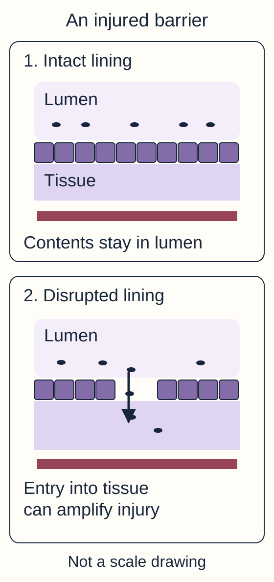
In the barrier diagram, distinguish the lumen, the lining, the deeper wall and the blood supply. Luminal contents normally remain separated from the tissues. Injury permits bacterial products and organisms to enter tissue, amplifying inflammation. Edema and impaired perfusion further compromise the bowel. Injury may remain limited, extend through the wall, or be followed by scarring. Perforation is one possible outcome, not an obligatory stage of every episode. [1]
Trace the lining before following material into tissue. The vessel represents blood supply, not a measured flow rate. [1]
Trace the damaged lining in the diagram. Predict why a local bowel problem could produce apnea and hypotension rather than only vomiting.
The consequence: intestinal injury can accompany systemic inflammation and sepsis, while fluid leakage into tissue reduces effective circulating volume. Abdominal and systemic findings therefore belong in the same assessment. Apnea, temperature instability, lethargy and impaired perfusion increase concern when feeding intolerance or abdominal distention is new. Bloody stools, tenderness, bilious emesis and abdominal wall discoloration add important information. Not all are present initially. [1] [2]
Would one larger gastric residual prove NEC?
No. Residuals and mild distention are nonspecific in premature infants. A stable infant with one isolated residual differs from an infant with progressive distention, new apnea and reduced activity. Routine residual measurements are not a stand-alone diagnostic test; assess the whole clinical course.
Apply this to a different host: a term infant with congenital heart disease and poor systemic perfusion can still develop intestinal injury consistent with NEC. Gestational age changes probability, but it is not an exclusion test. Conversely, a well infant with isolated blood in the stool needs assessment for other causes rather than automatic attribution to NEC. [4] [9]
Where is the gas, and what boundary has it crossed?
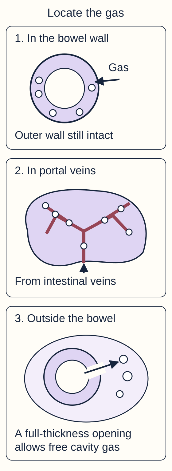
Dark areas on a radiograph do not all mean the same thing. Begin with location, not with how dramatic the image looks. Gas within the bowel lumen is expected. Pneumatosis intestinalis is gas within the bowel wall, seen as curving lines or small bubbles following the wall. In a compatible sick premature infant, it strongly supports definite NEC. Stool mixed with luminal gas can imitate a bubbly pattern, so an uncertain image needs experienced review and clinical correlation. [1]
Gas entering mesenteric venous drainage can reach the portal venous branches in the liver. Portal venous gas can appear as branching lucencies extending toward the hepatic periphery. It supports intestinal injury in this context, but does not demonstrate a hole through the bowel. It increases concern and warrants close reassessment; it is not, by itself, an instruction to resect bowel. [1] [6]
Pneumoperitoneum is gas outside the bowel in the peritoneal cavity. In an infant being treated for NEC, this indicates perforation until urgently assessed otherwise. Free gas may outline both sides of a bowel wall or form a large central oval on a supine film. A horizontal-beam view, such as left lateral decubitus, can show gas between the liver and the right abdominal wall. Tiny amounts may be difficult to see. The absence of visible free gas does not establish bowel viability. [1]
Use the three-compartment diagram as a tracing exercise. Keep the gas in the wall first. Then follow venous drainage toward the liver. Finally, change only one feature: add a full-thickness opening to the outside of the bowel. Predict which new compartment becomes accessible before checking the comparisons.
Trace each compartment and compare the intact outer wall with the full-thickness opening. Numbered panels label locations, not mandatory stages of disease. [1][6]Compare gas in the wall with gas in the liver
Wall gas is pneumatosis. Gas transported through mesenteric and portal veins produces a branching hepatic pattern. Neither finding requires a full-thickness opening into the peritoneal cavity.
Compare an intact outer wall with a full-thickness opening
The opening provides a route from the lumen to the peritoneal cavity. New free intraperitoneal gas changes the immediate concern to perforation and urgent surgical source control, alongside resuscitation.
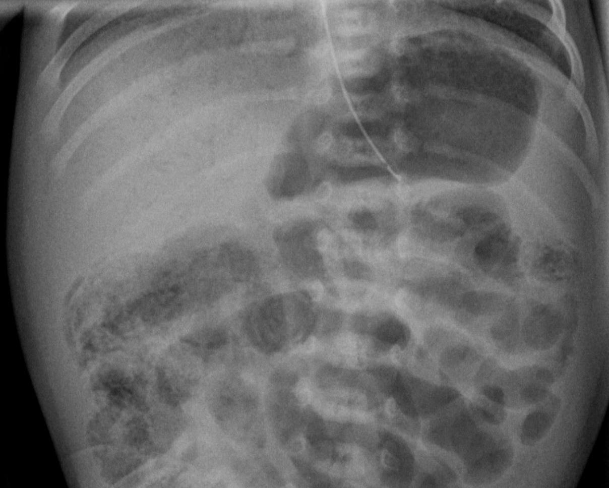
The visible rule: wall gas supports NEC; portal gas reflects a vascular route; free cavity gas raises the perforation emergency. These are anatomical distinctions, not a compulsory chronological ladder. The accompanying clinical radiograph illustrates bowel-wall gas and hepatic gas described by its source. It is not a demonstration that every dark area represents free air. [1]
Identify bowel-wall pneumatosis and portal venous gas, then compare the gas distribution with the compartment schematic. Check that a lucency lies in bowel wall rather than free peritoneal space. Image: Hellerhoff; original source; CC BY-SA 4.0.
Now apply the rule to an inconclusive film in a worsening infant. Continue evaluation and treatment based on clinical concern, obtain serial imaging, and consider expert bowel ultrasound. Ultrasound can assess wall thickness, peristalsis, perfusion, gas and fluid. Absent perfusion or complex fluid is concerning, but technique and the full clinical picture matter; no isolated ultrasound feature replaces surgical judgment. [1] [6]
Which alternative changes the next decision?
Start with the infant's course rather than the presence of blood alone. A premature infant who had been tolerating feeds and then develops systemic illness with pneumatosis has a different problem from a thriving infant with occasional blood streaks. Term birth, an early presentation or a negative first radiograph cannot independently exclude serious intestinal disease. [1] [4] [9]
Compare the pattern, then protect against the important error
Pattern
Useful distinction
PatternNEC
Useful distinctionIntestinal inflammation with systemic illness in a susceptible host; pneumatosis supports the diagnosis.
PatternSpontaneous intestinal perforation
Useful distinctionOften an earlier, focal perforation in an extremely low birthweight infant, with less typical pneumatosis. A provisional distinction must not delay surgical care.
Useful distinctionA generally healthy, growing infant with blood or mucus in stool; exclude fissure, infection and other bowel disease.
PatternMalrotation with volvulus
Useful distinctionBilious vomiting can signal intestinal obstruction and threatened blood supply, even before dramatic distention or radiographic changes.
PatternHirschsprung-associated enterocolitis
Useful distinctionObstructive history, delayed meconium, distention and systemic illness or explosive diarrhea warrant urgent specialist assessment.
Spontaneous intestinal perforation (SIP) is not simply another name for perforated NEC. SIP often involves a focal ileal defect rather than widespread inflammatory bowel necrosis. Timing, feeding history and imaging help, but overlap and diagnostic uncertainty remain. Both an infant with SIP and an infant with perforated NEC need urgent neonatal and surgical care. Their distinction can inform procedure selection and prognosis. [1] [3]
Compare two new presentations. One infant has blood-streaked stool, normal growth and a soft abdomen. Another has green emesis and progressive distention despite no pneumatosis. Which infant needs an obstruction-focused emergency assessment?
The consequence: the second pattern requires urgent assessment for malrotation and volvulus; waiting for NEC-specific gas would miss the relevant danger. In a stable infant, urgent upper gastrointestinal contrast imaging is commonly used to investigate suspected malrotation. Instability or peritonitis requires immediate surgical assessment rather than a delayed imaging sequence. The well infant's bleeding still needs evaluation, but a non-IgE food-protein reaction is a reasonable alternative. Clinician-guided elimination and later reintroduction can support that diagnosis; negative IgE tests do not exclude it. [4] [10]
Distinguish other neonatal vomiting patterns
Meconium ileus typically causes obstruction from the first days of life. Pyloric stenosis usually causes progressively forceful nonbilious vomiting rather than a new septic bowel syndrome. Intussusception is uncommon in neonates, but age alone cannot exclude it. Use timing, emesis character, examination and targeted imaging, not one symptom. [10]
Carry the comparison forward: negative blood cultures do not exclude NEC, and a positive culture does not prove that sepsis is the only problem. Clinical examination and intestinal imaging remain necessary when systemic infection and abdominal signs coexist. [1] [11]
Does the current stage describe the direction of travel?
Modified Bell staging organizes the strength of evidence and the severity of illness. It is a communication framework, not a requirement that an infant pass through each stage. The dangerous shortcut is to interpret every abnormal finding as perforation, or to assume that no free air means mild disease. [1] [2]
Modified Bell logic, without replacing bedside assessment
Category
Pattern
What it does not establish
CategoryI: suspected
PatternNonspecific intestinal and systemic findings; normal gas pattern or mild ileus. Gross rectal blood distinguishes IB from IA.
What it does not establishFeeding intolerance alone does not prove necrosis.
CategoryII: definite
PatternPneumatosis in a compatible illness; IIB adds findings such as mild metabolic acidosis, thrombocytopenia or portal venous gas.
What it does not establishDefinite NEC is not synonymous with perforation.
CategoryIII: advanced
PatternSevere systemic compromise, worsening abdominal findings, shock or coagulopathy. IIIA has no demonstrated perforation; IIIB has pneumoperitoneum.
What it does not establishIIIA can still require urgent surgery.
Read the direction of change
Compare two infants who both have no visible free air. One has improving perfusion and tenderness during treatment. The other has increasing support requirements, falling platelets, persistent acidosis and a discolored abdomen. The radiographic statement is identical; the evidence about bowel viability is not. Serial findings may justify operative assessment before perforation becomes visible. A fixed dilated loop is concerning when it persists, especially with deterioration, but is not independently diagnostic of necrotic bowel. [1]
Consider a six-hour trend: lactate rises from 2 to 6 mmol/L, platelets fall from 180 to 70 x 109/L, and urine output falls from 2 to 0.4 mL/kg/hour. Pneumatosis is less conspicuous. Predict whether the fading radiographic sign is enough to call this recovery.
The consequence: the worsening perfusion and laboratory pattern outweigh an isolated decrease in visible gas. Reassess resuscitation and request urgent surgical review for ongoing intestinal injury. These values are not operation cutoffs. Thrombocytopenia, acidosis or an abnormal loop considered alone is insufficient; concordant deterioration is the concern. [1] [6]
Could a normal platelet count rule out an early episode?
No. Platelets help characterize severity and trend, but there is no single laboratory test that excludes NEC. Interpret examination, imaging, blood gas, blood count, renal function and perfusion together.
Apply this to an infant whose first study was reassuring: new tenderness, bilious output or instability requires a fresh assessment. A previous normal image is a description of that earlier examination, not permanent protection against later disease. [1]
Which problem does each treatment address?
When NEC is clinically suspected, protect the bowel and stabilize the infant while investigating. Stop enteral feeds, decompress the stomach with an orogastric or nasogastric tube, obtain blood cultures and appropriate blood studies, and arrange prompt abdominal imaging. Start empiric intravenous antibiotics according to the level of concern and the local neonatal protocol. Obtaining cultures is important, but difficult sampling must not substantially delay treatment of an unstable infant. Respiratory and circulatory support proceed in parallel. [1] [2]
Bowel rest avoids introducing feeds into an acutely injured intestine. Gastric drainage reduces accumulated air and secretions above a poorly functioning bowel. Neither measure sterilizes tissue or restores dead bowel. Intravenous antibiotics address enteric organisms and systemic infection risk; treatment ordinarily provides broad enteric coverage, including anaerobic coverage according to disease severity and local policy. Culture results, local resistance, renal function and the clinical course guide the actual regimen. This lesson does not prescribe one universal drug combination, dose or duration. [1]
Edema does not guarantee adequate circulating volume. Intestinal inflammation and capillary leakage may produce a distended, heavier infant whose tissue perfusion is poor. Individualized fluid and electrolyte replacement, respiratory support and vasoactive treatment when indicated require neonatal expertise and repeated reassessment. Track urine output, perfusion, blood gas, electrolytes, platelets and coagulation alongside the abdominal examination. Establish parenteral nutrition when enteral nutrition must be withheld; account for line and metabolic complications during continued use. [1] [2]
Match treatment to its target: gastric drainage addresses distention, antibiotics address infection risk, and surgery addresses nonviable or perforated bowel. Predict which target remains untreated if an infant with free air receives only broader antibiotics.
The consequence: antibiotics cannot provide source control for necrotic perforated bowel. Pneumoperitoneum in this setting requires urgent pediatric surgical intervention while resuscitation continues. Progressive clinical deterioration despite intensive medical treatment can also warrant surgery without free air. Consultation begins early, not only after perforation. A center without the necessary neonatal surgery capability should coordinate transfer with the receiving team while stabilization continues. [1] [5]
Surgical decisions aim to control contamination and address nonviable bowel while preserving useful intestinal length. Laparotomy permits direct assessment and resection. Peritoneal drainage may be selected in particular circumstances, sometimes followed by laparotomy. The NEST randomized trial found no overall difference in death or neurodevelopmental impairment between the initial strategies in the combined study population, but a prespecified analysis suggested different treatment effects for presumed NEC versus isolated perforation. That result does not make either procedure universally best for every unstable infant. [3]
Why is a negative culture not permission to stop definite-NEC treatment?
The diagnosis concerns intestinal injury, not only bloodstream infection. An infant with convincing pneumatosis and a compatible illness can have negative blood cultures. Reassess treatment using the whole syndrome, cultures and trajectory rather than culture status alone.
Apply the target-based approach to a stable infant with pneumatosis and portal gas but no signs of threatened viability: intensive medical treatment and early surgical involvement are appropriate. Portal gas is not a substitute for the clinical reasons that justify an operation. [1] [6]
Has the bowel recovered enough to function?
Recovery is more than the disappearance of gas from an image. Look for improving perfusion, a reassuring abdominal examination, resolving ileus, improving laboratory abnormalities and an appropriate imaging course. Feed reintroduction and advancement are coordinated by the neonatal and surgical teams according to disease extent and recovery. Parenteral nutrition bridges the period of inadequate enteral intake; it should not become an automatic reason to prolong fasting after recovery. [1] [5]
A review comparing earlier with later feeding after NEC included only three observational studies and no randomized trials. It did not demonstrate increased recurrence or stricture with earlier refeeding, but it cannot establish a universally safe restart day. Avoid both extremes: restarting solely because one film improved, or insisting that every infant fast for the same fixed interval. The question is whether this infant's bowel and overall condition support the planned feeding trial. [7]
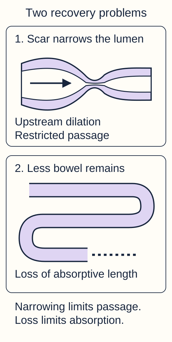
In the recovery diagram, compare a healed bowel segment with a wide lumen, a scarred segment with a narrow lumen, and a shortened remaining intestine after resection. Healing by scarring can cause a post-NEC stricture, including after medical treatment without surgery. A baby who initially improves and later develops increasing distention, bilious vomiting or difficulty tolerating feeds needs assessment for obstruction. This should not be dismissed as ordinary feeding intolerance. [5]
Follow the lumen through the narrowed segment, then compare the shortened intestine. Scarring can follow medical treatment; loss of bowel length follows extensive necrosis and resection. [1][5]
Trace the lumen across the narrowed segment. Predict what happens to contents upstream. Then compare a wide but shortened intestine: would the principal problem be the same?
The consequence: a stricture restricts passage and can produce upstream dilation. Extensive loss of bowel instead reduces absorptive capacity, contributing to short bowel syndrome, fluid losses, malabsorption and prolonged nutrition dependence. Both affect growth, but they call for different assessments. High ostomy output deserves attention to hydration and electrolytes, not simply an increase in offered calories. [1] [5]
Does a normal abdominal examination end follow-up?
No. Follow growth, nutrition, stool or ostomy output, and neurodevelopment. Gastrointestinal recovery does not eliminate the risk of developmental difficulties after severe neonatal illness. Early identification permits timely nutrition, rehabilitation and developmental support.
Prevention and family counseling also matter. Human milk and standardized neonatal feeding practices are important preventive approaches, but risk reduction is not a guarantee, and NEC is not evidence that a family caused the illness. Probiotics are not an automatic solution: product, population and regulatory safeguards matter. The FDA has warned about invasive, potentially fatal infection from organisms in probiotic products given to preterm infants. Do not translate a possible preventive benefit into a universal instruction for every extremely premature infant. [1] [8]
Apply the lesson after discharge: new bilious vomiting requires prompt evaluation; slow growth requires a nutrition and absorption assessment; developmental concerns deserve follow-up even when feeds are tolerated. Protecting the infant continues after the acute intestinal emergency has settled. [1] [5]
Apply the lesson
Case 1
Show answer and explanations for case 1
A. Continue feeds and obtain a milk-allergy evaluation (Why this does not fit)
A generally healthy growing infant with isolated rectal blood can have a food-protein reaction. New apnea, reduced activity and tenderness indicate systemic and intestinal illness, not isolated allergic bleeding.
Reasoning steps for option A
When might a food-protein assessment explain rectal symptoms?
A generally healthy growing infant with isolated rectal blood can have a food-protein reaction.
How does the current course differ?
New apnea, reduced activity and tenderness indicate systemic and intestinal illness, not isolated allergic bleeding.
B. Hold feeds, decompress and begin a neonatal NEC evaluation (Best answer)
Progressive distention and tenderness now coexist with new systemic abnormalities in a highly susceptible infant. No. Suspected NEC warrants bowel protection, empiric treatment under the neonatal protocol and serial assessment before diagnostic gas necessarily appears.
Reasoning steps for option B
What makes the new feeding problem concerning?
Progressive distention and tenderness now coexist with new systemic abnormalities in a highly susceptible infant.
Does the inconclusive first film justify waiting?
No. Suspected NEC warrants bowel protection, empiric treatment under the neonatal protocol and serial assessment before diagnostic gas necessarily appears.
C. Proceed directly to bowel resection for presumed perforation (Why this does not fit)
Perforation or progressive evidence of nonviable bowel despite medical support can justify surgery. No free air, refractory shock or peritonitis is supplied; begin urgent treatment and assessment rather than assuming a need for resection.
Reasoning steps for option C
What findings can justify urgent operative source control?
Perforation or progressive evidence of nonviable bowel despite medical support can justify surgery.
Has this presentation established that indication?
No free air, refractory shock or peritonitis is supplied; begin urgent treatment and assessment rather than assuming a need for resection.
D. Reduce feed volume and repeat the examination the next day (Why this does not fit)
Isolated feeding intolerance in an otherwise stable infant may be managed after a reassuring evaluation. The combination of progressive abdominal and systemic findings requires prompt evaluation and close serial monitoring.
Reasoning steps for option D
When might feeding modification be considered without a NEC syndrome?
Isolated feeding intolerance in an otherwise stable infant may be managed after a reassuring evaluation.
Why is next-day reassessment inadequate here?
The combination of progressive abdominal and systemic findings requires prompt evaluation and close serial monitoring.
Takeaway: An inconclusive initial film does not cancel a clinically concerning NEC presentation.
A. Perforated bowel is established despite the soft abdomen (Why this does not fit)
Peritoneal findings, free intraperitoneal gas or marked clinical deterioration would raise that concern. It does not demonstrate a wall defect or systemic deterioration.
Reasoning steps for option A
What would make perforation a leading concern?
Peritoneal findings, free intraperitoneal gas or marked clinical deterioration would raise that concern.
What does one residual show in this infant?
It does not demonstrate a wall defect or systemic deterioration.
B. Allergic proctocolitis is established by the milky appearance (Why this does not fit)
Blood or mucus in stool in an otherwise thriving infant can support that differential. No rectal bleeding is present, and a milky residual is not an allergy test.
Reasoning steps for option B
What presentation supports allergic proctocolitis?
Blood or mucus in stool in an otherwise thriving infant can support that differential.
Does the observed residual supply that pattern?
No rectal bleeding is present, and a milky residual is not an allergy test.
C. Normal feeding tolerance has been proved for subsequent days (Why this does not fit)
It describes the infant at this assessment. No. New distention, systemic illness or bleeding would require reassessment even after a reassuring examination.
Reasoning steps for option C
What does the reassuring examination describe?
It describes the infant at this assessment.
Can it guarantee a future course?
No. New distention, systemic illness or bleeding would require reassessment even after a reassuring examination.
D. The residual is nonspecific and requires clinical correlation (Best answer)
Progressive distention, tenderness, rectal blood or new systemic instability would increase concern. One residual cannot establish NEC; evaluate in context and continue appropriate clinical surveillance.
Reasoning steps for option D
What other findings would strengthen suspicion for NEC?
Progressive distention, tenderness, rectal blood or new systemic instability would increase concern.
What is justified without those findings?
One residual cannot establish NEC; evaluate in context and continue appropriate clinical surveillance.
Takeaway: An isolated residual is neither a diagnosis of NEC nor a guarantee against later illness.
A. Intensive medical NEC treatment with early surgical involvement (Best answer)
They indicate bowel-wall and portal venous gas, supporting definite NEC in this clinical setting. Bowel rest, decompression, intravenous antibiotics and close monitoring are appropriate with early surgical involvement; portal gas alone does not mandate resection.
Reasoning steps for option A
Where do the two gas patterns localize?
They indicate bowel-wall and portal venous gas, supporting definite NEC in this clinical setting.
What does the stable trajectory imply about the initial plan?
Bowel rest, decompression, intravenous antibiotics and close monitoring are appropriate with early surgical involvement; portal gas alone does not mandate resection.
B. Immediate laparotomy because portal gas demonstrates perforation (Why this does not fit)
Free gas in the peritoneal cavity supports a full-thickness bowel defect. No. It lies in venous drainage and does not by itself demonstrate perforation.
Reasoning steps for option B
Which gas compartment supports perforation?
Free gas in the peritoneal cavity supports a full-thickness bowel defect.
Is portal venous gas in that compartment?
No. It lies in venous drainage and does not by itself demonstrate perforation.
C. An allergy-directed feeding trial with serial stool assessment (Why this does not fit)
A well growing infant with isolated rectal blood can be evaluated for a food-protein reaction. New abdominal illness and bowel-wall gas support NEC, so active enteral feeding is withheld during acute treatment.
Reasoning steps for option C
When is an allergy-directed approach a reasonable consideration?
A well growing infant with isolated rectal blood can be evaluated for a food-protein reaction.
Why is that not the primary approach here?
New abdominal illness and bowel-wall gas support NEC, so active enteral feeding is withheld during acute treatment.
D. Observation for isolated perforation without enteric antimicrobial coverage (Why this does not fit)
Extremely premature infants can develop a focal perforation distinct from NEC. Pneumatosis with intestinal symptoms favors NEC, and concern for enteric infection requires appropriate intravenous coverage.
Reasoning steps for option D
Why does isolated perforation enter the neonatal differential?
Extremely premature infants can develop a focal perforation distinct from NEC.
What favors NEC and requires medical treatment here?
Pneumatosis with intestinal symptoms favors NEC, and concern for enteric infection requires appropriate intravenous coverage.
Takeaway: Portal venous gas heightens concern but does not replace the clinical indications for surgery.
A. Continue current treatment until an abdominal CT confirms necrosis (Why this does not fit)
Targeted imaging may clarify anatomy in an equivocal case. The described free intraperitoneal gas and deterioration already require urgent surgical source-control assessment.
Reasoning steps for option A
What can additional imaging contribute when findings are uncertain?
Targeted imaging may clarify anatomy in an equivocal case.
Why should CT not postpone action here?
The described free intraperitoneal gas and deterioration already require urgent surgical source-control assessment.
B. Treat the gas as portal venous gas and reassess after feeds (Why this does not fit)
It follows branching vessels within the liver. It is outside the liver and bowel lumen in the peritoneal cavity, not a portal branching pattern.
Reasoning steps for option B
How does portal gas appear?
It follows branching vessels within the liver.
Where is the new collection described?
It is outside the liver and bowel lumen in the peritoneal cavity, not a portal branching pattern.
C. Continue resuscitation and obtain urgent pediatric surgical source control (Best answer)
It is free peritoneal gas, indicating perforation in the current NEC setting. It reinforces the need for concurrent stabilization and urgent surgical intervention rather than further observation alone.
Reasoning steps for option C
What does gas between liver and abdominal wall represent on this view?
It is free peritoneal gas, indicating perforation in the current NEC setting.
What does worsening perfusion add to the decision?
It reinforces the need for concurrent stabilization and urgent surgical intervention rather than further observation alone.
D. Broaden antibiotics and defer surgery until blood cultures become positive (Why this does not fit)
They treat or prevent invasive infection from enteric organisms. Perforated necrotic bowel requires source control, and culture positivity is not a prerequisite.
Reasoning steps for option D
What target do antibiotics address?
They treat or prevent invasive infection from enteric organisms.
What target remains despite antibiotic coverage?
Perforated necrotic bowel requires source control, and culture positivity is not a prerequisite.
Takeaway: In a deteriorating infant with NEC, new free cavity gas requires urgent surgical action alongside resuscitation.
A. Fluoroscopic upper gastrointestinal series for malrotation (Why this does not fit)
It evaluates suspected malrotation in a stable infant with an appropriate obstructive presentation. The unresolved issue is inflammatory bowel injury with equivocal wall gas, not a supplied acute malrotation pattern.
Reasoning steps for option A
When is an upper gastrointestinal series especially important?
It evaluates suspected malrotation in a stable infant with an appropriate obstructive presentation.
What unresolved question dominates this case?
The unresolved issue is inflammatory bowel injury with equivocal wall gas, not a supplied acute malrotation pattern.
B. Expert bowel ultrasound with Doppler assessment (Best answer)
Luminal stool gas and wall gas can overlap in appearance, leaving the intestinal findings uncertain. It can assess wall gas, thickness, perfusion, peristalsis and fluid while serial clinical and radiographic reassessment continues.
Reasoning steps for option B
Why is the mottled radiograph insufficient?
Luminal stool gas and wall gas can overlap in appearance, leaving the intestinal findings uncertain.
How can ultrasound add information?
It can assess wall gas, thickness, perfusion, peristalsis and fluid while serial clinical and radiographic reassessment continues.
C. Milk-specific IgE testing for protein allergy (Why this does not fit)
It addresses sensitization relevant to immediate IgE-mediated allergy. Systemic illness raises concern for intestinal injury, and even allergic proctocolitis is generally non-IgE mediated.
Reasoning steps for option C
What does IgE testing assess?
It addresses sensitization relevant to immediate IgE-mediated allergy.
Why is it poorly matched to this infant?
Systemic illness raises concern for intestinal injury, and even allergic proctocolitis is generally non-IgE mediated.
D. Contrast enema to document post-inflammatory stricture (Why this does not fit)
Delayed obstructive symptoms after an apparent recovery suggest scarring and narrowing. This is an acute first inflammatory presentation, so evaluating bowel injury is more appropriate than testing for a late healed stricture.
Reasoning steps for option D
When would a post-NEC stricture be a leading concern?
Delayed obstructive symptoms after an apparent recovery suggest scarring and narrowing.
Does this timing support that target?
This is an acute first inflammatory presentation, so evaluating bowel injury is more appropriate than testing for a late healed stricture.
Takeaway: Equivocal radiographs can be supplemented by expert bowel ultrasound without pausing clinical treatment.
A. Preserved wall perfusion with active peristalsis (Why this does not fit)
They provide functional information about the bowel segment at the time of the study. It would be relatively reassuring rather than the strongest additional evidence of threatened viability, although follow-up remains necessary.
Reasoning steps for option A
What do perfusion and peristalsis indicate?
They provide functional information about the bowel segment at the time of the study.
How would preservation affect concern in this stable infant?
It would be relatively reassuring rather than the strongest additional evidence of threatened viability, although follow-up remains necessary.
B. A small amount of uncomplicated free fluid (Why this does not fit)
It can accompany several abdominal processes and is less specific than complex fluid. Simple fluid alone provides less evidence of severe intestinal injury than a poorly perfused segment with complex surrounding fluid.
Reasoning steps for option B
What can simple free fluid reflect?
It can accompany several abdominal processes and is less specific than complex fluid.
Why is it not the strongest adverse finding here?
Simple fluid alone provides less evidence of severe intestinal injury than a poorly perfused segment with complex surrounding fluid.
C. A decrease in visible intramural gas bubbles (Why this does not fit)
It measures the visibility of gas, not directly the viability of the bowel wall. No. It must be interpreted with examination, perfusion and other ultrasound findings.
Reasoning steps for option C
What does less visible wall gas measure?
It measures the visibility of gas, not directly the viability of the bowel wall.
Can this finding independently establish worsening viability?
No. It must be interpreted with examination, perfusion and other ultrasound findings.
D. Absent segmental perfusion with complex adjacent fluid (Best answer)
When technically confirmed, it suggests that the affected segment is not receiving detectable blood flow. Together these findings are associated with severe NEC outcomes and warrant urgent integrated surgical reassessment; they are not an isolated numerical operation rule.
Reasoning steps for option D
What additional information does absent perfusion provide?
When technically confirmed, it suggests that the affected segment is not receiving detectable blood flow.
Why does complex adjacent fluid strengthen concern?
Together these findings are associated with severe NEC outcomes and warrant urgent integrated surgical reassessment; they are not an isolated numerical operation rule.
Takeaway: A persistent loop becomes more concerning when functional imaging and the clinical trajectory also suggest severe injury.
A. Portal venous gas arriving from injured intestine (Best answer)
They are compatible with gas in the portal venous branches. Gas associated with intestinal injury can enter mesenteric venous drainage and reach the liver without a demonstrated peritoneal perforation.
Reasoning steps for option A
What anatomical pattern do peripheral branching hepatic lucencies follow?
They are compatible with gas in the portal venous branches.
How does the ileal wall finding explain this route?
Gas associated with intestinal injury can enter mesenteric venous drainage and reach the liver without a demonstrated peritoneal perforation.
B. Free peritoneal gas proving a bowel perforation (Why this does not fit)
It lies outside organs and bowel in the abdominal cavity. No. The gas follows branching intrahepatic structures rather than collecting outside the liver.
Reasoning steps for option B
Where would free peritoneal gas be located?
It lies outside organs and bowel in the abdominal cavity.
Does the supplied hepatic distribution match that location?
No. The gas follows branching intrahepatic structures rather than collecting outside the liver.
C. Biliary gas proving an ascending duct infection (Why this does not fit)
Biliary gas tends to be more central rather than finely branching toward the hepatic periphery. Peripheral hepatic gas with bowel-wall gas favors a portal route in intestinal injury, not proof of a primary duct infection.
Reasoning steps for option C
How does biliary gas usually distribute compared with portal gas?
Biliary gas tends to be more central rather than finely branching toward the hepatic periphery.
What better explains the described pattern and bowel findings?
Peripheral hepatic gas with bowel-wall gas favors a portal route in intestinal injury, not proof of a primary duct infection.
D. Luminal bowel gas projected over a normal liver (Why this does not fit)
Bowel gas can overlap another structure in a two-dimensional image. Fine branching lucencies extending to the liver periphery are not described as a normal bowel loop or stool pattern.
Reasoning steps for option D
Why can projection cause uncertainty on a single film?
Bowel gas can overlap another structure in a two-dimensional image.
What supplied detail favors a true hepatic vascular pattern?
Fine branching lucencies extending to the liver periphery are not described as a normal bowel loop or stool pattern.
Takeaway: Portal gas travels through venous drainage; it is not the same compartment as free peritoneal gas.
A. Necrotizing enterocolitis with extensive bowel necrosis (Why this does not fit)
Segments of inflammatory injury and nonviable bowel would support that diagnosis. The lesion is an isolated small defect with viable surrounding bowel rather than extensive inflammatory necrosis.
Reasoning steps for option A
What operative pattern would support extensive NEC?
Segments of inflammatory injury and nonviable bowel would support that diagnosis.
How does the supplied operative finding differ?
The lesion is an isolated small defect with viable surrounding bowel rather than extensive inflammatory necrosis.
B. Hirschsprung-associated enterocolitis with distal obstruction (Why this does not fit)
An obstructive history and aganglionic distal bowel would strengthen it. An isolated ileal defect is described without an obstructive distal disease pattern.
Reasoning steps for option B
What history and pathology would strengthen this alternative?
An obstructive history and aganglionic distal bowel would strengthen it.
What is actually documented at exploration?
An isolated ileal defect is described without an obstructive distal disease pattern.
C. Spontaneous intestinal perforation of the newborn (Best answer)
A single focal defect with otherwise viable bowel fits spontaneous intestinal perforation. It is compatible with SIP in an extremely low birthweight infant, although urgent intervention was required before diagnostic certainty.
Reasoning steps for option C
What pattern distinguishes isolated perforation from extensive NEC?
A single focal defect with otherwise viable bowel fits spontaneous intestinal perforation.
How does the early prefeeding presentation contribute?
It is compatible with SIP in an extremely low birthweight infant, although urgent intervention was required before diagnostic certainty.
D. Malrotation with volvulus and mesenteric infarction (Why this does not fit)
A twisted mesentery with compromised bowel blood supply would support it. No. A focal perforation without twisting or widespread ischemic bowel is reported.
Reasoning steps for option D
What structural finding would support volvulus?
A twisted mesentery with compromised bowel blood supply would support it.
Does the operation supply that abnormality?
No. A focal perforation without twisting or widespread ischemic bowel is reported.
Takeaway: SIP and NEC are distinct but potentially overlapping diagnostic considerations; perforation requires urgent care in either.
A. The absence of substantial feeding favors isolated milk-protein inflammation (Why this does not fit)
A generally well infant with compatible dietary exposure and isolated rectal bleeding can fit that diagnosis. Marked systemic perfusion risk with new intestinal illness and wall gas supports more serious bowel injury.
Reasoning steps for option A
What exposure and clinical pattern support a food-protein reaction?
A generally well infant with compatible dietary exposure and isolated rectal bleeding can fit that diagnosis.
Why does it not explain this case well?
Marked systemic perfusion risk with new intestinal illness and wall gas supports more serious bowel injury.
B. Impaired perfusion can support NEC despite term birth (Best answer)
Poor systemic perfusion can compromise intestinal blood supply. Neither excludes NEC when the infant has a compatible intestinal syndrome and pneumatosis.
Reasoning steps for option B
What relevant risk does the cardiac illness supply?
Poor systemic perfusion can compromise intestinal blood supply.
How should term birth and minimal feeding affect the diagnosis?
Neither excludes NEC when the infant has a compatible intestinal syndrome and pneumatosis.
C. Term birth makes congenital obstruction the necessary explanation (Why this does not fit)
Structural abnormalities can present early with feeding or abdominal symptoms. No. Cardiac-associated perfusion injury is a recognized NEC context, and the findings do not require congenital obstruction.
Reasoning steps for option C
Why should congenital obstruction remain in a neonatal differential?
Structural abnormalities can present early with feeding or abdominal symptoms.
Does gestational age make obstruction necessary here?
No. Cardiac-associated perfusion injury is a recognized NEC context, and the findings do not require congenital obstruction.
D. Wall gas confirms perforation regardless of hemodynamic status (Why this does not fit)
It establishes intramural rather than free peritoneal gas. A full-thickness opening is not demonstrated; severity and surgical need require the broader assessment.
Reasoning steps for option D
What does gas in the wall establish anatomically?
It establishes intramural rather than free peritoneal gas.
Why is perforation not established by this description?
A full-thickness opening is not demonstrated; severity and surgical need require the broader assessment.
Takeaway: Host risk includes perfusion disorders; neither term birth nor minimal feeding excludes NEC.
A. The negative IgE result excludes dietary protein-related inflammation (Why this does not fit)
It addresses IgE sensitization, not all forms of food-related inflammation. Food protein-induced allergic proctocolitis is usually non-IgE mediated.
Reasoning steps for option A
What type of immune response does specific IgE testing address?
It addresses IgE sensitization, not all forms of food-related inflammation.
Why does the result not exclude the leading dietary alternative?
Food protein-induced allergic proctocolitis is usually non-IgE mediated.
B. Rectal blood establishes definite NEC despite the reassuring examination (Why this does not fit)
Systemic illness, progressive abdominal abnormalities and supportive imaging strengthen concern for NEC. The infant is thriving and otherwise well; blood alone does not establish definite NEC.
Reasoning steps for option B
What makes NEC more likely than isolated allergic bleeding?
Systemic illness, progressive abdominal abnormalities and supportive imaging strengthen concern for NEC.
What is missing from this presentation?
The infant is thriving and otherwise well; blood alone does not establish definite NEC.
C. Assess for non-IgE proctocolitis with a supervised dietary trial (Best answer)
It supports food protein-induced allergic proctocolitis after evaluation for competing causes. It does not exclude this non-IgE condition; clinician-guided elimination and planned reintroduction can help assess the diagnosis.
Reasoning steps for option C
What does the combination of rectal blood and preserved well-being suggest?
It supports food protein-induced allergic proctocolitis after evaluation for competing causes.
How should the negative IgE result affect the plan?
It does not exclude this non-IgE condition; clinician-guided elimination and planned reintroduction can help assess the diagnosis.
D. The pattern supports volvulus and immediate exploratory laparotomy (Why this does not fit)
Bilious vomiting, obstruction or ischemic systemic deterioration would raise that concern. No. The current history supports a less acute differential while still requiring evaluation of bleeding.
Reasoning steps for option D
What presentation would raise urgent concern for volvulus?
Bilious vomiting, obstruction or ischemic systemic deterioration would raise that concern.
Does isolated intermittent bleeding in this thriving infant supply that pattern?
No. The current history supports a less acute differential while still requiring evaluation of bleeding.
Takeaway: A well infant with rectal blood can have a non-IgE food-protein reaction despite negative IgE testing.
A. Urgent upper gastrointestinal series with surgical involvement (Best answer)
Malrotation with volvulus remains a concern in a neonate with bilious vomiting. Urgent upper gastrointestinal contrast imaging can evaluate malrotation while the surgical team remains involved; deterioration requires immediate reassessment rather than waiting for imaging.
Reasoning steps for option A
What important diagnosis is not excluded by the nondiagnostic radiograph?
Malrotation with volvulus remains a concern in a neonate with bilious vomiting.
What investigation fits the currently stable condition?
Urgent upper gastrointestinal contrast imaging can evaluate malrotation while the surgical team remains involved; deterioration requires immediate reassessment rather than waiting for imaging.
B. A milk-protein elimination trial with outpatient reassessment (Why this does not fit)
An otherwise well infant with isolated rectal blood can fit that pathway. Bilious vomiting and feeding failure require prompt exclusion of a surgical obstruction.
Reasoning steps for option B
What symptoms fit an outpatient food-protein assessment better?
An otherwise well infant with isolated rectal blood can fit that pathway.
Why does this presentation require a different priority?
Bilious vomiting and feeding failure require prompt exclusion of a surgical obstruction.
C. Serial plain films until bowel-wall gas establishes NEC (Why this does not fit)
They can demonstrate evolving intestinal findings. The principal danger may be volvulus, which does not require pneumatosis to threaten intestinal perfusion.
Reasoning steps for option C
Why are serial films useful during suspected NEC?
They can demonstrate evolving intestinal findings.
Why is waiting for wall gas unsafe in this presentation?
The principal danger may be volvulus, which does not require pneumatosis to threaten intestinal perfusion.
D. Pyloric ultrasound with refeeding after a negative study (Why this does not fit)
Progressively forceful nonbilious vomiting is the usual pattern. Dark-green emesis is bilious and requires assessment of obstruction beyond the pylorus, including malrotation.
Reasoning steps for option D
What emesis pattern classically suggests pyloric stenosis?
Progressively forceful nonbilious vomiting is the usual pattern.
What changes the diagnostic priority here?
Dark-green emesis is bilious and requires assessment of obstruction beyond the pylorus, including malrotation.
Takeaway: A nondiagnostic abdominal radiograph does not dismiss neonatal bilious vomiting.
A. Resume full feeds and manage as uncomplicated overflow stool (Why this does not fit)
An obstructed infant may pass liquid stool, so diarrhea does not establish normal transit. Fever, lethargy and poor perfusion indicate a systemic enterocolitis syndrome rather than a benign stooling problem.
Reasoning steps for option A
Can distal obstruction produce overflow stool?
An obstructed infant may pass liquid stool, so diarrhea does not establish normal transit.
Why is uncomplicated overflow an inadequate explanation here?
Fever, lethargy and poor perfusion indicate a systemic enterocolitis syndrome rather than a benign stooling problem.
B. Arrange allergy testing and defer decompression until results return (Why this does not fit)
A well infant with isolated blood or mucus in stool can fit that differential. Known aganglionic obstruction with fever, distention and impaired perfusion requires urgent inpatient care.
Reasoning steps for option B
What clinical setting can justify a food-protein evaluation?
A well infant with isolated blood or mucus in stool can fit that differential.
What dominates this presentation instead?
Known aganglionic obstruction with fever, distention and impaired perfusion requires urgent inpatient care.
C. Treat as a routine viral illness with delayed surgical follow-up (Why this does not fit)
Infectious gastroenteritis can cause fever and diarrhea. The known Hirschsprung disease and obstructive abdominal findings make associated enterocolitis a serious concern requiring immediate specialist treatment.
Reasoning steps for option C
Why can infection enter the diarrhea differential?
Infectious gastroenteritis can cause fever and diarrhea.
Why is delay unsafe in this infant?
The known Hirschsprung disease and obstructive abdominal findings make associated enterocolitis a serious concern requiring immediate specialist treatment.
D. Begin resuscitation, decompression and antibiotics with surgery (Best answer)
In Hirschsprung disease, explosive diarrhea with distention and systemic illness suggests associated enterocolitis. Urgent inpatient resuscitation and specialist-directed decompression and antimicrobial treatment are needed even without free air.
Reasoning steps for option D
How does the underlying disease change the meaning of diarrhea?
In Hirschsprung disease, explosive diarrhea with distention and systemic illness suggests associated enterocolitis.
What does impaired perfusion require?
Urgent inpatient resuscitation and specialist-directed decompression and antimicrobial treatment are needed even without free air.
Takeaway: Diarrhea does not exclude obstruction or enterocolitis in an infant with Hirschsprung disease.
A. Definite IIA, followed by advanced IIIA (Why this does not fit)
Pneumatosis in a compatible illness supports definite disease. The initial assessment lacks that finding, and the later assessment lacks advanced systemic compromise.
Reasoning steps for option A
What separates definite stage II from suspected stage I?
Pneumatosis in a compatible illness supports definite disease.
What prevents this pair from fitting?
The initial assessment lacks that finding, and the later assessment lacks advanced systemic compromise.
B. Suspected IB, followed by definite IIA (Best answer)
With nonspecific systemic and intestinal signs, gross rectal blood fits suspected stage IB. It supports definite stage IIA rather than advanced disease.
Reasoning steps for option B
How is gross rectal blood classified without specific imaging?
With nonspecific systemic and intestinal signs, gross rectal blood fits suspected stage IB.
What does later pneumatosis add without severe abnormalities?
It supports definite stage IIA rather than advanced disease.
C. Suspected IA, followed by perforated IIIB (Why this does not fit)
Gross rectal blood separates IB from IA, while pneumoperitoneum identifies perforated IIIB. Gross blood is present initially and no free air is present later.
Reasoning steps for option C
Which findings distinguish IB and IIIB?
Gross rectal blood separates IB from IA, while pneumoperitoneum identifies perforated IIIB.
Which supplied findings contradict this pair?
Gross blood is present initially and no free air is present later.
D. Suspected IB, followed by advanced IIIA (Why this does not fit)
Severe systemic compromise and advanced abdominal findings can support IIIA without visible perforation. No. Normal perfusion, platelets and acid-base status do not provide the advanced pattern described by IIIA.
Reasoning steps for option D
What supports stage IIIA?
Severe systemic compromise and advanced abdominal findings can support IIIA without visible perforation.
Does pneumatosis alone establish that severity?
No. Normal perfusion, platelets and acid-base status do not provide the advanced pattern described by IIIA.
Takeaway: More specific imaging can establish definite NEC without establishing advanced or perforated disease.
A. Stage IIB; delay surgical reassessment until pneumoperitoneum appears (Why this does not fit)
Milder thrombocytopenia or metabolic acidosis can occur in IIB without refractory systemic compromise. Vasoactive-dependent hypotension, oliguria, coagulopathy and worsening abdominal findings indicate advanced disease that needs urgent reassessment.
Reasoning steps for option A
What systemic pattern fits IIB rather than III?
Milder thrombocytopenia or metabolic acidosis can occur in IIB without refractory systemic compromise.
Why is this infant beyond that limited pattern?
Vasoactive-dependent hypotension, oliguria, coagulopathy and worsening abdominal findings indicate advanced disease that needs urgent reassessment.
B. Stage IIIB; use the absent free gas to confirm a contained perforation (Why this does not fit)
Pneumoperitoneum supports IIIB. It does not confirm a contained perforation; severe disease without demonstrated perforation is consistent with IIIA.
Reasoning steps for option B
What radiographic finding defines perforated IIIB in modified Bell staging?
Pneumoperitoneum supports IIIB.
What can absent free gas actually establish here?
It does not confirm a contained perforation; severe disease without demonstrated perforation is consistent with IIIA.
C. Stage IIIA; obtain urgent assessment for operative source control (Best answer)
It fits advanced NEC without demonstrated perforation, commonly described as stage IIIA. Yes. Progressive deterioration despite treatment can indicate nonviable bowel before free air is visible.
Reasoning steps for option C
How should severe systemic deterioration without free air be classified?
It fits advanced NEC without demonstrated perforation, commonly described as stage IIIA.
Does that category permit an urgent operative assessment?
Yes. Progressive deterioration despite treatment can indicate nonviable bowel before free air is visible.
D. Stage I; treat the current film as evidence that definite NEC has resolved (Why this does not fit)
It changes what is visible on the current study. No. Clinical deterioration and prior pneumatosis are not reclassified as mild suspected disease because free air is absent.
Reasoning steps for option D
What does disappearance of a previously visible imaging sign mean?
It changes what is visible on the current study.
Can it erase definite NEC during worsening systemic illness?
No. Clinical deterioration and prior pneumatosis are not reclassified as mild suspected disease because free air is absent.
Takeaway: Advanced nonperforated NEC is not a reason to wait for perforation before surgical reassessment.
A. The laboratory pattern outweighs the fading gas and warrants urgent escalation (Best answer)
Together they indicate worsening systemic perfusion and illness rather than a reassuring isolated fluctuation. Gas visibility does not establish bowel viability; the worsening clinical trajectory warrants immediate reassessment of support and surgical source control.
Reasoning steps for option A
How do the laboratory and urine findings change the assessment?
Together they indicate worsening systemic perfusion and illness rather than a reassuring isolated fluctuation.
Why does less visible pneumatosis not reverse that conclusion?
Gas visibility does not establish bowel viability; the worsening clinical trajectory warrants immediate reassessment of support and surgical source control.
B. The radiographic improvement permits feeding despite the falling urine output (Why this does not fit)
The infant needs a clinically appropriate recovery pattern, not merely a less abnormal image. Increasing tenderness, rising lactate and falling urine output indicate ongoing deterioration.
Reasoning steps for option B
What needs to improve before feed reintroduction is considered?
The infant needs a clinically appropriate recovery pattern, not merely a less abnormal image.
C. The platelet decline alone establishes perforation and makes further assessment unnecessary (Why this does not fit)
They can support concern about disease severity and progression. They do not prove a wall defect, and integrated examination, perfusion and surgical assessment remain necessary.
Reasoning steps for option C
What can falling platelets contribute?
They can support concern about disease severity and progression.
What can they not determine by themselves?
They do not prove a wall defect, and integrated examination, perfusion and surgical assessment remain necessary.
D. The absence of free air permits observation until the next routine daily film (Why this does not fit)
No. Advanced nonperforated disease can threaten bowel viability. Several concordant signs show worsening perfusion and systemic illness despite treatment.
Reasoning steps for option D
Does a radiograph without free air exclude advanced NEC?
No. Advanced nonperforated disease can threaten bowel viability.
What requires action before routine imaging here?
Several concordant signs show worsening perfusion and systemic illness despite treatment.
Takeaway: Follow the infant, not the visibility of one gas pattern.
A. Negative blood cultures establish resistance to the empiric antibiotics (Why this does not fit)
An organism and susceptibility testing, or a relevant epidemiologic assessment, are needed. No. It cannot establish antimicrobial resistance.
Reasoning steps for option A
What information is required to identify bacterial resistance?
An organism and susceptibility testing, or a relevant epidemiologic assessment, are needed.
Does a culture with no growth supply a resistant isolate?
No. It cannot establish antimicrobial resistance.
B. Any negative culture makes the radiographic bowel findings nonspecific (Why this does not fit)
It samples the bloodstream for recoverable organisms. No. It does not change the anatomical location of gas or exclude intestinal injury.
Reasoning steps for option B
What does a blood culture sample?
It samples the bloodstream for recoverable organisms.
Does that directly invalidate convincing bowel-wall gas?
No. It does not change the anatomical location of gas or exclude intestinal injury.
C. Persistent ileus requires proof of bacteremia before NEC care continues (Why this does not fit)
Intestinal inflammation and injury can impair motility. No. Continuing assessment and treatment depend on the bowel and overall clinical course, not proof of bloodstream infection alone.
Reasoning steps for option C
Why can ileus persist during NEC?
Intestinal inflammation and injury can impair motility.
Is bacteremia required for that intestinal syndrome?
No. Continuing assessment and treatment depend on the bowel and overall clinical course, not proof of bloodstream infection alone.
D. Bloodstream testing does not exclude ongoing intestinal injury (Best answer)
No organism has grown from the sampled blood under the testing conditions. The compatible illness, pneumatosis and persistent abdominal abnormalities still support NEC; the neonatal team individualizes treatment using that course.
Reasoning steps for option D
What does culture negativity tell the team?
No organism has grown from the sampled blood under the testing conditions.
What disease evidence remains independent of that result?
The compatible illness, pneumatosis and persistent abdominal abnormalities still support NEC; the neonatal team individualizes treatment using that course.
Takeaway: Negative blood cultures do not erase a clinically and radiographically supported intestinal diagnosis.
A. Retain the current regimen and continue bowel rest (Why this does not fit)
It supports continuing acute NEC care, including bowel rest. The identified bloodstream organism is not covered, so antimicrobial treatment needs adjustment to active intravenous therapy under the neonatal protocol.
Reasoning steps for option A
What does ongoing intestinal injury require?
It supports continuing acute NEC care, including bowel rest.
What prevents retaining the current regimen?
The identified bloodstream organism is not covered, so antimicrobial treatment needs adjustment to active intravenous therapy under the neonatal protocol.
B. Change to active intravenous therapy and continue bowel rest (Best answer)
The team should select an active intravenous regimen accounting for organism, illness severity, renal function and local guidance. No. Persistent distention and pneumatosis support continuing bowel protection and serial reassessment.
Reasoning steps for option B
What does the susceptibility report require?
The team should select an active intravenous regimen accounting for organism, illness severity, renal function and local guidance.
Does changing antibiotics establish intestinal recovery?
No. Persistent distention and pneumatosis support continuing bowel protection and serial reassessment.
C. Change to active intravenous therapy and resume full feeds (Why this does not fit)
An active intravenous regimen addresses the documented coverage gap. Resuming full feeds is not justified while acute intestinal injury remains evident.
Reasoning steps for option C
Which part of this plan addresses the culture result?
An active intravenous regimen addresses the documented coverage gap.
Which part ignores the current bowel findings?
Resuming full feeds is not justified while acute intestinal injury remains evident.
D. Retain the current regimen and resume full feeds (Why this does not fit)
The current regimen is inactive against a clinically significant isolate. The bowel is still acutely affected, so neither retaining ineffective coverage nor resuming full feeds follows the evidence.
Reasoning steps for option D
What problem does the susceptibility report identify?
The current regimen is inactive against a clinically significant isolate.
What second problem remains unresolved?
The bowel is still acutely affected, so neither retaining ineffective coverage nor resuming full feeds follows the evidence.
Takeaway: Antimicrobial activity and bowel recovery are separate questions; correcting one does not establish the other.
A. Increased effective circulating volume causing pressure-driven diuresis (Why this does not fit)
Adequate perfusion may permit increased urinary fluid excretion. The infant instead has hypotension and oliguria, which do not demonstrate adequate effective circulating volume.
Reasoning steps for option A
How would preserved renal perfusion usually affect urine production during volume excess?
Adequate perfusion may permit increased urinary fluid excretion.
Why does that not reconcile the observations?
The infant instead has hypotension and oliguria, which do not demonstrate adequate effective circulating volume.
B. Isolated dietary calorie excess causing rapid tissue growth (Why this does not fit)
Tissue growth is not an adequate explanation for abrupt edema over hours. It does not explain worsening perfusion during an acute inflammatory bowel illness.
Reasoning steps for option B
How does nutritional growth usually differ from acute fluid accumulation?
Tissue growth is not an adequate explanation for abrupt edema over hours.
What else does calorie excess fail to explain?
It does not explain worsening perfusion during an acute inflammatory bowel illness.
C. Capillary leakage causing edema with reduced effective volume (Best answer)
Yes. Fluid can accumulate outside vessels while effective intravascular filling becomes inadequate. Intestinal inflammation and capillary leakage can produce edema alongside hypotension and oliguria, requiring individualized hemodynamic reassessment.
Reasoning steps for option C
Can total body fluid rise while useful circulating volume falls?
Yes. Fluid can accumulate outside vessels while effective intravascular filling becomes inadequate.
Why is that relevant in NEC?
Intestinal inflammation and capillary leakage can produce edema alongside hypotension and oliguria, requiring individualized hemodynamic reassessment.
D. Primary cardiac pump failure with isolated pulmonary fluid retention (Why this does not fit)
New ventricular dysfunction and a compatible cardiac assessment would support it. The supplied echocardiogram lacks new dysfunction, while inflammatory capillary leakage can explain both abdominal edema and poor perfusion.
Reasoning steps for option D
What evidence would support new pump failure?
New ventricular dysfunction and a compatible cardiac assessment would support it.
What makes another mechanism more directly supported?
The supplied echocardiogram lacks new dysfunction, while inflammatory capillary leakage can explain both abdominal edema and poor perfusion.
Takeaway: Edema and weight gain do not prove that the circulation is adequately filled.
A. Reduce distention; provide nutrients without enteral feeding (Best answer)
It reduces accumulated air and secretions contributing to distention during ileus. It supplies nutrients while adequate nutrition cannot safely be delivered through the acutely affected intestine.
Reasoning steps for option A
What does gastric drainage directly reduce?
It reduces accumulated air and secretions contributing to distention during ileus.
What separate problem does parenteral nutrition address?
It supplies nutrients while adequate nutrition cannot safely be delivered through the acutely affected intestine.
B. Sterilize the intestinal wall; close a full-thickness bowel defect (Why this does not fit)
Antimicrobials address infection risk, while a perforation may require surgical source control. No. Neither sterilizes injured tissue nor closes necrotic perforated bowel.
Reasoning steps for option B
What treatments address invasive infection and perforation?
Antimicrobials address infection risk, while a perforation may require surgical source control.
Can gastric drainage and parenteral nutrition replace those treatments?
No. Neither sterilizes injured tissue nor closes necrotic perforated bowel.
C. Restore mesenteric arterial flow; reverse all mucosal necrosis (Why this does not fit)
Appropriate circulatory support may improve perfusion of salvageable bowel. Drainage does not directly restore arterial flow, and providing nutrients does not reverse dead tissue.
Reasoning steps for option C
What can supportive care contribute to viable tissue?
Appropriate circulatory support may improve perfusion of salvageable bowel.
Why is this pair not a description of drainage and nutrition?
Drainage does not directly restore arterial flow, and providing nutrients does not reverse dead tissue.
D. Confirm portal venous gas; determine the need for bowel resection (Why this does not fit)
Imaging, examination, physiologic trends and surgical assessment supply that information. No. They are supportive treatments, not proof of portal gas or a stand-alone resection decision.
Reasoning steps for option D
What supplies anatomical and viability information?
Imaging, examination, physiologic trends and surgical assessment supply that information.
Do the two interventions constitute diagnostic tests?
No. They are supportive treatments, not proof of portal gas or a stand-alone resection decision.
Takeaway: Decompression and nutrition support have distinct roles and do not replace antibiotics or surgical source control.
A. The short resection carries the greater risk of losing absorptive capacity (Why this does not fit)
The amount and function of remaining intestine are important determinants. The infant with extensive small-bowel resection, not the infant with a short localized resection.
Reasoning steps for option A
What determines the effect of resection on absorption?
The amount and function of remaining intestine are important determinants.
Which infant has lost more potential absorptive tissue?
The infant with extensive small-bowel resection, not the infant with a short localized resection.
B. Both infants have equal nutritional risk once bloodstream infection resolves (Why this does not fit)
It establishes improvement in systemic infection and acute illness. The extent of intestinal loss remains different and can affect fluid absorption, growth and nutrition dependence.
Reasoning steps for option B
What does recovery from sepsis establish?
It establishes improvement in systemic infection and acute illness.
What independent difference remains?
The extent of intestinal loss remains different and can affect fluid absorption, growth and nutrition dependence.
C. The proximal stoma prevents clinically important fluid or electrolyte losses (Why this does not fit)
Fluid, electrolytes and incompletely absorbed nutrients can be lost. Less downstream intestine is available to reclaim those materials, so output and hydration need close monitoring.
Reasoning steps for option C
What can leave the body through an intestinal stoma?
Fluid, electrolytes and incompletely absorbed nutrients can be lost.
Why does a proximal stoma increase concern after extensive resection?
Less downstream intestine is available to reclaim those materials, so output and hydration need close monitoring.
D. The extensive resection increases prolonged nutrition dependence (Best answer)
The extensive resection leaves substantially less functional small bowel. Reduced absorptive capacity and possible high stoma losses increase concern for intestinal failure and prolonged specialized nutrition support.
Reasoning steps for option D
Which operation leaves less absorptive reserve?
The extensive resection leaves substantially less functional small bowel.
How does that affect the predicted recovery needs?
Reduced absorptive capacity and possible high stoma losses increase concern for intestinal failure and prolonged specialized nutrition support.
Takeaway: Successful source control does not erase the functional consequences of losing intestine.
A. The pooled comparison establishes noninferiority of drainage for NEC (Why this does not fit)
It does not by itself establish equivalence or noninferiority. The overall population combined different preoperative diagnoses, and an interaction was identified.
Reasoning steps for option A
What does a nonsignificant overall difference establish?
It does not by itself establish equivalence or noninferiority.
Why is that especially important for a family discussing NEC?
The overall population combined different preoperative diagnoses, and an interaction was identified.
B. The interaction limits applying one pooled average to both diagnoses (Best answer)
It summarizes the comparison across infants with two different preoperative diagnostic categories. It suggests that diagnosis can modify the treatment comparison, so NEC and isolated perforation should inform the surgical discussion separately without claiming a universal procedure.
Reasoning steps for option B
What does the combined result summarize?
It summarizes the comparison across infants with two different preoperative diagnostic categories.
What does the interaction add?
It suggests that diagnosis can modify the treatment comparison, so NEC and isolated perforation should inform the surgical discussion separately without claiming a universal procedure.
C. The NEC estimate proves a reduction in death considered alone (Why this does not fit)
The outcome combined death with neurodevelopmental impairment. A composite result does not separately prove a mortality benefit, and the supplied NEC confidence interval includes no effect.
Reasoning steps for option C
What outcome was measured in the presented comparison?
The outcome combined death with neurodevelopmental impairment.
What prevents the proposed conclusion?
A composite result does not separately prove a mortality benefit, and the supplied NEC confidence interval includes no effect.
D. The pooled percentages justify the same procedure for every infant (Why this does not fit)
It summarizes the average comparison in the enrolled population. Diagnostic interaction, anatomy, clinical stability and surgical judgment remain relevant to the individual infant.
Reasoning steps for option D
Why might a pooled estimate be useful?
It summarizes the average comparison in the enrolled population.
Why is it not an individual procedure-selection rule?
Diagnostic interaction, anatomy, clinical stability and surgical judgment remain relevant to the individual infant.
Takeaway: A pooled result does not erase diagnostic heterogeneity or establish procedural equivalence.
A. Begin a cautious neonatal feeding plan while adjusting parenteral support (Best answer)
The supplied clinical, intestinal and imaging course is consistent with recovery rather than active deterioration. The neonatal team can plan cautious reintroduction and advancement while parenteral support bridges incomplete intake; no single elapsed day replaces that assessment.
Reasoning steps for option A
What supports considering enteral nutrition again?
The supplied clinical, intestinal and imaging course is consistent with recovery rather than active deterioration.
How should feeds and intravenous nutrition be handled?
The neonatal team can plan cautious reintroduction and advancement while parenteral support bridges incomplete intake; no single elapsed day replaces that assessment.
B. Give full-volume feeds immediately because the latest image is reassuring (Why this does not fit)
It contributes to an overall recovery assessment. Feed tolerance must be re-established under the neonatal plan; one image does not establish immediate full absorptive and motility capacity.
Reasoning steps for option B
What does the favorable imaging contribute?
It contributes to an overall recovery assessment.
Why does it not authorize an abrupt full-volume restart?
Feed tolerance must be re-established under the neonatal plan; one image does not establish immediate full absorptive and motility capacity.
C. Prolong fasting for an identical interval in every recovered infant (Why this does not fit)
They provide practical treatment frameworks for groups of infants. Clinical severity and recovery vary, and observational refeeding data do not define one mandatory restart day for every infant.
Reasoning steps for option C
Why have fixed intervals appeared in protocols?
They provide practical treatment frameworks for groups of infants.
Why is a universal interval not established by the evidence?
Clinical severity and recovery vary, and observational refeeding data do not define one mandatory restart day for every infant.
D. Stop parenteral nutrition before testing any enteral feeding tolerance (Why this does not fit)
When sufficient enteral intake is tolerated and supplies the required nutrition. The infant has not yet demonstrated adequate enteral intake, so intravenous nutrition is adjusted as feeding succeeds.
Reasoning steps for option D
When can enteral feeding fully replace parenteral support?
When sufficient enteral intake is tolerated and supplies the required nutrition.
Why is stopping all support before the trial poorly matched here?
The infant has not yet demonstrated adequate enteral intake, so intravenous nutrition is adjusted as feeding succeeds.
Takeaway: Feed readiness depends on the recovery pattern; nutrition support changes with demonstrated tolerance.
A. A new episode of diffuse acute inflammatory bowel necrosis (Why this does not fit)
New systemic illness and active inflammatory intestinal findings would increase concern. Delayed symptoms after recovery with a fixed focal narrowing favor a scar-related obstruction rather than diffuse new necrosis.
Reasoning steps for option A
What would support a new acute NEC episode?
New systemic illness and active inflammatory intestinal findings would increase concern.
What better explains the supplied course and localization?
Delayed symptoms after recovery with a fixed focal narrowing favor a scar-related obstruction rather than diffuse new necrosis.
B. Adhesive obstruction created by a previous laparotomy (Why this does not fit)
Surgery can lead to bands that obstruct bowel. There was no prior abdominal operation, and the imaging localizes a fixed colonic narrowing.
Reasoning steps for option B
Why are adhesions considered after abdominal surgery?
Surgery can lead to bands that obstruct bowel.
What weakens that explanation in this infant?
There was no prior abdominal operation, and the imaging localizes a fixed colonic narrowing.
C. Post-inflammatory fibrosis causing a post-NEC stricture (Best answer)
Scarring can narrow the lumen, including after NEC managed without surgery. A fixed stricture restricts passage and produces upstream dilation, distention and bilious vomiting after an initially successful recovery.
Reasoning steps for option C
What complication can develop while injured bowel heals?
Scarring can narrow the lumen, including after NEC managed without surgery.
How does that explain the delayed symptoms?
A fixed stricture restricts passage and produces upstream dilation, distention and bilious vomiting after an initially successful recovery.
D. Food-protein inflammation producing isolated distal bleeding (Why this does not fit)
An otherwise well growing infant may have isolated blood or mucus in stool. A fixed narrowing with upstream dilation and bilious vomiting is an obstructive pattern, not isolated allergic rectal bleeding.
Reasoning steps for option D
What pattern is typical of allergic proctocolitis?
An otherwise well growing infant may have isolated blood or mucus in stool.
Why does it not explain this anatomical obstruction?
A fixed narrowing with upstream dilation and bilious vomiting is an obstructive pattern, not isolated allergic rectal bleeding.
Takeaway: Post-NEC strictures can follow medical treatment; no previous operation is required.
A. Water retention from inappropriate antidiuretic hormone secretion (Why this does not fit)
Water retention can produce hyponatremia without a primary large external sodium loss. Increasing ostomy losses, weight loss and poor perfusion point to extracellular fluid and sodium depletion rather than isolated water retention.
Reasoning steps for option A
What pattern often accompanies inappropriate antidiuresis?
Water retention can produce hyponatremia without a primary large external sodium loss.
What pattern is more directly demonstrated here?
Increasing ostomy losses, weight loss and poor perfusion point to extracellular fluid and sodium depletion rather than isolated water retention.
B. Extrarenal fluid and sodium losses exceeding replacement (Best answer)
It provides a route for substantial gastrointestinal water and sodium loss. They support depletion with renal sodium conservation; replacement and nutrition plans need reassessment by the neonatal team.
Reasoning steps for option B
What does the increasing ostomy output provide a route for?
It provides a route for substantial gastrointestinal water and sodium loss.
How do weight loss, poor perfusion and low urine sodium fit?
They support depletion with renal sodium conservation; replacement and nutrition plans need reassessment by the neonatal team.
C. Primary renal sodium wasting despite unchanged intestinal losses (Why this does not fit)
Urinary sodium loss would be inappropriately high for the depleted state. Urine sodium is low while ostomy output has clearly increased.
Reasoning steps for option C
What would inappropriate renal sodium loss tend to show?
Urinary sodium loss would be inappropriately high for the depleted state.
Which observations instead support gastrointestinal loss?
Urine sodium is low while ostomy output has clearly increased.
D. Excess intravascular volume diluting an otherwise normal sodium balance (Why this does not fit)
Weight gain and signs of fluid accumulation would be more compatible. The infant has weight loss, high external losses, delayed perfusion and oliguria.
Reasoning steps for option D
What would support uncomplicated volume excess?
Weight gain and signs of fluid accumulation would be more compatible.
Which findings point in the opposite direction?
The infant has weight loss, high external losses, delayed perfusion and oliguria.
Takeaway: High stoma output is a fluid and electrolyte problem as well as a nutritional one.
A. End specialized follow-up because abdominal symptoms have resolved (Why this does not fit)
It shows improvement in one important part of recovery. Poor growth and developmental concerns require assessment even when the abdomen is reassuring.
Reasoning steps for option A
What does successful feeding establish?
It shows improvement in one important part of recovery.
What unresolved issues remain in this visit?
Poor growth and developmental concerns require assessment even when the abdomen is reassuring.
B. Repeat bowel radiographs until the developmental findings resolve (Why this does not fit)
It can evaluate selected intestinal structural or acute disease questions. It cannot replace nutritional assessment or evaluation of developmental function.
Reasoning steps for option B
What can abdominal imaging assess?
It can evaluate selected intestinal structural or acute disease questions.
Why does it not address the current concerns by itself?
It cannot replace nutritional assessment or evaluation of developmental function.
C. Defer nutritional assessment until a new episode of intestinal illness (Why this does not fit)
Nutritional difficulties can persist without a new acute episode. The current slowing of weight gain is already a reason to evaluate intake, absorption and other contributors.
Reasoning steps for option C
Why is growth surveillance part of post-NEC care?
Nutritional difficulties can persist without a new acute episode.
What makes assessment appropriate now?
The current slowing of weight gain is already a reason to evaluate intake, absorption and other contributors.
D. Assess nutrition and growth while arranging developmental evaluation (Best answer)
Growth and developmental function both need attention. Multidisciplinary nutritional and developmental assessment can identify support needs before another intestinal emergency occurs.
Reasoning steps for option D
Which two domains remain affected despite tolerated feeds?
Growth and developmental function both need attention.
What follow-up matches those findings?
Multidisciplinary nutritional and developmental assessment can identify support needs before another intestinal emergency occurs.
Takeaway: Long-term care includes growth and development, not only recurrent abdominal symptoms.
A. An intended probiotic organism can itself cause invasive disease (Best answer)
The infant has sepsis with recovery of the organism from blood. The intentionally administered organism can be the invasive pathogen; an additional contaminating species is not required to explain the risk.
Reasoning steps for option A
What distinguishes this result from harmless intestinal colonization?
The infant has sepsis with recovery of the organism from blood.
What does the strain match show about the safety concern?
The intentionally administered organism can be the invasive pathogen; an additional contaminating species is not required to explain the risk.
B. The culture represents expected colonization because the species is marketed as beneficial (Why this does not fit)
Colonization is ordinarily considered at a nonsterile site such as the intestine. Sepsis and a matching bloodstream isolate indicate invasive disease, not merely intestinal carriage.
Reasoning steps for option B
Where would expected colonization occur?
Colonization is ordinarily considered at a nonsterile site such as the intestine.
Why is that interpretation inadequate here?
Sepsis and a matching bloodstream isolate indicate invasive disease, not merely intestinal carriage.
C. The infection proves that an unlisted contaminating species caused the illness (Why this does not fit)
Recovery and identification of an organism not intended to be in the product would support that explanation. The isolate matches the intended strain, so an extra contaminant is not required.
Reasoning steps for option C
What would support an unlisted contaminant as the pathogen?
Recovery and identification of an organism not intended to be in the product would support that explanation.
Which organism was identified in this case?
The isolate matches the intended strain, so an extra contaminant is not required.
D. The infection establishes that all probiotic preparations have identical efficacy against NEC (Why this does not fit)
No. Product efficacy and invasive-infection risk are different questions. This event supports a serious safety concern in a vulnerable population, not a universal efficacy conclusion for every preparation.
Reasoning steps for option D
Does one safety event compare preventive efficacy across products?
No. Product efficacy and invasive-infection risk are different questions.
What narrower lesson is supported?
This event supports a serious safety concern in a vulnerable population, not a universal efficacy conclusion for every preparation.
Takeaway: Potential preventive benefit does not make live organisms universally safe for extremely vulnerable infants.