Diagnose acute pancreatitis, trace its causes and complications, and choose reassessed fluids, early nutrition and procedures for the problem present now.
Two patients have the same lipase concentration. One is eating lunch; the other needs ventilatory support. What separates their care? Acute pancreatitis requires three different judgments: establish pancreatic inflammation, identify its cause, and follow the patient's physiology. The enzyme result helps with the first judgment, not the other two.
By the end, you should be able to justify selective imaging, explain how pancreatic injury affects distant organs, classify severity over time, and choose fluids, nutrition and procedures for a specific indication. The examples concern adults. Read continuously or use the section links; the explanations do not depend on completing practice.
Does every painful abdomen with a high lipase have pancreatitis?
Use two of three findings: characteristic acute upper abdominal pain, serum lipase or amylase at least three times the laboratory upper limit of normal, or characteristic pancreatic imaging. Persistent epigastric pain often radiates to the back and accompanies nausea, vomiting and tenderness. Neither pain location nor enzyme height grades severity. [1][2]
Compare two examples. A patient with characteristic pain has lipase 420 U/L when the upper limit is 60 U/L. That is 420 divided by 60, or seven times normal: two criteria are present without CT. A patient with kidney failure has lipase 420 U/L but no abdominal symptoms or characteristic imaging: only one criterion is present. Reduced clearance and several nonpancreatic abdominal diseases can produce increased enzyme concentrations. [1][7]
Count the independent diagnostic findings in each example before reading the explanation. Do amylase and lipase together count as two criteria?
Check the criterion count
No. Both belong to the same enzyme criterion. Two abnormal enzymes cannot substitute for compatible pain or pancreatic imaging.
The consequence is practical: the first patient needs etiologic evaluation and supportive care, not CT merely to repeat an established diagnosis. The second needs interpretation in context, not an automatic pancreatitis label. Acute coronary ischemia, perforation, mesenteric ischemia and obstruction remain important alternatives when the presentation does not fit. [7]
Obtain biliary ultrasound and liver tests to investigate a cause. Reserve contrast-enhanced CT or MRI for diagnostic uncertainty, deterioration, or failure to improve after initial care, commonly at 48 to 72 hours. Early CT can underestimate evolving necrosis. That timing is not a prohibition against immediate imaging when another abdominal emergency is suspected. Once pancreatitis is established, routine serial lipase measurements do not track recovery or direct discharge. [1]
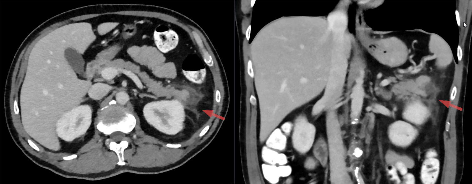
Find pancreatic-tail inflammatory change and adjacent fluid at the arrows. Compare the clinical timeline and other images before assigning acute versus chronic disease; the German and English source descriptions differ. Image: Hellerhoff; original source; CC BY-SA 4.0.
Transfer: severe characteristic pain with lipase only twice normal does not establish or exclude the diagnosis. Seek the missing evidence when suspicion remains high, especially with late presentation, prior pancreatic damage or marked lipemia. The accompanying clinical CT illustrates inflammation around the pancreatic tail, not a severity score. A single published image cannot establish the pictured patient's timing, cause or organ function.
Why search for a cause after the attack is already diagnosed?
The cause changes recurrence prevention and sometimes urgent treatment. Gallstones can obstruct the shared outlet transiently; bilirubin may normalize after passage while the gallbladder remains a source of future stones. Alcohol-associated disease involves acinar toxicity and susceptibility from multiple factors, not a specific diagnostic blood test. Take a nonjudgmental exposure history rather than assigning the cause from appearance or an isolated liver-test pattern. [1][6]
Investigate the trigger, then connect it to prevention
Evidence to seek
What it changes
Evidence to seekStones or sludge, biliary dilation, transient liver-test rise
What it changesAssess retained obstruction; plan cholecystectomy when appropriate.
Evidence to seekSustained heavy alcohol exposure, recurrent attacks, tobacco use
What it changesOffer alcohol-use treatment and smoking cessation.
Evidence to seekEarly triglycerides, diabetes control, pregnancy or triglyceride-raising drugs
What it changesCorrect metabolic drivers and arrange lipid follow-up.
Evidence to seekCalcium, medication timing, recent ERCP, trauma, infection or structural disease
What it changesAddress the demonstrated cause rather than stopping at an idiopathic label.
Measure triglycerides early because fasting can rapidly lower them. A concentration above 1,000 mg/dL strongly supports hypertriglyceridemia as the cause when a better explanation is absent. This is not a biological cutoff below which pancreatitis is impossible. Lipemic serum can interfere with amylase and lipase assays and produce misleadingly low results. Poorly controlled diabetes, pregnancy, obesity, alcohol and some medications can amplify an underlying lipid disorder. [3]
A patient has characteristic pain, CT evidence of pancreatic inflammation, triglycerides 2,600 mg/dL and lipase 1.7 times normal. Identify the two qualifying findings without counting the triglyceride concentration as a diagnostic criterion.
Check which evidence establishes the attack
Pain and characteristic imaging establish it. The triglyceride concentration supports the cause; assay interference can explain the unexpectedly modest enzyme result.
Insulin is appropriate for uncontrolled diabetes in this setting, with monitoring of glucose and potassium. It is not a universal pancreatitis treatment for patients with normal glucose. Plasma exchange lowers triglycerides quickly but has not established routine outcome benefit; selected severe or refractory situations warrant specialist discussion, not an automatic threshold-based order. [3]
For a suspected drug cause, assess timing, evidence for that drug, improvement after withdrawal and competing causes. Azathioprine and mercaptopurine have stronger evidence than many medications appearing on long association lists. Do not deliberately re-expose a patient to prove causation. If the initial evaluation is unrevealing, repeat ultrasound, MRI/MRCP or endoscopic ultrasound may find microlithiasis or structural disease. In an adult older than 40 with unexplained or recurrent attacks, consider an obstructing pancreatic tumor. Autoimmune disease, hypercalcemia, infection, trauma and post-ERCP injury also belong in a directed evaluation. [1][6]
Transfer: normalized bilirubin after gallstone pancreatitis does not mean recurrence prevention is unnecessary. Conversely, a triglyceride concentration measured after several days of minimal intake may underestimate the concentration that initiated the attack.
Which clock are you reading?
Different thresholds answer different questions. Three times normal is an enzyme criterion for diagnosis. More than 48 hours of organ failure defines severe pancreatitis. Roughly four weeks describes collection organization and often a safer opportunity for intervention. None substitutes for the others. [1][2]
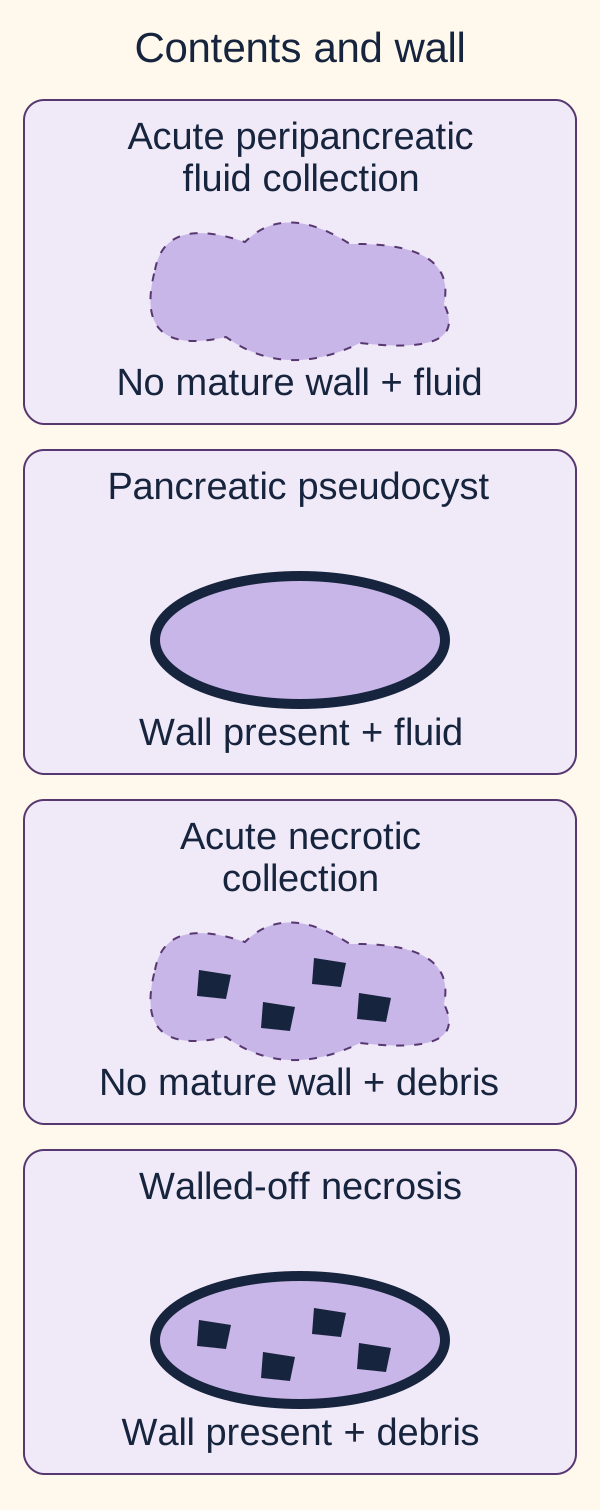
A collection is named by its contents, wall and time course, not simply its diameter. Interstitial edematous pancreatitis can produce a fluid-only collection. Necrotizing pancreatitis produces nonviable pancreatic or peripancreatic tissue; collections contain solid debris with variable fluid. Contrast nonenhancement supports parenchymal necrosis, but an early scan may not show its eventual extent. [2][7]
Identify contents before naming a collection. A mature wall changes the term, while symptoms or infection determine whether intervention is needed. These are conceptual cross-sections, not CT scans. [2][7]
Collection names when morphology and timing agree
Contents
Early, without a mature wall
Encapsulated, usually after four weeks
ContentsFluid without necrotic debris
Early, without a mature wallAcute peripancreatic fluid collection
Encapsulated, usually after four weeksPancreatic pseudocyst
ContentsNecrotic tissue, often mixed with fluid
Early, without a mature wallAcute necrotic collection
Encapsulated, usually after four weeksWalled-off necrosis
Use the collection diagram to classify an encapsulated lesion containing debris at six weeks. Then mentally change only the contents to homogeneous fluid after interstitial pancreatitis. Which name changes?
Check the first classification
Debris within an organized capsule after necrotizing pancreatitis supports walled-off necrosis.
Check the changed contents
Fluid without necrotic debris in the second setting supports a pseudocyst. A shared age or diameter does not make these collections interchangeable.
The consequence is procedural: solid debris may need a different drainage and debridement strategy from fluid alone. Do not drain an asymptomatic collection solely because it reaches a birthday or diameter. Infection, pain with persistent illness, obstruction or nutritional failure can justify intervention. In a stable patient, postponing nonurgent necrosis intervention until organization, preferably around four weeks, generally offers a safer approach. Deteriorating infected necrosis may need earlier drainage. [1][5]
Transfer: a day-10 heterogeneous collection without a wall is not a mature pseudocyst, even if large. A week-6 lesion without solid debris is not automatically necrosis. If morphology and elapsed time disagree, describe the uncertainty rather than forcing a calendar-based label.
How can a local pancreatic injury become a systemic illness?
Keep three processes separate: duct flow, intracellular injury and loss of functional reserve. Normally, acinar cells package protease precursors for delivery into the intestine. In acute pancreatitis, sustained intracellular calcium signaling, mitochondrial ATP failure, abnormal enzyme activation and inflammatory pathways injure acinar and ductal cells. Trypsin activation matters, but it is not the whole disease. Cytokines and endothelial injury can affect lungs, circulation and kidneys far from the pancreas. [6]
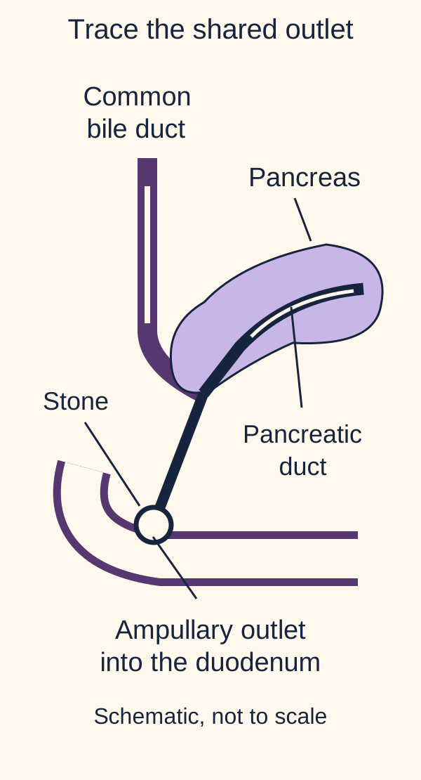
The pancreas lies largely behind the peritoneum. Its posterior position helps explain pain radiating to the back, while inflammatory fluid can extend into the lesser sac, paracolic gutters and pelvis. The anatomy diagram is a simplified duct map, not an exact depiction of every person's anatomy. Trace bile from the common bile duct and pancreatic secretion from the pancreatic duct to the duodenum. [5][7][11]
Follow each upstream route when the outlet is obstructed. Stone passage can restore drainage while pancreatic inflammation persists. Common-channel length and organ proportions are schematic. [1][9][11]
Trace the outlet, then change one condition
First place an imaginary stone at the shared ampullary outlet. With your finger or by reading the labels in order, trace backward along each upstream duct. Which two drainage routes can be affected?
Check the upstream effect
Both pancreatic outflow and biliary drainage can be obstructed at this shared outlet. This connects pancreatic pain to the possibility of jaundice.
Now suppose the stone passes and bilirubin falls. Keep pancreatic inflammation present. Predict whether ERCP necessarily reverses that inflammation.
Check the consequence of stone passage
It does not. With no continuing obstruction or cholangitis, there may be nothing for urgent biliary decompression to treat.
Finally keep the stone impacted and add fever, jaundice and circulatory instability. Which process now needs source control?
Check what now requires drainage
An infected obstructed biliary system requires urgent decompression and antibiotics. ERCP addresses cholangitis rather than directly treating every inflamed acinar cell.
The full relationship remains visible: a passed stone can leave inflammation behind; an infected retained obstruction adds a separate emergency. ACG recommends early ERCP, within 24 hours, for pancreatitis complicated by cholangitis. Resuscitation and antibiotics proceed while drainage is organized, not after a scheduled delay. [1]
Lipase-mediated fat breakdown releases fatty acids that can bind calcium as soaps, termed saponification. This can contribute to hypocalcemia, but calcium disturbances are multifactorial. Low albumin can lower total calcium without lowering ionized calcium; magnesium deficiency and systemic illness can also affect calcium regulation. Low calcium during an attack is not evidence that hypercalcemia caused it. Hemorrhagic exudate can produce flank ecchymosis (Grey Turner sign) or periumbilical ecchymosis (Cullen sign). These uncommon findings are neither required nor specific enough to establish pancreatitis. [7][8]
Transfer: recurrent injury can eventually reduce exocrine and endocrine reserve, causing maldigestion and diabetes. That loss of function differs from the premature activation driving an acute attack. New weight loss, greasy stools or persistent hyperglycemia after recovery warrants follow-up rather than being dismissed because pain improved. [6]
Development explains why duct anatomy varies: the ventral pancreatic bud forms the uncinate process and inferior head, while the dorsal bud forms the remainder. Their ducts normally unite; incomplete fusion can produce pancreas divisum, whose presence alone does not establish the cause of an attack. The neighboring spleen arises from mesoderm, not pancreatic endoderm. [9][10] Compare pancreas and spleen development with the functional consequences in chronic pancreatitis and exocrine insufficiency.
Can falling lipase coexist with worsening disease?
Yes. Severity follows organ function and its duration. Mild pancreatitis has no organ failure and no local or systemic complications. Moderately severe disease has transient organ failure, local complications or exacerbation of a preexisting comorbidity, without persistent organ failure. Severe disease has organ failure persisting more than 48 hours, in one or several systems. [2]
Assess cardiovascular, respiratory and renal function. A modified Marshall score of at least two in any of these systems indicates organ failure; examples include a PaO2/FiO2 ratio in the 201 to 300 band, or hypotension unresponsive to fluids. Interpret creatinine against baseline and after assessment of volume status. Simply receiving oxygen or having one low blood pressure does not by itself establish the classification. [2][7]
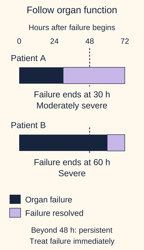
Transient organ failure supports moderately severe disease; organ failure lasting more than 48 hours defines severe disease. The clock begins with organ failure, not automatically with admission. Do not delay treatment while measuring duration. [2][7]
Compare the two organ-function timelines. Patient A's hypoxemic respiratory failure resolves after 30 hours. Patient B has respiratory failure continuously for 60 hours although lipase falls from twelve to four times normal. Classify each course, then identify the measurement that must not overrule the classification.
Check the two courses
A has transient organ failure and therefore moderately severe disease. B has persistent organ failure and severe disease. The falling lipase does not negate B's respiratory failure.
Track perfusion, mental status, urine output, oxygenation, BUN, hematocrit and creatinine repeatedly. Rising BUN and hemoconcentration can indicate fluid deficit and higher risk, but they are not automatic instructions for unlimited fluids. Systemic inflammatory response syndrome, or SIRS, signals risk and can occur without infection. Prediction scores such as BISAP support assessment but do not replace clinical reassessment or define the final Atlanta category. [1]
The consequence is to match observation and support to risk now. Organ failure or SIRS favors a monitored setting, and unstable or persistent organ failure requires critical care capability. Do not wait 48 hours to treat respiratory failure or shock; the 48-hour threshold classifies the course retrospectively. [1]
Transfer: a sterile necrotic collection without persistent organ failure is a local complication and supports moderately severe disease, not automatically severe disease. Conversely, severe organ failure can occur without a large necrotic collection. Imaging anatomy and physiological severity are related but distinct descriptions.
Which treatment addresses the problem present now?
Restore perfusion, control symptoms, feed when tolerated and treat demonstrated complications. Begin isotonic crystalloid, usually lactated Ringer solution, with moderate, frequently reassessed resuscitation. Hypovolemia may require a bolus; an adequate blood pressure does not end assessment. Recheck perfusion and congestion within the first six hours and repeatedly over the next 24 to 48 hours. Heart failure, kidney disease and older age increase the need for caution. [1]
In WATERFALL, aggressive resuscitation produced fluid overload in 20.5% compared with 6.3% under a moderate strategy, without a demonstrated reduction in progression to moderately severe or severe pancreatitis. This supports avoiding routine aggressive fluid loading; it does not justify withholding resuscitation from someone in shock. Treat fluid as an intervention with both benefit and harm, not a fixed number of liters required by the diagnosis. [4]
Compare a patient with dry mucosa, oliguria and rising BUN but clear lungs with one whose perfusion is adequate and who develops crackles and breathlessness after repeated boluses. Predict whether the same fluid order should continue in both.
Check the direction of reassessment
The first needs assessment and correction of intravascular deficit. The second needs automatic boluses stopped and urgent reassessment for congestion and other causes of respiratory decline. More fluid can worsen gas exchange once overload develops.
Nutrition and symptom relief are treatment
Provide effective analgesia, including opioids when needed, and antiemetics. In mild disease, offer a low-fat solid diet within 24 to 48 hours as tolerated rather than waiting for enzyme normalization or requiring a clear-liquid progression. Significant vomiting, ileus or obstruction changes what is feasible. If oral intake is inadequate but the gut is usable, enteral tube feeding is preferred to parenteral nutrition; a nasogastric route is often sufficient, with postpyloric feeding when clinically needed. Enteral nutrition supports the intestinal barrier. Reserve parenteral nutrition for inability to use or meet needs through the enteral route. [1][5]
Give each procedure and antimicrobial a specific indication
Cholangitis warrants antibiotics and urgent ERCP. Without cholangitis, routine early ERCP does not improve uncomplicated biliary pancreatitis. Continuing jaundice or progressive cholestasis requires evaluation for retained obstruction; MRCP or endoscopic ultrasound can assess an uncertain duct stone without diagnostic ERCP. A demonstrated retained obstruction prompts therapeutic planning. [1]
Do not prescribe prophylactic antibiotics for sterile pancreatitis or sterile necrosis. Treat cholangitis, pneumonia, urinary infection, bacteremia or another established infection on its own merits. Gas in a necrotic collection, bacteremia, sepsis or deterioration after initial stabilization can support suspected infected necrosis; absence of gas does not exclude infection. Routine fine-needle aspiration and antifungal prophylaxis are not required. [1][5]
Infected necrosis needs multidisciplinary care, antibiotics with appropriate tissue penetration and a source-control plan. A stable patient may improve while intervention is delayed for organization. Persistent sepsis or instability can require earlier drainage, often percutaneous, followed by escalation if needed. Prefer minimally invasive approaches when feasible rather than automatic early open necrosectomy. Sterile collections may also need intervention for persistent symptoms, obstruction or nutritional failure. [5]
After mild biliary pancreatitis, cholecystectomy during the same admission, preferably before discharge, prevents recurrence. Significant necrosis or collections may justify delay and individualized planning. Sphincterotomy can reduce recurrence when surgery is not feasible, but it is not a routine substitute in an operative candidate. Address alcohol use, tobacco, lipid disorders and causative medications, and arrange follow-up for unresolved causes or impaired pancreatic function. [1][3][6]
Transfer: a recovering patient who tolerates food and has stable organs does not need prolonged fasting because lipase remains high. A different patient who deteriorates during apparently successful resuscitation needs a new assessment of lungs, circulation, infection and complications, not reassurance from a lower lipase.
Apply the lesson
Case 1
Show answer and explanations for case 1
A. Mechanical obstruction not established; pancreatitis established (Why this does not fit)
A discrete transition point with obstipation supports mechanical obstruction, whereas the described pain is not characteristic pancreatic pain.
Reasoning steps for option A
Does the discrete small-bowel transition point permit saying obstruction is not established?
No. A transition point with proximal dilation, colic and obstipation supports mechanical obstruction, despite the alternative pancreatic label.
Does fourfold lipase elevation establish pancreatitis without characteristic pain or pancreatic CT findings?
No. It supplies only one pancreatitis criterion; neither the pain pattern nor CT provides a second criterion.
B. Mechanical obstruction established; pancreatitis not established (Best answer)
The symptoms and transition point establish obstruction. The enzyme result supplies only one pancreatitis criterion in this presentation.
Reasoning steps for option B
How many pancreatitis diagnostic criteria does this presentation meet?
Only the enzyme criterion is met: lipase is four times normal, but lower-abdominal colic is not characteristic pancreatic pain and CT shows no pancreatic abnormality.
What diagnostic conclusion follows from proven obstruction and only one pancreatic criterion?
Mechanical obstruction is established, while pancreatitis is not established despite the fourfold lipase elevation.
C. Mechanical obstruction established; pancreatitis also established (Why this does not fit)
The obstruction evidence is convincing, but lipase alone does not supply two pancreatitis criteria.
Reasoning steps for option C
Does lipase four times normal add a second independent criterion to the CT-proven obstruction?
No. The CT shows bowel obstruction but no pancreatic inflammation, and characteristic epigastric pain is absent; the enzyme result alone cannot establish pancreatitis.
How should the concurrent-pancreatitis claim be handled despite proven obstruction?
Reject the added pancreatic diagnosis: elevated lipase is only one criterion and can occur with reduced renal clearance or nonpancreatic illness.
D. Mechanical obstruction not established; pancreatitis not established (Why this does not fit)
Pancreatitis is not established, but the clinical and CT findings do support mechanical obstruction.
Reasoning steps for option D
Can the uncertain significance of lipase negate the transition point and obstipation?
No. These bowel findings independently support mechanical obstruction even though pancreatitis lacks a second qualifying criterion.
Which half of the proposed neither-diagnosis conclusion fails?
The obstruction half fails because a discrete transition point and obstipation support mechanical obstruction; pancreatitis remains unconfirmed.
Takeaway: Interpret pancreatic criteria and competing anatomical evidence independently [1,7].
A. Pancreatitis established; triglycerides probably incidental (Why this does not fit)
The diagnosis is established, but an early concentration of 2,600 mg/dL with no better cause is more than a minor incidental rise.
Reasoning steps for option A
Is an early triglyceride value of 2,600 mg/dL plausibly incidental when other triggers are absent?
No. This prefasting concentration far exceeds 1,000 mg/dL and strongly supports a triglyceride trigger, although it cannot prove causation alone.
Given pain and CT already establish pancreatitis, how should marked early triglycerides be interpreted?
A likely trigger rather than probably incidental, though the concentration alone cannot establish causation with certainty.
B. Pancreatitis established; triglycerides a likely trigger (Best answer)
Pain and imaging satisfy the diagnosis, and the marked early triglyceride concentration supports a cause when alternatives are not evident. Lipemic assay interference is possible, not proven in this sample.
Reasoning steps for option B
Do characteristic pain and pancreatic CT inflammation diagnose the attack despite lipase only 1.4 times normal?
Yes. They are two independent diagnostic criteria; lipemia may interfere with the assay but interference is not demonstrated in this sample.
What does the early triglyceride concentration contribute beyond the two diagnostic criteria?
At 2,600 mg/dL with no better cause, it supports a likely metabolic trigger; it is not itself a pancreatitis diagnostic criterion.
C. Pancreatitis unconfirmed; triglycerides probably incidental (Why this does not fit)
This discounts both independent pain-plus-imaging evidence and a strong metabolic exposure.
Reasoning steps for option C
Can the modest lipase erase both pain-plus-CT diagnostic evidence and the 2,600 mg/dL exposure?
No. Pain and characteristic imaging establish pancreatitis, while marked early hypertriglyceridemia supports its likely cause.
Which parts of an unconfirmed-and-incidental interpretation fail?
Pain plus pancreatic CT establish the attack, and marked prefasting triglycerides strongly support a causal contribution.
D. Pancreatitis unconfirmed; triglycerides a likely trigger (Why this does not fit)
The metabolic exposure is important, but the diagnosis is already supported by pain and imaging even with a modest enzyme result.
Reasoning steps for option D
Does probable triglyceride causation imply the pancreatic diagnosis remains unconfirmed?
No. Causal attribution and diagnosis are separate: pain and characteristic CT already meet two criteria without a threefold enzyme elevation.
Which part of the unconfirmed-but-trigger interpretation fails?
The unconfirmed diagnosis fails: characteristic pain and CT provide two criteria even though lipase is only 1.4 times normal.
Takeaway: Pain and imaging can establish the attack; early marked hypertriglyceridemia addresses its cause [1,3].
A. Drug contribution is excluded; investigate recurrent causes (Why this does not fit)
Further evaluation is appropriate, but earlier attacks do not rule out an additional medication trigger for the current episode.
Reasoning steps for option A
Do two attacks before azathioprine exposure exclude a contribution to the third attack three weeks after starting it?
No. The medication cannot explain earlier attacks but its timing permits an additional trigger for the current attack.
What investigation is needed even if the third attack may be drug-associated?
Continue evaluating recurrent causes, since the two confirmed attacks before azathioprine still lack an explanation.
B. Drug contribution remains possible; end the etiologic investigation (Why this does not fit)
The recent timing supports suspicion but does not explain the pre-exposure episodes.
Reasoning steps for option B
Can azathioprine started only before attack three explain the two prior confirmed attacks?
No. Temporal impossibility leaves the earlier recurrent disease unexplained even while the current drug contribution remains possible.
Why is ending the etiologic workup after holding azathioprine premature?
It cannot explain attacks predating prescription, and timing alone does not establish that all attacks share this trigger.
C. Drug contribution is excluded; end the etiologic investigation (Why this does not fit)
The chronology neither excludes a new contributor nor resolves the cause of the earlier episodes.
Reasoning steps for option C
Does holding azathioprine prove drug causation impossible and resolve recurrent etiology?
No. Withdrawal is prudent when suspected, but neither exclusion of the new trigger nor explanation of pre-exposure attacks follows.
What two conclusions remain unresolved despite stopping azathioprine?
Its contribution to the third attack remains possible, and the causes of the two pre-exposure episodes still require evaluation.
D. Drug contribution remains possible; investigate recurrent causes (Best answer)
The new exposure can contribute to the current attack, but cannot explain attacks before exposure, so other recurrent causes still need evaluation.
Reasoning steps for option D
Which chronology separates the suspected new drug trigger from the recurrent background?
Two attacks preceded exposure; a third followed initiation by three weeks. Drug contribution to the third remains possible while recurrent causes still warrant investigation.
What plan addresses both the possible current drug contribution and prior unexplained attacks?
Hold the suspected drug and continue recurrent-cause evaluation rather than assigning every episode to azathioprine.
Takeaway: A plausible new medication trigger and an unexplained recurrent background can coexist [1,6].
A. Pancreatic head; targeted endoscopic ultrasound (Best answer)
The downstream cutoff localizes the concern toward the head. EUS can assess a small obstructing lesion not apparent on cross-sectional imaging.
Reasoning steps for option A
Where is an obstruction if body and tail ducts dilate upstream of an abrupt cutoff near the duodenum?
Toward the pancreatic head, downstream of the dilated duct; persistent cutoff can signal an occult lesion despite no visible MRCP mass.
Which next test addresses an occult head lesion without a visible MRCP mass?
Targeted endoscopic ultrasound can examine small obstructing tissue lesions despite negative cross-sectional mass imaging.
B. Pancreatic head; diagnostic ERCP as the first test (Why this does not fit)
The location is plausible, but EUS can evaluate occult tissue disease without the added risks of diagnostic ERCP when no therapeutic duct indication is established.
Reasoning steps for option B
Would diagnostic ERCP be the lowest-risk first test for a head-side duct cutoff without a therapeutic target?
No. Targeted EUS can inspect for a small obstructing head lesion without starting with invasive duct instrumentation.
What makes first-line diagnostic ERCP less appropriate than targeted EUS here?
No therapeutic duct intervention is established; EUS evaluates occult head tissue without diagnostic ERCP risks.
C. Pancreatic tail; diagnostic ERCP as the first test (Why this does not fit)
This mislocalizes the obstruction and chooses invasive duct evaluation before targeted assessment of an occult head lesion.
Reasoning steps for option C
Does dilation in the body and tail place the obstructing cutoff within the tail?
No. Dilation is upstream; the cutoff near the duodenum localizes the concern toward the head, where EUS can inspect occult tissue.
What two corrections does the tail-and-ERCP plan need?
Localize the concern to the downstream head cutoff, then investigate occult tissue with targeted EUS rather than initial diagnostic ERCP.
D. Pancreatic tail; targeted endoscopic ultrasound (Why this does not fit)
EUS is reasonable for occult disease, but a downstream cutoff with dilation of the body and tail does not localize the obstruction to the tail.
Reasoning steps for option D
Does a reasonable EUS choice make the tail the suspected site of obstruction?
No. The upstream body-and-tail dilation ends at a downstream cutoff near the head; EUS should target that area.
How should the EUS target be changed in this otherwise reasonable test choice?
Aim at the pancreatic head near the downstream cutoff, not the dilated upstream tail.
Takeaway: Persistent unexplained upstream duct dilation in an older adult warrants focused evaluation of the downstream obstructing region [1,11].
A. Initially biliary only; afterward pancreatic only (Why this does not fit)
The isolated pancreatic neck lesion does not initially impair bile flow. The later shared-outlet stone threatens biliary as well as pancreatic drainage.
Reasoning steps for option A
Can a neck lesion isolated to the pancreatic duct initially obstruct the normal common bile duct?
No. The neck stricture impairs pancreatic outflow alone; the later ampullary stone at a shared outlet can impair both routes.
Which two route predictions fail in the biliary-first, pancreatic-only-after plan?
Initially pancreatic rather than biliary flow is impaired; after the shared-outlet stone both pancreatic and biliary drainage are threatened.
B. Initially biliary only; afterward pancreatic and biliary (Why this does not fit)
The later combined impairment is plausible, but the initial narrowing affects the pancreatic route, not the separate bile duct.
Reasoning steps for option B
Which duct is upstream of the original neck narrowing while the head duct and bile duct remain normal?
The body-and-tail pancreatic duct is dilated upstream, identifying initial pancreatic drainage impairment rather than biliary impairment.
What is the correct initial route despite the correctly predicted later combined impairment?
The initial pancreatic neck narrowing obstructs pancreatic outflow only, while the later ampullary obstruction adds biliary impairment.
C. Initially pancreatic only; afterward pancreatic only (Why this does not fit)
The initial route is correct. The added shared-outlet obstruction also impairs bile flow, so the later problem is not confined to pancreatic drainage.
Reasoning steps for option C
When a second stone blocks the shared ampullary outlet, can bile still drain normally through it?
No. The ampulla serves biliary as well as pancreatic flow; biliary impairment is added to the existing pancreatic neck lesion.
What must be added to the correct pancreatic-only initial prediction after the second stone?
Biliary impairment: the second stone blocks the shared ampullary outlet serving both routes.
D. Initially pancreatic only; afterward pancreatic and biliary (Best answer)
The neck lesion initially impairs pancreatic drainage. A second obstruction at the shared outlet adds biliary impairment while the original pancreatic narrowing remains.
Reasoning steps for option D
What changes when the ampullary stone is added without removing the pancreatic neck narrowing?
The initial isolated pancreatic outflow problem persists and biliary drainage is newly impaired at the shared outlet.
Which route change follows from the second obstruction while the first remains?
Drainage changes from pancreatic-only impairment to pancreatic and biliary impairment.
Takeaway: An isolated pancreatic duct lesion and a shared-outlet lesion have different upstream consequences [1,11].
A. A mild; B moderately severe (Why this does not fit)
B is classified correctly, but A had qualifying respiratory dysfunction even though it resolved quickly.
Reasoning steps for option A
Does A qualify as mild when 72/0.30 yields a P/F ratio of 240 for 18 hours?
No. A P/F of 240 is qualifying respiratory organ failure, albeit transient, which supports moderately severe disease; B has a symptomatic local complication.
What categories follow for A and B after excluding mild disease in A?
Both are moderately severe: A has respiratory failure resolving at 18 hours, and B has a symptomatic local complication.
B. A severe; B moderately severe (Why this does not fit)
B is classified correctly, but A did not have persistent organ failure beyond 48 hours.
Reasoning steps for option B
Can 18 hours of respiratory failure put A into the severe category?
No. Severe requires organ failure lasting more than 48 hours; A recovered at 18 hours, while B is moderately severe through a local collection.
How does the 48-hour duration rule change A, but not B, in the proposed severe/moderate pair?
A is moderately severe rather than severe because failure resolved in 18 hours; B is moderately severe through its local collection.
C. A moderately severe; B mild (Why this does not fit)
A is classified correctly, but a symptomatic local collection excludes mild disease in B.
Reasoning steps for option C
Can B be mild despite a symptomatic peripancreatic fluid collection and no organ failure?
No. A local complication supports moderately severe disease even without organ failure; A also qualifies because P/F was 240 transiently.
What classification follows for B despite never having organ failure?
Moderately severe, not mild, because the symptomatic peripancreatic collection is a local complication.
D. A moderately severe; B moderately severe (Best answer)
A had respiratory failure with a P/F ratio of 240 that resolved before 48 hours. B qualifies through a local complication, even without organ failure.
Reasoning steps for option D
By which distinct routes do A and B each reach moderately severe classification?
A has transient respiratory failure with P/F 240 for 18 hours; B has a symptomatic local collection without organ failure.
Why is neither patient severe despite each qualifying as moderately severe?
A's failure ended before 48 hours; B has no organ failure, only a symptomatic local collection.
Takeaway: Transient organ failure and local complications can independently produce moderately severe disease [2,7].
A does not have documented fluid-unresponsive cardiovascular failure or other complications. B has persistent cardiovascular failure for more than 48 hours.
Reasoning steps for option A
Does fluid-responsive 84 mm Hg pressure establish the same organ failure as 58 hours of vasopressors?
No. A rapidly corrects and has no subsequent complication; B has fluid-unresponsive cardiovascular failure persisting beyond 48 hours.
What completed-course labels follow despite falling lipase in both?
A is mild with no documented organ failure or complication; B is severe from fluid-unresponsive cardiovascular failure lasting 58 hours.
B. A severe; B severe (Why this does not fit)
B is severe; A lacks persistent organ failure and therefore cannot be classified as severe from the initial pressure alone.
Reasoning steps for option B
What persistent failure evidence is absent in A despite the initial systolic pressure of 84?
A responds promptly to fluid with no subsequent dysfunction, unlike B whose pressor-dependent shock lasts 58 hours.
Why cannot A be labeled severe along with B?
A's initial hypotension resolved promptly after fluid, so persistent organ failure is absent; B required vasopressors continuously for 58 hours.
C. A moderately severe; B severe (Why this does not fit)
B is correctly classified, but a single fluid-responsive pressure does not establish cardiovascular organ failure in A.
Reasoning steps for option C
Does A meet fluid-unresponsive cardiovascular failure simply because fluid was required?
No. Response to initial fluid means this episode does not document cardiovascular organ failure, even transiently; B has persistent failure.
What label follows for A when no organ failure or complication occurs?
Mild, not moderately severe; a single fluid-responsive pressure does not by itself establish organ failure.
D. A mild; B moderately severe (Why this does not fit)
A is correctly classified, but B's continuous 58-hour vasopressor requirement is persistent, not transient, failure.
Reasoning steps for option D
Is B's continuous 58-hour vasopressor need a transient complication?
No. Fluid-unresponsive shock for more than 48 hours defines persistent cardiovascular organ failure and severe pancreatitis.
How must B be reclassified when vasopressors continue for 58 hours?
Severe rather than moderately severe, because cardiovascular failure persists longer than 48 hours.
Takeaway: Confirm organ failure before timing it, and use its duration rather than enzyme trends to classify severity [2,7].
A does not need routine CT to confirm two diagnostic criteria, while B's deterioration warrants assessment rather than deferral.
Reasoning steps for option A
Why does A's six-hour lipase of seven times normal not demand confirmatory CT now?
Characteristic pain and qualifying lipase already establish two criteria; A improves and has no suspected alternative emergency, whereas B deteriorates.
Which proposed imaging assignment fits A's improvement and B's later deterioration?
Image B now to assess the new problem; do not scan improving A routinely just to confirm established pancreatitis.
B. B now; not routinely A (Best answer)
B's failure to improve and deterioration create a complication question. A already meets diagnostic criteria and is improving without a new imaging indication.
Reasoning steps for option B
What new imaging indication appears 60 hours into B's course?
Fever, worsening pain and inability to eat despite care raise a complication or alternative-diagnosis question; A has no such question.
Why does improving A not need the same cross-sectional assessment as deteriorating B?
A already has characteristic pain and sevenfold lipase elevation, is improving and has no suspected alternative emergency; there is no new diagnostic or complication question requiring routine imaging.
C. Both now (Why this does not fit)
B warrants imaging, but scanning A routinely adds no established diagnostic or complication question.
Reasoning steps for option C
Is having the same established pancreatic diagnosis a reason to scan both patients?
No. Routine early CT adds little in improving A, while selective cross-sectional imaging is warranted by B's deterioration.
Which unnecessary scan is included in the both-now plan?
A's: characteristic pain plus sevenfold lipase already establish the diagnosis, and A is improving without another concern.
D. Neither now (Why this does not fit)
Avoiding a routine scan fits A but not B, whose evolving symptoms warrant selective cross-sectional assessment.
Reasoning steps for option D
Does avoiding unnecessary early imaging for A justify deferring B's scan after deterioration?
No. B now has a specific complication question at 60 hours; normal creatinine and prior diagnostic certainty do not remove it.
Which missed scan is embedded in the neither-now plan?
B's: new fever, worsening pain and inability to eat at 60 hours call for selective cross-sectional assessment.
Takeaway: Imaging is selected for the question present now, not ordered or withheld solely by the pancreatitis label [1].
A. Perfusion currently adequate; congestion present (Best answer)
Pressure, urine output and mentation support adequate current perfusion. New crackles, hypoxemia and raised venous pressure after fluid loading support congestion, so further automatic loading should pause while the patient is reassessed.
Reasoning steps for option A
Do pressure 124/72, urine output 0.8 mL/kg/h and normal mentation exclude simultaneous fluid congestion?
No. They support current perfusion, while new crackles, hypoxemia and jugular distention after 3 L support congestion.
What should automatic bolusing do when perfusion appears adequate but congestion emerges?
Pause further automatic fluid loading and reassess oxygenation, circulation and possible causes of congestion.
B. Perfusion currently inadequate; congestion present (Why this does not fit)
The congestion interpretation fits, but current pressure, urine output and mentation do not demonstrate inadequate perfusion.
Reasoning steps for option B
Which supplied perfusion measurements contradict current inadequacy despite pulmonary congestion?
Blood pressure 124/72, urine output 0.8 mL/kg/h and normal mentation support adequate current perfusion, though continued reassessment is necessary.
How does the adequately perfused state alter the inadequate-perfusion half of this choice?
The current measurements do not demonstrate inadequate perfusion; congestion is plausible and warrants reassessment instead of automatic boluses.
C. Perfusion currently adequate; congestion absent (Why this does not fit)
The current perfusion interpretation fits, but new lung findings and raised venous pressure support congestion rather than its absence.
Reasoning steps for option C
Which new signs contradict a claim of no congestion after 3 L of crystalloid?
Crackles, hypoxemia and raised jugular venous pressure support pulmonary and venous congestion even with reassuring perfusion.
What does the raised venous pressure imply for the no-congestion half of this choice?
It contradicts absent congestion, especially with new crackles and hypoxemia following fluid loading.
D. Perfusion currently inadequate; congestion absent (Why this does not fit)
Both components conflict with the supplied pattern: current perfusion measures are reassuring, while new pulmonary and venous findings indicate congestion.
Reasoning steps for option D
Do the observations support either inadequate current perfusion or absence of congestion?
Neither: pressure, urine output and mentation are reassuring; crackles, hypoxemia and jugular distention favor congestion.
Which paired assessment replaces both unsupported halves?
Current perfusion appears adequate while congestion is present; monitor and reassess rather than reflexively add fluid.
Takeaway: Reassessed fluids require separate judgments about perfusion and congestion [1].
A. 14.2 percentage-point excess overload; withhold resuscitation fluid (Why this does not fit)
The arithmetic is correct, but withholding fluid ignores the current deficit and confuses avoiding aggressive protocols with avoiding resuscitation.
Reasoning steps for option A
Does a 14.2 percentage-point overload excess mean this oliguric patient should receive no fluid?
No. The trial warns against automatic aggressive loading, not correction of the deficit suggested by urine output 0.4 mL/kg/h and rising BUN.
Which patient findings still warrant resuscitation despite the excess overload risk?
Urine output 0.4 mL/kg/h, dry mucosa, tachycardia and rising BUN during minimal intake support a deficit; give isotonic fluid with repeated reassessment.
B. 3.3 percentage-point excess overload; withhold resuscitation fluid (Why this does not fit)
This confuses relative and absolute measures and also disregards the patient's hypovolemia indicators.
Reasoning steps for option B
Is 20.5% minus 6.3% equal to 3.3 percentage points, and does that justify withholding fluid?
No. The absolute difference is 14.2 points; about 3.3 is the rate ratio, and deficit signs still warrant reassessed resuscitation.
What treatment follows from oliguria and rising BUN despite avoiding aggressive protocols?
Give moderate, reassessed isotonic fluid for likely hypovolemia, not no fluid; monitor for congestion.
C. 3.3 percentage-point excess overload; give reassessed isotonic fluid (Why this does not fit)
Reassessed fluid is appropriate, but roughly 3.3 is the ratio of the event rates, not their absolute percentage-point difference.
Reasoning steps for option C
Which calculation produces the proposed 3.3 instead of the absolute overload difference?
20.5 divided by 6.3 is about 3.3, a relative ratio; subtraction gives 14.2 percentage points, while reassessed fluid remains appropriate.
How should the numerical error alter an otherwise appropriate fluid plan?
Retain reassessed isotonic fluid, but report the absolute overload excess as 14.2 percentage points rather than 3.3.
D. 14.2 percentage-point excess overload; give reassessed isotonic fluid (Best answer)
The absolute difference is 20.5 minus 6.3, or 14.2 percentage points. The patient has evidence of deficit; the trial argues against routine aggressive loading, not against correcting hypovolemia.
Reasoning steps for option D
What is the absolute overload increase, and what do clear lungs plus oliguria imply for fluid?
20.5 minus 6.3 equals 14.2 percentage points; urine output 0.4 mL/kg/h, tachycardia and rising BUN favor moderate reassessed isotonic fluid.
Does the trial support withholding fluid despite dry mucosa, oliguria and rising BUN?
No. It found more overload with aggressive rather than moderate fluid and no demonstrated reduction in disease progression. The current deficit still supports reassessed isotonic fluid rather than withholding resuscitation.
Takeaway: Fluid-trial harm supports moderate reassessment, not denial of treatment for hypovolemia [1,4].
A. Gastric enteral feeding; add supplemental parenteral nutrition (Why this does not fit)
Gastric delivery repeatedly provokes regurgitation, and tolerated jejunal feeding already meets needs. Neither the proposed route nor supplementation fits.
Reasoning steps for option A
Which repeated gastric-trial symptom contradicts the proposed gastric-plus-parenteral route?
Repeated gastric trials cause vomiting and witnessed regurgitation; tolerated jejunal feeding already meets needs.
What route and supplementation choices follow from these contrasting trials?
Use the tolerated jejunal route without parenteral supplementation; gastric regurgitation and adequate jejunal calories defeat both parts of this plan.
B. Jejunal enteral feeding; add supplemental parenteral nutrition (Why this does not fit)
Jejunal delivery fits the gastric intolerance, but it already meets nutritional needs; supplemental parenteral nutrition is unnecessary here.
Reasoning steps for option B
Why is jejunal rather than gastric delivery appropriate after these feeding trials?
Gastric feeding repeatedly causes vomiting and witnessed regurgitation, whereas jejunal infusion is tolerated.
Should parenteral nutrition be added to otherwise suitable jejunal feeding?
No. Jejunal feeding meets estimated needs, making the proposed supplementation unnecessary.
C. Jejunal enteral feeding; no supplemental parenteral nutrition (Best answer)
Repeated gastric intolerance favors the jejunal route. Its demonstrated adequate delivery means no supplemental parenteral nutrition is needed.
Reasoning steps for option C
Which route avoids witnessed regurgitation while meeting estimated needs?
Gastric trials cause regurgitation, while jejunal infusion is tolerated and meets estimated needs.
What nutrition plan follows if the jejunal route already meets estimated needs?
Continue jejunal enteral feeding without supplemental parenteral nutrition.
D. Gastric enteral feeding; no supplemental parenteral nutrition (Why this does not fit)
Avoiding unnecessary parenteral nutrition fits, but repeated vomiting and regurgitation make the gastric route a poor choice.
Reasoning steps for option D
Why is gastric feeding still unsuitable despite omitting parenteral nutrition?
Omitting supplementation is reasonable because jejunal feeding meets needs, but repeated vomiting and regurgitation rule against gastric delivery.
Is supplemental parenteral nutrition needed when the route is changed to tolerated jejunal feeding?
No. Jejunal infusion already meets estimated nutritional needs, so omitting parenteral supplementation is the supported part of this option.
Takeaway: Use the functional intestine, selecting postpyloric delivery for demonstrated gastric intolerance [1].
A. Early transmural drainage; do not await wall maturation (Why this does not fit)
Urgent source control fits refractory shock, but there is no mature wall or safe gastric apposition for the proposed transmural route.
Reasoning steps for option A
What anatomy makes early transmural drainage unsafe despite worsening shock?
A mature wall and safe gastric apposition are both absent; the accessible route is retroperitoneal percutaneous.
Which early drainage route should be pursued instead?
Seek prompt percutaneous drainage through the accessible retroperitoneal route rather than unsafe transmural access.
B. Delayed percutaneous drainage; await wall maturation (Why this does not fit)
The percutaneous route is anatomically feasible, but worsening shock despite treatment does not support waiting for encapsulation.
Reasoning steps for option B
Is a mature wall required to use the described retroperitoneal percutaneous route?
No. The supplied anatomy provides an accessible percutaneous route even though the collection lacks a mature wall; it does not provide safe gastric apposition for transmural access.
What timing should the feasible percutaneous route have?
Early source-control assessment and drainage are warranted because shock worsens despite treatment; do not wait merely for wall maturation.
C. Early percutaneous drainage; do not await wall maturation (Best answer)
Refractory shock requires prompt source-control assessment rather than a fixed wait. The available retroperitoneal access permits early percutaneous drainage within a step-up strategy.
Reasoning steps for option C
Which available route provides prompt source control without safe gastric apposition?
An accessible retroperitoneal percutaneous route is available, while no mature wall or safe apposition permits transmural drainage.
Does this route allow source control without waiting for a mature wall?
Yes. Early retroperitoneal percutaneous drainage fits refractory shock within a step-up strategy.
D. Delayed transmural drainage; await wall maturation (Why this does not fit)
Waiting despite refractory shock is unsafe, and the supplied anatomy does not provide a safe transmural target. A feasible percutaneous route is already available.
Reasoning steps for option D
Why do both waiting and transmural access fail in this deteriorating patient?
Which five-week morphology distinguishes walled-off necrosis from a pseudocyst?
A mature encapsulated collection containing fluid and solid debris after necrotizing pancreatitis identifies walled-off necrosis rather than a pure-fluid pseudocyst.
Why assess drainage rather than only continue assisted feeding?
Compression persists and the patient cannot meet nutritional needs despite that support, so intervention assessment is warranted.
D. Walled-off necrosis; continue feeding and reassess (Why this does not fit)
Morphology fits, but persistent nutritional failure makes drainage assessment appropriate.
Reasoning steps for option D
Why is continued feeding alone insufficient despite a correct WON label?
The mature debris-containing collection compresses the stomach and causes weight loss despite assisted enteral support.
What should replace continued feeding and reassessment alone?
Assess drainage for symptomatic stomach compression and nutritional failure, while retaining the correct walled-off necrosis label.
A. A: index-admission cholecystectomy; B: index-admission cholecystectomy (Why this does not fit)
The index-admission plan fits A, but B has extensive necrosis and a substantial collection that favor deferral and reassessment.
Reasoning steps for option A
What in B's week-2 anatomy makes simultaneous index-admission surgery inappropriate?
A has recovered from mild disease without collection; B has extensive necrosis and a large collection at week 2.
How should B's operation be timed relative to A's?
A receives index-admission cholecystectomy; defer and reassess B for interval surgery when inflammation and collection resolve or stabilize.
B. A: interval cholecystectomy; B: interval cholecystectomy (Why this does not fit)
The interval plan fits B, but A has recovered from uncomplicated mild disease and has no reason to defer recurrence prevention.
Reasoning steps for option B
Why does A's mild recovery make delaying both operations unnecessary?
Only B has extensive necrosis and a substantial collection; A is eating after uncomplicated mild disease.
Should A wait for interval surgery solely because B must wait?
No. A's uncomplicated mild recovery supports index-admission cholecystectomy while B needs individualized interval timing.
C. A: index-admission cholecystectomy; B: interval cholecystectomy (Best answer)
A should receive index-admission prevention after mild disease. B needs individualized interval surgery after inflammation and the collection resolve or stabilize.
Reasoning steps for option C
Which difference between A and B prevents applying the mild-disease surgical timetable to both?
A has recovered from mild disease without a collection; B has extensive necrosis and a large collection at week 2, which favor deferral and reassessment despite stability.
How does recurrence prevention remain definitive for both patients?
Perform cholecystectomy during A's index admission and reassess B for individualized interval cholecystectomy after inflammation and the collection resolve or stabilize.
D. A: index-admission cholecystectomy; B: definitive sphincterotomy (Why this does not fit)
Sphincterotomy is not a routine definitive replacement for cholecystectomy in a fit surgical candidate. B needs reassessed operative timing, and no current retained-duct target for ERCP is supplied.
Reasoning steps for option D
Why is B's sphincterotomy not a definitive substitute for surgery?
B has no cholangitis or retained stone and remains a fit surgical candidate, though necrosis and a collection affect timing.
What plan preserves definitive prevention for B without premature surgery?
Reassess for interval cholecystectomy once inflammation and collection settle; sphincterotomy is not routinely definitive for this fit patient.
Takeaway: Mild and necrotizing biliary attacks require different surgical timing [1].
A. Post-ERCP pancreatitis; supportive care and organ monitoring (Best answer)
Sustained pancreatic-type pain and lipase measured more than 24 hours after ERCP remaining above three times normal support post-ERCP pancreatitis. Documented clearance and falling bilirubin do not provide a current target for repeat ERCP.
Reasoning steps for option A
How does pain plus lipase five times normal at 30 hours distinguish pancreatitis?
New sustained pancreatic-type pain requiring admission and lipase over three times normal at 30 hours support post-ERCP pancreatitis.
Which care follows when no residual duct target is shown?
Supportive pancreatitis care with organ monitoring, without repeat ERCP despite the difficult initial procedure.
B. Post-ERCP perforation; repeat ERCP for duct assessment (Why this does not fit)
The examination and CT do not support perforation, and duct clearance was documented.
Reasoning steps for option B
What findings argue against perforation and another duct ERCP?
Examination and CT show no perforation, while documented stone clearance and falling bilirubin argue against a duct target.
What diagnosis and duct plan better fit than perforation with repeat ERCP?
Treat post-ERCP pancreatitis supportively and monitor organs; no perforation or residual obstruction is demonstrated.
C. Isolated post-ERCP hyperlipasemia; supportive care and organ monitoring (Why this does not fit)
Management is reasonable, but clinically significant persistent pain rules out isolated enzyme increase.
Reasoning steps for option C
Why is this not isolated asymptomatic post-ERCP hyperlipasemia?
This is symptomatic sustained pancreatic-type pain requiring admission with lipase five times normal after 24 hours, not an isolated enzyme rise.
Does supportive monitoring validate the isolated-hyperlipasemia diagnosis?
No. Supportive care and organ monitoring fit symptomatic post-ERCP pancreatitis, not a mere isolated enzyme rise.
D. Post-ERCP pancreatitis; repeat ERCP for residual obstruction (Why this does not fit)
The pancreatic syndrome fits, but documented duct clearance and falling bilirubin argue against another ERCP.
Reasoning steps for option D
What evidence makes residual obstruction an unsupported reason to repeat ERCP?
Stone clearance was documented and bilirubin is falling; no evidence establishes ongoing duct obstruction.
What should replace repeat duct instrumentation?
Provide supportive care and organ monitoring for post-ERCP pancreatitis unless new evidence establishes a duct target.
Takeaway: Confirm post-ERCP pancreatitis and avoid repeat duct instrumentation absent a target [1].
A. Retroperitoneal hemorrhage suspected; urgent bleeding assessment (Best answer)
The combined high-attenuation tracking fluid, falling hemoglobin and falling pressure raise concern for hemorrhage. Urgent assessment and resuscitation are needed even without visible active extravasation.
Reasoning steps for option A
How do high-attenuation tracking fluid and falling pressure support hemorrhage?
High-attenuation retroperitoneal fluid tracks toward ecchymoses as hemoglobin and blood pressure fall, suggesting hemorrhage.
What action is needed even if CT shows no active extravasation?
Urgently resuscitate and assess retroperitoneal bleeding; one scan cannot exclude intermittent or recently stopped hemorrhage.
B. Retroperitoneal hemorrhage suspected; interval imaging surveillance (Why this does not fit)
The suspected mechanism fits, but the falling pressure and hemoglobin require urgent assessment rather than scheduled surveillance.
Reasoning steps for option B
Why is interval surveillance unsafe with hemoglobin falling from 12 to 8 g/dL?
Hemoglobin falls from 12 to 8 g/dL with declining pressure, and high-attenuation fluid suggests bleeding despite absent visible extravasation.
How should declining pressure change the proposed surveillance plan?
Urgent hemorrhage assessment and resuscitation replace scheduled interval imaging despite absent active extravasation.
C. Consumptive coagulopathy suspected; urgent clotting-factor replacement (Why this does not fit)
Stable platelets, INR and fibrinogen do not support consumptive coagulopathy as the explanation. The imaging and circulatory changes instead raise concern for bleeding.
Reasoning steps for option C
What stable clotting measures undermine consumptive coagulopathy?
Stable platelets, INR and fibrinogen argue against consumption; tracking high-attenuation fluid with falling hemoglobin suggests hemorrhage.
What response fits better than empiric factor replacement?
Urgently assess and resuscitate suspected retroperitoneal hemorrhage; stable coagulation tests do not establish consumptive factor loss.
D. Inflammatory third spacing suspected; further crystalloid resuscitation (Why this does not fit)
Third spacing may lower pressure, and dilution can lower hemoglobin, but neither alone explains this combined high-attenuation tracking-fluid, blood-count and pressure pattern. A bleeding assessment is needed rather than attributing the deterioration only to third spacing.
Reasoning steps for option D
What finding is poorly explained by third spacing alone?
Third spacing may lower pressure and dilution may lower hemoglobin, but neither explains high-attenuation tracking fluid with this deterioration.
What response avoids missing possible hemorrhage?
Assess bleeding urgently while resuscitating; do not attribute the combined CT, hemoglobin and pressure changes solely to third spacing.
Takeaway: Imaging and laboratory trajectory raise concern for retroperitoneal hemorrhage, not a specific bleeding source [7].