Chronic Pancreatitis and Exocrine Pancreatic Insufficiency
Separate pancreatic structural injury from digestive failure, interpret fecal elastase, match enzymes to meals, and manage nutrition, pain, and diabetes.
A scarred pancreas can hurt, underdeliver digestive enzymes, and lose control of blood glucose. These are related problems, not interchangeable diagnoses. The central question is: which function is failing, what evidence establishes that failure, and which treatment actually reaches it?
By the end, you should be able to separate structural disease from exocrine pancreatic insufficiency (EPI), interpret a fecal elastase result, correct an ineffective enzyme prescription, and identify pain or complications that need more than enzyme replacement. The examples and practice cases are original educational scenarios, not patient records.
Does one damaged organ require one treatment?
Chronic pancreatitis is a persistent fibroinflammatory disorder, not simply an acute attack lasting longer. Repeated or sustained injury produces fibrosis, acinar loss, and distorted ducts. Premature digestive enzyme activation within the pancreas can contribute to injury; this is different from inadequate enzyme activity in the intestinal lumen causing maldigestion. The process can develop without a recognized prior acute episode. Pain, exocrine failure, and diabetes appear at different times; none is required to accompany every other feature. [1]
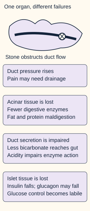
Trace the duct separately from the enzyme-producing tissue and endocrine islets; Sources [4] and [5].
Acinar cells supply digestive enzymes. Ductal cells supply bicarbonate-rich fluid, which helps carry enzymes and neutralize acidic gastric contents. Protein plugs, stones, and strictures can obstruct flow and raise upstream pressure. Fibrosis reduces enzyme delivery and bicarbonate secretion, while inflammatory and neural processes also contribute to pain. A duct procedure may address obstruction without restoring destroyed acinar tissue. [5]
EPI means that pancreatic enzyme activity in the intestine is insufficient for normal digestion. Too little enzyme production, poor delivery, and ineffective mixing can all contribute. Fat maldigestion produces oily, bulky stools and weight loss; protein-calorie malnutrition, bloating, and deficiencies of vitamins A, D, E, and K may follow. Large pancreatic reserve means visible steatorrhea is often late. Absence of oily stool does not guarantee normal nutrition. [2][6]
Predict what changes if a duct stone is cleared but substantial acinar loss remains. Then compare your prediction with the explanation.
Would successful drainage necessarily end the need for enzymes?
No. Better duct flow can reduce an obstructive component of pain, but it does not replace lost enzyme-producing tissue. EPI may persist. Match treatment to the failed function.
Now apply the distinction to a patient whose pain improves while weight continues to fall: assess intake and digestive function rather than interpreting pain relief as organ recovery. Islet injury adds another problem, pancreatogenic or type 3c diabetes. Loss of insulin and sometimes glucagon makes glucose management different from treating maldigestion. [4]
Alcohol exposure is not the entire explanation
Two people with similar calcifications may have different causes and prevention needs. Ask about alcohol and tobacco separately, the age of the first attack, family history, prior severe or recurrent acute pancreatitis, metabolic abnormalities, and possible obstruction. Alcohol is an important toxic driver; tobacco independently increases risk and promotes progression. Support alcohol abstinence and tobacco cessation rather than treating one as a substitute for the other. [1][5]
Compare patterns before choosing an etiologic evaluation
Why it mattersConsider genetic evaluation with counseling. PRSS1, SPINK1, CTRC, and CFTR involve trypsin regulation or ductal secretion; a susceptibility variant is not automatically a fully penetrant diagnosis.
PatternRecurrent attacks with high calcium or severe hypertriglyceridemia
Why it mattersInvestigate and treat metabolic drivers. Recurrent acute injury can contribute to lasting damage.
PatternFocal duct narrowing with upstream dilation
Why it mattersIdentify a stone, stricture, or mass. An exposure history does not exclude an obstructing neoplasm.
PatternDiffuse enlargement, duct narrowing, systemic inflammatory features
Why it mattersEvaluate autoimmune pancreatitis. Type 1 belongs to the IgG4-related disease spectrum; type 2 can occur with inflammatory bowel disease and need not have high IgG4.
Autoimmune disease has a distinct, potentially steroid-responsive treatment pathway, but neither a serum IgG4 result nor a casual steroid trial safely resolves an unexplained pancreatic mass. Specialist evaluation must address malignancy before immunosuppression. Pancreas divisum is also not a sufficient explanation by itself: its causal contribution is uncertain and depends on the broader phenotype. [5]
Compare a 24-year-old with attacks since childhood and two affected relatives with a 70-year-old whose first symptoms accompany focal duct cutoff and jaundice. Name the next question for each before checking.
Which patient most strongly needs a hereditary assessment?
The younger patient with recurrent disease across relatives. Counseling helps interpret susceptibility, implications for relatives, and risk discussions. A negative alcohol history alone would not establish a genetic cause.
What takes priority in the older patient?
Explain the focal obstruction, including possible malignancy. A smoking or drinking history must not end that evaluation. Imaging pattern and time course determine the immediate concern.
Transfer this approach to recurrent idiopathic pancreatitis without a family history: inherited susceptibility remains possible. Etiology directs prevention and counseling even when imaging already establishes structural disease. [1]
Can a scan tell you whether a meal is being digested?
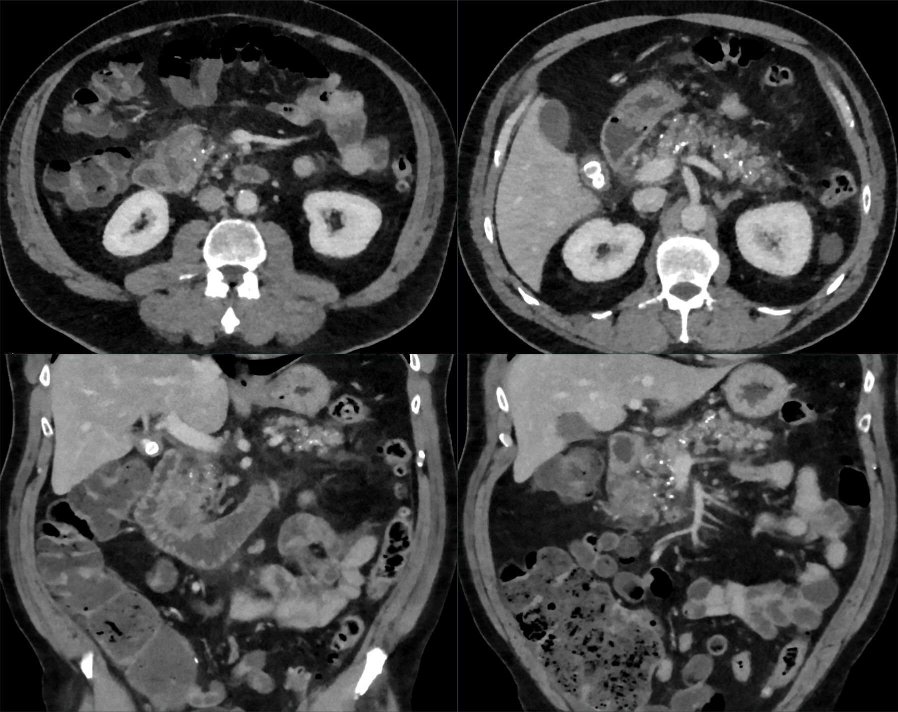
Imaging describes structure; function testing answers a different question. In suspected chronic pancreatitis, use pancreatic CT or MRI first. Established disease may show coarse calcifications, atrophy, duct dilation or irregularity, strictures, stones, and pseudocysts. Cross-sectional imaging also looks for masses and complications. A normal study does not settle every early or subtle presentation. [1]
Find the dense pancreatic calcifications anterior to the spine. The source describes chronic pancreatitis with superimposed acute groove inflammation, not an uncomplicated stable gland. This historical image demonstrates structural change, not EPI or a quantitative severity score. Image: Hellerhoff, 2024; original file; CC BY-SA 4.0. image, no added arrows or cropping.
MRI with MRCP maps ducts and fluid. If cross-sectional imaging leaves the diagnosis uncertain, EUS can identify subtler parenchymal or ductal changes, but minor nonspecific findings must not become a diagnosis by themselves. Secretin-enhanced MRCP is an additional option when suspicion stays high despite inconclusive cross-sectional imaging or EUS. ERCP is primarily therapeutic, not a routine diagnostic substitute for these studies. [1][5]
Consider two findings: a CT with calcifications and a formed-stool elastase of 65 micrograms/g in a patient with steatorrhea. Assign each to structure or function before continuing.
Which result directly supports digestive enzyme insufficiency?
The low formed-stool elastase, interpreted with steatorrhea, supports EPI. Calcifications establish structural injury in the appropriate setting but do not measure digestion. A structural diagnosis does not replace a functional assessment.
To transfer this distinction, consider a patient after pancreatic surgery who has weight loss and oily stools but no calcifications. EPI remains possible: chronic calcific pancreatitis is only one cause. Cystic fibrosis, pancreatic cancer, recurrent acute pancreatitis, and previous pancreatic surgery are high-risk settings; duodenal disease, including celiac disease, can also contribute. Do not require a calcified gland before assessing digestion. [2]
Is a low elastase value a pancreatic diagnosis?
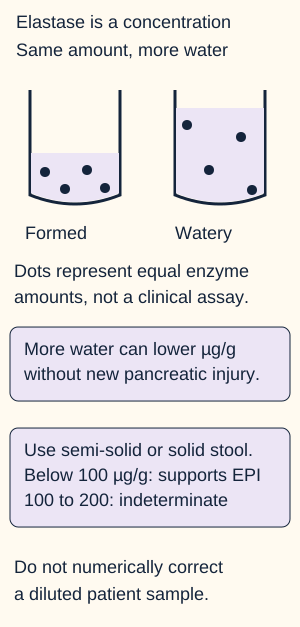
Fecal elastase is the preferred initial test for EPI, but it measures concentration in stool. Collect a semi-solid or solid specimen. Below 100 micrograms/g provides good evidence of EPI; 100 to 200 micrograms/g is indeterminate. A result above this interval is more reassuring in a low-risk person than in someone with strong evidence of maldigestion, because mild disease can be missed. [2][5]
Original concentration model. Extra water can lower measured elastase without new acinar destruction. The diagram does not provide a formula for correcting an individual result. Sources [2] and [5].
Put these results in order of diagnostic confidence: 65 micrograms/g in formed stool with weight loss and steatorrhea; 160 micrograms/g in formed stool with nonspecific bloating; 118 micrograms/g in watery stool with stable weight. Explain why the lowest number alone is not the full reasoning.
Which result supports EPI most strongly?
The 65 micrograms/g result combines a suitable specimen, a value below 100, and a compatible nutritional phenotype. The formed-stool value of 160 is indeterminate; it requires clinical reassessment, not automatic reassurance or lifelong treatment.
What should happen to the watery-stool result?
Obtain a suitable specimen and assess competing causes of watery diarrhea. Dilution can create a falsely low result. Normal CT cannot by itself exclude EPI, but this sample cannot reliably establish it either.
Elastase can be measured during pancreatic enzyme replacement therapy (PERT); the assay measures human elastase rather than the replacement enzymes. Do not stop a necessary treatment solely to collect it. Conversely, symptom improvement during an empiric enzyme trial is unreliable as a diagnostic test. Symptom fluctuation, dose, and timing can all affect that response. [2]
For a new patient with persuasive maldigestion but an equivocal result, reassess specimen quality, nutrition, structural disease, and nonpancreatic causes. Specialized direct function or breath testing may help where available. Quantitative fecal fat testing is rarely needed routinely and requires an appropriate high-fat intake during collection. Stool appearance alone cannot distinguish pancreatic disease from every intestinal cause. [2]
Make the enzymes meet the meal
Once EPI is diagnosed, replace the missing digestive activity. An adult initial regimen is at least 40,000 USP lipase units during each meal and about half that with snacks, then individualized to meal size, fat content, symptoms, and nutritional recovery. This is a starting framework, not a universal final dose or a pediatric prescription. Count lipase units, not merely capsules, because strengths differ. [2]
The enzymes must mix with food. With several capsules, distribute them through eating rather than taking the entire dose well after the meal. Current PERT preparations are porcine-derived. Non-enteric-coated preparations require acid suppression; for persistent symptoms on enteric-coated therapy, first check access, adherence, timing, and dose, then consider acid suppression and alternative diagnoses. [2][5]
Compare quantity with timing
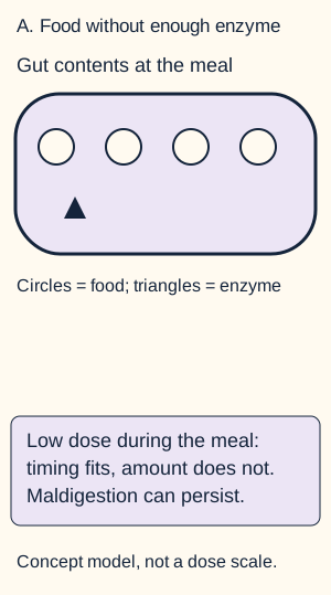
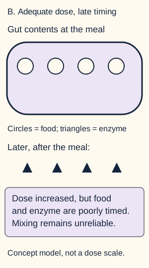
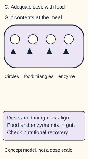
Start with the diagram below. Imagine changing only one feature at a time: more enzyme, then better timing. Predict whether each change guarantees adequate mixing. Open each optional answer to compare the resulting intestinal contents; close it to repeat the comparison. Nothing in the lesson depends on your answer.
Starting state: timing is appropriate, but the dose may be insufficient. The shapes are qualitative, not a numerical absorption model.What if the dose increases but is taken well after eating?
Changed state: increasing quantity alone does not ensure mixing with the meal.
The prescription looks stronger, but enzyme and food delivery are poorly aligned. Dose cannot substitute for timing.
What if the adequate dose is distributed during eating?
Changed state: enzyme and food delivery now coincide. The figure predicts better opportunity for digestion, not a guaranteed patient response.
Both quantity and timing now fit the task. Reassess symptoms and nutritional recovery rather than assuming that the prescription has finished treatment.
Worked comparison: a patient taking 10,000 lipase units per meal may remain undertreated even with correct timing. Increasing to an adequate adult starting dose does not solve taking it long after food. An adequate dose during eating addresses both errors. Enzymes do not repair fibrosis, and they should not be prescribed solely as reliable analgesia. [1][2]
Apply the same logic to a patient taking 40,000 units with each main meal but none with two daily snacks. Two 20,000-unit snacks add 40,000 units to the 120,000-unit meal total, for 160,000 units over that example day. This arithmetic illustrates coverage of eating occasions; it is not an individualized dose calculator.
Judge success by nutrition, not the stool alone
Use frequent smaller meals and adequate calories and protein, with a low-moderate fat diet rather than severe fat restriction. A fat-free diet may disguise steatorrhea while worsening energy intake. Involve a dietitian. Record weight or BMI, muscle status, symptoms, quality of life, and fat-soluble vitamin levels; provide appropriate vitamin supplementation and monitoring. Obtain baseline bone density and repeat DXA every 1 to 2 years in EPI. [2]
Follow reduction in steatorrhea, weight and muscle recovery, and vitamin improvement. Fecal elastase is not a target for titrating PERT. Persistent vitamin D depletion can raise parathyroid hormone, lower phosphate and impair bone mineralization even when serum calcium is preserved by compensation. Weakness or a fragility fracture therefore needs more than a reassuring stool history; biochemical findings suggest, but do not by themselves prove, osteomalacia. [9] If stool quality improves but weight still falls, check food intake, ongoing maldigestion, intestinal disease, small intestinal bacterial overgrowth, malignancy, and other complications. Do not keep escalating enzymes without reconsidering the problem. [5]
What still needs attention after digestion improves?
Pain, endocrine failure, and neighboring organs need their own plan. Begin pain care with exposure cessation, nutrition, and individualized nonopioid or selected adjunctive analgesia, while looking for an anatomical target. Pain without duct obstruction can involve inflammatory and neural mechanisms; drainage is not a universal pain treatment. [1][5]
For painful chronic pancreatitis with an obstructed main pancreatic duct, the 2024 ASGE guideline suggests surgical evaluation before endoscopic management in patients fit for surgery. This means multidisciplinary discussion of durability, anatomy, risks, and patient preference, not automatic total pancreatectomy. Endoscopy can be an initial approach for patients with contraindications to surgery or those preferring a less-invasive treatment when complete duct clearance is likely. Stone extraction, lithotripsy, stricture therapy, and selected pseudocyst drainage have specific roles. [3]
Compare persistent pain with a dilated duct containing an obstructing stone against isolated steatorrhea with no obstruction. Choose which problem has a potential drainage target.
Which patient needs a discussion of duct decompression?
The patient with painful duct obstruction. Enzyme replacement treats maldigestion in the second patient; it does not extract a stone in the first. Surgical fitness and likely duct clearance help select the route.
Endocrine failure changes the safety plan
Check fasting glucose and HbA1c at baseline and annually in chronic pancreatitis; impaired results warrant further evaluation, often with a 75-g oral glucose tolerance test. Diabetes secondary to pancreatic destruction is type 3c, but type 1 or type 2 diabetes can coexist. Low elastase alone does not classify diabetes. Advanced insulin deficiency often requires insulin. Impaired glucagon counterregulation, variable absorption, and inconsistent intake can make hypoglycemia difficult to prevent. Coordinate glucose therapy with reliable nutrition and PERT, and reassess when food absorption changes. [4]
Use anatomy when symptoms change
A pseudocyst or inflammatory narrowing can compress the stomach, duodenum, or bile duct, producing early satiety, vomiting, or jaundice.
Splenic vein thrombosis can redirect blood through short gastric collaterals, causing isolated gastric fundal varices even without cirrhosis. [7]
A pseudoaneurysm may cause major hemorrhage. Bleeding or hemodynamic instability requires urgent assessment and resuscitation; hemorrhagic pseudoaneurysms usually need angiographic embolization. A detected pseudoaneurysm also needs prompt specialist treatment assessment when it is not actively bleeding. Exclude vascular hazards before draining a nearby collection. [5]
Fever, rigors or hemodynamic deterioration with biliary obstruction suggests cholangitis: resuscitation, antibiotics and prompt biliary drainage take priority, including before starting immunosuppression for autoimmune disease. [10]
New jaundice, progressive weight loss, a new focal mass, or a changed pain pattern requires reassessment for obstruction, complications, and cancer rather than attribution to the old diagnosis. Chronic pancreatitis increases cancer risk, but this does not justify inventing a single surveillance schedule for every patient. Hereditary risk discussions belong in specialist care. [1][5]
If stools improve but recurrent hypoglycemia appears, which assessment cannot wait?
Review glucose treatment, intake, and absorption together. Loss of counterregulatory glucagon and insulin deficiency can coexist. Better digestion does not guarantee stable glucose; it changes the nutrient delivery that the diabetes regimen must accommodate.
Pause here and summarize three separate treatment targets: replace enzyme activity, address painful obstruction or neural pain, and manage endocrine and nutritional consequences. In a new presentation, ask which target explains the current change before repeating the previous treatment.
Independent practice
Case 1
Show answer and explanations for case 1
A. Use interval pancreatic imaging to determine whether the sibling inherited the familial risk (Why this does not fit)
Imaging can identify structural disease but cannot establish carrier status in an asymptomatic person; an inherited variant may be present before any phenotype.
Reasoning steps for option A
What could interval scans reveal in the asymptomatic sibling?
Scans could reveal structural pancreatic changes if disease develops; they do not identify a PRSS1 sequence variant.
Why would normal imaging fail to settle inherited risk in this family?
The mother carries the pathogenic variant without pancreatitis at age 60, showing that a carrier can lack an imaging phenotype.
B. Defer targeted testing because the unaffected mother makes the reported variant clinically uninformative (Why this does not fit)
Lack of disease in one carrier does not negate a clinically established pathogenic variant when penetrance is incomplete.
Reasoning steps for option B
How does the unaffected mother change the interpretation of this PRSS1 pedigree?
An unaffected carrier alongside affected carriers demonstrates incomplete penetrance, not absence of pathogenicity.
Does her lack of attacks justify withholding targeted testing from the sibling?
No. The familial variant is clinically confirmed as pathogenic, so counseled testing remains informative about inheritance despite variable disease expression.
C. Offer counseled targeted testing for susceptibility, not certainty of future pancreatitis (Best answer)
The unaffected carrier demonstrates incomplete penetrance. Targeted testing can identify the established familial variant, but genotype does not by itself establish current disease or guarantee future pancreatitis.
Reasoning steps for option C
What risk distinction should counseling draw from the affected grandfather and unaffected mother?
Both carry the same pathogenic variant, but only one developed pancreatitis; inheriting it does not make future disease certain.
What would a positive targeted result establish for this asymptomatic adult?
It would establish carriage of the familial PRSS1 variant and inherited susceptibility, not a diagnosis of current pancreatitis.
D. Offer targeted testing and describe a positive result as a diagnosis of current hereditary pancreatitis (Why this does not fit)
A pathogenic variant establishes genetic susceptibility, not current structural disease in an asymptomatic relative. Disease expression requires separate clinical assessment.
Reasoning steps for option D
Which part of offering targeted PRSS1 testing is appropriate for this sibling?
Testing for the confirmed familial variant can determine whether the sibling inherited that susceptibility.
Why would calling a positive result current hereditary pancreatitis overstate the evidence?
The sibling has no clinical phenotype, and the unaffected carrier mother shows that genotype and expressed pancreatic disease are not equivalent.
Takeaway: Distinguish incomplete penetrance, genetic susceptibility and an established clinical phenotype.
A. Begin meal-timed pancreatic enzymes and follow the clinical response (Why this does not fit)
Remote pancreatitis raises EPI risk, but elastase 125 in liquid stool may reflect dilution and magnesium supplies a competing osmotic cause. Response to enzymes would not reliably establish the diagnosis.
Reasoning steps for option A
Which findings might lead to an empiric enzyme prescription in this patient?
Remote recurrent pancreatitis raises EPI risk, and an elastase concentration of 125 micrograms/g is below the reassuring range.
Why would symptom response to enzymes not resolve this liquid-stool result?
The specimen may be diluted and magnesium preceded the watery diarrhea; enzyme response is not a reliable diagnostic test for EPI.
B. Review magnesium use and repeat elastase on semi-solid or formed stool (Best answer)
The watery specimen makes the borderline-low value unreliable; the temporal magnesium exposure deserves assessment without dismissing pancreatic risk.
Reasoning steps for option B
How should elastase 125 micrograms/g be interpreted when the specimen is liquid?
Even on suitable stool, 100 to 200 is indeterminate. Added stool water can lower the measured concentration further, so this sample does not establish EPI.
What should be reassessed before attributing the new watery stools to the pancreas?
Review the newly started magnesium citrate and repeat elastase using semi-solid or formed stool, preserving evaluation of the prior pancreatic risk.
C. Repeat elastase in another liquid specimen to assess reproducibility (Why this does not fit)
Repeated diluted specimens do not correct the preanalytic problem.
Reasoning steps for option C
Would a second liquid specimen test away the cause of a falsely low elastase?
No. Extra water can dilute elastase in both specimens, so reproducibility does not establish a valid concentration.
Which collection change would make repeat elastase more informative than another liquid sample?
Use semi-solid or formed stool rather than repeating the same dilution-prone collection condition.
D. Obtain pancreas-protocol CT to look for progression of chronic injury (Why this does not fit)
CT can assess structure, but it does not resolve whether this indeterminate concentration represents dilution or current exocrine dysfunction.
Reasoning steps for option D
What question could pancreas-protocol CT answer after remote recurrent pancreatitis?
It could assess structural progression, such as new pancreatic calcification, atrophy or ductal change.
Why is structural imaging not the immediate solution to elastase 125 in watery stool?
CT cannot determine whether that concentration is diluted or demonstrates digestive failure; medication review and a suitable specimen address the actual uncertainty.
Takeaway: Specimen quality and competing diarrhea etiologies govern interpretation; retest on suitable stool rather than diagnose EPI from a diluted result.
A. Two capsules during dinner and one capsule with each other meal and snack (Why this does not fit)
This covers food occasions but leaves breakfast and lunch at 20,000 units, below the usual adult starting meal dose in a patient losing weight.
Reasoning steps for option A
How many lipase units would breakfast and lunch receive under this dinner-heavy schedule?
One 20,000-unit capsule at each of those meals supplies 20,000 units, while two capsules at dinner supply 40,000.
Why does covering every eating occasion still leave this patient undertreated?
Breakfast and lunch remain below the usual adult starting meal dose of at least 40,000 units despite steatorrhea and a 4-kg weight loss.
B. One capsule during each meal and snack with an added proton-pump inhibitor (Why this does not fit)
Meal coverage improves, but the regimen remains below the usual adult meal starting dose. Acid suppression should not substitute for correcting the demonstrated dosing deficit.
Reasoning steps for option B
What lipase quantity does one capsule supply at each main meal, even with a proton-pump inhibitor?
The capsule still supplies 20,000 units, below the usual adult starting meal amount.
Why should acid suppression not replace a meal-dose correction in this enteric-coated regimen?
The demonstrated defects include too little lipase, late dosing and missed meals. A proton-pump inhibitor does not supply the missing meal units.
C. Two capsules 45 minutes after each main meal, with dietary fat reduction for snack symptoms (Why this does not fit)
The meal quantity improves, but late administration and absent snack coverage still limit enzyme-food mixing. Excessive fat restriction may worsen energy intake.
Reasoning steps for option C
Which dosing defect is corrected by two capsules after each main meal?
Two capsules supply 40,000 lipase units per main meal, correcting the meal quantity compared with a single capsule.
Which two delivery defects remain with dosing 45 minutes late and restricting snack fat?
Enzymes still arrive after the food, and the afternoon snack has no enzyme coverage. Fat restriction does not correct either mixing problem.
D. Two capsules during each main meal and one during the snack (Best answer)
This provides an adult starting dose of 40,000 units per meal and roughly half with snacks while aligning enzymes with food; subsequent titration depends on response.
Reasoning steps for option D
How do two meal capsules and one snack capsule translate into lipase units?
At 20,000 units per capsule, two provide 40,000 with a main meal and one provides 20,000 with the snack.
How does dosing during every meal and snack correct the original dinner-only schedule?
It aligns enzyme delivery with food and covers breakfast, lunch and the snack as well as dinner; stool, weight and nutrition then guide titration.
Takeaway: Interpret lipase units per eating occasion and timing together; reserve acid strategies and alternative-cause workup for persistent symptoms after an adequate regimen.
A. Pancreatic surgical evaluation for duct drainage before choosing an intervention (Best answer)
A fit patient with a dilated obstructed duct warrants surgical evaluation before selecting endoscopy; difficult clearance makes comparative durability important.
Reasoning steps for option A
What does the 12-mm impacted stone explain in a 9-mm upstream duct?
The stone supplies a mechanical obstruction associated with duct dilation and pain despite optimized medical treatment.
Why seek surgical evaluation before committing this fit patient to staged ERCP?
Likely repeated endoscopic sessions make durability and procedural burden important; surgical evaluation allows comparison before selecting an intervention.
B. Staged lithotripsy and ERCP as the initial intervention (Why this does not fit)
This can be reasonable if surgery is contraindicated or a less-invasive route is preferred, but likely repeated sessions and surgical fitness warrant a comparative surgical assessment first.
Reasoning steps for option B
How could staged lithotripsy and ERCP address this impacted head stone?
Fragmentation and extraction could clear the obstruction and decompress the upstream pancreatic duct.
Which patient and procedural features argue for surgical consultation before that endoscopic plan?
He is fit for surgery and clearance is expected to require repeated procedures. These favor comparing surgical drainage with endoscopy rather than choosing endoscopy first.
C. EUS-guided celiac plexus intervention as the initial intervention (Why this does not fit)
Celiac plexus intervention may palliate selected pain, but a fit patient with a dilated obstructed duct has a structural target that warrants evaluation for durable decompression.
Reasoning steps for option C
Which component of refractory pancreatic pain could a celiac plexus intervention target?
It may palliate the neural component of pain by acting on its nerve pathway.
What cause of pain would a celiac plexus intervention leave in place here?
The impacted stone would still obstruct the dilated duct; procedural assessment should address that drainable structural target.
D. Further titration of neuropathic-pain medication before procedural referral (Why this does not fit)
Adjunctive medication can help neural pain, but persistent disability after optimized medical therapy and a major duct obstruction favor procedural assessment.
Reasoning steps for option D
What could further neuropathic medication titration address despite alcohol abstinence?
It could reduce a neural pain component that persists after exposure cessation.
Why should medication titration not postpone procedural referral in this case?
Disabling pain persists despite optimized medical therapy, and the large impacted stone with upstream dilation provides an anatomical target for decompression.
Takeaway: Fitness, duct dilation and predicted clearance distinguish surgery-first evaluation from a possible endoscopic route; neither procedure restores lost acini.
A. Beta-cell insulin deficiency with preserved alpha-cell counterregulation (Why this does not fit)
Beta-cell loss fits the low C-peptide, but preserved glucagon cannot be assumed with destructive pancreatic disease and would omit an important contributor to recurrent hypoglycemia.
Reasoning steps for option A
What does low fasting C-peptide support about beta-cell function in this patient?
Endogenous insulin secretion is deficient, consistent with islet injury in longstanding calcific pancreatic disease.
Why is preserved glucagon rescue an unsafe assumption after destructive pancreatic disease?
Alpha-cell injury can accompany beta-cell loss, weakening counterregulation during skipped dinners. The episodes alone do not prove alpha-cell failure, but the risk should be anticipated.
B. Reduced renal insulin clearance with preserved alpha-cell counterregulation (Why this does not fit)
Normal renal function does not explain low C-peptide or the pancreatic phenotype.
Reasoning steps for option B
How could reduced renal clearance cause lows in someone receiving basal insulin?
If kidney function declined, administered insulin could persist longer and contribute to hypoglycemia.
Why does renal clearance not explain the proposed endocrine pattern with an estimated GFR of 96?
Renal function is normal, and reduced clearance does not account for low endogenous C-peptide in destructive pancreatic disease.
C. Predominant peripheral insulin resistance with compensatory hyperinsulinemia (Why this does not fit)
The low C-peptide and destructive pancreatic phenotype favor deficient endogenous secretion rather than compensatory hyperinsulinemia.
Reasoning steps for option C
What endogenous secretory response would compensatory hyperinsulinemia imply?
It would imply increased endogenous insulin output and accompanying C-peptide rather than the low fasting C-peptide described.
How do calcific disease, steatorrhea and weight loss weigh against predominantly compensated insulin resistance?
Together with low C-peptide, they favor pancreatic tissue destruction with insulin deficiency rather than predominant resistance with increased secretion.
D. Beta-cell insulin deficiency with impaired alpha-cell counterregulation (Best answer)
Acinar and islet destruction can coexist; insulin is needed for deficiency, while lost glucagon counterregulation plus skipped intake increases hypoglycemia risk.
Reasoning steps for option D
How do low C-peptide and progressive weight loss identify one endocrine treatment need?
They support deficient endogenous insulin secretion in destructive pancreatic disease, which can require insulin replacement.
What additional islet impairment makes unchanged basal insulin hazardous after a missed dinner?
Loss of alpha-cell glucagon counterregulation can weaken rescue from falling glucose. Combined with absent food intake and administered insulin, this increases hypoglycemia risk.
Takeaway: Destructive pancreatic disease with insulin deficiency also creates a risk of weak glucagon counterregulation; missed-meal hypoglycemia alone does not prove alpha-cell failure.
A. Treat established advanced chronic pancreatitis and use enzyme response to confirm the structural diagnosis (Why this does not fit)
EPI warrants treatment, but neither a low elastase nor response to replacement proves advanced structural chronic pancreatitis.
Reasoning steps for option A
What does formed-stool elastase 74 establish when oily stool and weight loss coexist?
A value below 100 micrograms/g in a suitable specimen with maldigestion supports an exocrine pancreatic deficit.
Why can neither that result nor improvement on PERT confirm advanced structural pancreatitis?
Elastase assesses function, and replacement response does not diagnose pancreatic structure. CT and MRCP have not shown definite chronic changes.
B. Defer enzymes until EUS establishes chronic pancreatitis and focus meanwhile on nonpancreatic diarrhea causes (Why this does not fit)
Further structural assessment is reasonable, but the valid functional result and maldigestion already support treatment; an EUS label is not a prerequisite.
Reasoning steps for option B
What unresolved question could EUS investigate after negative CT and MRCP?
It could look for subtler structural pancreatic abnormalities when the recurrent familial history sustains suspicion.
Why should supported EPI treatment not wait for an EUS diagnosis?
Formed-stool elastase 74 with oily stool and weight loss already supports digestive insufficiency; an additional structural label is not needed before replacing enzymes.
C. Start PERT for EPI and continue specialist assessment of the pancreatic structural diagnosis (Best answer)
Symptoms and a valid markedly low elastase support EPI now. Negative cross-sectional imaging leaves the cause and stage of structural injury unresolved rather than invalidating the functional deficit.
Reasoning steps for option C
Which evidence justifies treating digestion despite the unrevealing scans?
Steatorrhea, weight loss and elastase 74 in formed stool jointly support EPI, independent of whether advanced structural changes are visible.
What should continued specialist assessment resolve while PERT addresses the functional deficit?
The early recurrent attacks and affected relative warrant etiologic and structural assessment; the cause and stage of pancreatic injury remain unresolved.
D. Use diagnostic ERCP to identify an obstructed duct before deciding whether enzyme replacement is needed (Why this does not fit)
The established question is not a demonstrated therapeutic duct target. ERCP carries risk, while current EPI treatment need not wait for duct instrumentation.
Reasoning steps for option D
What would justify ERCP as a therapeutic procedure rather than simply another diagnostic test?
A demonstrated ductal target such as an obstruction needing intervention would provide a therapeutic purpose.
Why does the current CT/MRCP and elastase combination not justify diagnostic ERCP before PERT?
No therapeutic obstruction is identified, while a valid low elastase and maldigestion already support treatment. Duct instrumentation adds risk without being required for that decision.
Takeaway: Supported exocrine failure can be treated while the underlying structural diagnosis remains under evaluation.
A. Begin glucocorticoids now and reserve drainage for bilirubin that remains high after treatment (Why this does not fit)
Steroids treat autoimmune inflammation but do not replace prompt drainage of an infected obstructed biliary system.
Reasoning steps for option A
Which established findings would ordinarily support glucocorticoid treatment?
The core shows storiform fibrosis with IgG4-rich inflammation, salivary involvement is present, and the team has resolved the malignancy concern.
Why should a steroid trial not precede drainage once fever, rigors and hypotension develop?
The new systemic illness with worsening biliary obstruction suggests cholangitis. Steroids do not provide mechanical source control for an infected obstructed duct.
B. Perform prompt biliary drainage, then begin disease-directed glucocorticoids after infection control (Best answer)
The prior evidence supports type 1 autoimmune disease, but the new systemic illness with obstructed ducts suggests superimposed cholangitis. Source control takes priority over beginning immunosuppression.
Reasoning steps for option B
How does the new febrile illness change the significance of the bile-duct narrowing?
The narrowing is now accompanied by rigors, hypotension and worsening dilation, indicating superimposed obstructed infection rather than autoimmune inflammation alone.
What treatment sequence follows after fluids and antibiotics have been started?
Proceed with prompt therapeutic biliary drainage, then start disease-directed glucocorticoids after infection control rather than delaying source control for immunosuppression.
C. Continue antibiotics until cultures clear, then begin glucocorticoids before reassessing the need for drainage (Why this does not fit)
Antibiotics are important, but postponing source control in obstructed cholangitis risks persistent infection despite transient hemodynamic improvement.
Reasoning steps for option C
Why are antibiotics necessary but not sufficient with persistent biliary obstruction?
They treat the bacterial component, but an obstructed infected biliary system still requires drainage for source control.
Does a fluid-responsive blood pressure justify waiting for cultures to clear before drainage?
No. Hemodynamic improvement after fluids does not remove the obstruction; delaying available ERCP can leave the source of infection untreated.
D. Begin glucocorticoids together with antibiotics and schedule drainage after an initial inflammatory response (Why this does not fit)
The autoimmune diagnosis remains relevant, but an initial steroid-response strategy delays the mechanical source control required by the new infection.
Reasoning steps for option D
What two diseases does simultaneous steroid and antibiotic treatment attempt to address?
It attempts to treat the supported autoimmune process and the new bacterial biliary infection at the same time.
Why is scheduling drainage after a steroid response the wrong sequence here?
An inflammatory response cannot be relied on to control the infected mechanical obstruction. Prompt drainage takes priority before initiating disease-directed immunosuppression.
Takeaway: A supported autoimmune diagnosis does not supersede urgent treatment of an infected obstructed bile duct.
A. Contrast assessment of splenic venous drainage and short gastric collaterals (Best answer)
The fundal-only collateral pattern with preserved hepatic/portal findings localizes likely obstruction to splenic outflow near the inflamed tail; imaging can confirm patency and anatomy.
Reasoning steps for option A
How do fundal varices without esophageal varices help localize the venous problem?
With preserved liver function and a patent portal trunk, this distribution favors a regional outflow problem rather than generalized portal hypertension.
Why should contrast imaging follow splenic outflow and short gastric collaterals near the tail collection?
The inflamed pancreatic tail and hilar pseudocyst can compromise splenic venous drainage, redirecting flow through fundal collaterals. Dedicated imaging is needed to confirm the anatomy.
B. Assessment for diffuse hepatic sinusoidal disease and generalized portal hypertension (Why this does not fit)
Normal hepatic morphology/function and absent esophageal varices weaken generalized sinusoidal hypertension.
Reasoning steps for option B
How could diffuse hepatic sinusoidal disease produce gastric varices?
Generalized portal hypertension from hepatic resistance can drive portosystemic collateral formation, including varices.
Which supplied findings make that diffuse hepatic explanation less persuasive?
Normal liver contour and synthetic function, absent esophageal varices and an adjacent pancreatic-tail process favor a regional splenic outflow problem.
C. Repeat contrast assessment of the main portal vein for central obstruction (Why this does not fit)
The main portal vein is documented patent; a regional outflow problem better explains the isolated fundal collateral pattern.
Reasoning steps for option C
What venous mechanism would a central portal obstruction offer for varices?
Obstruction of the main portal vein could elevate upstream venous pressure and recruit collateral pathways.
Why would repeating the documented portal assessment miss the more likely localization?
The main portal vein is already patent, whereas splenic outflow beside the tail pseudocyst has not been assessed and better fits fundal-only varices.
D. Contrast assessment of superior mesenteric venous drainage and mesenteric collaterals (Why this does not fit)
Superior mesenteric outflow does not match the fundal short-gastric collateral distribution and the adjacent pancreatic-tail process as well as splenic outflow does.
Reasoning steps for option D
What complication could assessment of superior mesenteric drainage detect after regional inflammation?
It could detect mesenteric venous obstruction and associated collateral formation.
Why do the fundal distribution and splenic-hilar collection instead direct attention to splenic drainage?
Fundal short gastric collaterals align with compromised splenic outflow near the pancreatic tail, not primarily with the superior mesenteric venous territory.
Takeaway: Isolated fundal varices with a normal liver and patent portal trunk suggest segmental splenic venous obstruction; map vessels before an intervention.
A. Repeat formed-stool elastase to determine whether endogenous secretion has recovered (Why this does not fit)
Elastase measures secretion, not response to replacement, and cannot explain the changed pain pattern.
Reasoning steps for option A
What would repeat fecal elastase measure despite the ongoing 50,000-unit meal regimen?
It would measure endogenous pancreatic secretion, not the digestive activity supplied by replacement enzymes.
Why would that secretion result not explain the new 6-kg loss and persistent back-radiating pain?
It cannot identify a new mass or structural complication. The changed pain and decline despite verified intake and PERT require structural reassessment.
B. Quantitative fecal fat collection to measure residual fat malabsorption (Why this does not fit)
Residual malabsorption is possible, but formed stools and adequate enzyme coverage plus a changed pain pattern make structural red flags more urgent.
Reasoning steps for option B
What residual problem could a quantitative fecal fat collection identify?
It could measure persistent fat malabsorption even when the patient reports less stool oil.
Why does the changed pain pattern place imaging ahead of fecal fat quantification?
Persistent back-radiating pain with ongoing weight loss despite adequate intake and enzyme coverage raises concern for a pancreatic mass or complication, not just residual fat loss.
C. Contrast-enhanced pancreas-protocol CT to assess for occult masses and complications (Best answer)
Persistent weight loss and a changed pain pattern despite documented intake and PERT warrant structural reassessment, including malignancy; continue nutritional care.
Reasoning steps for option C
Why do documented calories and enzyme timing make the continuing weight loss concerning?
They weaken inadequate intake and missed replacement as sufficient explanations for the 6-kg decline despite improved stools.
What should contrast-enhanced pancreas-protocol CT assess after intermittent pain becomes persistent?
It should evaluate for a new pancreatic mass or structural complication that could explain the changed pain and nutritional decline.
D. Breath testing for small-intestinal bacterial overgrowth as the initial investigation (Why this does not fit)
Overgrowth can coexist with EPI, but it should not displace assessment of new systemic red flags.
Reasoning steps for option D
How could bacterial overgrowth contribute to weight loss in a patient with EPI?
Coexisting intestinal overgrowth can impair nutrition and cause gastrointestinal symptoms despite replacement therapy.
Why should breath testing not precede evaluation of this new persistent pancreatic-type pain?
The new pain pattern and substantial ongoing weight loss require assessment for malignancy or another structural complication before prioritizing an intestinal explanation.
Takeaway: Persistent weight loss with a changed pain pattern warrants structural reassessment even when fat-digestion symptoms improve.
A. Suspected primary hyperparathyroidism; pursue parathyroid localization before treating the severe vitamin deficiency (Why this does not fit)
Primary hyperparathyroidism can lower phosphate, but severe vitamin D depletion provides a strong secondary explanation for PTH with normal calcium. Correct secondary contributors before making that diagnosis or pursuing localization.
Reasoning steps for option A
Which two mineral results might initially suggest primary parathyroid overactivity?
PTH is elevated at 135 pg/mL and phosphate is low at 2.0 mg/dL, a combination that can occur with parathyroid excess.
Why should vitamin D 8 ng/mL be addressed before parathyroid localization?
Severe vitamin D depletion supplies a secondary stimulus for PTH despite calcium of 8.9 mg/dL. Primary disease should not be inferred before correcting secondary contributors.
B. Suspected age-related osteoporosis alone; prioritize antiresorptive therapy rather than correction of the mineral abnormalities (Why this does not fit)
Osteoporosis can cause fragility fractures but does not explain the vitamin D, phosphate and PTH pattern. Mineral delivery and possible impaired mineralization need assessment before treating this as uncomplicated osteoporosis.
Reasoning steps for option B
What feature of the presentation is compatible with osteoporosis?
A low-trauma fracture can occur in osteoporosis and signals reduced skeletal strength.
Which biochemical findings prevent osteoporosis alone from explaining this fracture?
Severe vitamin D depletion, low phosphate and elevated PTH with alkaline phosphatase indicate a mineral-metabolism problem that requires correction and assessment for impaired mineralization.
C. Suspected renal mineral disease; prioritize active vitamin D therapy for the presumed underlying kidney dysfunction (Why this does not fit)
Renal disease can alter PTH and mineral metabolism, but normal renal function and the marked nutritional vitamin deficiency favor a malabsorptive secondary process.
Reasoning steps for option C
What mechanism makes renal disease a consideration when PTH and bone abnormalities coexist?
Kidney dysfunction can disturb vitamin D and mineral metabolism, producing secondary hyperparathyroidism and skeletal disease.
Which results favor malabsorptive vitamin D depletion rather than a renal-directed treatment pathway?
Creatinine is normal, while 25-hydroxyvitamin D is markedly low in a patient with EPI. These findings favor a nutritional secondary process over renal dysfunction.
D. Suspected vitamin D-related mineralization failure with secondary PTH; restore vitamins and calcium and assess bone (Best answer)
Very low vitamin D, low phosphate and raised PTH fit secondary hyperparathyroidism that can preserve serum calcium. Normal calcium and less steatorrhea do not establish mineral or skeletal recovery; the pattern warrants assessment for impaired mineralization rather than claiming biopsy-proven osteomalacia.
Reasoning steps for option D
How can calcium remain 8.9 mg/dL when vitamin D is 8 ng/mL and phosphate is low?
Compensatory PTH can preserve serum calcium while promoting phosphate loss, so normal calcium does not demonstrate adequate vitamin delivery.
What do weakness, fracture and this mineral pattern warrant beyond stool improvement?
Restore vitamin and calcium delivery and assess skeletal disease, including suspected impaired mineralization. These findings raise concern but do not establish histologically proven osteomalacia.
Takeaway: Normal serum calcium can reflect compensation rather than adequate vitamin D delivery or normal mineralization.
A. Use a monitored glucocorticoid trial to test the IgG4-supported inflammatory diagnosis (Why this does not fit)
Steroid responsiveness is not an adequate substitute for malignancy assessment when tissue is nondiagnostic and imaging is increasingly concerning.
Reasoning steps for option A
What could a modest IgG4 rise and pancreatic head lesion suggest besides cancer?
Autoimmune pancreatitis can produce a mass-like lesion, obstructive jaundice and an IgG4 increase.
Why is a steroid trial insufficient with this enlarging lesion and scant aspirate?
IgG4 of 180 mg/dL is only modestly elevated, no other organ features are present, and the inadequate sample does not exclude malignancy behind the abrupt cutoff.
B. Repeat EUS-guided core acquisition with multidisciplinary malignancy assessment (Best answer)
Scant benign aspirate does not reliably exclude cancer in a high-risk evolving lesion. A modest IgG4 increase is insufficient to establish autoimmune disease; obtain better tissue and integrate it with the anatomical risk.
Reasoning steps for option B
How much cancer reassurance comes from scant benign inflammatory cells without a core?
The sample is nondiagnostic for excluding malignancy in an enlarging focal obstructive lesion; its benign cells do not establish adequate sampling.
What should be done when weak autoimmune evidence conflicts with progressive focal obstruction?
Obtain a better EUS-guided core and multidisciplinary malignancy assessment rather than allowing a modest IgG4 elevation to settle the cause.
C. Repeat MRI at a routine interval using the negative aspirate as reassurance (Why this does not fit)
The specimen was scant, not an adequate exclusion of malignancy. Waiting risks delaying diagnosis of a progressive focal obstructive lesion.
Reasoning steps for option C
What would be needed for a benign aspirate to justify a routine imaging interval?
The tissue assessment and clinical context would need to provide meaningful reassurance rather than an inadequate sample from a progressing lesion.
Which changes make routine MRI follow-up inadequate in this patient?
The head lesion is enlarging, the bile duct now has an abrupt cutoff and jaundice is progressive, so scant benign cells should not defer renewed tissue assessment.
D. Use the bilirubin response to biliary stenting to distinguish benign from malignant narrowing (Why this does not fit)
Both benign and malignant obstruction can improve after drainage. Biochemical response does not establish the underlying cause.
Reasoning steps for option D
What does a fall in bilirubin after biliary stenting demonstrate?
It demonstrates improved bile drainage across the obstruction, not the biological cause of that obstruction.
Why would that response fail to distinguish inflammation from cancer in the head lesion?
Both benign and malignant narrowings can drain better after stenting; tissue and multidisciplinary anatomical assessment are still required.
Takeaway: A weak negative sample and modest IgG4 increase cannot erase a high-risk evolving focal lesion.
A. A structured tobacco-cessation intervention (Best answer)
Tobacco independently promotes pancreatic disease and progression even after alcohol cessation. Addressing this ongoing exposure targets a modifiable driver.
Reasoning steps for option A
Which injurious exposure persists after 14 months without alcohol?
Smoking a pack daily continues, and tobacco is an independent risk for pancreatic disease progression.
Why does structured tobacco cessation address the MRCP progression more directly than symptom treatment?
It removes an ongoing modifiable injury driver, whereas analgesic adjuncts, enzyme replacement and fat restriction do not eliminate that exposure.
B. A daily antioxidant supplement regimen (Why this does not fit)
Antioxidants may be considered for selected pain symptoms, with limited benefit; they are not a substitute for cessation of an established progression exposure.
Reasoning steps for option B
What pancreatic symptom might a daily antioxidant regimen be intended to address?
Antioxidants are sometimes considered for selected pancreatic pain, with limited benefit.
Why is an antioxidant regimen not the clearest answer to ongoing duct injury here?
Pain is already tolerable, but daily tobacco exposure continues. Symptom-directed supplementation does not replace cessation of that progression risk.
C. Pancreatic enzyme treatment with all meals (Why this does not fit)
PERT treats digestive insufficiency when present. It is not established as prevention of tobacco-associated pancreatic injury in a patient without EPI evidence.
Reasoning steps for option C
Which findings would make meal-time pancreatic enzymes directly relevant?
Evidence of digestive insufficiency, such as steatorrhea or nutritional deficiencies, would support PERT for maldigestion.
Why does PERT not target this smoker's current prevention problem?
There is no steatorrhea or vitamin deficiency, and replacement enzymes are not established prevention for tobacco-associated pancreatic injury.
D. A more restrictive dietary-fat target (Why this does not fit)
Individualized nutrition is important, but aggressive fat restriction does not eliminate tobacco exposure and may compromise calorie intake.
Reasoning steps for option D
What could a more restrictive fat target change about this patient's meals?
It could alter dietary tolerance and calorie intake, but it would not remove the continued tobacco exposure.
Why is added fat restriction poorly matched to progressive ducts without maldigestion?
There is no current EPI phenotype to explain through diet, while restriction can compromise nutrition and leaves the independent smoking risk untreated.
Takeaway: Treat pain separately from tobacco-related progression prevention
A. Increase the three main-meal enzyme doses while keeping the supplement schedule unchanged (Why this does not fit)
Titration can help some incomplete responses, but raising enzymes at already covered meals does not cover the separate nutrient load from drinks.
Reasoning steps for option A
What nutrient intake is already covered by the three 50,000-unit doses?
The three main meals receive enzymes during eating, and the reduced stool oil suggests at least partial digestive benefit.
Why would increasing those doses leave nearly half the daily calories uncovered?
The two fat-containing nutrition drinks are consumed separately between meals without PERT; a larger meal dose does not supply enzymes during those later drinks.
B. Add a proton-pump inhibitor while keeping enzyme doses confined to main meals (Why this does not fit)
Acid strategies may help selected delivery problems, but cannot supply enzyme during the substantial uncovered intake described here.
Reasoning steps for option B
What delivery issue could a proton-pump inhibitor help in some incomplete PERT responses?
Acid suppression can improve enzyme activity in selected settings where acid exposure limits replacement effectiveness.
Why cannot acid suppression correct the specific gap in this food diary?
No enzyme is taken with the fat-containing drinks that supply nearly half the calories. Acid protection cannot replace an absent dose at those occasions.
C. Treat suspected bacterial overgrowth while continuing the current meal-only regimen (Why this does not fit)
Altered anatomy makes overgrowth possible, but the documented gap in nutrient coverage is a direct correctable explanation that should be addressed first.
Reasoning steps for option C
What aspect of pancreaticoduodenectomy makes bacterial overgrowth a plausible competitor?
Altered postoperative anatomy can predispose to overgrowth and persistent nutritional symptoms.
Which documented problem should be corrected before treating overgrowth as the explanation?
The substantial fat-containing supplement intake has no enzyme coverage, a direct correctable mismatch despite adequate main-meal dosing.
D. Add PERT during both nutrition drinks while continuing the meal regimen (Best answer)
Meal-related improvement suggests partial treatment success, while a large portion of caloric intake remains uncovered. Supplements containing nutrients need enzyme coverage rather than being treated as enzyme-free occasions.
Reasoning steps for option D
Why can improved stool oil coexist with weight loss despite adequate recorded calories?
Only main meals receive PERT, while nearly half the calories come from separate fat-containing drinks consumed without digestive enzyme replacement.
How should PERT be aligned with those two supplement occasions?
Provide enzyme coverage during both drinks as well as during the existing meals, then reassess weight and other nutritional outcomes.
Takeaway: Count caloric supplements as eating occasions when assessing whether PERT actually covers nutrient intake.
A. Arrange genetic counseling and a hereditary pancreatitis panel because attacks occurred before age 40 (Why this does not fit)
Young onset can justify genetic assessment if episodes remain unexplained, but a repeated severe modifiable metabolic exposure is already present.
Reasoning steps for option A
Why might recurrent pancreatitis at age 34 prompt a hereditary evaluation?
Young recurrent attacks can reflect inherited susceptibility, especially when a cause remains unexplained.
What established abnormality takes priority over an age-based genetic panel in this case?
Fasting triglycerides remain 1,180 mg/dL on two post-recovery draws, and HbA1c is 10.2%; these identify a severe modifiable metabolic risk and a contributor.
B. Coordinate glycemic control with dietary and pharmacologic triglyceride management (Best answer)
Repeated severe fasting hypertriglyceridemia is a plausible trigger; poorly controlled diabetes can aggravate it, so treat both rather than only the lipid number.
Reasoning steps for option B
What does triglyceride elevation on two fasting draws six weeks after recovery imply?
A persistent severe exposure above 1,000 mg/dL is present outside the acute attack, making hypertriglyceridemia a plausible recurrent trigger.
How does HbA1c 10.2% change the prevention plan beyond lowering the lipid number?
Poor glycemic control can aggravate hypertriglyceridemia. Coordinate diabetes treatment with dietary and pharmacologic triglyceride management for sustained prevention.
C. Pursue EUS for microlithiasis and plan cholecystectomy if biliary sludge is found (Why this does not fit)
Occult biliary disease can cause attacks, but negative ultrasound and repeated severe triglycerides plus uncontrolled diabetes favor metabolic intervention first.
Reasoning steps for option C
Why could EUS still find a biliary cause after a negative ultrasound?
Microlithiasis or sludge can be missed on routine ultrasonography, so negative ultrasound alone does not exclude a biliary trigger.
Why should that possibility not displace treatment of the documented metabolic driver?
Repeated severe fasting triglycerides with uncontrolled diabetes already provide a linked, actionable explanation; no biliary stone has been demonstrated.
D. Refer for endoscopic pancreatic duct therapy to reduce recurrence associated with MRCP irregularity (Why this does not fit)
Mild irregularity without obstruction is not a drainable cause; endoscopic duct intervention does not correct the metabolic insult.
Reasoning steps for option D
What duct finding would make endoscopic prevention of recurrent obstruction plausible?
An obstructing stone or stricture with a relevant ductal target could support therapeutic decompression.
Why does mild MRCP irregularity not provide that target here?
The main duct is neither obstructed nor dilated. Instrumentation would not correct the severe triglyceride exposure or the poor glucose control sustaining it.
Takeaway: Repeated severe triglycerides and uncontrolled diabetes demand linked metabolic prevention
A. After dietary review, increase meal and snack lipase and reassess stool frequency (Why this does not fit)
Dose review is reasonable for recurrent steatorrhea, but the new non-oily watery phenotype and unchanged meals make enzyme failure less likely.
Reasoning steps for option A
Which previous changes indicate benefit from the current meal and snack lipase doses?
Oily stool resolved, weight rose 3 kg and vitamin levels improved during three months of consistent treatment.
Why does the new watery non-oily pattern not primarily argue for more lipase?
Food and enzyme timing are unchanged, and symptoms began four days after magnesium hydroxide. The change fits a competing medication effect better than recurrent fat maldigestion.
B. After reviewing enteric-coated PERT, add a proton-pump inhibitor and reassess stools (Why this does not fit)
Acid suppression may aid selected persistent maldigestion, but does not directly test the new laxative exposure.
Reasoning steps for option B
When might adding acid suppression help someone taking enteric-coated PERT?
It may help selected persistent maldigestion after dose, access and food timing have been assessed.
What newly introduced factor would adding a proton-pump inhibitor fail to test?
It would not test magnesium hydroxide, the new osmotic laxative temporally linked to watery diarrhea after prior nutritional recovery.
C. After safety review, hold magnesium, continue PERT and reassess stool frequency (Best answer)
The timing and watery nonfatty phenotype, with prior nutritional recovery, favor a medication effect; a dechallenge tests it without losing EPI treatment.
Reasoning steps for option C
How does the onset four days after magnesium differ from the previous EPI symptoms?
The new stools are watery and non-oily, whereas the earlier oily stool and nutritional deficits improved on unchanged PERT.
Why hold the magnesium after safety review while maintaining replacement enzymes?
Withdrawal tests the new medication as the cause without abandoning established effective EPI treatment; reassess the stool pattern after that change.
D. Before medication changes, test stool for enteric pathogens and assess the results (Why this does not fit)
Infectious testing becomes appropriate with exposure, fever, blood or persistence; the current pattern favors medication review first.
Reasoning steps for option D
What would make enteric pathogen testing more urgent in a new diarrheal episode?
Fever, blood, relevant exposure or persistent symptoms would increase concern for infection and support stool testing.
Why does the current absence of inflammatory features favor medication review first?
There is no fever or blood, no recent antibiotics, and a new magnesium exposure closely precedes the watery stools. Testing before any medication change misses the strongest immediate explanation.
Takeaway: A new watery stool phenotype after magnesium warrants focused medication review
A. Catheter angiography with intent to embolize the implicated arterial lesion (Best answer)
Arterial continuity and extravasation identify a bleeding pseudoaneurysm; catheter embolization usually provides targeted control after resuscitation, with surgery when needed.
Reasoning steps for option A
What do continuity with the splenic artery and delayed contrast leakage identify?
An arterial-density sac connected to the splenic artery with increasing extravasation indicates a bleeding pseudoaneurysm.
Why is catheter angiography with embolization the appropriate definitive strategy after stabilization?
It can target the demonstrated arterial lesion, and embolization is promptly available. Surgery remains important if endovascular control fails or access is unsuitable.
B. Upper endoscopy for endoscopic treatment of a bleeding gastroduodenal ulcer (Why this does not fit)
Upper endoscopy is credible for hematemesis, but this CTA localizes an active arterial lesion outside the luminal ulcer pathway.
Reasoning steps for option B
Why would hematemesis normally make ulcer-directed upper endoscopy a plausible choice?
A bleeding gastroduodenal ulcer can cause vomiting of blood and is often treated endoscopically.
What CTA evidence redirects definitive treatment away from a luminal ulcer source?
The scan identifies a leaking sac contiguous with the splenic artery inside a peripancreatic collection, localizing an arterial lesion rather than demonstrating an ulcer.
C. Upper endoscopy for treatment of gastric fundal varices (Why this does not fit)
Fundal varices can accompany chronic pancreatitis, but venous collaterals do not explain arterial-phase sac filling and delayed extravasation.
Reasoning steps for option C
How could chronic pancreatitis lead to bleeding gastric fundal varices?
Splenic venous outflow obstruction can recruit fundal short gastric collaterals that may bleed.
Why does this scan instead require treatment of an arterial lesion?
Arterial-density filling of a splenic-artery-connected sac with delayed extravasation is not explained by venous fundal collaterals.
D. Immediate operative exploration and arterial ligation (Why this does not fit)
Surgery is important when embolization fails or anatomy precludes it, but accessible arterial hemorrhage usually favors targeted angiographic treatment first.
Reasoning steps for option D
When could operative exploration and arterial ligation be needed for a pancreatic pseudoaneurysm?
Surgery can provide hemorrhage control if embolization fails or the anatomy prevents endovascular treatment.
Which supplied circumstances favor attempted embolization before immediate surgery?
Blood pressure stabilized after resuscitation, CTA has localized the arterial lesion, and catheter embolization is promptly available.
Takeaway: Arterial-phase sac filling with extravasation requires prompt pseudoaneurysm control
A. Increase basal insulin to address the post-meal peaks (Why this does not fit)
A general increase in basal insulin may worsen overnight hypoglycemia and does not specifically match the larger meal-related nutrient load.
Reasoning steps for option A
What could explain higher post-meal glucose peaks after larger meals and consistent PERT?
Larger portions and more effective digestion can increase absorbed nutrient loads and change meal-related insulin requirements.
Why should those peaks not be treated with an indiscriminate basal insulin increase?
The patient also has overnight lows after missed suppers. More basal insulin can worsen those lows without specifically matching the new meal-related load.
B. Use CGM and food records to review basal and mealtime insulin separately (Best answer)
PERT and larger portions can change absorbed nutrients, but glycemic responses vary. Concurrent lows prohibit a blanket insulin increase; match adjustments to intake and monitoring.
Reasoning steps for option B
How should the new meal peaks be interpreted alongside changed eating and enzyme adherence?
Greater intake and more consistent nutrient digestion may contribute, but the glucose effect of PERT is not uniform and must be interpreted from the actual CGM pattern.
What do the skipped-supper lows require the insulin review to separate?
Review basal and meal-linked dosing separately using CGM and intake records; one blanket change would not fit both post-meal peaks and overnight lows.
C. Reduce every insulin dose by the same proportion to prevent nocturnal lows (Why this does not fit)
Uniform dose reduction may protect some fasting periods but worsen meal peaks. The discordant pattern needs separate assessment of meal-linked and basal insulin with intake.
Reasoning steps for option C
What part of the CGM pattern could reducing every insulin dose improve?
Less insulin exposure might reduce some hypoglycemia, including the overnight lows after missed suppers.
Why does a uniform reduction fail to address the other part of this glucose pattern?
It may worsen the already higher post-meal peaks. Meal-linked requirements and fasting-period dosing need separate assessment against intake.
D. Reduce pancreatic enzymes to return absorbed calories to the previous level (Why this does not fit)
Restricting effective enzyme treatment would sacrifice nutrition. Adjust glucose management to the altered nutrient intake and absorption rather than recreating maldigestion.
Reasoning steps for option D
Why might reducing PERT appear to lower the new meal-related glucose peaks?
Less effective digestion could reduce nutrient absorption and thereby change post-meal glucose delivery.
Why should maldigestion not be recreated to manage the CGM changes?
Withholding effective enzymes sacrifices nutrition. Adjust the glucose plan to actual intake and absorption while retaining needed EPI therapy.
Takeaway: Coordinate changing nutrient absorption and hypoglycemia risk rather than predict a uniform glucose direction
A. Trial meal-timed PERT and use the symptomatic response to establish the diagnosis (Why this does not fit)
Nonspecific EUS findings do not demonstrate acinar failure, particularly with stable weight and formed-stool elastase above 200.
Reasoning steps for option A
What do stable weight and formed-stool elastase 240 say about using PERT as a diagnostic trial?
They do not demonstrate EPI, and two minor nonspecific EUS findings do not establish a digestive enzyme deficit.
Why would symptom improvement during PERT not establish the cause of the recurrent attacks?
An enzyme response is not a reliable diagnostic test and cannot convert equivocal structural findings into a confirmed chronic pancreatic diagnosis.
B. Limit evaluation to nonpancreatic explanations for the continuing abdominal pain (Why this does not fit)
Normal CT and a fecal result that does not demonstrate EPI do not categorically exclude subtle early injury.
Reasoning steps for option B
Why are nonpancreatic causes of persistent pain worth considering after these studies?
CT is normal, EUS findings are minor and nonspecific, and elastase 240 does not demonstrate EPI, so other pain causes remain plausible.
Why should evaluation not be confined to those nonpancreatic causes?
Two prior pancreatic attacks remain incompletely explained. Normal CT and nondiagnostic function results do not categorically exclude subtle early injury or its causes.
C. Perform diagnostic ERCP to look for subtle pancreatic ductal abnormalities (Why this does not fit)
MRI/MRCP or further specialist evaluation can clarify anatomy without routine diagnostic ERCP risk.
Reasoning steps for option C
What unresolved anatomical question would diagnostic ERCP attempt to answer?
It would examine the pancreatic ducts for a structural explanation of recurrent attacks and persistent suspicion.
Why is routine diagnostic duct instrumentation not the preferred next step?
No therapeutic duct target is shown, and interval MRI/MRCP or specialist assessment can clarify anatomy without routine diagnostic ERCP risk.
D. Reassess causes between attacks and obtain MRI/MRCP for persistent suspicion (Best answer)
Minor EUS features do not establish chronic pancreatitis, and elastase 240 does not demonstrate EPI; recurrent attacks still merit etiologic and appropriately targeted structural follow-up.
Reasoning steps for option D
How should two nonspecific EUS findings be combined with elastase 240 and stable weight?
They establish neither chronic pancreatitis nor EPI; the evidence is insufficient for a definitive structural or functional label.
What gaps in the recurrent-attack evaluation remain despite that diagnostic restraint?
Triglycerides and calcium were tested only during an acute admission, and no interval duct-focused study has been done. Reassess etiology between attacks and select MRI/MRCP for persistent suspicion.
Takeaway: Equivocal structural findings and a result that does not demonstrate EPI call for etiologic follow-up rather than a definitive label.
A. Restore acid suppression; fecal elastase should normalize even if stool oil persists (Why this does not fit)
Acid protection may improve replacement activity, but it does not regenerate endogenous secretion. Persistent stool oil would not demonstrate clinical treatment success.
Reasoning steps for option A
Which formulation change makes restarting acid suppression mechanistically appropriate?
The replacement is now non-enteric-coated, and omeprazole was stopped, leaving enzymes without the previous acid protection.
Why would normalized elastase despite persistent stool oil be the wrong success criterion?
Acid protection improves replacement activity, not endogenous pancreatic secretion. Persistent oily stools would still suggest maldigestion even if an elastase result changed.
B. Double the lipase dose; stool oil should decrease, while fecal elastase may remain low (Why this does not fit)
The outcome distinction is reasonable, but the same dose worked before the formulation and acid-protection changes. Correct the new acid-protection deficit rather than treating this primarily as reduced dose.
Reasoning steps for option B
Why might doubling lipase be considered when oily stools recur?
Some incomplete replacement responses improve with more enzyme, and stool oil can decrease without endogenous elastase normalizing.
What within-patient comparison makes dose escalation the less targeted first correction?
The same prescribed units worked before removal of enteric coating and acid suppression. Timing and intake are unchanged, pointing to acid protection rather than reduced quantity.
C. Restore acid suppression; stool oil should decrease, while fecal elastase may remain low (Best answer)
The within-patient change points to acid exposure of unprotected enzymes rather than less prescribed lipase. Improved replacement can reduce maldigestion without restoring endogenous human elastase secretion.
Reasoning steps for option C
What changed in enzyme delivery despite unchanged lipase units and meal timing?
The formulation lost its enteric coating while omeprazole was discontinued, exposing replacement enzymes to gastric acid.
After restoring acid suppression, which outcome would support benefit without implying pancreatic regeneration?
Less stool oil would support improved digestion, while fecal elastase may remain low because it reflects endogenous secretion rather than replacement efficacy.
D. Double the lipase dose; fecal elastase should normalize as proof of replacement efficacy (Why this does not fit)
This neither addresses the demonstrated acid-protection change nor uses an appropriate response marker. Fecal elastase is not a target for replacement titration.
Reasoning steps for option D
Which protection problem would remain after doubling an uncoated enzyme dose alone?
The non-enteric-coated enzymes would still lack the acid suppression removed at the formulary change.
Why cannot fecal elastase normalization prove that the higher replacement dose works?
The assay measures endogenous human elastase, not the supplied pancreatic enzymes; symptoms and nutritional recovery assess treatment benefit.
Takeaway: Identify the changed delivery condition, then judge replacement by clinical outcomes rather than endogenous elastase normalization.
A. Distal common bile duct and splenic vein (Best answer)
Upstream biliary dilation places the cholestatic block distally near the pancreatic head. Fundal-only varices with a preserved liver and portal trunk suggest compromised splenic outflow near the tail.
Reasoning steps for option A
Where must a biliary lesion lie to dilate both intrahepatic and proximal extrahepatic ducts?
A lesion distal to those dilated segments fits the pattern, placing the likely obstruction in the distal common bile duct near the pancreatic head.
Which separate venous lesion explains fundal-only varices with a patent portal trunk?
Compromised splenic venous outflow near the pancreatic tail can recruit short gastric collaterals despite preserved liver findings.
B. Distal common bile duct and main portal vein (Why this does not fit)
The biliary localization fits, but the main portal vein is patent. The isolated fundal collateral pattern instead favors regional splenic outflow obstruction.
Reasoning steps for option B
Which half of a distal bile-duct and main portal-vein pairing fits the jaundice?
The distal common bile duct explains conjugated cholestasis with upstream intrahepatic and proximal extrahepatic dilation.
Why does the main portal-vein half fail to explain the supplied vascular findings?
The main portal vein is documented patent, and isolated fundal varices favor a regional splenic outflow problem rather than central portal obstruction.
C. Common hepatic duct at the hilum and splenic vein (Why this does not fit)
The venous localization fits, but a hilar block does not explain dilation of the downstream proximal extrahepatic duct as well as a distal bile-duct block.
Reasoning steps for option C
What does the splenic-vein half of a hilar-duct and splenic-vein pairing explain?
Regional splenic outflow impairment can produce isolated fundal varices with a preserved liver and patent portal trunk.
Why is a common hepatic duct block at the hilum less compatible with the biliary dilation?
The proximal extrahepatic duct downstream from a hilar block is also dilated. A distal common bile-duct lesion better explains both dilated regions.
D. Common hepatic duct at the hilum and main portal vein (Why this does not fit)
A hilar block does not explain the dilated downstream proximal common bile duct as well as a distal lesion, and the portal trunk is documented patent.
Reasoning steps for option D
What part of the duct-dilation map conflicts with a hilar common hepatic duct block?
The proximal extrahepatic duct is dilated in addition to the intrahepatic ducts, favoring an obstruction farther downstream than the hilum.
What independent vascular finding also conflicts with this two-structure choice?
The main portal vein is patent. The fundal-only varices instead point toward regional splenic drainage, so both localizations in this pairing are mismatched.
Takeaway: Map biliary flow and splenic venous flow separately when pancreatic disease causes more than one neighboring-organ complication.
A. Impaired intestinal bile delivery causes vitamin K malabsorption and reduced activation of vitamin K-dependent clotting factors (Why this does not fit)
Cholestasis can cause fat-soluble vitamin deficiency, but normal bilirubin and alkaline phosphatase with no biliary dilation favor the documented pancreatic maldigestion in this patient.
Reasoning steps for option A
How could inadequate bile delivery produce vitamin K-responsive coagulation abnormalities?
Poor bile delivery can impair fat-soluble vitamin absorption, depleting vitamin K needed for normal clotting-factor activation.
Which findings make biliary failure less likely than pancreatic maldigestion in this patient?
Bilirubin and alkaline phosphatase are normal, there is no biliary dilation, and pancreatic steatorrhea already supplies an absorption-related explanation.
B. Hepatocellular synthetic failure reduces production of clotting factors, prolonging prothrombin time (Why this does not fit)
Hepatic disease can prolong PT, but rapid correction after vitamin K and preserved other indices favor deficiency rather than major synthetic failure.
Reasoning steps for option B
Why could hepatic synthetic failure prolong the prothrombin time?
Reduced hepatic production of clotting factors can impair coagulation and raise INR.
Why does substantial INR correction within 24 hours of vitamin K argue against major synthetic failure?
The rapid response supports a replaceable vitamin deficiency, while normal albumin and other supplied liver indices make major hepatocellular failure less persuasive.
C. Disseminated coagulation consumes circulating clotting factors, producing the isolated prothrombin-time change (Why this does not fit)
Consumption commonly affects platelets/fibrinogen and is not expected to correct promptly with vitamin K alone.
Reasoning steps for option C
What would disseminated coagulation consumption tend to deplete besides clotting factors?
Consumptive coagulopathy commonly also affects platelets and fibrinogen rather than presenting solely as this isolated PT abnormality.
How do normal platelets and fibrinogen plus vitamin K correction weaken consumption here?
The preserved components and prompt correction after vitamin K alone favor deficiency over ongoing disseminated factor consumption.
D. Fat maldigestion depletes vitamin K, impairing hepatic gamma-carboxylation of vitamin K-dependent clotting factors (Best answer)
Steatorrhea supplies a route to vitamin K depletion, and prompt correction after replacement favors deficiency over irreversible hepatic synthesis failure.
Reasoning steps for option D
What does the rapid INR response identify about the coagulation defect?
It supports deficient vitamin K availability rather than major irreversible hepatic synthetic failure or continuing disseminated consumption.
How does pancreatic steatorrhea connect that deficiency to hepatic factor function?
Fat maldigestion reduces vitamin K uptake, limiting hepatic gamma-carboxylation of vitamin K-dependent clotting factors; replacement can restore that vitamin-dependent step.
Takeaway: Use vitamin K response and synthetic indices to interpret prolonged PT
A. Image-guided percutaneous catheter drainage (Why this does not fit)
Percutaneous drainage is useful when internal access is unavailable or for selected infected collections, but entails an external catheter and possible fistula here.
Reasoning steps for option A
What could percutaneous catheter drainage accomplish for the 6-cm collection?
It could decompress the collection and potentially relieve its pressure on the stomach.
Why does direct gastric apposition make an external catheter less attractive initially?
The mature collection has a favorable internal access route. Percutaneous drainage introduces an external catheter and possible fistula when internal drainage is feasible.
B. EUS-guided transgastric drainage (Best answer)
Mature simple collection plus mechanical gastric symptoms supports drainage; close gastric apposition permits internal EUS-guided drainage after vascular assessment.
Reasoning steps for option B
Which symptoms make this mature collection a treatment target rather than an incidental CT finding?
Compression of the posterior stomach accompanies postprandial vomiting and weight loss, indicating a mechanical complication.
What imaging features favor EUS-guided transgastric drainage?
The mature homogeneous collection abuts the stomach and has no solid debris or pseudoaneurysm; this supports internal drainage after assessment of safe vascular access.
C. ERCP with transpapillary pancreatic stenting (Why this does not fit)
Transpapillary therapy may help with a communicating duct leak, not reliably decompress a collection without demonstrated communication.
Reasoning steps for option C
What connection would allow a transpapillary pancreatic stent to treat a collection?
A demonstrated communicating duct leak can provide a target for transpapillary therapy.
Why is ERCP stenting less directly suited to this compressive collection?
No duct communication is demonstrated and there is no distal duct dilation, while direct gastric apposition provides an internal route to decompress the symptomatic collection.
D. Surgical cystogastrostomy (Why this does not fit)
Surgery remains an alternative if endoscopic access fails or anatomy requires it, but is not obligatory with favorable internal access.
Reasoning steps for option D
How could surgical cystogastrostomy relieve the vomiting and weight loss?
It creates internal drainage of the collection into the stomach, reducing the collection's mechanical effect.
Why is surgery not the preferred initial route with the supplied anatomy?
A mature simple collection directly against the stomach has favorable endoscopic access. Surgery remains an alternative when endoscopic access fails or anatomy requires it.
Takeaway: Choose internal drainage for a symptomatic mature collection with favorable access
A. Start a gluten-free diet, continue PERT and monitor nutrition (Best answer)
Concordant serology and villous injury support celiac disease, while independent pretreatment formed-stool elastase and steatorrhea support coexisting EPI; treat and monitor both.
Reasoning steps for option A
What do strong transglutaminase IgA and villous blunting indicate during gluten exposure?
With normal total IgA and compatible duodenal findings, the concordant serology and biopsy support celiac disease.
Why should pancreatic replacement continue when gluten-free treatment begins?
Pretreatment formed-stool elastase 62, steatorrhea and calcific disease independently support EPI. Treat both pancreatic maldigestion and the intestinal disease while monitoring nutrition.
B. Begin a gluten-free diet and discontinue PERT (Why this does not fit)
An intestinal diagnosis does not negate a separately supported pancreatic secretory deficit.
Reasoning steps for option B
Which newly established process would gluten withdrawal treat?
It would treat the gluten-associated enteropathy supported by positive serology and villous injury.
Why does that intestinal diagnosis not justify stopping PERT now?
A separate pretreatment formed-stool result below 100 with steatorrhea supports pancreatic insufficiency, and oiliness improved on replacement; celiac disease does not negate that evidence.
C. Increase PERT and follow nutritional response (Why this does not fit)
Enzyme escalation does not treat immune-mediated villous injury.
Reasoning steps for option C
Which continuing symptoms could prompt consideration of more pancreatic enzymes?
Persistent weight loss and diarrhea can occur when pancreatic replacement does not fully correct maldigestion.
What specific evidence shows why increasing PERT alone is insufficient here?
Strong gluten-exposed celiac serology and villous blunting identify immune-mediated intestinal injury, which enzyme escalation does not treat.
D. Treat small intestinal bacterial overgrowth and follow nutritional response (Why this does not fit)
Overgrowth can coexist but does not better account for strongly positive celiac serology with compatible histology.
Reasoning steps for option D
Why might intestinal bacterial overgrowth be considered despite improved stool oil?
Overgrowth can coexist with EPI and contribute to watery diarrhea and poor nutritional recovery.
Which paired findings favor celiac treatment over empiric overgrowth treatment as the main addition?
Strong tissue transglutaminase IgA and compatible villous injury during gluten intake directly support celiac disease rather than making overgrowth the best explanation.
Takeaway: Integrate independent pancreatic and duodenal evidence and treat both
A. Targeted predictive testing of unaffected relatives for the reported variant (Why this does not fit)
Predictive testing based on an uncertain variant risks misclassifying unaffected relatives; establish clinical validity and interpretation first.
Reasoning steps for option A
When is targeted predictive testing of unaffected relatives informative?
It can identify an established clinically meaningful familial variant after appropriate counseling.
Why does this consumer CFTR report not yet provide that basis for predictive testing?
The variant is of uncertain significance and has not been clinically established as causal; using it predictively risks misclassifying unaffected relatives.
B. Repeat accredited sweat chloride testing to determine whether the reported variant explains the familial attacks (Why this does not fit)
Repeat sweat testing can refine the CFTR phenotype, but physiology alone does not establish the molecular pathogenicity of a reported uncertain variant or explain familial causation.
Reasoning steps for option B
What can repeating accredited sweat chloride testing clarify after an intermediate result?
It can refine the evidence about CFTR-related physiology and the patient's clinical phenotype.
Why cannot sweat physiology alone establish that this particular uncertain variant caused the familial attacks?
A physiological result does not by itself determine molecular pathogenicity or familial causation; clinical confirmation and variant interpretation remain necessary.
C. Clinical genetics review for confirmation, classification and phenotype correlation (Best answer)
Clinical confirmation, current variant classification, phenotype and family context can refine risk; neither a VUS nor intermediate sweat chloride proves causality.
Reasoning steps for option C
What distinction separates the consumer variant report from an established inherited cause?
The report identifies an uncertain variant call, not a clinically confirmed pathogenic explanation for pancreatitis.
How should clinical genetics integrate the affected sibling and intermediate sweat chloride?
Use confirmation, variant classification, phenotype correlation and family context to assess risk without assuming causality or a predictable outcome for relatives.
D. Test the affected sibling for the variant to establish pathogenicity through matching results (Why this does not fit)
Family segregation can contribute to classification, but a matching result in one affected sibling cannot independently establish that a variant is pathogenic.
Reasoning steps for option D
How could testing the affected sibling contribute to interpreting the CFTR finding?
Shared or discordant results can provide segregation evidence within a broader clinical genetic assessment.
Why would a matching result in one affected sibling not establish pathogenicity by itself?
One familial match is not enough to convert a VUS into a pathogenic variant; confirmation, classification and phenotype evidence still have to be integrated.
Takeaway: Separate analytical confirmation from pathogenic classification and familial causality
A. Low elastase demonstrates inadequate replacement; restore calories only after increasing enzymes until the assay improves (Why this does not fit)
Nutritional restoration is needed, but waiting for elastase normalization uses the wrong treatment-response target and prolongs the calorie deficit.
Reasoning steps for option A
What is repeat formed-stool elastase 45 measuring after oily stools have resolved?
It is still measuring deficient endogenous pancreatic secretion, not the digestive efficacy of the prescribed replacement enzymes.
Why should restoration of calories not wait for enzyme escalation to improve that assay?
PERT need not normalize endogenous elastase, so waiting on that marker would prolong the documented 900-kcal deficit despite evidence of digestive benefit.
B. Low elastase does not prove PERT failure; restore calories with low-moderate fat and matched enzymes (Best answer)
PERT does not replace the endogenous elastase measured by the assay. Prior stool improvement suggests treatment benefit, while documented severe caloric restriction explains continuing nutritional risk and should be corrected.
Reasoning steps for option B
How can elastase remain 45 while the current PERT regimen reduces stool oil?
Replacement enzymes improve intestinal digestion without restoring the endogenous human elastase measured by the assay; the low result does not establish treatment failure.
What dietary change directly addresses the continuing weight and muscle loss?
Restore adequate calories with a low-moderate-fat diet and matched enzyme coverage instead of the nearly fat-free 900-kcal intake, while deficiencies and bone health are managed in parallel.
C. Low elastase does not demonstrate PERT failure; maintain the current near-fat-free diet to preserve stool control (Why this does not fit)
The test interpretation is sound, but the documented calorie and muscle losses make continued restrictive intake unsafe as the nutritional plan.
Reasoning steps for option C
Which part of interpreting low elastase as unrelated to PERT failure is correct?
Persistently low endogenous elastase does not measure replacement efficacy, and resolved stool oil suggests digestive benefit.
Why does that valid interpretation not justify maintaining the near-fat-free diet?
The documented intake is only 900 kcal with continuing weight and muscle loss. Stool control does not establish adequate nutrition or make that restriction acceptable.
D. Low elastase demonstrates inadequate replacement; intensify dietary fat restriction while raising enzyme doses (Why this does not fit)
This misinterprets endogenous elastase as a response marker and further worsens the already severe intake restriction.
Reasoning steps for option D
Why might elastase 45 appear to support increasing pancreatic treatment?
It documents a marked endogenous secretory deficit, but it does not show whether the current replacement dose is effective.
What two errors arise from increasing enzymes while further restricting dietary fat?
The plan mistakes elastase for a response marker and worsens an already severe calorie deficit. Nutritional restoration cannot be replaced by further restriction.
Takeaway: Interpret the assay separately from replacement response, then correct the documented nutritional deficit.