Use rectal symptoms, exposure history, tissue findings, and illness severity to distinguish proctitis causes and choose testing, treatment, and follow-up.
An inflamed rectum can feel full when it is nearly empty. That persistent urge to pass stool is tenesmus. It helps locate the problem, but it cannot tell you whether the cause is infection, ulcerative colitis, radiation injury, or inadequate blood flow.
Keep three questions separate: Where is the inflammation? What is causing it? How sick is the patient? Treating every red rectum with antibiotics misses chronic inflammatory disease. Treating every bloody rectum with corticosteroids can miss an infection. Shock, uncontrolled bleeding, or peritoneal signs require emergency assessment before a routine outpatient workup. [1][5][8][9]
Localize symptoms before naming a cause
Does diarrhea always mean the rectum is inflamed? No. Compare a person passing small amounts of mucus with a constant rectal urge to a person with watery stools and abdominal cramps but no rectal pain, discharge, or tenesmus. The first presentation localizes to the rectum; the second suggests an enteric illness without a rectal syndrome. [1]
Proctitis means rectal inflammation. Proctocolitis includes the rectum and more proximal colon, so rectal symptoms can accompany diarrhea and abdominal cramps. Symptoms suggest extent; examination and, when indicated, endoscopy establish it. CDC describes infectious proctitis using the distal 10 to 12 cm, while ACG describes ulcerative proctitis relative to the rectosigmoid junction and within 18 cm of the anal verge. These are different clinical frameworks, not a universal centimeter rule for every patient. [1][5]
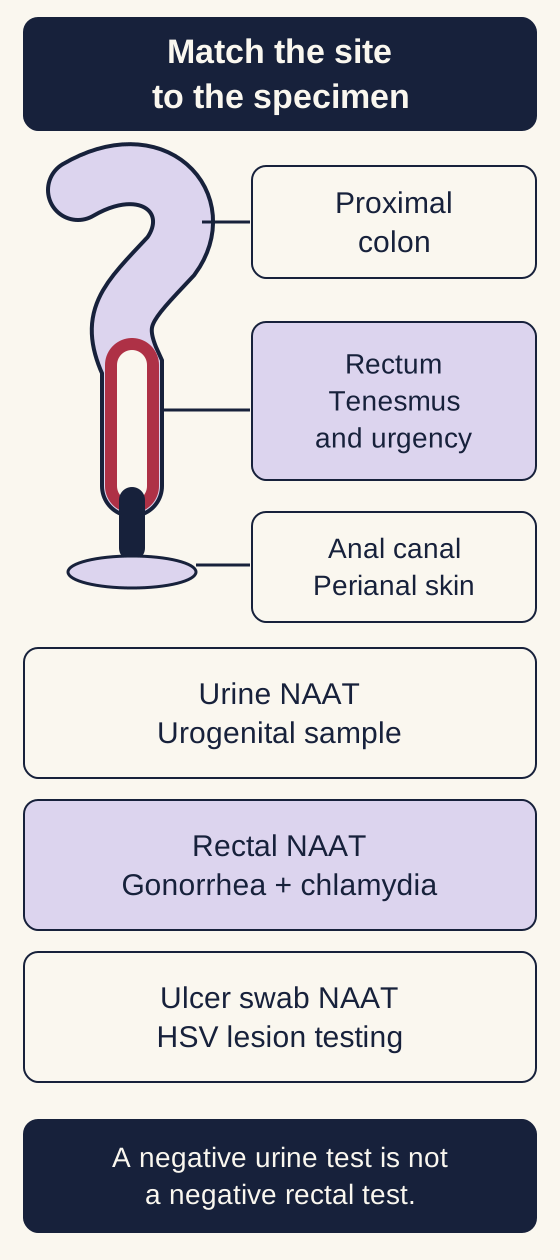
The scope map separates the perianal skin, anal canal, rectum, and proximal colon. Inspect skin for vesicles, ulcers, fissures, chancres, and warts. Anoscopy reveals distal mucosal exudate, ulcers, bleeding, and friability. A normal external inspection does not establish that the rectal mucosa is normal. [1]
Match the symptomatic site to its specimen. A urine NAAT cannot exclude infection at an unsampled rectal site. [1][4][1][4]
Try the specimen-to-question exercise: point to the compartment that produced each sample in the map. A urine sample asks about urogenital infection. A rectal swab asks about rectal infection. A swab from an ulcer asks about the lesion. Do not let a negative result from one compartment erase an unsampled compartment. Rectal gonorrhea and chlamydia testing uses a nucleic acid amplification test, abbreviated NAAT. [1][4]
Predict: urine testing is negative, but rectal discharge persists. What remains untested?
The rectal site remains untested. A negative urine NAAT does not exclude a rectal infection. Collect the site-specific sample rather than repeating only the urine test. [1][4]
The visible consequence is a different workup: isolated rectal symptoms call for rectal and lesion testing as appropriate; diarrhea and cramps can require stool evaluation as well. Neither purulent material nor an ulcer identifies one organism by appearance alone. HSV, syphilis, invasive chlamydia, CMV, trauma, and inflammatory disease can all enter the ulcer differential. [1][6]
Apply it elsewhere: after oral-anal exposure, watery diarrhea without rectal symptoms should prompt consideration of enteric pathogens, not automatic classification as proctitis. Conversely, normal stools between episodes do not negate persistent tenesmus and bloody rectal discharge. [1]
Ask precisely, examine safely, sample the right site
Can a patient's identity substitute for an exposure history? No. Ask privately and without judgment about receptive genital-anal, oral-anal, and digital-anal contact. Ask what contacted which site, when symptoms began, barrier use, partners, prior infections, and treatments. Also ask about inflammatory bowel disease, pelvic radiotherapy, immune suppression, HIV, recent antibiotics, and hypotension or vascular procedures. [1][5][8]
For example, a patient may report no penile-anal intercourse but describe oral-anal contact followed by cramps and bloody diarrhea. That history still changes the infectious differential. A person with established ulcerative colitis can also acquire a new rectal infection; the old diagnosis is not an exemption from testing. [1][5]
Explain the examination, obtain consent, offer a chaperone, and address pain. Inspect the perianal skin; perform a digital examination when safe and tolerated and anoscopy for suspected acute proctitis. Do not force an examination through severe pain or delay resuscitation to complete it. A focal swelling, a mass, marked tenderness, or peritoneal findings changes the immediate assessment. [1][7][9]
Build a short sampling plan before reading on. Rectal NAAT evaluates gonorrhea and chlamydia. Test an accessible fresh ulcer for HSV, preferably with lesion NAAT. Obtain syphilis serology and HIV testing; test other exposed sites rather than treating a urine result as a whole-body result. Inflammation on a rectal smear does not identify the pathogen, and rectal Gram stain is not a reliable stand-alone test to exclude gonorrhea. [1][3][4]
Predict: does a positive HSV blood antibody test prove the rectal ulcer is herpes?
No. Antibody can reflect a prior infection and does not locate the current disease. Testing the lesion is more directly informative. Do not mistake evidence of exposure for proof that a particular ulcer has that cause. [3]
Add stool studies for substantial diarrhea or suspected proctocolitis; enteric infection and an STI can coexist. Persistent unexplained symptoms, a mass, suspected inflammatory bowel disease, or concern for CMV can require endoscopy with appropriately directed biopsies. Suspected ulcerative colitis requires exclusion of infection, including C. difficile, and assessment of affected and unaffected mucosa. [1][5][6]
Apply it elsewhere: a patient whose symptoms continue despite correctly taken treatment needs reassessment, not an indefinite refill. Recheck adherence and new exposure, reconsider pathogens and noninfectious causes, and obtain culture with susceptibility testing when gonorrhea treatment failure is plausible. NAAT alone cannot provide antibiotic susceptibility. [1][4]
Treat the acute syndrome without pretending the cause is known
When should treatment start before the swab result returns? In acute sexually acquired proctitis, anorectal exudate on examination or polymorphonuclear leukocytes in secretions supports presumptive therapy. When anoscopy or Gram staining is unavailable, compatible acute symptoms plus receptive anal exposure also justify treatment while tests are pending. A nonspecific symptom alone does not make every proctitis infectious. [1]
For a nonpregnant adult without relevant contraindications, the CDC syndrome regimen is ceftriaxone 500 mg intramuscularly once plus doxycycline 100 mg orally twice daily for 7 days. Use ceftriaxone 1 g once when body weight is at least 150 kg. This covers likely gonorrhea and chlamydia while the cause is being resolved. Pregnancy, serious allergy, and complicated infection require a tailored regimen rather than automatic use of this teaching example. [1][4]
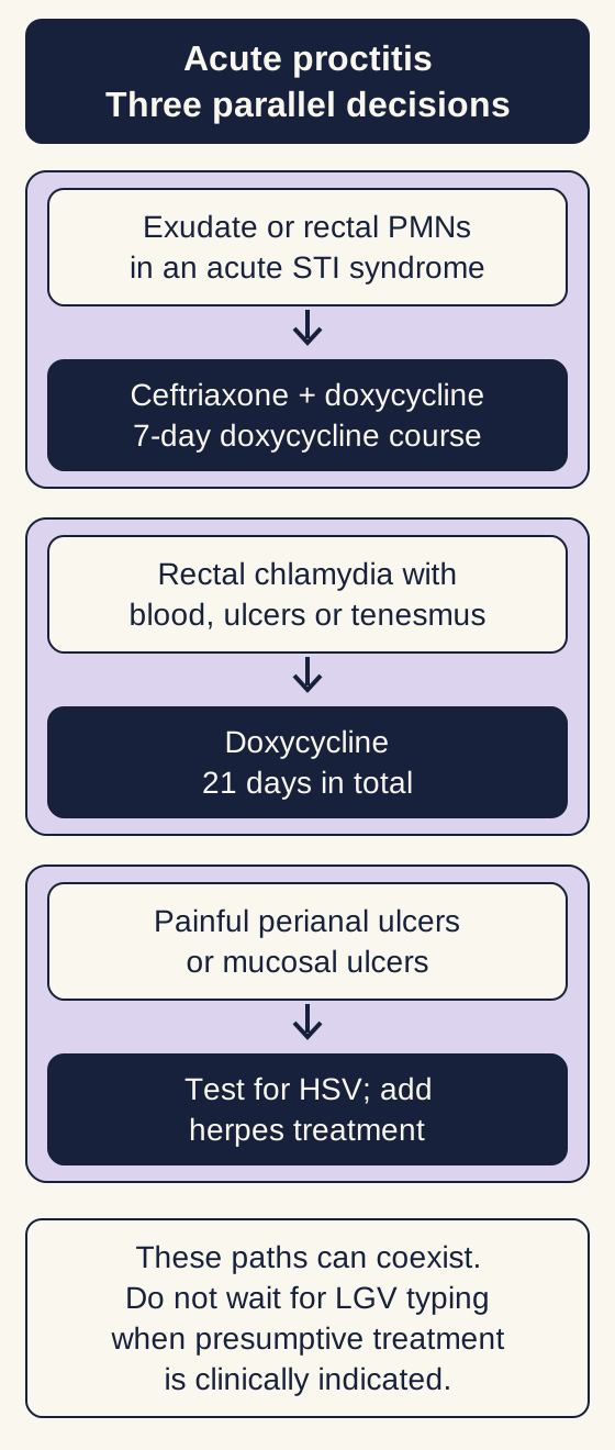
The treatment diagram separates three decisions rather than presenting one antibiotic ladder. First decide whether presumptive bacterial coverage is indicated. Next assess for invasive chlamydial disease. Finally ask whether ulcer findings also require herpes treatment. More than one pathway can apply to the same person. [1][2][3]
These are parallel decisions, not a mandatory sequence. The accompanying text supplies the unavailable-examination criterion, weight-based ceftriaxone dosing and individualized exceptions. A convincing LGV syndrome can be treated before genotyping. [1][2][3][4][1][2][3][4]
Change one finding: a positive rectal chlamydia test accompanied by bloody discharge, mucosal or perianal ulcers, or tenesmus supports presumptive lymphogranuloma venereum, or LGV. Extend doxycycline to a total of 21 days. Do not interpret a standard chlamydia NAAT as LGV serotyping. A convincing severe LGV syndrome can warrant presumptive LGV treatment at the initial visit; unavailable genotyping must not become a reason to delay. [1][2]
Predict: what does a painless early response change about an indicated 21-day LGV course?
Improvement does not convert invasive chlamydial disease into uncomplicated infection. Complete the indicated total course and reassess the patient rather than shortening therapy solely because symptoms improve. [2]
Painful perianal ulcers or mucosal ulcers seen on anoscopy warrant HSV testing and presumptive herpes treatment in the appropriate acute syndrome. An oral first-episode option is valacyclovir 1 g twice daily for 7 to 10 days; healing, renal function, and severity determine adjustments. Severe or disseminated disease requires hospital-level assessment and intravenous therapy. Herpes treatment supplements needed bacterial coverage; it does not replace it. In people with HIV, ulcerative or bloody acute proctitis warrants particular attention to both HSV and LGV. [1][3]
Syphilis requires stage-directed treatment, not an assumption that the empiric rectal regimen settles every infection. For primary or secondary syphilis without neurologic, ocular, or otic involvement, benzathine penicillin G 2.4 million units intramuscularly once is the standard adult regimen. Evaluate symptoms that would change that pathway. [10]
Apply it elsewhere: do not extrapolate a drug's urogenital indication to every rectal syndrome. The FDA's December 2025 zoliflodacin approval concerns uncomplicated urogenital gonorrhea; that is not a replacement indication for this CDC acute-proctitis regimen. Follow site-specific guidance and susceptibility information when treatment must be individualized. [1][4][12]
Use the host, time course, and tissue together
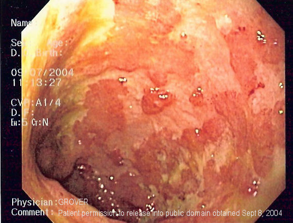
Does an ulcer settle the diagnosis? No. In the accompanying clinical photograph, the mucosa is irregular, with broad pale surface defects and red areas. Image: left-sided ulcerative colitis but explicitly warns that the appearance resembles Crohn's colitis. This is a real image of colonic disease, not proof of isolated proctitis and not a pathogen-identification test. [11]
Real left-colon endoscopy image. Image: ulcerative colitis but explicitly describes an appearance resembling Crohn colitis. This is not an image proving isolated proctitis, and it cannot establish a pathogen or complete disease extent. Image: [5][11] Samir at English Wikipedia; original source; CC BY-SA 3.0.
Describe before diagnosing: identify one surface abnormality, then name the history or test still needed. A single endoscopic frame cannot establish the full extent, chronicity, or cause. Continuous inflammation beginning in the rectum favors ulcerative colitis in a compatible history, but infection must be excluded and biopsies help establish chronic inflammatory disease. [1][5]
Use four contrasts. Acute rectal pain and discharge after exposure favor infection. Recurrent urgency and blood with continuous rectal inflammation and chronic histologic changes favor ulcerative proctitis. Bleeding months or years after pelvic radiation with fragile vascular mucosal changes suggests chronic radiation proctopathy. Abrupt pain and blood after severe hypotension or disruption of pelvic circulation raise concern for ischemic injury. None of these histories abolishes the others. [1][5][7][8]
For mild to moderately active ulcerative proctitis, rectal mesalamine, a 5-aminosalicylate, at 1 g daily is recommended for induction. A suppository delivers drug to rectal disease; more proximal disease may require a broader delivery strategy. Nonresponse warrants review of adherence, extent, diagnosis, and infection before escalation. A small anatomic extent does not guarantee mild disease. [5]
In marked immune suppression, particularly advanced HIV, CMV can cause substantial ulcers and bleeding. An undetectable plasma CMV PCR does not exclude tissue disease; a positive blood or tissue PCR alone does not prove invasive disease. Compatible ulcers plus histopathology, with immunohistochemistry when useful, establish a stronger diagnosis. Severe CMV gastrointestinal disease generally needs intravenous ganciclovir initially; oral valganciclovir is appropriate for selected mild disease or after adequate oral absorption is established. Specialist care includes renal and blood-count monitoring. [6]
Predict: does a prior diagnosis of ulcerative colitis make a new positive rectal infection irrelevant?
No. Chronic inflammatory disease and an infection can coexist. Treat the documented infection and reassess activity rather than assuming all symptoms are an inflammatory relapse. [1][5]
Apply it elsewhere: a new focal mass or persistent bleeding after pelvic radiotherapy requires evaluation for recurrent or new malignancy as well as radiation injury. A familiar treatment history is not a substitute for examining the actual lesion. [7]
Separate a local problem from a dangerous patient
Does tenesmus alone mean a transmural emergency? No. A stable person with mild symptoms, no substantial bleeding, no systemic illness, and reliable follow-up can often receive outpatient assessment and indicated treatment. Purulent discharge changes testing and antibiotic decisions, but by itself does not prove bowel necrosis. [1]
Compare that person with someone who is febrile, immunosuppressed, or unable to tolerate fluids and has extensive ulcers. That patient needs urgent in-person assessment, often hospital care depending on severity. Deep ulcer appearance does not itself prove full-thickness injury, but host vulnerability and systemic findings make casual follow-up unsafe. [6]
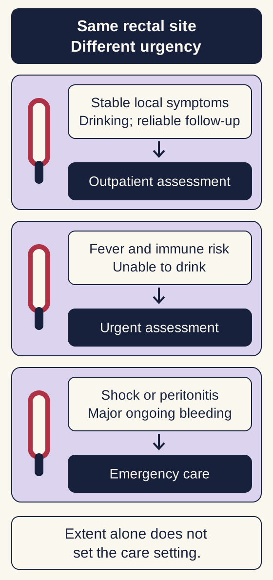
The urgency diagram is deliberately not a numerical score. Severe pain, heavy bleeding, rapidly worsening illness, or marked immune suppression lowers the threshold for escalation. Hypotension, ongoing major hemorrhage, peritoneal signs, suspected perforation, necrosis, or sepsis requires emergency care. Resuscitation and specialist assessment proceed alongside diagnostic work rather than after a routine office examination. [6][8][9]
This comparison is not a numerical triage score. Outpatient care requires otherwise appropriate severity, treatment and follow-up. A vulnerable host or deteriorating physiology changes the assessment setting even when the inflamed segment is short. [1][6][8][9][1][6][8][9]
Sort these by the needed setting: mild tenesmus with normal vital signs; fever with poor intake and severe immune suppression; abrupt pain with a rigid abdomen and shock. The consequence is respectively a prompt outpatient plan when otherwise appropriate, urgent assessment with likely inpatient care, and emergency resuscitation with surgical evaluation. The symptom's location stayed rectal; the danger changed because the host and physiology changed.
Predict: can protected rectal blood supply exclude ischemia after prolonged shock?
No. Rich collateral blood supply makes rectal ischemia unusual, not impossible. Severe low flow or vascular interruption can overcome that protection. Abrupt symptoms in that setting deserve urgent evaluation. [8]
Ischemic proctitis can occur after profound hypotension, aortoiliac disease, or injury to collateral circulation. Sharply demarcated dusky mucosa supports ischemia in context, but the whole clinical picture determines management. Nongangrenous injury may be managed without resection under close observation and perfusion support. Suspected transmural necrosis or perforation requires urgent surgical assessment. Avoid presenting endoscopy as a mandatory detour before treating shock or peritonitis. [8][9]
Apply it elsewhere: a short inflamed rectal segment in a patient who is becoming hypotensive is not automatically a mild outpatient illness. Anatomic extent and physiologic severity answer different questions. Conversely, a stable patient with discharge should not be told that the finding proves a surgical emergency.
Choose the next phase, not just the first prescription
When symptoms improve, is the entire treatment plan finished? Not necessarily. An infection needs completion of its indicated course, response assessment, and prevention of reinfection. Ulcerative proctitis needs a plan to maintain remission. Chronic radiation bleeding needs a plan for the injured tissue, not an endless course of empiric STI antibiotics. [1][5][7]
For gonorrhea or chlamydia, arrange partner evaluation and treatment for relevant contacts within the preceding 60 days. Abstain from sexual contact until the patient and partners have completed their indicated treatment and symptoms have resolved; after single-dose gonorrhea therapy, observe the 7-day interval. Retest for gonorrhea or chlamydia about 3 months after treatment to detect reinfection. A routine test of cure is not required after appropriately treated uncomplicated rectal gonorrhea, but pharyngeal gonorrhea needs a test of cure at 7 to 14 days. Persistent symptoms require reassessment now rather than waiting for the routine retest. [1][4]
Offer HIV testing and appropriate prevention counseling. Assess a recent potentially significant HIV exposure promptly for postexposure prophylaxis under current guidance, and discuss preexposure prophylaxis for ongoing risk. Include barrier use and hygiene after contact with fecal material. Explain that infection is about exposure and biology, not a moral judgment. [1]
Predict the second prescription: a person with mild ulcerative proctitis reaches remission on rectal mesalamine. Rectal mesalamine 1 g daily is also recommended for maintenance. Corticosteroids can have an induction role in selected refractory disease but are not maintenance therapy, even if the rectum is the only involved segment. Persisting activity despite appropriate treatment calls for reassessment and specialist escalation, not repeated switching among equivalent preparations without a plan. [5]
Check: which treatment goal remains after the visible bleeding of ulcerative proctitis stops?
Sustained steroid-free remission remains the goal. Continue an appropriate maintenance plan and monitor disease activity; symptom improvement is not a reason to substitute chronic prednisone. [5]
Radiation injury needs a different timetable. Symptoms during or shortly after treatment differ from delayed fragile vascular lesions that bleed months or years later. Acute mild symptoms can often be managed with symptom-focused care coordinated with the oncology team. [14] For chronic radiation-related rectal bleeding, specialist options include sucralfate retention enemas and endoscopic argon plasma coagulation. Formalin treatment or hyperbaric oxygen may be considered in selected cases. These are not interchangeable prescriptions for every patient; bleeding burden, prior treatment, mucosal integrity, expertise, and exclusion of malignancy guide selection. [7]
Apply it elsewhere: when bleeding continues despite a course designed for infection, first reconsider the cause and severity. When symptoms recur after a new exposure, reconsider reinfection. When inflammatory disease relapses after stopping maintenance, reconsider the long-term plan. The same word, recurrence, can therefore lead to three different decisions. [1][4][5]
Apply the lesson
For each patient, identify the involved site, use the discriminating findings, and choose the requested decision.
Case 1
Show answer and explanations for case 1
A. Repeat urine gonorrhea and chlamydia NAAT (Why this does not fit)
Urine NAAT addresses a urogenital infection and would fit urethral symptoms. This patient has a rectal syndrome without dysuria; repeating only urine leaves the relevant site unsampled.
Reasoning steps for option A
Which compartment would another urine NAAT sample?
The urethral/urogenital site, not the exudative rectum.
Why is repeating yesterday's negative urine assay unhelpful for this man's tenesmus?
He has no dysuria, and the symptomatic rectal mucosa remains untested despite the urine result.
B. Rectal gonorrhea and chlamydia NAAT (Best answer)
Tenesmus and mucosal exudate localize the symptomatic site to the rectum. The negative urine sample did not assess that site, so rectal NAAT directly tests the leading bacterial causes.
Reasoning steps for option B
Where do tenesmus and visible distal exudate localize the process?
They point to inflamed rectal mucosa after receptive anal exposure.
What specimen directly addresses gonorrhea and chlamydia despite a negative urine NAAT?
A rectal swab for gonorrhea and chlamydia NAAT samples the relevant infected site.
C. Stool multiplex enteric pathogen panel (Why this does not fit)
Stool pathogen testing is useful when diarrhea and cramps suggest an enteric syndrome. The supplied symptoms are distal and there is no diarrhea, making rectal gonorrhea and chlamydia testing the more direct study.
Reasoning steps for option C
When would a stool enteric panel better match this presentation?
Substantial diarrhea and abdominal cramps would suggest enteric illness or proctocolitis.
Does his isolated rectal urge and mucus support stool testing ahead of rectal STI NAAT?
No; without diarrhea or cramps, rectal sampling more directly addresses this exudative syndrome.
D. Serum herpes simplex virus antibody panel (Why this does not fit)
HSV can cause proctitis, so evidence of prior exposure may seem relevant. Blood antibody does not identify the cause of current rectal exudate; lesion NAAT is preferred when an ulcer is present.
Reasoning steps for option D
What can a serum HSV antibody result establish?
It can reflect previous HSV exposure, not prove the etiology of current rectal exudate.
What finding would instead justify lesion-directed HSV testing?
An accessible ulcer could be swabbed for HSV NAAT, but no external ulcer is seen here.
Takeaway: A negative result from an unconnected sampling site does not exclude rectal infection. [1] [3] [4]
A. Ceftriaxone once without oral antibiotics (Why this does not fit)
Ceftriaxone is appropriate treatment for uncomplicated rectal gonorrhea. Chlamydia has not been excluded in this patient, so ceftriaxone alone leaves a likely concurrent pathogen untreated.
Reasoning steps for option A
Which likely pathogen does an isolated ceftriaxone injection cover?
Gonorrhea, but it does not provide the needed treatment for possible chlamydia.
Why does pending rectal testing make ceftriaxone alone insufficient?
Her acute exposure-associated discharge warrants presumptive coverage while chlamydia remains unexcluded.
B. Doxycycline for 7 days without injection (Why this does not fit)
A seven-day doxycycline course treats uncomplicated chlamydia. The undifferentiated acute syndrome also warrants gonorrhea coverage, which doxycycline does not reliably provide.
Reasoning steps for option B
What does a seven-day doxycycline course accomplish?
It treats uncomplicated chlamydia, one component of the acute rectal STI syndrome.
Which organism remains inadequately covered if this patient receives only doxycycline?
Gonorrhea remains a concern while rectal results are pending and requires appropriate ceftriaxone coverage.
C. Cefixime once plus doxycycline for 7 days (Why this does not fit)
Oral cefixime is an alternative gonorrhea regimen when ceftriaxone cannot be administered. Ceftriaxone is available and there is no contraindication, so the preferred injectable regimen is indicated.
Reasoning steps for option C
Under what constraint would oral cefixime be considered for gonorrhea?
It is an alternative when ceftriaxone cannot be administered.
Does this 68-kg patient have a reason to substitute cefixime?
No; injectable ceftriaxone is available and she reports no allergy, so the preferred injection plus doxycycline fits.
D. Ceftriaxone once plus doxycycline for 7 days (Best answer)
Compatible acute symptoms plus receptive exposure justify presumptive treatment when anoscopy or Gram staining is unavailable. Ceftriaxone covers gonorrhea and seven days of doxycycline covers chlamydia while the rectal results are pending.
Reasoning steps for option D
What permits presumptive treatment without anoscopy or Gram stain today?
Acute compatible tenesmus and mucopurulent discharge after receptive anal contact meet the unavailable-examination criterion.
How should the regimen cover both unconfirmed rectal pathogens?
Give ceftriaxone 500 mg IM once at 68 kg plus doxycycline 100 mg orally twice daily for seven days pending results.
Takeaway: Unavailable anoscopy does not require delaying indicated presumptive therapy. [1] [4]
Rectal chlamydia plus bloody discharge and mucosal ulcers suggests invasive LGV disease. Treat presumptively for a total of 21 days; early improvement and unavailable genotyping do not justify shortening the course.
Reasoning steps for option A
What does positive rectal chlamydia with blood and ulcers suggest beyond uncomplicated infection?
It raises presumptive invasive lymphogranuloma venereum (LGV) proctitis.
How should three days of improvement affect the prescribed total duration?
It should not shorten treatment: complete 21 total days of doxycycline without waiting for unavailable genotyping.
B. 7 days in total (Why this does not fit)
Seven days is the usual doxycycline course for uncomplicated rectal chlamydia. The documented ulcers and bloody discharge identify a more invasive syndrome than uncomplicated infection.
Reasoning steps for option B
In which rectal chlamydia syndrome is seven days of doxycycline sufficient?
Uncomplicated chlamydial infection without invasive proctitis features.
Why do this man's bloody discharge and mucosal ulcers preclude that shorter course?
They support presumptive LGV even though he is improving after initial therapy.
C. 14 days in total (Why this does not fit)
Fourteen-day courses are used for some other genital tract infection syndromes. This case instead has the specific rectal chlamydia and severe proctitis combination for which the LGV course is 21 days.
Reasoning steps for option C
Does a generic positive rectal chlamydia NAAT specify LGV serovar?
No; the severe ulcerative clinical syndrome drives presumptive LGV treatment when typing is unavailable.
Why not stop doxycycline at day 14?
Fourteen days is not the indicated total for this presumptive LGV syndrome; the course is 21 days.
D. 28 days in total (Why this does not fit)
Longer doxycycline courses can be used for other staged infections such as selected syphilis regimens. Nothing supplied changes this presumptive LGV episode into that separate diagnosis or requires a routine 28-day course.
Reasoning steps for option D
What alternative infection could justify extending doxycycline beyond the 21-day LGV course?
Other staged infections such as selected syphilis presentations can have longer courses.
What in these results argues against routine 28-day doxycycline here?
Rectal chlamydia with bloody ulcerative proctitis points to a 21-day LGV course, not a separate staged infection.
Takeaway: Use the invasive rectal syndrome, not early symptom relief, to set the LGV treatment duration. [1] [2]
A. Ceftriaxone 500 mg intramuscularly once (Why this does not fit)
The 500 mg single dose is used for uncomplicated gonorrhea in patients below 150 kg. This patient weighs 154 kg, so that dose does not meet the weight-specific recommendation.
Reasoning steps for option A
For which weight range is ceftriaxone 500 mg IM once the uncomplicated-gonorrhea dose?
It applies below 150 kg.
How does his measured 154 kg change this otherwise single-dose plan?
The recommended one-time intramuscular ceftriaxone dose increases to 1 g.
B. Ceftriaxone 1 g intravenously daily for 7 days (Why this does not fit)
Daily parenteral ceftriaxone is used for certain disseminated gonococcal syndromes. There are no joint, skin, or systemic findings to justify treating this localized infection as disseminated disease.
Reasoning steps for option B
Which features would raise concern for disseminated gonococcal disease?
Joint symptoms, rash, fever, or other systemic manifestations would prompt a different assessment.
Why is seven days of daily IV ceftriaxone excessive here?
Only localized rectal discharge is documented, without joint, skin, or systemic findings.
C. Ceftriaxone 1 g intramuscularly once (Best answer)
The findings support uncomplicated rectal gonorrhea rather than disseminated infection. His weight is at least 150 kg, so the single intramuscular ceftriaxone dose is 1 g; chlamydia has been excluded.
Reasoning steps for option C
Do rectal gonorrhea without fever, rash, or joint symptoms suggest localized or disseminated infection?
It is uncomplicated localized rectal infection in the absence of dissemination signs.
Which weight-adjusted treatment is indicated after chlamydia is excluded?
Ceftriaxone 1 g intramuscularly once; the negative rectal chlamydia NAAT removes the doxycycline indication.
D. Ceftriaxone 500 mg once plus doxycycline for 7 days (Why this does not fit)
Adding doxycycline is appropriate when chlamydia has not been excluded. Chlamydia testing is negative and the ceftriaxone dose is still too low for this patient weight.
Reasoning steps for option D
When should doxycycline accompany gonorrhea treatment?
Add it when chlamydia has not been excluded.
What two details make 500 mg plus doxycycline mismatched here?
Rectal chlamydia is negative, and his 154-kg weight calls for 1 g rather than 500 mg of ceftriaxone.
Takeaway: For uncomplicated rectal gonorrhea, determine both the weight-based dose and whether chlamydia remains unexcluded. [1] [4]
A. Valacyclovir 500 mg once daily for ongoing suppression (Why this does not fit)
Once-daily low-dose valacyclovir can reduce recurrent genital herpes episodes. This is an untreated first episode, so a maintenance-style suppressive dose is not the appropriate initial regimen.
Reasoning steps for option A
What clinical situation is valacyclovir 500 mg daily suppression designed for?
It reduces recurrences in selected people with established recurrent HSV.
Why is a suppressive dose inadequate for his newly apparent painful ulcers?
This is a first clinical ulcer episode requiring initial treatment, not ongoing recurrence prevention.
B. Valganciclovir 900 mg twice daily for CMV disease (Why this does not fit)
Valganciclovir targets CMV and can be used in selected CMV gastrointestinal disease. The patient is not immunosuppressed and has acute painful perianal ulcers, which favor HSV rather than tissue-invasive CMV.
Reasoning steps for option B
What virus is valganciclovir intended to treat in gastrointestinal disease?
It targets CMV, particularly tissue disease in susceptible hosts.
Why is CMV-directed therapy a poor fit for these ulcers?
An immunocompetent, stable young man with acute painful perianal lesions more strongly suggests HSV than invasive CMV.
C. Valacyclovir 1 g twice daily for 7 to 10 days (Best answer)
Painful ulcers during an acute proctitis syndrome support presumptive HSV treatment. A first clinical episode in a stable adult with normal renal function is treated with an induction regimen, not a suppressive dose.
Reasoning steps for option C
What new finding adds an HSV treatment decision to ongoing bacterial coverage?
Painful perianal ulcers in acute proctitis support presumptive first-episode herpes therapy while lesion NAAT is pending.
Which first-episode HSV regimen fits his oral tolerance and normal kidney function?
Add valacyclovir 1 g orally twice daily for 7 to 10 days; it supplements rather than replaces indicated antibacterial care.
D. Doxycycline 100 mg twice daily for 21 total days (Why this does not fit)
An extended doxycycline course treats presumptive invasive rectal chlamydial infection. Rectal chlamydia testing is negative and doxycycline does not treat the suspected HSV ulcer disease.
Reasoning steps for option D
Which chlamydial pattern warrants a 21-day doxycycline course?
Rectal chlamydia with invasive LGV-type features such as bloody discharge or ulcers.
Why will extending doxycycline not treat these new lesions?
His rectal chlamydia NAAT is negative, and doxycycline has no activity against the suspected HSV ulcers.
Takeaway: An ulcerative herpes presentation can require treatment in addition to bacterial syndrome coverage. [1] [3] [6]
A. Benzathine penicillin G 2.4 million units intramuscularly once (Best answer)
The new rash with compatible ulcer history and positive serology supports early symptomatic syphilis. Without neurologic, ocular, or otic involvement, primary or secondary syphilis uses a single benzathine penicillin dose.
Reasoning steps for option A
How do the palmar rash, ulcer history, and dual-positive serology stage this infection?
They support symptomatic early syphilis, including secondary disease, rather than latent infection.
Which penicillin regimen treats early symptomatic syphilis without neurologic, ocular, or otic involvement?
Benzathine penicillin G 2.4 million units intramuscularly once is appropriate.
B. Benzathine penicillin G weekly for three doses (Why this does not fit)
Three weekly doses are used for late latent syphilis or infection of unknown duration. The current rash and ulcer establish an active early clinical stage rather than an unexplained latent serologic result.
Reasoning steps for option B
For which stage is a three-week benzathine penicillin schedule used?
Late latent syphilis or latent infection of unknown duration.
Why is this not merely latent seropositivity of uncertain duration?
He has a current diffuse palmar rash and compatible ulcer, providing clinical evidence of early symptomatic disease.
C. Aqueous crystalline penicillin G intravenously for 14 days (Why this does not fit)
Intravenous aqueous penicillin is used when syphilis involves the nervous system, eye, or ear. The stem specifically supplies no neurologic, visual, or hearing features requiring that treatment pathway.
Reasoning steps for option C
Which syphilis complications call for IV aqueous crystalline penicillin?
Neurosyphilis or ocular or otic syphilis requires that pathway.
Which examination and symptom findings argue against the IV syphilis treatment pathway?
Normal neurologic examination and absent visual and hearing symptoms favor uncomplicated early symptomatic syphilis.
D. Doxycycline 100 mg twice daily for 21 days (Why this does not fit)
A 21-day doxycycline course is standard treatment for LGV proctitis. The negative rectal chlamydia assay and positive syphilis evidence establish a different infection, and penicillin can be given.
Reasoning steps for option D
What rectal pathogen syndrome is treated with 21 days of doxycycline?
LGV-associated invasive chlamydial proctitis.
Why should this penicillin-eligible patient not receive LGV therapy for the identified infection?
Rectal chlamydia is negative while syphilis serology and palmar rash establish a penicillin-treatable diagnosis.
Takeaway: An ulcer can be syphilitic; stage the infection and check for organ involvement before selecting the regimen. [1] [2] [10]
A. Repeat plasma CMV PCR after a week of observation (Why this does not fit)
Blood PCR can detect circulating CMV and may be positive in advanced immune suppression. It neither establishes mucosal invasion nor reliably excludes it, so repeating blood testing delays the more direct tissue assessment.
Reasoning steps for option A
What does plasma CMV PCR measure rather than directly show?
Circulating viral DNA, not whether CMV is invading an ulcer's mucosa.
Can a second blood PCR resolve his large ulcers despite the first being undetectable?
No; absent viremia does not exclude compartmentalized gastrointestinal CMV in a patient with CD4 32.
B. Quantitative stool CMV PCR without tissue sampling (Why this does not fit)
PCR can detect CMV material shed into the gastrointestinal tract. Viral detection without compatible histopathologic changes cannot by itself establish invasive CMV colitis.
Reasoning steps for option B
What could a positive stool CMV PCR represent?
Viral material shed into the gut without proof of histologic mucosal invasion.
Why is stool viral quantity alone insufficient for this bloody ulcerative illness?
The needed distinction is invasive CMV at the sampled ulcer, which requires tissue assessment.
C. Serum CMV IgG with a repeat convalescent titer (Why this does not fit)
CMV IgG can identify prior exposure to this common virus. Prior exposure does not prove that CMV is causing these ulcers; the diagnostic question is tissue disease rather than serostatus.
Reasoning steps for option C
What question does serum CMV IgG answer?
Whether prior CMV exposure has occurred, not whether current ulcers are caused by CMV.
Why is a convalescent antibody titer less decisive than ulcer sampling here?
In advanced HIV with large rectosigmoid ulcers, serostatus cannot establish or exclude tissue-invasive disease.
D. Ulcer biopsy with CMV immunostaining (Best answer)
Severe cellular immune suppression with large ulcers raises concern for tissue-invasive CMV. Negative plasma PCR does not exclude local organ disease; tissue histology and immunohistochemistry assess whether CMV is invading the affected mucosa.
Reasoning steps for option D
Which combination makes local CMV disease plausible despite undetectable plasma DNA?
Untreated HIV with CD4 32, weight loss, bloody diarrhea, and large rectosigmoid ulcers.
How can sampling the rectosigmoid ulcers establish whether CMV is invading the mucosa?
Biopsy the ulcers for histology with CMV immunostaining; tissue findings supply stronger evidence than blood PCR.
Takeaway: In advanced HIV, distinguish CMV detected in a sample from CMV invading symptomatic tissue. [6]
A. Oral valganciclovir with laboratory monitoring (Why this does not fit)
Oral valganciclovir can be used for mild disease or after reliable oral absorption is established. The patient is vomiting and cannot retain medication, so oral delivery is not a dependable initial route.
Reasoning steps for option A
When is oral valganciclovir an acceptable CMV gastrointestinal option?
Selected mild disease or a step-down phase when oral intake and absorption are reliable.
Why is oral valganciclovir unreliable during his current vomiting?
Frequent vomiting prevents him from retaining medication during severe biopsy-confirmed disease.
B. Intravenous ganciclovir with laboratory monitoring (Best answer)
Biopsy establishes tissue-invasive CMV gastrointestinal disease. Severe symptoms and inability to absorb oral treatment favor intravenous ganciclovir initially, with renal and blood-count monitoring.
Reasoning steps for option B
What establishes that the ulcers require CMV-directed rather than empiric HSV treatment?
Characteristic biopsy inclusions plus positive CMV immunohistochemistry establish tissue-invasive CMV.
Which ganciclovir route and safety monitoring fit biopsy-confirmed CMV with vomiting?
Start intravenous ganciclovir with blood-count and renal-function monitoring, then reassess oral transition when feasible.
C. Intravenous acyclovir with laboratory monitoring (Why this does not fit)
Intravenous acyclovir treats severe herpes simplex or varicella-zoster disease. The biopsy identifies CMV, for which acyclovir is not the indicated treatment of this invasive gastrointestinal illness.
Reasoning steps for option C
Which severe viral infections would IV acyclovir address?
Severe HSV or varicella-zoster infection, not proven invasive CMV colitis.
How do the biopsy findings change the antiviral target?
CMV inclusions and immunostaining identify CMV, so acyclovir is not the appropriate initial agent.
D. Intravenous foscarnet with laboratory monitoring (Why this does not fit)
Foscarnet is an alternative for ganciclovir-resistant CMV or treatment-limiting intolerance. Neither resistance nor intolerance is supplied, so ganciclovir is the preferred initial treatment.
Reasoning steps for option D
When might IV foscarnet replace ganciclovir?
Documented or suspected resistance, or treatment-limiting ganciclovir intolerance, may justify it.
Which facts favor ganciclovir instead for this patient?
He has normal renal function and no prior intolerance or known resistant CMV; IV ganciclovir is the preferred initial approach.
Takeaway: A confirmed pathogen and reliable drug delivery are separate treatment decisions. [6]
A. Stool pathogen testing and culture (Best answer)
Diarrhea, cramps, and proximal colonic involvement indicate proctocolitis rather than isolated distal disease. Enteric pathogens can spread with oral-anal contact and are not excluded by negative gonorrhea and chlamydia NAATs.
Reasoning steps for option A
What do cramps, bloody diarrhea, and inflammation reaching 35 cm imply about extent?
The syndrome involves rectum and sigmoid, making it proctocolitis rather than isolated distal proctitis.
What untested infectious route does the oral-anal exposure suggest?
B. Repeat rectal gonorrhea NAAT as the only new study (Why this does not fit)
A rectal gonorrhea assay addresses one sexually transmitted cause of proctitis. Repeating only that assay does not evaluate the enteric organisms suggested by diarrhea and proximal colonic inflammation.
Reasoning steps for option B
Which specific organism would another rectal gonorrhea NAAT reassess?
It would repeat assessment for rectal gonorrhea alone.
Why does repeating gonorrhea NAAT miss pathogens suggested by diarrhea and inflammation to 35 cm?
Negative rectal STI tests do not evaluate enteric organisms causing fever, diarrhea, cramps, and proximal colonic involvement.
C. Serum inflammatory bowel disease antibody panel (Why this does not fit)
Inflammatory bowel disease can cause bloody diarrhea and continuous mucosal inflammation. Antibody panels do not reliably establish that diagnosis, and infection should be assessed in this acute exposure-associated illness.
Reasoning steps for option C
Could inflammatory bowel disease produce bloody continuous colitis?
Yes, but an antibody panel does not reliably establish its cause or exclude infection.
What should take priority in this acute post-exposure diarrheal illness?
Evaluate enteric pathogens in stool rather than attributing proctocolitis to IBD serology alone.
D. Urine gonorrhea and chlamydia NAAT as the only study (Why this does not fit)
Urine testing can assess an additional exposed urogenital site. It does not investigate the diarrheal colonic illness or the enteric pathogens implicated by oral-anal contact.
Reasoning steps for option D
What site would a urine gonorrhea/chlamydia NAAT investigate?
A urogenital site, if exposure there warrants testing.
Does that specimen address the colonic illness after oral-anal contact?
No; it cannot identify enteric pathogens responsible for bloody diarrhea and sigmoid inflammation.
Takeaway: Proximal inflammation and diarrhea broaden testing beyond the rectal STI panel. [1] [5]
A. Mesalamine tablets without rectal therapy (Why this does not fit)
Oral mesalamine can treat ulcerative colitis and may be useful with more extensive disease. For mild isolated proctitis, rectal delivery is preferred because it places the drug directly at the involved site.
Reasoning steps for option A
When does oral mesalamine become especially useful in ulcerative colitis?
More extensive colonic involvement may warrant oral or combined delivery.
Why is an oral-only approach less targeted for this distal 8-cm disease?
A rectal suppository delivers mesalamine directly to the mild, isolated inflamed rectum.
B. Prednisone tablets as first-line monotherapy (Why this does not fit)
Systemic corticosteroids can induce remission when appropriate first-line treatment fails or disease is more severe. This untreated mild rectal-limited presentation does not require starting with systemic corticosteroids.
Reasoning steps for option B
What clinical setting can justify systemic prednisone for induction?
More severe or refractory ulcerative disease may require systemic corticosteroids.
What makes first-line prednisone disproportionate here?
She is stable, has mild rectal-limited inflammation, and has not tried local mesalamine.
C. Mesalamine suppository 1 g daily (Best answer)
The chronic histology and negative infection evaluation support ulcerative proctitis. Mild inflammation confined to the rectum is appropriately treated with rectal mesalamine induction.
Reasoning steps for option C
Which findings support ulcerative proctitis over ongoing infectious proctitis?
Three months of urgency, continuous distal inflammation, chronic active biopsy changes, and negative infection testing.
Which mesalamine formulation and dose target mild inflammation confined to the distal 8 cm?
Mesalamine suppository 1 g daily treats mild disease confined to the rectum.
D. Azathioprine tablets as induction monotherapy (Why this does not fit)
Azathioprine can have a maintenance role in selected inflammatory bowel disease patients. It is not an appropriate rapid induction choice for newly diagnosed mild isolated proctitis.
Reasoning steps for option D
What is azathioprine's timing and role in selected IBD?
It can contribute to maintenance in selected patients but is not rapid induction monotherapy.
Why does her newly diagnosed mild distal colitis not warrant it now?
Localized untreated inflammation calls for rectal mesalamine induction, not slow-onset systemic immunomodulation.
Takeaway: Select induction therapy after establishing both the cause and the anatomic extent. [5]
A. Continue rectal hydrocortisone (Why this does not fit)
Topical corticosteroids can help induce remission in selected active distal disease. They are not recommended for maintenance, even when systemic exposure is lower than with oral prednisone.
Reasoning steps for option A
Can topical hydrocortisone help during active distal colitis?
It can be an induction option in selected active disease.
Why not continue it after documented remission?
Topical steroid exposure is still not an appropriate long-term maintenance strategy for this rectal-limited disease.
B. Continue rectal mesalamine (Best answer)
Her response establishes remission of rectal-limited ulcerative disease. Rectal mesalamine is appropriate maintenance treatment; remission is not a reason to replace it with chronic corticosteroids.
Reasoning steps for option B
How did bleeding and urgency respond to eight weeks of rectal mesalamine?
Eight weeks of rectal mesalamine resolved bleeding and urgency and was tolerated.
Which maintenance therapy preserves remission after tolerated rectal mesalamine induction?
Continue rectal mesalamine as maintenance rather than stopping effective therapy or substituting corticosteroids.
C. Continue oral budesonide MMX (Why this does not fit)
Budesonide MMX can be used for induction in selected active ulcerative colitis. Its lower systemic steroid exposure does not make it a recommended maintenance drug.
Reasoning steps for option C
When is oral budesonide MMX considered in ulcerative colitis?
It is a steroid formulation used for induction in selected active disease.
Does its lower systemic exposure make it suitable for this remission phase?
No; it is not recommended as maintenance when effective rectal mesalamine is available.
D. Continue oral prednisone (Why this does not fit)
Prednisone can suppress active inflammation during induction. Long-term prednisone is not a recommended remission-maintenance strategy and exposes the patient to steroid toxicity.
Reasoning steps for option D
Does oral prednisone have an induction or maintenance role in ulcerative colitis?
It can induce improvement in selected active inflammatory disease.
Why avoid chronic prednisone once rectal-limited colitis is in remission?
Chronic prednisone causes steroid toxicity and is not recommended to maintain remission.
Takeaway: Induction controls active inflammation; maintenance sustains remission without chronic corticosteroids. [5]
A. Serum anti-neutrophil cytoplasmic antibody testing (Why this does not fit)
Serologic patterns are sometimes associated with inflammatory bowel disease. They cannot establish that this new exposure-associated episode is an inflammatory relapse or exclude rectal infection.
Reasoning steps for option A
What can an IBD-associated ANCA pattern tell the clinician?
It may correlate with inflammatory bowel disease but cannot assign the cause of a new acute episode.
Why would serum serology miss the key alternative after this partner exposure?
It cannot exclude an acquired rectal STI causing pain and mucopurulent discharge.
B. Fecal calprotectin without pathogen testing (Why this does not fit)
Calprotectin can reflect intestinal inflammatory activity. It can also increase during infection, so it cannot resolve the cause of this acute syndrome by itself.
Reasoning steps for option B
Does fecal calprotectin elevation separate an IBD flare from infectious rectal inflammation?
No; it indicates intestinal inflammation, which can arise from infection as well as an IBD flare.
Can calprotectin alone distinguish this abrupt painful episode from his former painless urgency?
No; without pathogen sampling it cannot resolve the post-exposure infectious possibility.
C. Colonic transit testing with a stool diary (Why this does not fit)
Transit assessment can help evaluate selected functional bowel symptoms. Acute pain and purulent discharge after exposure suggest mucosal disease rather than an isolated transit disorder.
Reasoning steps for option C
What problems are colonic transit studies designed to assess?
Selected motility or functional bowel complaints, not acute mucopurulent rectal inflammation.
Why is a stool diary insufficient before changing anti-inflammatory therapy?
His new rectal pain and discharge after receptive contact point to mucosal infection needing site-specific evaluation.
D. Anoscopy and site-specific infectious testing (Best answer)
The abrupt painful discharge after a new exposure raises a competing infectious diagnosis. Established ulcerative proctitis does not exclude a superimposed STI, so examine and obtain rectal and lesion testing as appropriate.
Reasoning steps for option D
What change challenges the assumption that his known ulcerative proctitis has relapsed?
Abrupt pain and purulent discharge five days after a new receptive anal exposure differ from prior painless urgency.
Which examination and specimens should precede attribution solely to IBD?
Perform anoscopy and obtain rectal gonorrhea/chlamydia NAAT plus lesion testing if an ulcer is found; IBD does not prevent coinfection.
Takeaway: A known inflammatory diagnosis does not explain every subsequent rectal symptom. [1] [5]
A. Grade radiation injury from telangiectasias alone (Why this does not fit)
Telangiectasias after pelvic radiotherapy are compatible with chronic radiation proctopathy. That finding does not resolve the separate focal mass or weight loss and must not end the diagnostic workup.
Reasoning steps for option A
What can scattered telangiectasias four years after radiation explain?
They can reflect chronic radiation-related vascular injury and some rectal bleeding.
What finding makes grading radiation injury alone unsafe?
A separate irregular focal mass with weight loss and progressive tenesmus needs its own malignancy assessment.
B. Measure stool calprotectin to establish the cause (Why this does not fit)
Calprotectin may document intestinal inflammation. It cannot distinguish a mass from radiation injury or provide the tissue assessment needed for possible malignancy.
Reasoning steps for option B
What does fecal calprotectin measure in a bleeding patient?
It can signal intestinal inflammatory activity, not characterize a focal neoplastic lesion.
Why would it not settle this proctoscopic mass?
A stool marker cannot distinguish radiation change from malignancy or replace evaluation of the lesion.
C. Repeat urine STI NAAT to exclude infection (Why this does not fit)
STI testing is useful when relevant exposure or an acute infectious syndrome is present. A urine assay cannot explain or exclude malignancy in this progressive focal rectal lesion.
Reasoning steps for option C
When would urine STI testing inform a rectal complaint?
It could assess urogenital infection if exposure or urethral symptoms suggested that site.
Why does repeating a urine assay miss the urgent concern here?
It neither samples the rectal mass nor excludes cancer in a patient with weight loss and progressive symptoms.
D. Evaluate the focal mass for malignancy (Best answer)
Prior radiotherapy can explain diffuse vascular changes but does not explain every new lesion. A focal mass with progressive symptoms and weight loss requires a malignancy evaluation rather than attribution to radiation alone.
Reasoning steps for option D
Which lesion is not adequately explained by diffuse post-radiation telangiectasias?
The discrete irregular rectal mass is an independent alarm finding.
What diagnostic priority follows discovery of an irregular rectal mass with weight loss after radiotherapy?
Evaluate the focal mass for recurrent or new malignancy with appropriate specialist assessment and tissue diagnosis as indicated.
Takeaway: Radiation-associated mucosal changes do not exclude a separate rectal malignancy. [7]
A. Endoscopic balloon dilation (Why this does not fit)
Balloon dilation can address a clinically significant narrowing. The described lesion is bleeding vascular mucosa, not an obstructing stricture that needs dilation.
Reasoning steps for option A
What mechanical problem is balloon dilation intended to correct?
A clinically important stenosis or stricture causing narrowing.
What does this colonoscopy show instead of obstruction?
Diffuse bleeding telangiectasias without a narrowing, so dilation does not treat the vascular source.
B. Argon plasma coagulation (Best answer)
Delayed bleeding from fragile telangiectatic mucosa supports chronic radiation proctopathy. Persistent bleeding despite medical therapy makes specialist endoscopic coagulation a suitable vessel-directed option.
Reasoning steps for option B
How does the three-year delay after radiation and diffuse telangiectasia pattern identify the bleeding source?
They favor chronic radiation proctopathy with fragile mucosal vessels, after malignancy has been excluded.
What intervention follows continued bleeding and falling hemoglobin despite sucralfate?
An experienced endoscopist can apply argon plasma coagulation to bleeding telangiectasias, with procedural risk assessment.
C. Endoscopic mucosal resection (Why this does not fit)
Mucosal resection treats selected discrete mucosal neoplasms. The examination excludes a focal mass and describes diffuse telangiectasias rather than a resectable neoplastic target.
Reasoning steps for option C
What type of lesion is endoscopic mucosal resection meant to remove?
A selected focal mucosal neoplasm or discrete resectable lesion.
Why do diffuse telangiectasias without a focal mass argue against mucosal resection?
There is no mass; the target is widespread fragile vascular mucosa rather than a localized neoplasm.
D. Endoscopic rectal stent placement (Why this does not fit)
A stent can palliate selected obstructing colorectal lesions. There is no obstructing lesion here; stenting does not directly treat the radiation-related bleeding vessels.
Reasoning steps for option D
What clinical goal can a rectal stent serve?
It may palliate selected obstructing colorectal lesions.
Why would a stent not address this falling hemoglobin?
She has no obstructing mass or stenosis; it would not coagulate radiation-related bleeding vessels.
Takeaway: Match an endoscopic intervention to the lesion causing persistent bleeding. [7]
A. Argon plasma coagulation of the distal rectum (Why this does not fit)
Coagulation can control chronic radiation-related bleeding from telangiectatic vessels. The current examination lacks that vascular bleeding target and instead shows an acute inflammatory reaction during treatment.
Reasoning steps for option A
What finding would make argon plasma coagulation a targeted intervention?
Bleeding from chronic radiation telangiectasias would provide a vascular target.
Why is coagulation mismatched to this third week of radiotherapy?
She has mild diffuse acute inflammation without telangiectasias or substantial bleeding.
B. Long-term mesalamine maintenance for ulcerative colitis (Why this does not fit)
Mesalamine maintenance is useful for established ulcerative proctitis. The treatment-linked onset and absence of an established chronic inflammatory diagnosis do not justify labeling this episode ulcerative colitis.
Reasoning steps for option B
When is maintenance mesalamine appropriate for rectal inflammation?
It is used for established ulcerative proctitis to sustain remission.
Does new urgency during week three of irradiation establish chronic ulcerative colitis?
The new urgency began during pelvic irradiation, with no prior chronic inflammatory diagnosis and a plausible acute treatment injury.
C. Supportive care with oncology reassessment (Best answer)
Symptoms arising during radiotherapy with mild diffuse inflammation fit acute treatment-associated mucosal injury. With stable physiology and no hemorrhagic telangiectatic lesion, symptom support and oncology reassessment are appropriate rather than an invasive bleeding procedure.
Reasoning steps for option C
What does new urgency and diarrhea in the third week of pelvic irradiation suggest?
Symptoms emerging during the third week of pelvic radiotherapy with diffuse mild mucosal inflammation suggest acute radiation proctitis.
How should stable, mildly affected mucosa without a bleeding lesion be managed?
Provide symptom-directed supportive care and reassess with oncology rather than perform vascular ablation or urgent surgery.
D. Urgent rectal resection for full-thickness necrosis (Why this does not fit)
Resection can be necessary when radiation or ischemic injury causes irreversible full-thickness damage. There are no deep ulcers, systemic findings, or peritoneal signs supporting necrosis in this mild presentation.
Reasoning steps for option D
What would justify urgent resection for rectal injury?
Full-thickness necrosis or perforation with severe systemic or peritoneal findings could require surgery.
Do mild mucosal inflammation and preserved oral intake support radiation-induced transmural necrosis?
Normal intake, no fever, mild inflammation and no deep ulcers or peritoneal findings argue against it.
Takeaway: The timing and lesion determine whether radiation symptoms need supportive assessment or treatment of chronic bleeding. [7] [14]
A. Low flow overwhelms rectal collateral perfusion (Best answer)
The abrupt symptoms after profound hypotension and dusky mucosa support ischemic injury. The rectum has a rich collateral supply, but sufficiently severe low flow can overcome that protection.
Reasoning steps for option A
Why can ischemia affect a normally well-perfused rectum?
Its collateral arterial supply can be overwhelmed by sufficiently profound sustained hypotension.
Which observations identify low-flow injury in this patient?
Pain and hematochezia began abruptly after hypotension, and the distal mucosa is dusky with a sharp boundary.
B. Autoimmune inflammation extends continuously from the anus (Why this does not fit)
Ulcerative colitis can begin in the rectum and cause bleeding. The immediate relationship to hypotension and sharply demarcated dusky tissue is more consistent with ischemia than a new chronic inflammatory pattern.
Reasoning steps for option B
What presentation might support continuous autoimmune colitis?
Ulcerative colitis can involve the rectum and cause persistent bloody bowel symptoms.
Why does sharply demarcated dusky mucosa after hypotension favor ischemia over autoimmune colitis?
There were no preceding months of symptoms; a sudden low-flow event immediately preceded sharply demarcated dusky mucosa.
C. Radiation damages small vessels in a prior treatment field (Why this does not fit)
Late radiation injury can produce rectal bleeding through chronic vascular damage. There is no radiation history, and the symptoms began immediately after a low-flow event rather than after a radiation latency.
Reasoning steps for option C
What history would support radiation vascular damage?
Prior pelvic irradiation followed by a delayed rectal injury would support that mechanism.
Does the bleeding after hypotension have the radiation exposure and latency needed for radiation injury?
He has no pelvic radiation history, and bleeding arose directly after hypotension.
D. Invasive chlamydia produces chronic rectal inflammation (Why this does not fit)
LGV can cause ulcers, pain, blood, and severe proctitis. The supplied precipitant is profound hypotension with dusky demarcated mucosa, not evidence of chlamydial infection.
Reasoning steps for option D
Can invasive chlamydia produce painful bloody proctitis?
LGV may produce rectal ulceration, bleeding and severe pain.
What instead explains the distinct dusky segment?
Profound hypotension directly precedes a sharply bounded ischemic-appearing lesion, without evidence of chlamydial infection.
Takeaway: Collateral blood supply reduces rectal ischemia risk but cannot eliminate it during severe low flow. [8] [9]
A. Full bowel preparation followed by routine colonoscopy (Why this does not fit)
Endoscopy can characterize stable nonperforated mucosal disease. Bowel preparation and routine colonoscopy would delay definitive care in a patient with shock and evidence of perforation.
Reasoning steps for option A
Would routine colonoscopy fit stable mucosal inflammation or suspected rectal perforation?
It can characterize mucosal disease in a stable patient without suspected perforation.
Why should bowel preparation not delay care now?
Blood pressure of 78/44, rebound rigidity and extraluminal gas suggest perforation or necrosis needing immediate surgical evaluation.
B. Emergency surgical assessment for necrosis or perforation (Best answer)
Shock, peritoneal findings, and extraluminal gas indicate a threatened or established full-thickness complication. These findings require emergency surgical assessment alongside resuscitation rather than completion of a routine outpatient diagnostic sequence.
Reasoning steps for option B
What do rebound, shock and adjacent extraluminal gas imply?
They indicate a potentially full-thickness rectal injury with perforation rather than superficial proctitis.
What assessment must accompany resuscitation for shock, rigidity, and extraluminal rectal gas?
C. Outpatient rectal antibiotics while pathogen tests return (Why this does not fit)
Stable infectious proctitis can be treated while site-specific tests are pending. This patient has hemodynamic collapse and peritoneal signs that cannot be managed as an uncomplicated outpatient STI syndrome.
Reasoning steps for option C
When could empiric outpatient therapy address rectal discharge?
A stable uncomplicated infectious proctitis syndrome can be treated while rectal pathogen tests return.
Why is an outpatient antibiotic plan unsafe in this patient?
Hemodynamic collapse, a rigid tender abdomen and CT extraluminal gas require emergency management of a structural complication.
D. Topical mesalamine followed by reassessment next week (Why this does not fit)
Topical mesalamine treats mild to moderate ulcerative proctitis. Extraluminal gas and a rigid abdomen indicate an acute structural emergency rather than isolated mucosal inflammation suitable for delayed review.
Reasoning steps for option D
Which mucosal inflammatory condition would topical rectal mesalamine normally treat?
Mild to moderate ulcerative proctitis confined to mucosal inflammation.
Why do rigidity and extraluminal gas preclude a week-long trial of topical mesalamine?
Peritoneal signs and extraluminal gas signal an acute perforation or necrosis risk, not an uncomplicated inflammatory flare.
A. Outpatient treatment with prompt follow-up (Best answer)
The symptoms suggest a local infectious rectal syndrome without systemic danger signs. Stable vital signs, preserved intake, and reliable follow-up support outpatient treatment and clear return precautions.
Reasoning steps for option A
What pattern makes outpatient infectious-proctitis care reasonable?
Localized tenesmus and mucopurulent discharge after receptive exposure without systemic instability fit a treatable local syndrome.
Which vital-sign, intake, treatment-access, and follow-up findings support outpatient proctitis care?
Normal vital signs, oral intake, available empiric therapy and reliable follow-up support outpatient care with return precautions.
B. Hospital admission for presumed transmural necrosis (Why this does not fit)
Necrosis requires concern for deeper irreversible injury or severe systemic findings. Purulent discharge alone does not establish necrosis, and the stem supplies no shock, peritonitis, or major hemorrhage.
Reasoning steps for option B
Does purulent discharge establish transmural necrosis?
No; it points toward an infectious mucosal process, not necessarily deep tissue death.
What severe features are absent before considering admission for necrosis?
There is no hypotension, peritonitis, major hemorrhage or inability to drink.
C. Emergency surgery for the presence of tenesmus (Why this does not fit)
Surgery may be necessary for perforation, necrosis, or uncontrolled bleeding. Tenesmus localizes irritation to the rectum but is not by itself evidence of a surgical complication.
Reasoning steps for option C
What would make rectal symptoms a surgical emergency?
Perforation, gangrene or uncontrolled bleeding could require urgent surgery.
What does tenesmus indicate in this otherwise stable patient?
It signals rectal irritation but does not by itself demonstrate a structural surgical complication.
D. Observation without testing or treatment until fever develops (Why this does not fit)
Observation alone can be appropriate for selected self-limited noninfectious symptoms. This exposure-associated syndrome already warrants the indicated testing and treatment; fever is not required for action.
Reasoning steps for option D
Is fever required before testing suspected sexually acquired proctitis?
No; exposure-associated tenesmus and mucopurulent discharge already justify rectal testing and indicated empiric therapy.
Why is simple observation inadequate despite normal temperature?
Waiting for fever leaves a symptomatic possible STI untreated even though specimens and treatment are available.
Takeaway: A local rectal symptom does not automatically imply a transmural emergency. [1] [8]
A. Rectal test of cure now; retesting at 3 months (Why this does not fit)
Three-month retesting is appropriate after gonorrhea treatment. The early site-specific test is needed for the pharynx rather than routinely for the successfully treated rectal infection.
Reasoning steps for option A
What does three-month gonorrhea retesting accomplish?
It screens for reinfection after treatment, even when symptoms have resolved.
Which infected site needs an early cure test instead of routine rectal testing at day ten?
The documented pharyngeal infection needs a 7 to 14-day test of cure; routine early rectal test of cure is not the priority.
B. Both sites tested now; no later retesting (Why this does not fit)
An early test can assess treatment response at a site where this is recommended. Routine rectal test of cure is unnecessary here, and early negative results do not eliminate the need for later reinfection screening.
Reasoning steps for option B
Which infected site needs an early test of cure?
The pharyngeal site does, at 7 to 14 days after gonorrhea treatment.
Why is testing both sites now without later follow-up incomplete?
Uncomplicated successfully treated rectal infection needs no routine early cure test, while three-month retesting remains important for reinfection.
C. Neither site tested now; retesting at 3 months (Why this does not fit)
Three-month retesting is useful for detecting reinfection. Waiting that long omits the specific 7-to-14-day pharyngeal test-of-cure recommendation.
Reasoning steps for option C
Why might three-month retesting still be planned after symptom resolution?
Later testing detects repeat acquisition rather than simply confirming immediate cure.
Which pharyngeal gonorrhea follow-up window would be missed by waiting three months?
It misses the recommended pharyngeal cure test at day 10 within the 7 to 14-day window.
D. Pharyngeal test of cure now; retesting at 3 months (Best answer)
Successfully treated uncomplicated rectal gonorrhea does not routinely require a test of cure. Pharyngeal infection requires testing at 7 to 14 days, while later gonorrhea retesting addresses reinfection.
Reasoning steps for option D
How do follow-up requirements differ between pharyngeal and rectal gonorrhea?
The pharynx requires a test of cure at 7 to 14 days; uncomplicated successfully treated rectal disease does not routinely require one.
What further surveillance is needed after the pharyngeal result?
Retest for gonorrhea around three months to identify reinfection despite adequate initial therapy.
Takeaway: Site-specific tests of cure and later reinfection screening serve different purposes. [4]
A. Repeat gonorrhea NAAT without culture (Why this does not fit)
Another NAAT can again detect gonococcal nucleic acid. It will not identify antibiotic susceptibility, which is the requested information for possible treatment failure.
Reasoning steps for option A
What information can another rectal gonorrhea NAAT provide?
It detects gonococcal nucleic acid and can corroborate presence at the symptomatic site.
Why cannot NAAT alone answer the resistance question?
It supplies no viable isolate for antimicrobial susceptibility testing.
B. Mycoplasma genitalium NAAT without culture (Why this does not fit)
Additional pathogens can contribute to persistent proctitis and may warrant testing. This assay does not determine susceptibility of the gonococcus already detected in the symptomatic site.
Reasoning steps for option B
Could Mycoplasma genitalium be considered in a broader evaluation of persistent rectal discharge?
Yes, Mycoplasma genitalium may be considered in a broader persistent-proctitis evaluation.
Would its NAAT establish ceftriaxone resistance in detected gonorrhea?
No; it tests another organism and provides no gonococcal susceptibility result.
C. Gonococcal culture with susceptibility testing (Best answer)
Persistent symptoms after recommended therapy without intervening exposure raise concern for treatment failure. Culture provides an isolate for susceptibility testing, whereas NAAT alone cannot supply that information.
Reasoning steps for option C
Why consider gonococcal treatment failure rather than reflexively assume reinfection?
Discharge persists eight days after observed appropriate ceftriaxone with no intervening exposure, and rectal NAAT remains positive.
Which gonococcal test can supply an isolate for antimicrobial susceptibility testing?
Obtain gonococcal culture from the affected site and perform susceptibility testing on the isolate.
D. Chlamydia serovar testing without culture (Why this does not fit)
Serovar testing can help confirm LGV when chlamydia is detected. Chlamydia testing was negative, and this assay cannot investigate resistance in the documented gonococcal infection.
Reasoning steps for option D
When is chlamydia serovar testing informative?
When chlamydia is detected with a clinical syndrome suggesting LGV.
Why would chlamydia serotyping not investigate resistance after ceftriaxone-treated gonorrhea?
Rectal chlamydia was negative; serotyping cannot assess the confirmed gonococcus after ceftriaxone.
A. Presumptive doxycycline for 21 days (Why this does not fit)
A 21-day course treats symptomatic LGV disease. The patient has no invasive rectal syndrome, so the index diagnosis alone does not establish a need for that longer course in this asymptomatic partner.
Reasoning steps for option A
Who generally receives the 21-day doxycycline LGV course?
A patient with symptomatic invasive LGV disease receives the extended course.
Does asymptomatic recent partnership alone establish that syndrome?
No rectal pain, ulcers, bleeding or tenesmus are present, so the partner should not automatically receive the index patient's invasive-disease regimen.
B. Treatment only after a positive NAAT (Why this does not fit)
Waiting for an assay may appear reasonable when examination is normal. Recent contact with a diagnosed LGV patient warrants presumptive partner treatment despite absent symptoms.
Reasoning steps for option B
Can a normal examination exclude a transmissible chlamydial infection?
No; exposed partners can have asymptomatic infection.
Why not wait for the pending rectal NAAT?
Contact occurred within the 60-day partner interval, warranting presumptive treatment without waiting for a result.
C. Presumptive doxycycline for 7 days (Best answer)
The contact falls within the 60-day partner-management interval. An asymptomatic exposed partner receives a standard chlamydia regimen rather than automatically receiving the symptomatic index patient LGV course.
Reasoning steps for option C
Does the contact timing qualify for LGV partner management?
Contact 24 days before index symptom onset is within the preceding 60-day window.
Which presumptive regimen fits an asymptomatic nonpregnant partner?
Give standard chlamydia treatment with doxycycline for seven days, rather than a 21-day symptomatic LGV course.
D. Treatment only after symptoms appear (Why this does not fit)
Symptom-triggered treatment can appear sufficient for a clinically well patient. Chlamydial infection can be asymptomatic, so this strategy leaves a recently exposed partner untreated.
Reasoning steps for option D
Why is treatment contingent on symptoms a poor partner strategy?
Chlamydial infection and transmission may occur without rectal symptoms.
What specific exposure makes action necessary now?
Recent sexual contact with a confirmed LGV patient falls inside the 60-day presumptive-treatment period.
Takeaway: An asymptomatic exposed partner needs treatment but not automatically the invasive-disease regimen. [2]
A. Persistent ceftriaxone-resistant infection (Why this does not fit)
Resistance can cause infection to persist despite recommended therapy. The symptom-free interval and subsequent exposure to an untreated infected partner make reinfection the leading explanation on the supplied facts.
Reasoning steps for option A
What history would favor ceftriaxone-resistant persistence?
Ongoing infection without clearance or re-exposure after adequate therapy would raise concern.
How do symptom resolution and renewed contact with an untreated partner favor gonorrhea reinfection?
Symptoms completely resolved, then returned after renewed contact with an untreated partner who tested positive.
B. Residual nonviable bacterial nucleic acid (Why this does not fit)
NAAT can detect nucleic acid even when organisms are no longer viable. New symptoms six weeks after treatment with a documented new exposure are not adequately explained by residual material alone.
Reasoning steps for option B
Can NAAT detect dead gonococcal material?
Molecular tests can detect nucleic acid without proving viable organisms.
Why does new discharge after exposure six weeks later argue against residual gonococcal material alone?
Six weeks elapsed and new discharge followed a documented exposure to an infected untreated partner.
C. A noninfectious inflammatory relapse (Why this does not fit)
Inflammatory bowel disease can cause recurrent rectal symptoms. The new exposure, positive site-specific assay, and infected untreated partner support infection rather than a purely noninfectious relapse.
Reasoning steps for option C
Can bowel inflammation cause recurrent rectal complaints?
Noninfectious inflammatory disease can recur with urgency or bleeding.
Why is a purely inflammatory relapse less likely after renewed contact and another positive gonorrhea test?
Renewed exposure, positive rectal gonorrhea testing and the partner's positive test identify an infectious source.
D. Reinfection from an untreated partner (Best answer)
Initial symptom resolution followed by a new documented exposure supports reinfection. The untreated partner with a positive test supplies a direct explanation for a new episode rather than persistent treatment failure.
Reasoning steps for option D
How does complete initial symptom resolution affect interpretation of recurrent gonorrhea after re-exposure?
The first treated episode clinically cleared before a new symptomatic episode began.
How does an untreated gonorrhea-positive partner explain recurrence after renewed contact?
Renewed contact with a still-untreated, gonorrhea-positive partner supports reacquisition rather than primary treatment failure.
Takeaway: New exposure and partner treatment status matter when interpreting recurrence. [1] [4]
A. Residual nucleic acid from nonviable organisms (Best answer)
A molecular assay detects nucleic acid rather than proving viable infection. Testing less than four weeks after treatment can remain positive from nonviable material; this result alone does not establish failure in an adherent asymptomatic patient.
Reasoning steps for option A
Does a positive post-treatment chlamydia NAAT establish organism viability?
No; it detects chlamydial nucleic acid, which need not come from living organisms.
Why is the ten-day post-treatment result not proof of failure?
Testing within four weeks can detect residual nonviable material despite completed doxycycline, symptom resolution and no new exposure.
B. Persistent infection caused by doxycycline resistance (Why this does not fit)
Persistent symptoms and appropriately timed testing can prompt a treatment-failure evaluation. This patient is well, and the very early assay cannot by itself establish resistance or viable persistent infection.
Reasoning steps for option B
Which clinical and testing findings would justify evaluating persistent chlamydial infection after doxycycline?
Persistent or recurrent symptoms with appropriately timed testing would prompt further evaluation.
Can this early isolated NAAT establish doxycycline resistance?
No; the patient is well after adherence and the assay was taken just ten days after therapy.
C. Untreated invasive lymphogranuloma venereum (Why this does not fit)
LGV changes treatment duration when an invasive rectal syndrome is present. Neither the original presentation nor current symptoms support that syndrome, and early NAAT positivity does not identify an LGV serovar.
Reasoning steps for option C
Which features would prompt presumptive invasive LGV treatment?
Rectal chlamydia with severe proctitis, ulcers, bloody discharge or marked tenesmus supports invasive disease.
Does this isolated early result identify LGV?
There were no invasive features at diagnosis and early NAAT positivity does not establish an LGV serovar.
D. Reinfection after a new rectal exposure (Why this does not fit)
Reinfection is an important cause of later positive chlamydia tests. No intervening exposure is supplied, and an assay obtained this soon has a known alternative explanation.
Reasoning steps for option D
What evidence would support reinfection after chlamydia therapy?
An intervening sexual exposure followed by a new infection would make reacquisition plausible.
Why is reinfection less supported than residual nucleic acid ten days after doxycycline completion?
No new exposure occurred, symptoms resolved and the test was performed soon enough to detect residual nucleic acid.
Takeaway: Assay timing determines whether a positive post-treatment result can support a conclusion about persistent infection. [13]
A. Assign the diagnosis from ulcer shape alone (Why this does not fit)
Some mucosal patterns favor particular inflammatory diagnoses. The supplied irregular ulcers are not specific enough to establish a cause without the missing distribution and tissue information.
Reasoning steps for option A
Can an irregular ulcer pattern alone diagnose ulcerative colitis?
No; ulcerated mucosa has infectious and inflammatory causes and appearance alone is not etiologically specific.
What missing context prevents accepting the image label?
The isolated frame gives neither full disease distribution nor biopsy or pathogen results.
B. Assess extent, histology, and infection (Best answer)
Ulcerated mucosa can occur in inflammatory and infectious conditions, and a single frame cannot establish the full distribution. Integrating endoscopic extent, tissue findings, and infection evaluation is more discriminating than accepting an image label alone.
Reasoning steps for option B
Why is a single left-colon photograph insufficient?
It cannot establish proximal extent or reliably distinguish chronic colitis from an infectious mimic.
What combined assessment should precede a long-term inflammatory label?
Review endoscopic distribution, obtain or assess tissue histology and evaluate relevant infections.
C. Use serum antibodies as the definitive test (Why this does not fit)
Serologic markers may be associated with inflammatory bowel disease. They do not reliably establish this diagnosis or exclude an infectious mimic of an ulcerated colon.
Reasoning steps for option C
What can serum inflammatory bowel disease antibodies contribute?
They may be associated with inflammatory bowel disease but are not definitive diagnostic proof.
Can IBD-associated serum antibodies replace missing pathogen testing and tissue assessment in ulcerated colitis?
No; they cannot exclude an infectious ulcerating colitis or establish the disease extent.
D. Use calprotectin as the definitive test (Why this does not fit)
Calprotectin can support the presence of intestinal inflammation. It cannot by itself distinguish infection from chronic inflammatory disease or define the extent of involvement.
Reasoning steps for option D
What does fecal calprotectin measure indirectly?
It supports intestinal inflammation but not its specific cause.
Why would calprotectin not settle the cause or extent of colitis shown in one left-colon image?
It cannot distinguish infection from chronic inflammatory bowel disease or map how far the colitis extends.
Takeaway: Describe an endoscopic abnormality without converting one image into a complete etiologic diagnosis. [1] [5] [11]
A. Repeat stool molecular detection alone (Why this does not fit)
A molecular panel can identify enteric pathogen nucleic acid. Repeating detection alone does not supply the susceptibility information required to select a more targeted antimicrobial.
Reasoning steps for option A
What did the stool molecular panel establish?
It detected Shigella nucleic acid in a compatible febrile bloody-diarrhea syndrome.
Why is repeating the same detection insufficient after empiric treatment?
Another molecular result cannot determine which antimicrobial the organism is susceptible to.
B. Rectal chlamydia serovar testing (Why this does not fit)
Chlamydia serovar assessment can clarify LGV in a compatible rectal infection. Chlamydia testing is negative and the detected enteric organism, not an unconfirmed chlamydial serovar, is the immediate treatment target.
Reasoning steps for option B
When might chlamydia serotyping clarify rectal disease?
It may confirm LGV when rectal chlamydia is detected and invasive features are present.
Why does a negative chlamydia NAAT with stool Shigella detection argue against chlamydia serotyping?
Rectal chlamydia NAAT is negative while the stool panel identifies Shigella as the enteric target.
C. Serum inflammatory bowel disease antibodies (Why this does not fit)
Inflammatory bowel disease may cause similar symptoms and can remain in the differential. Serology cannot determine which antimicrobial will treat the identified enteric bacterium.
Reasoning steps for option C
Could inflammatory bowel disease be considered with bloody stools?
It remains a differential consideration when inflammation persists.
Can serum IBD antibodies guide Shigella antibiotic selection?
No; they provide no bacterial isolate or antimicrobial susceptibility information.
D. Stool culture with antimicrobial susceptibility (Best answer)
The diarrheal syndrome and stool assay identify an enteric bacterial target not assessed by the rectal STI assays. Culture provides an isolate for susceptibility testing when resistance may explain persistence after empiric therapy.
Reasoning steps for option D
Which organism best fits the febrile diarrhea despite negative rectal STI tests?
The stool panel identifies Shigella, an enteric pathogen capable of bloody diarrhea and urgency.
What laboratory step addresses persistence after empiric antibiotics?
Obtain stool culture and antimicrobial susceptibility testing to guide a targeted regimen.
Takeaway: A positive molecular pathogen result does not automatically provide the susceptibility needed for treatment. [1]
A. Proceed with segmental rectal resection (Why this does not fit)
Surgery can be necessary when ischemia causes transmural necrosis or perforation. Those features are not present, and the supplied assessment supports monitored conservative care rather than automatic resection.
Reasoning steps for option A
When would ischemic rectal injury require resection?
Transmural necrosis, perforation or clinical deterioration may require surgery.
Which CT, endoscopic, and clinical findings support observation rather than rectal resection after low flow?
CT shows no extraluminal gas, endoscopy no gangrene, the abdomen lacks peritoneal signs and symptoms are improving.
B. Induce remission with systemic corticosteroids (Why this does not fit)
Corticosteroids can treat active inflammatory bowel disease. The temporal relation to low flow and sharply bounded injury instead support ischemia, for which restoring perfusion is the immediate goal.
Reasoning steps for option B
Which inflammatory disease mechanism could justify systemic corticosteroids rather than perfusion support?
Active inflammatory bowel disease may require anti-inflammatory induction therapy.
Why is perfusion support more appropriate here?
Pain and bleeding began after sustained low flow and the lesion is sharply demarcated, consistent with ischemia rather than an immune flare.
C. Maintain perfusion with close inpatient reassessment (Best answer)
The low-flow onset and demarcated injury support ischemic proctitis. Improving nongangrenous injury without peritoneal findings can be managed conservatively under close observation, with escalation if deterioration occurs.
Reasoning steps for option C
What links the rectal lesion to an ischemic cause?
A sustained low-flow episode preceded pain and blood, followed by sharply bounded superficial mucosal injury.
How do restored perfusion, improving symptoms, and absence of gangrene guide care of rectal ischemia?
Normalized perfusion, improvement and no gangrene or peritonitis favor close inpatient conservative reassessment with escalation if worsening.
D. Ablate the injured mucosa with argon plasma (Why this does not fit)
Argon plasma coagulation can control selected chronic radiation-related vascular bleeding. The lesion is recent ischemic injury, not telangiectatic radiation bleeding, so ablation does not address the cause.
Reasoning steps for option D
What lesion is argon plasma coagulation designed to treat?
It may control bleeding from chronic radiation-associated telangiectatic vessels.
Why is argon plasma ablation mismatched to sharply demarcated rectal injury after low flow?
This is recent low-flow ischemic injury without a radiation vascular bleeding target; maintain perfusion instead.
Takeaway: Rectal ischemia does not always require resection, but conservative care requires close reassessment. [8] [9]
A. Treat uncomplicated chlamydia with 7 days of doxycycline (Why this does not fit)
Seven days treats uncomplicated rectal chlamydia. The current ulcers, bloody discharge, and severe tenesmus support the longer presumptive LGV course rather than uncomplicated infection treatment.
Reasoning steps for option A
For which rectal chlamydia presentation is seven-day doxycycline sufficient?
Uncomplicated rectal chlamydia without invasive proctitis features.
Which new features demand a longer presumptive course?
Positive rectal chlamydia accompanied by ulcers, bloody discharge and severe tenesmus suggests LGV.
B. Treat presumptive LGV with 21 days of doxycycline (Best answer)
Positive rectal chlamydia with ulcers and bloody discharge supports an invasive chlamydial syndrome. Chronic inflammatory histology does not negate a concurrent infection; treat the suspected LGV episode and reassess disease activity.
Reasoning steps for option B
Can chronic ulcerative-proctitis histology exclude a new STI?
No; established inflammatory bowel disease can coexist with a superimposed rectal infection.
What treatment addresses the new invasive syndrome before immunosuppression?
Start presumptive LGV treatment with 21 days of doxycycline and reassess underlying disease activity.
C. Treat the inflammatory relapse with prednisone alone (Why this does not fit)
Established ulcerative proctitis can recur and leave chronic changes on biopsy. That explanation does not account for or treat the new positive rectal pathogen test and invasive infectious presentation.
Reasoning steps for option C
Why could chronic inflammatory biopsy changes make prednisone seem plausible for this new rectal episode?
Prior ulcerative proctitis and persistent chronic inflammatory biopsy changes could suggest relapse.
Why is prednisone alone inadequate when rectal chlamydia accompanies ulcers and bloody discharge?
A positive rectal chlamydia NAAT with ulcers and bloody discharge identifies an untreated invasive infectious syndrome.
D. Wait for LGV genotyping before treating chlamydia (Why this does not fit)
Specific genotyping can confirm an LGV serovar. It is not needed to begin indicated presumptive therapy in a compatible severe rectal syndrome.
Reasoning steps for option D
What would LGV genotyping add?
It could identify an LGV-associated chlamydial serovar.
Should treatment await genotype confirmation in this patient?
No; severe compatible proctitis with positive rectal chlamydia warrants presumptive 21-day therapy now.
Takeaway: A positive infectious assessment and chronic inflammatory histology can both be clinically relevant. [1] [2] [5]
A. Combine oral mesalamine with a rectal mesalamine enema (Best answer)
Inflammation now extends beyond the reach expected from suppository-only treatment. For mild to moderately active left-sided disease, oral mesalamine plus rectal mesalamine is preferred to oral therapy alone and covers more than the rectal compartment.
Reasoning steps for option A
How does continuous inflammation to 40 cm change the previous rectal-limited disease classification?
Continuous active chronic colitis now reaches 40 cm, beyond the rectum into left-sided colon.
Which delivery strategy reaches both newly involved colon and active distal mucosa?
Combine oral mesalamine with a rectal mesalamine enema for mild to moderately active left-sided disease.
B. Continue the suppository as the sole treatment (Why this does not fit)
A suppository provides useful treatment for isolated rectal disease. The documented extension to 40 cm leaves a more proximal inflamed segment inadequately addressed by the previous delivery plan.
Reasoning steps for option B
What distribution does a mesalamine suppository primarily treat?
It delivers drug to the rectum and works well for isolated ulcerative proctitis.
Why is sole suppository use now insufficient?
Inflammation extends continuously to 40 cm despite adherence, beyond reliable suppository reach.
C. Use oral mesalamine and discontinue all rectal therapy (Why this does not fit)
Oral mesalamine reaches more proximal colonic disease. The rectum remains active, and combined oral and rectal treatment is preferred to oral therapy alone for this left-sided presentation.
Reasoning steps for option C
What benefit does oral mesalamine add when inflammation has extended from the rectum to 40 cm?
It treats the more proximal portion of the newly documented left-sided colitis.
Why should rectal mesalamine not be stopped?
The rectum remains inflamed, and combined oral plus rectal delivery is preferred to oral monotherapy.
D. Replace mesalamine with rectal hydrocortisone alone (Why this does not fit)
Topical corticosteroids can induce improvement in selected distal inflammatory disease. This does not provide the preferred combined mesalamine strategy for the newly documented left-sided extent.
Reasoning steps for option D
When can rectal corticosteroids be useful?
They may induce improvement in selected distal inflammatory flares.
Why is hydrocortisone alone not the preferred delivery plan?
The disease extends to 40 cm and calls for combined oral and rectal mesalamine rather than steroid-only distal treatment.
Takeaway: Reassess drug delivery when inflammatory disease extends beyond the rectum. [5]