Primary Sclerosing Cholangitis versus Primary Biliary Cholangitis
Compare duct injury in PSC and PBC, interpret imaging and antibodies, and separate treatment response, new complications, and disease-specific surveillance.
Why can two patients with itching and cholestatic liver tests need different investigations? Follow the injured ducts, assemble the diagnostic evidence, and separate treatment of today's problem from surveillance of future risk.
Start with the site and pattern of injury
Primary sclerosing cholangitis, or PSC, is a chronic inflammatory and fibrotic disease of the biliary tree. Fibrosis means scar formation. Larger intrahepatic and extrahepatic ducts can develop multiple narrow segments, with intervening ducts that appear normal or relatively dilated. This pattern can impair drainage and create a setting for bacterial cholangitis. [1]
Primary biliary cholangitis, or PBC, is an autoimmune disease primarily affecting small intrahepatic bile ducts. Immune injury progressively damages and removes these microscopic drainage channels. A routine scan of the large ducts may therefore be normal despite genuine cholestatic disease. Normal large-duct imaging is not the same as a normal liver. [2][6]
PSC is diagnosed more often in men, commonly in early to middle adulthood, and is strongly associated with inflammatory bowel disease, especially ulcerative colitis. Bowel disease is not a diagnostic requirement. PBC is diagnosed more often in middle-aged women and can coexist with autoimmune thyroid disease, Sjogren syndrome, or systemic sclerosis. [1][8] Sicca describes dry eyes or mouth. Sex and associated disease change the probability; they should not be used to exclude a disorder in someone outside the familiar profile. [1][6]
Both diseases can produce fatigue, pruritus, elevated alkaline phosphatase, progressive fibrosis, and later jaundice or portal complications. Symptom severity and the degree of fibrosis do not always change together. The shared word cholangitis also needs context. These chronic disease names are not automatically a diagnosis of an acute bacterial infection. [1][6]
Trace the drainage route
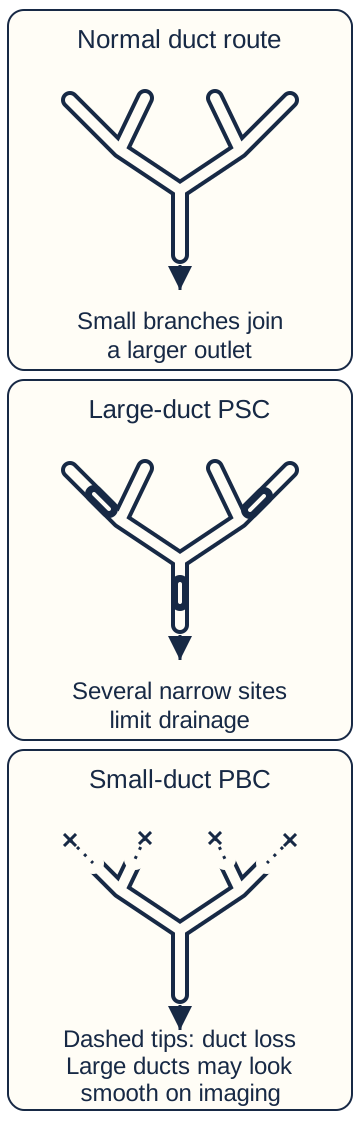
Bile passes from microscopic channels through small intrahepatic ducts into larger intrahepatic branches and the extrahepatic ducts before reaching the intestine. In the duct-map diagram, trace one branch to the outlet. PBC primarily damages the small branches; PSC can narrow several visible segments. A single distal stone or tumor can obstruct drainage upstream, but does not by itself explain multiple separated strictures. [1][8]
Trace a branch toward the outlet. Several separated visible narrowings suggest a different process from microscopic duct loss with smooth large ducts. Crosses and dashed tips indicate missing small ducts. [1][2][8]
A cholestatic pattern means that alkaline phosphatase, or ALP, is disproportionately raised relative to aminotransferases such as ALT. An accompanying increase in gamma-glutamyl transferase, or GGT, supports a hepatobiliary source of ALP. Compare each result with its own laboratory upper limit, not the raw numerical heights of different tests. Ultrasound helps assess obstruction before the more specific diagnostic routes below. [1][8]
Picture the outlet staying open while several microscopic side branches disappear. Predict what large-duct imaging can and cannot show.
Check the imaging prediction
The large ducts may still look smooth. Loss of microscopic ducts can impair bile handling without a visible large-duct obstruction. A normal large-duct picture does not exclude small-duct cholestasis.[2][8]
Now change the example: several visible branches contain separated narrow segments. The distribution, rather than itching alone, supports a sclerosing duct process. Clinical cases later ask you to apply this distinction without the diagram labels. [1]
Use high-quality duct imaging and know when tissue still helps
For suspected PSC, AASLD recommends high-quality MRI with MRCP as the first diagnostic imaging approach. The goal is to show the pattern and extent of strictures while excluding another cause of sclerosing cholangitis. A normal ultrasound or CT is not enough to rule out PSC. ERCP should not be performed simply as the routine diagnostic first step because it has avoidable procedural risks. [1]
When MRCP shows a typical large-duct pattern and the clinical context fits, liver biopsy is not routinely necessary. Histology can show concentric periductal fibrosis, sometimes described as onion-skin scarring, but sampling is limited and the classic appearance is not required in every specimen. A biopsy should answer a specific unresolved question rather than demonstrate a memorized image. [1]
A high-quality normal MRCP does not exclude small-duct PSC. Persistent cholestatic findings with appropriate clinical suspicion can justify liver biopsy to assess that possibility. An equivocal or technically poor MRCP is a different problem and may need repeat imaging at an experienced center rather than immediate labeling as small-duct disease. [1]
Measure serum IgG4 during the assessment because IgG4-related sclerosing cholangitis is an important alternative. Exclude secondary causes such as prior injury or other obstructive processes. Perinuclear antineutrophil antibodies and other autoantibodies may be present in PSC but are not sufficiently diagnostic by themselves. If the presentation suggests overlapping autoimmune hepatitis, biopsy and specialist interpretation can change treatment. [1]
Decide whether a negative scan was actually informative
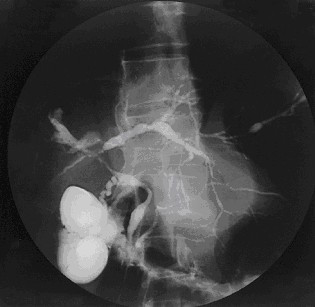
Magnetic resonance cholangiopancreatography, or MRCP, displays fluid-filled ducts without inserting an endoscope into the duct. Endoscopic retrograde cholangiopancreatography, or ERCP, can sample or treat a selected lesion but carries risks such as pancreatitis and cholangitis. The historical clinical image in this section is a contrast cholangiogram, not an MRCP and not a recommendation to use ERCP first. [1]
Image: a cholangiogram of PSC. Trace the irregular caliber of the opacified ducts. This is a historical contrast cholangiogram, not MRCP and not a reason to select routine diagnostic ERCP. Its limited resolution does not support lesion measurements, cancer exclusion, or disease staging. Image: Joy Worthington and Roger Chapman; original source; CC BY 2.0.
Compare two reports in persistent cholestasis: A, the ducts are clearly seen and smooth; B, motion prevents assessment of peripheral ducts. Which report can support a small-duct evaluation?
Interpret report A
A reliable normal MRCP leaves microscopic duct disease possible. Biopsy can assess small-duct PSC when suspicion persists. Normal visible ducts and absent duct disease are different statements.[1]
Interpret report B
The examination has not answered the visible-duct question. Repeat high-quality imaging at an experienced center can resolve the technical limitation; equivocal cases may also need expert-directed biopsy. An uninterpretable scan is not a negative scan.[1]
Transfer the distinction to a typical MRCP with a nonspecific biopsy: a limited tissue sample need not contain the classic concentric scar. Conversely, diffuse pancreatic enlargement, a long smooth distal stricture, and markedly high IgG4 should prompt evaluation for IgG4-related disease while excluding malignancy. IgG4 elevation by itself is not a definitive diagnosis. [1]
Combine cholestasis with specific antibodies or histology
AASLD describes PBC diagnosis using at least two of three categories. These are cholestatic biochemistry, principally elevated alkaline phosphatase; antimitochondrial antibodies or another PBC-specific antibody; and compatible histology showing nonsuppurative destructive cholangitis with interlobular duct injury. Nonsuppurative means the process is not defined by pus-forming bacterial inflammation. [2][6]
Antimitochondrial antibodies, or AMA, provide useful diagnostic evidence in the appropriate cholestatic setting. A major target is the E2 component of the pyruvate dehydrogenase complex, a mitochondrial enzyme complex. This antigen identity does not itself specify the location or stage of liver injury. [9] An isolated positive antibody without the expected biochemical or clinical context is not a complete diagnosis. When AMA is negative, PBC-specific antibodies such as sp100 or gp210 can provide diagnostic support. No single common demographic feature should substitute for these criteria. [2]
A patient with cholestatic liver tests and appropriate PBC-specific antibodies usually does not need a biopsy just to confirm what two categories already establish. Biopsy becomes more useful when the cause remains uncertain, antibodies do not clarify the diagnosis, or an overlap process is suspected. A marked aminotransferase elevation, including ALT above five times the upper limit in the guidance, should prompt consideration of another or overlapping disease. [2]
Exclude an obstructed large duct and review medications or other causes of cholestasis before attributing the entire pattern to autoimmunity. AMA-positive PBC can coexist with gallstones or an acquired liver injury. The aim is a diagnosis that explains the current findings, not an antibody result used to end every future investigation. [2][6]
Count evidence categories, not antibody names
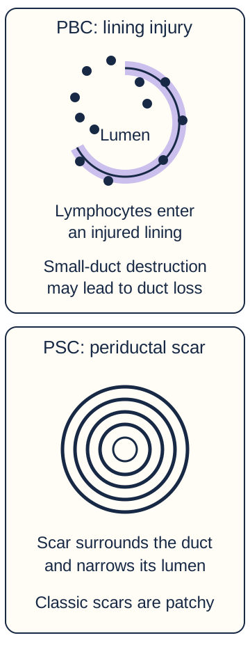
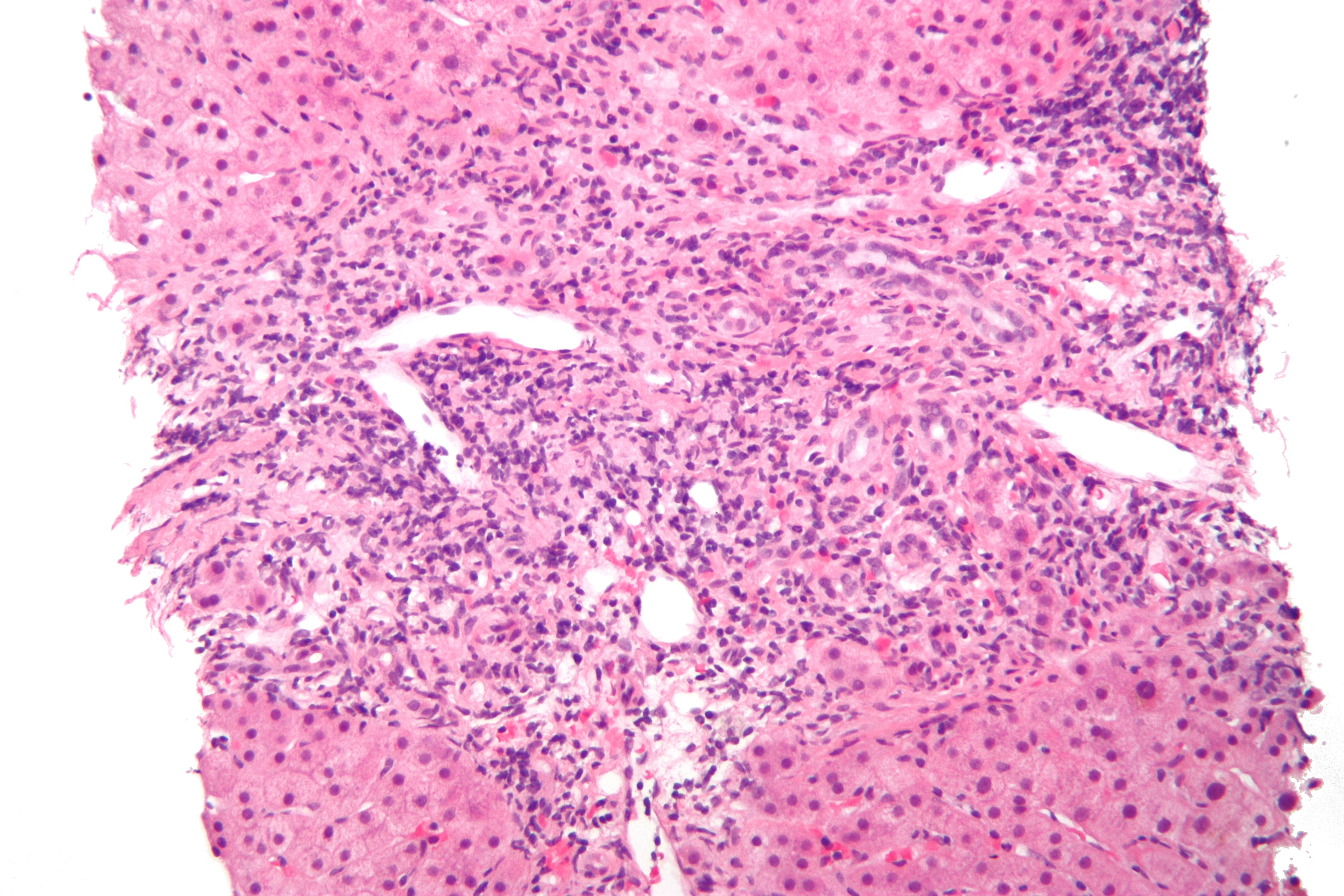
The classic florid duct lesion combines lymphocytic small-duct injury with a granulomatous reaction. Progressive duct loss is called ductopenia. Neither a florid lesion nor every stage of injury is visible in every specimen. Compare the original cross-sectional schematic with the real H&E micrograph: the micrograph shows portal inflammatory cells around an injured duct, not a guaranteed conspicuous granuloma or proof of cirrhosis. [8]
PBC injures small-duct epithelium; PSC may produce concentric periductal fibrosis. Compare the site of injury, not the drawn sizes. A real biopsy need not contain every classic feature. [1][8]
Image: PBC on H&E. Compare the duct lining and surrounding portal inflammation with the schematic. A conspicuous granuloma is not clearly demonstrated in this field. The historical file title uses cirrhosis, but this field alone does not establish the fibrosis stage. Image: Nephron; original source; CC BY-SA 3.0.
A patient has persistent ALP and GGT elevation, no obstruction on imaging, negative AMA, and positive gp210. Count the independent diagnostic categories before deciding whether biopsy is mandatory.
Count the categories in this example
There are two: biochemical cholestasis and a PBC-specific antibody. Negative AMA does not cancel positive gp210. Without an unresolved alternative or overlap question, biopsy is not required solely because AMA is negative. Two different evidence categories matter more than the number of positive antibody tests.[2]
Change just one feature: with repeatedly normal cholestatic enzymes, AMA alone is not a complete diagnosis and warrants biochemical follow-up. Change another feature: marked ALT elevation and increased IgG raise a hepatocellular overlap question that biopsy can address. These are different reasons to investigate, not exceptions that invalidate the diagnostic framework. [2][8]
Keep established first-line care separate from newer second-line evidence
Ursodeoxycholic acid, or UDCA, is recommended for PBC with abnormal liver enzymes at 13 to 15 mg/kg/day, regardless of histologic stage. The dose is based on body weight and must be prescribed in the available formulation by the treating team. Reassess the biochemical response after 12 months to help determine whether additional treatment is appropriate. [2]
A good response is a reason to continue useful therapy, not proof that the disease has disappeared. Ongoing review includes bilirubin, alkaline phosphatase, symptoms, and the broader risk of progression. Before calling a response inadequate, review the actual dose, adherence, competing disease, and clinical state. Changes in symptoms and changes in long-term disease risk are related but not identical outcomes. [2][6]
In the United States, elafibranor and seladelpar received accelerated approval for adults with inadequate response to UDCA, used with UDCA, or as monotherapy when UDCA cannot be tolerated. Their labels describe approval based on reduction in alkaline phosphatase. Improved survival or prevention of decompensation has not been demonstrated in those labels. Neither is recommended for decompensated cirrhosis, and each requires drug-specific monitoring and interaction review. [3][4]
Older guidance summaries still list obeticholic acid as a next step. On November 6, 2025, AASLD acknowledged its recent US market withdrawal and announced a guidance update. Do not copy the old drug sequence as a current US prescription pathway. Drug availability, licensing, and specialist choices vary by jurisdiction; the source version and population matter whenever treatment recommendations are compared. [2][5]
Check exposure before judging response
A 70-kg patient has a usual PBC UDCA range of 910 to 1,050 mg/day: 70 multiplied by 13 to 15. The prescribed formulation, current weight, dose actually taken, and binding interactions all affect exposure. Review these before calling persistent cholestasis a failure of adequately used UDCA. [2][8]
Hold the patient's weight at 70 kg, but change the recorded intake from 1,000 mg/day to 500 mg/day. Does an unchanged ALP establish resistance to a correctly used drug?
Check the exposure inference
No. Five hundred divided by 70 is about 7.1 mg/kg/day, below the usual range. Address the dosing barrier and reassess the actual exposure. A prescription in the record is not proof of the dose taken.[2][8]
For selected additional therapy, review the current drug-specific label rather than treating newer agents as interchangeable. Elafibranor requires particular attention to muscle injury and pregnancy precautions; both elafibranor and seladelpar require liver and bone-health safeguards. A lower ALP cannot override new ascites, variceal bleeding, or another decompensation event. [3][4]
Apply that separation to a different patient whose ALP normalizes but itching persists: continued disease-directed treatment and separate symptom care can both be appropriate. Neither persistent itching nor improved ALP alone establishes the entire disease course. [2][6]
Treat important problems without promising an unproven disease cure
PSC does not have a medical treatment established to improve transplant-free survival in the way a learner might infer from a falling enzyme result. Management therefore includes symptoms, obstruction, infection, associated bowel disease, progression assessment, and consideration of clinical trials. A normalizing alkaline phosphatase is useful information but is not a guarantee that scarring or cancer risk has disappeared. [1]
The 2022 AASLD guidance allows a selected UDCA trial at 13 to 23 mg/kg/day when alkaline phosphatase or GGT remains persistently elevated. Continuation depends on tolerance and a meaningful biochemical or symptom response within 12 months. This is not the same recommendation or dose range as routine first-line PBC therapy. High-dose UDCA at 28 to 30 mg/kg/day should be avoided because studies identified harm. [1]
A relevant PSC stricture is one associated with obstructive cholestasis or bacterial cholangitis, rather than merely an interesting narrow segment on a picture. New itching, jaundice, weight loss, worsening liver tests, recurrent infection, or a concerning imaging change can justify MRCP and focused ERCP. During ERCP for a relevant stricture, intraductal sampling for cytology and FISH helps assess malignancy as well as drainage. [1]
FISH examines chromosomal abnormalities in sampled cells; it complements rather than replaces cytology and the clinical picture. Negative brush cytology does not safely exclude malignancy when suspicion persists. Endoscopic dilation or selected stenting should be planned with expertise and device follow-up. A temporary stent without a removal plan can itself become a source of infection. [1]
Relieve a local obstruction without mistaking it for a cure
Fever with jaundice and suspected cholangitis needs prompt clinical assessment and antibiotics. Inadequate response to antibiotics, ongoing obstruction, or a tissue-sampling indication can require therapeutic ERCP. MRI/MRCP usually helps plan ERCP when feasible, but evaluation must match clinical urgency. Intraductal biopsy may complement brush cytology and FISH. Avoid assuming that a percutaneous route through a potential perihilar cancer is interchangeable with specialist-planned sampling, particularly when transplantation is being considered. [1]
Return to the duct map: one important narrowing is dilated, but several others remain. Predict the local and whole-liver effects separately.
Compare the two effects
Drainage above the treated narrowing can improve while disease elsewhere persists. Local decompression does not reverse the distributed cholangiopathy.[1]
Now consider a severe hilar stricture with negative brush cytology but FISH polysomy, meaning abnormal numbers of chromosomes in sampled cells. Persistent polysomy on repeat sampling, often reassessed at three months in the guidance pathway, strongly supports biliary neoplasia and probable cholangiocarcinoma. FISH alone cannot distinguish high-grade dysplasia from invasive carcinoma or establish metastatic stage. The discordant results need expert assessment, not a reassuring average of the tests. [1]
Match the cancer-screening plan to the disease and population
PSC is associated with cholangiocarcinoma and gallbladder cancer. AASLD recommends annual surveillance with abdominal imaging, preferably MRI/MRCP, with or without CA 19-9 for the appropriate adult large-duct PSC population. This recommendation does not apply routinely to patients younger than 18 or to small-duct PSC. Small-duct disease still needs follow-up because it can evolve into large-duct disease. [1]
CA 19-9 is an adjunct, not a stand-alone cancer test. Obstruction and inflammation can complicate its interpretation, and some patients do not produce it. New concerning symptoms or a relevant stricture require diagnostic evaluation now rather than waiting for the next surveillance date. Screening a stable patient and investigating a new abnormality are different tasks. [1]
For a PSC gallbladder polyp at most 8 mm, AASLD allows ultrasound monitoring every 6 months. A larger polyp should prompt consideration of cholecystectomy, preferably at an experienced center when liver disease is advanced. Operative risk, liver function, and the individual lesion matter. The size threshold is a decision aid, not an instruction to operate without assessing the patient. [1]
PSC also changes the bowel-surveillance plan. Colonoscopy with biopsies is recommended at diagnosis if IBD is not already known. In PSC with IBD, high-definition surveillance colonoscopy with biopsies begins at age 15 and is repeated every 1 to 2 years under AASLD guidance. For PBC, the 2018 guidance advises HCC imaging every 6 months in men and in patients with cirrhosis; PSC with cirrhosis follows the applicable HCC guidance as well. [1][2]
Change the finding, then change the purpose of the visit
Small-duct PSC is followed with MRI/MRCP every 3 to 5 years to assess progression to large-duct disease, or sooner for concerning changes. If visible-duct disease appears, the adult large-duct surveillance framework becomes relevant. In PSC without IBD on baseline biopsies, repeat ileocolonoscopy may be considered every five years or earlier for symptoms suggesting IBD. [1]
Follow-up depends on what is being investigated
Current situation
Purpose and response
Current situationPBC responding to UDCA
Purpose and responseContinue longitudinal care. HCC imaging every six months applies to men and patients with cirrhosis; biochemical response does not cancel those criteria. [2]
Current situationStable adult large-duct PSC
Purpose and responseAnnual hepatobiliary cancer surveillance, preferably MRI/MRCP, with or without CA 19-9. [1]
Current situationPSC with biopsy-established IBD
Purpose and responseAdd high-definition colonoscopy with biopsies every 1 to 2 years from the applicable starting age, even when bowel symptoms are quiet. [1]
Current situationNew PSC jaundice, weight loss, or relevant stricture
Purpose and responseInvestigate the change promptly. This is diagnostic assessment, not a reason to wait for routine surveillance. [1]
Start with the stable adult PSC row. Add new jaundice and a new hilar narrowing while keeping the last scheduled MRI date unchanged. Which part of the plan must change?
Reveal the consequence of the new finding
The patient now needs prompt diagnostic evaluation of the narrowing and drainage needs. The calendar does not override the new finding. Surveillance is for a stable risk; a new abnormality creates a diagnostic question.[1]
Try the reverse transfer: newly recognized quiet colitis changes colorectal surveillance, but does not by itself prove a new bile-duct cancer. Likewise, a growing gallbladder polyp above 8 mm prompts specialist surgical consideration, not automatic ERCP or an operation without liver-risk assessment. [1]
Treat symptoms and recognize when transplantation becomes relevant
Pruritus can be a major burden even before advanced liver failure. Cholestyramine or another bile-acid sequestrant is an initial option in the PBC guidance, with selected alternatives when it fails. These medicines can bind other drugs in the intestine, so dosing separation matters. The PBC guidance places UDCA at least 1 hour before or 4 hours after a sequestrant; individual newer drugs have their own label instructions. [2]
Linerixibat, an ileal bile acid transporter inhibitor, is now a US-labeled option specifically for cholestatic pruritus in adults with PBC. It addresses itching rather than replacing UDCA or proving prevention of cirrhosis. Avoid it with decompensated cirrhosis or prior or active decompensation events. [7]
The linerixibat label requires attention to diarrhea and dehydration, liver tests, fat-soluble vitamins, bleeding, bone health, and a 4-hour separation on either side of bile-acid binding resins. It is taken at least 30 minutes before any food or beverage other than water. Persistent diarrhea can require treatment interruption and evaluation for dehydration. [7][2][3][4]
Fatigue and sicca symptoms deserve their own assessment rather than assuming that a lower alkaline phosphatase will resolve everything. Review contributing conditions such as thyroid disease, anemia, sleep problems, or medication effects as clinically appropriate. Bone health, nutrition, fat-soluble vitamin status in substantial cholestasis, and portal-hypertension complications also belong in follow-up. [1][6]
Ascites, variceal bleeding, hepatic encephalopathy, worsening synthetic function, recurrent difficult cholangitis, or severe refractory symptoms can change the need for transplant-center assessment. In PSC, a carefully selected patient with perihilar cholangiocarcinoma may have a specialized neoadjuvant-treatment and transplant pathway. That is not a general transplant indication for every biliary cancer. [1][2]
Finish the comparison by separating three decisions. Which disease is established? Which current complication needs treatment? Which future risk needs surveillance? A patient may have a well-supported chronic diagnosis and still require urgent evaluation of a new infection or obstructing lesion. The best follow-up plan makes all three decisions visible. [1][2][6]
Predict what happens when less bile acid is recycled
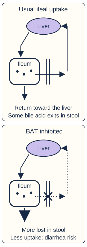
The recycling diagram follows bile acids into the intestine and back through the terminal ileal wall toward the liver. Linerixibat inhibits the ileal bile acid transporter, or IBAT, so less bile acid is reabsorbed and more is lost in stool. This is an intestinal transport action, not dilation of a liver stricture. The complete mechanism linking bile acids to itching is not fully established. [7]
The paired vertical lines mark the ileal wall and uptake crossing. Trace the return toward the liver, then compare the crossed crossing: inhibition of IBAT reduces reabsorption and increases bile-acid loss in stool. The wider exit arrow is qualitative, not a dose or flow measurement. Uptake inhibition can cause diarrhea and dehydration; it is not dilation of a biliary stricture. [7]
Trace the usual return arrow, then cover the IBAT crossing with a finger or follow the blocked crossing in the schematic. Predict which exit now carries more bile acid before opening the answer.
Check the direction of the transport change
More bile acid leaves through stool rather than returning through ileal uptake. Reduced reabsorption and increased fecal loss are opposite sides of the same transport change.[7]
Predict a consequence that needs a safety check
Diarrhea can cause dehydration. Persistent watery stools or lightheadedness need assessment, not reassurance that the medicine is working. Symptom benefit and treatment toxicity must be assessed separately.[7]
Transfer the mechanism to a medication schedule. If cholestyramine is taken at 08:00, UDCA at 07:00 fits its one-hour-before rule, but linerixibat at 07:00 does not fit its four-hour separation rule. Linerixibat at noon would be four hours after the resin; food and nonwater beverages must still wait at least 30 minutes. This is a timing example, not a prescription for an individual patient. [2][7]
Sicca symptoms may need artificial tears or saliva substitutes and specialist escalation when persistent. For fatigue, consider anemia, thyroid disease, sleep disorders, and medication effects rather than attributing every symptom to the ALP result. Severe refractory itching or recurrent difficult cholangitis may justify transplant-center assessment even when laboratory severity looks modest; referral is not a guarantee of listing or allocation priority. [1][6][8]
Apply the lesson to clinical cases
You can pause here and return to any case. Choose an answer when ready, then compare the explanations.
Case 1
Show answer and explanations for case 1
A. Persistent small-duct destruction after correction of incidental obstruction (Why this does not fit)
PBC can destroy interlobular ducts while leaving large-duct imaging smooth. The untreated multifocal visible strictures require a distributed large-duct process, not isolated PBC.
Reasoning steps for option A
What could produce small-duct loss with a normal large-duct study?
PBC can destroy interlobular ducts while leaving large-duct imaging smooth.
What finding is not explained by that process alone?
The untreated multifocal visible strictures require a distributed large-duct process, not isolated PBC.
B. Multifocal fibrosis persisting after relief of one stricture (Best answer)
The strictures involve multiple intrahepatic branches and the extrahepatic duct. Dilation treated one symptomatic hilar narrowing. The other fibrotic segments remain despite improved local drainage; this fits large-duct PSC.
Reasoning steps for option B
Where are the original abnormalities distributed?
The strictures involve multiple intrahepatic branches and the extrahepatic duct.
What was the anatomical reach of treatment?
Dilation treated one symptomatic hilar narrowing.
Why does the remaining pattern persist?
The other fibrotic segments remain despite improved local drainage; this fits large-duct PSC.
C. Continued obstruction by a solitary distal common-duct stone (Why this does not fit)
A distal stone can produce dilation upstream of a single obstructing site. Multiple separated strictures in both lobes persist after a different focal lesion was treated; a solitary distal stone does not explain that distribution.
Reasoning steps for option C
How can a distal stone change upstream ducts?
A distal stone can produce dilation upstream of a single obstructing site.
Why is that insufficient here?
Multiple separated strictures in both lobes persist after a different focal lesion was treated; a solitary distal stone does not explain that distribution.
D. Diffuse hepatocyte inflammation after resolution of duct injury (Why this does not fit)
A marked aminotransferase-predominant abnormality would raise a hepatocellular process. The supplied imaging still shows multiple duct strictures, so the duct injury has not resolved.
Reasoning steps for option D
What pattern would suggest predominant hepatocyte injury?
A marked aminotransferase-predominant abnormality would raise a hepatocellular process.
What remains structurally abnormal here?
The supplied imaging still shows multiple duct strictures, so the duct injury has not resolved.
Takeaway: Dilation relieves a selected site; it does not reverse fibrosis throughout the biliary tree.
A. Concentric scars narrowing several extrahepatic ducts (Why this does not fit)
Large-duct PSC can cause multifocal intrahepatic and extrahepatic strictures. High-quality MRCP lacks visible strictures, whereas the cholestasis and AMA support small intrahepatic duct injury.
Reasoning steps for option A
Which disorder often scars visible ducts?
Large-duct PSC can cause multifocal intrahepatic and extrahepatic strictures.
What observation argues against that localization?
High-quality MRCP lacks visible strictures, whereas the cholestasis and AMA support small intrahepatic duct injury.
B. Plasma-cell injury centered on the hepatocyte limiting plate (Why this does not fit)
Interface hepatitis injures hepatocytes at the portal-parenchymal boundary and can suggest autoimmune hepatitis. ALP and GGT are disproportionately abnormal with only mild ALT elevation and positive AMA, favoring PBC rather than predominant interface hepatitis.
Reasoning steps for option B
What does interface hepatitis chiefly affect?
Interface hepatitis injures hepatocytes at the portal-parenchymal boundary and can suggest autoimmune hepatitis.
Which supplied pattern points elsewhere?
ALP and GGT are disproportionately abnormal with only mild ALT elevation and positive AMA, favoring PBC rather than predominant interface hepatitis.
C. Granulomatous destruction of small interlobular bile ducts (Best answer)
ALP and GGT predominate over the mild ALT increase, indicating cholestasis. AMA supplies a second diagnostic category supporting PBC. The injured interlobular ducts are microscopic; a florid duct lesion can include granulomatous nonsuppurative destruction below MRCP resolution.
Reasoning steps for option C
Which laboratory compartment is most affected?
ALP and GGT predominate over the mild ALT increase, indicating cholestasis.
What does the antibody result add after imaging excludes obstruction?
AMA supplies a second diagnostic category supporting PBC.
Why can the duct study still look normal?
The injured interlobular ducts are microscopic; a florid duct lesion can include granulomatous nonsuppurative destruction below MRCP resolution.
D. Fibrosis surrounding a single distal common-duct outlet (Why this does not fit)
A distal obstruction impedes drainage of upstream visible ducts. Two imaging studies show no obstructing lesion, and the specific serology supports diffuse small-duct autoimmune injury.
Reasoning steps for option D
How might an outlet obstruction cause cholestasis?
A distal obstruction impedes drainage of upstream visible ducts.
Why does that not fit the complete evidence?
Two imaging studies show no obstructing lesion, and the specific serology supports diffuse small-duct autoimmune injury.
Takeaway: Microscopic interlobular duct destruction can produce cholestasis without visible large-duct strictures.
A. Ileocolonoscopy with mucosal biopsies now (Best answer)
PSC-associated bowel inflammation may be present without diarrhea or bleeding. No examination or biopsy of the colon has established whether IBD is present. Baseline ileocolonoscopy with biopsies assesses for IBD despite normal symptoms and C-reactive protein.
Reasoning steps for option A
Can PSC-associated IBD be clinically quiet?
PSC-associated bowel inflammation may be present without diarrhea or bleeding.
What information is missing?
No examination or biopsy of the colon has established whether IBD is present.
Which investigation changes the surveillance category?
Baseline ileocolonoscopy with biopsies assesses for IBD despite normal symptoms and C-reactive protein.
B. Fecal calprotectin followed by colonoscopy if raised (Why this does not fit)
Fecal calprotectin is a useful marker of intestinal inflammation. New PSC itself warrants ileocolonoscopy with biopsies; normal symptoms or a screening marker should not replace that baseline examination.
Reasoning steps for option B
What can fecal calprotectin detect?
Fecal calprotectin is a useful marker of intestinal inflammation.
Why is it not the preferred gatekeeper here?
New PSC itself warrants ileocolonoscopy with biopsies; normal symptoms or a screening marker should not replace that baseline examination.
C. CT enterography to identify occult intestinal inflammation (Why this does not fit)
Enterography can assess small-bowel inflammation and complications. PSC-associated colonic inflammation and microscopic disease require the recommended ileocolonoscopy and biopsies rather than an imaging substitute.
Reasoning steps for option C
Where can enterography be useful?
Enterography can assess small-bowel inflammation and complications.
What does this patient specifically need established?
PSC-associated colonic inflammation and microscopic disease require the recommended ileocolonoscopy and biopsies rather than an imaging substitute.
D. Annual symptom review with colonoscopy for new diarrhea (Why this does not fit)
New diarrhea or bleeding often prompts evaluation for intestinal disease. PSC can coexist with asymptomatic colitis, and delaying baseline biopsies could leave the higher colorectal-risk category unrecognized.
Reasoning steps for option D
When might symptoms usually trigger an IBD evaluation?
New diarrhea or bleeding often prompts evaluation for intestinal disease.
Why is waiting inappropriate for this risk group?
PSC can coexist with asymptomatic colitis, and delaying baseline biopsies could leave the higher colorectal-risk category unrecognized.
Takeaway: Ileocolonoscopy with biopsies can detect otherwise silent inflammation and determine the colorectal surveillance pathway.
A. Annual MRI/MRCP and CA 19-9 for large-duct cancer surveillance (Why this does not fit)
Annual hepatobiliary cancer surveillance applies to adults with large-duct PSC. The high-quality MRCP is normal and the diagnosis rests on biopsy, so this is small-duct PSC rather than the annual-surveillance population.
Reasoning steps for option A
Who is the usual annual imaging population?
Annual hepatobiliary cancer surveillance applies to adults with large-duct PSC.
Which supplied finding changes that category?
The high-quality MRCP is normal and the diagnosis rests on biopsy, so this is small-duct PSC rather than the annual-surveillance population.
B. Annual ERCP with intraductal cytology for occult strictures (Why this does not fit)
ERCP can obtain tissue and restore drainage when a relevant stricture or malignancy is suspected. No visible relevant stricture or new warning feature is supplied; routine invasive surveillance adds risk without establishing a needed intervention.
Reasoning steps for option B
When can ERCP add useful information?
ERCP can obtain tissue and restore drainage when a relevant stricture or malignancy is suspected.
Why is it not suitable for this follow-up?
No visible relevant stricture or new warning feature is supplied; routine invasive surveillance adds risk without establishing a needed intervention.
C. Biochemical follow-up without further imaging unless cirrhosis develops (Why this does not fit)
Biochemical follow-up helps assess cholestatic disease over time. Small-duct PSC can become large-duct PSC before cirrhosis, so periodic MRCP is used to detect that change.
Reasoning steps for option C
What do repeated liver tests contribute?
Biochemical follow-up helps assess cholestatic disease over time.
What would waiting for cirrhosis miss?
Small-duct PSC can become large-duct PSC before cirrhosis, so periodic MRCP is used to detect that change.
D. MRI/MRCP every 3 to 5 years for progression (Best answer)
A normal high-quality MRCP is compatible with biopsy-supported small-duct PSC. Some patients develop visible large-duct disease over time. AASLD recommends MRI/MRCP every 3 to 5 years for progression, with earlier diagnostic assessment for concerning changes; routine annual CCA surveillance is not recommended for small-duct disease.
Reasoning steps for option D
Does normal MRCP contradict all forms of PSC?
A normal high-quality MRCP is compatible with biopsy-supported small-duct PSC.
What change should follow-up detect?
Some patients develop visible large-duct disease over time.
Which interval and exception apply?
AASLD recommends MRI/MRCP every 3 to 5 years for progression, with earlier diagnostic assessment for concerning changes; routine annual CCA surveillance is not recommended for small-duct disease.
Takeaway: Monitor for conversion with MRI/MRCP every 3 to 5 years, or sooner when clinically indicated, rather than routine annual CCA surveillance for this phenotype.
A. Liver biopsy to establish small-duct PSC despite the limited scan (Why this does not fit)
Biopsy is useful when compatible cholestasis persists despite a normal high-quality MRCP. The ducts were not adequately imaged, so a limited scan cannot establish that large-duct disease is absent.
Reasoning steps for option A
When does biopsy establish a small-duct phenotype?
Biopsy is useful when compatible cholestasis persists despite a normal high-quality MRCP.
What prerequisite is missing?
The ducts were not adequately imaged, so a limited scan cannot establish that large-duct disease is absent.
B. Repeat high-quality MRI/MRCP at an experienced imaging center (Best answer)
Severe motion artifact prevented evaluation of relevant duct branches. The presence and distribution of large-duct strictures remain unknown. A repeat high-quality MRI/MRCP directly resolves the imaging problem before phenotype assignment.
Reasoning steps for option B
Was the first MRCP a trustworthy negative examination?
Severe motion artifact prevented evaluation of relevant duct branches.
What question remains unanswered?
The presence and distribution of large-duct strictures remain unknown.
What addresses that limitation without unnecessary instrumentation?
A repeat high-quality MRI/MRCP directly resolves the imaging problem before phenotype assignment.
C. Diagnostic ERCP to define the peripheral duct pattern (Why this does not fit)
Contrast cholangiography can depict biliary strictures. There is no supplied therapeutic or tissue-sampling indication, and repeat high-quality MRCP can address the technical failure without ERCP risk.
Reasoning steps for option C
What could ERCP show?
Contrast cholangiography can depict biliary strictures.
Why is it not the preferred next study in this stable patient?
There is no supplied therapeutic or tissue-sampling indication, and repeat high-quality MRCP can address the technical failure without ERCP risk.
D. Serum pANCA testing to decide whether further imaging is needed (Why this does not fit)
Perinuclear antineutrophil antibodies can occur in PSC and ulcerative colitis. The antibody is nonspecific and cannot determine whether visible strictures were missed on a motion-degraded scan.
Reasoning steps for option D
Why might pANCA seem relevant?
Perinuclear antineutrophil antibodies can occur in PSC and ulcerative colitis.
Can that result resolve the duct question?
The antibody is nonspecific and cannot determine whether visible strictures were missed on a motion-degraded scan.
Takeaway: Repeat high-quality MRI/MRCP at an experienced center before assigning a small-duct phenotype.
A. Large-duct PSC remains supported despite the nonspecific biopsy (Best answer)
Characteristic MRCP findings support the diagnosis after alternative causes are excluded. Concentric periductal fibrosis is patchy and may be absent from a limited tissue sample. Without a separate unresolved question such as autoimmune hepatitis overlap, repeat biopsy merely to find that scar is not routinely needed.
Reasoning steps for option A
How is typical large-duct PSC usually established?
Characteristic MRCP findings support the diagnosis after alternative causes are excluded.
Why need not the biopsy show the classic scar?
Concentric periductal fibrosis is patchy and may be absent from a limited tissue sample.
What does this mean for further biopsy?
Without a separate unresolved question such as autoimmune hepatitis overlap, repeat biopsy merely to find that scar is not routinely needed.
B. Small-duct PSC is favored because the characteristic scar was not sampled (Why this does not fit)
Small-duct PSC has normal cholangiography with compatible histology. This patient already has multifocal visible strictures; the absence of a sampled scar cannot convert large-duct disease into small-duct disease.
Reasoning steps for option B
What defines the small-duct imaging phenotype?
Small-duct PSC has normal cholangiography with compatible histology.
Why does that definition not apply here?
This patient already has multifocal visible strictures; the absence of a sampled scar cannot convert large-duct disease into small-duct disease.
C. Autoimmune hepatitis is favored because portal inflammation was present (Why this does not fit)
Disproportionate aminotransferase elevation, high IgG, and characteristic interface injury can support an overlap evaluation. The ALT elevation is mild, IgG is normal, and portal inflammation is nonspecific; these findings do not outweigh characteristic duct imaging.
Reasoning steps for option C
What findings would raise concern for autoimmune hepatitis overlap?
Disproportionate aminotransferase elevation, high IgG, and characteristic interface injury can support an overlap evaluation.
How does the supplied evidence differ?
The ALT elevation is mild, IgG is normal, and portal inflammation is nonspecific; these findings do not outweigh characteristic duct imaging.
D. The duct abnormalities require a second biopsy before PSC can be diagnosed (Why this does not fit)
Additional tissue may answer a specific unresolved diagnosis or characterize overlap. The characteristic MRCP pattern and excluded mimics provide the large-duct diagnosis without a requirement to sample concentric fibrosis.
Reasoning steps for option D
When is a second biopsy potentially useful?
Additional tissue may answer a specific unresolved diagnosis or characterize overlap.
What is already established in this case?
The characteristic MRCP pattern and excluded mimics provide the large-duct diagnosis without a requirement to sample concentric fibrosis.
Takeaway: Typical imaging supports large-duct PSC; absence of a classic patchy lesion in one specimen does not reverse that conclusion.
A. Assessment for PBC using AMA as the decisive test for the stricture (Why this does not fit)
AMA is useful in evaluating small-duct autoimmune cholestasis. A long distal visible stricture with diffuse pancreatic enlargement is not explained by PBC alone.
Reasoning steps for option A
Why is antibody testing familiar in cholestasis?
AMA is useful in evaluating small-duct autoimmune cholestasis.
What anatomical findings would it fail to explain here?
A long distal visible stricture with diffuse pancreatic enlargement is not explained by PBC alone.
B. Assessment for conventional PSC using pANCA as the deciding result (Why this does not fit)
PSC is a chronic cholangiopathy that can produce visible strictures. pANCA is not diagnostic, and the paired pancreatic enlargement and marked IgG4 elevation specifically require assessment for an alternative pancreatobiliary process.
Reasoning steps for option B
Why can PSC enter the differential?
PSC is a chronic cholangiopathy that can produce visible strictures.
Why is this proposed discriminator inadequate?
pANCA is not diagnostic, and the paired pancreatic enlargement and marked IgG4 elevation specifically require assessment for an alternative pancreatobiliary process.
C. Assessment for distal cancer based on CA 19-9 as the deciding result (Why this does not fit)
Painless jaundice and a distal stricture can be caused by malignancy. Obstruction can raise CA 19-9, and the pancreatic and IgG4 findings require integrated evaluation rather than a serum marker used as a definitive cancer test.
Reasoning steps for option C
Why must cancer remain under consideration?
Painless jaundice and a distal stricture can be caused by malignancy.
Why can CA 19-9 not settle the diagnosis?
Obstruction can raise CA 19-9, and the pancreatic and IgG4 findings require integrated evaluation rather than a serum marker used as a definitive cancer test.
D. Assessment for IgG4-related disease with malignancy exclusion (Best answer)
Diffuse pancreatic enlargement and a long biliary stricture form a pancreatobiliary pattern. Marked elevation strengthens suspicion for IgG4-related sclerosing cholangitis rather than a routine PSC label. IgG4 elevation alone is not definitive; expert imaging and appropriate tissue evaluation must distinguish IgG4-related disease from malignancy before treatment is assumed.
Reasoning steps for option D
Which abnormalities should be interpreted together?
Diffuse pancreatic enlargement and a long biliary stricture form a pancreatobiliary pattern.
What does the degree of IgG4 elevation contribute?
Marked elevation strengthens suspicion for IgG4-related sclerosing cholangitis rather than a routine PSC label.
Why is further assessment still necessary?
IgG4 elevation alone is not definitive; expert imaging and appropriate tissue evaluation must distinguish IgG4-related disease from malignancy before treatment is assumed.
Takeaway: Obtain specialist integrated imaging and tissue assessment to distinguish that process from malignancy before relying on immunosuppression.
A. The antibody establishes PBC and warrants immediate UDCA treatment (Why this does not fit)
AMA is a PBC-specific diagnostic category in an appropriate clinical setting. There is neither biochemical cholestasis nor compatible liver histology, so one antibody result alone does not establish PBC requiring treatment.
Reasoning steps for option A
What information can AMA provide?
AMA is a PBC-specific diagnostic category in an appropriate clinical setting.
Which diagnostic evidence is absent?
There is neither biochemical cholestasis nor compatible liver histology, so one antibody result alone does not establish PBC requiring treatment.
B. The antibody is uninterpretable without diagnostic ERCP (Why this does not fit)
ERCP can evaluate and treat a clinically important duct obstruction or obtain intraductal tissue. There is no biochemical or imaging evidence of obstruction, and ERCP is not a test to validate an isolated PBC-associated antibody.
Reasoning steps for option B
What problem can ERCP investigate?
ERCP can evaluate and treat a clinically important duct obstruction or obtain intraductal tissue.
Why is that not the missing category here?
There is no biochemical or imaging evidence of obstruction, and ERCP is not a test to validate an isolated PBC-associated antibody.
C. Continue biochemical follow-up without diagnosing active PBC (Best answer)
Only the antibody category is supplied; cholestatic enzymes remain normal. An isolated positive AMA does not establish active PBC. Periodic liver biochemical assessment can detect later cholestasis without treating the antibody result as a complete diagnosis.
Reasoning steps for option C
How many diagnostic categories are present?
Only the antibody category is supplied; cholestatic enzymes remain normal.
What conclusion should be avoided?
An isolated positive AMA does not establish active PBC.
What remains reasonable?
Periodic liver biochemical assessment can detect later cholestasis without treating the antibody result as a complete diagnosis.
D. Discontinue liver follow-up while liver-related symptoms remain absent (Why this does not fit)
Repeatedly normal cholestatic enzymes argue against currently established biochemical PBC. Cholestasis can emerge before symptoms in an AMA-positive patient, so periodic biochemical follow-up remains appropriate.
Reasoning steps for option D
Why might the normal tests seem to permit discharge from liver follow-up?
Repeatedly normal cholestatic enzymes argue against currently established biochemical PBC.
Why should future assessment not depend only on symptoms?
Cholestasis can emerge before symptoms in an AMA-positive patient, so periodic biochemical follow-up remains appropriate.
Takeaway: AMA alone does not establish active PBC; continue periodic biochemical assessment rather than automatically starting disease treatment.
A. Obtain a liver biopsy before starting treatment for antibody-negative PBC (Why this does not fit)
Biopsy helps when PBC-specific antibodies do not establish the cause or an overlap process is suspected. Anti-gp210 is a PBC-specific antibody, so this patient is AMA-negative but not negative for all diagnostic PBC serology.
Reasoning steps for option A
When can biopsy clarify antibody-negative cholestasis?
Biopsy helps when PBC-specific antibodies do not establish the cause or an overlap process is suspected.
Why is that not the situation supplied?
Anti-gp210 is a PBC-specific antibody, so this patient is AMA-negative but not negative for all diagnostic PBC serology.
B. Begin UDCA using the usual PBC weight-based dose (Best answer)
Disproportionate ALP and GGT elevation supports cholestasis after obstruction is excluded. Positive anti-gp210 supplies the PBC-specific antibody category. PBC can be diagnosed without mandatory biopsy, and UDCA at 13 to 15 mg/kg/day is the usual first-line treatment.
Reasoning steps for option B
What do the liver tests establish?
Disproportionate ALP and GGT elevation supports cholestasis after obstruction is excluded.
Does negative AMA eliminate the antibody category?
Positive anti-gp210 supplies the PBC-specific antibody category.
What follows from the two categories and absence of overlap findings?
PBC can be diagnosed without mandatory biopsy, and UDCA at 13 to 15 mg/kg/day is the usual first-line treatment.
C. Repeat liver tests in one year before assigning a cholestatic diagnosis (Why this does not fit)
An isolated PBC-associated antibody with normal liver tests may be followed biochemically. This patient has persistent symptomatic biochemical cholestasis rather than an isolated antibody result.
Reasoning steps for option C
When is observation of an antibody result reasonable?
An isolated PBC-associated antibody with normal liver tests may be followed biochemically.
Which necessary condition for that approach is absent?
This patient has persistent symptomatic biochemical cholestasis rather than an isolated antibody result.
D. Perform diagnostic ERCP before selecting a disease-specific treatment (Why this does not fit)
ERCP can treat an obstructing duct lesion or obtain intraductal tissue. Excluded obstruction, cholestatic enzymes, and anti-gp210 support microscopic autoimmune duct disease rather than a routine ERCP indication.
Reasoning steps for option D
What could ERCP address in a different presentation?
ERCP can treat an obstructing duct lesion or obtain intraductal tissue.
What does this presentation already support instead?
Excluded obstruction, cholestatic enzymes, and anti-gp210 support microscopic autoimmune duct disease rather than a routine ERCP indication.
Takeaway: Begin weight-based UDCA without requiring biopsy solely because AMA is absent.
A. Repeat AMA titer to quantify autoimmune bile-duct injury (Why this does not fit)
AMA helps establish PBC in an appropriate cholestatic setting. It does not define the new hepatocellular inflammatory pattern suggested by marked ALT elevation and increased IgG.
Reasoning steps for option A
What is the diagnostic role of AMA?
AMA helps establish PBC in an appropriate cholestatic setting.
What does a repeat titer fail to characterize here?
It does not define the new hepatocellular inflammatory pattern suggested by marked ALT elevation and increased IgG.
B. Repeat MRCP to search for additional large-duct strictures (Why this does not fit)
Imaging is important when new obstruction or visible duct disease is suspected. MRCP has excluded obstruction, while the disproportionate ALT and IgG findings require characterization of hepatocyte inflammation.
Reasoning steps for option B
When would duct imaging explain new deterioration?
Imaging is important when new obstruction or visible duct disease is suspected.
Why is another duct study not the highest-yield answer now?
MRCP has excluded obstruction, while the disproportionate ALT and IgG findings require characterization of hepatocyte inflammation.
C. Measure serum IgM to determine whether the UDCA response is adequate (Why this does not fit)
IgM can be increased in PBC and may accompany the cholestatic disorder. The ninefold ALT elevation and increased IgG raise possible autoimmune hepatitis overlap, which serum IgM cannot classify.
Reasoning steps for option C
Why might IgM be associated with PBC?
IgM can be increased in PBC and may accompany the cholestatic disorder.
What more consequential question would it leave unanswered?
The ninefold ALT elevation and increased IgG raise possible autoimmune hepatitis overlap, which serum IgM cannot classify.
D. Obtain liver histology to assess the predominant pattern of injury (Best answer)
ALT 360 divided by an upper limit of 40 is nine times the upper limit. The marked hepatocellular abnormality with increased IgG raises concern for autoimmune hepatitis overlap. Histology can establish interface injury and the predominant disease pattern before immunosuppression or other treatment is selected.
Reasoning steps for option D
How large is the ALT abnormality?
ALT 360 divided by an upper limit of 40 is nine times the upper limit.
What does the concurrent IgG increase suggest?
The marked hepatocellular abnormality with increased IgG raises concern for autoimmune hepatitis overlap.
Why would biopsy affect treatment?
Histology can establish interface injury and the predominant disease pattern before immunosuppression or other treatment is selected.
Takeaway: Liver biopsy can identify interface hepatitis and determine whether treatment for autoimmune hepatitis overlap is needed.
At 80 kg, 900 mg/day provides 11.25 mg/kg/day. The range of 13 to 15 mg/kg/day corresponds to 1,040 to 1,200 mg/day. A total of 1,200 mg/day fits the current weight and 300 mg formulation; a past prescription should be rechecked before escalating therapy.
Reasoning steps for option A
What dose per kilogram is she currently receiving?
At 80 kg, 900 mg/day provides 11.25 mg/kg/day.
What is the PBC target at the new weight?
The range of 13 to 15 mg/kg/day corresponds to 1,040 to 1,200 mg/day.
Which supplied daily total fits that range?
A total of 1,200 mg/day fits the current weight and 300 mg formulation; a past prescription should be rechecked before escalating therapy.
B. 900 mg per day (Why this does not fit)
At 60 kg, 900 mg/day provided 15 mg/kg/day. At 80 kg it provides only 11.25 mg/kg/day, so the current body weight changes the dose assessment.
Reasoning steps for option B
Why was 900 mg/day previously appropriate?
At 60 kg, 900 mg/day provided 15 mg/kg/day.
Why is the unchanged dose no longer in range?
At 80 kg it provides only 11.25 mg/kg/day, so the current body weight changes the dose assessment.
C. 1,600 mg per day (Why this does not fit)
At 80 kg, 1,600 mg/day provides 20 mg/kg/day. Twenty mg/kg/day falls within a selected PSC trial range, not the routine PBC range of 13 to 15 mg/kg/day.
Reasoning steps for option C
What daily dose per kilogram would this supply?
At 80 kg, 1,600 mg/day provides 20 mg/kg/day.
Why is that not the usual PBC starting range?
Twenty mg/kg/day falls within a selected PSC trial range, not the routine PBC range of 13 to 15 mg/kg/day.
D. 600 mg per day (Why this does not fit)
A simpler two-dose 300 mg schedule may appear easier to take. Six hundred mg/day at 80 kg gives 7.5 mg/kg/day and increases the underdosing rather than correcting it.
Reasoning steps for option D
What might lead someone to consider a lower total?
A simpler two-dose 300 mg schedule may appear easier to take.
Why would that worsen the identified problem?
Six hundred mg/day at 80 kg gives 7.5 mg/kg/day and increases the underdosing rather than correcting it.
Takeaway: At 80 kg, 13 to 15 mg/kg/day gives 1,040 to 1,200 mg/day; 1,200 mg/day fits the supplied formulation before labeling treatment failure.
A. Replace UDCA with a second-line PBC drug because pruritus proves nonresponse (Why this does not fit)
Additional disease-directed therapy may be appropriate after an inadequate UDCA biochemical response. ALP and bilirubin have normalized; persistent itching does not by itself demonstrate biochemical failure.
Reasoning steps for option A
Why can a new PBC drug be considered in some patients?
Additional disease-directed therapy may be appropriate after an inadequate UDCA biochemical response.
Which supplied finding argues against this explanation for the symptoms?
ALP and bilirubin have normalized; persistent itching does not by itself demonstrate biochemical failure.
B. Continue UDCA and assess separate treatment for cholestatic pruritus (Best answer)
The ALP and bilirubin response is favorable on an appropriate UDCA dose. Pruritus remains burdensome despite that biochemical response. Continue useful UDCA and select symptom-directed care with medication and liver-stage safeguards rather than equating itching with cure or nonresponse.
Reasoning steps for option B
What do the follow-up liver tests show?
The ALP and bilirubin response is favorable on an appropriate UDCA dose.
What problem has not been adequately addressed?
Pruritus remains burdensome despite that biochemical response.
How should the treatment goals be separated?
Continue useful UDCA and select symptom-directed care with medication and liver-stage safeguards rather than equating itching with cure or nonresponse.
C. Discontinue UDCA and treat pruritus because the disease has resolved (Why this does not fit)
Normal results can make an effective treatment response look like disappearance of the underlying disease. The results were obtained during effective therapy; biochemical improvement does not establish a cure or eliminate the need for continued PBC care.
Reasoning steps for option C
What might normalized enzymes appear to imply?
Normal results can make an effective treatment response look like disappearance of the underlying disease.
Why would stopping UDCA misinterpret those results?
The results were obtained during effective therapy; biochemical improvement does not establish a cure or eliminate the need for continued PBC care.
D. Arrange ERCP because persistent itching indicates a treatable large-duct lesion (Why this does not fit)
New or worsening pruritus can occur with biliary obstruction and warrants evaluation in context. Imaging shows no obstruction and liver tests have normalized, so no therapeutic ERCP target is supplied.
Reasoning steps for option D
When can itching accompany an obstructed duct?
New or worsening pruritus can occur with biliary obstruction and warrants evaluation in context.
What does the completed evaluation show here?
Imaging shows no obstruction and liver tests have normalized, so no therapeutic ERCP target is supplied.
Takeaway: Persistent itching requires its own treatment assessment rather than assuming either treatment failure or cure.
A. Add a second-line PBC agent to the present medication schedule (Why this does not fit)
A second-line agent may be considered after an inadequate response to appropriately used UDCA. She has not received the prescribed exposure consistently, so the current result does not establish failure of adequately used UDCA.
Reasoning steps for option A
When can additional PBC therapy be appropriate?
A second-line agent may be considered after an inadequate response to appropriately used UDCA.
What does the medication history show instead?
She has not received the prescribed exposure consistently, so the current result does not establish failure of adequately used UDCA.
B. Increase the prescribed UDCA dose to 1,800 mg each day (Why this does not fit)
Persistent cholestatic enzymes can prompt concern that the dose is too low. The prescribed 900 mg/day already equals 15 mg/kg/day; doubling the prescription does not address the observed missed doses and exceeds the usual PBC dose range.
Reasoning steps for option B
Why might a dose increase appear attractive?
Persistent cholestatic enzymes can prompt concern that the dose is too low.
Which distinction changes that calculation?
The prescribed 900 mg/day already equals 15 mg/kg/day; doubling the prescription does not address the observed missed doses and exceeds the usual PBC dose range.
C. Resolve the dosing barriers and reassess with adequate UDCA exposure (Best answer)
Nine hundred mg/day divided by 60 kg equals 15 mg/kg/day, within the PBC range. One 300 mg tablet on some days provides far less than the prescribed exposure. A feasible dosing plan and follow-up on adequate exposure are needed before the persistent ALP is attributed to drug nonresponse.
Reasoning steps for option C
What is the prescribed dose per kilogram?
Nine hundred mg/day divided by 60 kg equals 15 mg/kg/day, within the PBC range.
How does her actual use differ?
One 300 mg tablet on some days provides far less than the prescribed exposure.
What should be established before another agent is selected?
A feasible dosing plan and follow-up on adequate exposure are needed before the persistent ALP is attributed to drug nonresponse.
D. Replace UDCA with symptom-directed therapy for the next year (Why this does not fit)
Symptom-directed medicines can relieve burdens such as cholestatic pruritus when appropriately selected. Symptom treatment does not replace disease-directed UDCA, and the biochemical result was obtained without adequate use of that treatment.
Reasoning steps for option D
What role do symptom-directed medicines have?
They can relieve burdens such as cholestatic pruritus when appropriately selected.
Why would substitution leave this problem unresolved?
Symptom treatment does not replace disease-directed UDCA, and the biochemical result was obtained without adequate use of that treatment.
Takeaway: Address the dosing barriers and reassess on adequate UDCA exposure before classifying pharmacologic nonresponse.
A. The ALP response supports increasing seladelpar to reverse the ascites (Why this does not fit)
The ALP decrease demonstrated a biochemical response during treatment. The label does not establish seladelpar as treatment for decompensation, and new ascites requires assessment of progression rather than an unsupported higher dose.
Reasoning steps for option A
What did the ALP decline demonstrate?
It demonstrated a biochemical response during treatment.
Why does it not justify dose escalation for ascites?
The label does not establish seladelpar as treatment for decompensation, and new ascites requires assessment of progression rather than an unsupported higher dose.
B. The ALP response supports maintaining therapy while treating fluid retention (Why this does not fit)
A falling biochemical marker can suggest that an added medicine is helping its intended biochemical endpoint. Ascites is a decompensation event; the improved ALP does not override the label limitation for decompensated cirrhosis.
Reasoning steps for option B
Why might maintaining treatment seem reasonable?
A falling biochemical marker can suggest that an added medicine is helping its intended biochemical endpoint.
Which new finding changes its suitability?
Ascites is a decompensation event; the improved ALP does not override the label limitation for decompensated cirrhosis.
C. The bilirubin rise establishes direct drug toxicity as the cause of ascites (Why this does not fit)
Liver-test deterioration during treatment requires evaluation for drug-related injury. Ascites and rising bilirubin can also reflect disease progression or another complication; the supplied findings require assessment rather than proving a single cause.
Reasoning steps for option C
Why should drug injury enter the assessment?
Liver-test deterioration during treatment requires evaluation for drug-related injury.
Why is the cause not yet established?
Ascites and rising bilirubin can also reflect disease progression or another complication; the supplied findings require assessment rather than proving a single cause.
D. The ascites requires reassessment despite the biochemical response (Best answer)
New ascites signals decompensation requiring prompt assessment of the liver disease and current medicines. The seladelpar label states that improvement in survival or prevention of decompensation has not been demonstrated. Seladelpar is not recommended in decompensated cirrhosis, so continuation must be urgently reassessed rather than justified by ALP alone.
Reasoning steps for option D
What is the clinical significance of newly confirmed ascites?
It signals decompensation requiring prompt assessment of the liver disease and current medicines.
Does the ALP decrease establish protection against that outcome?
No. The seladelpar label states that improvement in survival or prevention of decompensation has not been demonstrated.
What does the label imply for this changed clinical state?
Seladelpar is not recommended in decompensated cirrhosis, so continuation must be urgently reassessed rather than justified by ALP alone.
Takeaway: New decompensation requires urgent liver-stage and medication reassessment; seladelpar is not recommended in decompensated cirrhosis.
A. Refer for advanced-liver-disease review rather than follow the old sequence (Best answer)
AASLD acknowledged US market withdrawal of obeticholic acid in November 2025. Ascites and variceal hemorrhage establish decompensation, a separate limitation for elafibranor and seladelpar. Advanced-liver-disease and transplant-center assessment should guide care instead of an automatic second-line drug substitution.
Reasoning steps for option A
Does the handout represent the current US OCA pathway?
No. AASLD acknowledged US market withdrawal of obeticholic acid in November 2025.
What does the ascites and variceal hemorrhage add?
They establish decompensation, a separate limitation for elafibranor and seladelpar.
What kind of reassessment is now needed?
Advanced-liver-disease and transplant-center assessment should guide care instead of an automatic second-line drug substitution.
B. Replace the handout recommendation with routine elafibranor initiation (Why this does not fit)
Elafibranor is a US-labeled option for selected adults with PBC and inadequate UDCA response. Ascites and variceal hemorrhage indicate decompensation, for which elafibranor is not recommended.
Reasoning steps for option B
Why is elafibranor relevant after an inadequate UDCA response?
It is a US-labeled option for selected adults with PBC and inadequate UDCA response.
Which supplied clinical feature limits this substitution?
Ascites and variceal hemorrhage indicate decompensation, for which elafibranor is not recommended.
C. Replace the handout recommendation with routine seladelpar initiation (Why this does not fit)
Seladelpar is a US-labeled option for selected adults with an inadequate UDCA response. Decompensated cirrhosis is outside its recommended use, so the clinical stage must direct a broader review.
Reasoning steps for option C
Why is seladelpar relevant to a current PBC discussion?
It is a US-labeled option for selected adults with an inadequate UDCA response.
Why is a routine switch not justified here?
Decompensated cirrhosis is outside its recommended use, so the clinical stage must direct a broader review.
D. Follow the handout using the prior obeticholic acid escalation schedule (Why this does not fit)
The handout accurately reflects a historical guidance recommendation. US OCA withdrawal supersedes the old sequence, and this patient also has decompensated disease requiring a different clinical assessment.
Reasoning steps for option D
Why might a published older sequence seem authoritative?
It accurately reflects a historical guidance recommendation.
Why does that historical recommendation not apply now?
US OCA withdrawal supersedes the old sequence, and this patient also has decompensated disease requiring a different clinical assessment.
Takeaway: Decompensation also limits newer PBC agents and warrants specialist and transplant-center assessment rather than routine second-line substitution.
A. Increase to 2,400 mg/day and judge benefit by the next ALP result (Why this does not fit)
At 80 kg, 2,400 mg/day is 30 mg/kg/day. High-dose PSC trials identified harm at 28 to 30 mg/kg/day despite biochemical improvement.
Reasoning steps for option A
What dose per kilogram would that provide?
At 80 kg, 2,400 mg/day is 30 mg/kg/day.
Why does a possible enzyme response not justify that dose?
High-dose PSC trials identified harm at 28 to 30 mg/kg/day despite biochemical improvement.
B. Keep the dose within the selected PSC range and reassess the trial (Best answer)
Sixteen hundred mg/day at 80 kg is 20 mg/kg/day, within the selected 13 to 23 mg/kg/day range. The proposed 30 mg/kg/day enters the high-dose range associated with harm. AASLD permits selected trials with tolerance and meaningful biochemical or symptom response reviewed within 12 months, not escalation to a harmful dose or a promise of survival benefit.
Reasoning steps for option B
Where does the current dose fall?
Sixteen hundred mg/day at 80 kg is 20 mg/kg/day, within the selected 13 to 23 mg/kg/day range.
What changes with the proposed prescription?
The proposed 30 mg/kg/day enters the high-dose range associated with harm.
How should continuation be judged?
AASLD permits selected trials with tolerance and meaningful biochemical or symptom response reviewed within 12 months, not escalation to a harmful dose or a promise of survival benefit.
C. Switch to the proposed dose because PSC uses the standard PBC regimen (Why this does not fit)
PBC generally uses 13 to 15 mg/kg/day. PSC has a separate conditional recommendation, and 30 mg/kg/day exceeds both the routine PBC range and the selected PSC range.
Reasoning steps for option C
What is routine first-line UDCA dosing in PBC?
PBC generally uses 13 to 15 mg/kg/day.
Why does that comparison not support the proposed prescription?
PSC has a separate conditional recommendation, and 30 mg/kg/day exceeds both the routine PBC range and the selected PSC range.
D. Continue 1,600 mg/day indefinitely because tolerance establishes benefit (Why this does not fit)
A medicine must be tolerated for continued use to be reasonable. Continuation also depends on a meaningful biochemical or symptom response within 12 months; tolerance alone does not demonstrate benefit.
Reasoning steps for option D
Why is tolerance relevant to a selected trial?
A medicine must be tolerated for continued use to be reasonable.
What additional requirement is missing from this plan?
Continuation also depends on a meaningful biochemical or symptom response within 12 months; tolerance alone does not demonstrate benefit.
Takeaway: Do not escalate into the harmful range; reassess a tolerated selected trial within 13 to 23 mg/kg/day and its response by 12 months.
A. Repeat MRCP at the annual surveillance date after antibiotics end (Why this does not fit)
Annual imaging is a surveillance strategy for clinically stable adults with large-duct PSC. Persistent jaundice, recurrent infection, and a new high-grade narrowing require diagnostic and therapeutic assessment now.
Reasoning steps for option A
What does annual MRI/MRCP address?
Annual imaging is a surveillance strategy for clinically stable adults with large-duct PSC.
Why is this no longer a routine surveillance situation?
Persistent jaundice, recurrent infection, and a new high-grade narrowing require diagnostic and therapeutic assessment now.
B. Increase UDCA before considering an invasive duct assessment (Why this does not fit)
A selected UDCA trial may be considered for persistently abnormal cholestatic enzymes in selected PSC patients. A new obstructing stricture with recurrent cholangitis requires drainage assessment and investigation of its cause rather than a medication trial as a substitute.
Reasoning steps for option B
What can a selected UDCA trial address?
It may be considered for persistently abnormal cholestatic enzymes in selected PSC patients.
What problem cannot be assumed to respond to that trial?
A new obstructing stricture with recurrent cholangitis requires drainage assessment and investigation of its cause rather than a medication trial as a substitute.
C. Perform ERCP for drainage assessment and intraductal cytology with FISH (Best answer)
The new stricture is associated with obstructive jaundice and recurrent cholangitis. New narrowing and weight loss raise concern for malignancy even without a discrete mass. Expert ERCP can restore drainage when needed and obtain intraductal cytology with FISH for cause assessment, with antimicrobial and device follow-up safeguards.
Reasoning steps for option C
Why is the narrowing clinically relevant?
It is associated with obstructive jaundice and recurrent cholangitis.
Why should tissue assessment accompany the drainage plan?
New narrowing and weight loss raise concern for malignancy even without a discrete mass.
What does the combined approach accomplish?
Expert ERCP can restore drainage when needed and obtain intraductal cytology with FISH for cause assessment, with antimicrobial and device follow-up safeguards.
D. Obtain a percutaneous liver biopsy to determine the fibrosis stage first (Why this does not fit)
A biopsy can clarify small-duct disease or overlapping hepatocellular injury. The current problem is a focal obstructing duct lesion requiring drainage and intraductal assessment, not an unexplained diffuse histologic stage.
Reasoning steps for option D
When is liver tissue useful in PSC?
A biopsy can clarify small-duct disease or overlapping hepatocellular injury.
Why would routine parenchymal staging not resolve the urgent question?
The current problem is a focal obstructing duct lesion requiring drainage and intraductal assessment, not an unexplained diffuse histologic stage.
Takeaway: Expert ERCP can address drainage while obtaining intraductal cytology and FISH; lack of a visible mass does not eliminate concern for malignancy.
A. The negative cytology supports routine annual surveillance of a benign stricture (Why this does not fit)
Cytology can contribute information about the sampled duct cells. Brush cytology has limited sensitivity, and persistent polysomy with a severe symptomatic stricture maintains substantial concern for neoplasia.
Reasoning steps for option A
Why is cytology reassuring when convincingly benign material is obtained?
Cytology can contribute information about the sampled duct cells.
Why does a negative result not settle this case?
Brush cytology has limited sensitivity, and persistent polysomy with a severe symptomatic stricture maintains substantial concern for neoplasia.
B. The absence of a mass makes inflammatory atypia the leading explanation (Why this does not fit)
Inflamed ducts can produce reactive atypia and confusing isolated results. Polysomy persists on repeat sampling in a severe stricture; infiltrative cholangiocarcinoma need not form a discrete MRI-visible mass.
Reasoning steps for option B
Why can inflammation complicate cell interpretation?
Inflamed ducts can produce reactive atypia and confusing isolated results.
What supplied evidence makes simple reassurance unsafe?
Polysomy persists on repeat sampling in a severe stricture; infiltrative cholangiocarcinoma need not form a discrete MRI-visible mass.
C. The repeat polysomy establishes metastatic cholangiocarcinoma (Why this does not fit)
FISH polysomy identifies chromosomal abnormalities that support biliary neoplasia in the appropriate context. It does not demonstrate metastatic spread or by itself distinguish high-grade dysplasia from invasive carcinoma; staging requires additional assessment.
Reasoning steps for option C
What does polysomy identify in sampled cells?
It identifies chromosomal abnormalities that support biliary neoplasia in the appropriate context.
What information does it not provide?
It does not demonstrate metastatic spread or by itself distinguish high-grade dysplasia from invasive carcinoma; staging requires additional assessment.
D. The combined findings support probable cholangiocarcinoma requiring further evaluation (Best answer)
Limited sampling sensitivity leaves malignancy possible despite a negative cytology report. Serial polysomy substantially strengthens concern for biliary neoplasia and probable cholangiocarcinoma. FISH does not provide a complete tissue or stage diagnosis, so specialist diagnostic and staging evaluation is needed rather than an automatic metastatic label.
Reasoning steps for option D
Does negative brush cytology reliably exclude malignancy?
No. Limited sampling sensitivity leaves malignancy possible despite a negative cytology report.
What does repeated polysomy add to this severe stricture?
Serial polysomy substantially strengthens concern for biliary neoplasia and probable cholangiocarcinoma.
What limit remains important?
FISH does not provide a complete tissue or stage diagnosis, so specialist diagnostic and staging evaluation is needed rather than an automatic metastatic label.
Takeaway: A severe stricture with serial polysomy strongly supports biliary neoplasia and probable cholangiocarcinoma, requiring specialist evaluation rather than routine surveillance.
A. Begin annual hepatobiliary imaging, preferably MRI/MRCP, with optional CA 19-9 (Best answer)
The previously normal cholangiogram now shows characteristic visible-duct strictures. The disease has progressed from small-duct to large-duct PSC. Annual abdominal imaging, preferably MRI/MRCP with or without CA 19-9, becomes the applicable hepatobiliary cancer-surveillance approach.
Reasoning steps for option A
What has changed since the original phenotype was assigned?
The previously normal cholangiogram now shows characteristic visible-duct strictures.
What phenotype follows?
The disease has progressed from small-duct to large-duct PSC.
What is the surveillance consequence in a stable adult?
Annual abdominal imaging, preferably MRI/MRCP with or without CA 19-9, becomes the applicable hepatobiliary cancer-surveillance approach.
B. Continue MRI/MRCP every 3 to 5 years until cirrhosis is documented (Why this does not fit)
Patients with small-duct PSC are monitored at that interval for large-duct progression. Progression is now demonstrated, and adult large-duct cancer surveillance is not contingent on cirrhosis.
Reasoning steps for option B
Which population uses MRI/MRCP every 3 to 5 years for phenotype reassessment?
Patients with small-duct PSC are monitored at that interval for large-duct progression.
Why is that interval no longer sufficient for the stated purpose?
Progression is now demonstrated, and adult large-duct cancer surveillance is not contingent on cirrhosis.
C. Begin annual diagnostic ERCP with brush cytology of each narrowed segment (Why this does not fit)
ERCP is useful for relevant strictures, concerning changes, or a defined therapeutic or tissue-sampling need. No symptomatic relevant stricture or suspicious lesion is supplied; routine surveillance is based on noninvasive imaging rather than annual instrumentation.
Reasoning steps for option C
When should ERCP be considered?
ERCP is useful for relevant strictures, concerning changes, or a defined therapeutic or tissue-sampling need.
Why does the supplied stable phenotype change not mandate it?
No symptomatic relevant stricture or suspicious lesion is supplied; routine surveillance is based on noninvasive imaging rather than annual instrumentation.
D. Measure CA 19-9 every year and obtain imaging when the marker rises (Why this does not fit)
CA 19-9 can be an adjunct to imaging during hepatobiliary cancer surveillance. The marker can be falsely reassuring in nonproducers and is not a stand-alone exclusion test, so annual imaging remains part of the surveillance plan.
Reasoning steps for option D
What can CA 19-9 contribute?
It can be an adjunct to imaging during hepatobiliary cancer surveillance.
Why can it not replace scheduled imaging?
The marker can be falsely reassuring in nonproducers and is not a stand-alone exclusion test, so annual imaging remains part of the surveillance plan.
Takeaway: Adult large-duct PSC warrants annual hepatobiliary cancer surveillance with abdominal imaging, preferably MRI/MRCP, with or without CA 19-9.
A. Diagnostic ERCP during the next month (Why this does not fit)
ERCP evaluates and treats the bile ducts and can obtain intraductal specimens. The supplied lesion is a small gallbladder polyp without a duct obstruction, so ERCP does not provide the recommended polyp surveillance.
Reasoning steps for option A
What anatomy does ERCP primarily assess?
ERCP evaluates and treats the bile ducts and can obtain intraductal specimens.
Why is it not a lesion-specific test here?
The supplied lesion is a small gallbladder polyp without a duct obstruction, so ERCP does not provide the recommended polyp surveillance.
B. Repeat gallbladder ultrasound in six months (Best answer)
The patient has PSC, so the PSC-specific gallbladder guidance applies. The 6-mm polyp is within the at-most-8-mm category. Ultrasound every six months can follow a lesion in this category, with reassessment for growth or new concerning features.
Reasoning steps for option B
Which population determines the size rule?
The patient has PSC, so the PSC-specific gallbladder guidance applies.
Where does 6 mm fall?
It is within the at-most-8-mm category.
What surveillance interval is supported?
Ultrasound every six months can follow a lesion in this category, with reassessment for growth or new concerning features.
C. Repeat gallbladder ultrasound in two years (Why this does not fit)
A small stable lesion may appear low risk in a general incidental-polyp framework. PSC changes the risk context; the guidance uses six-month ultrasound follow-up for polyps at most 8 mm.
Reasoning steps for option C
Why might a long interval be considered?
A small stable lesion may appear low risk in a general incidental-polyp framework.
Why is that interval not the PSC recommendation?
PSC changes the risk context; the guidance uses six-month ultrasound follow-up for polyps at most 8 mm.
D. Cholecystectomy before further imaging follow-up (Why this does not fit)
A PSC gallbladder polyp larger than 8 mm prompts consideration of cholecystectomy. This stable 6-mm lesion has no additional supplied suspicious feature, placing it within the ultrasound-monitoring category.
Reasoning steps for option D
When does surgery become a stronger consideration under this guidance?
A PSC gallbladder polyp larger than 8 mm prompts consideration of cholecystectomy.
Which finding supports surveillance rather than routine surgery here?
This stable 6-mm lesion has no additional supplied suspicious feature, placing it within the ultrasound-monitoring category.
Takeaway: A gallbladder polyp at most 8 mm in PSC may be monitored by ultrasound every six months.
A. Continue ultrasound every six months until the polyp reaches 15 mm (Why this does not fit)
Small gallbladder polyps can be monitored in selected PSC patients. The lesion is already larger than the PSC 8-mm threshold for consideration of cholecystectomy and has grown; waiting for an unrelated 15-mm rule misses that change.
Reasoning steps for option A
Why might size surveillance be considered?
Small gallbladder polyps can be monitored in selected PSC patients.
Why is the proposed threshold not appropriate here?
The lesion is already larger than the PSC 8-mm threshold for consideration of cholecystectomy and has grown; waiting for an unrelated 15-mm rule misses that change.
B. Schedule routine outpatient cholecystectomy without additional liver assessment (Why this does not fit)
The growing 10-mm polyp crosses the PSC threshold for considering cholecystectomy. Cirrhosis, thrombocytopenia, and splenomegaly indicate important operative risk that needs specialist assessment rather than being ignored.
Reasoning steps for option B
What makes surgical treatment relevant?
The growing 10-mm polyp crosses the PSC threshold for considering cholecystectomy.
What makes the proposed setting incomplete?
Cirrhosis, thrombocytopenia, and splenomegaly indicate important operative risk that needs specialist assessment rather than being ignored.
C. Obtain specialist surgical assessment that includes liver and portal-hypertension risk (Best answer)
At 10 mm, the polyp exceeds 8 mm, and documented growth increases concern. Thrombocytopenia and splenomegaly suggest clinically important portal hypertension in a patient with cirrhosis. Consider cholecystectomy at an experienced center while assessing liver function and operative risk; neither automatic delay nor unqualified routine surgery addresses both issues.
Reasoning steps for option C
How does the lesion compare with the PSC threshold?
At 10 mm it exceeds 8 mm, and documented growth increases concern.
What do the platelet count and splenomegaly add?
They suggest clinically important portal hypertension in a patient with cirrhosis.
How should these findings be reconciled?
Consider cholecystectomy at an experienced center while assessing liver function and operative risk; neither automatic delay nor unqualified routine surgery addresses both issues.
D. Perform ERCP with brush cytology before considering gallbladder treatment (Why this does not fit)
Intraductal brush cytology samples cells from a bile-duct stricture. The concerning lesion is a gallbladder polyp without a described duct target, so ERCP does not replace appropriate surgical risk assessment.
Reasoning steps for option D
What does intraductal brush cytology sample?
It samples cells from a bile-duct stricture.
Why does it not resolve the supplied lesion?
The concerning lesion is a gallbladder polyp without a described duct target, so ERCP does not replace appropriate surgical risk assessment.
Takeaway: Portal-hypertension findings warrant experienced hepatobiliary or transplant-center risk assessment rather than an unqualified routine operation.
A. Repeat high-definition colonoscopy with biopsies every 1 to 2 years (Best answer)
Chronic colitis establishes associated IBD even when bowel symptoms are minimal. At 25 years he is beyond the age-15 starting scope in AASLD guidance. High-definition surveillance colonoscopy with biopsies every 1 to 2 years is appropriate without waiting many years from the first recognized colitis.
Reasoning steps for option A
Does the absence of symptoms negate the biopsy?
No. Chronic colitis establishes associated IBD even when bowel symptoms are minimal.
Does this patient meet the age scope for intensified surveillance?
At 25 years he is beyond the age-15 starting scope in AASLD guidance.
What interval follows from PSC with IBD?
High-definition surveillance colonoscopy with biopsies every 1 to 2 years is appropriate without waiting many years from the first recognized colitis.
B. Begin surveillance eight years after the colitis diagnosis (Why this does not fit)
Disease-duration thresholds can influence surveillance in some IBD populations. PSC places associated IBD in a higher-risk category with surveillance every 1 to 2 years rather than a prolonged waiting period.
Reasoning steps for option B
Why might an eight-year interval be recalled?
Disease-duration thresholds can influence surveillance in some IBD populations.
What additional diagnosis changes that framework?
PSC places associated IBD in a higher-risk category with surveillance every 1 to 2 years rather than a prolonged waiting period.
C. Repeat colonoscopy in five years if bowel symptoms remain absent (Why this does not fit)
Patients without IBD at the baseline assessment may be reassessed at that interval or sooner for new symptoms. Biopsies have already established chronic colitis, so the PSC-IBD surveillance schedule applies.
Reasoning steps for option C
Which PSC group may be considered for five-year repeat ileocolonoscopy?
Patients without IBD at the baseline assessment may be reassessed at that interval or sooner for new symptoms.
Why is that not the group described?
Biopsies have already established chronic colitis, so the PSC-IBD surveillance schedule applies.
D. Use annual fecal immunochemical testing between symptom-triggered colonoscopies (Why this does not fit)
Fecal immunochemical testing can identify occult blood and is used in some colorectal-screening settings. PSC-associated IBD requires scheduled colonoscopic dysplasia surveillance with biopsies, which cannot be replaced by a symptom or stool-blood gate.
Reasoning steps for option D
What can fecal blood testing detect?
It can identify occult blood and is used in some colorectal-screening settings.
Why is it not a substitute here?
PSC-associated IBD requires scheduled colonoscopic dysplasia surveillance with biopsies, which cannot be replaced by a symptom or stool-blood gate.
Takeaway: High-definition surveillance colonoscopy with biopsies is repeated every 1 to 2 years rather than waiting for a conventional duration threshold.
A. Defer liver imaging until cirrhosis is identified (Why this does not fit)
Cirrhosis is an important HCC-surveillance indication. The patient is male, and the PBC guidance also advises six-month HCC imaging for men.
Reasoning steps for option A
Why is cirrhosis relevant to HCC surveillance?
Cirrhosis is an important HCC-surveillance indication.
What additional PBC criterion is supplied?
The patient is male, and the PBC guidance also advises six-month HCC imaging for men.
B. Obtain annual MRCP to screen for cholangiocarcinoma (Why this does not fit)
Adult large-duct PSC has a specific cholangiocarcinoma and gallbladder cancer-surveillance pathway. The patient has PBC, and the relevant guidance criterion is HCC surveillance in a man rather than substitution of the PSC pathway.
Reasoning steps for option B
Which disease commonly uses annual hepatobiliary imaging for that purpose?
Adult large-duct PSC has a specific cholangiocarcinoma and gallbladder cancer-surveillance pathway.
Why is that not the applicable answer here?
The patient has PBC, and the relevant guidance criterion is HCC surveillance in a man rather than substitution of the PSC pathway.
C. Stop surveillance while bilirubin and ALP remain normal (Why this does not fit)
Normal bilirubin and ALP support a favorable biochemical treatment response. They do not negate the male PBC HCC-surveillance criterion or demonstrate elimination of cancer risk.
Reasoning steps for option C
What do normal bilirubin and ALP suggest during treatment?
They support a favorable biochemical treatment response.
Why do they not end the stated surveillance indication?
They do not negate the male PBC HCC-surveillance criterion or demonstrate elimination of cancer risk.
D. Liver imaging every six months for HCC surveillance (Best answer)
The patient is a man with PBC. The guidance advises six-month HCC imaging for men and for patients with cirrhosis. The biochemical response is favorable clinical information but does not by itself cancel this surveillance indication.
Reasoning steps for option D
Which diagnosis and patient characteristic determine the applicable guidance?
He is a man with PBC.
Is established cirrhosis required under that PBC statement?
No. The guidance advises six-month HCC imaging for men and for patients with cirrhosis.
What does the treatment response change?
It is favorable clinical information but does not by itself cancel this surveillance indication.
Takeaway: AASLD PBC guidance advises HCC imaging every six months in men as well as in patients with cirrhosis.
A. UDCA 07:00; linerixibat 09:00 (Why this does not fit)
UDCA at 07:00 is one hour before the 08:00 resin. Linerixibat at 09:00 is only one hour after the resin, short of its four-hour separation requirement.
Reasoning steps for option A
Which part of this schedule satisfies a separation rule?
UDCA at 07:00 is one hour before the 08:00 resin.
Which part fails?
Linerixibat at 09:00 is only one hour after the resin, short of its four-hour separation requirement.
B. UDCA 07:00; linerixibat 12:00 (Best answer)
UDCA at 07:00 is one hour before the 08:00 cholestyramine. Linerixibat at noon is four hours after the resin and 45 minutes before her first food or nonwater beverage. Both resin-separation rules and the linerixibat requirement of at least 30 minutes before food or a beverage other than water are satisfied.
Reasoning steps for option B
Does the UDCA time satisfy its resin rule?
UDCA at 07:00 is one hour before the 08:00 cholestyramine, satisfying its before-resin interval.
Does the linerixibat time satisfy its resin rule?
Linerixibat at noon is four hours after the 08:00 resin, satisfying its after-resin interval.
Does the food and beverage timing also fit?
She has only water until the 12:45 meal. Noon is therefore 45 minutes before food or a beverage other than water, exceeding the required 30-minute interval.
C. UDCA 09:00; linerixibat 12:00 (Why this does not fit)
Linerixibat at noon is four hours after the 08:00 cholestyramine and precedes the meal by 45 minutes. UDCA at 09:00 is only one hour after the resin; the shorter one-hour allowance applies before, not after, the resin.
Reasoning steps for option C
Which dose is adequately separated from the resin?
Linerixibat at noon is four hours after the 08:00 cholestyramine and precedes the meal by 45 minutes.
Why does the other dose fail the stated guidance?
UDCA at 09:00 is only one hour after the resin; the shorter one-hour allowance applies before, not after, the resin.
D. UDCA 12:00; linerixibat 07:00 (Why this does not fit)
Noon is four hours after cholestyramine, so the UDCA timing fits. Linerixibat at 07:00 is only one hour before the resin; unlike UDCA, it needs four hours of separation in either direction.
Reasoning steps for option D
Does the UDCA time satisfy its requirement?
Noon is four hours after cholestyramine, so the UDCA timing fits.
Why is the linerixibat time still unsuitable?
Linerixibat at 07:00 is only one hour before the resin; unlike UDCA, it needs four hours of separation in either direction.
Takeaway: Linerixibat requires at least four hours on either side of the resin and at least 30 minutes before food or a beverage other than water.
A. Continue local follow-up until ascites or encephalopathy develops (Why this does not fit)
Ascites and encephalopathy are decompensation events that can support transplant assessment. Recurrent difficult cholangitis and severe refractory pruritus may justify assessment even before those events appear.
Reasoning steps for option A
Why are ascites and encephalopathy important?
They are decompensation events that can support transplant assessment.
Why are they not the only relevant triggers in PSC?
Recurrent difficult cholangitis and severe refractory pruritus may justify assessment even before those events appear.
B. Schedule repeated elective stenting despite the lack of a durable target (Why this does not fit)
Targeted endoscopic drainage can relieve a clinically important obstruction when a treatable target is present. The expert team has identified no remaining target expected to provide durable correction, so repeated instrumentation alone does not address the recurring burden.
Reasoning steps for option B
When can endoscopic drainage help?
It can relieve a clinically important obstruction when a treatable target is present.
What supplied limitation changes that approach?
The expert team has identified no remaining target expected to provide durable correction, so repeated instrumentation alone does not address the recurring burden.
C. Request transplant-center assessment of recurrent infection and refractory symptoms (Best answer)
Infections recur despite expert duct management and pruritus remains severe despite supervised therapy. Modest bilirubin and creatinine abnormalities do not capture all morbidity from cholangitis and refractory symptoms. A transplant center can assess candidacy and options; referral does not itself establish listing priority or guarantee transplantation.
Reasoning steps for option C
What shows that routine complication management has been insufficient?
Infections recur despite expert duct management and pruritus remains severe despite supervised therapy.
Why can the laboratory picture underestimate the need for referral?
Modest bilirubin and creatinine abnormalities do not capture all morbidity from cholangitis and refractory symptoms.
What is the appropriate scope of the next step?
A transplant center can assess candidacy and options; referral does not itself establish listing priority or guarantee transplantation.
D. Increase UDCA into the high-dose range before seeking advanced-care review (Why this does not fit)
Persistent PSC complications can prompt a search for additional disease-directed treatment. The high-dose range has been associated with harm in PSC and is not an established solution to recurrent cholangitis or refractory pruritus.
Reasoning steps for option D
Why might a medication escalation appear to offer another outpatient option?
Persistent PSC complications can prompt a search for additional disease-directed treatment.
Why is high-dose UDCA not a suitable prerequisite to referral?
The high-dose range has been associated with harm in PSC and is not an established solution to recurrent cholangitis or refractory pruritus.
Takeaway: Transplant-center assessment can be appropriate before overt decompensation, without implying automatic listing or a guaranteed allocation exception.
A. Less ileal bile-acid uptake, with diarrhea-related fluid depletion (Best answer)
Linerixibat inhibits the ileal bile acid transporter, reducing intestinal bile-acid reabsorption. More bile acid reaches the stool, and diarrhea is a recognized adverse effect. Orthostatic lightheadedness raises concern for dehydration; persistent diarrhea requires clinical assessment and may require treatment interruption rather than being dismissed as evidence of efficacy.
Reasoning steps for option A
What transport process does linerixibat inhibit?
It inhibits the ileal bile acid transporter, reducing intestinal bile-acid reabsorption.
What downstream change can follow?
More bile acid reaches the stool, and diarrhea is a recognized adverse effect.
Why does the lightheadedness matter?
It raises concern for dehydration; persistent diarrhea requires clinical assessment and may require treatment interruption rather than being dismissed as evidence of efficacy.
B. More ileal bile-acid uptake, with loss of free water into the intestine (Why this does not fit)
Watery diarrhea can account for orthostatic symptoms through dehydration. Linerixibat reduces rather than increases ileal bile-acid reabsorption.
Reasoning steps for option B
Why does intestinal fluid loss fit part of the presentation?
Watery diarrhea can account for orthostatic symptoms through dehydration.
What transport direction is incorrect?
Linerixibat reduces rather than increases ileal bile-acid reabsorption.
C. Less hepatic bile-acid export, with an obstructing extrahepatic plug (Why this does not fit)
An obstructed duct can impair bile flow and worsen cholestatic symptoms. The label mechanism is ileal uptake inhibition, and normal large-duct imaging plus new watery diarrhea does not supply an obstructing plug.
Reasoning steps for option C
Why could reduced bile delivery suggest biliary disease?
An obstructed duct can impair bile flow and worsen cholestatic symptoms.
Why does that not explain the drug-linked presentation?
The label mechanism is ileal uptake inhibition, and normal large-duct imaging plus new watery diarrhea does not supply an obstructing plug.
D. More renal bile-acid secretion, with diuresis-related fluid depletion (Why this does not fit)
Renal fluid loss can cause dehydration and orthostatic symptoms. The patient has new watery stools after an intestinal transporter inhibitor, not a supplied renal bile-acid secretory or diuretic mechanism.
Reasoning steps for option D
Why might diuresis be considered in someone who is lightheaded?
Renal fluid loss can cause dehydration and orthostatic symptoms.
What supplied symptom and drug action point elsewhere?
The patient has new watery stools after an intestinal transporter inhibitor, not a supplied renal bile-acid secretory or diuretic mechanism.
Takeaway: Diarrhea can cause dehydration and warrants prompt assessment and treatment interruption if persistent; relief of itching is not proof of improved liver survival.