Use peritoneal relationships to explain organ fixation, image orientation, operative neighbors, and the early paths of blood, gas and urine.
How can a major abdominal injury remain behind a nearly quiet abdominal examination? Start with the lining, not a memorized organ list. The peritoneum separates a mobile, serosa-covered gut from structures attached farther posteriorly. Follow that boundary and you can predict fixation, operative neighbors, and where a leak may first collect.
By the end, you should be able to orient an axial image, distinguish primary from secondary retroperitoneal anatomy, explain the important exceptions, and interpret posterior blood, gas or urine without assuming the compartment proves the cause.
Where is the lining on this cross-section?
Start at the vertebral body. On a conventional axial CT display viewed from the patient's feet, the patient's right appears on the viewer's left. The aorta is usually anterior to the spine and to the patient's left of the inferior vena cava, or IVC. The kidneys lie more laterally. Use the image's orientation markers before assigning a side; congenital variants and distorted anatomy require direct tracing. [1]
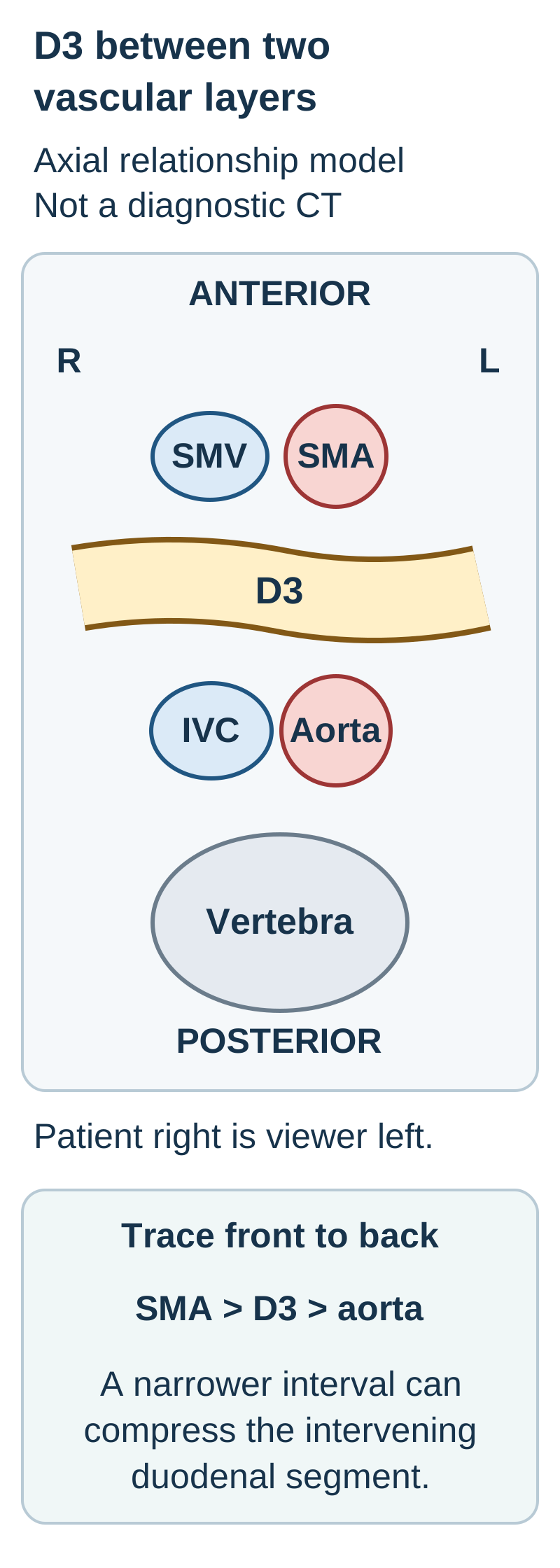
The third duodenal part, D3, travels from right to left anterior to the aorta and IVC, but posterior to the superior mesenteric artery and vein. In contrast, the pancreatic neck and body cross at a higher level; the uncinate process extends posterior to the superior mesenteric vessels. A composite sketch is not one literal CT slice. [2][8]
Trace it: on the D3 relationship diagram, travel from anterior to posterior through the superior mesenteric artery, duodenum and aorta. Predict which hollow structure is compressed when the interval between the artery and aorta narrows. Then locate the IVC using the patient's right, not the screen's right.
Trace the superior mesenteric artery (SMA), D3 and aorta from front to back. The superior mesenteric vein (SMV) is drawn on the patient right of the artery. This diagram exaggerates spacing and omits other organs; it is not a scan or a diagnostic measurement. [1][2][9]
The intervening bowel is D3. Loss of the cushioning fat can contribute to duodenal compression, but a narrow interval alone does not establish symptomatic obstruction. Symptoms, proximal dilation and the demonstrated transition must agree. This reasoning uses two relationships: what lies between the vessels, and what an obstructed segment does to the bowel upstream. [2]
Check your prediction with a different lesion
A collection centered just anterior to the vertebral body and behind D3 is closer to the great vessels than to the jejunal mesentery. Trace its continuity before naming its source. The same posterior position can contain blood, inflammatory fluid or a mass.
Carry it forward: if a renal mass distorts the normal image, first identify the spine and trace each vessel on neighboring sections. Do not reverse right and left because a familiar organ has been displaced.
Same adult location, different developmental histories
Does posterior position mean the organ developed there? No. Primary retroperitoneal structures developed outside the freely suspended gut mesentery. Secondary retroperitoneal organs began with a mesenteric attachment and later became applied to the posterior wall. These names describe developmental relationships, not the severity of an injury. [1][3]
Classify the relationship before predicting mobility
Relationship
Typical examples
What the attachment predicts
RelationshipPrimary retroperitoneal
Typical examplesKidneys, adrenal glands, abdominal ureters, aorta and IVC
What the attachment predictsPosterior location without a former free gut mesentery
RelationshipSecondary retroperitoneal
Typical examplesMost duodenum, pancreatic head and body, ascending and descending colon
What the attachment predictsBroad posterior attachment after developmental apposition
RelationshipIntraperitoneal
Typical examplesStomach, spleen, jejunum, ileum, transverse colon and sigmoid colon
What the attachment predictsExtensive serosal covering with mesenteric or ligamentous suspension
Intraperitoneal does not mean an organ floats inside the potential space between visceral and parietal peritoneum. Think of pressing a fist into a soft balloon without puncturing it: the surface wraps around the fist, but the fist is not inside the balloon's air. The analogy explains the covering, not the exact shape or strength of mesenteries. [1]
Compare two specimens: one has a renal capsule and perirenal fat; another has a colon and an apposed mesocolon that can be separated from posterior tissue. Both were posterior before dissection. Which one acquired its attachment during gut development?
The colon is secondary; the kidney is primary. Their adult positions overlap, but only the colon belongs to the suspended gut that became attached. A retained mesentery permits greater mobility, while ligaments, vessel pedicles and neighboring organs still constrain an intraperitoneal organ. [1][4]
Try the same distinction with a jejunal loop
A jejunal loop remains attached by a vascular mesentery rather than a broad posterior fusion surface. It is intraperitoneal even when it lies against the posterior abdomen on one scan. Contact on an image is not developmental fixation.
Carry it forward: a lesion's distance from the back is not enough to classify the organ. Establish its covering and attachment before predicting operative mobility or the earliest route of leakage.
How suspension becomes posterior attachment
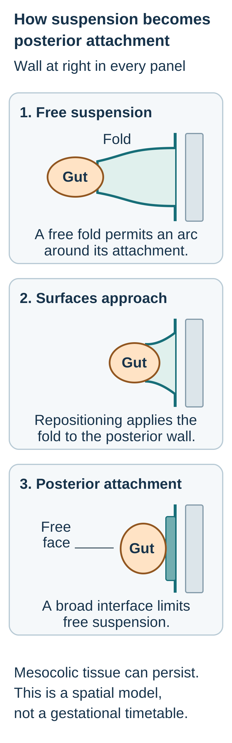
What changes when two peritoneal surfaces remain apposed? During differential growth and gut repositioning, including the usual rotation sequence, parts of the duodenum, pancreas and colon are applied to the posterior wall. Their mesenteric surfaces become incorporated into an attachment plane. The result is less free suspension, not a transformation into a kidney-like developmental origin. [3][4]
Read the fixation diagram vertically. First, a fold suspends the organ away from the wall. Second, the fold approaches the posterior lining. Third, the apposed tissues form a fascial attachment, leaving the anterior aspect facing the peritoneal compartment. This is a simplified spatial model, not a time-lapse of a specified gestational week.
Compare the arc possible around a free fold with the broad attached interface. Mesenteric tissue need not disappear when free suspension is lost. This schematic illustrates the classical attachment model without prescribing a week of gestation. [3][4]
Predict the difference: compare the first and third panels. Imagine gently lifting the organ anteriorly. In the first, the free fold permits an arc of mobility. In the third, a broad attachment must be separated before similar mobilization is possible. Name the anatomical change responsible before reading the explanation.
The free mesenteric suspension has become an apposed posterior attachment. In the colon, this is conventionally taught as secondary retroperitoneal fixation through fusion fascia. Modern operative observations show that mesocolic tissue remains continuous and can be mobilized along a plane. Avoid saying the entire mesocolon disappears. [4]
What would persistence of a free mesocolon predict?
The involved segment would have greater mobility than the usual attached ascending or descending colon. Its tissue identity would still be colon. A variation in fixation is not the same as failure of intestinal return, an open abdominal wall, or a primary retroperitoneal origin.
Carry it forward: when a surgeon separates the ascending colon from the posterior wall, the operation exposes an anatomical plane created during development. It does not make the original organ primary retroperitoneal, and the ureter behind that plane must still be identified and protected.
Use SAD PUCKER as a reminder, not a boundary map
Which part is actually posterior? The mnemonic can recall suprarenal glands, aorta and IVC, duodenum, pancreas, ureters, colon and kidneys. Its esophagus and rectum entries require regional qualification. The adult relationship is more useful than the letter alone. [1][2][5][8]
The exceptions that change an anatomical prediction
Structure
Useful distinction
StructureDuodenum
Useful distinctionThe proximal cap, the initial few centimeters, is mobile and intraperitoneal. The distal first part and D2-D4 are attached posteriorly. Do not assign the whole first part the cap's mobility. [9]
StructurePancreas
Useful distinctionMost of the gland is secondarily retroperitoneal. The tail reaches the splenic hilum within the splenorenal ligament, beside the splenic vessels. It is the important intraperitoneal exception. [8]
StructureColon
Useful distinctionAscending and descending colon are usually attached posteriorly. Transverse and sigmoid colon retain mesocolic suspension. Variants occur. [1][4]
StructureEsophagus
Useful distinctionThe thoracic esophagus belongs to the mediastinum, not the abdominal retroperitoneum. Specify the region of this long organ rather than labeling the entire esophagus retroperitoneal. [10]
StructureRectum
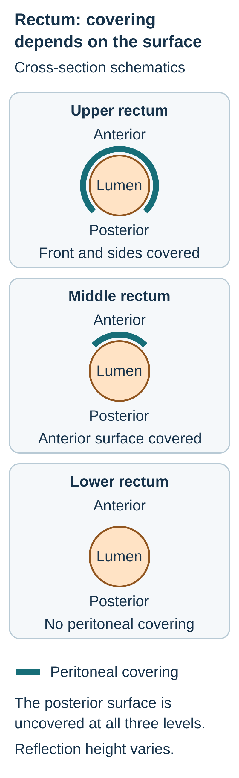
Useful distinctionDescribe pelvic extraperitoneal or subperitoneal relationships and the peritoneal reflection. The upper third has anterior and lateral covering, the middle third anterior covering, and the lower third no peritoneal covering. [5]
The rectal coverage diagram represents surfaces, not three completely separate compartments. A lesion's height and the wall it reaches both matter. The upper rectum's posterior surface is not serosa-covered merely because its anterior surface is. Individual reflection height varies, so a fixed distance from the anal verge cannot replace imaging or operative localization. [5]
Identify the surface before predicting where a full-thickness defect first communicates. The drawings are simplified surface maps, not precise thirds on an individual MRI; the peritoneal reflection varies. [5]
Predict the destination: compare a full-thickness anterior injury above the anterior peritoneal reflection with one below it. Which can directly enter the peritoneal cavity, and which first reaches pelvic extraperitoneal tissue?
The upper anterior injury can communicate directly with the peritoneal cavity. Below the reflection, the initial local pathway is extraperitoneal. Neither location determines how small, contained or clinically harmless the injury will be. [5]
Now transfer the exception to splenic surgery
A dissection beside the splenic vessels at the hilum can injure the pancreatic tail because both occupy the splenorenal attachment. A later pancreatic leak is explained by that local relationship, not by assuming the entire pancreas lies in the peritoneal cavity. [8]
Predict mobility before choosing an operative plane
Why does a sigmoid loop lift more readily than the descending colon? Its mesocolon remains a free suspension. The descending colon usually has a broad posterior attachment that limits its mobility until mobilization. Neither organ is completely without constraints: mesenteries carry vessels, nerves and lymphatics, and attachments vary among people. [1][4]
A practical comparison is a suspended curtain versus material laid against a backing sheet. The suspended part can swing around its attachment; the apposed part cannot do so until its interface is separated. The analogy describes freedom of suspension, not permission to pull on living tissue.
Choose the tissue that should stay behind: during left colon mobilization, compare the descending mesocolon, the ureter crossing the posterior wall, and the sigmoid mesocolon. Predict which structure is at risk if the dissection strays too deeply behind the intended plane.
The ureter belongs to the posterior urinary pathway, not to the mesocolic specimen. Staying in an identified plane helps separate mesocolon from retroperitoneal structures; inflammation, prior surgery and variant anatomy can obscure that plane. The same reasoning applies on the right, where the duodenum also becomes an important deep neighbor. [1][4][7]
Does respiratory displacement make a kidney intraperitoneal?
No. A kidney can change position with breathing while remaining within its retroperitoneal fascial relationships. Primary retroperitoneal does not mean absolutely immobile, and displacement does not create a free gut mesentery.
Carry it forward: two patients may have different colonic mobility without either scan being incorrectly labeled. Describe the observed attachment. Do not force variant anatomy into an absolute rule learned from a mnemonic.
Use the compartment to interpret a leak, not dismiss it
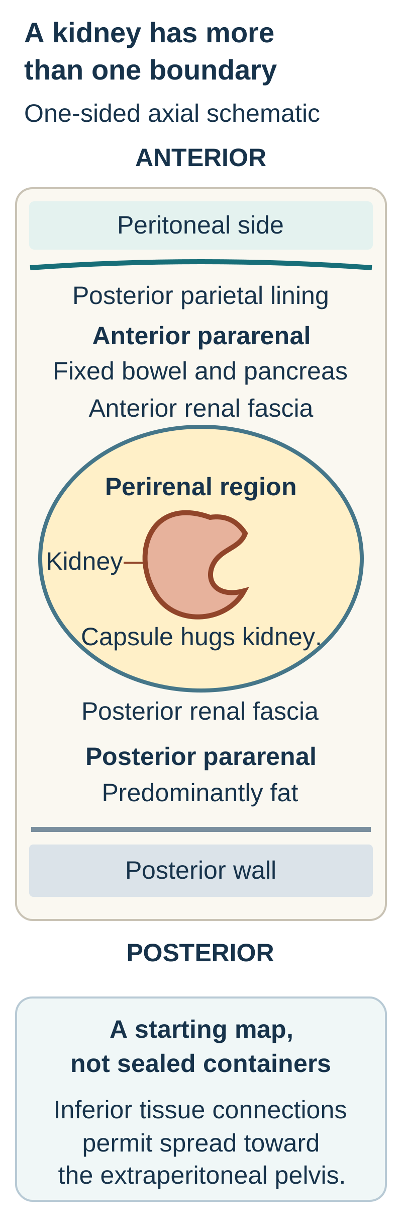
Where can fluid accumulate before it reaches the general peritoneal cavity? The conventional three-space model distinguishes an anterior pararenal region, a perirenal region and a posterior pararenal region. At renal levels, the anterior pararenal region lies between posterior parietal peritoneum and anterior renal fascia; it includes the fixed duodenum, much of the pancreas and the ascending or descending colon. The perirenal region contains the kidneys, adrenal glands, proximal urinary collecting structures, vessels and fat. The posterior pararenal region lies behind posterior renal fascia and is predominantly fat. Great vessels occupy the central retroperitoneum, not the kidney's capsule. [1]
The fascial-space diagram shows a useful starting map, not sealed containers. Renal fascia is separate from the fibrous capsule directly on the kidney. Fluid can extend along fascial planes and inferior connections into the pelvic extraperitoneal tissues. Cadaver injection studies demonstrated communications between these regions; their existence does not predict an identical route or rate in every living patient. [6]
The capsule directly hugs the renal surface; renal fascia encloses a larger region. This one-sided schematic omits central great vessels and many contents. The posterior-wall line represents its fascial covering. Axial boundaries do not imply sealed longitudinal compartments. [1][6]
Trace two possible leaks: begin at a fixed duodenal wall and then at the renal collecting system. For each, name the first nearby region and explain why fluid next to a kidney does not by itself establish renal injury.
A posterior fixed-duodenal leak can first enter the anterior pararenal region. A collecting-system leak can produce perirenal urine. Pancreatic or bowel fluid can also reach neighboring planes. Determine the source by continuity, organ injury and the appropriate contrast phase, rather than the final pool's location alone. [1][6][7]
Blood from a renal or vascular injury, gas from an injured fixed bowel segment, and urine from a ureteral disruption can initially remain extraperitoneal. Flank or back pain and limited early generalized peritoneal signs do not exclude serious injury. A negative focused abdominal ultrasound for free intraperitoneal fluid cannot exclude retroperitoneal hemorrhage. For a stable patient with suspected renal or ureteral injury, contrast-enhanced CT and appropriately delayed excretory images can answer questions that a single early phase cannot. An unstable patient requires urgent resuscitation and trauma assessment; do not delay time-critical care simply to complete a routine CT protocol. [1][7]
A posterior duodenal ulcer also has a vascular hazard: the gastroduodenal artery lies behind the proximal duodenum. Gastrointestinal bleeding can arise from arterial erosion without a retroperitoneal perforation. Posterior location is a relationship, not one inevitable complication. [2]
Compare gas beside the psoas with free intraperitoneal gas
Gas tracking along posterior fascial planes suggests an extraperitoneal route, while free gas in the peritoneal compartment describes a different distribution. Neither pattern alone identifies one organ, excludes communication across compartments, or determines treatment. Combine distribution with the wall defect, recent procedure, symptoms and physiological status.
Carry it forward: localize the center, trace a connection, identify the material, then check the patient's trajectory. Anatomy narrows the differential; it does not replace assessment of shock, sepsis or ongoing leakage.
Apply the relationships
Case 1
Show answer and explanations for case 1
A. The pyloric canal compressed between the liver and pancreas (Why this does not fit)
Pyloric obstruction dilates the stomach proximal to the duodenum. This transition is beyond the proximal duodenum and lies between the aorta and a midgut arterial branch.
Reasoning steps for option A
Why might pyloric narrowing cause vomiting and gastric dilation?
Pyloric obstruction dilates the stomach proximal to the duodenum.
Why does a transition beyond the proximal duodenum between the aorta and midgut artery rule out the pylorus?
This transition is beyond the proximal duodenum and lies between the aorta and a midgut arterial branch.
B. The jejunal origin compressed between the kidney and spleen (Why this does not fit)
The jejunal origin is near the left upper abdomen and continues from the duodenojejunal flexure. The demonstrated bowel segment is between the aorta and superior mesenteric artery, not between kidney and spleen.
Reasoning steps for option B
Where does the jejunal origin lie relative to the duodenojejunal flexure?
The jejunal origin is near the left upper abdomen and continues from the duodenojejunal flexure.
Why do the aorta and SMA, rather than the kidney and spleen, localize this narrowing elsewhere?
The demonstrated bowel segment is between the aorta and superior mesenteric artery, not between kidney and spleen.
C. The third duodenal part compressed between the aorta and SMA (Best answer)
D3 traverses anterior to the aorta and posterior to the superior mesenteric artery. Weight loss plus proximal dilation and a transition at this interval support a mechanically compressed D3.
Reasoning steps for option C
Which vessels bracket the third part of the duodenum?
D3 traverses anterior to the aorta and posterior to the superior mesenteric artery.
How do weight loss, upstream dilation, and the aortomesenteric transition implicate D3?
Weight loss plus proximal dilation and a transition at this interval support a mechanically compressed D3.
When does a narrow aortomesenteric interval establish symptomatic bowel compression?
Here, the aortomesenteric narrowing matches the D3 transition, proximal gastric and duodenal dilation, and postprandial vomiting after weight loss. A narrow vascular interval without matching obstruction findings would not suffice.
D. The second duodenal part compressed between the IVC and liver (Why this does not fit)
D2 is related to the pancreatic head and lies anterior to the IVC. D2 does not cross the aortomesenteric interval described at the transition.
Reasoning steps for option D
Where is D2 in relation to the pancreatic head and IVC?
D2 is related to the pancreatic head and lies anterior to the IVC.
Why is D2 inconsistent with the bowel crossing between the aorta and SMA?
D2 does not cross the aortomesenteric interval described at the transition.
Takeaway: A narrow vascular interval becomes clinically meaningful when it matches the bowel transition and upstream dilation.
A. Abdominal aorta with retroperitoneal hemorrhage (Best answer)
The aorta usually lies to the patient's left of the IVC and anterior to the vertebral body. Viewer right is patient left in the stated orientation, and the demonstrated leak enters posterior extraperitoneal tissue.
Reasoning steps for option A
Where is the aorta relative to the IVC and vertebral body?
The aorta usually lies to the patient's left of the IVC and anterior to the vertebral body.
How does viewer-right extravasation into posterior tissue identify the vessel and bleeding compartment?
Viewer right is patient left in the stated orientation, and the demonstrated leak enters posterior extraperitoneal tissue.
Why must image laterality and the hemorrhage compartment be evaluated separately?
Viewer right identifies patient left and supports the aorta as the leaking vessel; direct extravasation into posterior tissue identifies the retroperitoneal compartment. No free peritoneal fluid does not negate that demonstrated bleed.
B. Inferior vena cava with retroperitoneal hemorrhage (Why this does not fit)
The IVC usually lies to the patient's right of the aorta. The leaking vessel is on viewer right, which corresponds to patient left, reversing the position expected for the IVC.
Reasoning steps for option B
On which side of the aorta does the IVC usually lie?
The IVC usually lies to the patient's right of the aorta.
Why does a leak on viewer right in conventional axial orientation argue against IVC injury?
The leaking vessel is on viewer right, which corresponds to patient left, reversing the position expected for the IVC.
C. Abdominal aorta with free intraperitoneal hemorrhage (Why this does not fit)
An aortic leak can be life-threatening and may eventually enter more than one compartment. The scan directly describes the initial collection in posterior tissue rather than the peritoneal cavity.
Reasoning steps for option C
Can an aortic leak eventually reach multiple abdominal compartments?
An aortic leak can be life-threatening and may eventually enter more than one compartment.
Why does posterior extravasation without free fluid not establish initial intraperitoneal hemorrhage?
The scan directly describes the initial collection in posterior tissue rather than the peritoneal cavity.
D. Superior mesenteric vein with mesenteric hemorrhage (Why this does not fit)
Mesenteric venous injury can produce blood within mesenteric tissue. A vessel immediately in front of the spine and patient-left of the IVC is the aorta rather than the more anterior mesenteric vein.
Reasoning steps for option D
What kind of collection could a mesenteric venous injury produce?
Mesenteric venous injury can produce blood within mesenteric tissue.
Why does a leaking vessel anterior to the spine and left of the IVC favor aorta over mesenteric vein?
A vessel immediately in front of the spine and patient-left of the IVC is the aorta rather than the more anterior mesenteric vein.
Takeaway: Orient the image and establish the compartment separately; absence of free peritoneal fluid does not negate a demonstrated vascular leak.
A. Pancreatic neck with mesenteric vessels posterior to it (Why this does not fit)
The neck bridges anterior to the mesenteric venous confluence. The lesion is posterior to the mesenteric vessels and continuous with the lower head, which does not match the neck.
Reasoning steps for option A
What is the pancreatic neck's relation to the mesenteric venous confluence?
The neck bridges anterior to the mesenteric venous confluence.
Why does a lesion behind those vessels continuous with the inferior head not fit the neck?
The lesion is posterior to the mesenteric vessels and continuous with the lower head, which does not match the neck.
B. Pancreatic tail with splenic vessels within its ligament (Why this does not fit)
The tail reaches the splenic hilum in the splenorenal ligament. The described lesion remains by the pancreatic head and mesenteric vessels, far from the splenic hilum.
Reasoning steps for option B
Where does the pancreatic tail approach the splenic vessels?
The tail reaches the splenic hilum in the splenorenal ligament.
Why does a lesion beside the lower head and SMA not belong to the splenic-hilar tail?
The described lesion remains by the pancreatic head and mesenteric vessels, far from the splenic hilum.
C. Pancreatic body with the splenic artery along its upper border (Why this does not fit)
The body extends leftward across the posterior abdomen and relates to the splenic artery. An inferior head extension behind the mesenteric vessels is not the leftward body.
Reasoning steps for option C
What are the pancreatic body's course and splenic-artery relation?
The body extends leftward across the posterior abdomen and relates to the splenic artery.
Why is an extension from the lower head behind mesenteric vessels not pancreatic body?
An inferior head extension behind the mesenteric vessels is not the leftward body.
D. Uncinate process with mesenteric vessels anterior to it (Best answer)
The uncinate process projects from the lower head behind the superior mesenteric vessels. Continuity with the lower head and its posterior vascular position identify this process and the vessels in front of it.
Reasoning steps for option D
Which pancreatic projection lies behind the superior mesenteric vessels?
The uncinate process projects from the lower head behind the superior mesenteric vessels.
How does inferior-head continuity predict vessels anterior to this lesion?
Continuity with the lower head and its posterior vascular position identify this process and the vessels in front of it.
Why must the vascular relationship be assigned to the specific pancreatic subdivision rather than the gland as a whole?
The uncinate process lies behind the superior mesenteric vessels, unlike the pancreatic neck anterior to the venous confluence. The inferior-head continuity and posterior position must guide the operative prediction.
Takeaway: Pancreatic parts have different vascular relationships; a whole-gland rule cannot substitute for local anatomy.
A. A posterior pararenal hematoma not assessed by the ultrasound finding (Why this does not fit)
The posterior pararenal region lies behind posterior renal fascia. This collection surrounds the kidney inside renal fascia rather than lying behind it.
Reasoning steps for option A
Where is the posterior pararenal compartment relative to renal fascia?
The posterior pararenal region lies behind posterior renal fascia.
Why does blood surrounding the injured kidney inside the fascia exclude posterior pararenal localization?
This collection surrounds the kidney inside renal fascia rather than lying behind it.
B. A perirenal hematoma not excluded by the ultrasound finding (Best answer)
Perirenal blood surrounds a kidney within its fascial envelope. The renal laceration and collection location support this compartment, while a search for free peritoneal fluid tests a different space.
Reasoning steps for option B
Where does perirenal blood lie relative to the renal fascial envelope?
Perirenal blood surrounds a kidney within its fascial envelope.
Why can a negative free-peritoneal-fluid ultrasound coexist with this renal laceration and hematoma?
The renal laceration and collection location support this compartment, while a search for free peritoneal fluid tests a different space.
Why does absence of free intraperitoneal fluid fail to exclude a retroperitoneal renal injury?
The ultrasound assessed free intraperitoneal fluid, not blood surrounding the lacerated kidney within renal fascia; its negative result cannot exclude this perirenal hematoma.
C. A renal subcapsular hematoma excluded by the ultrasound finding (Why this does not fit)
A subcapsular collection lies directly beneath the fibrous renal capsule. The described collection occupies surrounding perirenal tissue, and absent peritoneal fluid would not exclude either type of renal bleeding.
Reasoning steps for option C
What separates a subcapsular hematoma from perirenal blood?
A subcapsular collection lies directly beneath the fibrous renal capsule.
Why do the collection surrounding the kidney and negative peritoneal study not establish or exclude subcapsular bleeding?
The described collection occupies surrounding perirenal tissue, and absent peritoneal fluid would not exclude either type of renal bleeding.
D. An anterior pararenal hematoma excluded by the ultrasound finding (Why this does not fit)
Anterior pararenal blood lies anterior to anterior renal fascia. The collection is inside the fascia, and a negative peritoneal-fluid examination cannot rule out retroperitoneal blood.
Reasoning steps for option D
Where would anterior pararenal blood sit relative to anterior renal fascia?
Anterior pararenal blood lies anterior to anterior renal fascia.
Why does an intrafascial renal collection persist as a concern despite a negative peritoneal-fluid study?
The collection is inside the fascia, and a negative peritoneal-fluid examination cannot rule out retroperitoneal blood.
Takeaway: A negative examination of one compartment cannot exclude injury in another compartment.
A. Second duodenal part into the anterior pararenal region (Best answer)
The major papilla opens in D2, a fixed segment associated with the pancreatic head. A posterior defect there can enter the region anterior to renal fascia without first entering the perirenal space.
Reasoning steps for option A
Which duodenal part contains the major papilla beside the pancreatic head?
The major papilla opens in D2, a fixed segment associated with the pancreatic head.
Where would gas from its posterior wall defect collect relative to anterior renal fascia?
A posterior defect there can enter the region anterior to renal fascia without first entering the perirenal space.
How do the papilla and renal fascial boundary together identify both source and early compartment?
The major papilla identifies D2, and gas outside the anterior renal fascia localizes the early leak to the anterior pararenal region rather than the perirenal space.
B. First duodenal cap into the perirenal region (Why this does not fit)
The duodenal cap lies near the pylorus and differs from the attached descending segment. The papilla localizes the defect to D2, and the gas is outside rather than inside anterior renal fascia.
Reasoning steps for option B
How does the duodenal cap's location differ from the papilla-bearing descending duodenum?
The duodenal cap lies near the pylorus and differs from the attached descending segment.
Why do the papillary defect and extrafascial gas exclude a D1-to-perirenal leak?
The papilla localizes the defect to D2, and the gas is outside rather than inside anterior renal fascia.
C. Pancreatic tail into the anterior pararenal region (Why this does not fit)
Pancreatic leakage can extend through posterior tissue planes. A visible bowel defect at the papilla localizes the initiating injury to D2 rather than the tail at the splenic hilum.
Reasoning steps for option C
Can pancreatic leakage track through posterior abdominal planes?
Pancreatic leakage can extend through posterior tissue planes.
Why does the visible papillary bowel defect favor D2 over a splenic-hilar pancreatic-tail source?
A visible bowel defect at the papilla localizes the initiating injury to D2 rather than the tail at the splenic hilum.
D. Proximal ureter into the perirenal region (Why this does not fit)
A urinary disruption can cause a perirenal or periureteral collection. Intact collecting systems and a visible duodenal wall defect favor enteric leakage outside renal fascia.
Reasoning steps for option D
What distribution can a proximal urinary-tract disruption produce?
A urinary disruption can cause a perirenal or periureteral collection.
Why do intact collecting systems and a papillary wall defect favor enteric gas outside renal fascia?
Intact collecting systems and a visible duodenal wall defect favor enteric leakage outside renal fascia.
Takeaway: Identify the injured segment from its landmark, then use the fascial boundary to name the receiving region.
A. A renal collecting-system rupture draining into the pelvis (Why this does not fit)
Urinary extravasation can track inferiorly along posterior tissue planes. The observed continuity begins at pancreatic inflammation, and delayed images supply no urinary leak.
Reasoning steps for option A
How could collecting-system urine spread toward the pelvis?
Urinary extravasation can track inferiorly along posterior tissue planes.
Why do a pancreatic starting point and absent delayed urinary leak argue against renal rupture?
The observed continuity begins at pancreatic inflammation, and delayed images supply no urinary leak.
B. Pancreatic fluid extending through connected tissue planes (Best answer)
Retroperitoneal fascial regions communicate through potential pathways, including inferior connections. The serial scans show continuity from a pancreatic collection toward the pelvis without a demonstrated urinary source.
Reasoning steps for option B
What inferior communications exist between retroperitoneal fascial planes?
Retroperitoneal fascial regions communicate through potential pathways, including inferior connections.
How does serial extension from the inflamed pancreatic body explain pelvic extraperitoneal fluid?
The serial scans show continuity from a pancreatic collection toward the pelvis without a demonstrated urinary source.
Why need pelvic fluid along a continuous fascial plane not originate in the pelvis or urinary tract?
Serial continuity from the pancreatic collection along inferior fascial planes explains the new pelvic fluid. Its final position does not establish a separate pelvic or urinary source, and delayed images show no urinary leak.
C. A separate pelvic source spreading upward to the pancreas (Why this does not fit)
Pelvic extraperitoneal disease can extend into connected abdominal tissue planes. The documented sequence starts near the inflamed pancreas and extends inferiorly rather than beginning in the pelvis.
Reasoning steps for option C
How could a primary pelvic extraperitoneal process spread upward?
Pelvic extraperitoneal disease can extend into connected abdominal tissue planes.
Why does the documented pancreas-to-pelvis sequence reverse that proposed direction?
The documented sequence starts near the inflamed pancreas and extends inferiorly rather than beginning in the pelvis.
D. Peritoneal fluid passing through a defect in each renal capsule (Why this does not fit)
A defect in a tissue boundary can permit fluid to enter a neighboring location. No capsular defect or peritoneal origin is described; interfascial extension does not require bilateral renal injury.
Reasoning steps for option D
What breach would the proposed route through both renal capsules require?
It would require a defect in each fibrous renal capsule to allow fluid across those boundaries; no such defects are described.
Why is bilateral renal-capsule disruption unnecessary for this observed interfascial spread?
No capsular defect or peritoneal origin is described; interfascial extension does not require bilateral renal injury.
Takeaway: Fluid beside a kidney or in the pelvis can originate from a different organ along a communicating plane.
A. Renal arterial branch into the posterior pararenal region (Why this does not fit)
Arterial injury can produce contrast leakage during vascular enhancement. Delayed filling continuous with a calyx identifies excreted urine, and the collection surrounds the kidney rather than lying behind its fascia.
Reasoning steps for option A
During which CT phase would an injured renal artery typically leak contrast?
Arterial injury can produce contrast leakage during vascular enhancement.
Why does delayed calyceal continuity within renal fascia indicate urine rather than arterial blood behind the fascia?
Delayed filling continuous with a calyx identifies excreted urine, and the collection surrounds the kidney rather than lying behind its fascia.
B. Pancreatic duct into the anterior pararenal region (Why this does not fit)
Pancreatic fluid may collect near the kidney after extending through tissue planes. Direct continuity from an opacified calyx establishes a urinary rather than pancreatic pathway.
Reasoning steps for option B
Can pancreatic fluid reach tissue near the kidney?
Pancreatic fluid may collect near the kidney after extending through tissue planes.
Why does direct filling through a calyceal defect defeat a pancreatic-duct explanation?
Direct continuity from an opacified calyx establishes a urinary rather than pancreatic pathway.
C. Renal collecting system into the perirenal region (Best answer)
Excretory-phase contrast enters urine within calyces and the renal pelvis. The calyceal defect connects that contrast to the surrounding perirenal collection.
Reasoning steps for option C
What fills the calyces during the excretory CT phase?
Excretory-phase contrast enters urine within calyces and the renal pelvis.
How does contrast passing through a calyceal defect localize this perirenal collection's source?
The calyceal defect connects that contrast to the surrounding perirenal collection.
How can delayed rather than early opacification identify a collection's contents?
The early collection has uncertain contents. Delayed filling with excreted contrast through the calyceal defect identifies a urinary leak into the perirenal region.
D. Duodenal lumen into the free peritoneal cavity (Why this does not fit)
A bowel perforation may permit luminal contents to leave the gut. The demonstrated contrast arrives through urinary excretion and a calyceal defect, not through the bowel lumen.
Reasoning steps for option D
What can a duodenal perforation release?
A bowel perforation may permit luminal contents to leave the gut.
Why does excretory-phase continuity with a calyx rule out a bowel-to-peritoneum pathway?
The demonstrated contrast arrives through urinary excretion and a calyceal defect, not through the bowel lumen.
Takeaway: The phase in which a collection opacifies can identify its contents when anatomy alone cannot.
A. Inferior mesenteric vein within the colonic mesentery (Why this does not fit)
A mesenteric vein can be encountered in a colectomy specimen. A venous leak does not account for urine-rich output and excretory contrast leaving a tube over the psoas.
Reasoning steps for option A
In which operative tissue might the inferior mesenteric vein be encountered?
The inferior mesenteric vein can be encountered in colonic mesenteric tissue during colectomy.
Why do urine-rich drainage and excretory contrast from the psoas-side tube exclude venous injury?
A venous leak does not account for urine-rich output and excretory contrast leaving a tube over the psoas.
B. Left renal pelvis within the renal hilum (Why this does not fit)
The renal pelvis collects urine before it enters the ureter. The pelvis is intact, and the leak occurs in a separate descending tube along the posterior wall.
Reasoning steps for option B
How is the renal pelvis connected to the ureter?
The renal pelvis collects urine before it enters the ureter.
Why does an intact pelvis exclude it as the source of leakage from a descending tube?
The pelvis is intact, and the leak occurs in a separate descending tube along the posterior wall.
C. Sigmoid colon within its mobile mesocolon (Why this does not fit)
An anastomotic or colonic leak can cause postoperative collections. Urine-rich fluid with direct excretory-phase leakage identifies urinary rather than colonic disruption.
Reasoning steps for option C
Could a colonic anastomotic leak produce a collection after left colectomy?
An anastomotic or colonic leak can cause postoperative collections.
Why do elevated drain creatinine and excretory-phase extravasation point away from colon?
Urine-rich fluid with direct excretory-phase leakage identifies urinary rather than colonic disruption.
D. Left ureter behind the mobilized mesocolon (Best answer)
The abdominal ureter descends on the posterior wall and must be protected during colonic mobilization. The fluid composition, excretory contrast and psoas relationship identify a ureteral injury behind the intended mesocolic plane.
Reasoning steps for option D
Where does the abdominal ureter run during left colonic mobilization?
The abdominal ureter descends on the posterior wall and must be protected during colonic mobilization.
How do the psoas location and urine-rich excretory leak implicate the ureter behind mesocolon?
The fluid composition, excretory contrast and psoas relationship identify a ureteral injury behind the intended mesocolic plane.
Why should fluid composition be combined with tubular anatomical continuity after colectomy?
High drain creatinine identifies urine, while excretory contrast from a descending tube over the psoas identifies the ureter. Together they distinguish ureteral injury from a postoperative colonic leak.
Takeaway: Use fluid identity and anatomical continuity together to distinguish a urinary leak from a nearby bowel complication.
A. The colon developed posteriorly without a mesentery; the sheet is renal fascia (Why this does not fit)
Primary retroperitoneal structures are not derived from a freely suspended gut segment. The observed sheet is attached to the colon and is mesocolic tissue, not evidence of a primary renal-type origin.
Reasoning steps for option A
What developmental feature distinguishes a primary retroperitoneal structure from a mesenteric colon?
Primary retroperitoneal structures are not derived from a freely suspended gut segment.
Why is the intact colonic sheet mesocolon rather than renal fascia?
The observed sheet is attached to the colon and is mesocolic tissue, not evidence of a primary renal-type origin.
B. The colon remained freely suspended; the posterior attachment is acquired scar (Why this does not fit)
Inflammation or surgery can produce acquired adhesions. A normal dissection plane between apposed mesocolon and posterior tissue does not require an acquired scar or a completely free original suspension.
Reasoning steps for option B
What can cause an acquired posterior intestinal adhesion?
Inflammation or surgery can produce acquired adhesions.
Why does a normal plane behind intact mesocolon not imply scar or a freely suspended colon?
A normal dissection plane between apposed mesocolon and posterior tissue does not require an acquired scar or a completely free original suspension.
C. The mesocolon became apposed posteriorly; a separable fascial interface persists (Best answer)
Secondary fixation can preserve mesocolic continuity while establishing a posterior attachment. An intact mesocolic sheet and a posterior dissection plane are compatible with secondary retroperitoneal anatomy.
Reasoning steps for option C
What happens to mesocolon during secondary posterior fixation?
Secondary fixation can preserve mesocolic continuity while establishing a posterior attachment.
How can an intact vascular mesocolic sheet coexist with a separable posterior fascial interface?
An intact mesocolic sheet and a posterior dissection plane are compatible with secondary retroperitoneal anatomy.
Why does secondary retroperitoneal fixation not mean the mesocolon entirely disappears?
Posterior apposition can fix the ascending colon while retaining continuous mesocolic tissue. The intact sheet and separable fascial plane are therefore compatible with secondary retroperitoneal fixation.
D. The sheet is serosa alone; mesocolic tissue was completely lost (Why this does not fit)
Visceral peritoneum covers the intestinal surface and differs from the mesocolic sheet containing connective tissue and vessels. The dissection preserves an intact mesocolon, which is not explained by identifying the sheet as serosal covering alone.
Reasoning steps for option D
How does visceral serosa differ from a vascular mesocolic sheet?
Visceral peritoneum covers the intestinal surface and differs from the mesocolic sheet containing connective tissue and vessels.
Why does preservation of the intact mesocolon refute a serosa-only explanation?
The dissection preserves an intact mesocolon, which is not explained by identifying the sheet as serosal covering alone.
Takeaway: Secondary fixation describes an attachment relationship, not the disappearance of every mesocolic tissue layer.
A. Less free suspension in the first; greater mobility in the second (Best answer)
A broad posterior attachment restricts the arc permitted by a free mesenteric fold. The only altered developmental feature is apposition becoming attachment, so mobility differs without changing intestinal tissue identity.
Reasoning steps for option A
How does a broad posterior attachment affect mesenteric freedom of movement?
A broad posterior attachment restricts the arc permitted by a free mesenteric fold.
Why would the attached model be less mobile while the unfused model retains mobility?
The only altered developmental feature is apposition becoming attachment, so mobility differs without changing intestinal tissue identity.
Why should a change only in posterior attachment not imply altered intestinal identity?
Both models otherwise develop normally and retain intestinal identity. The changed feature is posterior attachment, which restricts free suspension rather than introducing a different developmental defect.
B. Primary retroperitoneal origin in the first; secondary origin in the second (Why this does not fit)
Primary and secondary retroperitoneal labels describe different developmental histories. Both modeled segments began with mesentery; neither acquires a primary origin from subsequent fixation.
Reasoning steps for option B
How do primary and secondary retroperitoneal developmental histories differ?
Primary retroperitoneal structures did not begin as freely suspended gut segments; secondary fixation attaches a previously mesenteric gut segment posteriorly.
Why can neither initially mesenteric model be labeled primary retroperitoneal?
Both modeled segments began with mesentery; neither acquires a primary origin from subsequent fixation.
C. An abdominal wall defect in the first; normal intestinal return in the second (Why this does not fit)
Failure of normal return or closure can produce an abdominal wall abnormality. The modeled change concerns posterior attachment after development, not an interruption of return through the umbilical region.
Reasoning steps for option C
What developmental failures can create an abdominal wall defect?
Failure of normal return or closure can produce an abdominal wall abnormality.
Why does differing posterior apposition say nothing about abnormal umbilical return or closure?
The modeled change concerns posterior attachment after development, not an interruption of return through the umbilical region.
D. Loss of a vascular supply in the first; retained mesenteric vessels in the second (Why this does not fit)
Mesenteries carry the vessels supplying their intestinal segments. Normal apposition changes suspension but does not remove the intestinal blood supply.
Reasoning steps for option D
What travels within a developing intestinal mesentery?
Mesenteries carry the vessels supplying their intestinal segments.
Why does posterior fixation not eliminate the first model's mesenteric blood supply?
Normal apposition changes suspension but does not remove the intestinal blood supply.
Takeaway: Predict what the altered attachment changes without adding a different developmental defect.
A. A primary retroperitoneal segment constrained by renal fascia (Why this does not fit)
Renal fascia is associated with primary retroperitoneal renal structures. The described intestinal segment has a persistent mesocolon and is not contained within the renal fascial envelope.
Reasoning steps for option A
Is renal fascia an attachment for renal structures or a persistent vascular mesocolon?
Renal fascia is associated with primary retroperitoneal renal structures.
Why does a cecum-to-hepatic-flexure segment with a mesocolon not belong within renal fascia?
The described intestinal segment has a persistent mesocolon and is not contained within the renal fascial envelope.
B. A mobile ascending colon constrained by its mesenteric attachment (Best answer)
The ascending colon is usually posteriorly attached, but a persistent free mesocolon permits variation in mobility. Its cecum-to-hepatic-flexure course establishes its identity, while the vascular fold still anchors it.
Reasoning steps for option B
Which colonic segment extends from cecum to hepatic flexure?
The cecum-to-hepatic-flexure segment is the ascending colon. It is usually posteriorly attached, but a persistent free mesocolon can permit greater mobility.
How does a persistent vascular mesocolon permit its mobility yet limit its excursion?
Its cecum-to-hepatic-flexure course establishes its identity, while the vascular fold still anchors it.
Why should observed mesocolic attachment override the usual expectation of posterior fixation?
The cecum-to-hepatic-flexure course still identifies ascending colon. Its observed vascular mesocolon explains greater mobility and remaining anchorage despite the usual expectation of broad posterior fixation.
C. A transverse colon constrained by the greater omentum (Why this does not fit)
The transverse colon typically has mesocolic suspension and omental relationships. Its course should run between the hepatic and splenic flexures rather than between cecum and hepatic flexure.
Reasoning steps for option C
What is the usual suspension of the transverse colon?
The transverse colon typically has mesocolic suspension and omental relationships.
Why does a cecum-to-hepatic-flexure course exclude transverse colon and its omental attachment?
Its course should run between the hepatic and splenic flexures rather than between cecum and hepatic flexure.
D. A detached ascending colon constrained by postoperative adhesions (Why this does not fit)
Acquired adhesions can restrict an otherwise mobilized intestinal segment. There is an intact vascular suspension and no scar; the findings support a developmental fixation variant rather than detachment.
Reasoning steps for option D
What effect can postoperative adhesions have on an otherwise mobilized ascending colon?
Acquired adhesions can restrict an otherwise mobilized intestinal segment.
Why do a scar-free vascular mesocolon and normal rotation favor persistent developmental mobility instead?
There is an intact vascular suspension and no scar; the findings support a developmental fixation variant rather than detachment.
Takeaway: Identify the bowel by continuity, then describe its observed attachment instead of treating usual anatomy as universal.
A. Gastrosplenic ligament between stomach and spleen (Why this does not fit)
The gastrosplenic ligament carries short gastric and left gastro-omental vessels. The demonstrated defect is in the pancreatic tail beside the splenic vascular pedicle rather than in the gastric attachment.
Reasoning steps for option A
Which vessels normally run in the gastrosplenic ligament?
The gastrosplenic ligament carries short gastric and left gastro-omental vessels.
Why does a tail defect beside divided splenic vessels implicate another splenic attachment?
The demonstrated defect is in the pancreatic tail beside the splenic vascular pedicle rather than in the gastric attachment.
B. Hepatoduodenal ligament between liver and duodenum (Why this does not fit)
The hepatoduodenal ligament contains the portal triad near the proximal duodenum. Its right upper abdominal location does not match a distal pancreatic defect at the splenic hilum.
Reasoning steps for option B
Which structures occupy the hepatoduodenal ligament?
The hepatoduodenal ligament contains the portal triad near the proximal duodenum.
Why is a right-sided portal-triad attachment unrelated to the splenic-hilar pancreatic defect?
Its right upper abdominal location does not match a distal pancreatic defect at the splenic hilum.
C. Gastrocolic ligament between stomach and transverse colon (Why this does not fit)
Opening the gastrocolic attachment can provide access toward the pancreas through the lesser sac. Access toward the pancreas is not the same as the ligament directly containing its distal end and splenic vessels.
Reasoning steps for option C
How can division of the gastrocolic ligament give access to the pancreas?
Opening the gastrocolic attachment can provide access toward the pancreas through the lesser sac.
Why does access through the lesser sac not place the pancreatic tail in that ligament?
Access toward the pancreas is not the same as the ligament directly containing its distal end and splenic vessels.
D. Splenorenal ligament between spleen and posterior wall (Best answer)
The splenorenal ligament contains the pancreatic tail and splenic vessels. The distal pancreatic defect at the divided splenic vascular pedicle localizes the relevant operative attachment.
Reasoning steps for option D
Which splenic attachment contains both pancreatic tail and splenic vessels?
The splenorenal ligament contains the pancreatic tail and splenic vessels.
How does the enzyme-rich hilar collection beside divided vessels identify that operative attachment?
The distal pancreatic defect at the divided splenic vascular pedicle localizes the relevant operative attachment.
Why can dissection of a ligament damage the pancreatic tail despite the gland's broader retroperitoneal position?
The tail lies with the splenic vessels in the splenorenal ligament, unlike most of the retroperitoneal gland. Dividing that attachment can therefore injure the distal pancreas.
Takeaway: A structure at an operative attachment can be injured even when most of its organ lies in a different peritoneal relationship.
A. Splenic artery erosion at the pancreatic tail (Why this does not fit)
The splenic artery supplies the pancreatic body and tail near the superior pancreatic border. The demonstrated vessel is behind the proximal duodenum, not near the left-sided distal gland.
Reasoning steps for option A
Where does the splenic artery run along the pancreas?
The splenic artery supplies the pancreatic body and tail near the superior pancreatic border.
Why does bleeding behind the duodenal bulb not localize to the distal pancreatic tail?
The demonstrated vessel is behind the proximal duodenum, not near the left-sided distal gland.
B. Gastroduodenal artery erosion into the ulcer (Best answer)
The gastroduodenal artery is a posterior neighbor of the proximal duodenum. Luminal gastrointestinal bleeding from the described artery supports erosion without requiring a perforation into posterior tissue.
Reasoning steps for option B
Which artery passes behind the proximal duodenum?
The gastroduodenal artery is a posterior neighbor of the proximal duodenum.
Why do hematemesis and angiographic bleeding support arterial erosion without posterior perforation?
Luminal gastrointestinal bleeding from the described artery supports erosion without requiring a perforation into posterior tissue.
Why do the demonstrated bulbar arterial bleed and absent enteric leak support erosion without establishing perforation?
Angiography identifies arterial bleeding behind the bulb into the ulcer, explaining hematemesis and melena. Posterior ulcer location does not itself establish perforation, and no enteric leak or extraluminal gas is demonstrated.
C. Superior mesenteric artery rupture anterior to D3 (Why this does not fit)
The SMA lies anterior to D3 and supplies the midgut and inferior pancreaticoduodenal territory. The ulcer and angiographic bleeding are localized to the proximal duodenum rather than the D3 crossing.
Reasoning steps for option C
How does the SMA relate to the third duodenal part?
The SMA lies anterior to D3 and supplies the midgut and inferior pancreaticoduodenal territory.
Why does a proximal bulbar ulcer not implicate an artery crossing anterior to D3?
The ulcer and angiographic bleeding are localized to the proximal duodenum rather than the D3 crossing.
D. Right renal artery erosion into the collecting system (Why this does not fit)
Renal arterial injury can bleed into renal tissue or the urinary collecting system. The observed bleeding enters the gastrointestinal tract from a vessel behind the duodenal bulb, not the urinary pathway.
Reasoning steps for option D
Where can a renal arterial injury direct its bleeding?
Renal arterial injury can bleed into renal tissue or the urinary collecting system.
Why does luminal gastrointestinal bleeding behind the bulb not indicate renal collecting-system hemorrhage?
The observed bleeding enters the gastrointestinal tract from a vessel behind the duodenal bulb, not the urinary pathway.
Takeaway: Posterior duodenal location can imply vascular erosion; it does not establish extraperitoneal perforation.
A. Both lesions have the same free suspension because both lie in D1 (Why this does not fit)
D1 is a named segment spanning the proximal duodenum. A shared segment name does not eliminate the difference between its mobile cap and its attached distal portion.
Reasoning steps for option A
Does the D1 label imply identical suspension across its proximal and distal portions?
No. D1 includes both the mobile proximal cap and the posteriorly attached distal first part, so its name alone does not establish identical suspension.
Why does the serosa-covered cap remain more mobile than the attached distal D1 lesion?
A shared segment name does not eliminate the difference between its mobile cap and its attached distal portion.
B. The cap requires posterior fascial release before the distal first part (Why this does not fit)
A broad posterior attachment may need release during mobilization. The broad attachment is described at the distal lesion rather than the freely covered cap.
Reasoning steps for option B
What posterior plane may require release to mobilize the distal first part?
A broad posterior attachment may need release during mobilization.
Which lesion actually has the broad posterior attachment requiring release?
The broad attachment is described at the distal lesion rather than the freely covered cap.
C. The distal lesion requires release of a posterior attachment for comparable mobilization (Best answer)
The distal first part is attached posteriorly, unlike the proximal cap. The observed surfaces predict greater free mobility at the cap and a required attachment-plane dissection distally.
Reasoning steps for option C
How does the distal D1 posterior attachment differ from the cap’s covering?
The distal first part is attached posteriorly, unlike the proximal cap.
What dissection is predicted when mobilizing the distal lesion to match the cap?
The observed surfaces predict greater free mobility at the cap and a required attachment-plane dissection distally.
When comparing duodenal sites, why must the exact portion rather than D1 alone determine mobility?
The proximal cap has serosal covering on both major surfaces, whereas distal D1 has a broad posterior attachment. Distal mobilization therefore requires release that the cap does not.
D. The cap has a primary retroperitoneal origin despite its free covering (Why this does not fit)
Primary retroperitoneal structures originate outside a freely suspended gut mesentery. The cap is part of the gut, and its described covering supports intraperitoneal rather than primary retroperitoneal anatomy.
Reasoning steps for option D
What developmental relationship defines a primarily retroperitoneal organ?
Primary retroperitoneal structures originate outside a freely suspended gut mesentery.
Why does the cap’s serosal covering argue against a primary retroperitoneal origin?
The cap is part of the gut, and its described covering supports intraperitoneal rather than primary retroperitoneal anatomy.
Takeaway: Assign peritoneal relationships to the specific portion, not to an entire named organ segment by default.
A. Peritoneal cavity above; pelvic extraperitoneal tissue below (Best answer)
Peritoneum covers the anterior rectal surface above the reflection, while the surface below lies in extraperitoneal tissue. The same anterior wall defect reaches different receiving compartments because it lies on opposite sides of the reflection.
Reasoning steps for option A
How does anterior rectal peritoneal covering change at the reflection?
Peritoneum covers the anterior rectal surface above the reflection, while the surface below lies in extraperitoneal tissue.
Where does each anterior full-thickness defect initially empty above versus below that reflection?
The anterior defect above the reflection directly reaches the peritoneal cavity; the anterior defect below it initially reaches pelvic extraperitoneal tissue.
Why must the injured wall and reflection level both guide the receiving-compartment prediction?
Both defects involve the anterior wall, whose covering changes at the reflection. Above it, leakage can enter the peritoneal cavity; below it, leakage first enters extraperitoneal tissue.
B. Pelvic extraperitoneal tissue above; peritoneal cavity below (Why this does not fit)
Both peritoneal and extraperitoneal routes are possible in rectal injury. This ordering reverses the actual change in anterior peritoneal covering across the reflection.
Reasoning steps for option B
Can rectal injury involve either peritoneal or extraperitoneal tissue?
Both peritoneal and extraperitoneal routes are possible in rectal injury.
Why is the proposed extraperitoneal-above, peritoneal-below ordering reversed?
The anterior surface is peritoneum-covered above the reflection and uncovered below it, so the proposed compartment order is the reverse of the anatomical route.
C. Peritoneal cavity above; peritoneal cavity below (Why this does not fit)
A rectal injury above the reflection can directly communicate with the peritoneal cavity. The lower injury has no peritoneal surface at its anterior wall and initially enters local extraperitoneal tissue.
Reasoning steps for option C
How can the upper anterior rectal defect directly access the peritoneal cavity?
A rectal injury above the reflection can directly communicate with the peritoneal cavity.
Why does the lower anterior defect not initially share that peritoneal route?
The lower injury has no peritoneal surface at its anterior wall and initially enters local extraperitoneal tissue.
D. Pelvic extraperitoneal tissue above; pelvic extraperitoneal tissue below (Why this does not fit)
An injury to an uncovered rectal surface can initially leak extraperitoneally. The upper anterior injury is explicitly above the reflection and directly traverses a covered surface.
Reasoning steps for option D
What route follows injury of an uncovered rectal surface?
An injury to an uncovered rectal surface can initially leak extraperitoneally.
Why is the upper anterior defect not initially confined to pelvic extraperitoneal tissue?
The upper anterior injury is explicitly above the reflection and directly traverses a covered surface.
Takeaway: The wall involved and its relationship to the reflection determine the initial route; height alone is incomplete.
A. Both have crossed a serosal surface because their height is the same (Why this does not fit)
The upper rectum has more peritoneal covering than the lower rectum. Its posterior surface remains uncovered; height does not give every wall the same serosal relationship.
Reasoning steps for option A
Does upper rectal height guarantee serosa on every wall?
No. The upper rectum has anterior and lateral peritoneal covering, but its posterior wall remains uncovered.
Why does the posterior lesion not cross serosa despite sharing the anterior lesion’s height?
Its posterior surface remains uncovered; height does not give every wall the same serosal relationship.
B. The posterior lesion enters the peritoneal cavity before adjacent fat (Why this does not fit)
An injury through a serosal surface may directly enter the peritoneal compartment. The posterior upper rectal wall lacks that serosal covering and abuts extraperitoneal fat.
Reasoning steps for option B
What would penetration of a serosal rectal wall permit?
An injury through a serosal surface may directly enter the peritoneal compartment.
What tissue does this posterior upper rectal defect reach instead of the peritoneal cavity?
The posterior upper rectal wall lacks that serosal covering and abuts extraperitoneal fat.
C. The anterior lesion remains extraperitoneal while the posterior lesion crosses serosa (Why this does not fit)
A lower anterior rectal lesion can remain below the peritoneal reflection. These are upper rectal lesions, and the described anterior serosa is crossed, while the posterior surface lacks serosa.
Reasoning steps for option C
When could an anterior rectal lesion remain below the peritoneal reflection?
A lower anterior rectal lesion can remain below the peritoneal reflection.
At this upper rectal level, which wall actually crosses serosa?
These are upper rectal lesions, and the described anterior serosa is crossed, while the posterior surface lacks serosa.
D. The posterior lesion enters extraperitoneal fat; the anterior lesion crosses peritoneal covering (Best answer)
Upper rectal covering is anterior and lateral, not circumferential. The observed wall directions therefore produce different immediate relationships at the same rectal height.
Reasoning steps for option D
Which upper rectal surfaces have peritoneal covering?
Upper rectal covering is anterior and lateral, not circumferential.
How do the opposite wall directions change the two lesions’ immediate destinations?
The posterior lesion enters adjacent extraperitoneal fat, whereas the anterior lesion crosses peritoneal covering despite being at the same height.
Why is upper rectal peritoneum better mapped by wall surface than by a circumferential label?
Upper rectal peritoneum covers the anterior and lateral surfaces, not the posterior wall. The two lesions therefore encounter different tissues at the same height.
Takeaway: Rectal peritoneal covering is a surface map, not a ring around every upper rectal cross-section.
A. Abdominal retroperitoneum because the esophagus appears in SAD PUCKER (Why this does not fit)
The mnemonic is used to recall abdominal posterior structures but requires regional qualification. A thoracic defect above the diaphragm cannot be assigned to the abdominal retroperitoneum from a mnemonic alone.
Reasoning steps for option A
What regional limitation applies when using SAD PUCKER for the esophagus?
The mnemonic is used to recall abdominal posterior structures but requires regional qualification.
Why does a defect above the diaphragm not initially enter abdominal retroperitoneum?
A thoracic defect above the diaphragm cannot be assigned to the abdominal retroperitoneum from a mnemonic alone.
B. Mediastinum because the injured esophageal segment is thoracic (Best answer)
The thoracic esophagus travels through the mediastinum above the diaphragm. Direct gas and fluid around the demonstrated thoracic defect localize the initial spread to mediastinal tissue.
Reasoning steps for option B
Through what thoracic compartment does the esophagus pass?
The thoracic esophagus travels through the mediastinum above the diaphragm.
Where do gas and fluid immediately around this thoracic defect localize?
Direct gas and fluid around the demonstrated thoracic defect localize the initial spread to mediastinal tissue.
What anatomical region should be identified before applying an abdominal mnemonic to an esophageal leak?
Identify the affected esophagus as thoracic and above the diaphragm. Its surrounding leak is mediastinal, not in an abdominal retroperitoneal compartment.
C. Peritoneal cavity because the esophagus joins the stomach (Why this does not fit)
The distal esophagus meets the stomach after traversing the diaphragm. Continuity with the stomach does not place a thoracic wall defect inside the abdominal peritoneal compartment.
Reasoning steps for option C
Where does the distal esophagus join the stomach relative to the diaphragm?
The distal esophagus meets the stomach after traversing the diaphragm.
Why does that junction not put the thoracic perforation in the peritoneal cavity?
Continuity with the stomach does not place a thoracic wall defect inside the abdominal peritoneal compartment.
D. Perirenal space because the posterior esophagus is near the spine (Why this does not fit)
The perirenal space is a posterior abdominal region around the kidney. Posterior position alone does not establish renal fascial containment, particularly above the diaphragm.
Reasoning steps for option D
Where is the renal fascial perirenal space situated?
The perirenal space is a posterior abdominal region around the kidney.
Why is posterior proximity to the spine insufficient to assign this thoracic gas to perirenal space?
Posterior position alone does not establish renal fascial containment, particularly above the diaphragm.
Takeaway: Use the named anatomical region before applying an abdominal mnemonic to a long organ.
A. Ascending colon with a posterior mesocolic attachment (Why this does not fit)
The ascending colon is usually applied to the right posterior abdominal wall. The imaged loop connects descending colon to rectum and has a free vascular fold rather than the stated broad attachment.
Reasoning steps for option A
What normal posterior-wall relationship characterizes the ascending colon?
The ascending colon is usually applied to the right posterior abdominal wall.
Why do the loop’s descending-to-rectal endpoints and vascular fold exclude ascending colon?
The imaged loop connects descending colon to rectum and has a free vascular fold rather than the stated broad attachment.
B. Transverse colon with an attached renal fascial envelope (Why this does not fit)
The transverse colon is usually suspended by its mesocolon. Its endpoints do not match the described loop, and renal fascia is not its suspensory attachment.
Reasoning steps for option B
How is the transverse colon normally suspended?
The transverse colon is usually suspended by its mesocolon.
Why do the loop’s endpoints and attachment fail to support transverse colon within renal fascia?
Its endpoints do not match the described loop, and renal fascia is not its suspensory attachment.
C. Sigmoid colon with a persistent free mesocolon (Best answer)
The sigmoid connects descending colon to rectum and retains a mesocolon. A suspended loop can rotate around its attachment more readily than a broadly attached descending segment.
Reasoning steps for option C
What bowel segment links descending colon to rectum on a retained mesocolon?
The sigmoid connects descending colon to rectum and retains a mesocolon.
How does that narrow vascular suspension permit rotation compared with the broadly attached descending colon?
A suspended loop can rotate around its attachment more readily than a broadly attached descending segment.
How should bowel continuity and mesocolic suspension be combined when identifying a twisted loop?
Continuity from descending colon to rectum identifies sigmoid colon. Its retained free mesocolon permits rotation around the vascular attachment more readily than the broadly attached descending colon.
D. Descending colon with complete mesocolic tissue loss (Why this does not fit)
The descending colon is usually less freely mobile because of posterior attachment. The involved loop lies beyond it, and posterior fixation does not mean all mesocolic tissue has disappeared.
Reasoning steps for option D
How does posterior attachment limit normal descending-colon mobility?
The descending colon is usually less freely mobile because of posterior attachment.
Why does the twist involve the segment beyond descending colon without implying total mesocolic loss?
The involved loop lies beyond it, and posterior fixation does not mean all mesocolic tissue has disappeared.
Takeaway: Trace a loop to identify it, then connect its suspension to the observed rotation.
A. Subcapsular first; perirenal second (Best answer)
The fibrous capsule directly covers the kidney, while renal fascia encloses a larger region containing perirenal fat. A collection beneath the capsule differs from one outside the capsule but still inside renal fascia.
Reasoning steps for option A
How do fibrous renal capsule and renal fascia enclose different regions?
The fibrous capsule directly covers the kidney, while renal fascia encloses a larger region containing perirenal fat.
Which collection is under the capsule, and which lies in fat within the fascial envelope?
The first, flattening crescent lies beneath the fibrous capsule and is subcapsular. The second occupies fat outside the capsule but within renal fascia and is perirenal.
Which enclosing boundary distinguishes subcapsular from perirenal trauma collections?
The fibrous capsule directly covers the kidney and encloses the subcapsular crescent beneath it. Renal fascia surrounds a larger space containing the second collection in perirenal fat.
B. Perirenal first; posterior pararenal second (Why this does not fit)
Perirenal and posterior pararenal regions are separated by posterior renal fascia. The first is specifically beneath the fibrous capsule, and the second remains within rather than behind renal fascia.
Reasoning steps for option B
What fascial boundary separates perirenal from posterior pararenal tissue?
Perirenal and posterior pararenal regions are separated by posterior renal fascia.
Why is neither the subcapsular crescent nor the intrafascial fat collection posterior pararenal?
The first is specifically beneath the fibrous capsule, and the second remains within rather than behind renal fascia.
C. Anterior pararenal first; subcapsular second (Why this does not fit)
Anterior pararenal tissue lies outside the anterior renal fascia. Neither description places the first collection outside renal fascia, and surrounding fat is not beneath the kidney capsule.
Reasoning steps for option C
Where is anterior pararenal tissue relative to anterior renal fascia?
Anterior pararenal tissue lies outside the anterior renal fascia.
Why cannot surrounding renal fat be called subcapsular or the first crescent anterior pararenal?
Neither description places the first collection outside renal fascia, and surrounding fat is not beneath the kidney capsule.
D. Posterior pararenal first; anterior pararenal second (Why this does not fit)
The pararenal regions lie on opposite sides outside the renal fascial envelope. Both described collections remain inside that envelope, with only the first lying beneath the fibrous capsule.
Reasoning steps for option D
Where do pararenal compartments sit relative to the renal fascial envelope?
The pararenal regions lie on opposite sides outside the renal fascial envelope.
Why do the capsule-level and intrafascial collections both contradict pararenal locations?
Both described collections remain inside that envelope, with only the first lying beneath the fibrous capsule.
Takeaway: Renal capsule and renal fascia are different boundaries; identify which one surrounds the collection.
A perirenal collection lies within the renal fascial envelope. The entire renal envelope is displaced anteriorly and the collection is explicitly behind its posterior fascia.
Reasoning steps for option A
What boundary encloses a perirenal collection?
A perirenal collection lies within the renal fascial envelope.
Why does displacement of the whole renal envelope exclude a collection inside it?
The entire renal envelope is displaced anteriorly and the collection is explicitly behind its posterior fascia.
B. Psoas muscle (Why this does not fit)
A psoas abscess occupies or expands the muscle itself. The muscle outline remains preserved while the collection occupies the fascial region in front of the body wall.
Reasoning steps for option B
How would a collection centered in psoas affect the muscle?
A psoas abscess occupies or expands the muscle itself.
Why does the preserved psoas outline favor a fascial space anterior to the body wall?
The muscle outline remains preserved while the collection occupies the fascial region in front of the body wall.
C. Anterior pararenal space (Why this does not fit)
The anterior pararenal space lies anterior to anterior renal fascia. The demonstrated collection is behind posterior renal fascia, on the opposite side of the renal envelope.
Reasoning steps for option C
On which side of anterior renal fascia is anterior pararenal space?
The anterior pararenal space lies anterior to anterior renal fascia.
Why does a collection behind posterior renal fascia exclude that anterior compartment?
The demonstrated collection is behind posterior renal fascia, on the opposite side of the renal envelope.
D. Posterior pararenal space (Best answer)
This region lies between posterior renal fascia and the fascial covering of the posterior wall. Both the boundary description and anterior displacement of the intact renal envelope locate the collection here.
Reasoning steps for option D
Which two fascial boundaries bracket posterior pararenal space?
This region lies between posterior renal fascia and the fascial covering of the posterior wall.
How does forward displacement of kidney and perirenal fat confirm this posterior location?
Both the boundary description and anterior displacement of the intact renal envelope locate the collection here.
Why do the described fascial boundaries and renal-envelope displacement outweigh proximity to the kidney?
The collection is outside and behind posterior renal fascia, pushing the intact renal envelope forward. Its position in front of the posterior-wall fascia identifies posterior pararenal rather than perirenal tissue.
Takeaway: Use fascial boundaries and displacement together to locate a collection rather than naming it from the nearest organ.
A. The initial quiet abdomen establishes an uncomplicated postoperative collection (Why this does not fit)
Some small postoperative collections cause limited symptoms. A quiet early peritoneal examination does not establish harmlessness, particularly when a urinary leak and worsening systemic findings are demonstrated.
Reasoning steps for option A
Can a small postoperative collection initially produce limited symptoms?
Some small postoperative collections cause limited symptoms.
Why do urine-rich ureteral leakage and later systemic decline defeat reassurance from a quiet abdomen?
A quiet early peritoneal examination does not establish harmlessness, particularly when a urinary leak and worsening systemic findings are demonstrated.
B. The later fever establishes entry of the leak into the peritoneal cavity (Why this does not fit)
Intraperitoneal leakage can cause inflammation and systemic illness. Fever can accompany an extraperitoneal complication; the new symptoms do not prove a change of compartment.
Reasoning steps for option B
Can intraperitoneal contamination cause fever and inflammation?
Intraperitoneal leakage can cause inflammation and systemic illness.
Why does fever alone fail to prove this ureteral leak has entered the peritoneal cavity?
Fever can accompany an extraperitoneal complication; the new symptoms do not prove a change of compartment.
C. An extraperitoneal leak initially spared the general peritoneum but remains clinically serious (Best answer)
Urine can initially collect outside the peritoneal cavity after ureteral disruption. The early examination fits that route, while the later deterioration requires prompt reassessment rather than reassurance from the original examination.
Reasoning steps for option C
Where may urine initially collect after an extraperitoneal ureteral disruption?
Urine can initially collect outside the peritoneal cavity after ureteral disruption.
How do initially absent guarding and later fever, tachycardia, and pain fit a serious extraperitoneal leak?
The early examination fits that route, while the later deterioration requires prompt reassessment rather than reassurance from the original examination.
Why do absent early guarding and later systemic deterioration not support a benign ureteral leak?
An extraperitoneal urinary leak can initially spare the general peritoneum, limiting guarding. Later fever, tachycardia and worsening pain require prompt reassessment despite that early examination.
D. The pelvic extension disproves the ureter as the source of the collection (Why this does not fit)
Pelvic collections can arise from several organs. The demonstrated ureteral continuity and urinary composition support a urinary source, and posterior tissue planes can extend into the pelvis.
Reasoning steps for option D
Can a pelvic collection have multiple potential sources?
Pelvic collections can arise from several organs.
Why do ureteral continuity, urinary composition, and posterior pelvic spread favor ureteral injury?
The demonstrated ureteral continuity and urinary composition support a urinary source, and posterior tissue planes can extend into the pelvis.
Takeaway: Limited early peritoneal signs describe a possible distribution, not a safe prognosis.
A. Both are intraperitoneal because each changes position (Why this does not fit)
Intraperitoneal organs often have greater freedom of suspension. Displacement with respiration does not give the kidney a mesentery or change its relationship to renal fascia.
Reasoning steps for option A
Does intraperitoneal mesenteric suspension often permit independent movement?
Intraperitoneal organs often have greater freedom of suspension.
Why does kidney movement together with renal fascia not confer a mesentery?
Displacement with respiration does not give the kidney a mesentery or change its relationship to renal fascia.
B. The kidney remains retroperitoneal; the jejunum retains mesenteric suspension (Best answer)
A primary retroperitoneal organ can undergo limited displacement, whereas jejunum is suspended by mesentery. The kidney keeps its fascial relationships while the jejunal loop demonstrates a different form of attachment and mobility.
Reasoning steps for option B
Can a primary retroperitoneal kidney move with respiration while jejunum moves on mesentery?
A primary retroperitoneal organ can undergo limited displacement, whereas jejunum is suspended by mesentery.
How do their distinct fascial and vascular attachments explain the observed movements?
The kidney keeps its fascial relationships while the jejunal loop demonstrates a different form of attachment and mobility.
Why should respiratory displacement not be equated with free mesenteric mobility?
The kidney moves with its renal fascia and remains retroperitoneal; the jejunal loop moves on its vascular mesentery. Displacement alone does not replace these distinct attachment relationships.
C. The kidney is secondarily retroperitoneal; the jejunum is primarily retroperitoneal (Why this does not fit)
Secondary fixation develops when a previously mesenteric gut segment becomes attached posteriorly. Kidneys do not arise as such gut segments, and a normally mesenteric jejunum is not primarily retroperitoneal.
Reasoning steps for option C
What developmental event produces secondary retroperitoneal fixation of gut?
Secondary fixation develops when a previously mesenteric gut segment becomes attached posteriorly.
Why are neither the kidney nor this normally mesenteric jejunum classified as proposed?
Kidneys do not arise as such gut segments, and a normally mesenteric jejunum is not primarily retroperitoneal.
D. Both are secondarily retroperitoneal despite their different attachments (Why this does not fit)
Secondary fixation involves gut segments that initially had mesenteric suspension. The kidney is not derived from a suspended gut segment, and the jejunum retains free mesentery rather than a secondary posterior attachment.
Reasoning steps for option D
What prior mesenteric relationship is required for secondary retroperitoneal fixation?
Secondary fixation involves gut segments that initially had mesenteric suspension.
Why do kidney origin and persistent jejunal mesentery refute secondary fixation of both?
The kidney is not derived from a suspended gut segment, and the jejunum retains free mesentery rather than a secondary posterior attachment.
Takeaway: Relative mobility supports anatomical interpretation, but motion alone does not define a peritoneal category.
A. Noncontrast abdominal CT with a urinary-stone protocol (Why this does not fit)
Noncontrast CT is useful for urinary calculi and can depict some collections. The unresolved questions are traumatic parenchymal enhancement and urinary extravasation, which need intravenous contrast and excretory assessment.
Reasoning steps for option A
What can a noncontrast stone-protocol CT show in the urinary tract?
Noncontrast CT is useful for urinary calculi and can depict some collections.
Why do suspected renal parenchymal trauma and urinary leakage demand contrast and delayed images?
The unresolved questions are traumatic parenchymal enhancement and urinary extravasation, which need intravenous contrast and excretory assessment.
B. Arterial-phase abdominal CT angiography without later images (Why this does not fit)
An arterial phase can help identify vascular injury or active arterial bleeding. A vascular study alone does not establish whether contrast-containing urine escapes from the collecting system during excretion.
Reasoning steps for option B
What does an arterial CT phase assess after trauma?
An arterial phase can help identify vascular injury or active arterial bleeding.
Which suspected collecting-system complication could arterial-only images miss?
A vascular study alone does not establish whether contrast-containing urine escapes from the collecting system during excretion.
C. Retrograde CT cystography focused on the bladder (Why this does not fit)
Retrograde cystography evaluates suspected bladder rupture. The supplied findings localize concern to the flank and upper urinary tract rather than a demonstrated bladder injury.
Reasoning steps for option C
Which injury is evaluated with retrograde CT cystography?
Why do flank tenderness and gross hematuria call for upper-tract rather than bladder-focused imaging?
The supplied findings localize concern to the flank and upper urinary tract rather than a demonstrated bladder injury.
D. Contrast-enhanced abdominal and pelvic CT with delayed excretory images (Best answer)
Contrast-enhanced CT evaluates renal injury, and delayed images can show collecting-system or ureteral leakage. Sustained stability permits imaging; gross hematuria and flank injury remain concerning despite a negative search for free peritoneal fluid.
Reasoning steps for option D
Which CT phases assess renal tissue and contrast-containing urinary extravasation?
Contrast-enhanced CT evaluates renal injury, and delayed images can show collecting-system or ureteral leakage.
Why do stability and gross hematuria warrant this study despite absent free fluid on ultrasound?
Sustained stability permits imaging; gross hematuria and flank injury remain concerning despite a negative search for free peritoneal fluid.
Why must delayed excretory images supplement renal trauma CT when collecting-system disruption is suspected?
Delayed excretory images can show contrast-containing urine escaping the collecting system or ureter, a question not resolved by early renal enhancement alone. The patient has sustained stability for this evaluation.
Takeaway: Match the imaging phases to the organ and the suspected type of leak after adequate physiological stabilization.
A. Descending colon into the anterior pararenal region (Best answer)
The descending colon is usually posteriorly attached and lies anterior to the renal fascial region. Its endpoints identify the bowel segment, and the posterior defect enters tissue anterior to renal fascia.
Reasoning steps for option A
Which colonic segment runs from splenic flexure toward sigmoid colon?
The descending colon runs from the splenic flexure toward the sigmoid colon and is usually posteriorly attached anterior to the renal fascial region.
Why does a posterior defect there yield gas anterior to left renal fascia?
Its endpoints identify the bowel segment, and the posterior defect enters tissue anterior to renal fascia.
How do segment endpoints and the renal fascial boundary jointly locate this perforation?
The splenic-flexure-to-sigmoid course identifies descending colon. Gas posterior to its defect but anterior to left renal fascia localizes the early leak to the anterior pararenal region.
B. Sigmoid colon into the perirenal region (Why this does not fit)
The sigmoid colon continues from the descending colon and has a mesocolon. The defect is upstream of the sigmoid, and the gas lies outside rather than inside the renal fascial envelope.
Reasoning steps for option B
How is sigmoid colon suspended downstream of descending colon?
The sigmoid colon continues from the descending colon and has a mesocolon.
Why do the upstream defect and extrafascial gas exclude sigmoid-to-perirenal spread?
The defect is upstream of the sigmoid, and the gas lies outside rather than inside the renal fascial envelope.
C. Transverse colon into the posterior pararenal region (Why this does not fit)
The transverse colon lies between the hepatic and splenic flexures. The damaged segment is beyond the splenic flexure, and the gas is anterior rather than posterior to renal fascia.
Reasoning steps for option C
Between which flexures does transverse colon extend?
The transverse colon lies between the hepatic and splenic flexures.
Why do the post-splenic defect and gas anterior to renal fascia refute transverse-to-posterior-pararenal spread?
The damaged segment is beyond the splenic flexure, and the gas is anterior rather than posterior to renal fascia.
D. Ascending colon into the anterior pararenal region (Why this does not fit)
The ascending colon is normally attached on the right and can leak into anterior pararenal tissue. The compartment is plausible, but the specified left-sided bowel continuity identifies descending rather than ascending colon.
Reasoning steps for option D
Where is ascending colon normally attached, and can it leak anterior pararenally?
The ascending colon is normally attached on the right and can leak into anterior pararenal tissue.
Why does left-sided splenic-to-sigmoid continuity identify descending rather than ascending colon?
The compartment is plausible, but the specified left-sided bowel continuity identifies descending rather than ascending colon.
Takeaway: Bowel continuity identifies the segment; its posterior relationship predicts the initial receiving region.
A. Renal origin established by proximity to the kidney (Why this does not fit)
Perirenal and nearby collections can result from renal disease. Proximity is weaker evidence than direct enteric communication, and the urinary study shows no leak.
Reasoning steps for option A
Can a collection near the kidney originate from renal pathology?
Perirenal and nearby collections can result from renal disease.
Why do direct enteric contrast passage and no urinary extravasation outweigh renal proximity?
Proximity is weaker evidence than direct enteric communication, and the urinary study shows no leak.
B. A second independent urinary leak despite normal delayed images (Why this does not fit)
More than one organ can be injured in a complex abdominal process. No second urinary communication is demonstrated; direct passage from the colon already explains this collection.
Reasoning steps for option B
Can separate urinary and enteric injuries coexist?
More than one organ can be injured in a complex abdominal process.
Why is an independent urinary leak unsupported when delayed images are negative and colon communicates directly?
No second urinary communication is demonstrated; direct passage from the colon already explains this collection.
C. Colonic origin established by demonstrated enteric continuity (Best answer)
Enteric contrast outside a bowel defect demonstrates communication with that bowel lumen. The posterior colonic defect connects directly to the collection while delayed urinary images do not demonstrate extravasation.
Reasoning steps for option C
What does contrast exiting through a bowel-wall defect establish?
Enteric contrast outside a bowel defect demonstrates communication with that bowel lumen.
How do posterior descending-colon communication and negative urinary images identify this collection’s source?
The posterior colonic defect connects directly to the collection while delayed urinary images do not demonstrate extravasation.
Why is directly observed organ-to-collection continuity stronger than neighboring-organ proximity?
Enteric contrast passes directly from the descending-colon defect into this collection, while delayed urinary images show no extravasation. That demonstrated connection supports a colonic source more strongly than renal proximity.
D. Primary pancreatic origin established by posterior fluid spread (Why this does not fit)
Pancreatic fluid can spread through posterior fascial pathways. A possible route does not outweigh the directly visualized descending-colon communication.
Reasoning steps for option D
Can pancreatic fluid follow posterior fascial pathways?
Pancreatic fluid can spread through posterior fascial pathways.
Why is a hypothetical pancreatic route weaker than observed contrast passage from descending colon?
A possible route does not outweigh the directly visualized descending-colon communication.
Takeaway: Direct continuity is stronger source evidence than the identity of the nearest organ.
A. A freely suspended duodenal loop is compressed; leakage begins intraperitoneally (Why this does not fit)
The proximal duodenal cap is more mobile and differs from the attached remainder. The horizontal great-vessel crossing identifies D3 rather than the cap; both the free-suspension assumption and the proposed initial compartment conflict with the findings.
Reasoning steps for option A
How does the proximal duodenal cap differ in mobility from the attached remainder?
The proximal duodenal cap is more mobile and differs from the attached remainder.
Why does the great-vessel crossing and posterior gas exclude a freely suspended, intraperitoneally leaking cap?
The horizontal great-vessel crossing identifies D3 rather than the cap; both the free-suspension assumption and the proposed initial compartment conflict with the findings.
B. A fixed duodenal segment is compressed against the spine; leakage begins posteriorly (Best answer)
D3 is attached posteriorly and lies in front of the great vessels and vertebral column. An anterior impact can compress this fixed segment, and its posterior wall injury can initially leak outside the general peritoneal cavity.
Reasoning steps for option B
What structures lie immediately behind the fixed horizontal duodenum?
D3 is attached posteriorly and lies in front of the great vessels and vertebral column.
How do handlebar compression and posterior wall injury explain both the trauma and extraperitoneal gas?
An anterior impact can compress this fixed segment, and its posterior wall injury can initially leak outside the general peritoneal cavity.
Why must fixed D3 compression and the initial posterior leak route be considered together?
D3 fixation allows anterior handlebar force to compress it against posterior structures. Its posterior wall defect explains local extraperitoneal gas without requiring early generalized peritoneal findings.
C. A fixed duodenal segment is compressed against the spine; leakage begins intraperitoneally (Why this does not fit)
Fixation makes a duodenal segment vulnerable to compression against posterior structures. This accounts for the injury mechanism but not the demonstrated posterior gas distribution with little free peritoneal fluid.
Reasoning steps for option C
Why can posterior fixation predispose duodenum to compression against the spine?
Fixation makes a duodenal segment vulnerable to compression against posterior structures.
Why does posterior gas with little free fluid contradict initial intraperitoneal leakage?
This accounts for the injury mechanism but not the demonstrated posterior gas distribution with little free peritoneal fluid.
D. A freely suspended duodenal loop is compressed; leakage begins posteriorly (Why this does not fit)
A posterior bowel-wall defect can initially release material outside the general peritoneal cavity. The receiving region fits, but D3 at the great-vessel crossing is attached posteriorly rather than freely suspended like the cap.
Reasoning steps for option D
Where may a posterior duodenal wall defect initially release gas?
A posterior bowel-wall defect can initially release material outside the general peritoneal cavity.
Why does the posterior leak route not imply that D3 is freely suspended?
The receiving region fits, but D3 at the great-vessel crossing is attached posteriorly rather than freely suspended like the cap.
Takeaway: Fixation can explain traumatic compression and a posterior leak without requiring early generalized peritoneal findings.