Clotting cascade: localize the defect, then explain the phenotype
Use thrombin-centered physiology, PT/aPTT patterns, mixing studies, bleeding phenotypes, anticoagulant targets, and DIC trends to localize hemostatic disorders.
A prolonged clotting test is not a diagnosis. The central task is to decide where coagulation is failing, then ask whether a factor is missing, blocked, consumed, or intentionally inhibited. By the end of this lesson, you should be able to connect a bleeding pattern and PT/aPTT result to the next discriminating test without treating the old pathway diagram as literal in-vivo anatomy.
Start with thrombin, not two isolated pathways
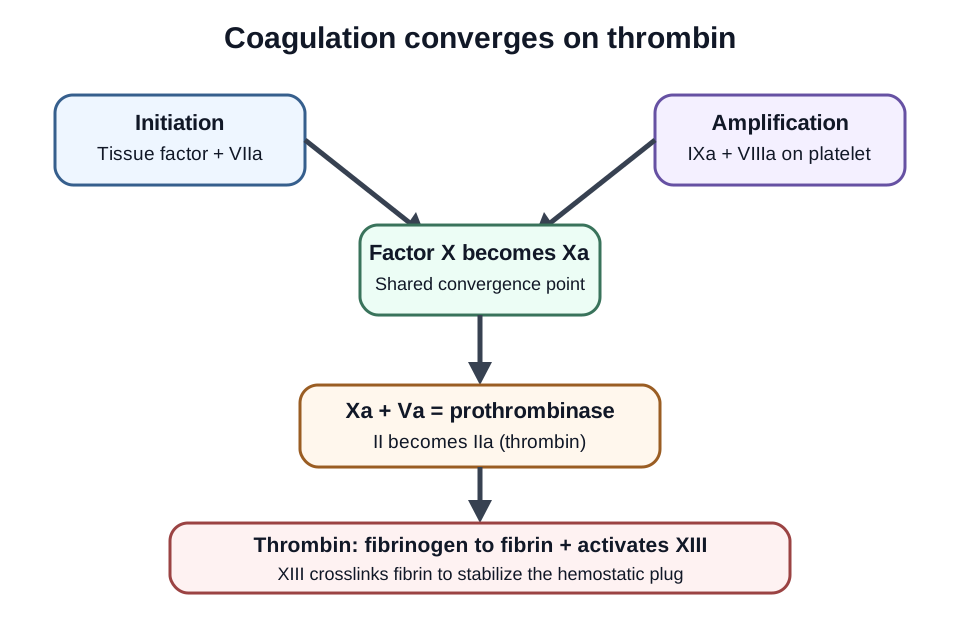
Common misconception: the intrinsic and extrinsic pathways operate as two separate pipes that independently make a clot. The classic diagram is useful for laboratory localization, but physiologic hemostasis is better understood as tissue-factor initiation on one cell surface, amplification, and a larger thrombin burst on activated platelets. [1]
Vessel injury exposes tissue factor to factor VII/VIIa. Early activated factor X helps generate a small amount of thrombin. Thrombin then activates platelets and the cofactors V and VIII. On the platelet surface, IXa with VIIIa forms tenase, which generates Xa. Xa with Va forms prothrombinase, which converts prothrombin, factor II, into much more thrombin. Thrombin cleaves fibrinogen, factor I, into fibrin and activates factor XIII, which stabilizes fibrin by crosslinking it. [1]
The classic pathway split is useful for testing, but both routes converge on Xa and thrombin; platelet-surface complexes amplify the response. [1]
Learner action: trace this sequence once from injury to stable fibrin: tissue factor plus VIIa, then Xa, then IIa, then fibrin, then XIII. Now trace the amplification route through IXa plus VIIIa and Xa plus Va. The visible consequence is that VIII and V are cofactors that accelerate enzyme complexes, while X and II are protease precursors whose activated forms directly drive thrombin generation.
Calcium and phospholipid surfaces support several coagulation complexes. This explains why a list of Roman numerals is less useful than remembering where the major enzyme complexes assemble. It also explains why severe VIII or IX deficiency impairs the thrombin burst even though tissue-factor initiation can still begin. [1][3]
Natural anticoagulant systems restrain the thrombin response. Activated protein C, with protein S, inactivates Va and VIIIa. Factor V Leiden makes factor V relatively resistant to activated protein C and is a hereditary venous thrombophilia, not a bleeding-factor deficiency. The prothrombin G20210A variant is another hereditary thrombophilia associated with increased venous thromboembolism risk. Finding either variant does not by itself dictate every anticoagulation decision; testing is useful only in selected clinical settings. [16]
Application: a child with recurrent hemarthroses, normal platelets, normal PT, and prolonged aPTT has a defect in the amplification side tested by aPTT. A factor VIII or IX assay can separate hemophilia A from B; the clinical bleeding pattern alone cannot. [3]
Use PT and aPTT as localization tests
Common misconception: a prolonged screening test directly measures bleeding risk. PT and aPTT instead test reagent-dependent routes to fibrin formation in plasma. PT is especially sensitive to factor VII plus the shared factors X, V, II, and fibrinogen. aPTT is especially sensitive to XII, XI, IX, VIII plus the shared factors. Reagent sensitivity and factor level matter, so real results do not obey a perfect binary map. [2]
A practical first pass is still powerful. Isolated PT prolongation suggests a problem centered on factor VII or a process that first affects it, such as vitamin K antagonism. Isolated aPTT prolongation suggests VIII, IX, XI, XII, heparin effect, lupus anticoagulant, or another inhibitor. Prolongation of both tests raises a shared-factor defect, multiple-factor deficiency, major anticoagulant effect, liver-related coagulopathy, or DIC. [2]
Screening pattern and first localization
PT
aPTT
First localization
Important exception
PTLong
aPTTNormal
First localizationVII or early multiple-factor effect
Important exceptionVerify specimen and medication context
PTNormal
aPTTLong
First localizationVIII, IX, XI, XII or inhibitor/drug
Important exceptionXII deficiency can be striking without bleeding
PTLong
aPTTLong
First localizationX, V, II, fibrinogen or broad acquired process
Important exceptionInterpret with platelets, fibrinogen, drugs, and illness
PTNormal
aPTTNormal
First localizationScreen does not exclude a hemostatic disorder
Important exceptionPlatelet/VWF disease and XIII deficiency remain possible
Learner action: imagine a patient with delayed postoperative bleeding but normal PT and aPTT. The correct consequence is not to declare coagulation normal. Factor XIII acts after the routine plasma clot endpoint and can be severely deficient while both screening times remain normal. A quantitative factor XIII activity assay is required when the phenotype fits. [8]
Application: if a screening result is unexpected, verify the specimen and drug exposure before ordering a wide panel. Then use the pattern to select focused studies. Mixing studies can help classify unexplained prolongation, but they are not a final diagnosis and are affected by anticoagulants, time, temperature, and assay method. [2]
Try it here · Checkpoint 1 of 3
Make your prediction before reading the choices. A first attempt is just a starting point.
Case 6
Show answer and explanations for case 6
A. Treat as hemophilia A with perioperative factor VIII replacement (Why this does not fit)
Hemophilia A causes factor VIII deficiency and a bleeding phenotype. This patient instead has severe factor XII deficiency with no personal or family bleeding history.
Reasoning steps for option A
What makes hemophilia A a tempting label for an aPTT of 92 seconds?
Hemophilia A is the most familiar cause of an isolated, correcting aPTT prolongation.
Which data contradict hemophilia A in this man?
Factor XII is below 1% and he has no personal or family bleeding history, so factor VIII replacement has no target.
B. Factor XII deficiency explains the prolonged assay without predicting surgical bleeding (Best answer)
Factor XII is required for contact activation in the aPTT assay, so severe deficiency explains the prolonged result. Congenital factor XII deficiency does not predict excessive surgical bleeding.
Reasoning steps for option B
Which assay result explains the very long aPTT?
Factor XII activity below 1% with a correcting mix identifies a contact-factor deficiency that the aPTT reagent depends on.
Why does this result not predict surgical bleeding?
Factor XII is not needed for hemostasis in vivo, which fits his lack of any bleeding history.
C. Treat factor XII deficiency as a thrombophilia with long-term anticoagulation (Why this does not fit)
Experimental factor XII biology does not make congenital deficiency an automatic indication for anticoagulation. The patient has no thrombotic history and the finding does not establish a treatment target.
Reasoning steps for option C
Why do some people link factor XII deficiency with thrombosis?
Experimental work shows factor XII contributes to thrombus formation, and older reports suggested a clotting tendency.
What stops long-term anticoagulation from being justified here?
He has no thrombotic history, and congenital XII deficiency is not an established indication for anticoagulation.
D. Give plasma before surgery to shorten the aPTT (Why this does not fit)
Plasma can shorten an abnormal aPTT by supplying factor XII, but the laboratory number is not the clinical problem here. Transfusion would expose an asymptomatic patient to risk without a bleeding indication.
Reasoning steps for option D
Why might plasma look like a fix before surgery?
Plasma supplies factor XII and would shorten the abnormal aPTT.
What makes plasma an unnecessary exposure?
The aPTT value is not a bleeding risk in XII deficiency, so transfusion adds risk without a clinical indication.
Takeaway: Factor XII deficiency is a high-yield example of a striking laboratory abnormality without a matching bleeding phenotype.
Let the bleeding phenotype refine the laboratory map
Common misconception: all inherited bleeding disorders look alike. Deep muscle bleeding and hemarthroses suggest a severe coagulation-factor disorder such as hemophilia, whereas epistaxis, gingival bleeding, easy bruising, and heavy menstrual bleeding more often suggest primary-hemostatic disorders such as von Willebrand disease. The pattern guides testing but does not replace it. [3][4]
Hemophilia A is factor VIII deficiency and hemophilia B is factor IX deficiency. Both are usually X-linked and can produce the same isolated aPTT pattern and deep-tissue phenotype. Specific factor activity testing distinguishes them. Severity and bleeding history matter more than the Roman numeral alone. [3]
Von Willebrand factor supports platelet adhesion and carries factor VIII in plasma. Modern VWD diagnosis uses VWF antigen plus a platelet-dependent VWF activity assay, with factor VIII and selected additional testing as needed. The old teaching that a prolonged bleeding time or a simple failed ristocetin test defines VWD is not current diagnostic practice. [4]
Learner action: compare two patients. Patient A has recurrent hemarthroses and factor VIII activity of 2%. Patient B has lifelong epistaxis and heavy menstrual bleeding with reduced VWF antigen and platelet-dependent VWF activity. The consequence is different localization: A has a coagulation-factor deficiency; B has a VWF disorder affecting primary hemostasis and sometimes factor VIII stability. [3][4]
Desmopressin can be useful for selected patients with VWD when an adequate response is expected or demonstrated, but it is not a universal treatment for every VWD subtype or every bleeding setting. Management depends on subtype, bleeding severity, procedure, contraindications, and prior response. [5]
Factor XII creates a different lesson. Severe deficiency can markedly prolong aPTT without causing a clinical bleeding phenotype. Do not infer that the laboratory value itself requires factor replacement. The historical claim that congenital XII deficiency itself establishes a clinical thrombophilia is not sufficiently supported for an individual patient. [7]
When a time is prolonged, ask missing factor or inhibitor
Common misconception: one 1:1 mix gives a universal yes-or-no answer. In a mixing study, patient plasma is combined with normal pooled plasma and the clotting test is repeated using a validated local method. Substantial correction supports a factor deficiency; persistent prolongation supports an inhibitor or anticoagulant effect. Laboratories use defined interpretation criteria rather than visual guesswork. [2]
A time-dependent factor VIII inhibitor can be harder. Some acquired hemophilia A samples correct more on the immediate mix and become more abnormal after incubation because the autoantibody progressively neutralizes factor VIII. Other inhibitors, lupus anticoagulants, and drug interference can complicate this pattern, so factor VIII activity and inhibitor testing remain essential when the phenotype suggests acquired hemophilia. [6]
How to read a mixing result
Observation
Interpretation to test
Next evidence
ObservationCorrection by validated criteria
Interpretation to testFactor deficiency becomes more likely
Next evidenceTargeted factor activity testing
ObservationLittle or no correction
Interpretation to testInhibitor or anticoagulant becomes more likely
Next evidenceDrug history, lupus anticoagulant studies, factor assays
ObservationGreater prolongation after incubation
Interpretation to testTime-dependent inhibitor becomes important
Next evidenceFactor VIII activity and Bethesda-type inhibitor assay when appropriate
Learner action: a previously well 72-year-old develops large spontaneous soft-tissue hematomas. PT is normal, aPTT is 68 seconds, VIII activity is very low, and the incubated mix fails to correct. The visible consequence is a focused evaluation for acquired hemophilia A rather than an inherited hemophilia label based on aPTT alone. [6]
Application: a patient with no bleeding history, normal PT, isolated prolonged aPTT, and a correcting study could have XI or XII deficiency, among other possibilities. A factor assay and clinical history determine whether the abnormal test carries bleeding relevance. [2][7]
Try it here · Checkpoint 2 of 3
Make your prediction before reading the choices. A first attempt is just a starting point.
Case 8
Show answer and explanations for case 8
A. Congenital hemophilia B (Why this does not fit)
Hemophilia B is factor IX deficiency and usually presents much earlier; the stem documents low VIII activity and an inhibitory pattern.
Reasoning steps for option A
Why might a hemophilia be considered in a man with hematomas and a long aPTT?
Isolated aPTT prolongation with serious soft-tissue bleeding looks like the classic hemophilia pattern.
Which findings exclude congenital hemophilia B?
He had no bleeding for 72 years, factor VIII rather than IX is low, and the incubated mix fails to correct.
B. Factor XII deficiency (Why this does not fit)
Factor XII deficiency does not cause this new severe bleeding phenotype and would not explain factor VIII activity of 3%.
Reasoning steps for option B
What makes factor XII deficiency a possible explanation for the long aPTT?
Factor XII deficiency can produce a striking isolated aPTT prolongation.
Why cannot factor XII deficiency explain this man's illness?
It causes no bleeding and would correct on mixing, while he has severe hematomas, VIII at 3%, and a noncorrecting mix.
C. Von Willebrand disease type 1 (Why this does not fit)
Type 1 VWD usually produces a mucosal phenotype and partial VWF deficiency; it does not usually produce this late-onset severe VIII-inhibitor pattern.
Reasoning steps for option C
Why could VWD be considered when factor VIII is low?
VWD can reduce factor VIII because VWF protects it in plasma.
Which features argue against type 1 VWD?
Type 1 VWD is lifelong and mucosal, and it does not produce a noncorrecting incubated mix with VIII at 3% in a previously normal older man.
D. Acquired hemophilia A (Best answer)
New bleeding without a lifelong history, isolated aPTT prolongation, very low VIII activity, and an inhibitory incubated mix support acquired factor VIII autoantibody disease.
Reasoning steps for option D
Which history points to an acquired rather than inherited disorder?
Large spontaneous hematomas began at age 72 with no lifelong bleeding history.
Which laboratory findings identify acquired hemophilia A?
An isolated long aPTT, factor VIII at 3%, and an incubated mix that stays prolonged indicate a factor VIII autoantibody.
Takeaway: Acquired hemophilia A should be considered when new bleeding accompanies isolated aPTT prolongation and low factor VIII.
Map anticoagulants to the protein they inhibit and the test that can answer the clinical question
Common misconception: every anticoagulant has one routine clotting time that accurately reports its intensity. Unfractionated heparin often uses aPTT or anti-Xa according to the local protocol. Low-molecular-weight heparin usually needs no routine monitoring, with anti-Xa reserved for selected situations. Direct oral anticoagulants generally do not need routine laboratory monitoring, and ordinary PT/aPTT results may be insensitive or reagent dependent. [9][15]
Unfractionated heparin accelerates antithrombin-mediated inhibition of thrombin (IIa) and factor Xa. If aPTT does not respond as expected, do not assume antithrombin deficiency immediately. High factor VIII, inflammation, thrombocytosis, antiphospholipid antibodies, dosing issues, and true antithrombin deficiency can all contribute to apparent heparin resistance. An anti-Xa assay can be useful when aPTT is discordant. [9]
Warfarin reduces functional vitamin K-dependent factors II, VII, IX, and X as well as proteins C and S. PT/INR is used for monitoring. Factor VII declines relatively quickly, helping explain why PT may become abnormal early even before the full antithrombotic effect is established. Fluconazole can increase warfarin effect through CYP inhibition, so INR needs close monitoring during the interaction. [14]
Learner action: place each drug at its dominant target: dabigatran at thrombin, apixaban/rivaroxaban at Xa, warfarin at vitamin K-dependent factor production, and heparin through antithrombin. The consequence is that a normal INR does not exclude clinically important dabigatran or factor Xa inhibitor exposure. [12][15]
Dabigatran has a specific reversal agent, idarucizumab, when reversal is needed for emergency surgery or life-threatening or uncontrolled bleeding. [12] U.S. teaching about Andexxa requires an update: the FDA reported that commercial U.S. sales ended December 22, 2025 after concluding that serious risks outweighed benefits. For major factor Xa inhibitor bleeding in the United States, use the current local emergency protocol; contemporary guidance describes prothrombin complex concentrates as an option when specific reversal is unavailable. [13][15]
DIC is a dynamic consumption process, not one laboratory threshold
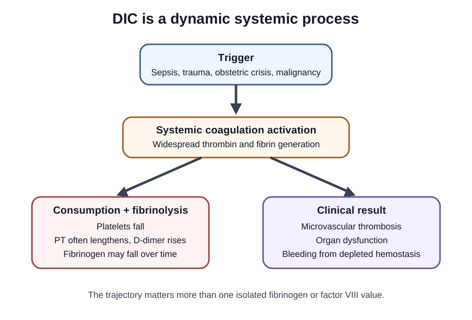
Common misconception: DIC means every coagulation factor is simply low at one moment. DIC is systemic activation of coagulation in the setting of an underlying disorder such as sepsis, major trauma, obstetric catastrophe, or malignancy. Fibrin formation can obstruct the microcirculation while platelets and coagulation proteins are consumed, producing simultaneous thrombosis and bleeding. [10]
DIC is a changing systemic process. Serial platelets, fibrin-related markers, coagulation times, fibrinogen, and clinical context are interpreted together. [10][11]
Typical overt DIC findings include falling platelets, prolonged PT and often aPTT, increased fibrin-related markers such as D-dimer, and declining fibrinogen. Fibrinogen is an acute-phase reactant and can be normal early, so a single normal value does not exclude DIC. Serial results and the clinical context matter. Schistocytes can appear when red cells cross fibrin-rich microvasculature but are not required for diagnosis. [10]
Learner action: compare a septic patient whose platelets fall from 180 to 55 x10^9/L, PT lengthens, D-dimer rises sharply, and fibrinogen falls over six hours with a cirrhotic patient whose abnormal tests are stable. The visible consequence is that trajectory plus the underlying illness carries more information than one isolated factor value.
Do not use factor VIII as a single separator between DIC and liver-related coagulopathy. A 2024 study found that factor VIII levels did not distinguish these conditions in the studied population. In a patient with liver disease, even the standard ISTH DIC score may perform differently, so diagnosis needs the clinical syndrome plus serial laboratory evidence rather than a memorized VIII rule. [11]
Treatment begins with the underlying cause. Blood components are generally used for bleeding or high-risk procedures rather than to normalize numbers in an otherwise stable patient. Platelet and plasma support depends on bleeding risk and the laboratory pattern; fibrinogen replacement is considered when clinically important hypofibrinogenemia accompanies bleeding. Repeat testing matters because DIC can change quickly. [10]
Application: if a patient with septic shock begins oozing from lines while platelets fall and fibrin markers rise, integrate all of those findings before assigning DIC. If an older adult instead has new large hematomas, isolated aPTT prolongation, low factor VIII, and an inhibitory mixing pattern, that is a different process and needs an acquired hemophilia pathway. [6][10]
Try it here · Checkpoint 3 of 3
Make your prediction before reading the choices. A first attempt is just a starting point.
Case 18
Show answer and explanations for case 18
A. Classify both profiles as liver-related coagulopathy because factor VIII is high (Why this does not fit)
Factor VIII can be high in chronic liver disease and in inflammatory states. A high value therefore cannot classify both patients as liver related.
Reasoning steps for option A
Why might a high factor VIII seem to point to liver disease?
Factor VIII is made largely outside hepatocytes and often stays high in cirrhosis while other factors fall.
Why cannot a high VIII classify both patients?
Factor VIII also rises in inflammation, so septic patient B can have a high value despite DIC.
B. Factor VIII alone cannot separate DIC from liver-related coagulopathy; compare clinical context and serial trajectory (Best answer)
The studied populations showed overlapping factor VIII levels in DIC and liver disease. Stable chronic findings versus rapidly worsening septic consumption provide the more discriminating evidence.
Reasoning steps for option B
How do factor VIII values compare between the two patients?
Both are elevated, at 165% and 150%, and studies show overlapping VIII levels in DIC and liver disease.
What evidence separates the two conditions instead?
Stable chronic values in cirrhosis versus rapidly worsening coagulopathy in septic shock distinguish liver disease from DIC.
C. Classify patient B as non-DIC because factor VIII is preserved (Why this does not fit)
Preserved or elevated factor VIII does not reliably exclude DIC, especially during inflammation. Patient B's septic trajectory remains strongly compatible with consumption.
Reasoning steps for option C
Why might a preserved factor VIII seem to argue against DIC?
Consumption in DIC should lower clotting factors, including VIII.
Why does that reasoning fail in patient B?
Inflammation raises factor VIII, so a normal or high value does not exclude DIC in a rapidly worsening septic patient.
D. Diagnose inherited hemophilia A in patient B from the lower factor VIII value (Why this does not fit)
Both factor VIII values are elevated rather than deficient. Hemophilia A would not explain thrombocytopenia and a broad acquired coagulopathy.
Reasoning steps for option D
Why might the lower VIII value in patient B draw attention?
Hemophilia A is defined by low factor VIII activity.
Why does hemophilia A not fit patient B?
B's factor VIII of 150% is above normal rather than deficient, and hemophilia A leaves the PT and platelet count normal, whereas B has both abnormal.
Takeaway: Factor VIII is not a reliable single discriminator between DIC and liver-related coagulopathy.
The deep-tissue bleeding pattern, X-linked family pattern, isolated aPTT prolongation, and measured VIII activity all support hemophilia A.
Reasoning steps for option A
Which findings point to a coagulation-factor disorder rather than a platelet problem?
Recurrent hemarthroses after minor trauma are deep-tissue bleeds, and an isolated long aPTT with normal PT and platelets localizes to the intrinsic factors.
What result names the missing factor in this boy?
Factor VIII activity of 2% identifies severe factor VIII deficiency, and the affected maternal uncle fits X-linked hemophilia A.
B. Hemophilia B from factor IX deficiency (Why this does not fit)
Hemophilia B can look identical clinically, but the stem directly documents severe factor VIII deficiency rather than IX deficiency.
Reasoning steps for option B
Why is hemophilia B a reasonable first thought for this boy?
It produces the same X-linked inheritance, hemarthroses, and isolated aPTT prolongation, so the phenotype alone cannot separate it.
Which measured value separates the two hemophilias here?
The assay shows factor VIII at 2%; factor IX deficiency would need a low IX level, which is not what was measured.
C. Von Willebrand disease (Why this does not fit)
VWD can lower factor VIII and cause mucosal bleeding, but recurrent hemarthroses with factor VIII activity of 2% and an X-linked family pattern more strongly support hemophilia A.
Reasoning steps for option C
What makes VWD tempting when factor VIII is low?
VWF carries factor VIII in plasma, so VWD can lower factor VIII activity and prolong aPTT.
Why does the bleeding pattern argue against VWD?
VWD usually causes mucosal bleeding, whereas recurrent hemarthroses with VIII at 2% and an affected maternal uncle fit severe hemophilia A.
D. Factor XII deficiency (Why this does not fit)
Factor XII deficiency can markedly prolong aPTT, but it does not explain recurrent hemarthroses or a factor VIII activity of 2%.
Reasoning steps for option D
Why might factor XII deficiency come to mind with a long aPTT?
Factor XII deficiency can prolong the aPTT dramatically because the test depends on contact activation.
Which clinical feature rules out factor XII deficiency as the explanation?
Factor XII deficiency does not cause bleeding, so it cannot explain hemarthroses or a factor VIII level of 2%.
Takeaway: Specific factor activity separates hemophilia A from clinically similar intrinsic-pathway disorders.
A. Congenital hemophilia A (Why this does not fit)
Hemophilia A usually causes deeper bleeding and isolated severe factor VIII deficiency; the stem instead documents reduced VWF quantity and activity.
Reasoning steps for option A
Why might hemophilia A be considered when factor VIII is reduced?
A low factor VIII activity is the defining laboratory feature of hemophilia A.
Which findings make hemophilia A the weaker fit for this woman?
Her bleeding is mucosal, and both VWF antigen and platelet-dependent VWF activity are reduced, so the mild VIII drop is secondary to low VWF.
B. Congenital factor XI deficiency (Why this does not fit)
Factor XI deficiency can cause procedure-related bleeding, but it does not explain reduced VWF antigen and activity.
Reasoning steps for option B
What makes factor XI deficiency plausible after dental bleeding?
Factor XI deficiency typically causes bleeding after procedures such as dental extraction.
Which laboratory results cannot be explained by factor XI deficiency?
Factor XI deficiency does not lower VWF antigen or platelet-dependent VWF activity.
C. Von Willebrand disease (Best answer)
The mucosal bleeding phenotype plus reduced VWF antigen and platelet-dependent activity with secondary VIII reduction fits VWD.
Reasoning steps for option C
Which bleeding history points toward primary hemostasis?
Lifelong epistaxis, heavy menstrual bleeding, and dental bleeding form a mucosal pattern typical of a platelet or VWF disorder.
What result confirms VWD rather than a platelet count problem?
The platelet count is normal while VWF antigen and platelet-dependent activity are both reduced, with factor VIII mildly lowered as a consequence.
D. Congenital factor XII deficiency (Why this does not fit)
Factor XII deficiency does not cause this bleeding phenotype and does not reduce VWF antigen or activity.
Reasoning steps for option D
Why might factor XII deficiency be raised in a bleeding workup?
It is a classic cause of an unexpectedly long aPTT during coagulation screening.
Why does factor XII deficiency fail to explain this patient?
Factor XII deficiency prolongs the aPTT yet leaves people clinically free of bleeding, and VWF levels are untouched, so her mucosal bleeding and low VWF results remain unexplained.
Takeaway: Modern VWD diagnosis relies on VWF-specific testing interpreted with the bleeding history.
A. Use desmopressin without relying on the prior response test (Why this does not fit)
Desmopressin responsiveness varies by VWD subtype and patient. This patient has useful response data, so ignoring it would discard the evidence that makes the plan individualized.
Reasoning steps for option A
Why is desmopressin itself a sensible choice for this patient?
The patient has type 1 VWD, the subtype most likely to respond to desmopressin, and no contraindication is listed.
What is lost by ignoring the prior response test?
Desmopressin response varies between patients, and the documented adequate VWF and VIII rise is the evidence that justifies the plan.
B. Use factor VIII concentrate alone because the platelet count is normal (Why this does not fit)
A normal platelet count does not correct impaired VWF-mediated adhesion. Replacement strategy must address the VWF disorder and the procedure rather than platelet number alone.
Reasoning steps for option B
What could make factor VIII concentrate seem adequate here?
Factor VIII is carried by VWF and may fall in VWD, and the platelet count is normal.
Why does a normal platelet count not solve the problem?
The defect is VWF-mediated platelet adhesion, which a normal count does not correct and factor VIII alone does not replace.
C. Proceed without hemostatic therapy because the dental procedure is minor (Why this does not fit)
Dental procedures can still provoke clinically important mucosal bleeding in VWD. The documented responsive therapy should be incorporated rather than dismissing risk from the procedure label.
Reasoning steps for option C
Why might no hemostatic therapy seem acceptable before dental work?
Dental procedures are minor, and type 1 VWD is often the mildest subtype.
What risk makes skipping therapy inappropriate?
The mouth is a mucosal site where VWD bleeding is common, and a proven responsive therapy is available.
D. Use desmopressin because prior testing demonstrated an adequate response (Best answer)
Prior testing showed an adequate VWF and factor VIII response, and no contraindication is present. Under these supplied conditions, desmopressin is an appropriate procedure plan rather than a universal rule for VWD.
Reasoning steps for option D
Which fact in the history makes desmopressin predictable for this patient?
Supervised testing already showed an adequate rise in both VWF and factor VIII after desmopressin.
What other condition must hold before using the prior response?
There must be no contraindication, which the vignette confirms, so desmopressin is an individualized, evidence-based plan.
Takeaway: Desmopressin is a selective VWD therapy, not a universal rule.
A. Prioritize lupus anticoagulant testing as the explanation for the corrected mix (Why this does not fit)
Lupus anticoagulants usually produce an inhibitor pattern with incomplete correction, although method effects can vary. Convincing correction makes targeted deficiency testing the more direct next step.
Reasoning steps for option A
What makes lupus anticoagulant a common suspect for an isolated long aPTT?
Lupus anticoagulant frequently prolongs the aPTT in otherwise well patients.
Which mixing result argues against it?
A lupus anticoagulant usually fails to correct fully, but this mix corrected by validated criteria, favoring a factor deficiency.
B. Proceed directly to a factor VIII inhibitor titer for the isolated aPTT (Why this does not fit)
A factor VIII inhibitor is important when bleeding, low factor VIII, and an inhibitor pattern support it. The corrected mix alone does not identify factor VIII or establish an inhibitor.
Reasoning steps for option B
Why could a factor VIII inhibitor be on the list for an isolated long aPTT?
Acquired factor VIII inhibitors cause isolated aPTT prolongation and can be dangerous.
Why is an inhibitor titer premature here?
The mix corrected and no bleeding or low factor VIII is described, so nothing yet points to an inhibitor.
C. Order targeted factor assays for the corrected mixing pattern (Best answer)
Normal pooled plasma corrected the aPTT by the laboratory's validated criteria, favoring replacement of missing factor activity. Targeted assays can then determine whether factor VIII, IX, XI, or XII is reduced.
Reasoning steps for option C
What does correction on a 1:1 mix tell you?
Normal pooled plasma replaced the missing activity, so a factor deficiency is more likely than an inhibitor, and no anticoagulant is involved.
Which tests follow from that classification?
Targeted assays of the aPTT-sensitive factors VIII, IX, XI, and XII identify which one is low.
D. Repeat PT alone before evaluating the prolonged aPTT (Why this does not fit)
A normal PT helps localize the abnormality but does not resolve an unexplained aPTT. The corrected mixing result should guide focused factor evaluation.
Reasoning steps for option D
Why might repeating the PT seem like a careful step?
Confirming that the PT is normal supports an isolated intrinsic-pathway problem.
Why does repeating PT fail to advance the workup?
The PT is already known to be normal; the corrected mix now calls for factor-specific testing of the aPTT.
Takeaway: A mixing study classifies a prolonged test; it does not name the diagnosis.
A. Quantify a factor VIII inhibitor with a Bethesda-type assay (Best answer)
The partial immediate correction followed by greater prolongation after incubation is compatible with time-dependent inhibition. Low factor VIII makes a factor VIII inhibitor assay the most direct confirmatory step.
Reasoning steps for option A
What does worsening after incubation suggest about the mixing result?
Partial early correction followed by greater prolongation fits a time-dependent inhibitor that acts slowly.
Why is a Bethesda-type assay the most direct test?
Factor VIII is low, so quantifying a factor VIII inhibitor tests the leading concern directly.
B. Measure factor XIII activity before inhibitor testing (Why this does not fit)
Factor XIII deficiency can cause delayed bleeding while routine PT and aPTT remain normal. It does not explain this isolated prolonged aPTT, low factor VIII, or time-dependent mixing pattern.
Reasoning steps for option B
Why could factor XIII be raised in a patient with a bleeding workup?
Factor XIII deficiency causes bleeding that routine screening misses.
Which findings make factor XIII testing a detour here?
Factor XIII deficiency leaves the aPTT normal and cannot explain low factor VIII or a time-dependent mixing pattern.
C. Base interpretation on the immediate correction and defer factor-specific testing (Why this does not fit)
Immediate correction can underestimate a time-dependent inhibitor. The later prolongation and low factor VIII require continued factor-specific evaluation.
Reasoning steps for option C
What makes the immediate correction reassuring at first glance?
Early correction on mixing usually suggests a simple factor deficiency.
Which later result overrides that first impression?
The aPTT lengthened after incubation and factor VIII is low, which is how a slow-acting VIII inhibitor behaves.
D. Prioritize lupus anticoagulant confirmation over factor VIII inhibitor testing (Why this does not fit)
Lupus anticoagulant can complicate phospholipid-dependent assays, but it does not by itself explain low factor VIII in a bleeding phenotype. Factor VIII inhibitor testing directly addresses both the kinetic and factor findings.
Reasoning steps for option D
Why is lupus anticoagulant a reasonable consideration with an abnormal mix?
Lupus anticoagulant is a common cause of incomplete mixing correction in phospholipid-dependent tests.
What finding does lupus anticoagulant fail to explain?
It does not lower factor VIII activity, whereas a factor VIII inhibitor explains both the low level and the incubation effect.
Takeaway: Time and temperature can expose inhibitors that an immediate mix understates.
A. Severe factor XII deficiency causing recurrent bleeding (Why this does not fit)
Factor XII deficiency does not cause a bleeding phenotype and a simple deficiency would usually correct on mixing.
Reasoning steps for option A
Why is factor XII deficiency tempting for a persistent long aPTT?
Factor XII deficiency produces a long aPTT without other abnormalities.
Which two findings argue against factor XII deficiency?
Deficiency would correct on mixing and does not cause bleeding, yet this mix barely corrects.
B. Lupus anticoagulant (Best answer)
A lupus anticoagulant can prolong aPTT in vitro while being associated clinically with thrombosis rather than bleeding; confirmatory antiphospholipid testing is required.
Reasoning steps for option B
What combination of findings seems contradictory in this woman?
The aPTT is prolonged, yet she has had venous thrombosis rather than bleeding.
How does lupus anticoagulant resolve the contradiction?
It prolongs phospholipid-dependent tests in vitro and fails to correct on mixing, while in vivo it is associated with thrombosis.
C. Hemophilia A carrier status with severe factor VIII deficiency (Why this does not fit)
Severe factor VIII deficiency would produce a deficiency pattern and bleeding concern, not the supplied thrombotic history plus inhibitor-like mix.
Reasoning steps for option C
Why might carrier status for hemophilia A be considered?
Female carriers can have low factor VIII and a longer aPTT.
What pattern would factor VIII deficiency produce instead?
A deficiency corrects on mixing and relates to bleeding, not to thrombosis with a noncorrecting mix.
D. Isolated factor VII deficiency (Why this does not fit)
Factor VII deficiency primarily affects PT, not isolated aPTT.
Reasoning steps for option D
Why could factor VII deficiency be raised in any unexplained clotting-time abnormality?
It is a well-known inherited factor deficiency detected on screening.
Which screening pattern excludes it here?
Factor VII deficiency prolongs the PT, and her PT is normal with an isolated aPTT change.
Takeaway: An inhibitor-like aPTT pattern can occur in a prothrombotic disorder; bleeding history remains essential.
A. Escalate heparin for presumed antithrombin deficiency before measuring drug effect (Why this does not fit)
Antithrombin deficiency can reduce heparin responsiveness, but it is only one possible cause. Escalating before measuring heparin effect risks overdosing when inflammation and high factor VIII are shortening the aPTT.
Reasoning steps for option A
Why is antithrombin deficiency a reasonable cause of heparin resistance?
Heparin works through antithrombin, so low antithrombin reduces its effect.
What risk comes from escalating before measuring?
Inflammation and high factor VIII can shorten the aPTT despite adequate heparin, so more heparin could cause overdose.
B. Use PT/INR as the primary quantitative heparin assay (Why this does not fit)
PT/INR is designed chiefly for vitamin K antagonist monitoring. It is not the standard quantitative assay for unfractionated heparin.
Reasoning steps for option B
Why might PT/INR seem like an alternative monitoring test?
When the aPTT is unreliable, clinicians look for another clotting time.
Why is PT/INR unsuitable for heparin?
PT/INR is built for vitamin K antagonist monitoring and is not a quantitative unfractionated heparin assay.
C. Give plasma to lengthen aPTT before adjusting heparin (Why this does not fit)
Changing a laboratory time with plasma does not establish the actual heparin effect. Management should measure anticoagulant activity and investigate the cause of discordance.
Reasoning steps for option C
Why might plasma be considered when heparin seems ineffective?
Plasma supplies antithrombin and coagulation factors that could change the aPTT.
Why does plasma not address the actual question?
Changing the aPTT with plasma does not measure how much heparin effect is present.
D. Check a validated heparin anti-Xa level and measure antithrombin activity (Best answer)
A heparin anti-Xa assay can clarify drug effect when inflammation and high factor VIII make aPTT unreliable. Measuring antithrombin helps evaluate one biologic cause without assuming it.
Reasoning steps for option D
Which findings make the aPTT unreliable in this patient?
Severe inflammation and markedly raised factor VIII shorten the aPTT independently of heparin.
How does the combined plan address both concerns?
An anti-Xa level measures actual heparin effect, and antithrombin activity tests one biologic cause without assuming it.
Takeaway: Apparent heparin resistance has multiple causes; anti-Xa testing can clarify discordant aPTT results.
A. Administer idarucizumab now to neutralize dabigatran before emergency neurosurgery (Best answer)
Recent dosing, renal impairment, and a markedly prolonged thrombin time support clinically important dabigatran effect. Idarucizumab specifically binds dabigatran and is indicated when urgent reversal is needed for life-threatening bleeding or emergency surgery.
Reasoning steps for option A
Which findings show clinically important dabigatran effect?
Dabigatran was taken three hours ago, stage 4 CKD slows its renal clearance, and the thrombin time is markedly prolonged.
Why is idarucizumab the right intervention now?
It binds dabigatran specifically and is indicated for life-threatening bleeding or emergency surgery, both present here.
B. Give intravenous vitamin K to restore vitamin K-dependent factor synthesis (Why this does not fit)
Vitamin K reverses reduced synthesis from vitamin K antagonism. It does not bind dabigatran or provide immediate neutralization of a direct thrombin inhibitor.
Reasoning steps for option B
Why might vitamin K be reached for in anticoagulant bleeding?
Vitamin K is the familiar reversal step for warfarin-associated bleeding.
Why does vitamin K not help with dabigatran?
Dabigatran directly inhibits thrombin; vitamin K only restores synthesis of vitamin K-dependent factors.
C. Give protamine to neutralize a presumed heparin effect (Why this does not fit)
Protamine neutralizes unfractionated heparin and partially neutralizes low-molecular-weight heparin. The medication and assay pattern point to dabigatran rather than heparin.
Reasoning steps for option C
What makes protamine tempting in a bleeding anticoagulated patient?
Protamine is a fast, specific reversal agent for heparin.
Which facts point away from heparin?
The drug taken is dabigatran, and protamine does not bind a direct thrombin inhibitor.
D. Use plasma as the specific neutralizer of dabigatran (Why this does not fit)
Plasma supplies coagulation factors but does not specifically remove or neutralize dabigatran. The urgent problem is an active direct thrombin inhibitor.
Reasoning steps for option D
Why might plasma seem reasonable before emergency surgery?
Plasma replaces coagulation factors in many bleeding coagulopathies.
Why does plasma fail to neutralize dabigatran?
Plasma adds clotting factors, but circulating dabigatran still inhibits the thrombin they generate; only a specific binder removes the drug quickly.
Takeaway: Recent dabigatran exposure plus impaired clearance can require idarucizumab before an emergency hemostatic intervention.
A. Keep Andexxa as the routine first-line U.S. reversal protocol (Why this does not fit)
The FDA reported that U.S. commercial sales ended December 22, 2025 after its safety review. A September 2026 protocol cannot treat the product as routinely marketed without qualification.
Reasoning steps for option A
Why might a hospital keep Andexxa in its factor Xa inhibitor protocol?
It was the specific reversal agent for apixaban and rivaroxaban and may still appear in older protocols.
What 2025 regulatory change makes that protocol outdated?
The FDA reported that U.S. commercial sales ended December 22, 2025 after its safety review.
B. U.S. Andexxa sales ended in December 2025; follow the hospital's current emergency protocol (Best answer)
The FDA reported that serious risks outweighed benefits and that U.S. commercial sales ended December 22, 2025. A 2026 hospital should therefore follow its current locally available reversal pathway, which typically uses 4-factor prothrombin complex concentrate.
Reasoning steps for option B
What FDA finding underlies the current U.S. position on Andexxa?
The FDA reported that serious risks outweighed benefits and that U.S. commercial sales ended December 22, 2025.
What should a September 2026 protocol do instead?
It should follow the hospital's current emergency pathway, which typically uses 4-factor prothrombin complex concentrate.
C. Use the normal INR to withhold reversal treatment (Why this does not fit)
Routine PT/INR can be insensitive to apixaban. A normal INR cannot determine that clinically important drug effect is absent during life-threatening bleeding.
Reasoning steps for option C
Why might a normal INR seem reassuring in a patient taking apixaban?
A normal INR usually indicates no clinically important vitamin K antagonist effect.
Why cannot a normal INR exclude apixaban effect?
PT/INR is insensitive to apixaban, so important drug effect can be present with a normal result.
D. Give vitamin K as the direct apixaban neutralizer (Why this does not fit)
Vitamin K restores vitamin K-dependent factor synthesis after antagonism. It does not directly neutralize apixaban.
Reasoning steps for option D
Why could vitamin K be listed in an anticoagulant bleeding protocol?
Vitamin K is standard for warfarin-associated bleeding.
Why does vitamin K not neutralize apixaban?
Apixaban directly inhibits factor Xa; vitamin K only restores vitamin K-dependent factor synthesis.
Takeaway: Current anticoagulant teaching must reflect the December 2025 U.S. Andexxa safety and market change.
A. Use the normal PT and aPTT to defer further drug assessment (Why this does not fit)
PT and aPTT responses to factor Xa inhibitors are reagent dependent and can remain normal despite clinically relevant exposure. Normal routine tests should not end the assessment when the drug level matters.
Reasoning steps for option A
Why might normal PT and aPTT seem to close the question?
Normal screening tests usually argue against a significant anticoagulant effect.
Why are normal screens unreliable for apixaban?
PT and aPTT responses to factor Xa inhibitors depend on the reagent and can stay normal despite relevant drug levels.
B. Use INR as the routine dose-adjustment test for apixaban (Why this does not fit)
INR standardizes warfarin-related PT, not apixaban intensity. Apixaban dosing is not routinely titrated to INR.
Reasoning steps for option B
Why might INR be proposed for monitoring an oral anticoagulant?
INR is the familiar monitoring test for warfarin.
Why does INR not guide apixaban dosing?
INR standardizes the PT for vitamin K antagonists; apixaban is not titrated to INR.
C. Use a drug-calibrated anti-Xa assay when the apixaban level would change urgent care (Best answer)
Direct oral anticoagulants do not need routine monitoring, but an appropriately calibrated anti-Xa assay can estimate apixaban when the result will change urgent management.
Reasoning steps for option C
When does measuring apixaban matter?
Routine monitoring is not needed, but an urgent bleeding decision can depend on how much drug is present.
Which test estimates apixaban directly?
An anti-Xa assay calibrated for apixaban estimates the drug level when normal screening tests cannot.
D. Use thrombin time as the quantitative assay for apixaban (Why this does not fit)
Thrombin time is highly sensitive to direct thrombin inhibition such as dabigatran. It is not the preferred quantitative assay for apixaban.
Reasoning steps for option D
What makes thrombin time appealing as a sensitive drug test?
Thrombin time is highly sensitive to direct thrombin inhibitors.
Why is it the wrong assay for apixaban?
Apixaban inhibits factor Xa, not thrombin, so thrombin time does not quantify it.
Takeaway: Routine clotting times cannot reliably exclude factor Xa inhibitor exposure.
A. Continue DIC assessment with serial platelet, PT, D-dimer, and fibrinogen trends (Best answer)
DIC is dynamic, and fibrinogen can remain in range early because it is an acute-phase reactant. Serial results and the whole septic syndrome are more informative than one current value.
Reasoning steps for option A
Which findings already support DIC despite a normal fibrinogen?
Septic shock with thrombocytopenia, a prolonged PT, and a very high D-dimer indicates coagulation activation and consumption.
Why is fibrinogen of 360 mg/dL not reassuring?
Fibrinogen is an acute-phase reactant that can stay in range early, so serial trends are more informative.
B. Defer DIC assessment until fibrinogen falls below the reference range (Why this does not fit)
A low fibrinogen supports consumption but is not required early in DIC. Waiting for it to fall could miss an evolving multi-parameter pattern.
Reasoning steps for option B
Why might a low fibrinogen seem necessary for DIC?
Falling fibrinogen is a classic sign that clotting factors are being consumed.
What is the danger of waiting for it to drop?
Fibrinogen may stay normal early in sepsis, so waiting would miss an evolving multi-parameter pattern.
C. Shift the evaluation to isolated factor XIII deficiency (Why this does not fit)
Factor XIII deficiency typically leaves PT and aPTT normal and does not explain thrombocytopenia or a very high D-dimer. The septic consumption pattern points elsewhere.
Reasoning steps for option C
Why might factor XIII deficiency be considered in a bleeding-risk evaluation?
Factor XIII deficiency is a classic cause that routine screens miss.
Which results point away from factor XIII deficiency?
Factor XIII deficiency leaves the PT normal and does not cause thrombocytopenia or a very high D-dimer.
D. Attribute thrombocytopenia to immune destruction despite the coagulation pattern (Why this does not fit)
Immune thrombocytopenia can lower platelets but usually does not prolong PT or markedly raise fibrin turnover. The additional coagulation abnormalities require a broader consumption evaluation.
Reasoning steps for option D
Why could immune destruction be proposed for the low platelets?
Immune thrombocytopenia is a common cause of an isolated low platelet count.
What coagulation findings require a broader explanation?
Immune destruction does not prolong the PT or produce a very high D-dimer, which point to consumption.
Takeaway: A normal fibrinogen value does not exclude early or evolving DIC.
A. Give plasma and platelets now to correct the abnormal PT and platelet count (Why this does not fit)
Component therapy can be appropriate for active bleeding or an invasive procedure. In a nonbleeding patient, correcting numbers alone does not address the cause and exposes the patient to transfusion risk.
Reasoning steps for option A
Why might transfusion seem logical with platelets of 42 and a long PT?
Plasma and platelets would correct both abnormal values.
Why is correcting the numbers not indicated here?
The patient is not bleeding and not having a procedure, so transfusion adds risk without treating the cause.
B. Give factor VIII concentrate as the initial replacement product (Why this does not fit)
DIC is not an isolated factor VIII deficiency, and factor VIII level is not the relevant treatment target. Product choice should follow the clinical bleeding risk and the measured deficits.
Reasoning steps for option B
Why might factor replacement be considered in DIC?
DIC consumes clotting factors, so replacing one could seem direct.
Why is factor VIII concentrate the wrong product?
DIC is not an isolated factor VIII deficiency, and factor VIII level is not the treatment target.
C. Treat sepsis first; reserve platelet or plasma support for active bleeding or a planned high-risk procedure (Best answer)
The cornerstone of DIC care is treatment of the underlying cause. Platelet and plasma support is generally reserved for active bleeding or a procedure with substantial bleeding risk.
Reasoning steps for option C
What is the main driver of this patient's DIC?
Bacterial sepsis is the trigger, so treating it addresses the cause of consumption.
When are platelets or plasma added?
Support is generally reserved for active bleeding or a procedure with substantial bleeding risk, neither of which is present.
D. Hold repeat coagulation studies unless bleeding develops (Why this does not fit)
DIC can evolve quickly even before bleeding is visible. Serial clinical and laboratory reassessment is needed to detect progression and guide support.
Reasoning steps for option D
Why might repeat testing seem unnecessary without bleeding?
A stable nonbleeding patient may appear not to need further laboratory checks.
Why should serial studies continue?
DIC can progress quickly before bleeding appears, and trends guide when support becomes needed.
Takeaway: In DIC, treat the cause and transfuse for clinical indications rather than laboratory normalization alone.
A. Factor VII-sensitive tissue-factor route with preserved shared-factor activity (Best answer)
The isolated correcting PT points toward a deficiency on the PT-sensitive side, and factor VII activity is markedly reduced. Preserved factors II, V, and X argue against a shared-factor defect.
Reasoning steps for option A
Which result localizes the defect to the PT-sensitive side?
The PT is isolated and prolonged, and it corrects on mixing, which fits a deficiency.
Which factor results pin down the defect?
Factor VII is markedly reduced while factors II, V, and X are normal, so the shared pathway is intact.
B. A platelet-adhesion defect (Why this does not fit)
Platelet-adhesion disorders can cause mucosal bleeding but do not usually prolong PT. The measured low factor VII provides a direct plasma-coagulation localization.
Reasoning steps for option B
Why might a platelet-adhesion defect be considered in a bleeding workup?
Platelet-adhesion disorders are a common cause of mucosal bleeding.
Why does a platelet defect not explain the laboratory pattern?
Platelet disorders do not prolong the PT, and a low factor VII has been measured directly.
C. A shared-factor deficiency affecting both screening pathways (Why this does not fit)
Severe deficiencies of X, V, II, or fibrinogen generally prolong both PT and aPTT. Those shared factors are normal here.
Reasoning steps for option C
Why might a shared-factor deficiency be suspected with a long PT?
Factors X, V, and II are part of the pathway measured by the PT.
Which results exclude a shared-factor deficiency?
Shared-factor defects prolong both PT and aPTT, but the aPTT is normal and II, V, and X are normal.
D. A factor XIII defect after initial fibrin formation (Why this does not fit)
Factor XIII acts after initial fibrin formation, so isolated deficiency leaves PT and aPTT normal. It cannot explain the measured factor VII reduction.
Reasoning steps for option D
What makes factor XIII a possible hidden bleeding cause?
Factor XIII deficiency is a coagulation defect that screening tests can miss.
Why does factor XIII not fit this patient?
It leaves PT normal and cannot explain the measured low factor VII.
Takeaway: Isolated correcting PT prolongation with low VII localizes to the tissue-factor-sensitive portion of the assay map.
A. An isolated intrinsic-pathway defect causing the aPTT abnormality (Why this does not fit)
Deficiencies of factors VIII, IX, or XI primarily affect aPTT when isolated. They do not explain why both PT and aPTT are prolonged while factor V activity is severely reduced.
Reasoning steps for option A
Why might an intrinsic-pathway defect explain the long aPTT?
Deficiencies of factors VIII, IX, or XI are common causes of aPTT prolongation.
Which results rule out an intrinsic defect?
VIII, IX, and XI are near normal, and an intrinsic defect would not prolong the PT.
B. Shared factor V deficiency affecting both tests (Best answer)
Factor V is part of prothrombinase in the shared pathway, so severe deficiency can prolong both screening tests.
Reasoning steps for option B
Why do both screening tests lengthen together?
Factor V acts in prothrombinase, the shared step measured by both PT and aPTT.
Which measurement confirms the shared-factor defect?
Factor V activity is 8% while the pathway-specific factors are near normal.
C. An isolated factor VII defect should prolong both PT and aPTT equally (Why this does not fit)
Isolated VII deficiency primarily prolongs PT, not both tests.
Reasoning steps for option C
Why might factor VII be considered with a prolonged PT of 26 seconds?
Factor VII deficiency is the classic cause of PT prolongation.
Why does factor VII deficiency not fit?
It prolongs the PT alone, and factor VII is near normal here while the aPTT is also long.
D. Factor XII deficiency should prolong both PT and aPTT (Why this does not fit)
XII deficiency primarily affects aPTT and does not explain severe factor V deficiency.
Reasoning steps for option D
Why might factor XII be considered with a prolonged aPTT of 59 seconds?
Factor XII deficiency is a common cause of a very long aPTT.
Why does factor XII not explain the full pattern?
XII deficiency does not prolong the PT and cannot explain factor V at 8%.
Takeaway: Shared-factor defects can prolong both PT and aPTT because both tests converge before fibrin formation.
A. Warfarin, through reduced vitamin K-dependent factor synthesis (Why this does not fit)
Warfarin reduces vitamin K-dependent factor function and primarily raises PT/INR. It does not directly inhibit thrombin or typically create this disproportionate thrombin-time pattern.
Reasoning steps for option A
Why might warfarin be suspected in a hospital bleed with an incomplete drug list?
Warfarin is a common anticoagulant and it prolongs the PT.
Which result is out of proportion for warfarin?
Warfarin does not inhibit thrombin directly, so it would not cause an extremely prolonged thrombin time.
B. Apixaban, through direct factor Xa inhibition (Why this does not fit)
Apixaban directly inhibits factor Xa and can be assessed with an appropriately calibrated anti-Xa assay when measurement matters. The absent factor Xa signal and extreme thrombin-time prolongation point away from apixaban.
Reasoning steps for option B
Why is apixaban a reasonable suspect in an anticoagulated inpatient?
Apixaban is widely used and can mildly prolong PT and aPTT.
Which two results exclude apixaban?
The calibrated Xa assay shows no factor Xa inhibitor, and apixaban does not markedly prolong the thrombin time.
C. Dabigatran, through direct thrombin inhibition (Best answer)
Dabigatran directly inhibits thrombin, so thrombin time is highly sensitive to its presence. The negative factor Xa assay helps separate it from a factor Xa inhibitor.
Reasoning steps for option C
Which test result points to a thrombin inhibitor?
The thrombin time is extremely prolonged while PT and aPTT are only mildly to moderately affected.
How does the Xa assay narrow the choice?
No factor Xa inhibitor is measured, so direct thrombin inhibition by dabigatran explains the pattern.
D. Clopidogrel, through platelet P2Y12 inhibition (Why this does not fit)
Clopidogrel impairs platelet P2Y12 signaling rather than plasma coagulation-factor activity. It does not directly cause an extreme thrombin-time prolongation.
Reasoning steps for option D
Why might clopidogrel be linked to gastrointestinal bleeding?
Clopidogrel is a common antithrombotic associated with GI bleeding.
Why does clopidogrel not fit the laboratory results?
It inhibits platelet P2Y12 signaling and does not prolong the thrombin time.
Takeaway: An extreme thrombin-time effect with no calibrated factor Xa signal points toward a direct thrombin inhibitor.
A. A stable factor VIII deficiency partially corrected by pooled plasma (Why this does not fit)
A simple factor deficiency should continue to correct when normal plasma supplies the missing factor. Loss of correction during incubation points to progressive inhibition instead.
Reasoning steps for option A
Why might a simple factor VIII deficiency seem to explain the early correction?
Pooled plasma supplies factor VIII, so the immediate mix corrects.
What result excludes a stable deficiency?
A simple deficiency stays corrected, but here the aPTT becomes prolonged again after incubation.
B. Residual heparin effect that intensifies during incubation (Why this does not fit)
Heparin can prolong aPTT and complicate mixing studies. The low factor VIII plus time-dependent loss of correction in a bleeding patient supports a factor VIII inhibitor more directly.
Reasoning steps for option B
Why could residual heparin be considered after surgery?
Postoperative patients may receive heparin, which prolongs the aPTT and disturbs mixing studies.
Which findings point to a factor VIII inhibitor instead?
Low factor VIII and time-dependent loss of correction in a bleeding patient fit a VIII autoantibody.
C. A lupus anticoagulant causing persistent phospholipid-dependent interference independent of factor VIII level (Why this does not fit)
Lupus anticoagulants can cause inhibitor-type noncorrection, but they are more often associated with thrombosis than bleeding. The low factor VIII and incubation-dependent worsening require specific factor VIII inhibitor testing.
Reasoning steps for option C
Why is lupus anticoagulant a common cause of abnormal mixing?
It interferes with phospholipid-dependent tests and can prevent correction.
What features make lupus anticoagulant a weak fit?
It is associated mainly with thrombosis, not a large hematoma, and it does not lower factor VIII.
D. A time-dependent factor VIII inhibitor that progressively neutralizes factor VIII (Best answer)
Some factor VIII autoantibodies neutralize factor VIII progressively during incubation. This explains early correction followed by delayed prolongation in a patient with new bleeding.
Reasoning steps for option D
What does early correction followed by later prolongation show?
The inhibitor needs time to act, so pooled factor VIII is neutralized progressively during incubation.
Which clinical features support a factor VIII autoantibody?
A new large hematoma with low factor VIII fits acquired hemophilia A caused by a time-dependent inhibitor.
Takeaway: Delayed loss of mixing correction can reflect time-dependent factor VIII inhibitor activity.
A. VWF antigen, platelet-dependent VWF activity, and factor VIII (Best answer)
Modern VWD diagnosis uses VWF-specific antigen and activity testing, interpreted with factor VIII and the clinical history.
Reasoning steps for option A
Why is VWD the leading concern despite normal screens?
Heavy menstrual bleeding suggests a mucosal bleeding disorder, and VWD can leave PT, aPTT, and platelets normal.
Which tests evaluate VWD directly?
VWF antigen and platelet-dependent VWF activity measure quantity and function, with factor VIII added for interpretation.
B. Repeat PT and aPTT before ordering VWF-specific tests (Why this does not fit)
Repeating normal screening tests does not evaluate VWF quantity or platelet-dependent VWF function. The phenotype warrants VWF-specific assays.
Reasoning steps for option B
Why might repeating PT and aPTT seem careful?
Screening tests can vary, and a repeat might reveal an abnormality.
Why does a repeat not answer the question?
PT and aPTT do not measure VWF quantity or platelet-dependent VWF function.
C. Order platelet function screening without VWF antigen or activity (Why this does not fit)
Platelet function testing can help selected bleeding evaluations, but omitting VWF antigen and activity misses the suspected disorder's core measurements. Start with the tests matched to the phenotype.
Reasoning steps for option C
Why might platelet function testing seem relevant to mucosal bleeding?
Platelet function disorders also cause mucosal bleeding with normal counts.
What is missed by skipping VWF assays?
VWD is the leading suspect, and its core measurements are VWF antigen and activity.
D. Use bleeding time as the initial functional test for VWD (Why this does not fit)
Bleeding time is poorly standardized and is not the modern diagnostic test for VWD. VWF antigen and activity assays provide the relevant quantity and functional data.
Reasoning steps for option D
Why might bleeding time be proposed?
It was historically used to assess primary hemostasis.
Why is bleeding time not the modern test?
It is poorly standardized and insensitive, while VWF antigen and activity assays give specific data.
Takeaway: Normal PT and aPTT do not rule out VWD; test VWF quantity and function when the phenotype supports it.
A. Factor V activity is absent, causing failure of prothrombinase (Why this does not fit)
Inherited factor V deficiency is a bleeding disorder with reduced prothrombinase activity; factor V Leiden is a gain-of-function thrombophilia phenotype.
Reasoning steps for option A
Why might a factor V abnormality be imagined as absent activity?
Factor V Leiden is a factor V gene variant, and factor V deficiency is also a factor V disorder.
How does true factor V deficiency differ from Leiden?
Deficiency reduces prothrombinase and causes bleeding, whereas Leiden causes thrombosis.
B. Factor VIII is neutralized by an acquired autoantibody (Why this does not fit)
A factor VIII inhibitor causes acquired hemophilia with bleeding and isolated aPTT prolongation, not factor V Leiden thrombophilia.
Reasoning steps for option B
Why might an antibody against a clotting factor be considered?
Autoantibodies can alter coagulation factor function.
Why does a factor VIII autoantibody not fit this man?
It causes acquired hemophilia with bleeding and a long aPTT, not an unprovoked DVT.
C. Activated protein C resistance of factor V (Best answer)
Factor V Leiden alters factor V so activated protein C inactivates it less effectively, favoring persistent procoagulant activity.
Reasoning steps for option C
What does activated protein C normally do to factor Va?
Activated protein C cleaves and inactivates factor Va to limit thrombin generation.
How does factor V Leiden cause thrombosis?
The variant resists activated protein C cleavage, so factor Va stays active longer and favors clotting.
D. Prothrombin synthesis falls because vitamin K recycling is blocked (Why this does not fit)
That describes vitamin K antagonism by warfarin, not factor V Leiden.
Reasoning steps for option D
Why might reduced prothrombin synthesis be considered?
A. It is equivalent to severe factor II deficiency and usually causes hemarthroses (Why this does not fit)
Severe factor II deficiency impairs thrombin generation and causes bleeding; G20210A is associated with thrombophilia.
Reasoning steps for option A
Why might a prothrombin variant be mistaken for a prothrombin deficiency?
Both involve factor II, the prothrombin gene.
How do their clinical effects differ?
Severe factor II deficiency causes bleeding, while G20210A raises prothrombin levels and causes thrombosis.
B. Use the variant result alone to choose indefinite anticoagulation (Why this does not fit)
Anticoagulation duration depends on the clinical VTE setting and bleeding risk. The prothrombin variant alone does not determine a universal treatment duration.
Reasoning steps for option B
Why might the variant seem to settle anticoagulation duration?
It is an inherited cause of higher thrombosis risk, which might suggest lifelong treatment.
What actually decides the duration of anticoagulation?
The clinical VTE setting and bleeding risk decide it; the variant alone does not.
C. It is diagnosed by an isolated prolonged aPTT (Why this does not fit)
The variant is not defined by an isolated aPTT prolongation and is established by appropriate genetic testing when indicated.
Reasoning steps for option C
Why might an aPTT be linked to a prothrombin gene variant?
Prothrombin participates in the pathways measured by clotting times.
How is G20210A actually identified?
It is detected by genetic testing and does not cause an isolated aPTT prolongation.
D. Hereditary thrombophilia with increased venous thromboembolism risk (Best answer)
The prothrombin G20210A variant is a hereditary thrombophilia associated with increased venous thromboembolism risk.
Reasoning steps for option D
What does the G20210A variant do?
The variant raises plasma prothrombin levels, increasing thrombin generation.
How is this classified clinically?
It is a hereditary thrombophilia associated with increased venous thromboembolism risk, as in her prior thrombosis.
Takeaway: Prothrombin G20210A is a venous thrombophilia, not a prothrombin deficiency syndrome.