Distinguish Chiari I from Chiari II, localize syrinx findings, interpret imaging, and choose observation or surgical evaluation without rigid rules.
An MRI reports low cerebellar tonsils. Does that explain the headache, and does it require surgery? Those are separate questions. Start with the pattern of symptoms, the space around the foramen magnum, and the spinal cord.
The number of millimeters describes anatomy. Symptoms, cerebrospinal fluid flow, and a syrinx determine its clinical importance.
Find a section
The posterior fossa contains the cerebellum and brainstem. Its lower opening, the foramen magnum, surrounds the junction of the medulla and spinal cord. Cerebrospinal fluid, or CSF, circulates through the subarachnoid spaces around these structures. The fourth ventricle is between the brainstem and cerebellum. Its median and lateral apertures connect it to the subarachnoid space. Crowding here can interfere with pulsatile CSF flow without behaving like a completely sealed drain.
Chiari I primarily describes inferior displacement of the cerebellar tonsils. A commonly used MRI threshold is about 5 mm below the basion-opisthion line, also called McRae's line. Borderline descent needs interpretation with tonsil shape, crowding, symptoms, and syringomyelia. Low tonsils alone do not establish a symptomatic syndrome. Chiari I can be found in children or adults and can involve a relatively small posterior fossa. It is not exclusively an adult disorder with an otherwise invariably normal skull base. [1]
Three posterior fossa patterns, compared by what is displaced
Chiari I
Cerebellar tonsils extend below the skull base opening. Look for reduced surrounding CSF space and a spinal syrinx.
Chiari II
A complex hindbrain malformation includes inferior displacement of the vermis and brainstem, with an elongated, low fourth ventricle. Open myelomeningocele is the characteristic partner.
Dandy-Walker malformation
Vermian underdevelopment and rotation accompany cystic enlargement of the fourth ventricle. The posterior fossa is classically enlarged rather than crowded downward.
This is a spatial comparison, not a scan or a scale drawing. Tonsillar position, vermian development, ventricular shape, and the spinal defect distinguish the patterns. [11]
The enlarged fossa is a classical Dandy-Walker appearance, not a requirement that decides every case. Modern imaging assessment emphasizes the underdeveloped inferior vermis, its rotation and relationship to the fourth ventricle, and other specific developmental landmarks. Fossa size alone cannot distinguish all posterior fossa cystic malformations. [13]
Read the developmental landmarks. The vermis is the midline cerebellar region; caudal means its lower end. The fastigial recess is the apex of the fourth-ventricular roof. The tela is a thin membrane associated with the choroid plexus. A blunted recess, an unpaired lower vermian lobule and displacement of the tela/plexus away from the vermis help distinguish the refined Dandy-Walker pattern from other posterior fluid spaces. [13]
Chiari II often includes a small posterior fossa, a beaked tectum, and cervicomedullary distortion. The traditional name Arnold-Chiari is especially associated with type II. Neither type is simply a more advanced stage of the other. A child with Chiari I does not progress into Chiari II. Fetal CSF loss through an open spinal defect contributes to the developmental anatomy of type II; it is more than tissue being pulled by a visible sac. [4][7]
Case 7
Show answer and explanations for case 7
A. Dandy-Walker malformation (Why this does not fit)
Make one prediction at a time. Earlier explanations stay available.
What does neural tissue in the open spinal defect indicate?
It describes myelomeningocele, the characteristic spinal partner of Chiari II.
Why might this option seem plausible: Dandy-Walker malformation?
Vermian and fourth-ventricular abnormalities occur in Dandy-Walker malformation.
Which supplied finding decides whether it fits?
The downwardly distorted brainstem and elongated low fourth ventricle with an open spinal defect favor Chiari II rather than the rotated, underdeveloped vermis and cystic ventricular expansion of Dandy-Walker.
Which finding needs attention before a long anatomical discussion?
Stridor may signal impaired airway function and requires immediate clinical assessment. Chiari II combines a characteristic open spinal defect with complex hindbrain distortion; it is not childhood Chiari I.
Read the complete explanation
Vermian and fourth-ventricular abnormalities occur in Dandy-Walker malformation. The downwardly distorted brainstem and elongated low fourth ventricle with an open spinal defect favor Chiari II rather than the rotated, underdeveloped vermis and cystic ventricular expansion of Dandy-Walker.
B. Chiari I malformation (Why this does not fit)
Make one prediction at a time. Earlier explanations stay available.
What does neural tissue in the open spinal defect indicate?
It describes myelomeningocele, the characteristic spinal partner of Chiari II.
Why might this option seem plausible: Chiari I malformation?
Chiari I also places cerebellar tissue below the foramen magnum.
Which supplied finding decides whether it fits?
Isolated tonsillar descent does not explain this complex vermian, brainstem and ventricular distortion with myelomeningocele.
Which finding needs attention before a long anatomical discussion?
Stridor may signal impaired airway function and requires immediate clinical assessment. Chiari II combines a characteristic open spinal defect with complex hindbrain distortion; it is not childhood Chiari I.
Read the complete explanation
Chiari I also places cerebellar tissue below the foramen magnum. Isolated tonsillar descent does not explain this complex vermian, brainstem and ventricular distortion with myelomeningocele.
C. Isolated aqueductal obstruction (Why this does not fit)
Make one prediction at a time. Earlier explanations stay available.
What does neural tissue in the open spinal defect indicate?
It describes myelomeningocele, the characteristic spinal partner of Chiari II.
Why might this option seem plausible: Isolated aqueductal obstruction?
Aqueductal obstruction can cause hydrocephalus in a newborn.
Which supplied finding decides whether it fits?
It does not unify the open neural defect with the specific displaced hindbrain anatomy described.
Which finding needs attention before a long anatomical discussion?
Stridor may signal impaired airway function and requires immediate clinical assessment. Chiari II combines a characteristic open spinal defect with complex hindbrain distortion; it is not childhood Chiari I.
Read the complete explanation
Aqueductal obstruction can cause hydrocephalus in a newborn. It does not unify the open neural defect with the specific displaced hindbrain anatomy described.
D. Chiari II malformation (Best answer)
Make one prediction at a time. Earlier explanations stay available.
What does neural tissue in the open spinal defect indicate?
It describes myelomeningocele, the characteristic spinal partner of Chiari II.
Why might this option seem plausible: Chiari II malformation?
Chiari II is closely associated with open myelomeningocele and complex hindbrain displacement.
Which supplied finding decides whether it fits?
The low vermis, distorted medulla and elongated fourth ventricle match this pattern; stridor makes brainstem and airway assessment urgent.
Which finding needs attention before a long anatomical discussion?
Stridor may signal impaired airway function and requires immediate clinical assessment. Chiari II combines a characteristic open spinal defect with complex hindbrain distortion; it is not childhood Chiari I.
Read the complete explanation
Chiari II is closely associated with open myelomeningocele and complex hindbrain displacement. The low vermis, distorted medulla and elongated fourth ventricle match this pattern; stridor makes brainstem and airway assessment urgent.
Takeaway: Chiari II combines a characteristic open spinal defect with complex hindbrain distortion; it is not childhood Chiari I.
Separate the headache from the spinal cord findings
Brief occipital or upper cervical pain triggered by coughing or straining is a characteristic Chiari I headache. Altered pressure transmission across a crowded craniocervical junction helps explain that trigger. A long throbbing headache with photophobia may instead be migraine, even when low tonsils are present. The history matters because decompression does not reliably treat every headache in a person with Chiari anatomy. [2]
Gait ataxia, vertigo, nystagmus with a downward fast phase, dysphagia, hoarseness, or sleep-related breathing problems can accompany hindbrain dysfunction. New apnea, aspiration, rapidly worsening weakness, or impaired consciousness needs urgent assessment. Do not assume every symptom arises from the measured tonsillar descent, particularly when the examination and imaging disagree.
A syrinx is inside the cord
Syringomyelia is a fluid cavity within spinal cord tissue. Disturbed CSF dynamics at the foramen magnum can promote its formation and growth, but the precise fluid mechanics are complex. It is not always simple enlargement of a patent central canal or direct drainage of the fourth ventricle into the cord. Other causes include spinal cord trauma, arachnoid scarring, tethering, and intramedullary tumor. [5][10]
Central crossing fibers Pain and temperature fibers cross in the anterior white commissure. Injury produces bilateral segmental loss, often over the shoulders and arms.
Anterior horns Extension into motor neuron cell bodies can cause segmental weakness, wasting, and reduced reflexes. Intrinsic hand wasting suggests involvement of lower cervical segments, particularly C8-T1.
Lateral long tracts Expansion into corticospinal pathways can produce spastic legs and extensor plantar responses below the cavity.
Posterior columns Vibration and joint position may remain intact early because these pathways lie farther posteriorly.
The useful distinction is dissociated sensory loss. Loss of pain and temperature with preserved vibration suggests selective pathway injury. A syrinx need not produce a perfect symmetric cape, and advanced disease can involve additional modalities. A C4-C5 cavity does not automatically explain intrinsic hand wasting, nor does a cavity confined to cervical levels guarantee normal legs. Descending tracts pass through that cervical cord on their way to lower levels. [5][7]
Case 1
Show answer and explanations for case 1
A. A lesion of both cervical posterior columns (Why this does not fit)
Make one prediction at a time. Earlier explanations stay available.
Which sensory modalities differ?
Pain and temperature are lost over both shoulders and arms, while vibration and position remain intact.
Why might this option seem plausible: A lesion of both cervical posterior columns?
Posterior columns carry sensory information from the limbs.
Which supplied finding decides whether it fits?
They carry vibration and position, which remain intact here; they do not explain the selective pain and temperature loss.
Would tonsillar descent alone explain this sensory map?
No. The sensory map points to cord involvement, so image the cord as well as the junction. Dissociated segmental sensation directs attention to a central cord lesion, even when the first scan shows a hindbrain abnormality.
Read the complete explanation
Posterior columns carry sensory information from the limbs. They carry vibration and position, which remain intact here; they do not explain the selective pain and temperature loss.
B. A central cervical spinal cord cavity (Best answer)
Make one prediction at a time. Earlier explanations stay available.
Which sensory modalities differ?
Pain and temperature are lost over both shoulders and arms, while vibration and position remain intact.
Why might this option seem plausible: A central cervical spinal cord cavity?
A cavity near the center of the cord can interrupt sensory fibers as they cross.
Which supplied finding decides whether it fits?
Bilateral segmental pain and temperature loss with preserved posterior-column sensation fits a syrinx associated with the crowded junction.
Would tonsillar descent alone explain this sensory map?
No. The sensory map points to cord involvement, so image the cord as well as the junction. Dissociated segmental sensation directs attention to a central cord lesion, even when the first scan shows a hindbrain abnormality.
Read the complete explanation
A cavity near the center of the cord can interrupt sensory fibers as they cross. Bilateral segmental pain and temperature loss with preserved posterior-column sensation fits a syrinx associated with the crowded junction.
C. A lesion of both brachial plexuses (Why this does not fit)
Make one prediction at a time. Earlier explanations stay available.
Which sensory modalities differ?
Pain and temperature are lost over both shoulders and arms, while vibration and position remain intact.
Why might this option seem plausible: A lesion of both brachial plexuses?
Plexus injury can affect sensation in both upper limbs.
Which supplied finding decides whether it fits?
A plexus pattern follows peripheral nerve territories and commonly involves several sensory modalities; the dissociated, segmental pattern favors the cord.
Would tonsillar descent alone explain this sensory map?
No. The sensory map points to cord involvement, so image the cord as well as the junction. Dissociated segmental sensation directs attention to a central cord lesion, even when the first scan shows a hindbrain abnormality.
Read the complete explanation
Plexus injury can affect sensation in both upper limbs. A plexus pattern follows peripheral nerve territories and commonly involves several sensory modalities; the dissociated, segmental pattern favors the cord.
D. A lesion of the lateral medulla on one side (Why this does not fit)
Make one prediction at a time. Earlier explanations stay available.
Which sensory modalities differ?
Pain and temperature are lost over both shoulders and arms, while vibration and position remain intact.
Why might this option seem plausible: A lesion of the lateral medulla on one side?
A lateral medullary lesion can disrupt pain and temperature pathways.
Which supplied finding decides whether it fits?
A unilateral brainstem lesion does not fit the bilateral shoulder-and-arm segmental pattern; its sensory distribution is different.
Would tonsillar descent alone explain this sensory map?
No. The sensory map points to cord involvement, so image the cord as well as the junction. Dissociated segmental sensation directs attention to a central cord lesion, even when the first scan shows a hindbrain abnormality.
Read the complete explanation
A lateral medullary lesion can disrupt pain and temperature pathways. A unilateral brainstem lesion does not fit the bilateral shoulder-and-arm segmental pattern; its sensory distribution is different.
Takeaway: Dissociated segmental sensation directs attention to a central cord lesion, even when the first scan shows a hindbrain abnormality.
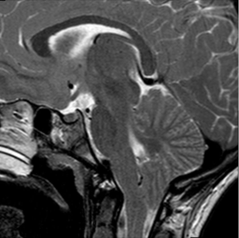
Find the junction before measuring. On this sagittal T2 image, anterior is left and posterior is right. Identify the brainstem, cerebellum and bright CSF, then inspect the inferior cerebellar tissue at the skull opening. This single slice does not measure CSF flow or establish a treatment indication. Select the image to enlarge the whole frame.
MRI establishes the hindbrain anatomy and detects a syrinx. If only a brain or cervical study is available, additional brain and spine imaging can help identify hydrocephalus, more extensive syringomyelia, or other relevant pathology. A sagittal measurement should be accompanied by assessment of crowding, brainstem position, and the whole clinical picture. Cine MRI can evaluate CSF flow, but its ability to predict benefit from surgery is uncertain. It is an adjunct rather than a stand-alone operation test. [1]
A fluid syrinx generally follows CSF signal, appearing bright on T2 and dark on T1. Report its maximum diameter separately from its longitudinal extent. A 15 mm long cavity and a 15 mm wide cavity are very different findings. An enhancing intramedullary component, unusual expansion, or an unexplained new deficit warrants investigation for an additional process. Surrounding T2 signal alone does not prove cancer, and a stable cavity does not rule out every cause of neurological decline. [7]
Low tonsils can accompany abnormal intracranial pressure, including intracranial hypotension with brain sag or raised pressure. Orthostatic headache, papilledema, ventricular changes, or a mass requires investigation of that mechanism rather than automatic labeling as congenital Chiari I. Lumbar puncture is not treatment for Chiari crowding. Consensus guidance lists Chiari malformation and tonsillar-herniation risk among contraindications to routine lumbar puncture: even small CSF withdrawal can be dangerous in susceptible anatomy. If CSF sampling is essential for a separate indication, specialist assessment of imaging and herniation risk is required before deciding whether and how to proceed. This CSF-withdrawal question is distinct from obstetric neuraxial-anesthesia planning. [7][8][16]
A remote uncomplicated head injury does not establish post-traumatic syringomyelia. The stronger clinical setting is prior spinal cord injury with subsequent scarring and altered CSF circulation. Similarly, new weakness years after decompression requires examination and reassessment of the junction, cord, and alternative neurological causes. Do not diagnose invisible tethering solely because surgery occurred in the past. [5][3]
New trauma is a new assessment. Acute neck pain with objective weakness or sensory change needs urgent spinal-injury evaluation and appropriate precautions. CT assesses fractures and alignment; it does not exclude every cord injury. In adults with a neurological abnormality that could arise from the cord, obtain MRI after CT even when CT shows no abnormality. [17]
Case 14
Show answer and explanations for case 14
A. A has shortened without narrowing and can continue planned review; B needs expedited neurosurgical reassessment (Best answer)
Make one prediction at a time. Earlier explanations stay available.
Which absolute measurement changed in each patient?
A's length halved with diameter unchanged; B's diameter increased with length unchanged.
Why might this option seem plausible: A has shortened without narrowing and can continue planned review; B needs expedited neurosurgical reassessment?
The dimensions and clinical courses answer different parts of the decision.
Which supplied finding decides whether it fits?
A's maximum diameter is unchanged despite shorter length; B's increased diameter, expansion and objective deterioration together change the urgency.
Would first seeing a 2 mm cavity establish that it had grown?
No. Growth requires a comparable prior study. Compare like dimensions, then integrate neurological trajectory; an unchanged length does not cancel dangerous widening.
Read the complete explanation
The dimensions and clinical courses answer different parts of the decision. A's maximum diameter is unchanged despite shorter length; B's increased diameter, expansion and objective deterioration together change the urgency.
B. A has narrowed and can end cavity surveillance; B needs expedited review for widening with weakness (Why this does not fit)
Make one prediction at a time. Earlier explanations stay available.
Which absolute measurement changed in each patient?
A's length halved with diameter unchanged; B's diameter increased with length unchanged.
Why might this option seem plausible: A has narrowed and can end cavity surveillance; B needs expedited review for widening with weakness?
B does need prompt reassessment, and A has some radiographic reduction.
Which supplied finding decides whether it fits?
A has shortened without narrowing, and partial reduction is not complete resolution; this interpretation misreads the dimension and overstates the follow-up implication.
Would first seeing a 2 mm cavity establish that it had grown?
No. Growth requires a comparable prior study. Compare like dimensions, then integrate neurological trajectory; an unchanged length does not cancel dangerous widening.
Read the complete explanation
B does need prompt reassessment, and A has some radiographic reduction. A has shortened without narrowing, and partial reduction is not complete resolution; this interpretation misreads the dimension and overstates the follow-up implication.
C. A can continue planned review; B can repeat imaging on the same routine schedule because its length is stable (Why this does not fit)
Make one prediction at a time. Earlier explanations stay available.
Which absolute measurement changed in each patient?
A's length halved with diameter unchanged; B's diameter increased with length unchanged.
Why might this option seem plausible: A can continue planned review; B can repeat imaging on the same routine schedule because its length is stable?
Longitudinal extent is one useful imaging measurement.
Which supplied finding decides whether it fits?
B already has maximum-diameter expansion and objective neurological decline; unchanged length does not justify retaining a routine schedule.
Would first seeing a 2 mm cavity establish that it had grown?
No. Growth requires a comparable prior study. Compare like dimensions, then integrate neurological trajectory; an unchanged length does not cancel dangerous widening.
Read the complete explanation
Longitudinal extent is one useful imaging measurement. B already has maximum-diameter expansion and objective neurological decline; unchanged length does not justify retaining a routine schedule.
D. A needs expedited surgical reassessment for persistent diameter; B needs expedited review for expanding diameter (Why this does not fit)
Make one prediction at a time. Earlier explanations stay available.
Which absolute measurement changed in each patient?
A's length halved with diameter unchanged; B's diameter increased with length unchanged.
Why might this option seem plausible: A needs expedited surgical reassessment for persistent diameter; B needs expedited review for expanding diameter?
A persistent cavity can require ongoing assessment, and B has clear deterioration.
Which supplied finding decides whether it fits?
The stable small maximum diameter and normal examination in A do not have the same urgency as B’s enlarging cavity with progressive deficits.
Would first seeing a 2 mm cavity establish that it had grown?
No. Growth requires a comparable prior study. Compare like dimensions, then integrate neurological trajectory; an unchanged length does not cancel dangerous widening.
Read the complete explanation
A persistent cavity can require ongoing assessment, and B has clear deterioration. The stable small maximum diameter and normal examination in A do not have the same urgency as B’s enlarging cavity with progressive deficits.
Takeaway: Compare like dimensions, then integrate neurological trajectory; an unchanged length does not cancel dangerous widening.
An incidental, asymptomatic Chiari I without syringomyelia generally does not need prophylactic surgery. Follow-up depends on age, symptoms, associated findings, and specialist assessment. Children may need surveillance through growth. Avoid promising inevitable deterioration or lifelong freedom from symptoms. New symptoms should trigger reassessment. [2][7]
A small stable asymptomatic cavity can sometimes be observed, but a new syrinx deserves neurosurgical assessment. Progressive enlargement, substantial cord distension, neurological decline, or scoliosis in a child changes the balance even without a classic headache. Pediatric consensus discusses syrinx diameter and growth as relevant factors, not a universal adult rule that every 2 mm change mandates surgery. [7]
For disabling symptoms attributable to Chiari, progressive deficits, or concerning syringomyelia, posterior fossa decompression aims to improve space and CSF circulation. Bone decompression and decompression with duraplasty are both accepted approaches. The choice considers anatomy, syrinx, age, expected benefit, and complications such as CSF leak. Fusion addresses demonstrated craniocervical instability in selected patients; it is not the routine treatment for a syrinx. Steroids do not correct mechanical crowding. [1][3]
A 2026 cluster-randomized trial enrolled 162 patients aged 21 years or younger with tonsillar descent of at least 5 mm and syrinx diameter 3.0-9.9 mm. With versus without duraplasty, six-month complications were 14% versus 6% (adjusted odds ratio 2.59; 95% CI 0.86-7.84). At 24 months, mean syrinx reduction was 3.08 versus 1.22 mm and repeat decompression occurred in 3% versus 14%; quality-of-life changes were similar. The complication estimate is uncertain: lack of statistical significance does not establish equal risk. These results inform individual selection within the studied population, not a universal operation rule. [14]
Symptoms and cavity size can improve on different schedules after surgery. Many syrinxes shrink, but incomplete collapse does not by itself prove treatment failure. A persistent cavity requires planned clinical and MRI review. CNS guidance allows consideration of another intervention when there is no radiographic improvement after 6 to 12 months, with earlier reassessment for deterioration. Do not promise that a persistent cavity is harmless or that another operation is inevitable. [3]
Activity and pregnancy require the actual risk profile
CNS guidance does not recommend preventive activity restriction for asymptomatic Chiari I without a syrinx. Symptoms, an existing cavity, postoperative status, and collision exposure require individualized advice. Playing violin is not equivalent to a sustained Valsalva maneuver. There is no defensible universal ban on every sport after decompression. [2]
The 2026 OAA consensus advises that delivery and anesthesia for asymptomatic or neurologically stable Chiari I, with or without a syrinx, can usually follow obstetric considerations and patient preference. Arrange obstetric and anesthetic review during pregnancy. New or worsening neurological symptoms require multidisciplinary assessment; evidence is more limited for severe presentations. Earlier observational delivery data also support avoiding blanket cesarean or general-anesthesia rules in appropriately selected patients. [6][15]
Case 4
Show answer and explanations for case 4
A. Continue surveillance for A; assess B for isolated syrinx drainage as the initial target (Why this does not fit)
Make one prediction at a time. Earlier explanations stay available.
What differs despite similar hindbrain anatomy?
A has stable normal function and cavity width; B has progressive weakness, spastic gait and widening.
Why might this option seem plausible: Continue surveillance for A; assess B for isolated syrinx drainage as the initial target?
The different trajectories support surveillance for A and intervention assessment for B.
Which supplied finding decides whether it fits?
For a Chiari-related cavity with junctional crowding, treating the surrounding CSF disturbance is the initial causal target; isolated drainage leaves that mechanism unaddressed.
Would cavity drainage alone correct the demonstrated junctional crowding?
No. The causal Chiari-directed goal is to improve space and surrounding CSF circulation. Use trajectory to select urgency and the obstructed CSF region to select the treatment goal.
Read the complete explanation
The different trajectories support surveillance for A and intervention assessment for B. For a Chiari-related cavity with junctional crowding, treating the surrounding CSF disturbance is the initial causal target; isolated drainage leaves that mechanism unaddressed.
B. Assess both for junctional decompression on the basis of their similar tonsillar descent (Why this does not fit)
Make one prediction at a time. Earlier explanations stay available.
What differs despite similar hindbrain anatomy?
A has stable normal function and cavity width; B has progressive weakness, spastic gait and widening.
Why might this option seem plausible: Assess both for junctional decompression on the basis of their similar tonsillar descent?
Junctional decompression can address disturbed CSF circulation.
Which supplied finding decides whether it fits?
The measurement does not make A's stable, asymptomatic profile equivalent to B's progressive neurological loss and cavity expansion.
Would cavity drainage alone correct the demonstrated junctional crowding?
No. The causal Chiari-directed goal is to improve space and surrounding CSF circulation. Use trajectory to select urgency and the obstructed CSF region to select the treatment goal.
Read the complete explanation
Junctional decompression can address disturbed CSF circulation. The measurement does not make A's stable, asymptomatic profile equivalent to B's progressive neurological loss and cavity expansion.
C. Continue surveillance for A; promptly assess B for decompression of the crowded junction (Best answer)
Make one prediction at a time. Earlier explanations stay available.
What differs despite similar hindbrain anatomy?
A has stable normal function and cavity width; B has progressive weakness, spastic gait and widening.
Why might this option seem plausible: Continue surveillance for A; promptly assess B for decompression of the crowded junction?
Clinical progression determines urgency, while anatomy identifies the treatment target.
Which supplied finding decides whether it fits?
A is stable; B has progressive myelopathy with cavity growth, supporting prompt neurosurgical assessment aimed at improving space and CSF circulation.
Would cavity drainage alone correct the demonstrated junctional crowding?
No. The causal Chiari-directed goal is to improve space and surrounding CSF circulation. Use trajectory to select urgency and the obstructed CSF region to select the treatment goal.
Read the complete explanation
Clinical progression determines urgency, while anatomy identifies the treatment target. A is stable; B has progressive myelopathy with cavity growth, supporting prompt neurosurgical assessment aimed at improving space and CSF circulation.
D. Continue surveillance for both until the cavity length increases as well as its diameter (Why this does not fit)
Make one prediction at a time. Earlier explanations stay available.
What differs despite similar hindbrain anatomy?
A has stable normal function and cavity width; B has progressive weakness, spastic gait and widening.
Why might this option seem plausible: Continue surveillance for both until the cavity length increases as well as its diameter?
Serial imaging is useful in deciding management.
Which supplied finding decides whether it fits?
B already has worsening neurological function and substantial diameter expansion; unchanged length would not justify waiting for a second dimension to worsen.
Would cavity drainage alone correct the demonstrated junctional crowding?
No. The causal Chiari-directed goal is to improve space and surrounding CSF circulation. Use trajectory to select urgency and the obstructed CSF region to select the treatment goal.
Read the complete explanation
Serial imaging is useful in deciding management. B already has worsening neurological function and substantial diameter expansion; unchanged length would not justify waiting for a second dimension to worsen.
Takeaway: Use trajectory to select urgency and the obstructed CSF region to select the treatment goal.
In Chiari II, protect breathing and check CSF diversion
Myelomeningocele involves meninges and neural tissue in an open spinal defect. Prenatal ultrasound can show the spinal lesion and associated cranial findings; maternal alpha-fetoprotein may be increased in open neural tube defects. An abnormal screening result requires further assessment rather than establishing the diagnosis by itself. [12] After birth, protect the exposed tissue and arrange specialist closure. Fetal repair is an option for selected pregnancies at experienced centers, not an interchangeable procedure for every fetus. It has maternal and fetal risks as well as potential benefits. [4][9]
Hydrocephalus is common and may require CSF diversion. Increasing head circumference, a tense fontanelle, vomiting, altered alertness, and abnormal downward gaze can indicate raised pressure. Brainstem dysfunction may appear as stridor, apnea, weak cry, or poor swallowing. Stabilize the airway when needed and assess hydrocephalus or shunt malfunction before attributing deterioration to an isolated hindbrain compression problem. Shunt failure can occur without striking ventricular enlargement. [4]
Persistent hindbrain descent on MRI in a clinically stable child does not require surgery simply to normalize the picture. Conversely, persistent brainstem compromise after adequate CSF diversion may require decompression. New leg weakness, changing foot deformity, pain, scoliosis, or deteriorating bladder function during growth also raises concern for tethered cord. These findings need assessment against the child's baseline rather than being dismissed as inevitable consequences of the original defect. [4]
Cough headache plus dissociated arm sensation calls for junction and cord imaging. Incidental low tonsils call for clinical correlation. Progressive myelopathy calls for prompt surgical assessment. In a shunted child with Chiari II and new symptoms, assess the shunt and breathing first.
Case 12
Show answer and explanations for case 12
A. Prioritize posterior-fossa flow imaging for recurrent brainstem compression (Why this does not fit)
Make one prediction at a time. Earlier explanations stay available.
Which reassuring-looking finding is insufficient here?
Similar ventricular size does not independently exclude shunt failure.
Why might this option seem plausible: Prioritize posterior-fossa flow imaging for recurrent brainstem compression?
Chiari II can cause apnea through brainstem dysfunction.
Which supplied finding decides whether it fits?
Vomiting and reduced alertness in a shunted child also raise concern for diversion failure; that cause requires urgent assessment even with unchanged ventricle size.
What if brainstem symptoms persist after adequate CSF diversion is established?
The team may then need to assess persistent hindbrain compression and whether decompression is appropriate. In a deteriorating shunted child, clinical signs can outweigh an unchanged ventricular measurement.
Read the complete explanation
Chiari II can cause apnea through brainstem dysfunction. Vomiting and reduced alertness in a shunted child also raise concern for diversion failure; that cause requires urgent assessment even with unchanged ventricle size.
B. Prioritize EEG for seizures presenting as apnea and reduced responsiveness (Why this does not fit)
Make one prediction at a time. Earlier explanations stay available.
Which reassuring-looking finding is insufficient here?
Similar ventricular size does not independently exclude shunt failure.
Why might this option seem plausible: Prioritize EEG for seizures presenting as apnea and reduced responsiveness?
Seizures can cause episodic apnea and altered responsiveness.
Which supplied finding decides whether it fits?
The combined vomiting and neurological deterioration in a shunted child make CSF-diversion and pressure assessment the immediate priority in this scenario.
What if brainstem symptoms persist after adequate CSF diversion is established?
The team may then need to assess persistent hindbrain compression and whether decompression is appropriate. In a deteriorating shunted child, clinical signs can outweigh an unchanged ventricular measurement.
Read the complete explanation
Seizures can cause episodic apnea and altered responsiveness. The combined vomiting and neurological deterioration in a shunted child make CSF-diversion and pressure assessment the immediate priority in this scenario.
C. Prioritize spinal MRI for an enlarging syrinx (Why this does not fit)
Make one prediction at a time. Earlier explanations stay available.
Which reassuring-looking finding is insufficient here?
Similar ventricular size does not independently exclude shunt failure.
Why might this option seem plausible: Prioritize spinal MRI for an enlarging syrinx?
A syrinx can contribute to neurological dysfunction in Chiari II.
Which supplied finding decides whether it fits?
It does not best unify the acute vomiting, reduced alertness and apnea; shunt and pressure assessment must come first.
What if brainstem symptoms persist after adequate CSF diversion is established?
The team may then need to assess persistent hindbrain compression and whether decompression is appropriate. In a deteriorating shunted child, clinical signs can outweigh an unchanged ventricular measurement.
Read the complete explanation
A syrinx can contribute to neurological dysfunction in Chiari II. It does not best unify the acute vomiting, reduced alertness and apnea; shunt and pressure assessment must come first.
D. Urgently assess shunt function and the clinical pressure state (Best answer)
Make one prediction at a time. Earlier explanations stay available.
Which reassuring-looking finding is insufficient here?
Similar ventricular size does not independently exclude shunt failure.
Why might this option seem plausible: Urgently assess shunt function and the clinical pressure state?
Shunt failure can occur without striking ventricular enlargement.
Which supplied finding decides whether it fits?
The new clinical deterioration remains concerning despite similar ventricular dimensions, so CSF diversion cannot be cleared by that image comparison alone.
What if brainstem symptoms persist after adequate CSF diversion is established?
The team may then need to assess persistent hindbrain compression and whether decompression is appropriate. In a deteriorating shunted child, clinical signs can outweigh an unchanged ventricular measurement.
Read the complete explanation
Shunt failure can occur without striking ventricular enlargement. The new clinical deterioration remains concerning despite similar ventricular dimensions, so CSF diversion cannot be cleared by that image comparison alone.
Takeaway: In a deteriorating shunted child, clinical signs can outweigh an unchanged ventricular measurement.
The first would cause an upper motor neuron pattern below the lesion, whereas the second would cause lower motor neuron signs in the legs; both are the reverse of the supplied patterns.
Must the cavity reach the lumbar cord to produce the leg pattern?
No. Cervical descending-tract injury can affect motor control below the cavity. Separate segmental lower motor neuron injury from long-tract upper motor neuron injury below a cervical cavity.
Read the complete explanation
Both structures participate in motor function. The first would cause an upper motor neuron pattern below the lesion, whereas the second would cause lower motor neuron signs in the legs; both are the reverse of the supplied patterns.
B. Cervical posterior columns; cervical corticospinal tracts (Why this does not fit)
Make one prediction at a time. Earlier explanations stay available.
How do the arm and leg motor signs differ?
The hands show wasting and reduced reflexes; the legs show clonus and extensor plantar responses.
Descending corticospinal injury can explain the spastic legs.
Which supplied finding decides whether it fits?
Posterior-column injury would affect vibration and position, not cause denervation-type hand wasting and reduced reflexes.
Must the cavity reach the lumbar cord to produce the leg pattern?
No. Cervical descending-tract injury can affect motor control below the cavity. Separate segmental lower motor neuron injury from long-tract upper motor neuron injury below a cervical cavity.
Read the complete explanation
Descending corticospinal injury can explain the spastic legs. Posterior-column injury would affect vibration and position, not cause denervation-type hand wasting and reduced reflexes.
C. Lower cervical anterior horns; cervical posterior columns (Why this does not fit)
Make one prediction at a time. Earlier explanations stay available.
How do the arm and leg motor signs differ?
The hands show wasting and reduced reflexes; the legs show clonus and extensor plantar responses.
Anterior-horn injury fits the hand lower motor neuron pattern.
Which supplied finding decides whether it fits?
Posterior-column injury would impair vibration and position, not explain leg clonus and extensor plantar responses.
Must the cavity reach the lumbar cord to produce the leg pattern?
No. Cervical descending-tract injury can affect motor control below the cavity. Separate segmental lower motor neuron injury from long-tract upper motor neuron injury below a cervical cavity.
Read the complete explanation
Anterior-horn injury fits the hand lower motor neuron pattern. Posterior-column injury would impair vibration and position, not explain leg clonus and extensor plantar responses.
D. Lower cervical anterior horns; cervical corticospinal tracts (Best answer)
Make one prediction at a time. Earlier explanations stay available.
How do the arm and leg motor signs differ?
The hands show wasting and reduced reflexes; the legs show clonus and extensor plantar responses.
A cavity can affect segmental motor neurons and then adjacent descending pathways.
Which supplied finding decides whether it fits?
Hand wasting and reduced reflexes localize to motor-neuron pools; subsequent leg spasticity localizes to descending motor tracts above the leg segments.
Must the cavity reach the lumbar cord to produce the leg pattern?
No. Cervical descending-tract injury can affect motor control below the cavity. Separate segmental lower motor neuron injury from long-tract upper motor neuron injury below a cervical cavity.
Read the complete explanation
A cavity can affect segmental motor neurons and then adjacent descending pathways. Hand wasting and reduced reflexes localize to motor-neuron pools; subsequent leg spasticity localizes to descending motor tracts above the leg segments.
Takeaway: Separate segmental lower motor neuron injury from long-tract upper motor neuron injury below a cervical cavity.
A. Treat the headache phenotype and arrange individualized follow-up of the tonsillar finding (Best answer)
Make one prediction at a time. Earlier explanations stay available.
Which history detail differs from typical Chiari cough headache?
These attacks last hours with photophobia and have no cough trigger.
Why might this option seem plausible: Treat the headache phenotype and arrange individualized follow-up of the tonsillar finding?
Low tonsils can coexist with a primary headache disorder.
Which supplied finding decides whether it fits?
The prolonged photophobic attacks lack the characteristic brief cough trigger, and there is no syrinx or neurological deficit to make prophylactic decompression appropriate.
Would a new objective deficit change this plan?
Yes. A new neurological deficit or syrinx would require reassessment rather than continued attribution to a primary headache alone. Match the headache phenotype and associated findings before attributing pain to low tonsils.
Read the complete explanation
Low tonsils can coexist with a primary headache disorder. The prolonged photophobic attacks lack the characteristic brief cough trigger, and there is no syrinx or neurological deficit to make prophylactic decompression appropriate.
B. Arrange elective neurosurgical review for headache attributed to Chiari I (Why this does not fit)
Make one prediction at a time. Earlier explanations stay available.
Which history detail differs from typical Chiari cough headache?
These attacks last hours with photophobia and have no cough trigger.
Why might this option seem plausible: Arrange elective neurosurgical review for headache attributed to Chiari I?
Chiari I can be associated with headache, and referral is appropriate when symptoms fit.
Which supplied finding decides whether it fits?
The hours-long photophobic phenotype without cough provocation, crowding or a syrinx favors headache-directed care rather than starting from a Chiari attribution.
Would a new objective deficit change this plan?
Yes. A new neurological deficit or syrinx would require reassessment rather than continued attribution to a primary headache alone. Match the headache phenotype and associated findings before attributing pain to low tonsils.
Read the complete explanation
Chiari I can be associated with headache, and referral is appropriate when symptoms fit. The hours-long photophobic phenotype without cough provocation, crowding or a syrinx favors headache-directed care rather than starting from a Chiari attribution.
C. Obtain cine CSF-flow imaging to decide whether to begin headache treatment (Why this does not fit)
Make one prediction at a time. Earlier explanations stay available.
Which history detail differs from typical Chiari cough headache?
These attacks last hours with photophobia and have no cough trigger.
Why might this option seem plausible: Obtain cine CSF-flow imaging to decide whether to begin headache treatment?
Flow imaging may contribute to selected Chiari assessments.
Which supplied finding decides whether it fits?
It is not a prerequisite for treating a clinically recognizable migraine-like syndrome with a normal examination and no associated cord lesion.
Would a new objective deficit change this plan?
Yes. A new neurological deficit or syrinx would require reassessment rather than continued attribution to a primary headache alone. Match the headache phenotype and associated findings before attributing pain to low tonsils.
Read the complete explanation
Flow imaging may contribute to selected Chiari assessments. It is not a prerequisite for treating a clinically recognizable migraine-like syndrome with a normal examination and no associated cord lesion.
D. Use a headache diary and avoid exertion while observing for a cough trigger (Why this does not fit)
Make one prediction at a time. Earlier explanations stay available.
Which history detail differs from typical Chiari cough headache?
These attacks last hours with photophobia and have no cough trigger.
Why might this option seem plausible: Use a headache diary and avoid exertion while observing for a cough trigger?
A diary can clarify timing and triggers.
Which supplied finding decides whether it fits?
The established prolonged photophobic attacks already support active headache-directed care; preventive exertion avoidance does not address that phenotype.
Would a new objective deficit change this plan?
Yes. A new neurological deficit or syrinx would require reassessment rather than continued attribution to a primary headache alone. Match the headache phenotype and associated findings before attributing pain to low tonsils.
Read the complete explanation
A diary can clarify timing and triggers. The established prolonged photophobic attacks already support active headache-directed care; preventive exertion avoidance does not address that phenotype.
Takeaway: Match the headache phenotype and associated findings before attributing pain to low tonsils.
A. Extension into posterior columns at C8-T1 cord segments (Why this does not fit)
Make one prediction at a time. Earlier explanations stay available.
Which muscles are selectively affected?
Intrinsic hand muscles are weak and wasted while proximal shoulder and elbow actions are preserved.
Why might this option seem plausible: Extension into posterior columns at C8-T1 cord segments?
These segments are relevant to hand function.
Which supplied finding decides whether it fits?
Posterior-column injury impairs vibration and position, rather than causing denervation-type hand wasting.
Does the word cervical establish the correct segment?
No. The cord level and involved tissue must match the actual motor and sensory findings. Match the affected motor-neuron segments to the weak muscles; a nearby imaging label is not enough.
Read the complete explanation
These segments are relevant to hand function. Posterior-column injury impairs vibration and position, rather than causing denervation-type hand wasting.
B. Extension into anterior horns at C8-T1 cord segments (Best answer)
Make one prediction at a time. Earlier explanations stay available.
Which muscles are selectively affected?
Intrinsic hand muscles are weak and wasted while proximal shoulder and elbow actions are preserved.
Why might this option seem plausible: Extension into anterior horns at C8-T1 cord segments?
Hand intrinsic muscles depend on lower cervical and upper thoracic motor-neuron pools.
Which supplied finding decides whether it fits?
The segmental hand findings call for assessment of these levels; the reported C4-C5 cavity alone is an incomplete explanation.
Does the word cervical establish the correct segment?
No. The cord level and involved tissue must match the actual motor and sensory findings. Match the affected motor-neuron segments to the weak muscles; a nearby imaging label is not enough.
Read the complete explanation
Hand intrinsic muscles depend on lower cervical and upper thoracic motor-neuron pools. The segmental hand findings call for assessment of these levels; the reported C4-C5 cavity alone is an incomplete explanation.
C. Extension into anterior horns at C4-C5 cord segments (Why this does not fit)
Make one prediction at a time. Earlier explanations stay available.
Which muscles are selectively affected?
Intrinsic hand muscles are weak and wasted while proximal shoulder and elbow actions are preserved.
Why might this option seem plausible: Extension into anterior horns at C4-C5 cord segments?
Anterior-horn injury produces a lower motor neuron pattern.
Which supplied finding decides whether it fits?
These more rostral motor pools do not match selective intrinsic hand wasting with preserved shoulder and elbow function.
Does the word cervical establish the correct segment?
No. The cord level and involved tissue must match the actual motor and sensory findings. Match the affected motor-neuron segments to the weak muscles; a nearby imaging label is not enough.
Read the complete explanation
Anterior-horn injury produces a lower motor neuron pattern. These more rostral motor pools do not match selective intrinsic hand wasting with preserved shoulder and elbow function.
D. Extension into lateral corticospinal tracts at C8-T1 cord segments (Why this does not fit)
Make one prediction at a time. Earlier explanations stay available.
Which muscles are selectively affected?
Intrinsic hand muscles are weak and wasted while proximal shoulder and elbow actions are preserved.
Why might this option seem plausible: Extension into lateral corticospinal tracts at C8-T1 cord segments?
The lower cervical level is anatomically relevant to descending motor control.
Which supplied finding decides whether it fits?
Corticospinal injury produces upper motor neuron signs below the lesion, not the segmental wasting pattern in the hand motor-neuron pools.
Does the word cervical establish the correct segment?
No. The cord level and involved tissue must match the actual motor and sensory findings. Match the affected motor-neuron segments to the weak muscles; a nearby imaging label is not enough.
Read the complete explanation
The lower cervical level is anatomically relevant to descending motor control. Corticospinal injury produces upper motor neuron signs below the lesion, not the segmental wasting pattern in the hand motor-neuron pools.
Takeaway: Match the affected motor-neuron segments to the weak muscles; a nearby imaging label is not enough.
A. The cervical lesion interrupts descending motor axons bound for lower cord levels (Best answer)
Make one prediction at a time. Earlier explanations stay available.
What identifies the leg findings as an upper motor neuron pattern?
Clonus and extensor plantar responses accompany the new spasticity.
Why might this option seem plausible: The cervical lesion interrupts descending motor axons bound for lower cord levels?
Motor pathways to the legs pass through the cervical cord before reaching lower motor neurons.
Which supplied finding decides whether it fits?
Their interruption produces upper motor neuron signs below the lesion, so the cavity need not extend into the lumbar enlargement.
Would a normal lumbar cord image exclude a cervical cause of leg weakness?
No. Descending motor pathways to the legs travel through the cervical cord. Long tracts connect a cervical lesion to leg signs; cord symptoms do not stop at the cavity's lowest visible level.
Read the complete explanation
Motor pathways to the legs pass through the cervical cord before reaching lower motor neurons. Their interruption produces upper motor neuron signs below the lesion, so the cavity need not extend into the lumbar enlargement.
B. The cervical lesion interrupts crossing anterolateral-system sensory axons (Why this does not fit)
Make one prediction at a time. Earlier explanations stay available.
What identifies the leg findings as an upper motor neuron pattern?
Clonus and extensor plantar responses accompany the new spasticity.
Why might this option seem plausible: The cervical lesion interrupts crossing anterolateral-system sensory axons?
Crossing pain-temperature fibers are vulnerable near a central cavity.
Which supplied finding decides whether it fits?
Their injury explains dissociated arm sensation, not the new leg clonus and extensor plantar responses.
Would a normal lumbar cord image exclude a cervical cause of leg weakness?
No. Descending motor pathways to the legs travel through the cervical cord. Long tracts connect a cervical lesion to leg signs; cord symptoms do not stop at the cavity's lowest visible level.
Read the complete explanation
Crossing pain-temperature fibers are vulnerable near a central cavity. Their injury explains dissociated arm sensation, not the new leg clonus and extensor plantar responses.
C. The cervical lesion interrupts posterior-column sensory axons from the legs (Why this does not fit)
Make one prediction at a time. Earlier explanations stay available.
What identifies the leg findings as an upper motor neuron pattern?
Clonus and extensor plantar responses accompany the new spasticity.
Why might this option seem plausible: The cervical lesion interrupts posterior-column sensory axons from the legs?
Posterior-column injury may affect balance and the awareness of leg position.
Which supplied finding decides whether it fits?
It does not directly account for clonus and extensor plantar responses, which indicate descending motor pathway dysfunction.
Would a normal lumbar cord image exclude a cervical cause of leg weakness?
No. Descending motor pathways to the legs travel through the cervical cord. Long tracts connect a cervical lesion to leg signs; cord symptoms do not stop at the cavity's lowest visible level.
Read the complete explanation
Posterior-column injury may affect balance and the awareness of leg position. It does not directly account for clonus and extensor plantar responses, which indicate descending motor pathway dysfunction.
D. The cervical lesion interrupts ascending spinocerebellar pathways (Why this does not fit)
Make one prediction at a time. Earlier explanations stay available.
What identifies the leg findings as an upper motor neuron pattern?
Clonus and extensor plantar responses accompany the new spasticity.
Why might this option seem plausible: The cervical lesion interrupts ascending spinocerebellar pathways?
These pathways help coordinate lower-limb movement.
Which supplied finding decides whether it fits?
Their injury would favor ataxia rather than the supplied spastic upper motor neuron pattern.
Would a normal lumbar cord image exclude a cervical cause of leg weakness?
No. Descending motor pathways to the legs travel through the cervical cord. Long tracts connect a cervical lesion to leg signs; cord symptoms do not stop at the cavity's lowest visible level.
Read the complete explanation
These pathways help coordinate lower-limb movement. Their injury would favor ataxia rather than the supplied spastic upper motor neuron pattern.
Takeaway: Long tracts connect a cervical lesion to leg signs; cord symptoms do not stop at the cavity's lowest visible level.
A. A: plan elective decompression; B: urgently assess airway and brainstem function (Why this does not fit)
Make one prediction at a time. Earlier explanations stay available.
Which infant has continuing physiological compromise after CSF treatment?
B still has stridor and apnea; A has normal feeding, breathing and stable head growth.
Why might this option seem plausible: A: plan elective decompression; B: urgently assess airway and brainstem function?
B’s ongoing airway signs require urgent reassessment, and decompression can treat selected Chiari II presentations.
Which supplied finding decides whether it fits?
A’s stable feeding, breathing and head growth do not show persistent brainstem compromise requiring elective decompression.
If adequate diversion had not been established in B, what would also be urgent?
Assessment of shunt function and pressure, alongside airway stabilization, before attributing the problem solely to hindbrain compression. After adequate CSF treatment, persistent brainstem compromise and persistent malformation anatomy lead to different next decisions.
Read the complete explanation
B’s ongoing airway signs require urgent reassessment, and decompression can treat selected Chiari II presentations. A’s stable feeding, breathing and head growth do not show persistent brainstem compromise requiring elective decompression.
B. A: continue surveillance; B: urgently assess airway and brainstem function, including decompression candidacy (Best answer)
Make one prediction at a time. Earlier explanations stay available.
Which infant has continuing physiological compromise after CSF treatment?
B still has stridor and apnea; A has normal feeding, breathing and stable head growth.
Why might this option seem plausible: A: continue surveillance; B: urgently assess airway and brainstem function, including decompression candidacy?
Persistent anatomy must be separated from persistent physiological compromise.
Which supplied finding decides whether it fits?
A is clinically stable despite the malformation; B's ongoing apnea after adequate CSF treatment warrants urgent specialist reassessment and possible decompression.
If adequate diversion had not been established in B, what would also be urgent?
Assessment of shunt function and pressure, alongside airway stabilization, before attributing the problem solely to hindbrain compression. After adequate CSF treatment, persistent brainstem compromise and persistent malformation anatomy lead to different next decisions.
Read the complete explanation
Persistent anatomy must be separated from persistent physiological compromise. A is clinically stable despite the malformation; B's ongoing apnea after adequate CSF treatment warrants urgent specialist reassessment and possible decompression.
C. A: continue surveillance; B: adjust diversion settings and monitor the breathing response (Why this does not fit)
Make one prediction at a time. Earlier explanations stay available.
Which infant has continuing physiological compromise after CSF treatment?
B still has stridor and apnea; A has normal feeding, breathing and stable head growth.
Why might this option seem plausible: A: continue surveillance; B: adjust diversion settings and monitor the breathing response?
Changes in diversion can help when CSF treatment is inadequate.
Which supplied finding decides whether it fits?
Adequate diversion has already been established here; continuing apnea requires urgent airway and broader brainstem assessment rather than making diversion adjustment the next assumed solution.
If adequate diversion had not been established in B, what would also be urgent?
Assessment of shunt function and pressure, alongside airway stabilization, before attributing the problem solely to hindbrain compression. After adequate CSF treatment, persistent brainstem compromise and persistent malformation anatomy lead to different next decisions.
Read the complete explanation
Changes in diversion can help when CSF treatment is inadequate. Adequate diversion has already been established here; continuing apnea requires urgent airway and broader brainstem assessment rather than making diversion adjustment the next assumed solution.
D. A: continue surveillance; B: obtain outpatient feeding and sleep studies before further neurosurgical review (Why this does not fit)
Make one prediction at a time. Earlier explanations stay available.
Which infant has continuing physiological compromise after CSF treatment?
B still has stridor and apnea; A has normal feeding, breathing and stable head growth.
Why might this option seem plausible: A: continue surveillance; B: obtain outpatient feeding and sleep studies before further neurosurgical review?
Feeding and sleep assessment can characterize chronic symptoms.
Which supplied finding decides whether it fits?
Recurrent apnea and stridor after adequate CSF treatment require urgent coordinated reassessment; outpatient characterization should not delay it.
If adequate diversion had not been established in B, what would also be urgent?
Assessment of shunt function and pressure, alongside airway stabilization, before attributing the problem solely to hindbrain compression. After adequate CSF treatment, persistent brainstem compromise and persistent malformation anatomy lead to different next decisions.
Read the complete explanation
Feeding and sleep assessment can characterize chronic symptoms. Recurrent apnea and stridor after adequate CSF treatment require urgent coordinated reassessment; outpatient characterization should not delay it.
Takeaway: After adequate CSF treatment, persistent brainstem compromise and persistent malformation anatomy lead to different next decisions.
A. A fits a Dandy-Walker phenotype; B does not establish that phenotype from size alone (Best answer)
Make one prediction at a time. Earlier explanations stay available.
Which fetus has abnormal vermian development and fourth-ventricular relationships?
A does; B has a normal vermis and fourth ventricle despite the larger fossa.
Why might this option seem plausible: A fits a Dandy-Walker phenotype; B does not establish that phenotype from size alone?
Vermian and fourth-ventricular relationships are central to modern phenotype assessment.
Which supplied finding decides whether it fits?
A has the relevant developmental abnormalities despite normal fossa size; B's enlarged fossa and fluid space lack those supplied vermian and fourth-ventricular changes.
Does this establish a precise alternative diagnosis for B?
No. It shows why size alone is insufficient; the complete imaging differential still needs assessment. Read vermian and fourth-ventricular development before using posterior-fossa size as a diagnostic shortcut.
Read the complete explanation
Vermian and fourth-ventricular relationships are central to modern phenotype assessment. A has the relevant developmental abnormalities despite normal fossa size; B's enlarged fossa and fluid space lack those supplied vermian and fourth-ventricular changes.
B. B fits a Dandy-Walker phenotype; A is excluded by normal posterior-fossa size (Why this does not fit)
Make one prediction at a time. Earlier explanations stay available.
Which fetus has abnormal vermian development and fourth-ventricular relationships?
A does; B has a normal vermis and fourth ventricle despite the larger fossa.
Why might this option seem plausible: B fits a Dandy-Walker phenotype; A is excluded by normal posterior-fossa size?
An enlarged posterior fossa is a classical association.
Which supplied finding decides whether it fits?
Size alone is insufficient: A's vermian and fourth-ventricular abnormalities matter, and normal size does not exclude the modern phenotype.
Does this establish a precise alternative diagnosis for B?
No. It shows why size alone is insufficient; the complete imaging differential still needs assessment. Read vermian and fourth-ventricular development before using posterior-fossa size as a diagnostic shortcut.
Read the complete explanation
An enlarged posterior fossa is a classical association. Size alone is insufficient: A's vermian and fourth-ventricular abnormalities matter, and normal size does not exclude the modern phenotype.
C. Both fit a Dandy-Walker phenotype because both have retrocerebellar fluid (Why this does not fit)
Make one prediction at a time. Earlier explanations stay available.
Which fetus has abnormal vermian development and fourth-ventricular relationships?
A does; B has a normal vermis and fourth ventricle despite the larger fossa.
Why might this option seem plausible: Both fit a Dandy-Walker phenotype because both have retrocerebellar fluid?
Retrocerebellar fluid can be part of this phenotype.
Which supplied finding decides whether it fits?
It is not specific by itself; the normal vermis and fourth ventricle in B require a different differential.
Does this establish a precise alternative diagnosis for B?
No. It shows why size alone is insufficient; the complete imaging differential still needs assessment. Read vermian and fourth-ventricular development before using posterior-fossa size as a diagnostic shortcut.
Read the complete explanation
Retrocerebellar fluid can be part of this phenotype. It is not specific by itself; the normal vermis and fourth ventricle in B require a different differential.
D. Neither fits a Dandy-Walker phenotype because neither combines all classical size features (Why this does not fit)
Make one prediction at a time. Earlier explanations stay available.
Which fetus has abnormal vermian development and fourth-ventricular relationships?
A does; B has a normal vermis and fourth ventricle despite the larger fossa.
Why might this option seem plausible: Neither fits a Dandy-Walker phenotype because neither combines all classical size features?
Classical descriptions often included posterior-fossa enlargement.
Which supplied finding decides whether it fits?
Requiring that feature would incorrectly dismiss A's characteristic vermian and fourth-ventricular relationships.
Does this establish a precise alternative diagnosis for B?
No. It shows why size alone is insufficient; the complete imaging differential still needs assessment. Read vermian and fourth-ventricular development before using posterior-fossa size as a diagnostic shortcut.
Read the complete explanation
Classical descriptions often included posterior-fossa enlargement. Requiring that feature would incorrectly dismiss A's characteristic vermian and fourth-ventricular relationships.
Takeaway: Read vermian and fourth-ventricular development before using posterior-fossa size as a diagnostic shortcut.
A. Standing spinal radiographs to characterize the scoliosis curve (Why this does not fit)
Make one prediction at a time. Earlier explanations stay available.
Do the new findings point only to a headache disorder?
No. Painless hand burns and progressive scoliosis raise concern for associated spinal cord disease.
Why might this option seem plausible: Standing spinal radiographs to characterize the scoliosis curve?
Radiographs can characterize an evolving spinal curvature.
Which supplied finding decides whether it fits?
They do not evaluate an intramedullary cause that could connect the scoliosis with dissociated hand sensation.
Why is repeating the millimeter measurement insufficient?
It would not show the existence, diameter or extent of a spinal syrinx. A child with Chiari I and new cord-related findings needs cord assessment; age does not convert Chiari I into Chiari II.
Read the complete explanation
Radiographs can characterize an evolving spinal curvature. They do not evaluate an intramedullary cause that could connect the scoliosis with dissociated hand sensation.
B. Electrodiagnostic testing of the upper-limb peripheral nerves (Why this does not fit)
Make one prediction at a time. Earlier explanations stay available.
Do the new findings point only to a headache disorder?
No. Painless hand burns and progressive scoliosis raise concern for associated spinal cord disease.
Why might this option seem plausible: Electrodiagnostic testing of the upper-limb peripheral nerves?
Peripheral lesions can cause hand sensory complaints.
Which supplied finding decides whether it fits?
Bilateral painless burns together with scoliosis in this hindbrain setting favor a cord complication that MRI assesses directly.
Why is repeating the millimeter measurement insufficient?
It would not show the existence, diameter or extent of a spinal syrinx. A child with Chiari I and new cord-related findings needs cord assessment; age does not convert Chiari I into Chiari II.
Read the complete explanation
Peripheral lesions can cause hand sensory complaints. Bilateral painless burns together with scoliosis in this hindbrain setting favor a cord complication that MRI assesses directly.
C. MRI of the spinal cord to assess for syringomyelia (Best answer)
Make one prediction at a time. Earlier explanations stay available.
Do the new findings point only to a headache disorder?
No. Painless hand burns and progressive scoliosis raise concern for associated spinal cord disease.
Why might this option seem plausible: MRI of the spinal cord to assess for syringomyelia?
Chiari I can occur in childhood and can be associated with a spinal syrinx.
Which supplied finding decides whether it fits?
New sensory abnormalities and scoliosis make cord imaging relevant even though the original diagnosis was based on brain MRI.
Why is repeating the millimeter measurement insufficient?
It would not show the existence, diameter or extent of a spinal syrinx. A child with Chiari I and new cord-related findings needs cord assessment; age does not convert Chiari I into Chiari II.
Read the complete explanation
Chiari I can occur in childhood and can be associated with a spinal syrinx. New sensory abnormalities and scoliosis make cord imaging relevant even though the original diagnosis was based on brain MRI.
D. Cine MRI of CSF flow at the craniovertebral junction (Why this does not fit)
Make one prediction at a time. Earlier explanations stay available.
Do the new findings point only to a headache disorder?
No. Painless hand burns and progressive scoliosis raise concern for associated spinal cord disease.
Why might this option seem plausible: Cine MRI of CSF flow at the craniovertebral junction?
Flow assessment may add information about the known junctional abnormality.
Which supplied finding decides whether it fits?
It would not by itself characterize the cord complication suggested by the new hand and spinal findings; the cord has never been imaged.
Why is repeating the millimeter measurement insufficient?
It would not show the existence, diameter or extent of a spinal syrinx. A child with Chiari I and new cord-related findings needs cord assessment; age does not convert Chiari I into Chiari II.
Read the complete explanation
Flow assessment may add information about the known junctional abnormality. It would not by itself characterize the cord complication suggested by the new hand and spinal findings; the cord has never been imaged.
Takeaway: A child with Chiari I and new cord-related findings needs cord assessment; age does not convert Chiari I into Chiari II.
A. Repeat cervical CT with three-dimensional bone reconstructions (Why this does not fit)
Make one prediction at a time. Earlier explanations stay available.
What remains unexplained after CT?
Acute bilateral hand weakness and sensory change persist despite no identified bony injury.
Why might this option seem plausible: Repeat cervical CT with three-dimensional bone reconstructions?
CT is useful for fracture and alignment assessment.
Which supplied finding decides whether it fits?
The initial CT did not identify a bony injury; persistent neurological abnormalities require cord and soft-tissue assessment rather than bone reconstructions alone.
Does a negative CT exclude a clinically important cord injury?
No. CT and MRI answer different questions; persistent neurological abnormalities warrant MRI and urgent specialist assessment. Persistent neurological abnormalities after trauma require cord assessment even when CT shows no fracture.
Read the complete explanation
CT is useful for fracture and alignment assessment. The initial CT did not identify a bony injury; persistent neurological abnormalities require cord and soft-tissue assessment rather than bone reconstructions alone.
B. Urgent MRI of the cervical spine (Best answer)
Make one prediction at a time. Earlier explanations stay available.
What remains unexplained after CT?
Acute bilateral hand weakness and sensory change persist despite no identified bony injury.
Why might this option seem plausible: Urgent MRI of the cervical spine?
MRI can assess the cord and soft tissues when neurological findings persist after CT.
Which supplied finding decides whether it fits?
The acute bilateral deficits after trauma require this evaluation even with a known stable cavity; immobilization and urgent clinical care continue.
Does a negative CT exclude a clinically important cord injury?
No. CT and MRI answer different questions; persistent neurological abnormalities warrant MRI and urgent specialist assessment. Persistent neurological abnormalities after trauma require cord assessment even when CT shows no fracture.
Read the complete explanation
MRI can assess the cord and soft tissues when neurological findings persist after CT. The acute bilateral deficits after trauma require this evaluation even with a known stable cavity; immobilization and urgent clinical care continue.
C. Cine MRI limited to the craniovertebral junction (Why this does not fit)
Make one prediction at a time. Earlier explanations stay available.
What remains unexplained after CT?
Acute bilateral hand weakness and sensory change persist despite no identified bony injury.
Why might this option seem plausible: Cine MRI limited to the craniovertebral junction?
The known Chiari-syrinx relationship may make junctional flow seem the next target.
Which supplied finding decides whether it fits?
The new trauma-associated neck pain and deficits require evaluation for acute cervical cord injury, not just the pre-existing flow mechanism.
Does a negative CT exclude a clinically important cord injury?
No. CT and MRI answer different questions; persistent neurological abnormalities warrant MRI and urgent specialist assessment. Persistent neurological abnormalities after trauma require cord assessment even when CT shows no fracture.
Read the complete explanation
The known Chiari-syrinx relationship may make junctional flow seem the next target. The new trauma-associated neck pain and deficits require evaluation for acute cervical cord injury, not just the pre-existing flow mechanism.
D. Flexion-extension cervical radiographs (Why this does not fit)
Make one prediction at a time. Earlier explanations stay available.
What remains unexplained after CT?
Acute bilateral hand weakness and sensory change persist despite no identified bony injury.
Why might this option seem plausible: Flexion-extension cervical radiographs?
Dynamic views can address instability in selected circumstances.
Which supplied finding decides whether it fits?
They do not assess the spinal cord and are not the appropriate next study for acute neurological deficits after trauma.
Does a negative CT exclude a clinically important cord injury?
No. CT and MRI answer different questions; persistent neurological abnormalities warrant MRI and urgent specialist assessment. Persistent neurological abnormalities after trauma require cord assessment even when CT shows no fracture.
Read the complete explanation
Dynamic views can address instability in selected circumstances. They do not assess the spinal cord and are not the appropriate next study for acute neurological deficits after trauma.
Takeaway: Persistent neurological abnormalities after trauma require cord assessment even when CT shows no fracture.
A. Plan elective decompression for radiographic syringomyelia (Why this does not fit)
Make one prediction at a time. Earlier explanations stay available.
Can this first spinal scan establish that the cavity has grown?
No. There is no earlier cord study for a valid comparison.
Why might this option seem plausible: Plan elective decompression for radiographic syringomyelia?
A syrinx can influence Chiari surgical selection.
Which supplied finding decides whether it fits?
A first tiny, nonexpansile cavity with a normal examination does not establish progression or by itself determine surgery.
Which change would strengthen the case for intervention?
Progressive neurological loss or convincing enlargement with cord distension would change the assessment. Newly detected is not the same as enlarging; symptoms, cord expansion and serial comparison matter.
Read the complete explanation
A syrinx can influence Chiari surgical selection. A first tiny, nonexpansile cavity with a normal examination does not establish progression or by itself determine surgery.
B. Classify the finding as a persistent central canal and use symptom-led follow-up without further characterization (Why this does not fit)
Make one prediction at a time. Earlier explanations stay available.
Can this first spinal scan establish that the cavity has grown?
No. There is no earlier cord study for a valid comparison.
Why might this option seem plausible: Classify the finding as a persistent central canal and use symptom-led follow-up without further characterization?
A tiny central channel can represent a persistent canal rather than pathological syringomyelia.
Which supplied finding decides whether it fits?
That interpretation needs specialist characterization of the actual study; the 2 mm measurement alone should not decide the follow-up plan.
Which change would strengthen the case for intervention?
Progressive neurological loss or convincing enlargement with cord distension would change the assessment. Newly detected is not the same as enlarging; symptoms, cord expansion and serial comparison matter.
Read the complete explanation
A tiny central channel can represent a persistent canal rather than pathological syringomyelia. That interpretation needs specialist characterization of the actual study; the 2 mm measurement alone should not decide the follow-up plan.
C. Specialist characterization and a clinical and imaging follow-up plan (Best answer)
Make one prediction at a time. Earlier explanations stay available.
Can this first spinal scan establish that the cavity has grown?
No. There is no earlier cord study for a valid comparison.
Why might this option seem plausible: Specialist characterization and a clinical and imaging follow-up plan?
A small cavity may represent a stable finding that can be observed after assessment.
Which supplied finding decides whether it fits?
The absence of expansion or symptoms supports planned evaluation rather than an automatic emergency operation, while the lack of earlier imaging leaves growth unknown.
Which change would strengthen the case for intervention?
Progressive neurological loss or convincing enlargement with cord distension would change the assessment. Newly detected is not the same as enlarging; symptoms, cord expansion and serial comparison matter.
Read the complete explanation
A small cavity may represent a stable finding that can be observed after assessment. The absence of expansion or symptoms supports planned evaluation rather than an automatic emergency operation, while the lack of earlier imaging leaves growth unknown.
D. Classify the finding as interval syrinx growth and arrange expedited operative assessment (Why this does not fit)
Make one prediction at a time. Earlier explanations stay available.
Can this first spinal scan establish that the cavity has grown?
No. There is no earlier cord study for a valid comparison.
Why might this option seem plausible: Classify the finding as interval syrinx growth and arrange expedited operative assessment?
An enlarging cavity can change urgency.
Which supplied finding decides whether it fits?
There is no earlier spinal study, so newly detected cannot be converted into demonstrated growth.
Which change would strengthen the case for intervention?
Progressive neurological loss or convincing enlargement with cord distension would change the assessment. Newly detected is not the same as enlarging; symptoms, cord expansion and serial comparison matter.
Read the complete explanation
An enlarging cavity can change urgency. There is no earlier spinal study, so newly detected cannot be converted into demonstrated growth.
Takeaway: Newly detected is not the same as enlarging; symptoms, cord expansion and serial comparison matter.
A. Plan further CSF diversion adjustment to reduce the persistent hindbrain descent (Why this does not fit)
Make one prediction at a time. Earlier explanations stay available.
Which clinical functions are reassuring now?
Feeding, breathing, head growth and the examination are stable after hydrocephalus treatment.
Why might this option seem plausible: Plan further CSF diversion adjustment to reduce the persistent hindbrain descent?
Hydrocephalus treatment can improve physiology and sometimes associated anatomy.
Which supplied finding decides whether it fits?
Stable head growth, feeding and breathing provide no supplied evidence of inadequate diversion; the persistent malformation alone does not identify a pressure-treatment target.
Would new apnea make the old stable scan sufficient reassurance?
No. A new clinical change requires urgent assessment of breathing and CSF diversion. Persistent Chiari II anatomy and active brainstem compromise are different treatment questions.
Read the complete explanation
Hydrocephalus treatment can improve physiology and sometimes associated anatomy. Stable head growth, feeding and breathing provide no supplied evidence of inadequate diversion; the persistent malformation alone does not identify a pressure-treatment target.
B. Arrange elective decompression planning on the basis of the residual fourth-ventricular abnormality (Why this does not fit)
Make one prediction at a time. Earlier explanations stay available.
Which clinical functions are reassuring now?
Feeding, breathing, head growth and the examination are stable after hydrocephalus treatment.
Why might this option seem plausible: Arrange elective decompression planning on the basis of the residual fourth-ventricular abnormality?
Decompression can be relevant to persistent brainstem compromise.
Which supplied finding decides whether it fits?
The infant has no supplied ongoing feeding or airway dysfunction; anatomy must be interpreted alongside the physiological response.
Would new apnea make the old stable scan sufficient reassurance?
No. A new clinical change requires urgent assessment of breathing and CSF diversion. Persistent Chiari II anatomy and active brainstem compromise are different treatment questions.
Read the complete explanation
Decompression can be relevant to persistent brainstem compromise. The infant has no supplied ongoing feeding or airway dysfunction; anatomy must be interpreted alongside the physiological response.
C. Continue surveillance for pressure and brainstem changes (Best answer)
Make one prediction at a time. Earlier explanations stay available.
Which clinical functions are reassuring now?
Feeding, breathing, head growth and the examination are stable after hydrocephalus treatment.
Why might this option seem plausible: Continue surveillance for pressure and brainstem changes?
Chiari II requires continuing clinical assessment after CSF treatment.
Which supplied finding decides whether it fits?
Stable feeding, breathing and head growth support monitoring rather than an operation aimed only at normalizing the MRI.
Would new apnea make the old stable scan sufficient reassurance?
No. A new clinical change requires urgent assessment of breathing and CSF diversion. Persistent Chiari II anatomy and active brainstem compromise are different treatment questions.
Read the complete explanation
Chiari II requires continuing clinical assessment after CSF treatment. Stable feeding, breathing and head growth support monitoring rather than an operation aimed only at normalizing the MRI.
D. Prioritize serial measurement of hindbrain descent to guide the next intervention (Why this does not fit)
Make one prediction at a time. Earlier explanations stay available.
Which clinical functions are reassuring now?
Feeding, breathing, head growth and the examination are stable after hydrocephalus treatment.
Why might this option seem plausible: Prioritize serial measurement of hindbrain descent to guide the next intervention?
MRI documents the congenital malformation and can support follow-up.
Which supplied finding decides whether it fits?
The clinically useful surveillance also tracks pressure, shunt function, feeding and breathing; descent measurements alone do not determine the next treatment.
Would new apnea make the old stable scan sufficient reassurance?
No. A new clinical change requires urgent assessment of breathing and CSF diversion. Persistent Chiari II anatomy and active brainstem compromise are different treatment questions.
Read the complete explanation
MRI documents the congenital malformation and can support follow-up. The clinically useful surveillance also tracks pressure, shunt function, feeding and breathing; descent measurements alone do not determine the next treatment.
Takeaway: Persistent Chiari II anatomy and active brainstem compromise are different treatment questions.
A. Describe similar complication risk and better quality of life with duraplasty, based on the interval and greater shrinkage (Why this does not fit)
Make one prediction at a time. Earlier explanations stay available.
Does the patient resemble the studied population?
Yes: age, tonsillar descent and cavity diameter fall within the stated eligibility range.
Why might this option seem plausible: Describe similar complication risk and better quality of life with duraplasty, based on the interval and greater shrinkage?
The interval crosses an odds ratio of 1, and the cavity-size outcome favors duraplasty.
Which supplied finding decides whether it fits?
Crossing 1 does not establish equivalent safety, and the trial reports similar quality-of-life changes despite different cavity outcomes.
Would the nonsignificant complication result justify saying the procedures are equally safe?
No. The interval includes both little difference and appreciably greater odds; absence of significance does not establish equivalence. An imprecise complication estimate and a radiographic benefit require separate interpretation; neither proves equal safety or better quality of life.
Read the complete explanation
The interval crosses an odds ratio of 1, and the cavity-size outcome favors duraplasty. Crossing 1 does not establish equivalent safety, and the trial reports similar quality-of-life changes despite different cavity outcomes.
B. Describe an established increase in complications but better cavity and repeat-surgery outcomes with duraplasty (Why this does not fit)
Make one prediction at a time. Earlier explanations stay available.
Does the patient resemble the studied population?
Yes: age, tonsillar descent and cavity diameter fall within the stated eligibility range.
Why might this option seem plausible: Describe an established increase in complications but better cavity and repeat-surgery outcomes with duraplasty?
The observed complication rate is higher, while the other stated outcomes favor duraplasty.
Which supplied finding decides whether it fits?
The wide adjusted interval leaves the complication comparison uncertain; the point estimate alone does not establish a difference.
Would the nonsignificant complication result justify saying the procedures are equally safe?
No. The interval includes both little difference and appreciably greater odds; absence of significance does not establish equivalence. An imprecise complication estimate and a radiographic benefit require separate interpretation; neither proves equal safety or better quality of life.
Read the complete explanation
The observed complication rate is higher, while the other stated outcomes favor duraplasty. The wide adjusted interval leaves the complication comparison uncertain; the point estimate alone does not establish a difference.
C. Describe uncertain complication risk and better quality of life with duraplasty, based on greater cavity reduction (Why this does not fit)
Make one prediction at a time. Earlier explanations stay available.
Does the patient resemble the studied population?
Yes: age, tonsillar descent and cavity diameter fall within the stated eligibility range.
Why might this option seem plausible: Describe uncertain complication risk and better quality of life with duraplasty, based on greater cavity reduction?
The complication estimate is imprecise and cavity reduction favors duraplasty.
Which supplied finding decides whether it fits?
Cavity size is a separate endpoint: it does not establish a quality-of-life advantage when reported changes were similar.
Would the nonsignificant complication result justify saying the procedures are equally safe?
No. The interval includes both little difference and appreciably greater odds; absence of significance does not establish equivalence. An imprecise complication estimate and a radiographic benefit require separate interpretation; neither proves equal safety or better quality of life.
Read the complete explanation
The complication estimate is imprecise and cavity reduction favors duraplasty. Cavity size is a separate endpoint: it does not establish a quality-of-life advantage when reported changes were similar.
D. Discuss the cavity and repeat-surgery advantages alongside uncertain complication risk, without promising better quality of life (Best answer)
Make one prediction at a time. Earlier explanations stay available.
Does the patient resemble the studied population?
Yes: age, tonsillar descent and cavity diameter fall within the stated eligibility range.
Why might this option seem plausible: Discuss the cavity and repeat-surgery advantages alongside uncertain complication risk, without promising better quality of life?
This patient resembles the trial population and several clinically relevant outcomes differ.
Which supplied finding decides whether it fits?
The wide complication interval does not establish equal risk, while the separately similar quality-of-life results limit what can be promised from greater shrinkage.
Would the nonsignificant complication result justify saying the procedures are equally safe?
No. The interval includes both little difference and appreciably greater odds; absence of significance does not establish equivalence. An imprecise complication estimate and a radiographic benefit require separate interpretation; neither proves equal safety or better quality of life.
Read the complete explanation
This patient resembles the trial population and several clinically relevant outcomes differ. The wide complication interval does not establish equal risk, while the separately similar quality-of-life results limit what can be promised from greater shrinkage.
Takeaway: An imprecise complication estimate and a radiographic benefit require separate interpretation; neither proves equal safety or better quality of life.
A. Routine preventive restriction is not recommended; reassess if symptoms develop (Best answer)
Make one prediction at a time. Earlier explanations stay available.
What two findings make this the profile addressed by the activity recommendation?
He is asymptomatic and spinal imaging shows no syrinx.
Why might this option seem plausible: Routine preventive restriction is not recommended; reassess if symptoms develop?
Current CNS guidance addresses asymptomatic Chiari I without a syrinx.
Which supplied finding decides whether it fits?
This student's symptom-free state and normal cord imaging match that profile; it is not a guarantee of zero risk or a rule for every athletic situation.
Would a residual syrinx after surgery describe the same evidence group?
No. That is a different profile requiring individualized assessment. Advice for asymptomatic Chiari I without a syrinx should not be generalized to symptomatic or postoperative patients with a cavity.
Read the complete explanation
Current CNS guidance addresses asymptomatic Chiari I without a syrinx. This student's symptom-free state and normal cord imaging match that profile; it is not a guarantee of zero risk or a rule for every athletic situation.
B. Limit activity to walking while obtaining elective assessment for preventive decompression (Why this does not fit)
Make one prediction at a time. Earlier explanations stay available.
What two findings make this the profile addressed by the activity recommendation?
He is asymptomatic and spinal imaging shows no syrinx.
Why might this option seem plausible: Limit activity to walking while obtaining elective assessment for preventive decompression?
A structural hindbrain finding can prompt concern about exertion.
Which supplied finding decides whether it fits?
This asymptomatic, syrinx-free profile does not establish a preventive surgical indication or a need to restrict ordinary jogging.
Would a residual syrinx after surgery describe the same evidence group?
No. That is a different profile requiring individualized assessment. Advice for asymptomatic Chiari I without a syrinx should not be generalized to symptomatic or postoperative patients with a cavity.
Read the complete explanation
A structural hindbrain finding can prompt concern about exertion. This asymptomatic, syrinx-free profile does not establish a preventive surgical indication or a need to restrict ordinary jogging.
C. Suspend jogging pending a cine CSF-flow study to establish exercise tolerance (Why this does not fit)
Make one prediction at a time. Earlier explanations stay available.
What two findings make this the profile addressed by the activity recommendation?
He is asymptomatic and spinal imaging shows no syrinx.
Why might this option seem plausible: Suspend jogging pending a cine CSF-flow study to establish exercise tolerance?
Cine imaging can assist selected assessments of CSF circulation.
Which supplied finding decides whether it fits?
Current guidance does not require that study before ordinary activity for every asymptomatic patient without a syrinx.
Would a residual syrinx after surgery describe the same evidence group?
No. That is a different profile requiring individualized assessment. Advice for asymptomatic Chiari I without a syrinx should not be generalized to symptomatic or postoperative patients with a cavity.
Read the complete explanation
Cine imaging can assist selected assessments of CSF circulation. Current guidance does not require that study before ordinary activity for every asymptomatic patient without a syrinx.
D. Use low-impact activity until interval MRI confirms stable tonsillar position (Why this does not fit)
Make one prediction at a time. Earlier explanations stay available.
What two findings make this the profile addressed by the activity recommendation?
He is asymptomatic and spinal imaging shows no syrinx.
Why might this option seem plausible: Use low-impact activity until interval MRI confirms stable tonsillar position?
Imaging surveillance may be individualized.
Which supplied finding decides whether it fits?
The recommendation for this profile does not make serial anatomical stability a prerequisite for ordinary jogging; new symptoms would warrant reassessment.
Would a residual syrinx after surgery describe the same evidence group?
No. That is a different profile requiring individualized assessment. Advice for asymptomatic Chiari I without a syrinx should not be generalized to symptomatic or postoperative patients with a cavity.
Read the complete explanation
Imaging surveillance may be individualized. The recommendation for this profile does not make serial anatomical stability a prerequisite for ordinary jogging; new symptoms would warrant reassessment.
Takeaway: Advice for asymptomatic Chiari I without a syrinx should not be generalized to symptomatic or postoperative patients with a cavity.
A. A: pause jogging pending cine flow assessment; B: obtain individualized collision assessment (Why this does not fit)
Make one prediction at a time. Earlier explanations stay available.
What differs despite both patients having normal examinations?
A is syrinx-free and asks about ordinary exercise; B is postoperative with a residual cavity and collision exposure.
Why might this option seem plausible: A: pause jogging pending cine flow assessment; B: obtain individualized collision assessment?
Both CSF flow and collision exposure may be relevant in selected evaluations.
Which supplied finding decides whether it fits?
B needs individualized review, but A’s asymptomatic syrinx-free profile does not require routine preventive restriction pending cine imaging.
Does individualized assessment mean every activity must be banned?
No. It means the cited population-level advice cannot supply automatic clearance for a different profile. Match both the clinical profile and the activity before transferring a recommendation.
Read the complete explanation
Both CSF flow and collision exposure may be relevant in selected evaluations. B needs individualized review, but A’s asymptomatic syrinx-free profile does not require routine preventive restriction pending cine imaging.
B. A: no routine preventive restriction, with symptom review; B: obtain individualized collision assessment (Best answer)
Make one prediction at a time. Earlier explanations stay available.
What differs despite both patients having normal examinations?
A is syrinx-free and asks about ordinary exercise; B is postoperative with a residual cavity and collision exposure.
Why might this option seem plausible: A: no routine preventive restriction, with symptom review; B: obtain individualized collision assessment?
Activity advice depends on the population and exposure addressed.
Which supplied finding decides whether it fits?
A matches the syrinx-free guidance; B's residual cavity, postoperative state and collision exposure prevent automatic extension of that advice.
Does individualized assessment mean every activity must be banned?
No. It means the cited population-level advice cannot supply automatic clearance for a different profile. Match both the clinical profile and the activity before transferring a recommendation.
Read the complete explanation
Activity advice depends on the population and exposure addressed. A matches the syrinx-free guidance; B's residual cavity, postoperative state and collision exposure prevent automatic extension of that advice.
C. A: continue ordinary jogging; B: begin a graded return to contact training after conditioning (Why this does not fit)
Make one prediction at a time. Earlier explanations stay available.
What differs despite both patients having normal examinations?
A is syrinx-free and asks about ordinary exercise; B is postoperative with a residual cavity and collision exposure.
Why might this option seem plausible: A: continue ordinary jogging; B: begin a graded return to contact training after conditioning?
Normal current examinations and symptom improvement can support rehabilitation.
Which supplied finding decides whether it fits?
B’s residual cavity and collision exposure need individualized medical clearance; conditioning and headache relief do not establish that clearance.
Does individualized assessment mean every activity must be banned?
No. It means the cited population-level advice cannot supply automatic clearance for a different profile. Match both the clinical profile and the activity before transferring a recommendation.
Read the complete explanation
Normal current examinations and symptom improvement can support rehabilitation. B’s residual cavity and collision exposure need individualized medical clearance; conditioning and headache relief do not establish that clearance.
D. A: use low-impact activity until repeat MRI; B: use symptoms to guide return to collision play (Why this does not fit)
Make one prediction at a time. Earlier explanations stay available.
What differs despite both patients having normal examinations?
A is syrinx-free and asks about ordinary exercise; B is postoperative with a residual cavity and collision exposure.
Why might this option seem plausible: A: use low-impact activity until repeat MRI; B: use symptoms to guide return to collision play?
Symptoms and surveillance contribute useful follow-up information.
Which supplied finding decides whether it fits?
A does not need routine preventive restriction for this profile, while B’s normal symptoms cannot alone settle collision safety with a residual syrinx.
Does individualized assessment mean every activity must be banned?
No. It means the cited population-level advice cannot supply automatic clearance for a different profile. Match both the clinical profile and the activity before transferring a recommendation.
Read the complete explanation
Symptoms and surveillance contribute useful follow-up information. A does not need routine preventive restriction for this profile, while B’s normal symptoms cannot alone settle collision safety with a residual syrinx.
Takeaway: Match both the clinical profile and the activity before transferring a recommendation.
A. A: an intramedullary neoplasm; B: Chiari-related disturbed CSF circulation (Best answer)
Make one prediction at a time. Earlier explanations stay available.
What differs beyond both patients having a larger cavity?
A has a new enhancing solid nodule; B has worsening junctional obstruction with a CSF-signal cavity and no solid component.
Why might this option seem plausible: A: an intramedullary neoplasm; B: Chiari-related disturbed CSF circulation?
Different upstream or intrinsic processes can produce a spinal fluid cavity.
Which supplied finding decides whether it fits?
A’s new solid nodule needs its own investigation; B’s fluid-only cavity and worsening junctional obstruction instead support assessment of the Chiari-related CSF mechanism.
Does T2 brightness alone establish a spinal tumor?
No. A’s solid enhancing nodule drives the concern; B’s fluid signal must be interpreted with its upstream CSF findings. A new solid enhancing cord component needs its own explanation; a known Chiari malformation is not an exclusion test for another lesion.
Read the complete explanation
Different upstream or intrinsic processes can produce a spinal fluid cavity. A’s new solid nodule needs its own investigation; B’s fluid-only cavity and worsening junctional obstruction instead support assessment of the Chiari-related CSF mechanism.
B. A: Chiari-related disturbed CSF circulation; B: Chiari-related disturbed CSF circulation (Why this does not fit)
Make one prediction at a time. Earlier explanations stay available.
What differs beyond both patients having a larger cavity?
A has a new enhancing solid nodule; B has worsening junctional obstruction with a CSF-signal cavity and no solid component.
Both patients have a known Chiari malformation and larger cavities.
Which supplied finding decides whether it fits?
That common history fits B but does not account for A’s new enhancing solid nodule; the cases should not receive an identical causal attribution.
Does T2 brightness alone establish a spinal tumor?
No. A’s solid enhancing nodule drives the concern; B’s fluid signal must be interpreted with its upstream CSF findings. A new solid enhancing cord component needs its own explanation; a known Chiari malformation is not an exclusion test for another lesion.
Read the complete explanation
Both patients have a known Chiari malformation and larger cavities. That common history fits B but does not account for A’s new enhancing solid nodule; the cases should not receive an identical causal attribution.
C. A: an intramedullary neoplasm; B: inflammatory myelitis (Why this does not fit)
Make one prediction at a time. Earlier explanations stay available.
What differs beyond both patients having a larger cavity?
A has a new enhancing solid nodule; B has worsening junctional obstruction with a CSF-signal cavity and no solid component.
Why might this option seem plausible: A: an intramedullary neoplasm; B: inflammatory myelitis?
A’s focal enhancing lesion raises concern for neoplasm, and inflammation can cause cord signal change.
Which supplied finding decides whether it fits?
B’s described signal follows CSF in a cavity, without a solid or enhancing component, and the worsening junctional obstruction supplies a more coherent mechanism than inflammatory edema.
Does T2 brightness alone establish a spinal tumor?
No. A’s solid enhancing nodule drives the concern; B’s fluid signal must be interpreted with its upstream CSF findings. A new solid enhancing cord component needs its own explanation; a known Chiari malformation is not an exclusion test for another lesion.
Read the complete explanation
A’s focal enhancing lesion raises concern for neoplasm, and inflammation can cause cord signal change. B’s described signal follows CSF in a cavity, without a solid or enhancing component, and the worsening junctional obstruction supplies a more coherent mechanism than inflammatory edema.
D. A: inflammatory myelitis; B: Chiari-related disturbed CSF circulation (Why this does not fit)
Make one prediction at a time. Earlier explanations stay available.
What differs beyond both patients having a larger cavity?
A has a new enhancing solid nodule; B has worsening junctional obstruction with a CSF-signal cavity and no solid component.
Inflammation can enhance, while B has evidence of an upstream CSF disturbance.
Which supplied finding decides whether it fits?
The well-defined solid nodule beside A’s cavity is more concerning for a focal intrinsic lesion requiring neoplasm-directed investigation than a generic inflammatory explanation.
Does T2 brightness alone establish a spinal tumor?
No. A’s solid enhancing nodule drives the concern; B’s fluid signal must be interpreted with its upstream CSF findings. A new solid enhancing cord component needs its own explanation; a known Chiari malformation is not an exclusion test for another lesion.
Read the complete explanation
Inflammation can enhance, while B has evidence of an upstream CSF disturbance. The well-defined solid nodule beside A’s cavity is more concerning for a focal intrinsic lesion requiring neoplasm-directed investigation than a generic inflammatory explanation.
Takeaway: A new solid enhancing cord component needs its own explanation; a known Chiari malformation is not an exclusion test for another lesion.
A. Continue symptom-led follow-up and defer imaging and neurosurgical reassessment for an additional year (Why this does not fit)
Make one prediction at a time. Earlier explanations stay available.
Which postoperative concern persists despite symptom improvement?
The syrinx has not decreased on comparable serial studies over nine months.
Why might this option seem plausible: Continue symptom-led follow-up and defer imaging and neurosurgical reassessment for an additional year?
Clinical stability and headache improvement are reassuring components of follow-up.
Which supplied finding decides whether it fits?
Nine months of absent cavity improvement is within the interval when further treatment may be considered; deferring reassessment for another year would postpone the mechanism-based review that is appropriate now.
What would justify earlier reassessment than a planned interval?
New neurological deterioration would require earlier evaluation. No radiographic improvement after 6 to 12 months is a reason to reconsider management, not an automatic operation rule.
Read the complete explanation
Clinical stability and headache improvement are reassuring components of follow-up. Nine months of absent cavity improvement is within the interval when further treatment may be considered; deferring reassessment for another year would postpone the mechanism-based review that is appropriate now.
B. Plan repeat decompression with a dural procedure based on the persistent cavity (Why this does not fit)
Make one prediction at a time. Earlier explanations stay available.
Which postoperative concern persists despite symptom improvement?
The syrinx has not decreased on comparable serial studies over nine months.
Why might this option seem plausible: Plan repeat decompression with a dural procedure based on the persistent cavity?
Further decompression may be considered for a non-improving syrinx.
Which supplied finding decides whether it fits?
The nine-month interval supports reconsideration, but the operation should be selected after reviewing the current anatomy and CSF circulation.
What would justify earlier reassessment than a planned interval?
New neurological deterioration would require earlier evaluation. No radiographic improvement after 6 to 12 months is a reason to reconsider management, not an automatic operation rule.
Read the complete explanation
Further decompression may be considered for a non-improving syrinx. The nine-month interval supports reconsideration, but the operation should be selected after reviewing the current anatomy and CSF circulation.
C. Plan direct syrinx drainage as the next intervention (Why this does not fit)
Make one prediction at a time. Earlier explanations stay available.
Which postoperative concern persists despite symptom improvement?
The syrinx has not decreased on comparable serial studies over nine months.
Why might this option seem plausible: Plan direct syrinx drainage as the next intervention?
Direct syrinx procedures have a role in selected persistent cases.
Which supplied finding decides whether it fits?
Reassessment of the junction and flow is needed before choosing direct drainage over other approaches.
What would justify earlier reassessment than a planned interval?
New neurological deterioration would require earlier evaluation. No radiographic improvement after 6 to 12 months is a reason to reconsider management, not an automatic operation rule.
Read the complete explanation
Direct syrinx procedures have a role in selected persistent cases. Reassessment of the junction and flow is needed before choosing direct drainage over other approaches.
D. Reassess the junction, CSF flow and serial findings with neurosurgery to consider further treatment (Best answer)
Make one prediction at a time. Earlier explanations stay available.
Which postoperative concern persists despite symptom improvement?
The syrinx has not decreased on comparable serial studies over nine months.
Why might this option seem plausible: Reassess the junction, CSF flow and serial findings with neurosurgery to consider further treatment?
CNS guidance permits considering additional intervention when a syrinx shows no radiographic improvement after 6 to 12 months.
Which supplied finding decides whether it fits?
At nine months, planned reassessment is appropriate; it neither mandates another operation nor declares the residual cavity benign.
What would justify earlier reassessment than a planned interval?
New neurological deterioration would require earlier evaluation. No radiographic improvement after 6 to 12 months is a reason to reconsider management, not an automatic operation rule.
Read the complete explanation
CNS guidance permits considering additional intervention when a syrinx shows no radiographic improvement after 6 to 12 months. At nine months, planned reassessment is appropriate; it neither mandates another operation nor declares the residual cavity benign.
Takeaway: No radiographic improvement after 6 to 12 months is a reason to reconsider management, not an automatic operation rule.
A. A: use obstetric preferences after review; B: retain the usual plan with more frequent outpatient symptom monitoring (Why this does not fit)
Make one prediction at a time. Earlier explanations stay available.
Which patient's neurological trajectory differs from the stable group?
B has progressive swallowing and motor dysfunction despite a similarly sized cavity.
Why might this option seem plausible: A: use obstetric preferences after review; B: retain the usual plan with more frequent outpatient symptom monitoring?
Stable patients can often follow ordinary obstetric considerations.
Which supplied finding decides whether it fits?
B’s progressive choking and weakness warrant prompt multidisciplinary reassessment of the plan, beyond increasing routine symptom checks.
Does this establish one anesthetic technique for every symptomatic patient?
No. The mechanism, severity, obstetric needs and patient preferences must be assessed together. Stable Chiari I can follow individualized obstetric preferences after review; new neurological dysfunction requires prompt multidisciplinary reassessment.
Read the complete explanation
Stable patients can often follow ordinary obstetric considerations. B’s progressive choking and weakness warrant prompt multidisciplinary reassessment of the plan, beyond increasing routine symptom checks.
B. A: obtain neurosurgical clearance before selecting delivery or anesthesia; B: obtain prompt multidisciplinary assessment (Why this does not fit)
Make one prediction at a time. Earlier explanations stay available.
Which patient's neurological trajectory differs from the stable group?
B has progressive swallowing and motor dysfunction despite a similarly sized cavity.
Why might this option seem plausible: A: obtain neurosurgical clearance before selecting delivery or anesthesia; B: obtain prompt multidisciplinary assessment?
Neurological expertise can be valuable when risk is uncertain, and B clearly needs it.
Which supplied finding decides whether it fits?
Current consensus permits usual obstetric and anesthetic planning after review in a stable profile like A; routine neurosurgical clearance is not established as a prerequisite for every stable patient.
Does this establish one anesthetic technique for every symptomatic patient?
No. The mechanism, severity, obstetric needs and patient preferences must be assessed together. Stable Chiari I can follow individualized obstetric preferences after review; new neurological dysfunction requires prompt multidisciplinary reassessment.
Read the complete explanation
Neurological expertise can be valuable when risk is uncertain, and B clearly needs it. Current consensus permits usual obstetric and anesthetic planning after review in a stable profile like A; routine neurosurgical clearance is not established as a prerequisite for every stable patient.
C. A: use obstetric indications and preference after review; B: promptly reassess neurologically with the multidisciplinary team (Best answer)
Make one prediction at a time. Earlier explanations stay available.
Which patient's neurological trajectory differs from the stable group?
B has progressive swallowing and motor dysfunction despite a similarly sized cavity.
Why might this option seem plausible: A: use obstetric indications and preference after review; B: promptly reassess neurologically with the multidisciplinary team?
Current guidance separates stable patients from those with new or worsening neurological symptoms.
Which supplied finding decides whether it fits?
A fits the stable group with or without syrinx; B's new bulbar and motor dysfunction requires reassessment before fixing the delivery and anesthetic plan.
Does this establish one anesthetic technique for every symptomatic patient?
No. The mechanism, severity, obstetric needs and patient preferences must be assessed together. Stable Chiari I can follow individualized obstetric preferences after review; new neurological dysfunction requires prompt multidisciplinary reassessment.
Read the complete explanation
Current guidance separates stable patients from those with new or worsening neurological symptoms. A fits the stable group with or without syrinx; B's new bulbar and motor dysfunction requires reassessment before fixing the delivery and anesthetic plan.
D. A: use obstetric indications after review; B: plan cesarean with general anesthesia while awaiting neurological assessment (Why this does not fit)
Make one prediction at a time. Earlier explanations stay available.
Which patient's neurological trajectory differs from the stable group?
B has progressive swallowing and motor dysfunction despite a similarly sized cavity.
Why might this option seem plausible: A: use obstetric indications after review; B: plan cesarean with general anesthesia while awaiting neurological assessment?
Avoiding expulsive effort or a particular anesthetic may be considered for selected severe presentations.
Which supplied finding decides whether it fits?
B’s evolving condition first requires timely assessment of mechanism and severity; fixing delivery mode and anesthesia in advance of that assessment is premature.
Does this establish one anesthetic technique for every symptomatic patient?
No. The mechanism, severity, obstetric needs and patient preferences must be assessed together. Stable Chiari I can follow individualized obstetric preferences after review; new neurological dysfunction requires prompt multidisciplinary reassessment.
Read the complete explanation
Avoiding expulsive effort or a particular anesthetic may be considered for selected severe presentations. B’s evolving condition first requires timely assessment of mechanism and severity; fixing delivery mode and anesthesia in advance of that assessment is premature.
Takeaway: Stable Chiari I can follow individualized obstetric preferences after review; new neurological dysfunction requires prompt multidisciplinary reassessment.
A. Progression of cervical syringomyelia (Why this does not fit)
Make one prediction at a time. Earlier explanations stay available.
Which new findings extend beyond a change in foot shape?
Leg strength and bladder control have also worsened.
Why might this option seem plausible: Progression of cervical syringomyelia?
A cervical syrinx can cause long-tract leg findings and warrants consideration in Chiari.
Which supplied finding decides whether it fits?
The growth-related combination of foot deformity, lower-limb decline and new bladder dysfunction after lumbosacral repair particularly directs assessment toward tethering at the repair site.
Does a reassuring shunt assessment establish that the spinal cord is safe?
No. Shunt function and symptomatic tethering are separate questions. New leg and bladder decline after myelomeningocele repair calls for spinal and urological reassessment, including possible tethered cord.
Read the complete explanation
A cervical syrinx can cause long-tract leg findings and warrants consideration in Chiari. The growth-related combination of foot deformity, lower-limb decline and new bladder dysfunction after lumbosacral repair particularly directs assessment toward tethering at the repair site.
B. Symptomatic tethering of the repaired lumbosacral cord (Best answer)
Make one prediction at a time. Earlier explanations stay available.
Which new findings extend beyond a change in foot shape?
Leg strength and bladder control have also worsened.
Why might this option seem plausible: Symptomatic tethering of the repaired lumbosacral cord?
Tethering may become clinically important as a child grows after myelomeningocele repair.
Which supplied finding decides whether it fits?
New lower-limb and urological deterioration in this setting warrants assessment for tethered cord rather than assuming all neurological change comes from the hindbrain.
Does a reassuring shunt assessment establish that the spinal cord is safe?
No. Shunt function and symptomatic tethering are separate questions. New leg and bladder decline after myelomeningocele repair calls for spinal and urological reassessment, including possible tethered cord.
Read the complete explanation
Tethering may become clinically important as a child grows after myelomeningocele repair. New lower-limb and urological deterioration in this setting warrants assessment for tethered cord rather than assuming all neurological change comes from the hindbrain.
C. Recurrent hydrocephalus from shunt malfunction (Why this does not fit)
Make one prediction at a time. Earlier explanations stay available.
Which new findings extend beyond a change in foot shape?
Leg strength and bladder control have also worsened.
Why might this option seem plausible: Recurrent hydrocephalus from shunt malfunction?
Hydrocephalus can cause neurological deterioration in a shunted child.
Which supplied finding decides whether it fits?
The reassuring shunt assessment and the evolving lower-limb/urological pattern favor targeted spinal reassessment rather than attributing the change to recurrent pressure.
Does a reassuring shunt assessment establish that the spinal cord is safe?
No. Shunt function and symptomatic tethering are separate questions. New leg and bladder decline after myelomeningocele repair calls for spinal and urological reassessment, including possible tethered cord.
Read the complete explanation
Hydrocephalus can cause neurological deterioration in a shunted child. The reassuring shunt assessment and the evolving lower-limb/urological pattern favor targeted spinal reassessment rather than attributing the change to recurrent pressure.
D. Compressive lower-limb neuropathy related to orthopedic deformity (Why this does not fit)
Make one prediction at a time. Earlier explanations stay available.
Which new findings extend beyond a change in foot shape?
Leg strength and bladder control have also worsened.
Why might this option seem plausible: Compressive lower-limb neuropathy related to orthopedic deformity?
Deformity or orthotic pressure can cause a peripheral motor deficit.
Which supplied finding decides whether it fits?
A focal peripheral neuropathy would not adequately explain concurrent new bladder deterioration and the broader progressive pattern.
Does a reassuring shunt assessment establish that the spinal cord is safe?
No. Shunt function and symptomatic tethering are separate questions. New leg and bladder decline after myelomeningocele repair calls for spinal and urological reassessment, including possible tethered cord.
Read the complete explanation
Deformity or orthotic pressure can cause a peripheral motor deficit. A focal peripheral neuropathy would not adequately explain concurrent new bladder deterioration and the broader progressive pattern.
Takeaway: New leg and bladder decline after myelomeningocele repair calls for spinal and urological reassessment, including possible tethered cord.
A. A: foramen magnum; B: the cervical cavity and adjacent cord (Why this does not fit)
Make one prediction at a time. Earlier explanations stay available.
Where do the upstream findings differ?
A has a crowded junction with impaired flow; B has injury-site arachnoid scarring and an uncrowded junction.
Why might this option seem plausible: A: foramen magnum; B: the cervical cavity and adjacent cord?
The cavity itself is the site of B’s neurological injury, and A has a junctional obstruction.
Which supplied finding decides whether it fits?
For B, the supplied injury-site scarring provides an upstream CSF mechanism that should be assessed rather than focusing the causal evaluation on the cervical cavity alone.
Would remote head trauma without cord injury or spinal findings establish B's mechanism?
No. The inference depends on the actual spinal injury and relevant regional findings. A similar spinal cavity does not imply a shared cause; identify the relevant site of disturbed CSF dynamics.
Read the complete explanation
The cavity itself is the site of B’s neurological injury, and A has a junctional obstruction. For B, the supplied injury-site scarring provides an upstream CSF mechanism that should be assessed rather than focusing the causal evaluation on the cervical cavity alone.
B. A: the cervical cavity and adjacent cord; B: the injured thoracic subarachnoid region (Why this does not fit)
Make one prediction at a time. Earlier explanations stay available.
Where do the upstream findings differ?
A has a crowded junction with impaired flow; B has injury-site arachnoid scarring and an uncrowded junction.
Why might this option seem plausible: A: the cervical cavity and adjacent cord; B: the injured thoracic subarachnoid region?
Both cervical cavities can directly injure cord tissue, and B’s thoracic scarring is relevant.
Which supplied finding decides whether it fits?
A’s cough syndrome, crowding and impaired junctional flow identify an upstream cause at the foramen magnum; the cavity is the consequence needing explanation.
Would remote head trauma without cord injury or spinal findings establish B's mechanism?
No. The inference depends on the actual spinal injury and relevant regional findings. A similar spinal cavity does not imply a shared cause; identify the relevant site of disturbed CSF dynamics.
Read the complete explanation
Both cervical cavities can directly injure cord tissue, and B’s thoracic scarring is relevant. A’s cough syndrome, crowding and impaired junctional flow identify an upstream cause at the foramen magnum; the cavity is the consequence needing explanation.
C. A: the cervical cavity and adjacent cord; B: the cervical cavity and adjacent cord (Why this does not fit)
Make one prediction at a time. Earlier explanations stay available.
Where do the upstream findings differ?
A has a crowded junction with impaired flow; B has injury-site arachnoid scarring and an uncrowded junction.
Why might this option seem plausible: A: the cervical cavity and adjacent cord; B: the cervical cavity and adjacent cord?
Both patients share the same visible cavity location and new neurological loss.
Which supplied finding decides whether it fits?
The shared consequence does not identify the same causal region: A has junctional obstruction, while B has injury-site spinal scarring.
Would remote head trauma without cord injury or spinal findings establish B's mechanism?
No. The inference depends on the actual spinal injury and relevant regional findings. A similar spinal cavity does not imply a shared cause; identify the relevant site of disturbed CSF dynamics.
Read the complete explanation
Both patients share the same visible cavity location and new neurological loss. The shared consequence does not identify the same causal region: A has junctional obstruction, while B has injury-site spinal scarring.
D. A: foramen magnum; B: subarachnoid region around the injured thoracic segment (Best answer)
Make one prediction at a time. Earlier explanations stay available.
Where do the upstream findings differ?
A has a crowded junction with impaired flow; B has injury-site arachnoid scarring and an uncrowded junction.
Why might this option seem plausible: A: foramen magnum; B: subarachnoid region around the injured thoracic segment?
Different obstructed CSF regions can contribute to a similar cavity pattern.
Which supplied finding decides whether it fits?
A's cough syndrome and crowded junction support Chiari-related disturbance; B's delayed ascending symptoms and local scarring support a post-traumatic spinal mechanism.
Would remote head trauma without cord injury or spinal findings establish B's mechanism?
No. The inference depends on the actual spinal injury and relevant regional findings. A similar spinal cavity does not imply a shared cause; identify the relevant site of disturbed CSF dynamics.
Read the complete explanation
Different obstructed CSF regions can contribute to a similar cavity pattern. A's cough syndrome and crowded junction support Chiari-related disturbance; B's delayed ascending symptoms and local scarring support a post-traumatic spinal mechanism.
Takeaway: A similar spinal cavity does not imply a shared cause; identify the relevant site of disturbed CSF dynamics.
A. Repeat cervical MRI with additional flow assessment at the operated junction (Why this does not fit)
Make one prediction at a time. Earlier explanations stay available.
Which new finding provides more localization than the word weakness?
The T10 sensory level, together with spastic legs, identifies a spinal cord syndrome.
Why might this option seem plausible: Repeat cervical MRI with additional flow assessment at the operated junction?
A cervical lesion can occasionally produce a falsely localizing thoracic sensory level.
Which supplied finding decides whether it fits?
The cervical study is already unchanged and the thoracic region remains unexamined; extending assessment to that region better addresses the new pattern than repeating the known region first.
Does the examination prove a specific thoracic tumor or tethering?
No. It directs investigation; the cause still requires assessment. Stable imaging of a known lesion does not explain away a new neurological localization.
Read the complete explanation
A cervical lesion can occasionally produce a falsely localizing thoracic sensory level. The cervical study is already unchanged and the thoracic region remains unexamined; extending assessment to that region better addresses the new pattern than repeating the known region first.
B. Obtain electrodiagnostic studies of the lower-limb peripheral nerves (Why this does not fit)
Make one prediction at a time. Earlier explanations stay available.
Which new finding provides more localization than the word weakness?
The T10 sensory level, together with spastic legs, identifies a spinal cord syndrome.
Why might this option seem plausible: Obtain electrodiagnostic studies of the lower-limb peripheral nerves?
Peripheral disease can contribute to leg weakness or sensory complaints.
Which supplied finding decides whether it fits?
Spasticity and a reproducible trunk sensory level favor a cord process and make targeted cord imaging the more direct next investigation.
Does the examination prove a specific thoracic tumor or tethering?
No. It directs investigation; the cause still requires assessment. Stable imaging of a known lesion does not explain away a new neurological localization.
Read the complete explanation
Peripheral disease can contribute to leg weakness or sensory complaints. Spasticity and a reproducible trunk sensory level favor a cord process and make targeted cord imaging the more direct next investigation.
C. Re-localize clinically and obtain targeted thoracic cord imaging (Best answer)
Make one prediction at a time. Earlier explanations stay available.
Which new finding provides more localization than the word weakness?
The T10 sensory level, together with spastic legs, identifies a spinal cord syndrome.
Why might this option seem plausible: Re-localize clinically and obtain targeted thoracic cord imaging?
A trunk sensory level and leg upper motor neuron findings direct assessment toward the spinal cord.
Which supplied finding decides whether it fits?
The newly implicated region has not been imaged; targeted reassessment can identify an additional lesion without inventing its cause.
Does the examination prove a specific thoracic tumor or tethering?
No. It directs investigation; the cause still requires assessment. Stable imaging of a known lesion does not explain away a new neurological localization.
Read the complete explanation
A trunk sensory level and leg upper motor neuron findings direct assessment toward the spinal cord. The newly implicated region has not been imaged; targeted reassessment can identify an additional lesion without inventing its cause.
D. Obtain flexion-extension imaging of the craniovertebral junction (Why this does not fit)
Make one prediction at a time. Earlier explanations stay available.
Which new finding provides more localization than the word weakness?
The T10 sensory level, together with spastic legs, identifies a spinal cord syndrome.
Why might this option seem plausible: Obtain flexion-extension imaging of the craniovertebral junction?
Instability can be relevant in selected postoperative neurological presentations.
Which supplied finding decides whether it fits?
No instability features are supplied, while the new cord syndrome and unexamined thoracic region provide a more immediate imaging target.
Does the examination prove a specific thoracic tumor or tethering?
No. It directs investigation; the cause still requires assessment. Stable imaging of a known lesion does not explain away a new neurological localization.
Read the complete explanation
Instability can be relevant in selected postoperative neurological presentations. No instability features are supplied, while the new cord syndrome and unexamined thoracic region provide a more immediate imaging target.
Takeaway: Stable imaging of a known lesion does not explain away a new neurological localization.
A. Acquired downward displacement associated with reduced CSF volume or pressure (Best answer)
Make one prediction at a time. Earlier explanations stay available.
What changed between the two scans?
Both the tonsillar position and generalized brain position changed after the puncture.
Why might this option seem plausible: Acquired downward displacement associated with reduced CSF volume or pressure?
CSF-volume loss can produce brain sag and acquired low tonsils.
Which supplied finding decides whether it fits?
The new orthostatic headache, temporal relationship to puncture and previously normal anatomy support this pressure-volume mechanism rather than proving a congenital malformation.
Is lumbar puncture therefore a treatment for pre-existing Chiari crowding?
No. CSF withdrawal can increase herniation risk in susceptible anatomy and requires specialist risk assessment when sampling is essential. New low tonsils with orthostatic headache and brain sag require evaluation for an acquired CSF-volume disorder, not automatic congenital labeling.
Read the complete explanation
CSF-volume loss can produce brain sag and acquired low tonsils. The new orthostatic headache, temporal relationship to puncture and previously normal anatomy support this pressure-volume mechanism rather than proving a congenital malformation.
B. Congenital Chiari I becoming symptomatic around the time of puncture (Why this does not fit)
Make one prediction at a time. Earlier explanations stay available.
What changed between the two scans?
Both the tonsillar position and generalized brain position changed after the puncture.
Why might this option seem plausible: Congenital Chiari I becoming symptomatic around the time of puncture?
A previously unnoticed hindbrain abnormality can come to attention during a new headache evaluation.
Which supplied finding decides whether it fits?
The earlier normal MRI and new generalized brain sag after puncture argue for an acquired CSF-volume mechanism rather than a congenital explanation.
Is lumbar puncture therefore a treatment for pre-existing Chiari crowding?
No. CSF withdrawal can increase herniation risk in susceptible anatomy and requires specialist risk assessment when sampling is essential. New low tonsils with orthostatic headache and brain sag require evaluation for an acquired CSF-volume disorder, not automatic congenital labeling.
Read the complete explanation
A previously unnoticed hindbrain abnormality can come to attention during a new headache evaluation. The earlier normal MRI and new generalized brain sag after puncture argue for an acquired CSF-volume mechanism rather than a congenital explanation.
C. Raised intracranial pressure with acquired tonsillar descent (Why this does not fit)
Make one prediction at a time. Earlier explanations stay available.
What changed between the two scans?
Both the tonsillar position and generalized brain position changed after the puncture.
Why might this option seem plausible: Raised intracranial pressure with acquired tonsillar descent?
Raised pressure can produce low tonsils in some patients.
Which supplied finding decides whether it fits?
The post-puncture orthostatic pattern and generalized brain sag favor reduced CSF volume rather than a raised-pressure mechanism.
Is lumbar puncture therefore a treatment for pre-existing Chiari crowding?
No. CSF withdrawal can increase herniation risk in susceptible anatomy and requires specialist risk assessment when sampling is essential. New low tonsils with orthostatic headache and brain sag require evaluation for an acquired CSF-volume disorder, not automatic congenital labeling.
Read the complete explanation
Raised pressure can produce low tonsils in some patients. The post-puncture orthostatic pattern and generalized brain sag favor reduced CSF volume rather than a raised-pressure mechanism.
D. Posterior-fossa mass effect causing acquired tonsillar descent (Why this does not fit)
Make one prediction at a time. Earlier explanations stay available.
What changed between the two scans?
Both the tonsillar position and generalized brain position changed after the puncture.
Why might this option seem plausible: Posterior-fossa mass effect causing acquired tonsillar descent?
A space-occupying posterior-fossa lesion can cause downward displacement.
Which supplied finding decides whether it fits?
The supplied MRI describes generalized brain sag rather than a focal mass, and the puncture-related orthostatic onset points toward CSF-volume loss.
Is lumbar puncture therefore a treatment for pre-existing Chiari crowding?
No. CSF withdrawal can increase herniation risk in susceptible anatomy and requires specialist risk assessment when sampling is essential. New low tonsils with orthostatic headache and brain sag require evaluation for an acquired CSF-volume disorder, not automatic congenital labeling.
Read the complete explanation
A space-occupying posterior-fossa lesion can cause downward displacement. The supplied MRI describes generalized brain sag rather than a focal mass, and the puncture-related orthostatic onset points toward CSF-volume loss.
Takeaway: New low tonsils with orthostatic headache and brain sag require evaluation for an acquired CSF-volume disorder, not automatic congenital labeling.