Localize cervical root disease by integrating overlapping motor, reflex, sensory, electrodiagnostic, and imaging patterns, then recognize urgent mimics.
A cervical root diagnosis is a convergence problem, not a one-finding lookup. Start by asking whether weakness, reflex change, sensory symptoms, electrodiagnostic data, and imaging point to the same anatomic level. The strongest answer explains the whole pattern while accounting for normal findings.
Localize with independent channels
On a narrow screen, scroll inside this figure to read every label.
Cervical roots C5 through C7 exit above the same-numbered vertebra; C8 exits below C7. Compare each root with its vertebral exit.
Image: Bone Wizardry. Reference: OpenStax. Reference license: CC BY 4.0. No source outline was copied.
Begin with motor function because one root supplies muscles carried by several named nerves. A C7 process can weaken elbow extension through the radial nerve and forearm pronation or wrist flexion through the median nerve. A radial neuropathy cannot explain that nonradial weakness, so the cross-nerve pattern is more useful than any single weak muscle.[1][2]
Reflexes add a second channel, but each arc has overlapping root input. The biceps is mainly C5-C6, the brachioradialis is C5-C6 and often emphasizes C6, and the triceps is predominantly C7 with C6-C8 contributions. A depressed reflex matters most when it agrees with the motor pattern; a normal reflex can persist when overlap or mild disease preserves the arc.[8]
Question
Finding that favors a root
Finding that favors a named nerve
QuestionMotor
Finding that favors a rootWeak muscles share a root but use different peripheral nerves
Finding that favors a named nerveWeak muscles follow one nerve across more than one root
QuestionReflex
Finding that favors a rootLoss agrees with the suspected root and other channels
Finding that favors a named nerveLoss fits the involved nerve while noninvolved root muscles remain strong
QuestionSensory study
Finding that favors a rootDistal sensory responses are usually preserved
Finding that favors a named nerveA response carried by the injured nerve is reduced
Sensory landmarks are screening zones, not painted borders. C8 commonly includes the little finger, T1 is represented along the medial forearm with person-to-person variation, and T2 reaches the axilla; overlap prevents a rigid one-point diagnosis.[6] Spurling reproduction of familiar arm symptoms raises the probability of radiculopathy because specificity is higher than sensitivity, but it does not prove the level, and a negative test does not exclude disease.[7]
Case 1
Show answer and explanations for case 1
A. Right radial neuropathy at the spiral groove (Why this does not fit)
Can a spiral-groove radial lesion denervate pronator teres and flexor carpi radialis?
No; both muscles are carried by the median nerve.
Would a spiral-groove lesion usually reduce the triceps reflex or affect cervical paraspinals?
No; the lesion is distal to triceps and outside the paraspinal supply.
Read the complete option explanation
A spiral-groove lesion can weaken finger extension but usually spares triceps and cannot account for median-innervated pronator teres and flexor carpi radialis abnormalities. The normal radial sensory response and paraspinal finding also oppose this localization.
B. Right posterior interosseous neuropathy (Why this does not fit)
Does a posterior interosseous lesion reach triceps or its reflex arc?
No; it begins well distal to the triceps branch.
Can it explain median-innervated pronation and wrist-flexion abnormalities?
No; those findings cross outside the posterior interosseous nerve.
Read the complete option explanation
A posterior interosseous lesion is motor-predominant and distal to the triceps branch. It does not explain reduced triceps reflex, pronator teres involvement, flexor carpi radialis involvement, or cervical paraspinal denervation.
C. Right C7 radiculopathy (Best answer)
What root is shared by triceps, pronator teres, and flexor carpi radialis?
All carry substantial C7 input.
Why do their different named nerves matter?
Radial and median involvement points proximal to the terminal nerve split.
Which findings place the lesion at the root?
Preserved sensory responses and matching cervical paraspinal denervation support a preganglionic C7 lesion.
Read the complete option explanation
The weak and abnormal muscles share substantial C7 input but travel in radial and median nerves. Reduced triceps reflex, preserved sensory responses, neck-to-middle-finger symptoms, and paraspinal denervation converge on a preganglionic C7 process.
D. Right posterior cord plexopathy (Why this does not fit)
Which abnormal muscles lie outside the posterior cord?
Pronator teres and flexor carpi radialis travel through the median nerve from other cord contributions.
What stated finding also favors a root over a cord lesion?
Cervical paraspinal denervation occurs proximal to the plexus.
Read the complete option explanation
A posterior cord lesion can affect triceps and finger extensors, but pronator teres and flexor carpi radialis leave through the median nerve from another cord. The stated abnormalities cross the posterior cord boundary.
E. Right middle-trunk plexopathy (Why this does not fit)
Could a middle-trunk lesion resemble a C7 motor pattern?
Yes, so motor distribution alone does not finish the localization.
What evidence tips this case toward C7 root?
Normal distal sensory responses plus a matching paraspinal abnormality favor a preganglionic site.
Read the complete option explanation
A middle-trunk lesion can resemble C7 disease, but the preserved distal sensory responses and cervical paraspinal denervation favor a lesion proximal to the dorsal root ganglion. The stem supplies no postganglionic sensory abnormality.
Takeaway: C7 becomes convincing when median and radial C7 muscles, the triceps reflex, sensory studies, and paraspinal sampling tell the same story.
Build a root pattern without pretending it is pure
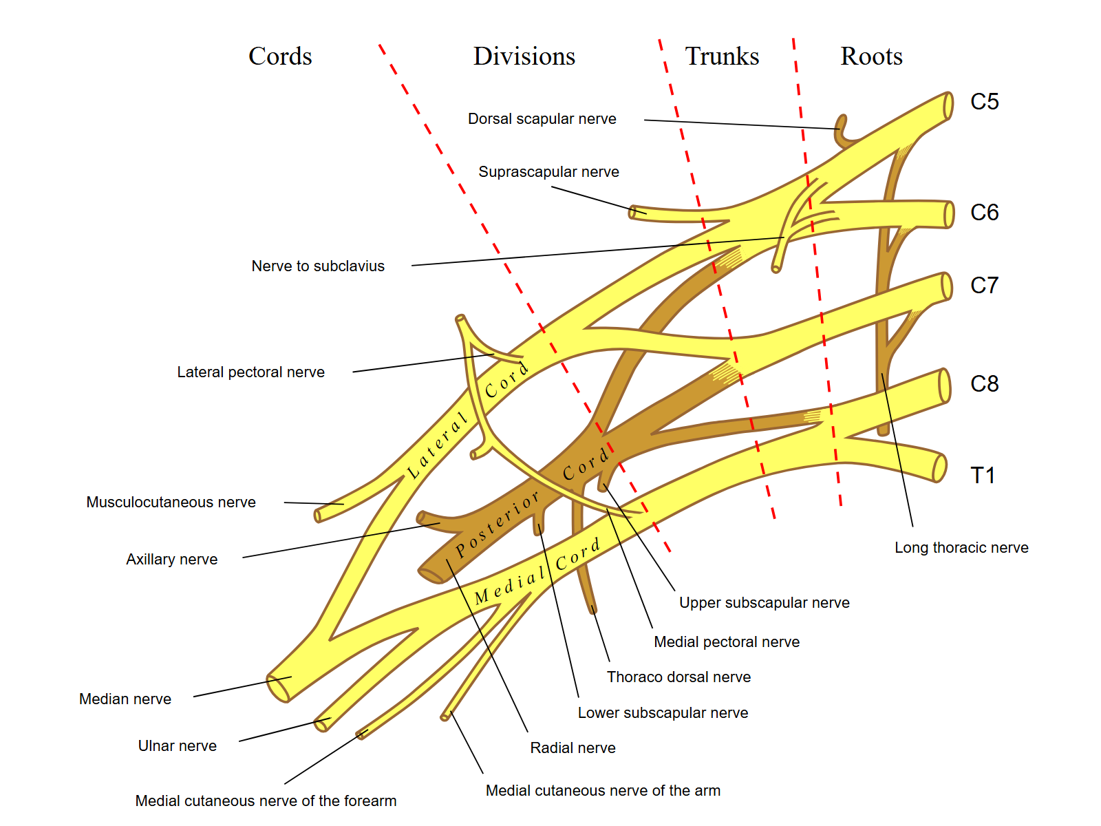
Open the complete image at full sizeFollow the roots from right to left as they combine before forming terminal nerves. A root lesion and a terminal-nerve lesion therefore need not weaken the same set of muscles. This is an anatomical reference, not patient imaging. Mattopaedia; vectorization by Captain-n00dle; simplification by MissMJ, after Gray's Anatomy. Image: Source record. Public domain. Rendered the original vector to a 1600 by 1200 PNG on white. Geometry and labels ; no photographic upscaling, cropping or extra annotation.
On a narrow screen, scroll inside this figure to read every label.
Approximate dorsal hand anchors help organize a root examination, but sensory overlap means no finger location proves a root lesion by itself.
Root contributions overlap, so use a working pattern rather than a memorized one-muscle rule. C5 commonly affects shoulder abduction and external rotation plus elbow flexion; C6 often affects elbow flexion, brachioradialis function, wrist extension, and forearm pronation; C7 often affects elbow extension, wrist flexion, and finger extension. C8 and T1 contribute broadly to finger flexion and intrinsic hand function, with C8 more often represented at the little finger and T1 along the medial forearm.[1][8]
No routinely reliable isolated deep tendon reflex identifies C8 or T1. That statement does not mean that no reflex pathway contains those roots; it means bedside practice lacks a dependable, isolated C8 or T1 reflex equivalent to the usual biceps, brachioradialis, or triceps tests. Lower-root localization therefore depends more heavily on a distributed motor examination, sensory pattern, and targeted electrodiagnostic testing.
C5-C6 zone
Compare deltoid, infraspinatus, biceps, brachioradialis, pronation, and wrist extension. Their axillary, suprascapular, musculocutaneous, radial, and median nerve supply lets one root-level process cross several nerve territories.
C7 zone
Pair triceps and finger extension with pronator teres or flexor carpi radialis. A radial lesion may weaken the extensors, but it should not reproduce the full median-plus-radial pattern.
C8-T1 zone
Test median, ulnar, and radial representatives rather than hand intrinsics alone. Flexor pollicis longus, abductor pollicis brevis, first dorsal interosseous, and extensor indicis help expose a root or lower-trunk pattern that crosses named nerves.
Exit anatomy explains why disc level and root label usually differ below the upper cervical region. C1-C7 roots exit above the same-numbered vertebra, so the C6 root exits through the C5-6 foramen and the C7 root through C6-7. C8 exits between C7 and T1, so a C7-T1 disc or foraminal process often affects C8.[1] These are common relationships, not permission to ignore the actual examination.
Separate roots from nerves, plexus, cord, and shoulder
A mononeuropathy follows one named nerve. Radial neuropathy at the spiral groove weakens wrist and finger extension while sparing triceps; posterior interosseous neuropathy causes motor-predominant finger extension weakness without a cutaneous sensory deficit. A posterior cord lesion reaches radial and axillary territories, whereas a C7 root process can include nonradial C7 muscles such as pronator teres or flexor carpi radialis.[9]
Plexopathy is postganglionic, so an affected sensory nerve action potential can be reduced. A lower-trunk process may weaken median, ulnar, and radial C8-T1 muscles and reduce medial antebrachial cutaneous or ulnar sensory responses; an upper-trunk process can involve suprascapular, axillary, musculocutaneous, and radial functions with abnormal sensory responses. Root disease usually preserves those distal responses and can involve paraspinal muscles.[2][9]
Shoulder disease is tested rather than assumed. Painful or restricted passive range of motion, impingement findings, and weakness limited by pain favor a local shoulder process. Weak deltoid plus infraspinatus plus biceps, a concordant reflex change, normal passive motion, and neck-to-arm symptoms support a root pattern over an isolated axillary, suprascapular, or rotator cuff lesion.
Use electrodiagnosis as anatomy sampled over time
On a narrow screen, scroll inside this figure to read every label.
The dorsal root ganglion relationship helps interpret a sensory nerve action potential. Preserved sensory responses can support a proximal root process, but electrodiagnostic interpretation uses the full pattern.
A typical root lesion lies proximal to the dorsal root ganglion, leaving the ganglion cell and distal sensory axon intact. Distal sensory nerve action potentials are therefore usually preserved in cervical radiculopathy. An abnormal response should prompt consideration of a postganglionic plexus or peripheral nerve lesion, a coexisting neuropathy, or an unusual lesion at or distal to the ganglion rather than automatic rejection of all root disease.[2]
Needle examination gains specificity when abnormalities appear in at least two muscles supplied by the same root but different peripheral nerves. Triceps plus pronator teres plus flexor carpi radialis is much harder for one radial lesion to explain than triceps alone. The laboratory must sample a distribution chosen from the clinical hypothesis, not simply every nearby muscle.[2]
Timing controls sensitivity. Active denervation may appear in paraspinals before distal limb muscles, and a study during the first days of symptoms can be normal even when the clinical localization is persuasive. Purely sensory or predominantly demyelinating root irritation may also lack the axonal loss that needle electromyography detects, so a normal study narrows the evidence but never erases a coherent presentation.[2]
Paraspinal activity is supportive only in context. Fibrillation potentials can occur without symptomatic radiculopathy, especially with age or prior spinal procedures, and isolated paraspinal abnormalities do not identify a complete root syndrome. Pair them with limb findings, sensory studies, timing, and the examination before assigning a level.[2]
Case 18
Show answer and explanations for case 18
A. Flexor carpi ulnaris and abductor digiti minimi (Why this does not fit)
Which nerve carries both muscles in this pair?
The ulnar nerve carries flexor carpi ulnaris and abductor digiti minimi.
Why does the pair fail the localization goal?
It does not cross beyond the nerve already suspected.
Read the complete option explanation
Both muscles remain within the ulnar nerve. Their weakness can occur with a proximal ulnar neuropathy and does not demonstrate a root pattern crossing named nerves.
B. Flexor pollicis longus and extensor indicis (Best answer)
What named nerve carries flexor pollicis longus?
Flexor pollicis longus travels in the median nerve.
Which nerve supplies extensor indicis?
The radial nerve supplies extensor indicis through the posterior interosseous branch.
Which member supplies the clearer C8 weighting?
Extensor indicis is a C8-predominant digit extensor.
How should flexor pollicis longus be weighted?
Its C8-T1 contribution is variable, so it adds median lower-root support.
Read the complete option explanation
Extensor indicis is radial-innervated and provides the clearer C8 weighting. Flexor pollicis longus is median-innervated with variable C8-T1 contribution, so the pair extends the syndrome beyond the ulnar nerve while using extensor indicis as the stronger C8 anchor.
C. Abductor pollicis brevis and first dorsal interosseous (Why this does not fit)
Do APB and FDI cross named nerves?
Yes, they cross median and ulnar nerves.
How are their root contributions distributed?
Abductor pollicis brevis can be T1-predominant, while first dorsal interosseous has mixed C8-T1 input.
What C8 anchor is missing from this pair?
Neither muscle supplies the clearer digit-extensor weighting of extensor indicis.
Read the complete option explanation
This pair crosses median and ulnar nerves, but abductor pollicis brevis can be T1-predominant and first dorsal interosseous has mixed C8-T1 input. It broadens the lesion beyond ulnar territory without the clearer C8 digit-extensor anchor supplied by extensor indicis.
D. Triceps and pronator teres (Why this does not fit)
Which root dominates triceps and pronator teres?
Both are weighted toward C7.
Why is that pair too high?
It does not match the little-finger and intrinsic-hand syndrome.
Read the complete option explanation
This pair crosses radial and median nerves but is weighted toward C7. It would identify a higher myotomal process rather than confirm C8.
E. Deltoid and biceps (Why this does not fit)
Which roots dominate deltoid and biceps?
They are weighted toward C5-C6.
Why is the pair nonconcordant?
It tests an upper-root distribution rather than a C8-predominant lower-root pattern.
Read the complete option explanation
This pair crosses axillary and musculocutaneous nerves and is weighted toward C5-C6. It does not match the lower-root symptoms in the stem.
Takeaway: Extensor indicis provides clearer C8 weighting while flexor pollicis longus adds variable C8-T1 median-nerve support to an ulnar-appearing deficit.
MRI shows structure, not culpability. Foraminal narrowing and disc abnormalities occur in people without matching symptoms, while clinically important irritation may be more convincing on examination than on an early or imperfect study. The side, root, level, and mechanism on imaging must agree with the motor, reflex, sensory, and electrodiagnostic pattern before the image is treated as the cause.[3][4]
Use the exit relationship as a first translation: C5-6 often affects C6, C6-7 often affects C7, and C7-T1 often affects C8. Then test that prediction. A left C5-6 foraminal lesion does not explain right triceps and flexor carpi radialis weakness with a reduced right triceps reflex, even if the report uses alarming language.
Imaging urgency depends on the clinical context. Persistent symptoms after an appropriate conservative interval, progressive objective weakness, suspected myelopathy, or red flags for infection or malignancy justify earlier MRI. Suspected infection or malignancy often requires a contrast-capable protocol, whereas uncomplicated acute radicular symptoms without red flags commonly begin with clinical care and reassessment.[1][3]
Read the report backward. Name the clinical side and root first, predict the responsible foramen, and only then ask whether the image supplies a matching lesion. This protects against anchoring on incidental degeneration.
Choose the next action from trajectory and danger
An uncomplicated presentation has a coherent radicular pattern, stable or improving function, and no cord, infection, trauma, or malignancy warning. Initial care can remain conservative with tolerated activity, symptom control, education, and planned neurologic reassessment. The follow-up is part of the plan because a changing deficit changes the decision.[1]
Progressive objective weakness deserves prompt imaging and specialist assessment even if pain is modest. Mixed segmental and long-tract findings require urgent evaluation for cervical cord compression. Fever, immunosuppression, injection drug use, recent bacteremia, severe unremitting pain, or inflammatory abnormalities raise concern for spinal infection; known cancer, weight loss, night pain, or a progressive focal deficit raises concern for malignant compression.[1][3]
A lower-trunk pattern deserves a wider field of view when the story is progressive or nonmechanical. Shoulder and medial forearm pain, C8-T1 weakness across several nerves, abnormal postganglionic sensory responses, true Horner syndrome, or an apical opacity should redirect evaluation toward the brachial plexus and chest. Ptosis without miosis, anhidrosis, lower-trunk findings, or thoracic evidence is not enough.[10][11]
Final bedside check
Name the weak actions and their peripheral nerves.
Find the root shared across those nerves.
Ask whether reflex and sensory evidence agree.
Use sensory studies and paraspinals to separate preganglionic from postganglionic disease.
Match MRI side and foramen to the clinical root.
Screen trajectory, cord signs, infection, and malignancy before choosing urgency.
Case 27
Show answer and explanations for case 27
A. Same-day cervical MRI and emergency spine assessment (Best answer)
Which finding is segmental?
Triceps weakness with reflex loss supplies a C7-level finding.
Which findings are long-tract or autonomic?
Leg hyperreflexia, plantar extension, gait decline, hand clumsiness, and urinary urgency implicate the cord.
What urgency follows from combining them?
Possible cervical cord compression requires same-day MRI and spine assessment.
Read the complete option explanation
Triceps weakness and reflex loss provide a segmental cervical finding, while leg hyperreflexia, an extensor plantar response, gait decline, hand clumsiness, and urinary change indicate cord involvement. Possible cervical myeloradiculopathy requires urgent structural evaluation.
B. Same-week outpatient cervical MRI and spine clinic (Why this does not fit)
Does this plan target the correct anatomy?
Yes, it targets the cervical spine.
Why is the timing inadequate?
Worsening gait and new urinary symptoms make outpatient delay unsafe.
Read the complete option explanation
This plan targets the correct region but underestimates the pace and breadth of cord findings. Worsening gait and new urinary urgency make an outpatient delay inappropriate.
C. Brain MRI and neurology assessment first (Why this does not fit)
Could brain disease produce upper motor neuron signs?
Yes, brain disease can produce those signs.
What finding makes the cervical cord more coherent?
The segmental triceps reflex loss localizes the same syndrome to the cervical region.
Read the complete option explanation
Brain imaging is a real consideration for upper motor neuron signs, but it does not unify the segmental triceps reflex loss with bilateral leg findings as well as a cervical cord lesion.
D. Electrodiagnostic testing before any imaging (Why this does not fit)
What can electrodiagnosis characterize?
It can characterize root, plexus, or peripheral nerve physiology.
What can it not exclude promptly?
It cannot exclude structural cervical cord compression.
Read the complete option explanation
Electrodiagnosis can characterize the root component but cannot exclude structural cord compression. Requiring it first would delay the test that addresses the dangerous localization.
E. Cervical epidural injection with close follow-up (Why this does not fit)
When is a cervical epidural injection considered?
It is considered for selected radicular pain without urgent neurologic danger.
Which findings prohibit that pathway first?
Long-tract, gait, and urinary changes require cord evaluation.
Read the complete option explanation
An injection can be considered for selected radicular pain without dangerous progression. It is not an appropriate first action when examination shows long-tract, gait, and bladder involvement.
Takeaway: Segmental arm loss plus long-tract, gait, and urinary findings requires same-day cervical cord evaluation.
Each case requires at least two steps: first infer the anatomic pattern, then use a discriminating finding to reject the closest alternative or select the safest next action. Treat normal findings as active evidence only when the case states them.
Case 2
Show answer and explanations for case 2
A. Median sensory response to digit II (Why this does not fit)
What territory would make the median digit II response the direct study?
A median sensory deficit involving the index finger would make it the direct study.
Which finding defeats that choice here?
The sensory loss and weak extensors stay within the radial nerve while median motor functions are normal.
Read the complete option explanation
A median response can be reduced by a median neuropathy or broader plexus process, but the weakness and dorsal first-web sensory loss stay within the radial nerve. Median-innervated pronation and wrist flexion are normal.
B. Ulnar sensory response to digit V (Why this does not fit)
What does the ulnar digit V response sample?
It samples sensory axons serving the little finger through the ulnar nerve.
Why is it not the predicted abnormal response?
No ulnar sensory or motor finding appears in this radial-pattern syndrome.
Read the complete option explanation
An ulnar response evaluates a different named nerve and does not account for wrist and finger extension weakness. The stem supplies no ulnar sensory or motor deficit.
C. Superficial radial sensory response (Best answer)
Where is the lesion relative to the triceps branch?
Normal elbow extension places the radial lesion distal to the triceps branch.
Which branch carries dorsal first-web sensation?
The superficial radial nerve carries that cutaneous territory.
What response follows from postganglionic radial sensory injury?
The symptomatic-side superficial radial sensory amplitude is likely reduced.
Read the complete option explanation
The motor deficit lies in the radial nerve distal to the triceps branch, while dorsal first-web numbness places the lesion proximal to the superficial radial sensory branch. An axonal radial lesion at the spiral groove can therefore reduce this response.
D. Medial antebrachial cutaneous response (Why this does not fit)
What does the medial antebrachial cutaneous response test?
It samples medial-cord and lower-trunk sensory fibers outside the radial nerve.
What case feature argues against that target?
The weakness is radial while nonradial lower-root functions are preserved.
Read the complete option explanation
This response samples medial-cord and lower-trunk sensory fibers. A lower-trunk process would not selectively weaken radial wrist and finger extension while sparing nonradial C8-T1 functions.
E. Lateral antebrachial cutaneous response (Why this does not fit)
Which nerve continues as the lateral antebrachial cutaneous nerve?
The musculocutaneous nerve continues as this lateral forearm sensory branch.
Why is that response not expected to fall?
The case contains no elbow-flexion or lateral-forearm sensory deficit.
Read the complete option explanation
This is the terminal sensory branch of the musculocutaneous nerve and is useful in upper-trunk or musculocutaneous localization. The stated deficit follows radial motor and sensory anatomy instead.
Takeaway: A radial motor deficit distal to triceps plus dorsal first-web sensory loss predicts an abnormal superficial radial sensory response.
Which C7 functions outside the radial nerve remain normal?
Pronator teres and flexor carpi radialis are strong.
Which proximal C7 channel is also preserved?
Triceps function and the triceps reflex are normal.
Read the complete option explanation
A C7 process should be tested across radial and median muscles and often affects the triceps reflex. Preserved pronation, wrist flexion, triceps function, and sensation narrow the deficit to a distal motor branch.
B. Radial neuropathy at the spiral groove (Why this does not fit)
Would a spiral-groove lesion commonly spare cutaneous sensation?
No; the superficial radial sensory branch is usually at risk.
What wrist pattern suggests a more distal motor branch?
Preserved extension with radial deviation reflects selective extensor weakness.
Read the complete option explanation
A spiral-groove lesion commonly adds superficial radial sensory symptoms and broader wrist extension weakness. The intact sensation and radial deviation with extension indicate selective posterior interosseous involvement.
C. Posterior cord plexopathy (Why this does not fit)
Would a posterior cord lesion isolate finger and thumb extension?
No; it should threaten proximal radial and axillary functions.
Which normal muscles make a posterior cord site too high?
Triceps and brachioradialis are normal.
Read the complete option explanation
A posterior cord lesion would affect proximal radial or axillary functions, not only distal finger and thumb extension. The normal triceps and brachioradialis make that site too proximal.
D. Posterior interosseous neuropathy (Best answer)
Which actions fail in this case?
Finger and thumb extension, supplied by posterior interosseous motor branches, fail.
Why is sensation intact?
The posterior interosseous nerve has no cutaneous sensory territory.
Why does wrist extension deviate radially?
Radial wrist extensors are relatively spared while ulnar-side extension is weaker.
Read the complete option explanation
The posterior interosseous nerve supplies finger and thumb extensors but carries no cutaneous sensation. Preserved wrist extension with radial deviation reflects relative sparing of radial wrist extensors outside or proximal to the affected branch.
E. Lower-trunk plexopathy (Why this does not fit)
Would lower-trunk disease remain within a distal radial motor branch?
No; it should cross median, ulnar, and radial C8-T1 territories.
Which expected lower-trunk findings are absent?
Hand intrinsic, finger-flexor, and medial sensory abnormalities are not present.
Read the complete option explanation
A lower-trunk lesion would not isolate radial finger extensors. It should involve C8-T1 muscles in median and ulnar territories and may alter medial forearm or ulnar sensory responses.
Takeaway: Finger extension failure without sensory loss, with preserved radially deviated wrist extension, is the classic motor pattern of posterior interosseous neuropathy.
Does one C7 root explain deltoid weakness and lateral-shoulder sensory loss?
No; those are chiefly upper-root and axillary findings.
What sensory-study feature argues against a root?
Both axillary and radial postganglionic responses are reduced.
Read the complete option explanation
C7 disease can weaken triceps and finger extensors, but it does not explain axillary sensory loss and deltoid weakness as one root pattern. Abnormal postganglionic sensory responses also oppose an isolated root lesion.
B. Radial nerve at the axilla (Why this does not fit)
Can a radial nerve lesion weaken deltoid?
No; deltoid is supplied by the axillary nerve.
What shared structure lies before radial and axillary nerves separate?
The posterior cord.
Read the complete option explanation
A radial lesion cannot weaken deltoid or reduce an axillary sensory response. Those findings require involvement before the radial and axillary nerves separate.
C. Upper trunk (Why this does not fit)
Which motor function should upper-trunk injury commonly affect?
Elbow flexion through biceps is commonly involved.
What pattern dominates instead?
Axillary plus broad radial deficits with preserved biceps point to the posterior cord.
Read the complete option explanation
An upper-trunk lesion emphasizes C5-C6 functions such as shoulder abduction, external rotation, and elbow flexion. The prominent triceps and distal radial pattern with preserved biceps is a poorer fit.
D. Lower trunk (Why this does not fit)
Which functions dominate lower-trunk lesions?
C8-T1 finger flexion and hand intrinsic functions across several nerves.
Do this patient's deltoid and triceps findings fit that center?
No; they localize much higher and posteriorly.
Read the complete option explanation
A lower-trunk lesion emphasizes C8-T1 hand and finger functions across several nerves. Deltoid, triceps, and radial sensory involvement with spared ulnar intrinsics points elsewhere.
E. Posterior cord (Best answer)
Which two terminal nerves are jointly affected?
Axillary and radial territories are both involved.
Which functions from other cords are spared?
Biceps, pronation, wrist flexion, and ulnar intrinsic function remain normal.
What supports a postganglionic plexus site?
Low axillary and radial sensory responses with normal paraspinals.
Read the complete option explanation
Deltoid and axillary sensation join proximal and distal radial deficits within the posterior cord distribution. Sparing of median and ulnar functions, low axillary and radial sensory responses, and normal paraspinals support a postganglionic cord lesion.
Takeaway: Combined axillary and radial deficits with postganglionic sensory loss localize to the posterior cord.
Median, ulnar, and radial nerves are all represented.
What does preservation of all sampled sensory responses imply?
The lesion is proximal to the dorsal root ganglion.
Which finding directly supports a root site?
A lower cervical paraspinal muscle is abnormal.
Read the complete option explanation
Weakness crosses median, ulnar, and radial nerves while retaining a common lower-root contribution. Preserved sensory responses and paraspinal involvement place the process proximal to the dorsal root ganglion and favor C8.
B. Ulnar neuropathy at the elbow (Why this does not fit)
Which weak muscles lie outside the ulnar nerve?
Flexor pollicis longus, abductor pollicis brevis, and extensor indicis.
What sensory result also weakens the ulnar-neuropathy hypothesis?
The ulnar sensory response is normal.
Read the complete option explanation
An ulnar lesion cannot explain flexor pollicis longus, abductor pollicis brevis, or extensor indicis weakness. It may also reduce the ulnar sensory response, which is preserved here.
C. Median neuropathy at the wrist (Why this does not fit)
Can a wrist-level median lesion affect ulnar intrinsics or extensor indicis?
No; both lie outside that distal median territory.
Can it create paraspinal denervation?
No; paraspinals are proximal to the plexus and wrist.
Read the complete option explanation
A wrist-level median lesion cannot weaken ulnar intrinsics or extensor indicis. It also should not produce lower cervical paraspinal abnormalities.
D. Lower-trunk plexopathy (Why this does not fit)
Would a lower-trunk plexopathy cross several named nerves?
Yes, so its motor pattern can resemble this case.
Which paired findings instead support a root?
Preserved ulnar and medial forearm sensory responses plus paraspinal denervation.
Read the complete option explanation
A lower-trunk lesion can cross these nerves, but postganglionic medial antebrachial cutaneous or ulnar sensory abnormalities would support it. Normal sensory responses plus paraspinal involvement favor a root.
E. T1 radiculopathy (Why this does not fit)
Which sensory landmark is stated?
Symptoms radiate to the little finger, a common C8 landmark.
What would strengthen T1 instead?
A medial-forearm pattern with more intrinsic-dominant weakness would better support T1.
Read the complete option explanation
C8 and T1 overlap in the hand, but the little-finger symptoms and broad finger flexor and extensor pattern are more consistent with C8 in this case. T1 is better supported by a medial forearm pattern and a more intrinsic-dominant motor distribution.
Takeaway: Median, ulnar, and radial lower-root muscles with preserved sensory responses identify a preganglionic C8 pattern better than an ulnar lesion.
A. Median sensory response to digit II (Why this does not fit)
Which skin territory does the median digit II response test?
It tests the index-finger territory through the median nerve.
Why is it a weak lower-trunk discriminator here?
It does not sample the medial forearm fibers that lie outside the ulnar nerve.
Read the complete option explanation
A median response may detect a separate median lesion or diffuse neuropathy, but it is not the proximal lower-trunk sensory marker that best separates an ulnar lesion from plexopathy.
B. Superficial radial sensory response (Why this does not fit)
Which named nerve carries the superficial radial response?
The radial nerve carries this dorsolateral hand sensory response.
Why is it not the best extension test?
The competing plexus lesion is centered in the medial C8-T1 territory.
Read the complete option explanation
This response samples radial sensory fibers and can help assess a broader neuropathy. It does not directly test the medial lower-trunk territory that lies outside the ulnar nerve.
C. Dorsal ulnar cutaneous response (Why this does not fit)
Where does the dorsal ulnar cutaneous branch arise?
It arises from the ulnar nerve proximal to the wrist.
What can it distinguish better than lower-trunk disease?
It can help separate an elbow-level ulnar lesion from a wrist-level ulnar lesion.
Read the complete option explanation
This response can help distinguish an ulnar lesion at the elbow from one at the wrist. Because it remains within the ulnar nerve, it cannot by itself show that the process extends into the lower trunk.
D. Medial antebrachial cutaneous response (Best answer)
Which plexus structure gives rise to the medial antebrachial cutaneous nerve?
It arises from the medial cord and carries lower-trunk input.
What result fits an isolated ulnar lesion?
The medial antebrachial cutaneous response remains preserved.
What result would broaden the localization?
A side-specific reduction would support lower-trunk or medial-cord involvement.
Read the complete option explanation
This sensory nerve arises from the medial cord and carries lower-trunk input outside the ulnar nerve. A preserved response supports an isolated ulnar lesion, whereas a side-specific reduction would raise concern for lower-trunk or medial-cord involvement.
E. Lateral antebrachial cutaneous response (Why this does not fit)
Which plexus region contributes to the lateral antebrachial cutaneous response?
This musculocutaneous branch predominantly samples upper-trunk sensory output.
Why does it miss the competing lesion?
The question concerns medial lower-trunk fibers rather than lateral forearm fibers.
Read the complete option explanation
This response reflects musculocutaneous and upper-trunk sensory anatomy. It is a real plexus study, but it does not interrogate the C8-T1 medial territory competing with an ulnar neuropathy.
Takeaway: The medial antebrachial cutaneous response tests lower-trunk sensory fibers that an isolated ulnar neuropathy should spare.
Does the cross-nerve motor pattern resemble C8 root disease?
Yes; median, ulnar, and radial lower-root muscles are involved.
What finding relocates the lesion distal to the ganglion?
Ulnar and medial antebrachial cutaneous sensory responses are reduced.
Read the complete option explanation
C8 disease can cross peripheral nerves, but it usually leaves distal sensory responses intact because the lesion is proximal to the dorsal root ganglion. The abnormal ulnar and medial forearm responses point postganglionically.
B. Ulnar neuropathy (Why this does not fit)
Which weak actions cannot come from an ulnar lesion?
Median thumb flexion and radial index extension lie outside the ulnar nerve.
What does medial forearm sensory loss add?
It extends beyond the ulnar cutaneous territory.
Read the complete option explanation
An ulnar lesion cannot explain median thumb flexion or radial index extension abnormalities. The motor pattern is broader than one named nerve.
C. Posterior cord lesion (Why this does not fit)
Does the posterior cord contain median and ulnar lower-root fibers?
No; those fibers leave through other cords.
Which sensory response is especially incompatible with posterior cord disease?
The medial antebrachial cutaneous response derives from the medial cord pathway.
Read the complete option explanation
A posterior cord lesion does not explain median and ulnar lower-root weakness or a reduced medial antebrachial cutaneous response. The distribution is centered in the lower trunk.
D. Cervical myelopathy (Why this does not fit)
Are long-tract signs supplied?
No; the stem gives a peripheral sensorimotor distribution.
Can cervical myelopathy selectively reduce these peripheral sensory responses?
No; the low postganglionic responses point to plexus or nerve disease.
Read the complete option explanation
The findings form a postganglionic lower-plexus sensorimotor pattern without stated long-tract signs. Myelopathy would require cord evidence beyond this peripheral distribution.
E. Lower-trunk brachial plexopathy (Best answer)
How broad is the motor involvement?
It spans median, ulnar, and radial C8-T1 muscles.
What establishes postganglionic localization?
Both ulnar and medial antebrachial cutaneous responses are reduced.
What finding reduces support for a root?
Cervical paraspinal muscles are normal.
Read the complete option explanation
The weakness spans median, ulnar, and radial lower-root muscles, while reduced ulnar and medial antebrachial cutaneous responses support postganglionic sensory involvement. Normal paraspinals further support the plexus over the roots.
Takeaway: A broad C8-T1 motor pattern plus reduced medial antebrachial cutaneous or ulnar sensory responses localizes to the lower trunk.
Could C5 root disease produce the stated motor distribution?
Yes; the motor pattern crosses several C5-C6 peripheral nerves.
What evidence places the lesion beyond the root?
Lateral antebrachial cutaneous and superficial radial sensory responses are reduced while paraspinals are normal.
Read the complete option explanation
A C5 process can produce much of the motor pattern, but distal sensory responses should usually remain preserved and paraspinal involvement may be found. The postganglionic sensory abnormalities favor plexus injury.
B. Upper-trunk brachial plexopathy (Best answer)
Which terminal nerve territories are involved?
Suprascapular, axillary, musculocutaneous, and radial functions are abnormal.
What do the low sensory responses establish?
They show postganglionic injury affecting upper-trunk output.
How do normal paraspinals refine the site?
They reduce support for a proximal C5-C6 root lesion.
Read the complete option explanation
The deficit crosses suprascapular, axillary, musculocutaneous, and radial territories that share upper-trunk input. Reduced sensory responses and normal paraspinals place the lesion distal to the dorsal root ganglion.
C. Axillary neuropathy (Why this does not fit)
Which abnormal muscles lie outside the axillary nerve?
Infraspinatus, biceps, and brachioradialis.
Can an axillary lesion lower the lateral antebrachial cutaneous response?
No; that response is carried by the musculocutaneous pathway.
Read the complete option explanation
An axillary lesion explains deltoid weakness but not infraspinatus, biceps, brachioradialis, or the lateral forearm sensory response. The syndrome crosses several terminal nerves.
D. Suprascapular neuropathy (Why this does not fit)
Which action fits suprascapular neuropathy?
External rotation through infraspinatus can be weak.
Which findings exceed that nerve?
Deltoid, biceps, brachioradialis, and two distal sensory responses are also abnormal.
Read the complete option explanation
A suprascapular lesion can weaken external rotation but cannot explain deltoid, elbow flexion, brachioradialis, or distal sensory abnormalities. Its territory is too narrow.
E. Rotator cuff tear (Why this does not fit)
Can a rotator cuff tear denervate biceps and brachioradialis?
No; those are neurologic findings outside the cuff.
Can it reduce peripheral sensory responses?
No; the abnormal sensory potentials demonstrate neural injury.
Read the complete option explanation
A cuff tear may impair shoulder actions but cannot cause biceps and brachioradialis denervation or reduced lateral antebrachial cutaneous and radial sensory responses. The data demonstrate neurologic injury.
Takeaway: Cross-nerve C5-C6 weakness with abnormal postganglionic sensory responses and normal paraspinals favors upper-trunk plexopathy.
A. Low lateral antebrachial and radial sensory responses with normal paraspinals (Why this does not fit)
What do low lateral forearm and radial sensory responses establish?
They establish postganglionic sensory axon involvement in upper-trunk territories.
What does normal paraspinal sampling add?
It removes a finding that would otherwise support a preganglionic root lesion.
Read the complete option explanation
The motor pattern can occur with upper-trunk disease, and reduced postganglionic sensory responses with spared paraspinals would support that plexus localization rather than a root.
B. Normal lateral antebrachial and radial sensory responses with cross-nerve denervation and abnormal paraspinals (Best answer)
Which named nerves carry the abnormal upper-root muscles?
Axillary, suprascapular, and musculocutaneous nerves carry the tested muscles.
What do preserved distal sensory responses imply?
They favor a lesion proximal to the dorsal root ganglion.
What does cervical paraspinal denervation add?
It supplies direct proximal support for C5 radiculopathy.
Read the complete option explanation
Deltoid, infraspinatus, and biceps abnormalities would cross axillary, suprascapular, and musculocutaneous nerves while sharing upper-root input. Preserved distal sensory responses and matching cervical paraspinal denervation favor a preganglionic C5 process.
C. Low axillary sensory response with denervation limited to deltoid (Why this does not fit)
Which muscle and sensory branch fit this pattern?
Deltoid and the axillary sensory branch fit an axillary neuropathy.
Which stem deficits remain unexplained?
External rotation, elbow flexion, and the biceps reflex lie outside an isolated axillary lesion.
Read the complete option explanation
This is a coherent axillary neuropathy pattern and can explain shoulder abduction weakness. It cannot account for external-rotation and elbow-flexion abnormalities across other nerves.
D. Normal sensory responses with denervation limited to infraspinatus (Why this does not fit)
Which nerve supplies infraspinatus?
The suprascapular nerve supplies infraspinatus.
Why is isolated infraspinatus denervation too narrow?
It cannot explain deltoid, biceps, and biceps-reflex abnormalities.
Read the complete option explanation
Isolated infraspinatus denervation would favor a suprascapular lesion or focal branch lesion. It does not explain deltoid, biceps, and reflex findings as one process.
E. Normal sensory responses with normal limb and paraspinal sampling (Why this does not fit)
Can a normal study occur in a mild root lesion?
Yes, especially when axonal loss is limited or the study is early.
Does a normal study favor root over upper trunk?
No, because it supplies no positive preganglionic discriminator.
Read the complete option explanation
A normal study can occur in mild or early radiculopathy, but it would not positively distinguish a root from an upper-trunk lesion. The question asks for the pattern that most strongly favors the root.
Takeaway: Cross-nerve upper-root denervation, preserved distal sensory responses, and paraspinal involvement favor C5 radiculopathy over upper-trunk plexopathy.
Right triceps weakness and a reduced right triceps reflex indicate a C7-level process.
Which findings are long tract?
Brisk legs, an extensor plantar response, impaired tandem gait, and hand clumsiness implicate the cord.
What single cervical framework includes both?
Cord compression with segmental root-level involvement produces a myeloradicular syndrome.
Read the complete option explanation
Triceps weakness and reflex loss supply a segmental C7 pattern, while brisk legs, an extensor plantar response, gait impairment, and hand clumsiness are long-tract findings. Their coexistence requires a cervical myeloradicular localization.
B. Isolated C7 radiculopathy (Why this does not fit)
What can isolated C7 radiculopathy explain?
It can explain triceps weakness and reflex reduction.
Which findings require a central lesion?
Leg hyperreflexia, a pathologic plantar response, and gait impairment.
Read the complete option explanation
An isolated root lesion can explain the arm findings but not bilateral leg hyperreflexia, a pathologic plantar response, or gait dysfunction. Those findings place the cord in danger.
C. Radial neuropathy (Why this does not fit)
Can a radial lesion alter the plantar response?
No; it is a peripheral upper-limb lesion.
Can it explain bilateral brisk legs and gait change?
No; those require corticospinal tract involvement.
Read the complete option explanation
A radial lesion might weaken elbow extension only if very proximal, but it cannot generate lower-extremity upper motor neuron signs. It also would not unify hand clumsiness and gait change.
D. Length-dependent polyneuropathy (Why this does not fit)
What reflex pattern is common in length-dependent polyneuropathy?
Distal reflexes are often reduced rather than brisk.
Does a generalized neuropathy explain a focal triceps-plus-long-tract pattern?
No; the findings converge in the cervical cord region.
Read the complete option explanation
Polyneuropathy more often reduces distal reflexes and produces a symmetric distal sensory pattern. It does not explain a focal reduced triceps reflex with pathologic long-tract signs.
E. Intrinsic shoulder disease (Why this does not fit)
Can shoulder disease reduce the triceps reflex?
No; the reflex arc is neurologic.
Can it produce an extensor plantar response?
No; that is a central motor pathway sign.
Read the complete option explanation
Shoulder disease cannot cause a reduced triceps reflex, extensor plantar response, or brisk legs. The neurologic examination directly indicates cervical cord and segmental involvement.
Takeaway: Segmental arm loss plus long-tract signs requires priority evaluation for cervical myeloradiculopathy.
Which expected C5 actions are not the center of this case?
Shoulder abduction and external rotation are not the dominant deficits.
Read the complete option explanation
This foramen transmits the C5 root. A C5 lesion can affect shoulder abduction and external rotation more prominently than the thumb, wrist-extension, and brachioradialis pattern supplied here.
B. C5-6 foramen (Best answer)
Which root pattern does the examination establish?
Thumb symptoms, elbow flexion, wrist extension, and the brachioradialis reflex point to C6.
Where does C6 exit?
C6 exits through the C5-6 foramen.
What target therefore matches side and level?
The right C5-6 foramen is concordant.
Read the complete option explanation
The examination first localizes to C6 across musculocutaneous and radial functions. Because C1-C7 roots exit above the same-numbered vertebra, C6 traverses the C5-6 foramen.
C. C6-7 foramen (Why this does not fit)
Which root exits through C6-7?
The C7 root exits through the C6-7 foramen.
Which C7 functions are preserved?
Triceps strength and the triceps reflex are preserved.
Read the complete option explanation
This foramen transmits C7. A concordant C7 syndrome would emphasize triceps, finger extension, wrist flexion, and the triceps reflex rather than the supplied C6-weighted pattern.
D. C7-T1 foramen (Why this does not fit)
Which root exits through C7-T1?
The C8 root exits through the C7-T1 foramen.
What lower-root pattern is absent?
Finger-flexor, index-extensor, and intrinsic-hand deficits are absent.
Read the complete option explanation
This foramen transmits C8. C8 disease is better supported by finger flexion, thumb flexion, index extension, intrinsic hand weakness, and little-finger symptoms.
E. T1-2 foramen (Why this does not fit)
Which spinal region follows the below-vertebra naming rule here?
Thoracic roots below C8 follow the lower-spine relationship.
Why is T1-2 too low?
It cannot explain a C6-weighted upper-limb pattern.
Read the complete option explanation
This level would not explain a C6-weighted upper-limb syndrome. The extra cervical root is C8 at C7-T1; below that level, thoracic roots follow the lower-spine naming relationship.
Takeaway: Infer the root from the examination first, then map C6 to the C5-6 exit foramen.
Which root most strongly contributes to flexor carpi radialis?
Flexor carpi radialis is weighted toward C6-C7.
Why would its weakness reduce level specificity?
It would add a higher-root median finding rather than targeted lower-root evidence.
Read the complete option explanation
Flexor carpi radialis is median-innervated but is weighted toward C6-C7. Its weakness would broaden a C7 pattern rather than add the most useful lower-root confirmation.
B. Brachioradialis (Why this does not fit)
Which roots dominate brachioradialis?
Brachioradialis is predominantly C5-C6.
Does another radial muscle add the missing nerve territory?
No, because extensor indicis already supplies radial evidence.
Read the complete option explanation
Brachioradialis is radial-innervated and weighted toward C5-C6. It does not add the median-innervated lower-root evidence needed to strengthen concordance at C7-T1.
C. Flexor pollicis longus (Best answer)
Which root exits through the C7-T1 foramen?
The C8 root exits through C7-T1.
Which nerve supplies flexor pollicis longus?
The median nerve supplies flexor pollicis longus.
Does flexor pollicis longus identify C8 by itself?
No, its root contribution varies across C8-T1.
How should its weakness be used here?
It adds median-innervated lower-root evidence to the radial and ulnar pattern.
Read the complete option explanation
The C8 root exits through C7-T1, which supplies the level discriminator. Flexor pollicis longus is a median-innervated lower-root muscle with variable C8-T1 contribution, so its weakness strengthens the radial and ulnar pattern without acting as an absolute C8 marker by itself.
D. Deltoid (Why this does not fit)
Which root dominates deltoid?
Deltoid is predominantly a C5 muscle through the axillary nerve.
Why would deltoid weakness be discordant?
It would broaden the process to a much higher root.
Read the complete option explanation
Deltoid adds axillary-nerve evidence but is weighted toward C5. Its weakness would suggest a higher or broader process rather than a concordant C8 lesion.
E. Triceps (Why this does not fit)
Which root and reflex are represented by triceps?
Triceps and its reflex predominantly represent C7.
What does the normal triceps examination already show?
It bounds the supplied deficit below the main C7 distribution.
Read the complete option explanation
Triceps is predominantly C7 and already has a normal examination and reflex in this case. New triceps weakness would create adjacent-level involvement rather than selectively strengthen C8.
Takeaway: At C7-T1, the foramen identifies C8 while flexor pollicis longus adds median-innervated lower-root evidence to the radial and ulnar pattern.
A. C7 radiculopathy with carpal tunnel syndrome (Why this does not fit)
Which part of this pair fits the motor findings?
The C7 root component fits median and radial muscle abnormalities with paraspinal involvement.
Which supplied sensory finding is left unexplained?
Carpal tunnel syndrome does not explain focal dorsal first-web numbness with a low superficial radial response.
Read the complete option explanation
The root component fits the cross-nerve motor and paraspinal findings, but a median lesion at the wrist does not explain focal dorsal first-web numbness and a low superficial radial response.
B. Radial-groove neuropathy with ulnar neuropathy (Why this does not fit)
Which abnormality could the radial component explain?
A radial lesion can affect radial motor and sensory fibers.
Do radial and ulnar lesions explain the entire needle pattern?
No, median-innervated muscles and cervical paraspinals are also abnormal.
Read the complete option explanation
A radial-groove lesion can affect radial motor and sensory fibers, but adding an ulnar lesion still does not explain denervation in median-innervated pronator teres and flexor carpi radialis plus cervical paraspinals.
C. Middle-trunk plexopathy with superficial radial neuropathy (Why this does not fit)
Why is the middle trunk initially plausible?
It carries C7 fibers that later enter different peripheral nerves.
Which convergent findings favor a root over that trunk?
Preserved median sensory conduction and cervical paraspinal denervation favor a preganglionic C7 process.
Read the complete option explanation
A middle-trunk lesion can involve C7 fibers across several nerves and the added sensory lesion fits the radial response. However, the preserved median sensory response and cervical paraspinal denervation favor a preganglionic root process over the plexus component.
D. C7 radiculopathy with superficial radial neuropathy (Best answer)
What unifies the cross-nerve weakness and paraspinal denervation?
A C7 root process can affect both median and radial motor outputs.
Does that root lesion alone explain the focal low radial sensory response?
Not well, because the distal sensory response is usually preserved in a preganglionic lesion.
Which additional lesion fits the focal cutaneous findings?
A superficial radial sensory lesion explains dorsal first-web numbness and the reduced response.
Why can the radial motor study remain normal?
The additional superficial radial lesion affects a cutaneous sensory branch rather than radial motor fibers.
Read the complete option explanation
The cross-nerve C7 motor pattern and paraspinal denervation support a root lesion. The separate focal dorsal first-web sensory deficit and low superficial radial response, with normal radial motor conduction, support an additional superficial radial sensory lesion.
E. Posterior-cord plexopathy with median neuropathy (Why this does not fit)
Why could two terminal territories suggest this pair?
Posterior-cord and median lesions can affect radial and median muscles.
Which finding is not explained by either component?
Cervical paraspinal denervation lies proximal to the plexus and terminal nerves.
Read the complete option explanation
Posterior-cord and median lesions can together affect radial and median muscles, but neither explains cervical paraspinal denervation. The focal radial sensory findings plus a convergent preganglionic C7 motor pattern are better explained by the keyed pair.
Takeaway: A side-specific abnormal SNAP does not erase a convincing root pattern; it can reveal a superimposed postganglionic lesion.
It stays within the competing radial territory and is weighted toward C5-C6.
Read the complete option explanation
Brachioradialis is supplied by the radial nerve and is weighted toward C5-C6. An abnormality would not cross the radial nerve boundary or specifically reinforce C7.
B. Extensor indicis (Why this does not fit)
Which distal branch supplies extensor indicis?
The posterior interosseous branch of the radial nerve supplies it.
Why does another radial muscle not settle the question?
A radial lesion can affect both finger extensors without crossing a named-nerve boundary.
Read the complete option explanation
Extensor indicis is a radial posterior interosseous muscle with C7-C8 contribution. It would extend the radial motor pattern but still could be explained by a radial lesion.
C. Flexor carpi radialis (Best answer)
Which nerve supplies flexor carpi radialis?
The median nerve supplies flexor carpi radialis.
Which root contribution links it to the stem?
It carries substantial C7 input.
What localization follows from radial plus median C7 loss?
A lesion proximal to terminal nerve separation becomes much more likely.
Read the complete option explanation
Flexor carpi radialis carries substantial C7 input through the median nerve. Its abnormality would join radial triceps and finger extensors across a different named nerve, which is the key root-level discriminator.
D. Abductor pollicis brevis (Why this does not fit)
Which roots dominate abductor pollicis brevis?
Abductor pollicis brevis is weighted toward C8-T1.
Why is that not targeted confirmation?
Its abnormality would introduce a lower-root process rather than reinforce C7.
Read the complete option explanation
Abductor pollicis brevis is median-innervated but weighted toward C8-T1. Its abnormality would suggest a lower-root or separate median process rather than targeted C7 confirmation.
E. Flexor carpi ulnaris (Why this does not fit)
Which roots dominate flexor carpi ulnaris?
Flexor carpi ulnaris is weighted toward C8-T1.
What would its weakness imply?
It would broaden the syndrome below C7 instead of confirming the suspected level.
Read the complete option explanation
Flexor carpi ulnaris is ulnar-innervated and weighted toward C8-T1. It crosses the nerve boundary but not at the root level needed to confirm this C7-weighted syndrome.
Takeaway: For suspected C7 radiculopathy, a median-innervated C7 muscle such as flexor carpi radialis adds more than another radial muscle.
A. Biceps and brachioradialis denervation with preserved sensory responses (Best answer)
Which named nerves carry biceps and brachioradialis?
Musculocutaneous and radial nerves carry them.
What shared root fits both muscles and the reflex examination?
Their shared C5-C6 input fits the clinical C6 localization.
What do preserved sensory responses add?
They favor a preganglionic lesion after limb denervation has had time to appear.
Read the complete option explanation
Biceps and brachioradialis cross musculocutaneous and radial nerves while sharing C5-C6 input that matches the examination. Preserved distal sensory responses support a preganglionic root process, and later sampling allows limb denervation to emerge.
B. Brachioradialis and wrist-extensor denervation with a low radial sensory response (Why this does not fit)
Do these abnormal muscles cross a named-nerve boundary?
No, because both remain in the radial nerve.
What does the low radial sensory response imply?
It favors a postganglionic radial lesion.
Read the complete option explanation
These muscles and the sensory response remain within the radial nerve. The pattern would favor a postganglionic radial lesion rather than support C6 across different named nerves.
C. Deltoid and biceps denervation with low lateral forearm and radial sensory responses (Why this does not fit)
Can deltoid and biceps reflect upper-root injury?
Yes, they cross axillary and musculocutaneous nerves.
What shifts this pattern toward the plexus?
Low lateral forearm and radial sensory responses show postganglionic injury.
Read the complete option explanation
Cross-nerve upper-root motor loss is plausible, but reduced postganglionic sensory responses would favor an upper-trunk plexopathy over a preganglionic C6 lesion.
D. Supraspinatus and infraspinatus denervation with preserved sensory responses (Why this does not fit)
Which nerve supplies supraspinatus and infraspinatus?
The suprascapular nerve supplies both muscles.
Which clinical deficit remains outside that nerve?
Wrist-extension weakness remains unexplained.
Read the complete option explanation
Both muscles are supplied by the suprascapular nerve. This pattern would support a focal suprascapular lesion and would not explain wrist-extension weakness or two reduced reflexes.
E. Abductor pollicis brevis denervation with a low median sensory response (Why this does not fit)
Which localization fits APB denervation with a low median response?
A distal median neuropathy is a plausible fit.
Why is it discordant with the stem?
APB is lower-root weighted and does not explain the C6 motor-reflex pattern.
Read the complete option explanation
This is a distal median pattern weighted toward C8-T1 and could reflect median neuropathy. It is anatomically remote from the C6 motor-reflex syndrome.
Takeaway: An early normal limb study can later become diagnostic when same-root abnormalities appear across different nerves and sensory responses stay preserved.
A. Denervation in triceps and flexor carpi radialis (Best answer)
Which named nerves carry triceps and flexor carpi radialis?
Radial and median nerves carry them.
Which root strongly contributes to both?
C7 strongly contributes to both muscles.
Why does this rescue the paraspinal finding?
A concordant cross-nerve limb distribution gives the proximal activity clinical specificity.
Read the complete option explanation
Triceps and flexor carpi radialis share substantial C7 input but travel in radial and median nerves. A concordant cross-nerve limb pattern would give the isolated paraspinal activity the distributional support it currently lacks.
B. Denervation in triceps and extensor digitorum (Why this does not fit)
Which named nerve carries both proposed muscles?
The radial nerve carries triceps and extensor digitorum.
What competing localization remains possible?
A proximal radial lesion can affect both without requiring C7 root disease.
Read the complete option explanation
Both muscles are radial-innervated. This could reflect a proximal radial lesion and supplies less root-specific evidence than abnormalities crossing into a nonradial C7 muscle.
C. Denervation in first dorsal interosseous and abductor digiti minimi (Why this does not fit)
Which named nerve carries both hand muscles?
The ulnar nerve carries first dorsal interosseous and abductor digiti minimi.
Which myotomal level do they emphasize?
They emphasize C8-T1 rather than C7.
Read the complete option explanation
Both muscles are ulnar-innervated and weighted toward C8-T1. They would support an ulnar or lower-root process, not the proposed C7 level.
D. Denervation in deltoid and infraspinatus (Why this does not fit)
Which root pattern links deltoid and infraspinatus?
They form an upper-root pattern centered on C5.
Why does that not corroborate the stated level?
It would support C5 physiology rather than C7.
Read the complete option explanation
These muscles cross axillary and suprascapular nerves but are weighted toward C5. They could support an upper-root process, not C7 concordance.
E. Fibrillation potentials in bilateral cervical paraspinal muscles only (Why this does not fit)
Does bilateral paraspinal activity create a limb distribution?
No, because the limb examination and limb needle samples remain normal.
Why is postoperative age context still important?
Older postoperative paraspinals can show activity without symptomatic radiculopathy.
Read the complete option explanation
More isolated paraspinal activity still lacks a matching limb distribution and may remain nonspecific in an older postoperative patient. Bilaterality would not create C7 specificity.
Takeaway: Paraspinal activity becomes persuasive when same-root limb abnormalities cross named peripheral nerves.
No; C4-5 carries C5, while the abnormal muscles are C7-weighted.
Which normal muscles argue against C5?
Biceps and brachioradialis are normal.
Read the complete option explanation
C4-5 contains the exiting C5 root. The normal biceps and brachioradialis and the abnormal C7-weighted radial and median muscles oppose C5 as the target.
B. C5-6 foramen (Why this does not fit)
Which root exits through C5-6?
The C5-6 foramen carries C6, above the sampled C7-weighted pattern.
Which expected C6 representatives are spared?
Biceps and brachioradialis are spared.
Read the complete option explanation
C5-6 contains the exiting C6 root. C6 disease would emphasize biceps, brachioradialis, and wrist extension more than the bounded triceps, pronator, and wrist-flexor pattern.
They bound the pattern between C5-C6 and C8-T1 representatives.
Which foramen carries C7?
C7 exits through the C6-7 foramen.
Read the complete option explanation
The abnormal muscles share C7 across radial and median nerves, adjacent myotomes are spared, and sensory responses fit a preganglionic process. The C7 root exits through the C6-7 foramen.
D. C7-T1 foramen (Why this does not fit)
Does C7-T1 fit the sampled C7-weighted muscles?
No; C7-T1 carries C8, below the sampled C7-weighted pattern.
Which C8-T1 samples are normal?
First dorsal interosseous and abductor pollicis brevis are normal.
Read the complete option explanation
C7-T1 contains the exiting C8 root. Normal first dorsal interosseous and abductor pollicis brevis reduce support for a C8-weighted lower-root syndrome.
E. T1-2 foramen (Why this does not fit)
What pattern would make T1-2 relevant?
A thoracic or strongly T1-weighted syndrome would make it relevant.
Why does the present pattern sit higher?
The abnormalities are bounded to C7 arm muscles.
Read the complete option explanation
A T1-2 lesion would not account for a bounded C7 motor pattern. The hand-intrinsic samples that would raise concern for a lower root are normal.
Takeaway: Cross-nerve C7 physiology predicts a concordant lesion at the C6-7 exit foramen.
A. A: lower-trunk plexopathy; B: lower-cervical radiculopathy (Why this does not fit)
What do patient A's normal sensory responses imply?
Normal ulnar and medial antebrachial cutaneous responses in patient A favor a preganglionic site.
What does patient B's reduced lower-trunk sensation imply?
It favors a postganglionic lesion, so the proposed order is reversed.
Read the complete option explanation
This reverses the preganglionic and postganglionic evidence. Patient A has preserved distal sensory responses and paraspinal involvement, whereas patient B has lower-trunk sensory axon loss with paraspinal sparing.
B. A: ulnar neuropathy; B: medial-cord plexopathy (Why this does not fit)
Can an ulnar lesion explain patient A's full motor set?
No, because median and radial lower-root muscles are also abnormal.
Why is medial cord incomplete for patient B?
Radial lower-root involvement points proximal to the medial cord at the lower trunk.
Read the complete option explanation
Patient A has median and radial lower-root muscle abnormalities that an ulnar lesion cannot produce. Patient B also involves radial lower-root output, which extends proximal to a medial-cord-only pattern and favors the lower trunk.
C. A: lower-cervical radiculopathy; B: lower-trunk plexopathy (Best answer)
Which muscle gives the shared pattern its clearest C8 weighting?
Extensor indicis is the clearest C8-weighted muscle in the set.
Why does the motor set not prove one exact root?
Flexor pollicis longus, abductor pollicis brevis, and first dorsal interosseous have C8-T1 overlap.
What localizes patient A?
Preserved sensory responses plus paraspinal denervation favor lower-cervical radiculopathy.
What localizes patient B?
Reduced lower-trunk sensory responses plus paraspinal sparing favor lower-trunk plexopathy.
Read the complete option explanation
Both motor patterns cross radial, median, and ulnar lower-root territories. Extensor indicis gives the set C8 predominance, but the remaining muscles have C8-T1 overlap. Preserved sensory responses with abnormal paraspinals support lower-cervical radiculopathy in patient A, while reduced lower-trunk sensory responses with spared paraspinals support lower-trunk plexopathy in patient B.
D. A: posterior-cord lesion; B: ulnar neuropathy (Why this does not fit)
Can the posterior cord explain median and ulnar weakness?
No, because those outputs arise outside the posterior cord.
Can an ulnar lesion explain median and radial weakness?
No, because it cannot cross those named-nerve boundaries.
Read the complete option explanation
Neither localization explains the full cross-nerve motor set. Posterior-cord disease misses median and ulnar muscles, while ulnar neuropathy misses median and radial muscles.
E. A: C7 radiculopathy; B: T1 radiculopathy (Why this does not fit)
Which finding makes C7 a poor fit?
Extensor indicis supplies clearer C8 weighting within the distal lower-root pattern.
Can a pure T1 root lesion reduce distal sensory responses?
Usually not, because a root lesion is preganglionic.
Read the complete option explanation
Extensor indicis makes the lower-cervical pattern C8-predominant rather than C7, but the mixed C8-T1 muscle set does not justify an isolated T1 label. Reduced distal sensory responses in patient B also argue against a pure preganglionic root lesion.
Takeaway: A C8-predominant lower-cervical motor pattern localizes to root or lower trunk only after sensory and paraspinal evidence are integrated.
A. Normal superficial radial SNAP; reduced recruitment in extensor indicis and extensor carpi ulnaris with brachioradialis spared (Best answer)
What does intact hand sensation predict for the superficial radial SNAP?
The cutaneous sensory response should remain normal.
Which sampled motor targets should show reduced recruitment?
Extensor indicis and extensor carpi ulnaris are posterior interosseous targets.
What does spared brachioradialis add?
It bounds the motor lesion distal to the proximal radial branches.
Read the complete option explanation
This pair matches posterior interosseous neuropathy. The superficial radial cutaneous branch and proximal brachioradialis branch are spared, while two posterior interosseous motor targets show reduced recruitment.
B. Reduced superficial radial SNAP; reduced recruitment in brachioradialis and extensor indicis with triceps spared (Why this does not fit)
What does a reduced superficial radial SNAP imply?
The lesion lies proximal to the superficial radial sensory branch.
Which close alternative matches the full proposed pair?
Brachioradialis and extensor indicis involvement with triceps sparing fits a spiral-groove radial lesion.
Read the complete option explanation
This is a coherent spiral-groove radial pattern because the lesion is proximal to the superficial radial and brachioradialis branches while sparing triceps. It conflicts with intact hand sensation and strong brachioradialis in the stem.
C. Normal superficial radial SNAP; reduced recruitment in triceps, pronator teres, and extensor digitorum (Why this does not fit)
Can a C7 root lesion preserve the superficial radial SNAP?
Yes, a preganglionic lesion usually preserves distal sensory responses.
Why does the proposed needle pattern miss this examination?
Triceps, pronation, and wrist flexion are preserved rather than showing cross-nerve C7 loss.
Read the complete option explanation
This cross-nerve needle pattern with a preserved sensory response supports C7 radiculopathy. The stem instead preserves triceps, pronation, and wrist flexion while confining weakness to posterior interosseous actions.
D. Reduced superficial radial and axillary SNAPs; reduced recruitment in deltoid, triceps, and extensor indicis (Why this does not fit)
What do reduced radial and axillary SNAPs support?
They support a postganglionic posterior-cord process.
Why does the proposed needle pattern conflict with the stem?
Deltoid and triceps involvement would be broader than the selective distal radial motor deficit.
Read the complete option explanation
This sensory and needle combination supports posterior-cord plexopathy across axillary and radial territories. The examination preserves triceps and shows a selective distal radial motor deficit rather than a broad posterior-cord syndrome.
E. Normal superficial radial SNAP; reduced recruitment in extensor indicis, flexor pollicis longus, and lower cervical paraspinals (Why this does not fit)
What localization does extensor indicis plus flexor pollicis longus and paraspinal denervation support?
That cross-nerve pattern supports lower-cervical radiculopathy.
Which stated findings make that root pattern less coherent?
Thumb and finger flexion plus hand intrinsic strength are preserved.
Read the complete option explanation
This cross-nerve pattern with paraspinal involvement supports lower-cervical radiculopathy. The stem preserves thumb and finger flexion plus intrinsic strength and confines the deficit to the posterior interosseous motor distribution.
Takeaway: Posterior interosseous neuropathy predicts both a preserved superficial radial SNAP and reduced recruitment in posterior interosseous muscles with proximal radial muscles spared.
The examination emphasizes triceps, wrist flexion, the middle finger, and the triceps reflex.
Read the complete option explanation
A right C5-6 lesion would predict right C6 disease, not the C7-weighted triceps, wrist-flexor, middle-finger, and triceps-reflex pattern.
B. Left C5-6 foramen (Why this does not fit)
Why is the severe reported lesion tempting?
Severe wording can draw attention even when anatomy is discordant.
Which two mismatches defeat it?
The lesion is left-sided and predicts C6 while symptoms are right C7.
Read the complete option explanation
This is the reported severe lesion, but the left examination is normal and the level predicts C6. Report severity cannot repair both the side and root mismatch.
C. Left C6-7 foramen (Why this does not fit)
Which root exits through left C6-7?
The left C7 root exits through that foramen.
What mismatch remains?
The patient has no left-sided deficit.
Read the complete option explanation
C6-7 is the correct exit level for C7, but the clinical syndrome is right-sided. A left lesion would remain nonconcordant.
D. Right C6-7 foramen (Best answer)
What root does the examination identify?
The cross-nerve motor, sensory, and reflex pattern identifies right C7.
Which exit level should a right C7 pattern predict?
The right C6-7 foramen carries the C7 root.
What target therefore matches both axes?
The right C6-7 foramen matches side and level.
Read the complete option explanation
The examination first localizes to right C7 across radial and median functions. C7 exits through the C6-7 foramen, so this is the side and level that should be reviewed or imaged.
E. Central C4-5 canal (Why this does not fit)
What findings would make a central canal target more likely?
Long-tract, gait, or multilevel findings would raise concern for the cord.
Which pattern is actually present?
The examination is an isolated right C7 lower motor neuron pattern.
Read the complete option explanation
A central lesion might be relevant if cord or multilevel findings were present. It does not explain an isolated right C7 lower motor neuron pattern as directly as the right C6-7 foramen.
Takeaway: When MRI and examination disagree, use the clinical root and cervical exit rule to choose the next side-specific foramen.
A. Reduced triceps reflex with weak flexor carpi radialis (Best answer)
Which root-reflex channel is abnormal?
The reduced triceps reflex supports a C7-weighted segment.
Which nonradial muscle adds independent evidence?
Median-innervated flexor carpi radialis adds C7 motor evidence.
What does the pair add beyond Spurling testing?
It supplies a coherent root-level motor-reflex distribution.
Read the complete option explanation
The reflex implicates the C7-weighted triceps arc, while flexor carpi radialis adds median-innervated C7 motor loss. Together they supply independent root-level evidence beyond symptom provocation.
B. Low superficial radial response with weak finger extension (Why this does not fit)
What does a low superficial radial response establish?
It establishes postganglionic radial sensory involvement.
Why can this pair reflect a mimic?
Finger extension and radial sensation can both be affected by radial neuropathy.
Read the complete option explanation
This pair stays within a postganglionic radial distribution and can occur with radial neuropathy. It does not add the nonradial motor or preganglionic evidence needed for C7.
C. Low medial antebrachial response with weak thumb flexion (Why this does not fit)
What does a low medial antebrachial response imply?
It implies postganglionic lower-trunk or medial-cord sensory injury.
Which root level does thumb flexion emphasize?
Thumb flexion is weighted toward C8 rather than C7.
Read the complete option explanation
This pair points toward a postganglionic lower-trunk process with C8-T1 emphasis. It does not match the middle-finger C7 syndrome.
D. Painful passive abduction with weak supraspinatus (Why this does not fit)
What does painful passive shoulder motion suggest?
It suggests intrinsic shoulder pathology.
Which focal structure does supraspinatus weakness implicate?
It implicates the rotator cuff or suprascapular nerve rather than C7.
Read the complete option explanation
Painful shoulder motion and focal supraspinatus weakness support intrinsic shoulder or suprascapular pathology. They do not provide a C7 motor-reflex distribution.
E. Leg hyperreflexia with extensor plantar responses (Why this does not fit)
What do leg hyperreflexia and extensor plantar responses indicate?
They indicate long-tract dysfunction below a cervical lesion.
Why does that reject an isolated root label?
An isolated C7 root cannot produce bilateral upper motor neuron signs.
Read the complete option explanation
These findings indicate long-tract involvement and require a cervical cord localization. They would make the process more than an isolated C7 root lesion.
Takeaway: A positive Spurling maneuver gains root-level meaning only when independent motor and reflex findings form a coherent distribution.
How much extra biologic time has elapsed by day 8 or 9?
Only one or two days have passed since the initially normal study.
Why is that interval insufficient?
Limb denervation is still unlikely to be reliably detectable.
Read the complete option explanation
One or two additional days are unlikely to allow enough distal axonal degeneration to become detectable in limb muscles. The clinical C6 hypothesis should not be rejected during this early interval.
B. At 10-14 days from onset (Why this does not fit)
Can proximal limb muscles become abnormal at 10-14 days?
Yes, some proximal muscles can already show denervation.
Why is that not the preferred interval for this plan?
The examination also targets distal limb muscles, where spontaneous activity may take longer to develop.
Read the complete option explanation
Some proximal limb muscles may already show denervation at 10-14 days. Distal limb abnormalities can lag, so this interval is less suitable for the planned comprehensive proximal-and-distal examination.
C. At 3-4 weeks from onset (Best answer)
Does a normal first-week limb study exclude the clinical root pattern?
No, spontaneous denervation activity may not yet be detectable.
Which part of the planned sampling can lag?
Distal limb muscles may develop abnormalities later than proximal muscles.
Which practical repeat interval accommodates both groups?
A comprehensive examination around 3-4 weeks is generally recommended, while acknowledging individual variation.
Read the complete option explanation
A comprehensive needle examination is generally scheduled around 3-4 weeks after injury so spontaneous denervation activity has time to develop in proximal and distal limb muscles. This is a practical sampling interval, not a claim that earlier studies can never be abnormal.
D. At 8-10 weeks from onset (Why this does not fit)
Can an 8-10 week study still show abnormalities?
Yes, denervation or reinnervation may remain visible.
Why is that delay unnecessary here?
Earlier confirmation could affect care while the patient is being reassessed.
Read the complete option explanation
A study at 8-10 weeks can still be diagnostic and may show denervation or reinnervation. It is not the earliest useful choice, and waiting that long is unnecessary when confirmation would affect current care.
E. At 6 months from onset (Why this does not fit)
Can a six-month study show evidence of prior axonal injury?
Yes, chronic reinnervation changes may still be visible.
Why is six months not the best repeat interval?
It delays useful confirmation long after limb denervation could first improve yield.
Read the complete option explanation
A six-month study may show chronic reinnervation, but it delays useful confirmation far beyond the interval when limb denervation can first improve diagnostic yield. It is not the best repeat timing when the result may change current care.
Takeaway: After a negative first-week study in a stable patient, a comprehensive proximal-and-distal needle examination around 3-4 weeks can improve yield. Earlier proximal and later chronic abnormalities remain possible.
A. Normal medial forearm response; three-nerve denervation; abnormal paraspinals (Why this does not fit)
What does three-nerve C8-T1 denervation establish?
It establishes a broad lower-root motor distribution.
Which findings place that distribution preganglionically?
A normal medial forearm response plus abnormal paraspinals favors roots rather than lower trunk.
Read the complete option explanation
The three-nerve lower-root pattern is plausible, but a normal medial antebrachial cutaneous response and abnormal paraspinals favor a preganglionic C8-T1 root process rather than lower-trunk plexopathy.
B. Low ulnar response; ulnar-only denervation; normal medial forearm response (Why this does not fit)
Does a low ulnar response show postganglionic injury?
Yes, but it remains within one named sensory nerve.
What keeps the pattern at the ulnar nerve?
Denervation is ulnar-only while the medial forearm response remains normal.
Read the complete option explanation
This pattern remains within the ulnar nerve and can reflect ulnar neuropathy. A normal medial antebrachial cutaneous response and lack of median or radial C8-T1 abnormalities fail to establish lower-trunk involvement.
C. Low median response; APB-only denervation; normal ulnar response (Why this does not fit)
Which focal lesion fits a low median response and APB denervation?
This isolated median pattern can arise from a distal median neuropathy.
Which supplied territories remain unexplained?
Ulnar, radial, medial forearm, and sympathetic findings remain unexplained.
Read the complete option explanation
This pattern favors a focal median neuropathy affecting abductor pollicis brevis. It does not account for the supplied medial forearm, ulnar, radial, and sympathetic syndrome.
D. Low medial forearm response; three-nerve denervation; normal paraspinals (Best answer)
What does a low medial forearm response support?
It supports postganglionic injury in a lower-trunk sensory territory.
Why must the motor sample cross three nerves?
Median, ulnar, and radial C8-T1 abnormalities place the lesion before terminal nerve separation.
How do spared paraspinals refine this lower-trunk pattern?
It favors lower-trunk plexus over a preganglionic root lesion.
Read the complete option explanation
A reduced medial antebrachial cutaneous response supplies postganglionic lower-trunk sensory evidence. C8-T1 abnormalities across median, ulnar, and radial nerves with spared paraspinals reinforce plexus localization.
E. Normal sensory responses; triceps-pronator denervation; abnormal paraspinals (Why this does not fit)
Which root pattern links triceps and pronator teres?
They form a C7-weighted motor pattern.
Why does the sensory-paraspinal combination not rescue it?
It supports a root site but at the wrong level for the supplied C8-T1 syndrome.
Read the complete option explanation
Preserved sensory responses and abnormal paraspinals favor a root process, but triceps and pronator teres form a C7-weighted pattern. They do not match the lower-trunk C8-T1 syndrome.
Takeaway: Pancoast localization requires converging lower-trunk, sympathetic, and thoracic evidence; ptosis alone is never enough.
A. Active conservative care with scheduled neurologic reassessment (Best answer)
What makes the current course low risk?
Symptoms are brief and stable with a normal neurologic examination.
What does active conservative care address?
It supports tolerated function while symptoms are managed.
Why schedule reassessment?
New weakness, persistence, or red flags would change the plan.
Read the complete option explanation
The short, stable course has no objective deficit or red flag. Activity as tolerated and symptom management are appropriate, while scheduled reassessment provides a defined check for persistence, new weakness, or cord signs.
B. Cervical MRI without contrast now (Why this does not fit)
When does MRI become more useful?
Persistence, objective progression, or red flags increase its value.
What problem can immediate imaging create here?
Incidental degeneration may be mistaken for the symptomatic cause.
Read the complete option explanation
MRI is a legitimate test when symptoms persist, deficits progress, or red flags appear. At two stable weeks with a normal neurologic examination, imaging can expose incidental degeneration without improving the initial risk decision.
C. Electrodiagnostic testing now (Why this does not fit)
When is electrodiagnosis especially useful?
It helps when localization is unclear or a peripheral mimic matters.
Why is day 14 a weak routine choice here?
Early sensitivity is limited and there is no deficit to characterize.
Read the complete option explanation
Electrodiagnosis is useful when localization is unclear or a mimic matters, but early sensitivity is limited and there is no objective deficit to characterize. It need not precede a monitored conservative trial.
D. Diagnostic selective C6 root injection now (Why this does not fit)
When can a selective root injection be considered?
It can be considered for selected persistent concordant radicular pain.
What prerequisite is missing?
The patient has not completed a monitored conservative course.
Read the complete option explanation
A targeted injection may be considered for selected persistent, concordant radicular pain. The current brief uncomplicated course has not yet justified an invasive diagnostic procedure.
E. Spine surgery consultation now (Why this does not fit)
What usually triggers early surgical assessment?
Progressive or severe loss, concordant compression, or refractory symptoms triggers it.
Which trigger is present here?
None is present in this stable normal examination.
Read the complete option explanation
Surgical assessment is appropriate for progressive or severe neurologic loss, concordant compression, or refractory symptoms. None of those thresholds is supplied in this presentation.
Takeaway: Stable acute radicular symptoms without objective loss begin with active conservative care and a scheduled neurologic check.
A. Continue active conservative care and recheck in two weeks (Why this does not fit)
Which data show that the course is no longer stable?
Elbow and finger extension have declined while the triceps reflex disappeared.
Why is another routine interval unsafe?
Objective motor progression already requires escalation.
Read the complete option explanation
This would be reasonable for a stable course, but serial weakness in two C7-weighted actions and new reflex loss show objective progression. The trajectory shortens the evaluation timeline.
B. Repeat electrodiagnostic testing at 3-4 weeks (Why this does not fit)
What can later electrodiagnosis add?
It can characterize axonal loss and exclude some mimics.
Why should MRI not wait for it?
Strength is actively declining in a coherent root distribution.
Read the complete option explanation
A later study may characterize axonal loss, but physiologic confirmation should not delay structural evaluation of a rapidly worsening motor deficit.
C. Obtain cervical MRI and prompt spine assessment (Best answer)
What localization do the serial findings form?
They form a progressive right C7 motor-reflex pattern.
What should MRI determine?
MRI should identify a side- and level-concordant compressive cause.
Why is specialist assessment prompt?
Progressive weakness may require treatment beyond routine conservative care.
Read the complete option explanation
The change from mild to marked weakness across a C7 motor-reflex distribution is the decisive finding. Prompt MRI should seek a concordant compressive cause, and specialist assessment should address the progressive deficit.
D. Perform a diagnostic C7 selective root injection (Why this does not fit)
What can a selective root injection address?
It can help selected patients with persistent concordant radicular pain.
What makes it the wrong first action?
It does not define the cause of active motor deterioration.
Read the complete option explanation
A selective injection can have a role in persistent concordant pain, but it does not replace imaging and specialist assessment when strength is actively declining.
E. Obtain shoulder MRI and sports assessment (Why this does not fit)
Which finding might justify shoulder imaging?
A shoulder-localized painful or mechanical examination might justify it.
Which current finding points elsewhere?
A newly absent triceps reflex places the deficit in a neurologic C7 pathway.
Read the complete option explanation
Shoulder evaluation is legitimate when the examination localizes there, but a new absent triceps reflex with elbow and finger-extension decline forms a neurologic C7 pattern.
Takeaway: Progressive objective motor loss warrants prompt cervical imaging and specialist assessment even without cord or infection signs.
A. Cervical MRI without IV contrast (Why this does not fit)
What can noncontrast MRI show?
It can show edema, compression, and many structural abnormalities.
What does contrast add in suspected infection?
Contrast improves assessment of epidural and inflammatory enhancement.
Read the complete option explanation
Noncontrast MRI can identify important structural changes and may be used when contrast is contraindicated. In suspected infection, adding contrast better evaluates epidural and inflammatory enhancement.
B. Cervical CT with IV contrast (Why this does not fit)
When is contrast-enhanced CT useful?
It is useful when MRI cannot be performed or is unavailable.
Why is MRI preferred here?
MRI better depicts the epidural space, cord, roots, and infected soft tissues.
Read the complete option explanation
Contrast-enhanced CT is a legitimate alternative when MRI cannot be performed, but it is less sensitive for the epidural space, spinal cord, and nerve-root soft tissues central to this presentation.
C. Cervical CT without IV contrast (Why this does not fit)
What is noncontrast CT strongest at showing?
It is strongest for bone detail and gross destruction.
Which urgent anatomy is less well assessed?
The epidural space and neural soft tissues are less well assessed.
Read the complete option explanation
Noncontrast CT can define bone and gross destruction but is not the preferred first study for suspected spinal infection with neurologic loss.
D. Cervical MRI without and with IV contrast (Best answer)
Which features raise concern for infection?
Injection exposure, fever, constant pain, and inflammatory abnormalities raise concern.
Which feature adds neural urgency?
New arm weakness suggests neural or epidural involvement.
Which study covers both needs?
Cervical MRI without and with contrast covers both needs.
Read the complete option explanation
Fever, injection exposure, inflammatory abnormalities, constant pain, and new weakness raise concern for discitis, osteomyelitis, or epidural infection. Contrast-capable cervical MRI best assesses the neural and infectious extent during emergency evaluation.
E. Brachial plexus MRI without and with IV contrast (Why this does not fit)
What does brachial plexus MRI evaluate?
It evaluates postganglionic plexus structure.
Why is the first target more proximal?
Systemic infection with neck pain requires exclusion of spinal and epidural disease.
Read the complete option explanation
Plexus MRI can evaluate a postganglionic lesion, but the systemic infection pattern and neck pain require exclusion of spinal and epidural disease first.
Takeaway: Suspected cervical spinal infection with neurologic loss calls for emergency evaluation and MRI without and with contrast.
A. Brachial plexus MRI without and with contrast (Why this does not fit)
When is plexus MRI the direct first target?
It is direct when examination and sensory studies favor postganglionic plexus disease.
Which findings point proximal to the plexus here?
Preserved sensory responses and paraspinal denervation support a root.
Read the complete option explanation
Plexus MRI is appropriate when postganglionic plexus disease is favored. Here, preserved sensory responses and paraspinal denervation support a preganglionic C8 process, so the cervical spine is the more direct first target.
B. Cervical MRI without and with contrast (Best answer)
What root pattern crosses the abnormal muscles?
Median, radial, and ulnar weakness forms a C8-weighted pattern.
What makes the process preganglionic?
Preserved lower-trunk sensory responses and paraspinal denervation make it preganglionic.
Why add contrast-capable cervical MRI?
Known cancer and progressive loss require assessment of tumor-related neural and vertebral disease.
Read the complete option explanation
The cross-nerve lower-root pattern, preserved sensory responses, and paraspinal abnormality localize to C8. Known malignancy, unremitting pain, and progressive weakness make contrast-capable cervical MRI the appropriate initial study for foraminal, vertebral, epidural, or other tumor-related disease.
C. Cervical CT with IV contrast (Why this does not fit)
What does cervical CT show well?
It shows cortical bone and gross osseous destruction well.
Which structures are better assessed with MRI?
Roots, marrow, epidural tissue, cord, and soft tissues are better assessed with MRI.
Read the complete option explanation
CT can define bone and may be used when MRI is unavailable or contraindicated. MRI better evaluates the root, epidural space, spinal cord, marrow, and soft-tissue extent needed here.
D. Whole-body FDG PET/CT (Why this does not fit)
What role can FDG PET/CT play?
It can contribute to systemic staging and metabolic assessment.
Why is it not the first regional study?
It does not define the localized cervical neural anatomy as directly as MRI.
Read the complete option explanation
PET/CT can contribute to systemic staging, but it is not the best first study for defining the regional neural anatomy behind a progressive, examination-localized C8 deficit.
E. Repeat electrodiagnostic testing in three weeks (Why this does not fit)
Could repeat electrodiagnosis show progression?
Yes, it could show evolving axonal loss.
Why is waiting inappropriate?
The malignancy warning pattern already requires prompt structural imaging.
Read the complete option explanation
Repeat physiology may describe evolution, but current root localization is already supported and the oncologic warning pattern requires prompt structural imaging rather than delayed confirmation.
Takeaway: Cancer history plus progressive, preganglionic C8 findings directs prompt contrast-capable cervical MRI before slower confirmatory testing.
It can evaluate foraminal, canal, and cord disease.
Which supplied lesion would remain uncharacterized?
The new apical opacity would remain uncharacterized.
Read the complete option explanation
Cervical MRI can evaluate a competing foraminal or cord lesion, but it does not directly characterize the new apical opacity that unifies the lower-trunk and sympathetic findings.
B. MRI brachial plexus (Why this does not fit)
What can brachial plexus MRI define?
It can define the extent of neural invasion.
Why is it not the first structural study?
The visible apical mass first needs thoracic characterization.
Read the complete option explanation
Plexus MRI can define neural invasion and may follow promptly, but the visible apical thoracic lesion first requires cross-sectional chest characterization.
C. CT angiography of the neck (Why this does not fit)
When is neck CT angiography a priority in Horner syndrome?
It is a priority when an acute vascular cause such as arterial dissection is suspected.
Which pattern points away from that pathway?
Progressive lower-trunk loss plus an apical opacity points to a thoracic mass.
Read the complete option explanation
Vascular imaging is appropriate in selected acute Horner presentations, especially when arterial dissection is suspected. The progressive lower-trunk deficit and apical opacity instead point to a thoracic mass.
D. Whole-body FDG PET/CT (Why this does not fit)
What can whole-body FDG PET/CT contribute?
It can contribute to staging after a tumor is identified.
Why does it follow direct chest characterization?
It is less direct for defining the local anatomy of the new opacity.
Read the complete option explanation
PET/CT may be used for staging after a suspicious tumor is characterized. It is not the most direct first study for defining the anatomy of a newly seen apical lesion.
E. Contrast-enhanced chest CT (Best answer)
Which neurologic pattern is present?
Cross-nerve C8-T1 weakness with medial forearm pain indicates lower-trunk involvement.
Which sympathetic pattern is present?
Ptosis, miosis, and facial anhidrosis establish Horner syndrome.
Which study directly evaluates the unifying lesion?
Contrast-enhanced chest CT directly characterizes the apical process.
Read the complete option explanation
The lower-trunk motor pattern, true Horner syndrome, smoking history, and apical opacity converge on a superior sulcus process. Contrast-enhanced chest CT directly characterizes the mass and guides coordinated thoracic and oncologic evaluation.
Takeaway: A lower-trunk syndrome plus true Horner findings and an apical opacity makes contrast-enhanced chest CT the first structural study.