Charcot-Bouchard Aneurysm: From Small-Vessel Injury to Deep Hemorrhage
Connect deep brain hemorrhage to arteriolar injury, localize its effects, and distinguish likely small-vessel disease from proven microaneurysm rupture.
A patient suddenly loses strength on one side, and CT shows blood deep inside the brain. How does injury to a tiny artery produce this deficit, and how much of the cause can imaging actually establish? Learn to connect the vessel wall, the blood compartment, and the injured pathway without treating a familiar pattern as microscopic proof.
By the end, you should be able to distinguish a likely deep perforator hemorrhage from its major alternatives, predict deficits and complications from location, and explain why a negative angiogram does not establish a Charcot-Bouchard aneurysm. The patient examples are original educational scenarios, not patient records.
What does the bright region establish?
A 69-year-old with documented hypertension for two decades develops left-sided weakness over minutes. Noncontrast CT shows a right putaminal hematoma extending toward the internal capsule. This establishes intracerebral hemorrhage: blood has accumulated within brain tissue. The location and history favor hypertension-associated small vessel disease. They do not show the ruptured vessel wall. [1][2]
Acute clotted blood usually attenuates X-rays more than brain tissue and therefore appears hyperdense, or brighter, on noncontrast CT. Contrast is not required to detect a substantial acute hematoma. Nearby lower-density tissue may represent edema. Read the collection's shape, center, and extension before assigning a cause. A hemorrhage can cross into a ventricle or the subarachnoid space without changing where it began. [7]
Locate the intracerebral blood and trace its extension into the ventricles. In this postpartum patient, investigate the cause rather than assigning chronic hypertension or a particular small-vessel lesion from CT alone. Image: Glitzy queen00, public domain; source and rights [10].
Try the image: First locate a collection within tissue. Then trace the bright material inside the normally fluid-filled ventricular spaces. Predict what could happen if blood obstructs cerebrospinal fluid passage.
Check the compartment prediction
Ventricular blood can obstruct cerebrospinal fluid circulation and produce hydrocephalus. Ventricular extension is a complication, not evidence that the original hemorrhage was a large-artery aneurysm. Identify the center and the extension separately.
Now change the example: a patient with atrial fibrillation has a documented middle cerebral artery infarct, then develops new blood within that injured territory two days later. Hemorrhagic transformation becomes a major explanation, including when the blood is in the basal ganglia. A deep location alone does not cancel the preceding ischemic event. [7]
Do not turn brightness into an absolute rule. Calcification and a hyperdense thrombosed artery can also look bright; the latter is blood inside an occluded vessel, not necessarily blood within tissue. Early ischemic CT can be normal or show subtle loss of gray-white differentiation. There is no fixed waiting interval during which all ischemic scans are normal, and CT brightness alone cannot precisely date a bleed. [7][16]
Does an injured small artery have only one outcome?
The key relationship is between wall integrity and lumen patency. Chronic hypertension is associated with degeneration of small penetrating arteries and arterioles. Smooth muscle loss, hyaline or fibrous wall change, and lipid-laden macrophages are part of the pathology traditionally called lipohyalinosis. Fibrinoid necrosis can accompany severe vascular injury; it is not a mutually exclusive alternative reserved only for a brief blood pressure crisis. [1]
Follow the opening through the vessel and compare wall integrity. Compare the opening through the vessel with the integrity of its wall. The two outcomes are alternatives, not mandatory consecutive stages. [1][2]
If the lumen becomes occluded, the dependent small territory loses perfusion and may develop a lacunar infarct. If the wall fails, blood enters surrounding brain and produces a hematoma. Small penetrating arteries have limited collateral support, making occlusion consequential. A pressure-damaged vascular bed can therefore produce both ischemic and hemorrhagic disease over a lifetime. [1][2]
A Charcot-Bouchard aneurysm is a small focal dilation of an arteriole, classically linked to hypertension-associated deep hemorrhage. It is a useful pathologic concept, but not an obligatory intermediate in every such hemorrhage. These lesions can also occur with cerebral amyloid angiopathy. A modern autopsy series identified them rarely and did not establish them as the usual source of large hematomas. [1]
Keep size language precise: the parent arterioles are usually less than 300 micrometers across, while reported dilation sizes and definitions vary substantially. Do not memorize a universal 50 to 200 micrometer aneurysm diameter or a universal angiographic resolution cutoff. Routine vascular imaging generally does not establish these microscopic wall lesions, and absence of a visible aneurysm is not histologic confirmation. [1]
Predict the consequence of one injured arteriole
Picture a damaged, thick-walled deep arteriole with a small remaining lumen. Which event would produce a downstream region short of blood flow? Choose one outcome to predict, then open its explanation. These are alternatives, not required successive stages.
Prediction: the remaining lumen closesThe lumen is blocked; tissue beyond it receives less blood and may infarct. Closure does not require an aneurysm.Prediction: a weak focal wall rupturesBlood escapes through the wall into surrounding tissue. A focal microaneurysm may be present, but hemorrhage does not require one.
Reference comparison: Closure reduces flow beyond the vessel and causes ischemia; rupture puts blood outside the vessel and causes hemorrhage. Both follow arteriolar injury, but neither must occur before the other. Close either disclosure to reset your prediction; the comparison remains available.
Apply the comparison: A patient with an old small deep infarct later develops a putaminal hemorrhage. The earlier ischemia does not contradict small vessel disease as the substrate of the later bleed. The shared substrate is vascular injury; whether tissue loses incoming blood or receives escaped blood determines the immediate lesion.
Try it here · Checkpoint 1 of 3
Make your prediction before reading the choices. A first attempt is just a starting point.
Case 2
Show answer and explanations for case 2
A. Perforator occlusion with infarction; delayed hemorrhagic conversion of that same infarct (Why this does not fit)
Conversion occurs within an infarct. The current hemorrhage is remote in time and location from the documented capsular infarct.
Reasoning steps for option A
Which archived MRI findings support the occlusive-infarction half of the delayed-conversion option?
Capsular restricted diffusion without susceptibility evidence of blood supports an ischemic infarct, later leaving a small cavity.
Why do the two-year interval and separate putaminal location argue against conversion of that same infarct?
Hemorrhagic conversion occurs within previously infarcted tissue. The new blood is not in the old capsular lesion and represents a separate event.
B. Perforator occlusion with infarction; new perforator wall failure with tissue bleeding (Best answer)
The archived diffusion-positive, blood-negative lesion evolved into a lacune. The new, spatially separate collection is a hemorrhage, representing a different consequence of small-vessel injury.
Reasoning steps for option B
How does the old blood-negative capsular lesion becoming a fluid-filled cavity support a prior perforator infarct?
The initial diffusion abnormality indicates ischemic injury, and the later cavity is consistent with a healed small deep infarct rather than documented old bleeding.
What does new putaminal blood away from the old cavity imply about the second small-vessel event?
The new lesion reflects blood escaping through a vessel wall into tissue, not loss of flow alone. Occlusion and rupture can be different outcomes of the same vascular substrate.
C. Perforator wall failure with bleeding; new perforator occlusion with infarction (Why this does not fit)
The first study documents ischemic rather than hemorrhagic injury, while the new scan shows tissue blood rather than an isolated occlusive infarct.
Reasoning steps for option C
What contradicts labeling the original capsular event as perforator wall failure with bleeding?
The original MRI showed restricted diffusion but no blood on susceptibility imaging, favoring infarction rather than a hemorrhagic event.
What contradicts labeling the current putaminal event as isolated perforator occlusion?
The current scan demonstrates a hematoma. Tissue blood requires a hemorrhagic event, so this option reverses the earlier and current processes.
D. Perforator wall failure with bleeding; recurrent bleeding from that same old cavity (Why this does not fit)
A cavity can follow prior bleeding, but the archived MRI favors infarction. The new collection is also not centered on that cavity.
Reasoning steps for option D
Why is the old cavity alone insufficient to establish a prior perforator hemorrhage?
A cavity can follow more than one injury, but the archived blood-negative, diffusion-positive study specifically supports an old infarct here.
Where would recurrent bleeding from that old cavity need to be centered?
It would need to involve the old capsular cavity. The new hematoma is in a separate putaminal location, contradicting recurrence from that site.
Takeaway: Remote lacunar infarction and a new deep hemorrhage can reflect different small-vessel outcomes.
Deep hemorrhage causes deficits by disrupting adjacent functional tissue, not because all deep structures do the same job. Basal-ganglia structures such as the putamen and globus pallidus can be involved. The putamen is a classic site; blood or edema extending into the neighboring internal capsule can interrupt tightly packed descending motor fibers. A right-sided lesion above their crossing in the lower medulla generally produces left-sided weakness. Sensory pathway involvement can add contralateral numbness. [2]
Original territory schematic. Connections identify useful supply relationships, not complete arterial boundaries or a frequency ranking. Thalamic supply must not be assigned wholesale to the lenticulostriate arteries. [2][8]
Putamen and capsule: lenticulostriate branches of the middle cerebral artery supply much of the basal ganglia and adjacent capsule. Capsular supply also includes other arteries, including the anterior choroidal artery. A focal motor deficit reflects the tract affected, not direct control of strength by the putamen.
Thalamus: posterior cerebral and posterior communicating arterial branches supply distinct thalamic territories. Hemorrhage can cause prominent contralateral sensory loss, with weakness if nearby pathways are involved. The thalamus borders the third ventricle, so extension into the ventricular system matters.
Pons: basilar perforator injury can affect bilateral long tracts, gaze pathways, and arousal systems. Large pontine hemorrhages may cause coma, very small reactive pupils, and severe bilateral weakness. A locked-in syndrome is not the required presentation of pontine hemorrhage.
Cerebellum: small branches of cerebellar arteries can be affected. Vomiting, gait instability, and limb dysmetria may predominate while limb strength remains intact. Cerebellar hemisphere signs are generally ipsilateral.
Pontine injury depends on extent. Abducens fascicular injury impairs ipsilateral abduction; nuclear injury impairs conjugate gaze. Extensive ventral pontine injury can abolish speech and limb output while preserving awareness. Purposeful eye-coded answers demonstrate awareness despite severe motor-output failure. [19][20]
The capsule's arterial supply and packed fiber organization explain why a small deep lesion can have large effects. Compare arterial territory with functional pathway rather than treating them as interchangeable. [8][12][13]
Slurred articulation is dysarthria; impaired language production or comprehension is aphasia. A deep motor-pathway lesion can impair speech muscles without disrupting language. Test these separately rather than calling every speech complaint a cortical sign. [13]
Predict laterality: Compare a right internal-capsule lesion with a right cerebellar hemisphere lesion. Which produces left limb weakness, and which produces inaccurate right finger-to-nose testing?
Check the two localizations
The right capsule interrupts descending motor output before its major crossing, producing left weakness. The right cerebellar hemisphere disrupts coordination on the right. Laterality depends on the pathway, not simply the side of the scan.
Transfer that rule to an alert patient with abrupt vomiting, right dysmetria, and preserved strength. A right cerebellar lesion fits better than a large pontine lesion with bilateral tract and consciousness effects. Imaging still distinguishes ischemia from hemorrhage. These findings localize dysfunction; they do not prove the vessel pathology.
Deep territories favor arteriolosclerosis, but hypertension-associated hemorrhage is not confined to them. A lobar bleed deserves a search for amyloid angiopathy and other causes even when hypertension is present. Neither a rigid location ranking nor an absolute deep-versus-surface rule substitutes for the whole pattern. [1][2]
Try it here · Checkpoint 2 of 3
Make your prediction before reading the choices. A first attempt is just a starting point.
Case 4
Show answer and explanations for case 4
A. Anterior choroidal and middle cerebral capsular systems (Why this does not fit)
These arteries supply the capsule and adjacent structures, with variation. They are a poorer broad fit for this sensory-predominant hematoma centered medial to the capsule.
Reasoning steps for option A
Which nearby white-matter structure receives anterior choroidal and middle cerebral contributions?
The internal capsule receives contributions from these arterial systems, with territorial variation.
Why is a capsular arterial explanation a poorer fit for this sensory-predominant hemorrhage?
The hematoma is centered in deep gray matter medial to the capsule, favoring thalamus rather than the capsular white matter supplied by the proposed systems.
B. Basilar and anterior inferior cerebellar branch systems (Why this does not fit)
These branches are relevant to pontine and cerebellar territories, rather than a supratentorial thalamic center.
Reasoning steps for option B
Which anatomic region would basilar and anterior inferior cerebellar branches chiefly direct attention toward?
Those branches direct attention toward pontine and cerebellar territories in the posterior fossa.
How does the gray-matter center medial to the right internal capsule challenge a pontine or cerebellar arterial assignment?
That center is supratentorial and favors the thalamus. Its location and sensory-predominant syndrome do not match a posterior fossa center.
C. Posterior cerebral and posterior communicating perforator systems (Best answer)
The findings favor thalamus, where posterior cerebral and posterior communicating perforators provide major contributions. Exact branches vary by territory.
Reasoning steps for option C
Which deep gray structure best explains marked left sensory loss and a hemorrhage medial to the right capsule?
The right thalamus fits both the sensory-predominant examination and the medial gray-matter location.
Which parent arterial contributions fit that thalamic center without assigning every territory to one branch?
Posterior cerebral and posterior communicating perforators provide major thalamic contributions. The exact supplying branch depends on the thalamic territory.
D. Middle cerebral and anterior cerebral perforator systems (Why this does not fit)
These anterior-circulation perforators supply important basal ganglia territories. The medial location and sensory-predominant syndrome instead point toward thalamus.
Reasoning steps for option D
Which deep territories make middle cerebral and anterior cerebral perforators plausible competitors?
These anterior-circulation perforators supply important basal-ganglia territories, making them relevant to some deep hemorrhages.
What separates this lesion from a typical basal-ganglia perforator assignment?
The putamen is not the center. Sensory predominance and gray matter medial to the capsule instead favor thalamus and its posterior cerebral and posterior communicating contributions.
Takeaway: Deep location does not make every hemorrhage a lenticulostriate event.
Ask which combination of blood compartment, prior lesions, time course, and vascular findings explains the event. Age and blood pressure modify probability but rarely decide the answer alone. A high pressure measured during an acute stroke does not establish decades of hypertension. [2]
Blood compartment is an anatomic observation, not a complete etiologic diagnosis. More than one compartment can contain blood from the same event. [7]
Small vessel disease: distribution over time
Hypertension-associated arteriolosclerosis commonly produces deep hematomas, deep microbleeds, and lacunar infarcts. In contrast, cerebral amyloid angiopathy deposits amyloid-beta in cortical and leptomeningeal vessel walls and favors lobar hemorrhage. Susceptibility-sensitive MRI can reveal prior microbleeds and cortical superficial siderosis, meaning residual blood products along the brain surface. A strictly lobar pattern is particularly informative. [2][3]
CAA becomes more common with age, may coexist with Alzheimer disease, and is associated with APOE epsilon-2 and epsilon-4 variants. Dementia, a particular genotype, or absence of hypertension is not required for clinical suspicion. Congo red with apple-green birefringence supports amyloid in tissue, but routine brain biopsy is not the default diagnostic step. Boston criteria version 2.0 support a clinical-MRI category of probable CAA in eligible patients age 50 or older after competing causes are excluded.
Their MRI requirements include absence of deep hemorrhagic lesions. A mixed deep and lobar pattern should not be forced into that category; it also does not prove that no amyloid is present. [1][2][3]
For probable CAA, Boston version 2.0 requires an eligible presentation plus either at least two strictly lobar hemorrhagic lesions, or one such lesion and a qualifying white-matter feature. Qualifying features are severe centrum-semiovale perivascular spaces or a multispot white-matter hyperintensity pattern. Deep hemorrhagic lesions and competing causes must be absent. Cerebellar hemorrhagic lesions count as neither lobar nor deep for these criteria. This is a classification rule, not a complete list of where amyloid can exist. [3]
Larger vascular lesions: seek the architecture
A ruptured saccular aneurysm classically produces subarachnoid blood in basal cisterns or fissures, with abrupt severe headache and sometimes meningismus. Common branch-point sites include the anterior communicating region, internal carotid-posterior communicating junction, middle cerebral bifurcation, and basilar tip. A posterior communicating region aneurysm can compress the ipsilateral third nerve, causing ptosis, a down-and-out eye, and a dilated pupil. It is not a general feature of every aneurysm. [7][9][11]
Saccular aneurysms reflect interacting wall, hemodynamic, and inherited factors, rather than a universally present congenital sac. Hypertension, smoking, family history, autosomal dominant polycystic kidney disease, and connective tissue disorders such as vascular Ehlers-Danlos syndrome are relevant. Rupture may also cause intraparenchymal or ventricular blood, so parenchymal extension does not exclude an aneurysm. A confirmed aneurysmal subarachnoid hemorrhage requires urgent specialist source assessment and treatment, not the assumption that every hemorrhage follows the same care pathway. [4][7][9]
An arteriovenous malformation shunts arterial blood to veins through an abnormal vascular network without a normal intervening capillary bed. A nidus and early venous filling support that architecture. Seizures or hemorrhage may lead to detection. Presentation is often in younger adults, but deep location, older age, or hypertension cannot exclude it. Do not equate a port-wine facial stain with a typical high-flow AVM: Sturge-Weber syndrome involves abnormal small vessels of the skin, eye, and brain and is a different vascular disorder. [5][6][7]
A negative angiogram changes probability, not histology
CT angiography evaluates a visible vascular source and may show contrast leakage that predicts hematoma expansion. It is not the same test as noncontrast CT, and a contrast spot is not necessarily a saccular aneurysm. When initial vascular imaging is negative, MRI can investigate small vessel disease, cavernous malformation, or tumor. Selected patients need catheter angiography or later repeat imaging because acute blood can obscure an underlying lesion. [2][7]
Compare two reports: Both say "no aneurysm or vascular nidus on CTA." One patient is 76 with longstanding hypertension and deep microbleeds; the other is 28 with no hypertension history and a lobar hematoma. Predict whether the same report should end both investigations.
Check the interpretation
The older patient's whole pattern supports arteriolosclerosis but does not prove a microaneurysm. The younger patient's unexplained hemorrhage retains a stronger need for additional structural investigation. A negative test must be interpreted against the pretest pattern and the lesions it can miss.
As a further application, headache evolving over days with bilateral thalamic edema and hemorrhage should raise concern for deep cerebral venous thrombosis. CT or MR venography assesses the suspected drainage obstruction; unilateral presentations also occur. [15] Territorial infarction before bleeding suggests hemorrhagic transformation. Persistent edema or an enhancing lesion as a hematoma resolves warrants investigation for an underlying mass rather than automatic attribution to hypertension. [7][17]
A crescentic extra-axial collection suggests subdural blood rather than a deep perforator source. [7] Posterior reversible encephalopathy syndrome usually emphasizes vasogenic edema in a compatible clinical setting, but hemorrhage can occur; it cannot be dismissed solely because blood is present. Read compartment and surrounding tissue together. [14]
Try it here · Checkpoint 3 of 3
Make your prediction before reading the choices. A first attempt is just a starting point.
Case 10
Show answer and explanations for case 10
A. Amyloid angiopathy is favored; the probable CAA category is not met (Why this does not fit)
Failure to meet the category is correct. Nevertheless, the dominant deep distribution and hypertension make arteriolosclerosis the stronger favored substrate; superficial findings can raise concern for coexistence without establishing it.
Reasoning steps for option A
Which features weaken amyloid angiopathy as the favored explanation for this putaminal hemorrhage?
Longstanding hypertension, the current deep hematoma and numerous pontine, thalamic and basal-ganglia microbleeds make arteriolosclerosis the stronger explanation for the dominant pattern.
Is the not-met Boston classification in this amyloid-favored option nevertheless accurate?
Yes. Deep hemorrhagic lesions prevent the probable-CAA clinical-MRI category. That accurate classification does not make amyloid the favored substrate for the extensive deep disease.
B. Amyloid angiopathy is favored; the probable CAA category is also met (Why this does not fit)
The lobar markers and siderosis are relevant to CAA, but neither overrides the deep-lesion exclusion nor makes amyloid the best explanation for the dominant deep pattern.
Reasoning steps for option B
How should two lobar microbleeds and a short segment of superficial siderosis be weighed against numerous deep lesions?
The superficial markers raise concern for possible coexisting amyloid pathology, but they do not outweigh the dominant deep pattern supporting arteriolosclerosis.
Can those lobar markers override the deep-lesion exclusion for probable CAA under Boston version 2.0?
No. Deep hemorrhagic lesions remain an exclusion for that clinical-MRI category, so the claim that probable CAA is also met is incorrect.
C. Arteriolosclerosis is favored; the probable CAA category is also met (Why this does not fit)
The favored substrate fits, but the deep lesions violate the no-deep-hemorrhagic-lesion requirement for probable CAA by these criteria.
Reasoning steps for option C
Why does the arteriolosclerosis half of this option fit the chronic imaging pattern?
The current putaminal hemorrhage and widespread old deep microbleeds in a patient with longstanding hypertension support a deep arteriolosclerotic substrate.
What specific MRI finding prevents pairing that substrate with a met probable-CAA category?
The deep hemorrhagic lesions violate the requirement that such lesions be absent. Additional lobar markers do not remove that exclusion.
D. Arteriolosclerosis is favored; the probable CAA category is not met (Best answer)
The current deep hemorrhage and extensive deep markers favor arteriolosclerosis. Deep hemorrhagic lesions prevent this MRI probable-CAA category, although coexisting amyloid pathology remains possible.
Reasoning steps for option D
Which distribution makes arteriolosclerosis the favored substrate despite some superficial blood products?
A putaminal hematoma with numerous pontine, thalamic and basal-ganglia microbleeds is predominantly deep and fits hypertension-associated arteriolosclerosis.
What does failing the Boston probable-CAA category establish about possible coexisting amyloid pathology?
It establishes only that the deep-lesion exclusion prevents this clinical-MRI classification. It does not prove amyloid is absent; the superficial markers may still raise concern for coexistence.
Takeaway: A clinical-MRI category can be unavailable without biologically excluding a coexisting pathology.
A hematoma is not static. Expansion, edema, ventricular obstruction, and pressure on nearby brain can worsen consciousness and focal deficits. New pupillary asymmetry or declining alertness demands urgent reassessment, not another attempt to name a microscopic lesion. Serial neurological examinations and repeat imaging are part of acute care. [2]
A large supratentorial mass can cause transtentorial herniation and third-nerve compression, producing an enlarged pupil on the side of compression. This is a different explanation for pupillary change from a nearby PCom aneurysm. The evolving examination and scan distinguish them. [21]
Consider a patient with a small cerebellar hemorrhage who is initially alert. Hours later, vomiting increases and alertness falls; repeat CT shows fourth-ventricular compression and hydrocephalus. The visible consequence is impaired fluid passage in a confined posterior fossa, with a risk of brainstem compression. Preserved limb strength on arrival did not establish a benign course.
Predict the urgent target: Would treating only nausea address this deterioration, or must the obstructed fluid pathway and posterior fossa mass be assessed?
Check the consequence
The new hydrocephalus and pressure effects require immediate neurocritical and neurosurgical assessment. Symptom treatment does not correct obstruction. Location and trajectory can matter as much as the first examination.
Cerebellar hemorrhage with neurological deterioration, brainstem compression, obstructive hydrocephalus, or volume at least 15 mL meets the 2022 guideline recommendation for immediate evacuation with or without ventricular drainage. Ventricular drainage is recommended when hydrocephalus contributes to reduced consciousness. These are urgent complications, not findings to observe while waiting for microscopic confirmation. [2]
Ventricular drainage does not replace indicated cerebellar hematoma evacuation: treating hydrocephalus alone leaves the posterior fossa mass. The evacuation recommendation aims to reduce mortality and does not guarantee functional recovery. [2]
Scope of this care advice: These are selected 2022 recommendations for spontaneous ICH, not a universal plan for every disorder with intracranial blood. Aneurysmal SAH, venous thrombosis, hemorrhagic transformation, tumor and PRES need cause-specific management. [2]
Initial priorities include emergency stroke care, airway and circulation support when needed, urgent imaging, medication review, and rapid reversal of clinically significant anticoagulation. For example, warfarin-associated ICH with INR at least 2 calls for four-factor prothrombin complex concentrate rather than plasma when available, together with intravenous vitamin K. This is treatment of the bleeding risk, not proof of why the vessel was vulnerable. [2]
For mild to moderate spontaneous ICH with presenting systolic pressure 150 to 220 mm Hg, the 2022 AHA/ASA guideline supports smooth lowering toward 140 and maintenance around 130 to 150 mm Hg; lowering below 130 may be harmful. Large or severe hemorrhage, threatened perfusion, or surgical decompression requires individualized management because the same evidence cannot simply be extrapolated. A universal instruction to normalize pressure immediately is unsafe. [2]
When intracranial pressure rises with stable mean arterial pressure, estimated cerebral perfusion pressure (MAP minus ICP) falls. Stable clot volume does not exclude new pressure from hydrocephalus. Brief stereotyped episodes or unexplained fluctuating consciousness can warrant continuous EEG even when repeat CT is unchanged. [2]
Corticosteroids do not treat ICH-related intracranial pressure, and routine antiseizure medication is not beneficial without evidence of seizures. After stabilization, rehabilitation, reliable medication access, and sustained blood pressure control address future risk. A long-term target near 130/80 mm Hg is reasonable for many survivors, individualized to the patient. [2]
Return to the opening decision: A deep hematoma and chronic hypertension support a small vessel explanation, but the immediate decisions depend on expansion, affected pathways, fluid obstruction, and correct exclusion of competing causes. Explain what is observed, what is likely, and what remains unproven. Pause here and summarize those three categories before independent practice.
Apply the clinical patterns
Case 1
Show answer and explanations for case 1
A. Arteriolosclerotic wall injury is demonstrated; the precise ruptured lesion remains unlocalized (Best answer)
The morphology supports chronic small-vessel injury, but neither a dilation nor the actual rupture point was recovered. Sampling cannot exclude an unsampled lesion.
Reasoning steps for option A
What arteriolar substrate is supported by smooth-muscle loss, hyaline thickening and focal fibrinoid injury?
These sampled wall changes support arteriolosclerotic small-vessel injury in the patient with longstanding hypertension.
How does failure to recover a dilation or a wall continuous with the hematoma limit the arteriolosclerosis report?
The report can identify the injured vascular substrate but cannot localize the actual rupture. An unsampled focal lesion remains possible.
B. Amyloid-associated wall injury is demonstrated; the precise ruptured lesion remains unlocalized (Why this does not fit)
Amyloid angiopathy is a relevant hemorrhagic substrate, but the stated stain and wall findings do not demonstrate amyloid deposition.
Reasoning steps for option B
What does negative Congo red staining mean for the claim that amyloid-associated wall injury was demonstrated?
The sampled vessels do not demonstrate amyloid deposition. Degenerative wall changes alone are not evidence of amyloid.
Does the unrecovered rupture site make an amyloid-associated substrate the supported diagnosis?
No. Uncertainty about the rupture site applies independently of the substrate; the demonstrated morphology favors arteriolosclerosis, while sampling cannot exclude amyloid elsewhere.
C. A ruptured microaneurysm is demonstrated; background arteriolar injury remains unclassified (Why this does not fit)
A deep hypertensive pattern makes this traditional mechanism relevant, but association cannot substitute for an actually recovered dilation and rupture point.
Reasoning steps for option C
Which recovered vessel features would be needed to substantiate the proposed ruptured microaneurysm?
A focal arteriolar dilation and evidence linking its wall failure to the hemorrhage would be needed. Neither is recovered in the serial sections.
Can a putaminal hemorrhage in a hypertensive patient substitute for direct evidence of microaneurysm rupture?
No. That association makes a small-vessel mechanism plausible but does not demonstrate a ruptured dilation; the background arteriolar injury is already classifiable.
D. Thrombotic perforator occlusion is demonstrated; the tissue hemorrhage represents secondary conversion (Why this does not fit)
Occlusion can accompany small-vessel disease, but no thrombus or preceding infarct is supplied. Wall injury alone does not establish this sequence.
Reasoning steps for option D
Do the serial sections establish the thrombotic perforator occlusion proposed in this report?
No intraluminal thrombus or occluded perforator is described. Wall degeneration alone does not demonstrate a thrombotic event.
What prior tissue event is missing before the putaminal blood can be called secondary hemorrhagic conversion?
An infarct preceding blood in the same territory would support conversion. No preceding infarct is supplied, so this sequence is not demonstrated.
Takeaway: Pathology can establish an arteriolar substrate without recovering the rupture site.
A. Left-sided extinction to simultaneous stimulation (Why this does not fit)
Extinction suggests higher-order attentional dysfunction, often involving right hemispheric association networks. The supplied spread is centered in capsular white matter.
Reasoning steps for option A
What function is disturbed by left-sided extinction during simultaneous stimulation?
Extinction reflects impaired higher-order attention, often involving right hemispheric association networks, rather than simple loss of primary sensation alone.
Does the described lateral spread from the third-ventricle-adjacent sensory center most directly predict an attentional deficit?
The spread favors adjacent capsular white matter containing descending motor pathways. New weakness is more directly predicted than extinction from this extension.
B. Left face-arm-leg weakness (Best answer)
The initial syndrome and medial location favor thalamic sensory injury. Lateral spread into the posterior capsule can recruit descending motor fibers and produce new contralateral weakness.
Reasoning steps for option B
Where does left face-arm-leg sensory loss with preserved power and a right deep gray center beside the third ventricle localize?
The combination favors the right thalamus, a sensory relay next to the third ventricle.
How can lateral spread from the right thalamus produce new left face-arm-leg weakness?
Spread into adjacent capsular motor fibers disrupts descending output above its major crossing. Right-sided pathway injury therefore produces contralateral weakness.
C. Left homonymous visual-field loss (Why this does not fit)
A left field defect can follow right retrolenticular or optic-radiation injury. The described spread instead enters the motor-containing capsule anterior to those visual pathways.
Reasoning steps for option C
Which right-sided pathways would need injury to produce the proposed left homonymous field loss?
Right retrolenticular or optic-radiation pathways can produce a left homonymous field defect.
Why is a visual-field defect a poorer prediction than weakness for the supplied thalamic extension?
The described direct lateral spread favors motor-containing capsular white matter rather than posterior visual pathways, making new contralateral weakness the stronger prediction.
D. Left limb dysmetria with normal power (Why this does not fit)
Sensory loss can impair coordination, but it is already present. The newly recruited capsular motor pathway predicts a new motor deficit rather than isolated dysmetria.
Reasoning steps for option D
How could the patient's existing loss of position sensation contribute to dysmetria?
Impaired sensory feedback can already make limb targeting inaccurate, so dysmetria could reflect the initial sensory deficit rather than a newly injured pathway.
Why does preserved power in the proposed new dysmetria pattern fit poorly with extension into capsular motor fibers?
Recruiting descending motor fibers predicts new weakness. Isolated dysmetria with normal power does not best capture the additional deficit caused by that spread.
Takeaway: Locate the initial lesion, then identify the adjacent pathway recruited by expansion.
A. Right cerebellar injury followed by obstruction of cerebrospinal fluid outflow (Best answer)
Ipsilateral dysmetria with relatively preserved power supports cerebellar injury. Fourth-ventricular compression, upstream enlargement and periventricular fluid change support obstructive hydrocephalus.
Reasoning steps for option A
How do right dysmetria, truncal instability and near-normal strength localize the initial posterior fossa injury?
They favor right cerebellar dysfunction: coordination is impaired on the lesion side while major descending motor output is relatively preserved.
What connects fourth-ventricular compression to enlarged lateral and third ventricles with periventricular low attenuation?
Blocked CSF passage produces upstream ventricular enlargement and periventricular fluid change, supporting obstructive hydrocephalus after the cerebellar hemorrhage.
B. Right pontine injury followed by obstruction of a deep cerebral vein (Why this does not fit)
A pontine lesion can cause ataxia, but the original pattern favors cerebellum. The later ventricular pattern supports a mechanical CSF-flow problem rather than deep venous thrombosis.
Reasoning steps for option B
Why is right pontine injury a weaker initial localization than right cerebellar injury in this examination?
Ataxia can occur with pontine injury, but prominent dysmetria and truncal instability with near-normal strength favor a cerebellar center in this case.
Does obstruction of a deep cerebral vein best explain a compressed fourth ventricle with upstream ventricular enlargement?
No. The scan directly identifies a CSF-flow bottleneck and its upstream effects, rather than a demonstrated deep venous drainage obstruction.
C. Right cerebellar injury followed by obstruction of a deep cerebral vein (Why this does not fit)
The first localization fits, but deep venous obstruction does not best explain this compressed-outlet and upstream-ventricle pattern.
Reasoning steps for option C
Which initial examination findings support the right cerebellar component of the venous-obstruction option?
Right limb dysmetria, inability to sit steadily and relatively preserved strength support ipsilateral cerebellar dysfunction.
Which part of the follow-up scan contradicts making deep venous obstruction the principal complication?
The fourth ventricle is compressed while the lateral and third ventricles enlarge. This outlet-and-upstream pattern points to obstructed CSF passage rather than a deep cerebral vein.
D. Left pontine injury followed by obstruction of cerebrospinal fluid outflow (Why this does not fit)
The hydrocephalus mechanism fits. Left pontine injury is a poorer fit for the initial right cerebellar syndrome without crossed cranial-nerve or substantial motor findings.
Reasoning steps for option D
Which new scan findings support the CSF-outflow component despite the proposed left pontine localization?
Fourth-ventricular compression, enlargement above it and periventricular low attenuation support obstructive hydrocephalus.
Why does the initial right-sided coordination syndrome not best support a left pontine center?
The supplied examination lacks crossed cranial-nerve or substantial motor findings and instead favors right cerebellar injury. A correct hydrocephalus mechanism does not rescue the mismatched initial localization.
Takeaway: Localize the posterior fossa syndrome and separately interpret the ventricular pattern.
A. Right abducens fascicle and right descending motor fibers (Best answer)
Isolated right abduction failure favors a sixth-nerve fascicular lesion over a complete nuclear gaze lesion. Right corticospinal injury above decussation accounts for left weakness.
Reasoning steps for option A
Why does preserved left-eye adduction during attempted right gaze favor a right abducens fascicular lesion?
Only right-eye abduction fails. A complete right abducens nuclear lesion would impair conjugate right gaze, including the coordinated movement of the other eye.
Which side of the pontine descending motor pathway explains left limb weakness?
The right descending motor fibers are above the medullary crossing, so their injury produces left-sided weakness and matches the right pontine hemorrhage.
B. Right abducens nucleus and both descending motor pathways (Why this does not fit)
Nuclear injury would threaten conjugate gaze, and bilateral descending motor injury would fit bilateral rather than predominantly left weakness.
Reasoning steps for option B
What additional eye-movement failure would a complete right abducens nuclear lesion predict?
It would impair conjugate right gaze, not just right-eye abduction. Normal left-eye adduction on attempted right gaze argues against that complete nuclear pattern.
How does useful right limb power challenge the proposed injury to both descending motor pathways?
Substantial bilateral pathway injury would predict bilateral weakness. Predominantly left weakness instead supports right-sided descending motor injury.
C. Left abducens fascicle and right descending motor fibers (Why this does not fit)
The motor laterality fits, but a left abducens fascicular lesion would impair left-eye abduction.
Reasoning steps for option C
Which eye would fail to abduct after injury to the left abducens fascicle?
The left eye would have impaired abduction. This patient instead has right-eye abduction failure, so the proposed fascicular side is wrong.
Does the right descending motor component still explain the patient's limb findings?
Yes. Right corticospinal injury above decussation explains left weakness, but the correct motor side cannot compensate for the incorrect ocular localization.
D. Right abducens fascicle and left descending motor fibers (Why this does not fit)
The fascicular ocular localization fits isolated right abduction failure. Left descending motor injury above the medullary crossing would instead cause right body weakness, contrary to the examination.
Reasoning steps for option D
Which part of a right-abducens-fascicle and left-motor-fiber pairing matches the gaze examination?
The right abducens fascicle matches isolated right abduction failure with preserved movement of the other eye.
What body weakness would left descending motor injury in the pons produce instead of the observed deficit?
Above the medullary crossing, a left motor-pathway lesion produces right body weakness. The observed left weakness requires right-sided motor-pathway injury.
Takeaway: Compare monocular abduction with conjugate gaze, then apply corticospinal laterality.
A. Hemorrhagic cerebral metastasis (Why this does not fit)
A hemorrhagic tumor can be obscured by acute blood. The compact hemosiderin-rim pattern, little edema and lack of a residual nodular mass are less supportive of metastasis here.
Reasoning steps for option A
Which residual MRI features would make an underlying hemorrhagic metastasis more concerning after the clot recedes?
A residual nodular enhancing mass or disproportionate surrounding edema would strengthen concern for tumor, but neither is described here.
How does the compact mixed-signal core with a complete hemosiderin rim affect the metastasis comparison?
It favors a blood-product-containing cavernous malformation over metastasis in this setting. Negative acute CTA alone would not settle the tumor question.
B. Cavernous malformation (Best answer)
A mixed-age blood-product core and hemosiderin rim favor cavernous malformation. A low-flow lesion may not appear as a shunting abnormality on CTA.
Reasoning steps for option B
What lesion is favored by a mixed-signal blood-product core surrounded by a complete hemosiderin rim?
That follow-up morphology favors a cavernous malformation, especially with little surrounding edema and no residual enhancing nodule.
Why is a negative CTA compatible with the favored cavernous malformation?
A cavernous malformation is a low-flow lesion and need not produce a visible high-flow shunt on CTA. Its MRI morphology can remain informative despite inconspicuous angiography.
C. Small arteriovenous malformation (Why this does not fit)
A small AVM can be obscured on acute imaging, so negative CTA is not absolute exclusion. The follow-up morphology without a visible high-flow architecture favors cavernoma.
Reasoning steps for option C
Does the negative acute CTA conclusively exclude the proposed small arteriovenous malformation?
No. Acute blood can obscure a small vascular lesion, so the negative study is not an absolute exclusion.
Which later MRI findings favor cavernous malformation over the remaining small-AVM possibility?
The compact mixed-signal core and complete hemosiderin rim, without described high-flow architecture, favor cavernous malformation as the better explanation.
D. Calcified postinfectious granuloma (Why this does not fit)
Calcification can cause susceptibility effects, but it does not best explain the mixed blood-product core and complete hemosiderin rim after a recent symptomatic hemorrhage.
Reasoning steps for option D
Why can a calcified granuloma enter the differential for an abnormal susceptibility signal?
Calcification can cause susceptibility effects, so susceptibility alone does not establish blood products or a vascular malformation.
Why is calcification alone a weaker explanation for this lesion after a recent symptomatic hemorrhage?
The mixed blood-product core with a complete hemosiderin rim better accounts for the hemorrhagic morphology than a calcified postinfectious granuloma.
Takeaway: An angiographically inconspicuous hemorrhagic lesion can still have informative MRI morphology.
A. Pial arteriovenous fistula without a nidus (Why this does not fit)
A pial fistula can produce early venous drainage, but its direct connection lacks the intervening nidus described here.
Reasoning steps for option A
What connecting structure distinguishes a pial arteriovenous fistula from the described arterial tangle?
A pial fistula has a direct arterial-to-venous connection without an intervening nidus. The angiogram instead shows a compact intraparenchymal tangle.
Can early venous filling alone distinguish a pial fistula from a nidus-containing AVM?
No. Both can shunt blood early into veins; the demonstrated intervening tangle makes a nidus-containing AVM the better classification here.
B. Cavernous malformation with slow intralesional flow (Why this does not fit)
A cavernous malformation can bleed but does not usually produce the angiographic high-flow feeder-nidus-early-vein pattern.
Reasoning steps for option B
What flow behavior is expected from the proposed cavernous malformation?
A cavernous malformation has slow intralesional flow and generally does not produce a high-flow arterial shunt with an early draining vein.
Which angiographic sequence argues against cavernous malformation in this lobar hemorrhage?
Several pial feeders enter a parenchymal tangle before a vein fills early. That feeder-nidus-vein sequence supports AVM architecture rather than a low-flow cavernous lesion.
C. Dural arteriovenous fistula with cortical drainage (Why this does not fit)
A dural fistula can drain into cortical veins, but the supplied shunting focus is an intraparenchymal tangle supplied by pial arteries rather than a dural connection.
Reasoning steps for option C
Where would the shunting connection of the proposed dural fistula be centered?
It would be centered at the dura, although its drainage can reach cortical veins.
Why does the source of the early venous filling matter more than the fact that a vein drains the lesion?
The early filling arises from an intraparenchymal nidus supplied by pial arteries, not a dural connection. Cortical venous drainage alone does not make a lesion a dural fistula.
D. Parenchymal arteriovenous malformation with a nidus (Best answer)
Multiple pial feeders, an intraparenchymal nidus and early venous drainage characterize an AVM. This describes architecture, not a quantified individual rupture risk.
Reasoning steps for option D
What is the compact intraparenchymal tangle between the pial feeders and the draining vein called?
It is a nidus, the intervening abnormal vascular network characteristic of a parenchymal arteriovenous malformation.
What does early filling of the vein after blood traverses that nidus demonstrate?
It demonstrates arteriovenous shunting through the malformation. The architecture identifies an AVM but does not by itself quantify this patient's future rupture risk.
Takeaway: Early venous drainage must be interpreted with the location and architecture of the connection.
A. Cortical and leptomeningeal small vessels; possible amyloid angiopathy (Why this does not fit)
Possible CAA is a lower clinical-MRI category. Multiple qualifying strictly lobar hemorrhagic lesions without deep lesions support the probable category here.
Reasoning steps for option A
Which blood-vessel compartment makes the cortical and leptomeningeal portion of the possible-CAA option plausible?
Sporadic cerebral amyloid angiopathy characteristically affects cortical and leptomeningeal small vessels, matching a strictly lobar hemorrhagic distribution.
Why is possible CAA less appropriate than probable CAA for this 79-year-old's MRI pattern?
Recurrent lobar hemorrhage, several strictly lobar microbleeds and superficial siderosis supply multiple qualifying hemorrhagic markers. With no deep lesions or alternative cause, the findings support the higher probable category.
B. Cortical and leptomeningeal small vessels; probable amyloid angiopathy (Best answer)
The age, qualifying presentation, multiple strictly lobar hemorrhagic markers and exclusions support probable CAA under Boston criteria version 2.0. That is a clinical-MRI category, not histological confirmation.
Reasoning steps for option B
What vessel bed connects the recurrent lobar hemorrhage, lobar microbleeds and surface siderosis?
The distribution points toward cortical and leptomeningeal small vessels, the characteristic bed of sporadic amyloid angiopathy.
Which Boston version 2.0 requirements support probable CAA without tissue in this case?
The patient is older than 50, has a qualifying spontaneous hemorrhage and multiple strictly lobar hemorrhagic lesions, with no deep hemorrhagic lesions or identified competing cause. These support a clinical-MRI diagnosis, not histological confirmation.
C. Cortical and leptomeningeal small vessels; definite amyloid angiopathy (Why this does not fit)
The vessel bed fits, but definite pathological confirmation is not supplied by MRI. No tissue is available in this case.
Reasoning steps for option C
Do strictly lobar microbleeds and superficial siderosis supply the tissue evidence needed for definite CAA?
No. They are imaging markers; the case explicitly provides no brain tissue, so they cannot establish the definite pathological category.
How should the otherwise fitting cortical and leptomeningeal vessel assignment be paired with diagnostic certainty?
The vessel bed fits CAA, but the supplied evidence supports probable CAA by clinical-MRI criteria rather than definite CAA.
D. Deep penetrating arterioles; probable amyloid angiopathy (Why this does not fit)
The probable clinical-MRI category fits the stated Boston version 2.0 findings, but sporadic CAA characteristically involves cortical and leptomeningeal small vessels, not the deep perforator bed named in this option.
Reasoning steps for option D
Does the probable-CAA component of the deep-penetrating-arteriole option fit the supplied MRI criteria?
Yes. The age, recurrent lobar bleeding, multiple strictly lobar markers and absence of deep lesions or another cause support probable CAA.
Why is the proposed deep perforator bed inconsistent with the characteristic vascular compartment of sporadic CAA?
Sporadic CAA characteristically involves cortical and leptomeningeal vessels. Assigning the disease to deep penetrating arterioles mismatches the vessel bed despite naming the appropriate certainty category.
Takeaway: Identify the affected vessel compartment separately from the certainty allowed by the evidence.
A. Right lenticulostriate arteriolar rupture (Why this does not fit)
A lenticulostriate source fits deep basal-ganglia blood, but not the dominant cisternal pattern with a focal ipsilateral compressive third-nerve syndrome.
Reasoning steps for option A
What hemorrhage center would make a right lenticulostriate arteriolar source a stronger fit?
A deep basal-ganglia hematoma would fit a lenticulostriate source better than the dominant basal cisternal blood described here.
Why does the right pupil-involving ocular deficit further weaken a lenticulostriate explanation?
Right ptosis, mydriasis and impaired adduction suggest nearby third-nerve compression. Without uncal distortion, a deep perforator source does not best unite that focal deficit with cisternal bleeding.
B. Right internal carotid-posterior communicating junction aneurysm (Best answer)
The right pupil-involving third-nerve deficit localizes near the right posterior communicating region. Aneurysmal rupture can produce cisternal blood with parenchymal extension.
Reasoning steps for option B
Which nerve and side are implicated by right ptosis, a dilated right pupil and impaired right adduction?
These findings identify a right pupil-involving third-nerve deficit, consistent with compression near the right carotid-posterior communicating junction.
How can a right carotid-posterior communicating junction aneurysm explain both cisternal blood and a temporal tissue component?
Rupture can produce subarachnoid blood with parenchymal extension. The small temporal component therefore does not exclude the aneurysmal source that fits the ipsilateral third-nerve findings.
C. Left internal carotid-posterior communicating junction aneurysm (Why this does not fit)
The site can compress the third nerve, but a left-sided lesion does not best match right pupillary and ocular findings.
Reasoning steps for option C
Which side of third-nerve compression would a left carotid-posterior communicating junction aneurysm most directly predict?
It would more directly predict left-sided third-nerve findings, whereas the ptosis, pupil enlargement and adduction deficit are on the right.
Does the cisternal hemorrhage pattern resolve the side mismatch in the left-sided aneurysm option?
No. Cisternal blood supports an aneurysmal source but does not outweigh the right-sided compressive ocular localization favoring the right junction.
D. Right middle cerebral bifurcation aneurysm (Why this does not fit)
An MCA aneurysm can cause sylvian and adjacent temporal blood. The ipsilateral compressive third-nerve pattern more specifically points to the carotid-posterior communicating junction.
Reasoning steps for option D
Which component of the scan makes a right middle cerebral bifurcation aneurysm a plausible competitor?
An MCA bifurcation aneurysm can produce sylvian and nearby temporal parenchymal blood, so the temporal component makes it a relevant alternative.
What additional localizing finding favors the carotid-posterior communicating junction over the MCA bifurcation?
The right pupil-involving third-nerve deficit more specifically fits compression near the right posterior communicating junction, particularly without uncal distortion from mass effect.
Takeaway: Combine compartment with a local compressive deficit; parenchymal extension does not exclude aneurysmal SAH.
A. Acute superficial venous thrombus; nonarterial cortical venous congestion (Why this does not fit)
A thrombosed cortical vein can be dense, but the arterial course and matching MCA-type syndrome and early tissue change favor an arterial event.
Reasoning steps for option A
What makes a superficial venous thrombus a possible mimic of a dense intracranial vessel?
A thrombosed cortical vein can be hyperdense, so density alone does not establish an arterial thrombus.
Which location and tissue findings favor an arterial rather than superficial venous process here?
The density follows the proximal left sylvian arterial course, and aphasia, right face-arm weakness and early insular change fit a left MCA territory rather than a nonarterial venous pattern.
B. Primary insular tissue hemorrhage; secondary compression of adjacent cortex (Why this does not fit)
Primary tissue blood would form a parenchymal collection rather than the supplied tubular arterial-course density. The surrounding subtle early change is more compatible with ischemia.
Reasoning steps for option B
What shape would be more supportive of primary insular tissue hemorrhage than the described tubular density?
A parenchymal blood collection would be centered in tissue rather than conforming to a proximal arterial course. No rounded tissue collection is present.
How does subtle loss of insular gray-white distinction at 35 minutes affect the tissue-hemorrhage interpretation?
It supports early ischemic tissue change in the symptomatic territory. The lack of a compressive tissue hematoma makes secondary cortical compression a poorer explanation.
C. Acute middle cerebral arterial thrombus; cortical and deep arterial ischemia (Best answer)
The new vessel-shaped density favors luminal thrombus rather than stable wall calcium. Aphasia, face-arm weakness and early insular change fit a left MCA process capable of affecting cortical and deep territories; vascular imaging is still needed.
Reasoning steps for option C
Why does the new tubular sylvian hyperdensity favor an acute MCA thrombus over stable arterial calcification?
It follows the arterial course and was absent three days earlier, supporting newly dense material within the lumen rather than longstanding wall calcium.
Why does this proximal MCA process threaten cortex as well as deep arterial territories?
The proximal artery supplies both downstream cortical branches and deep perforators. Aphasia and insular change support cortical involvement; the deep territory is also threatened, although vascular imaging is still needed.
D. Acute middle cerebral arterial thrombus; isolated deep perforator ischemia (Why this does not fit)
The acute thrombus interpretation fits. However, a proximal MCA process threatens cortical as well as deep tissue, and aphasia with early insular change is not explained by an isolated deep perforator lesion.
Reasoning steps for option D
Which findings support acute MCA thrombus even though this option limits the threatened tissue to deep perforators?
The new vessel-shaped density along the proximal left sylvian arterial course supports an acute luminal arterial thrombus.
Which clinical and CT findings are not adequately explained by isolated deep perforator ischemia?
Aphasia and early insular gray-white loss indicate cortical involvement. Restricting the threatened territory to deep perforators ignores both those findings and the proximal arterial location.
Takeaway: Interpret the dense structure first, then connect the clinical syndrome to its downstream territory.
A. Lobar amyloid-associated hemorrhage with adjacent tissue dysfunction (Why this does not fit)
CAA can produce lobar hemorrhage, but this blood follows the exact cortical and deep distribution of a previously demonstrated arterial infarct.
Reasoning steps for option A
What makes the cortical blood superficially compatible with a lobar amyloid-associated hemorrhage?
CAA can cause lobar hemorrhage, so cortical blood is relevant to that differential when considered without the earlier study.
Why does the prior blood-free MCA infarct argue against a new primary amyloid-associated event as the best explanation?
The later hemorrhage follows the same previously infarcted cortical and deep territory. That temporal and spatial match is better explained by hemorrhagic transformation than a separate primary lobar bleed.
B. Hemorrhagic transformation of the established arterial infarct (Best answer)
A blood-free infarct precedes hemorrhage in the same spatial distribution. This supports transformation, including its deep components, without proving an embolic source or requiring a particular treatment exposure.
Reasoning steps for option B
What establishes that arterial infarction preceded bleeding in the left MCA territory?
The first MRI showed restricted diffusion in the cortex and deep gray territory with no blood on susceptibility imaging, documenting a blood-free ischemic lesion first.
What does hemorrhage appearing three days later within that same territory imply?
It supports hemorrhagic transformation of the established infarct, including its deep components. The sequence does not by itself prove the embolic source or require a particular treatment exposure.
C. Primary deep arteriolar hemorrhage with secondary surrounding ischemia (Why this does not fit)
A primary hemorrhage can injure surrounding tissue, but the documented blood-free infarct was present first and involved both cortical and deep MCA territory.
Reasoning steps for option C
What order of events would primary deep arteriolar hemorrhage with secondary ischemia require?
The tissue hemorrhage would need to precede the surrounding ischemic injury, rather than follow a documented blood-free infarct.
How do the initial MRI and day-three CT contradict that hemorrhage-first sequence?
The initial study already demonstrated cortical and deep MCA ischemia without blood. Blood appeared later in that same distribution, reversing the order proposed by this option.
D. Hemorrhage from a malformation centered within the ischemic territory (Why this does not fit)
A malformation can bleed, but the supplied serial pattern is more directly explained by bleeding into an established infarct. Additional vascular investigation may still be appropriate if other findings suggest a lesion.
Reasoning steps for option D
Is there a demonstrated vascular malformation to connect directly to the new hemorrhage?
No malformation architecture is supplied. A malformation can bleed, but its presence is not established by blood within an ischemic territory alone.
Which serial imaging relationship provides a more direct explanation than an unshown malformation?
A blood-free territorial infarct is followed by hemorrhage in the same tissue. This supports transformation, while additional vascular investigation remains appropriate if other findings raise structural concern.
Takeaway: Temporal ordering and spatial overlap distinguish hemorrhagic transformation from primary tissue bleeding.
A. Antiseizure treatment for an electrographic seizure (Why this does not fit)
Seizures can coexist and require evaluation when suspected. The new ventricular obstruction provides a direct structural explanation that antiseizure treatment does not correct.
Reasoning steps for option A
What new structural finding competes with seizure as the explanation for progressive somnolence?
Blood obstructs the third ventricular outlet, with new lateral ventricular enlargement and periventricular low attenuation, supporting symptomatic obstructive hydrocephalus.
Would treating an electrographic seizure directly correct the demonstrated ventricular obstruction?
No. Antiseizure treatment addresses abnormal electrical activity, not blocked CSF passage. Suspected seizures still merit evaluation, but they do not replace treatment of this structural problem.
B. Ventricular catheter drainage of cerebrospinal fluid (Best answer)
The interval ventricular pattern supports acute obstructive hydrocephalus contributing to reduced consciousness. Urgent neurosurgical ventricular drainage directly addresses that CSF-flow problem.
Reasoning steps for option B
Which interval changes identify hydrocephalus rather than enlargement of the thalamic hematoma?
The tissue collection is unchanged, while ventricular outlet obstruction, enlarged lateral ventricles and periventricular fluid change are new and accompany reduced consciousness.
How does ventricular catheter drainage address the newly demonstrated cause of deterioration?
It drains CSF to relieve the obstructive hydrocephalus contributing to somnolence. This pattern warrants urgent neurosurgical assessment for drainage.
C. Surgical evacuation of the unchanged thalamic collection (Why this does not fit)
Evacuation targets the tissue hematoma. The newly demonstrated change is ventricular obstruction despite stable parenchymal volume, so drainage targets the most direct cause.
Reasoning steps for option C
Which lesion would surgical evacuation of the thalamic collection directly remove?
It would target the parenchymal hematoma, whose volume has not changed between the two scans.
Why is evacuating that unchanged collection less directly targeted than ventricular drainage in this comparison?
The new demonstrable problem is obstructed CSF circulation with upstream ventricular enlargement. Drainage addresses that change more directly than removal of the stable tissue collection.
D. Osmotic treatment to reduce tissue water around the collection (Why this does not fit)
Osmotic treatment may serve as a temporary measure for elevated pressure, but it does not remove the demonstrated CSF-flow obstruction.
Reasoning steps for option D
What pressure-related target does osmotic treatment act on around the thalamic collection?
It can reduce tissue water and may temporarily help elevated intracranial pressure, but it does not drain obstructed ventricular CSF.
Why does the compressed CSF pathway require more than an osmotic pressure measure?
The ventricular outlet is obstructed and the lateral ventricles have enlarged. A temporary tissue-water intervention does not remove that mechanical problem, whereas ventricular drainage directly addresses it.
Takeaway: Compare what changed before selecting the intervention that targets the new mechanical problem.
A. Revision of the functioning ventricular drainage catheter (Why this does not fit)
A blocked drain can leave hydrocephalus untreated, but the ventricles have decompressed. The remaining demonstrated problem is posterior fossa mass effect.
Reasoning steps for option A
What evidence indicates that the ventricular drainage catheter has already relieved the hydrocephalus?
The previously enlarged supratentorial ventricles have decompressed after drainage, demonstrating an effect on the CSF problem.
Why would revising that functioning catheter fail to target the persistent posterior fossa problem?
Repeat imaging still shows crowding and brainstem compression from the cerebellar hematoma. Catheter revision does not remove that compressive mass.
B. Immediate evacuation of the cerebellar hematoma (Best answer)
Drainage addressed the hydrocephalus but left a compressive posterior fossa mass. Deterioration and brainstem compression support immediate evacuation even below 15 mL; drainage may accompany evacuation.
Reasoning steps for option B
Which mechanical problem remains after successful supratentorial ventricular decompression?
The cerebellar hematoma continues to crowd the posterior fossa and compress the brainstem while responsiveness worsens.
Why is immediate cerebellar evacuation supported even though the hematoma is only 10 mL?
Neurological deterioration and brainstem compression are surgical indications independent of the 15 mL volume criterion. Evacuation addresses the remaining mass; ventricular drainage alone has not done so.
C. Reduction of arterial pressure as the principal intervention (Why this does not fit)
Blood-pressure management is important in ICH care but does not remove an already compressive posterior fossa mass. It should not substitute for indicated surgery.
Reasoning steps for option C
Which visible cause of decline would arterial pressure reduction leave in place?
The compressive cerebellar mass would remain in the crowded posterior fossa despite blood-pressure treatment.
How should blood-pressure control relate to surgery when brainstem compression and deterioration persist?
It remains part of ICH care but should not substitute for indicated evacuation. Making pressure reduction the principal intervention fails to remove the demonstrated mass.
D. Escalation of osmotic treatment as the principal intervention (Why this does not fit)
Osmotic treatment can be a temporary pressure measure. It does not replace evacuation when a cerebellar hematoma causes ongoing deterioration and brainstem compression.
Reasoning steps for option D
What limited role could escalating osmotic treatment have during this posterior fossa deterioration?
Osmotic treatment can serve as a temporary pressure measure while definitive management is arranged.
Why does that temporary effect not replace evacuation after the ventricles have decompressed?
The patient still has progressive decline and persistent brainstem compression from the hematoma. The remaining compressive mass requires evacuation rather than reliance on osmotic treatment alone.
Takeaway: Normalization of ventricular size does not mean that posterior fossa compression has been relieved.
A. Intermittent seizures remain a leading concern; the short normal recording completes their evaluation (Why this does not fit)
The seizure interpretation fits, but a brief interictal sample does not adequately assess intermittent electrographic events occurring hours apart.
Reasoning steps for option A
Which features make intermittent seizures a concern despite unchanged CT findings?
Repeated stereotyped episodes of unresponsiveness lasting 30 to 60 seconds, with return to the preceding examination, suggest an intermittent electrical process rather than a sustained structural change.
Why does a normal ten-minute recording between spells not complete the seizure evaluation?
The EEG did not sample a spell and covered only a short period between events separated by hours. It can therefore miss intermittent electrographic seizures.
B. Intermittent seizures remain a leading concern; a longer continuous recording is reasonable (Best answer)
Stereotyped episodes with recovery and stable structural imaging favor an intermittent electrical process. A short recording between events can miss it; continuous EEG of at least 24 hours is reasonable under the cited guideline.
Reasoning steps for option B
How do repeated brief spells with recovery and no new structural or supplied metabolic explanation support longer seizure monitoring?
The recurrent, stereotyped time course keeps intermittent seizures a leading concern even when hematoma, ventricles and mass effect remain unchanged.
What sampling change addresses the limitation of the normal ten-minute interepisode EEG?
Continuous EEG samples a much longer interval in which an intermittent event may occur. At least 24 hours is reasonable under the cited guideline when seizures are suspected in this setting.
C. A persistent structural pressure increase is the leading concern; the short normal EEG completes evaluation (Why this does not fit)
Neither conclusion follows: the pattern does not best support a sustained structural pressure increase, and a normal brief EEG cannot establish either pressure status or absence of intermittent seizures.
Reasoning steps for option C
Why is a persistent structural pressure increase a weaker explanation for the 30-to-60-second spells?
The patient repeatedly returns to the preceding examination, and CT shows no new hematoma, ventricular or mass-effect change. This pattern favors an intermittent process, without proving pressure is normal.
Can a brief normal EEG either establish pressure status or exclude seizures occurring outside the recording?
No. EEG does not measure intracranial pressure, and a normal interepisode sample can miss seizures. It therefore cannot complete evaluation on either premise proposed here.
D. A persistent structural pressure increase is the leading concern; a longer EEG recording is reasonable (Why this does not fit)
Raised pressure can affect consciousness, but the stable scan and brief stereotyped episodes with full return favor an intermittent process. Longer EEG is reasonable because seizures remain a concern, not because EEG measures structural pressure.
Reasoning steps for option D
Which observed time-course feature argues against making sustained structural pressure the leading explanation?
Brief stereotyped episodes followed by recovery are more compatible with an intermittent process than a persistent structural deterioration, especially with unchanged CT.
Why is longer EEG reasonable even though the structural-pressure explanation is not favored?
Longer EEG is reasonable to detect suspected intermittent seizures. It is not a test that measures the proposed structural pressure increase.
Takeaway: Interpret the clinical time course and the sampling limits of a short negative test separately.
A. Target about 140 mm Hg and maintain approximately 130 to 150 mm Hg (Best answer)
For selected mild-to-moderate ICH with presenting SBP 150 to 220, this target is safe and may be reasonable. Smooth sustained control matters; the recommendation is not a universal mandate for severe ICH.
Reasoning steps for option A
Which features place this patient within the mild-to-moderate ICH population addressed by the 2022 blood-pressure recommendation?
The patient is awake with a moderate deficit, a 12 mL hemorrhage and no severe mass effect or planned decompression. Repeated SBP of 188 mm Hg lies within the specified presenting range of 150 to 220 mm Hg.
What target and maintenance range match that recommendation for this eligible patient?
Smooth, monitored lowering toward 140 mm Hg with maintenance around 130 to 150 mm Hg matches the recommendation. It is a qualified approach for this population, not a universal target for severe hemorrhage.
B. Target about 160 mm Hg and maintain approximately 150 to 170 mm Hg (Why this does not fit)
This is a plausible less intensive approach, but it is not the stated recommended target range for the supplied eligible population.
Reasoning steps for option B
How does targeting 160 mm Hg differ from the cited target for an awake patient with moderate ICH and SBP 188 mm Hg?
It is a less intensive target than approximately 140 mm Hg, the target specified for the eligible population in this question.
Does maintaining 150 to 170 mm Hg reproduce the recommended maintenance interval?
No. The cited interval is approximately 130 to 150 mm Hg. The proposed range is higher; that mismatch, rather than a claim that every less intensive plan is unsafe, makes it the wrong answer here.
C. Target about 180 mm Hg and maintain approximately 170 to 190 mm Hg (Why this does not fit)
This leaves pressure close to the presenting range rather than following the cited acute target for an eligible mild-to-moderate hemorrhage.
Reasoning steps for option C
How much would an SBP target of 180 mm Hg reduce this patient's repeated presenting pressure of 188 mm Hg?
It would reduce SBP by only about 8 mm Hg, leaving it close to the presenting level rather than moving toward the cited target of 140 mm Hg.
Why does maintaining 170 to 190 mm Hg fail to match the applicable acute ICH plan?
That interval includes the presenting SBP and remains above the recommended maintenance range of approximately 130 to 150 mm Hg for this selected population.
D. Target about 120 mm Hg and maintain approximately 110 to 130 mm Hg (Why this does not fit)
The more intensive target includes SBP below 130, which is potentially harmful in the population described by the guideline.
Reasoning steps for option D
Which potentially harmful pressure range is included in a maintenance plan of 110 to 130 mm Hg?
It includes SBP below 130 mm Hg, which the cited guideline identifies as potentially harmful in the mild-to-moderate ICH population described.
Why is lowering toward 120 mm Hg not preferable simply because it is a lower pressure?
More intensive lowering is not automatically better during acute ICH. For this eligible patient, the recommendation targets about 140 mm Hg while maintaining approximately 130 to 150 mm Hg.
Takeaway: Apply a blood-pressure recommendation only to the population and clinical severity it addresses.
A. Platelet transfusion, correcting a new consumptive thrombocytopenia that prolongs the INR (Why this does not fit)
The platelet count is unchanged, and INR primarily reflects the plasma coagulation pathway rather than platelet quantity. This does not explain the observed reversal and rebound.
Reasoning steps for option A
What supplied laboratory finding argues against new consumptive thrombocytopenia as the explanation for the INR rebound?
The platelet count is unchanged. No new fall in platelet number is supplied to support the proposed consumptive thrombocytopenia.
Why would platelet replacement not explain correction and recurrence of the warfarin-related INR abnormality?
INR reflects plasma coagulation activity rather than platelet quantity. The response to factor replacement and later rebound point toward coagulation-factor availability, not a demonstrated platelet deficit.
B. Plasma replacement, correcting progressive loss of coagulation factors into an enlarging hematoma (Why this does not fit)
Plasma can replace deficient factors when appropriate, but the stable hematoma does not support progressive factor loss into an expanding bleed as the supplied explanation.
Reasoning steps for option B
Which imaging observation weakens the claim that factors are being progressively lost into an enlarging hematoma?
The hematoma is stable. The proposed explanation depends on enlargement and ongoing factor loss into the collection, neither of which is demonstrated.
Why does the INR sequence favor a durability problem over the proposed need for plasma because of hematoma expansion?
PCC initially corrected INR from 3.7 to 1.2, but the correction was not sustained. Without demonstrated expansion, persistent warfarin-related impairment of factor synthesis is a stronger explanation than the proposed factor-loss mechanism.
C. Intravenous vitamin K, supporting renewed synthesis after temporary replacement of deficient factors (Best answer)
The initial normalization shows effective factor replacement, but subsequent rebound suggests that warfarin-impaired synthesis remains active. Missing IV vitamin K is a strong explanation; urgent reassessment and further reversal follow the clinical protocol.
Reasoning steps for option C
What does the initial INR fall from 3.7 to 1.2 establish about the administered PCC?
It achieved rapid correction through replacement of deficient coagulation factors. The later INR rise therefore follows successful initial replacement rather than a complete failure to reverse anticoagulation.
How could missing intravenous vitamin K account for INR rising again to 2.1 without another warfarin dose?
Replacement factors provide temporary correction, while vitamin K supports renewed factor synthesis and sustained reversal. Omission is a strong explanation for rebound, not proof from the incomplete record; the recurrence requires urgent reassessment under the reversal protocol.
D. Additional prothrombin complex concentrate, correcting an initial replacement dose that never achieved reversal (Why this does not fit)
Additional factor replacement can be needed during urgent reassessment, but the initial INR 1.2 shows that the first dose did achieve rapid reversal. The trajectory points to failure to sustain that correction.
Reasoning steps for option D
Which measured INR contradicts the claim that the initial PCC dose never achieved reversal?
The INR of 1.2 immediately after PCC shows that rapid correction was achieved. The subsequent rise cannot be described as an initial dose that never corrected INR.
Does the possibility of giving further PCC make the proposed failed-initial-dose explanation correct?
No. Additional factor replacement may be considered during urgent reassessment, but the observed course points to failure to sustain correction, making missing vitamin K support a stronger explanation.
Takeaway: A rapidly corrected INR does not establish durable reversal when the underlying synthesis problem persists.
A. Stable mineral deposits; greater concern for chronic tissue irritation (Why this does not fit)
Mineral deposits should be visible without intravenous contrast and should not enlarge over the delayed contrast series.
Reasoning steps for option A
Would stable mineral deposits be expected to appear only after contrast when absent on the preceding noncontrast CT?
No. Mineral deposits should already be visible on the noncontrast series, rather than first appearing as contrast-dependent foci.
How does enlargement of these intrahaematomal foci on delayed images challenge a chronic mineral-deposit interpretation?
Stable mineral deposits should not enlarge over the contrast-imaging interval. New, growing foci favor accumulating contrast leakage rather than a chronic source of tissue irritation.
B. Ongoing contrast leakage; greater concern for downstream arterial infarction (Why this does not fit)
Progressive contrast accumulation supports leakage, but its immediate imaging implication is increased hematoma-expansion risk. A downstream arterial infarct requires evidence of impaired arterial perfusion rather than this sign alone.
Reasoning steps for option B
Which part of the leakage-with-arterial-infarction option is supported by the serial contrast images?
Ongoing leakage is supported because foci absent before contrast appear within the hematoma and enlarge on delayed images.
Why does that leakage not specifically establish downstream arterial infarction as the near-term concern?
Extravasation indicates escaping contrast and raises concern for hematoma expansion. Downstream arterial infarction requires evidence of impaired perfusion, which this leakage sign alone does not provide.
C. Retained venous contrast; greater concern for delayed venous infarction (Why this does not fit)
A retained vascular contrast column is a relevant imaging consideration, but growing intrahaematomal foci are more consistent with extravasation than simply delayed venous transit.
Reasoning steps for option C
What would a retained venous contrast explanation attribute the delayed density to?
It would attribute the density to contrast remaining within a venous channel because of delayed transit, rather than contrast escaping into the hematoma.
Why do growing foci inside the hematoma favor extravasation over retained venous contrast?
Their progressive intrahaematomal accumulation is more consistent with leakage. That evolution does not by itself demonstrate venous outflow obstruction or establish delayed venous infarction.
D. Ongoing contrast leakage; greater concern for hematoma expansion (Best answer)
New contrast foci that enlarge on delayed imaging support active extravasation. This is associated with expansion risk, not certainty of growth or proof of a particular microscopic vessel lesion.
Reasoning steps for option D
What do contrast-only foci that enlarge inside the hematoma demonstrate about contrast behavior over time?
They support active extravasation, with contrast accumulating outside the vessel in the hematoma rather than remaining a stable preexisting density.
What near-term risk does active extravasation raise, and what does it leave unproven?
It raises the risk of hematoma expansion. It does not make expansion inevitable or prove rupture of a particular microscopic lesion such as a Charcot-Bouchard aneurysm.
Takeaway: Use the behavior of contrast over time, then distinguish increased expansion risk from inevitable expansion.
A. Perfusion pressure falls from 80 to 60 mm Hg; perihematomal tissue edema is the leading mechanism (Why this does not fit)
The calculation is correct, and tissue edema can raise pressure. The newly demonstrated outlet obstruction and upstream ventricular enlargement instead favor a CSF-flow mechanism here.
Reasoning steps for option A
Is the proposed fall from 80 to 60 mm Hg consistent with MAP 95 and ICP rising from 15 to 35 mm Hg?
Yes. The supplied formula gives 95 minus 15, or 80 mm Hg, initially and 95 minus 35, or 60 mm Hg, later.
Why is perihematomal tissue edema not the strongest demonstrated explanation for the pressure rise?
Edema can increase pressure, but the new scan specifically shows blood obstructing CSF passage and enlargement of the lateral ventricles. That interval ventricular change favors obstruction as the leading mechanism.
B. Perfusion pressure falls from 80 to 60 mm Hg; ventricular obstruction is the leading mechanism (Best answer)
The pressure difference falls from 80 to 60. The new ventricular obstruction identifies a likely reason for rising ICP despite stable tissue blood volume; no universal treatment threshold is implied.
Reasoning steps for option B
How does the 20 mm Hg rise in ICP change calculated perfusion pressure when MAP stays 95 mm Hg?
Calculated perfusion pressure falls by 20 mm Hg, from 80 to 60 mm Hg, because the higher intracranial pressure is subtracted from the same mean arterial pressure.
Which new CT findings explain falling perfusion pressure despite similar tissue hematoma volume?
New blood blocks ventricular CSF passage and enlarges both lateral ventricles, supporting obstructive hydrocephalus as a driver of rising ICP. The calculation does not establish a universal treatment threshold.
C. Perfusion pressure remains near 80 mm Hg; ventricular obstruction is the leading mechanism (Why this does not fit)
The ventricular mechanism fits, but rising ICP with unchanged MAP lowers the calculated perfusion pressure.
Reasoning steps for option C
Which findings support the ventricular-obstruction half of the unchanged-perfusion-pressure option?
The new blood obstructs ventricular CSF passage and the lateral ventricles enlarge while the tissue collection remains similar in size.
Why can perfusion pressure not remain at 80 mm Hg when ICP reaches 35 mm Hg and MAP remains 95 mm Hg?
Subtracting the new ICP gives 95 minus 35, or 60 mm Hg. The unchanged estimate incorrectly ignores the measured rise in intracranial pressure.
D. Perfusion pressure remains near 80 mm Hg; perihematomal tissue edema is the leading mechanism (Why this does not fit)
Neither the stable pressure estimate nor the favored tissue-edema mechanism best fits the measured pressure rise and new obstructive ventricular pattern.
Reasoning steps for option D
What arithmetic error underlies retaining a perfusion pressure of 80 mm Hg after ICP rises?
It effectively retains the old ICP of 15 mm Hg. Using the measured new ICP of 35 with MAP 95 instead gives 60 mm Hg.
Which interval anatomic change also argues against assigning the pressure rise chiefly to perihematomal edema?
The scan shows new ventricular CSF obstruction with upstream enlargement. This directly demonstrated fluid-flow problem is a stronger explanation than the proposed tissue-edema mechanism.
Takeaway: Stable hematoma volume does not guarantee stable pressure or perfusion when CSF circulation changes.
A. Posterior reversible encephalopathy; repeat MRI after controlled blood-pressure treatment (Why this does not fit)
PRES can affect deep structures and can bleed. The deep venous distribution and progressive hemorrhagic thalamic edema make venographic assessment more immediately directed than assuming a blood-pressure-mediated syndrome.
Reasoning steps for option A
Can hemorrhage or deep thalamic involvement by itself exclude posterior reversible encephalopathy syndrome?
No. PRES can involve deep structures and can have hemorrhagic components, so those findings alone do not rule it out.
Why is waiting for repeat MRI after blood-pressure treatment less directed than venographic assessment in this presentation?
Progressive headache with bilateral asymmetric hemorrhagic thalamic swelling and a substantial increased-diffusion component raises concern for deep venous congestion. The case does not establish a blood-pressure-mediated syndrome that should replace investigation of that drainage system.
B. Artery-of-Percheron infarction; arterial angiography of the posterior circulation (Why this does not fit)
A single perforator variant can cause bilateral thalamic arterial infarction, so bilaterality is not exclusive to veins. The slower course and hemorrhagic swelling with increased diffusion favor venous congestion here.
Reasoning steps for option B
Why is an artery-of-Percheron infarct a relevant alternative for bilateral thalamic abnormalities?
A single arterial perforator variant can supply both thalami, so bilateral thalamic injury is not exclusive to venous disease.
Which temporal and diffusion features make a purely arterial infarct a weaker explanation here?
Symptoms progress over three days, and the hemorrhagic swelling includes substantial increased rather than restricted diffusion. This vasogenic component and course favor impaired venous drainage over the proposed arterial infarction.
C. Deep cerebral venous thrombosis; MR venography of the deep veins and straight sinus (Best answer)
Progressive symptoms and hemorrhagic deep edema with a substantial vasogenic component favor impaired deep venous drainage. MR venography can evaluate the internal cerebral veins and straight sinus.
Reasoning steps for option C
What mechanism links progressive headache with bilateral hemorrhagic thalamic swelling and increased diffusion?
Impaired deep cerebral venous drainage can produce venous congestion and vasogenic edema, with hemorrhagic areas. The distribution and slower progression make deep venous thrombosis a leading concern.
Which vascular structures should MR venography assess to investigate the suspected thalamic drainage problem?
It should evaluate the deep cerebral veins, including the internal cerebral veins, and the straight sinus. Venography targets the suspected outflow obstruction rather than an arterial supply lesion.
D. Deep cerebral venous thrombosis; MR angiography focused on intracranial arteries (Why this does not fit)
The suspected mechanism fits the hemorrhagic venous-congestion pattern, but arterial-focused angiography does not adequately evaluate the internal cerebral veins and straight sinus. A venographic study targets the relevant drainage system.
Reasoning steps for option D
Which part of the deep-venous-thrombosis and arterial-MRA pairing fits the MRI pattern?
Deep venous thrombosis fits the progressive hemorrhagic thalamic edema with a substantial vasogenic component.
Why is MR angiography focused on intracranial arteries not the appropriate matching investigation?
An arterial-focused study does not adequately evaluate the internal cerebral veins and straight sinus. A venographic study is needed to address the proposed deep venous drainage obstruction.
Takeaway: Bilateral thalamic abnormalities require arterial and venous thinking; diffusion and time course help select the next study.
A. Right abduction weakness with a normally reactive pupil (Why this does not fit)
Abduction weakness suggests sixth-nerve dysfunction and can accompany raised pressure as a less localizing sign. It does not specifically match the third nerve beside the displaced uncus.
Reasoning steps for option A
Which cranial nerve dysfunction would isolated right abduction weakness with a reactive pupil suggest?
It suggests right sixth-nerve dysfunction. This can accompany raised intracranial pressure as a less localizing sign.
Why is that sixth-nerve pattern less specific for the supplied right medial temporal displacement?
The displaced uncus threatens the adjacent ipsilateral third nerve. Isolated abduction weakness does not match the expected third-nerve pupillary and extraocular pattern.
B. Right mild ptosis with a small reactive pupil (Why this does not fit)
Mild ptosis with miosis suggests sympathetic pathway dysfunction. It differs from the parasympathetic and somatic third-nerve pattern expected from uncal compression.
Reasoning steps for option B
Which autonomic pattern is suggested by mild ptosis with a small reactive pupil?
Mild ptosis with miosis suggests sympathetic pathway dysfunction, rather than loss of the third nerve's pupillary constrictor function.
How would expected right third-nerve compression differ from this small-pupil pattern?
Third-nerve compression can produce pupillary dilation with ptosis and impaired third-nerve eye movements. A small pupil therefore does not best match the predicted effect of the right uncal displacement.
C. Right ptosis with a dilated pupil and a down-and-out eye (Best answer)
The displacement describes right uncal herniation, which commonly compresses the ipsilateral third nerve early. Ptosis, pupillary dilation and impaired third-nerve extraocular actions fit that mechanism.
Reasoning steps for option C
Which nerve is most directly threatened when the right medial temporal lobe displaces across the tentorial edge?
This describes right uncal herniation, which commonly compresses the adjacent right third nerve early.
How does right third-nerve compression produce the proposed ptosis, dilated pupil and down-and-out eye?
It impairs eyelid elevation, pupillary constriction and the extraocular actions carried by the third nerve. The resulting pattern matches the side and nerve threatened by the displacement.
D. Left ptosis with a dilated pupil and a down-and-out eye (Why this does not fit)
This is a third-nerve pattern, but the usual early compression is ipsilateral to the herniating temporal lobe. Contralateral patterns can occur and are not the expected first localization here.
Reasoning steps for option D
Does left ptosis with a dilated pupil and a down-and-out eye identify the right type of cranial-nerve deficit?
It identifies a third-nerve pattern, so the type of nerve dysfunction fits uncal compression.
Why is its left-sided location a poorer prediction for the expected early effect of this right-sided displacement?
Early compression is usually ipsilateral to the herniating temporal lobe, favoring the right third nerve here. Contralateral patterns can occur, but they are not the expected first localization supplied by this scan.
Takeaway: Predict the compressed nerve from the displacement, then predict its ocular findings and side.
A. Awareness is not demonstrated; descending motor and speech-output pathways are severely impaired (Why this does not fit)
Severe motor-output injury fits the lesion and examination, but it does not negate the positive evidence of awareness provided by reliable eye-coded answers to novel questions.
Reasoning steps for option A
Which part of the awareness-not-demonstrated and motor-output-injury option matches the ventral pontine lesion?
Severe injury to descending motor and speech-output pathways fits the bilateral ventral pontine hemorrhage and absent limb movement and speech.
What positive finding contradicts the claim that awareness has not been demonstrated?
Repeated accurate eye-coded answers to novel questions demonstrate purposeful communication after sedatives have cleared. Severe motor-output failure does not negate that evidence of awareness.
B. Awareness is demonstrated; descending motor and speech-output pathways are severely impaired (Best answer)
Consistent accurate responses to novel commands demonstrate awareness despite absent speech and limb output. Severe bilateral motor-output injury in the ventral pons fits a locked-in presentation.
Reasoning steps for option B
Why are accurate upward and downward eye-coded answers to novel questions evidence of awareness?
The patient repeatedly uses an agreed code to provide correct new answers, demonstrating purposeful understanding and communication despite absent speech and limb output.
Which principal functional injury explains absent speech and limb movement despite that preserved communication?
Severe bilateral descending motor and speech-output pathway injury in the ventral pons fits a locked-in presentation. Failure of those output channels is distinct from loss of awareness.
C. Awareness is demonstrated; cortical language comprehension is the principal impaired system (Why this does not fit)
The patient understands and answers novel questions through an alternative output. That argues against language comprehension being the principal explanation for absent speech.
Reasoning steps for option C
What does correctly answering novel questions through eye movements show about the proposed comprehension deficit?
It shows that the patient can understand the questions well enough to answer accurately, arguing against impaired cortical language comprehension as the principal explanation.
Why should absent speech be attributed chiefly to motor output rather than comprehension in this ventral pontine lesion?
The patient communicates meaningfully through a preserved alternative channel while speech and limb movement are absent. Together with the ventral pontine location, this favors motor-output failure rather than a primary language-comprehension disorder.
D. Awareness is not demonstrated; ascending arousal pathways are the principal impaired system (Why this does not fit)
Lack of speech and limb response can obscure awareness, but the reliable eye-coded answers provide positive evidence against this interpretation.
Reasoning steps for option D
What would the claim that ascending arousal failure is the principal problem fail to explain?
It would fail to explain repeated purposeful eye-coded answers after sedative clearance, which provide positive evidence that awareness is present.
How does the lesion location help separate impaired arousal from the observed loss of limb and speech responses?
Bilateral ventral pontine injury can severely disrupt descending motor output. The preserved accurate communication indicates that absent speech and limb movement should not be equated with absent awareness.
Takeaway: Absent limb and speech output is not equivalent to absent awareness.
A. A favors uncomplicated hematoma evolution; B warrants investigation for an underlying neoplasm (Best answer)
Concordant resolution of blood, edema and smooth enhancement in A supports organizing hematoma. Discordant persistent edema and nodularity in B reveal a structural concern that warrants neoplasm evaluation, without proving tumor histology.
Reasoning steps for option A
What does parallel reduction of clot, surrounding edema and smooth enhancement in patient A favor?
Concordant resolution favors uncomplicated evolution of an organizing hematoma rather than a newly emerging mass-like concern.
Why does patient B's shrinking clot with persistent edema and a discrete enhancing nodule warrant neoplasm investigation?
The edema and nodularity persist disproportionately as blood clears, revealing a structural concern not explained by the resolving clot alone. This warrants investigation for tumor but does not establish its histology.
B. Both trajectories favor uncomplicated hematoma evolution without a new structural concern (Why this does not fit)
Organizing hematoma can enhance, but persistent disproportionate edema and a discrete nodule in B should not be treated as equivalent to the resolving pattern in A.
Reasoning steps for option B
Which patient's follow-up findings support the proposed uncomplicated-hematoma interpretation?
Patient A's blood, edema and smooth peripheral enhancement all diminish together, supporting uncomplicated hematoma evolution.
Which findings prevent extending that reassuring interpretation to patient B?
B develops a discrete enhancing nodule and persistent disproportionate edema despite clot contraction. These are new structural concerns rather than the same concordant resolution seen in A.
C. A warrants investigation for an underlying neoplasm; B favors uncomplicated hematoma evolution (Why this does not fit)
This reverses the supplied evolution. A shows concordant resolution, while B retains a discrete mass-like concern as the blood clears.
Reasoning steps for option C
What in patient A's trajectory argues against assigning A the greater new neoplasm concern?
The clot, edema and smooth enhancement decrease together. That concordant change favors organizing hematoma rather than an emerging residual mass.
Why is classifying patient B as uncomplicated evolution the reverse of the supplied evidence?
The contracting clot exposes a discrete enhancing nodule while disproportionate edema persists. B, not A, has the trajectory that raises a new concern for an underlying neoplasm.
D. Both trajectories raise comparable concern for an underlying hemorrhagic neoplasm (Why this does not fit)
Both patients need clinical follow-up, but the new structural concern is greater in B. The concordantly diminishing findings in A do not raise comparable concern on the supplied evidence.
Reasoning steps for option D
Does enhancement after hemorrhage make the tumor concern comparable in A and B?
No. Smooth enhancement diminishing with the clot in A differs from a discrete nodule becoming visible with persistent edema in B.
How should the two trajectories change the relative concern for an underlying hemorrhagic neoplasm?
Concern is greater in B because mass-like findings persist as the blood clears. Both patients need clinical follow-up, but A's concordant resolution does not create comparable concern on the supplied evidence.
Takeaway: Compare the direction of change in blood, edema and enhancement rather than treating any enhancement as tumor.
A. Subarachnoid blood within a widened cistern (Why this does not fit)
Cisternal or sulcal blood follows subarachnoid spaces. The described collection is centered within deep brain tissue.
Reasoning steps for option A
What anatomic shape would be expected for blood within a widened cistern?
Subarachnoid blood would follow a cisternal or sulcal space rather than form a rounded collection centered in deep gray matter.
Which supplied location rules against a cistern as the center of this hyperdensity?
The rounded collection is centered in the putamen and displaces the adjacent ventricle. That is a tissue-centered pattern, not a collection following a subarachnoid cistern.
B. Arterial thrombus with downstream tissue hypodensity (Why this does not fit)
A luminal thrombus is vessel-shaped rather than a rounded putaminal collection. The rim here surrounds tissue blood, rather than defining the downstream territory of an isolated dense artery.
Reasoning steps for option B
How would an arterial luminal thrombus differ in shape from the rounded putaminal hyperdensity?
A luminal thrombus would conform to a vessel. The described rounded tissue collection does not have that vessel-shaped configuration.
Why does the lower-attenuation rim not best represent the downstream territory of an isolated arterial thrombus?
The low attenuation surrounds a tissue-centered collection that displaces the ventricle. This supports edema around parenchymal blood rather than a downstream ischemic territory defined by a dense artery.
C. Parenchymal blood with surrounding edema (Best answer)
A rounded tissue-centered hyperdensity with a surrounding lower-attenuation rim supports parenchymal blood and edema. This description does not identify the microscopic vessel source.
Reasoning steps for option C
Which features identify the putaminal hyperdensity as parenchymal blood rather than ventricular or arterial density?
It is a rounded collection centered within deep brain tissue and pushes the nearby ventricle aside rather than filling it or following a vessel.
What does the lower-attenuation rim add to the tissue-blood interpretation, and what remains unidentified?
The rim supports surrounding edema. Together the findings establish a blood-compartment and tissue-response pattern, but they do not identify the microscopic vessel source.
D. Ventricular blood with obstructive enlargement (Why this does not fit)
The ventricle is displaced by an adjacent tissue collection rather than serving as the center of the blood described. Ventricular extension could coexist but is not the supplied primary pattern.
Reasoning steps for option D
What would a ventricular-blood interpretation require regarding the center of the collection?
The blood would be centered within the ventricular space. Here the ventricle is displaced by a neighboring putaminal collection rather than filled by it.
Does displacement of that ventricle establish intraventricular blood with obstructive enlargement?
No. Displacement indicates an adjacent tissue mass in this description, not demonstrated blood obstructing the ventricle. Ventricular extension could coexist but is not the primary pattern supplied.
Takeaway: Establish the blood compartment before attributing the cause.
A. Prioritize catheter angiography for both; treat their residual structural probabilities as similar (Why this does not fit)
Both patients may require individualized investigation, but the supplied age, location and chronic markers do not support treating their residual probabilities as similar.
Reasoning steps for option A
Which differences argue against treating A and B as having similar residual structural probabilities?
A is 78 with longstanding hypertension, putaminal blood and numerous old deep microbleeds. B is 26 with unexplained lobar blood and no chronic small-vessel markers, leaving a stronger structural concern in B.
Does the possibility of investigating both patients justify assigning equal priority to catheter angiography?
No. Investigation can be individualized for both, but B lacks A's supporting small-vessel explanation. The same negative studies therefore do not establish equivalent residual concern.
B. Continue noninvasive follow-up for both; treat negative studies as similarly reassuring (Why this does not fit)
Negative noninvasive studies reduce some possibilities but are not equally reassuring across these different starting probabilities. Follow-up alone may leave an important structural cause insufficiently assessed in B.
Reasoning steps for option B
Why are negative CTA and MRI/MRA not equally reassuring in the young lobar case and the older deep-marker case?
The patients begin with different explanatory patterns. A has substantial evidence for chronic deep small-vessel disease, while B's young unexplained lobar hemorrhage retains concern for an occult structural lesion.
What concern makes noninvasive follow-up alone a weaker shared strategy for these two patients?
In B, a structural vascular cause may remain insufficiently assessed despite negative noninvasive imaging. Catheter angiography is reasonable under the cited framework rather than treating both evaluations as similarly reassuring.
C. Prioritize catheter angiography for A; individualize further arterial investigation for B (Why this does not fit)
The older patient has more supporting evidence for deep small-vessel disease. The younger lobar case, not A, retains the stronger unresolved structural concern.
Reasoning steps for option C
What evidence in patient A argues against placing A ahead of B solely for unresolved arterial investigation?
A's longstanding hypertension, putaminal hemorrhage and old deep microbleeds provide a stronger small-vessel explanation than the available findings in B.
Which patient's remaining pattern more strongly supports prioritizing catheter angiography?
B is young with a lobar hemorrhage and no identified small-vessel, mass or vascular explanation. This reverses the proposed priority, while allowing further individualized investigation of A when indicated.
D. Prioritize catheter angiography for B; individualize further arterial investigation for A (Best answer)
The young unexplained lobar hemorrhage retains meaningful structural concern after negative noninvasive studies; catheter angiography is reasonable under the cited investigation framework. The older deep-marker pattern gives a stronger small-vessel explanation, without forbidding further investigation when indicated.
Reasoning steps for option D
Why does patient B retain a meaningful structural concern after negative CTA and MRI/MRA?
A lobar hemorrhage at age 26 without hypertension or chronic small-vessel markers remains unexplained. Negative noninvasive studies do not exclude every occult vascular cause in that setting.
How should that residual concern change investigation priorities without declaring A's vascular workup universally complete?
Prioritize catheter angiography for B under the cited investigation framework. A's deep chronic markers support a small-vessel explanation, but further arterial investigation remains individualized and no microscopic rupture lesion is thereby proven.
Takeaway: The meaning of a negative angiographic study depends on the prior clinical and imaging pattern.

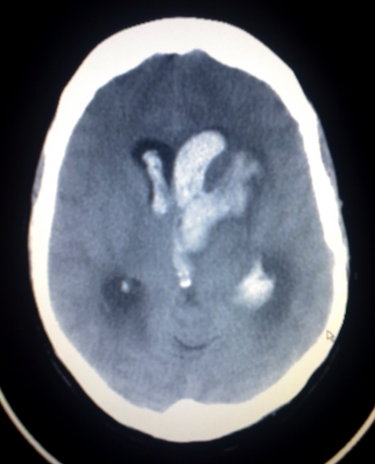
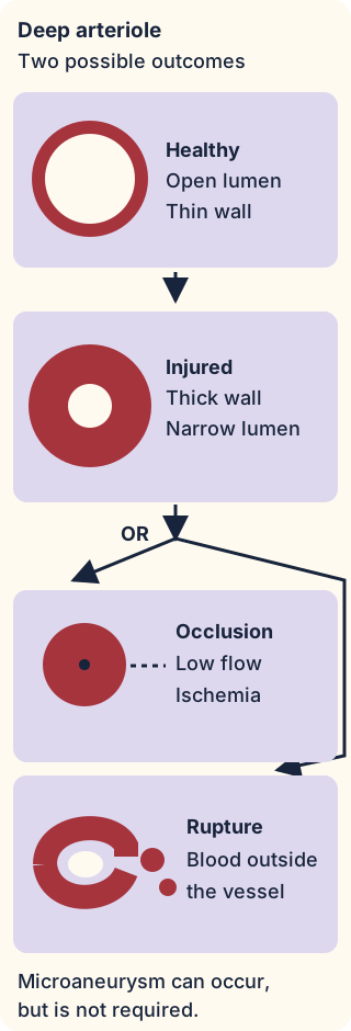
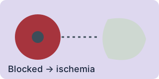
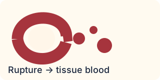
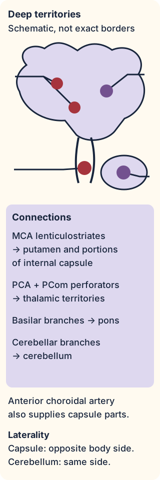
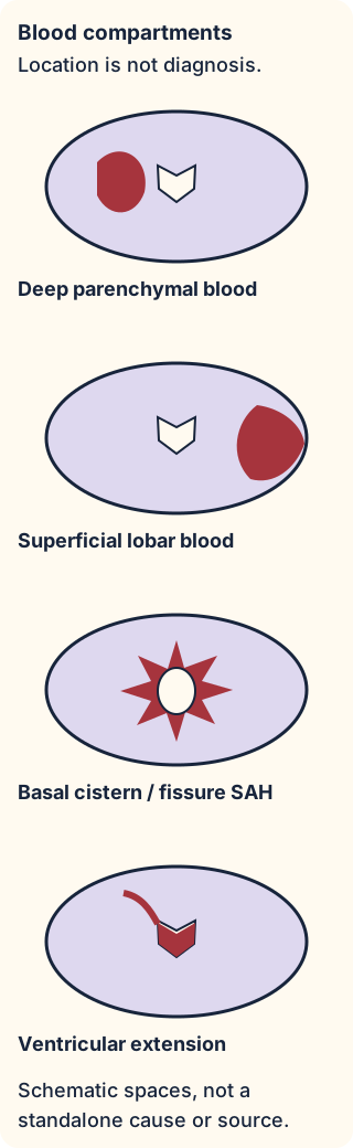
.jpg)