Cerebral Cortex 4 Lobes, 1 Brain, Every Function Mapped
Four lobes carry every function the brain performs. Kill a lobe, kill its function. Know the map, read the deficit.
Frontal · doParietal · feelTemporal · hearOccipital · see
Chapter II · The Opening Case
Robert, 68, in the ED
One vignette. Pick the diagnosis. The lobes will explain themselves.
Bedside · 02:14
Robert is 68. Two hours ago he stopped mid-sentence at dinner. Now in the ED he says "dog... walk... yesterday" and stops, frustrated. He hands you the cup when you ask for it. He points to the ceiling when you ask. He follows three-step commands. He just cannot get the words out.
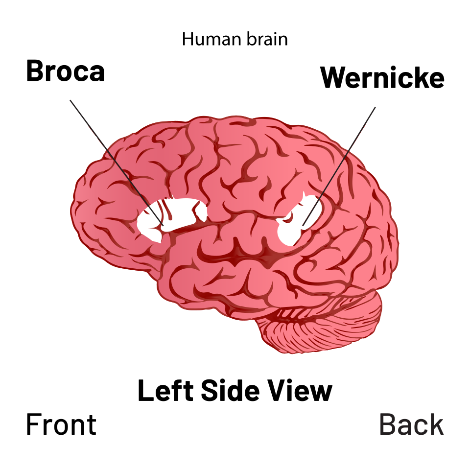
Broca aphasia. The clue is halting, telegraphic speech with intact comprehension. He understands every word you say (he hands you the cup, he points, he follows commands), so the back of the temporal lobe is fine. The problem is up front: the dominant inferior frontal gyrus (Broca area) cannot assemble the motor program for speech. That is why he is so frustrated. He knows what he wants to say. The wiring just will not fire.
Mnemonic: Broca = Broken speech. Wernicke = Wordy nonsense. The frustrated patient is always Broca.
Chapter III · The Lateral Atlas
Tap a Lobe. Read its Real Estate.
Lateral view of the dominant (left) hemisphere. Four lobes, color-coded. Tap any lobe to expose its territory.
Lateral view · Left hemisphere · Anterior at left
Tap any lobe
Lobe
Lobe name
tagline
Area
Function
Pearl
Chapter IV · The Real Estate
Four Lobes, Four Deep Dives
Each lobe is a neighborhood. Below are the streets, the houses, and the one block that always gets robbed on the clinical medicine.
4
Precentral gyrus
Primary motor cortex. MCA covers face and arm; ACA covers leg.
8
Frontal eye field
Voluntary saccades to the opposite side. Destructive lesion makes both eyes look toward the lesion.
44/45
Broca area
Dominant inferior frontal gyrus. Broken output with preserved comprehension.
3/1/2
Postcentral gyrus
Primary somatosensory cortex. Same homunculus logic as motor, one sulcus behind.
22
Wernicke region
Dominant superior temporal language comprehension. Fluent output, no meaning.
17
Primary visual cortex
Occipital calcarine cortex. PCA stroke causes contralateral homonymous hemianopia, often with macular sparing.
Frontal lobeAnterior to central sulcus
Do, decide, speak
Precentral gyrus · area 4
Primary motor cortex
Voluntary movement to the contralateral body. The homunculus is vascular: MCA face and arm, ACA leg. Hit the strip, lose the opposite body part.
Prefrontal cortex
Personality · executive function
Judgment, concentration, orientation, impulse control, planning. Damage gives disinhibition, poor judgment, and primitive reflexes. The classic anchor is Phineas Gage: same person outside, different person inside.
Dominant inferior frontal gyrus
Broca area
Motor speech production. Left-sided in nearly every right-hander. Lesion = Broca aphasia: speech is broken, comprehension is fine.
Frontal eye field · area 8
Gaze + motor planning
Frontal eye field drives saccadic eye movement to the opposite side. Destroy the right FEF and both eyes deviate right, toward the lesion.
PearlPrecentral = motor. One finger anterior to the central sulcus = M1. One finger posterior = S1. The whole exam pivots on which side of that one sulcus the lesion lives.
Parietal lobePosterior to central sulcus
Feel, locate, calculate
Postcentral gyrus · S1
Primary somatosensory cortex
Touch, pressure, proprioception, pain, temperature from the contralateral body. Mirror image of M1, just on the other side of the sulcus.
Angular gyrus
Visual association + language bridge
Where vision becomes meaning: reading, writing, naming what you see. Sits at the parietal-temporal-occipital junction.
Dominant (left) parietal
Gerstmann syndrome
Damage to dominant inferior parietal gives four signs together: agraphia (cannot write), acalculia (cannot calculate), finger agnosia (cannot name fingers), left-right confusion.
Non-dominant (right) parietal
Hemineglect
Spatial integration lives on the right. Lesion = left hemispatial neglect: eats only the right half of the plate or draws only the right side of a clock despite intact primary vision and sensation.
PearlLeft parietal = language and math. Right parietal = space. The patient who ignores the left half of the world has a right parietal lesion. The patient who cannot write or calculate has a left.
Temporal lobeInferior to lateral fissure
Hear, smell, remember, understand
Superior temporal · Heschl gyrus
Primary auditory cortex
Sound and music, input from the medial geniculate body of the thalamus. Bilateral representation, so a one-sided cortex lesion does not cause deafness.
Pyriform cortex · uncus
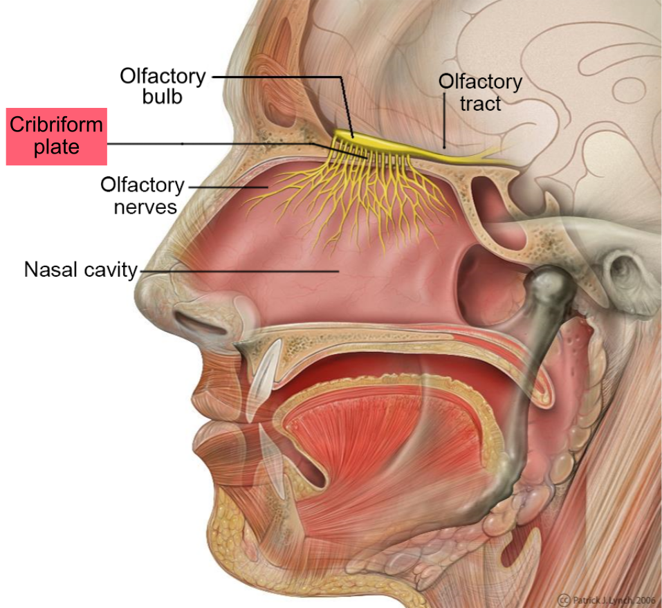
Olfactory cortex
Smell processing. Destruction gives ipsilateral anosmia. Irritation can produce psychomotor temporal lobe seizures with smells, sights, or sounds that are not there.
Limbic structures
Amygdala + hippocampus
Amygdala = fear and emotion. Hippocampus = memory consolidation. Bilateral amygdala injury can produce Kluver-Bucy: hyperphagia, hyperorality, hypersexuality, and visual agnosia.
Dominant superior temporal gyrus
Wernicke area
Receptive speech. Lesion = Wernicke aphasia: speech is fluent but meaningless, and comprehension is gone. Patient does not realize anything is wrong.
PearlTemporal lobe seizure = auditory hallucinations, deja vu, sudden fear. The amygdala fires the dread, the hippocampus mislabels the memory, the auditory cortex hears voices that are not there.
Occipital lobePosterior pole
See
Calcarine sulcus · V1
Primary visual cortex
Light and vision from the lateral geniculate body of the thalamus. Each side processes the contralateral visual hemifield.
Visual association cortex
Object recognition + motion
Sits around V1. Damage can produce visual agnosia. Bilateral occipital cortex injury can produce cortical blindness even when the eyes and optic nerves are structurally intact.
PCA stroke
Contralateral homonymous hemianopia
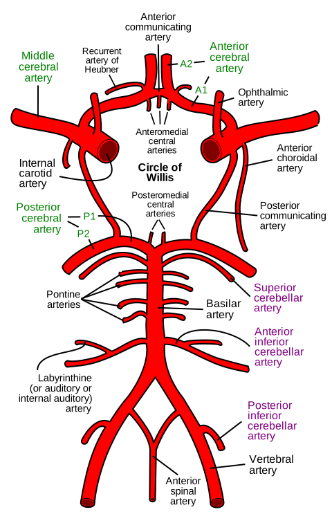
Posterior cerebral artery feeds occipital cortex. Stroke gives the opposite half of vision missing in both eyes, with macular sparing (the macula is dual-fed by both the PCA and the MCA).
Thalamic input
LGN → V1
LGN = light. MGN = music. Two thalamic relays, two cortices, two senses. Mix them up and you mix up auditory vs visual deficits.
PearlMacular sparing in a stroke = occipital. The macula has dual blood supply, so even a complete PCA stroke leaves the central dot of vision intact while everything around it goes dark.
Olfactory nerve and cribriform plate · anosmia localizer · tap to expand
Chapter V · The Aphasia Map
Six Aphasias, One Map
Tap an aphasia. Watch the lesion light up on the cortex. Read the fluency, comprehension, and repetition pattern.
Broca: front-and-down lesion. Speech motor program is offline.
Broca aphasia
motor / non-fluent / expressive
Fluency
Out
Comp.
Intact
Repeat
Out
LesionDominant inferior frontal gyrus (Broca area). Usually a superior-division MCA stroke on the left.
PatternHalting, telegraphic, frustrated. Patient understands every word you say but cannot get the words out. "Broca = Broken speech."
Chapter VI · The Discriminator
Broca vs Wernicke
If you only learn two aphasias, learn these. Side by side. Know which patient is frustrated and which one has no idea anything is wrong.
Frontal · Motor · Non-fluent
Broca aphasia
"Broca = Broken speech"
Lesion
Dominant inferior frontal gyrus. Sits just in front of the motor strip for face and tongue.
Intact. Follows commands, points to named objects.
Repeat
Impaired. Cannot repeat phrases.
Awareness
Knows it. Patient is frustrated, often tearful.
Other
Often paired with contralateral face and arm weakness (motor strip is right next door).
The frustrated patient is always Broca. If they grab your arm and look like they want to hit you, that is the frontal lobe trying to talk.
Temporal · Sensory · Fluent
Wernicke aphasia
"Wernicke = Wordy nonsense"
Lesion
Dominant superior temporal gyrus. Sits behind the lateral fissure.
Speech
Fluent but meaningless. Word salad, neologisms, paraphasic errors. The grammar sounds right; the content is gibberish.
Comp.
Absent. Does not follow commands. Cannot point to named objects.
Repeat
Impaired. Cannot repeat phrases.
Awareness
None. Patient is unaware, often cheerful, often agitated when corrected.
Other
Usually no major weakness (lesion is posterior to motor strip). May have a superior quadrantanopia from Meyer loop.
The cheerful gibberish is always Wernicke. If the patient is talking a mile a minute and the family looks horrified, that is the temporal lobe failing to comprehend what it is saying.
Reference diagram · Broca + Wernicke on lateral cortex · tap to expand
Chapter VII · The Arc Between Them
Arcuate Fasciculus · The Bridge that Breaks
Broca and Wernicke are not islands. A white-matter cable arcs between them: the arcuate fasciculus. Cut the cable and you get conduction aphasia.
Conduction aphasia
Speech fluent. Comp intact. Cannot repeat.
Cut the white-matter cable between Broca and Wernicke and the patient can still speak (Broca is intact) and still understand (Wernicke is intact), but they cannot pass a message from comprehension to production. Ask them to repeat "no ifs, ands, or buts". They will fail. That is the clinical medicine-favorite test.
Broca and Wernicke share a coast. The MCA waters both. A massive MCA stroke that takes out the whole language strip = global aphasia: nothing in, nothing out, no repetition.
Chapter VIII · The Sky and the Floor
Pie in the Sky vs Pie on the Floor
The optic radiation is a fan that splits as it leaves the LGN. The lower bundle dives forward into the temporal lobe (Meyer\'s loop). The upper bundle skims through the parietal lobe. Cut one bundle, lose one quadrant. Same hemisphere. Opposite quadrant. Same MCA.
Lateral view · Left hemisphere · Eye anterior
Visual field (patient\'s view) · Right eye | Left eye
Right eye
Left eye
Pick a lesion
Tap temporal or parietal above. The visual field shades the missing pie wedge.
Temporal · Meyer
PIE in the SKY
Lower bundle, upper quadrant lost
Parietal · Dorsal
PIE on the FLOOR
Upper bundle, lower quadrant lost
Lesion site
Pick one to start
Tract
Field cut
Why
Cause
Pearl
ACA, MCA, PCA territory map · homunculus and macular sparing anchor · tap to expand
Chapter IX · The Drill
Thirty-Two Cortical Localization Vignettes
Original clinical cases. Cross out traps with right-click or long-press, highlight clues with double-click or double-tap, then reveal the reasoning chain after each answer.
Vignette 1 of 25
Medically reviewed by Kaitlyn Cocuzzo, MD and Fatima Ali, DO
Bone Wizardry is an independent educational resource for visual learning in the medical sciences. It is not affiliated with, endorsed by, or sponsored by any licensing or examination board, contains no real or recalled examination questions, and does not guarantee any educational or examination outcome.