Basilar artery occlusion: follow the tissue, preserve the person
Connect basilar anatomy to crossed deficits, awareness, visual syndromes, urgent imaging, reperfusion selection and cause-directed recovery after stroke.
A person can understand every word and still be unable to speak or lift a finger. Another person with an obstruction in the same arterial system may become sleepy and lose vision while retaining limb strength. The central question is which neural functions lose their blood supply. Follow the arterial branches to the injured tissue, then use the examination to decide what must happen next.
By the end, you should be able to distinguish a pontine motor-output syndrome from a rostral brainstem syndrome, recognize dangerous transient symptoms, choose imaging that answers the unresolved question, and explain why reperfusion eligibility requires more than a clock. Sudden double vision, speech difficulty, severe imbalance, weakness or reduced responsiveness needs emergency stroke assessment. This lesson is educational, not an individual treatment plan. [1][2]
How can one lesion affect the face and the opposite side of the body?
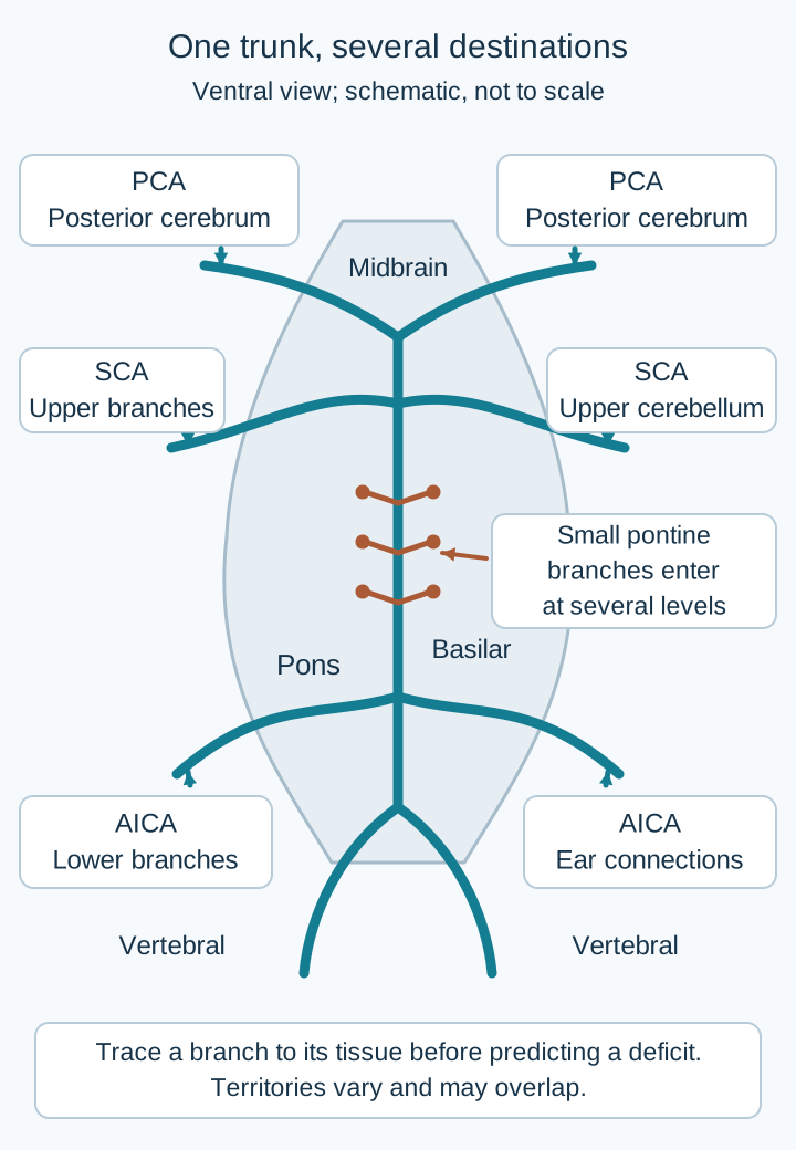
The two vertebral arteries join near the pontomedullary junction to form the basilar artery. It ascends along the front of the pons and ends at the posterior cerebral arteries, or PCAs. Its small paramedian and circumferential branches supply different pontine regions. Larger branches include the anterior inferior cerebellar artery, or AICA, and superior cerebellar artery, or SCA. Branch territories overlap and vary; the arterial map is a guide, not a guarantee that every listed deficit appears. [2][6][17]
First separate the front from the back of the pons. The ventral portion contains descending corticospinal fibers for the limbs and corticobulbar fibers for facial and bulbar function. The dorsal tegmentum contains cranial nerve nuclei, gaze circuitry and ascending pathways. The abducens and facial nuclei are not in the pontine base, although their exiting fibers can be affected by more ventral lesions. The medial lemniscus carries already-crossed body vibration and position information; the more lateral spinothalamic pathway carries already-crossed body pain and temperature information. [6]
Short circumferential branches reach ventrolateral pontine tissue. Depending on their extent, these infarcts can combine contralateral weakness or sensory loss with ipsilateral ataxia from affected cerebellar connections. AICA-region injury can involve the middle cerebellar peduncle; rostral cerebellar connections are important in SCA patterns. Assign the deficits to the actual structures rather than demand every feature of a named syndrome. [6][17]
In the arterial drawing, trace a left paramedian branch toward the descending motor fibers. Those corticospinal fibers have not yet crossed in the lower medulla. Injury here can weaken the right arm and leg. An accompanying left abducens fascicular lesion impairs left-eye abduction. A left abducens nuclear lesion instead disrupts conjugate gaze toward the left, because the nucleus coordinates both eyes. A neighboring pontine gaze-center lesion can also impair gaze toward that side. [6]
Trace the continuous arterial paths to distinguish the trunk from individual branches. Small-branch involvement, collateral supply and anatomical variation determine the actual deficits. [2][6][17]Predict the side: left-eye abduction failure with right-sided weakness
The eye finding points to the left sixth-nerve pathway. The body motor fibers cross below the pons, so right-sided weakness is compatible with a left pontine lesion. Use the cranial nerve finding to identify the side, then check the long tract.
Now compare three lateral patterns. AICA territory injury can combine vertigo, ipsilateral hearing loss, facial weakness, facial sensory loss, ataxia and opposite-side body pain-temperature loss. Facial weakness implicates cranial nerve VII; facial sensation implicates cranial nerve V. Hearing can be affected because the labyrinthine artery commonly arises from AICA. AICA infarction can therefore resemble an inner-ear disorder at first. [17]
SCA territory injury tends to emphasize ipsilateral cerebellar ataxia, sometimes with lateral rostral pontine sensory findings; the facial motor and auditory features characteristic of caudal lateral pontine injury are often absent. Posterior inferior cerebellar artery, or PICA, and vertebral territory injury can affect the lateral medulla instead. Hoarseness and dysphagia from nucleus ambiguus involvement favor that medullary localization. Horner syndrome and crossed pain-temperature findings can occur at more than one lateral brainstem level and do not distinguish AICA from PICA by themselves. [6][17]
Transfer the map: with right facial numbness, right hearing loss and left body pain-temperature loss, identify a right lateral pontine pattern before naming the likely vascular territory. With hoarseness and palatal weakness instead of hearing and facial motor findings, reconsider a lateral medullary site. Neither pattern proves that the entire basilar trunk is occluded.
Can awareness survive loss of motor output?
Proximal and middle basilar occlusions can compromise pontine branches on both sides. Bilateral corticospinal injury can cause quadriparesis or quadriplegia; corticobulbar injury can prevent intelligible speech and effective swallowing. Facial paresis and impaired horizontal gaze may accompany the motor deficits. The length of the obstruction, perforator involvement and collateral supply matter more than a rigid proximal-versus-middle checklist. [2][6]
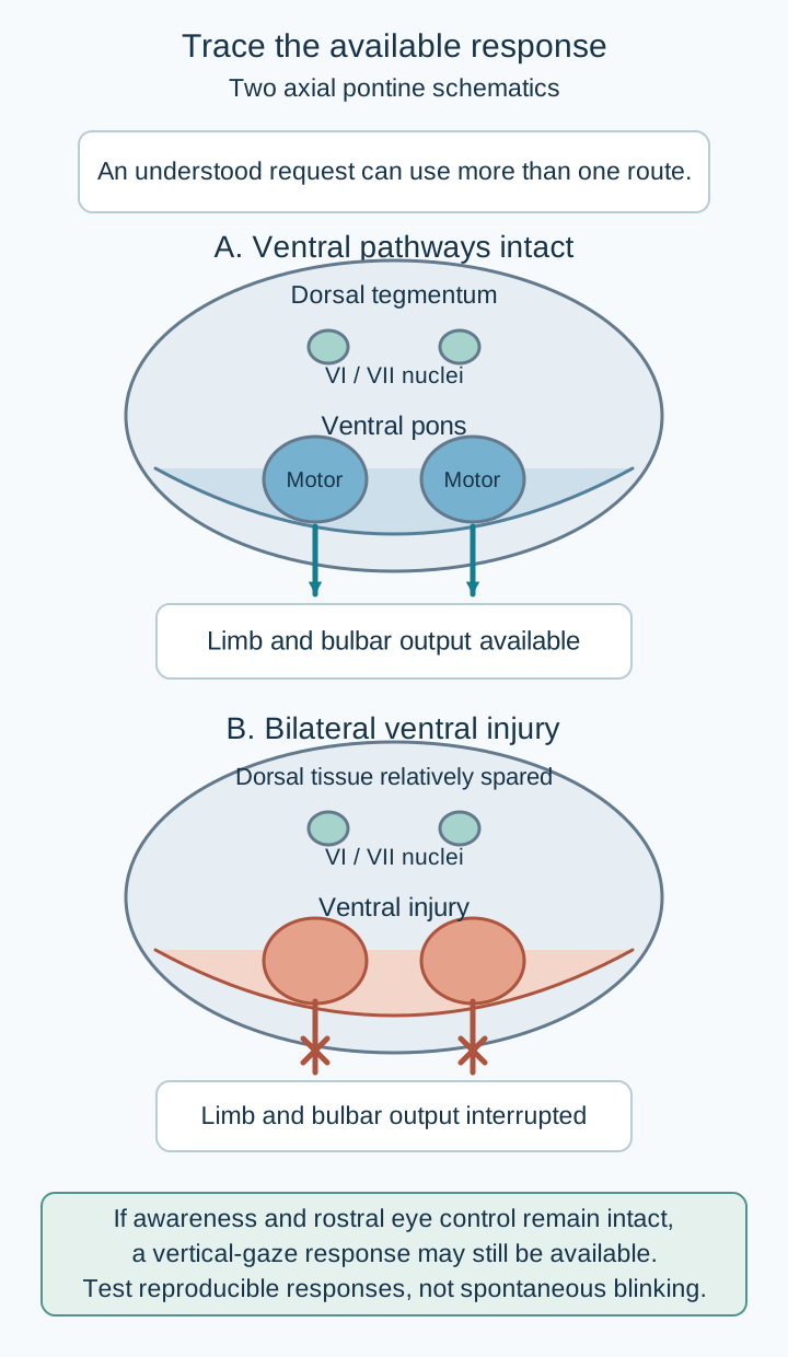
The classic locked-in pattern occurs when bilateral ventral pontine injury interrupts motor output while the systems supporting awareness remain sufficiently intact. Vertical gaze is controlled predominantly in the rostral midbrain and can remain available, as can blinking. A patient may answer reliably with those responses despite anarthria and severe limb paralysis. Failure to speak or produce a limb response is not proof of unconsciousness.[5][6]
Use the cross-sectional drawing as a pathway test. Trace a request from an aware patient toward the ventral limb and speech pathways, then toward the rostral vertical-gaze system. Predict which response survives a lesion confined to the ventral pons. The different destinations explain why the same request can fail through the limbs yet succeed through the eyes. This is a localization model, not a prediction that every pontine lesion has identical boundaries. [5][6]
Predict which response remains available when a request cannot reach limb and bulbar motor output. The two states and native prediction disclosure form a pathway-comparison exercise. Awareness is assessed through reproducible responses; dorsal sparing and vertical gaze are not guaranteed in every pontine lesion. [5][6]Test the model: what changes when dorsal arousal systems are also injured?
Preserved motor-independent communication is no longer assured. Extension into arousal-related brainstem structures can impair consciousness as well as motor output. The contrast explains why a patient with a larger pontine injury may be comatose while another with severe ventral injury remains aware. Lesion extent separates loss of output from loss of awareness.
At the bedside, request reproducible responses rather than infer awareness from spontaneous blinking. Agree on a simple yes/no signal and test it with commands and questions whose answers are known. Allow time to respond and account for vision, hearing, language and medication effects. Classical, incomplete and total locked-in variants exist; total variants may lack an observable eye response and need specialist assessment. Absence of vertical gaze does not by itself prove coma. [5]
Coma lacks wakefulness and demonstrable awareness. Unresponsive wakefulness syndrome has sleep-wake cycling without reproducible behavioral evidence of awareness. Akinetic mutism chiefly concerns reduced initiation rather than the bilateral descending-tract interruption of classic locked-in syndrome. Reproducible command-following is incompatible with brain death; brain-death determination is a separate formal process, not a conclusion drawn from immobility. Pontine hemorrhage can also cause profound paralysis or impaired consciousness, so the examination alone does not distinguish ischemia from bleeding. [1][5][6]
Transfer the model: a patient who correctly uses upward gaze to answer changing questions is communicating. Protect the airway when necessary and continue urgent stroke care, but address the patient directly and preserve that communication channel. A normal-looking lack of limb response must not erase demonstrated awareness.
Why can a distal obstruction disturb vision more than strength?
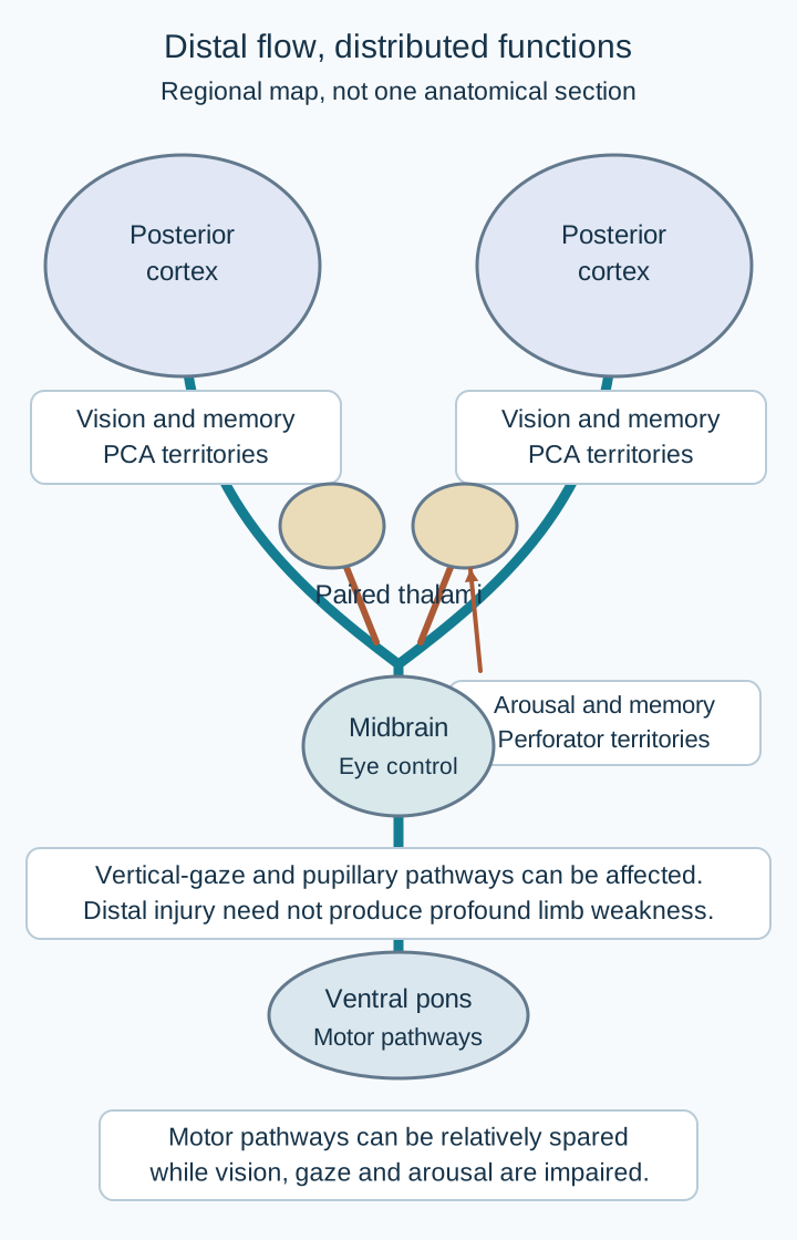
The distal basilar region connects to branches supplying the rostral brainstem, thalami and PCA territories. A lesion here can affect vertical gaze and pupillary pathways in the midbrain, arousal-related thalamic networks, occipital visual cortex and medial temporal memory structures. Somnolence, abnormal eye findings, visual-field loss, amnesia or agitated behavior may dominate while limb power is relatively preserved. This combination is often called top-of-the-basilar syndrome. [7]
Trace the distal branches in the distribution drawing to three separate destinations: midbrain eye-control structures, paired thalami, and posterior visual cortex. Predict the result of injuring all three while leaving the ventral pons relatively intact. Sleepiness plus vertical-gaze impairment plus a visual-field defect fits those destinations better than an isolated motor-output syndrome. Sudden amnesia or unusual behavior with focal eye or visual findings deserves vascular evaluation, not an automatic psychiatric explanation. [7]
This is a regional map, not one anatomical section. Relate each destination to vision, memory, arousal or ocular function, then predict why a distal event can spare much of limb strength. [7][8]Predict the visual response: bilateral occipital injury with intact pupillary pathways
The patient can have cortical blindness despite reactive pupils. The light reflex uses retinal input and midbrain connections without requiring an intact visual cortex. Reactive pupils do not prove conscious vision is intact.
Not every vertical-gaze deficit constitutes the full dorsal midbrain syndrome sometimes called Parinaud syndrome. Nor does somnolence plus vertical-gaze impairment prove a basilar-tip clot. An artery of Percheron variant can supply both paramedian thalami from a single perforating artery. Its occlusion can produce bilateral thalamic infarcts, with or without rostral midbrain injury, despite a patent basilar trunk and major PCAs. Imaging must distinguish the tissue pattern from the large-vessel target. [8]
Pupillary abnormalities and an absent oculocephalic response raise concern for brainstem dysfunction but are not precise stand-alone labels for an occlusion level. The reflex depends on multiple vestibular, pontine and midbrain structures and can be affected by confounding exposures. Check medications and the rest of the examination while obtaining urgent imaging. Atrial fibrillation suggests an embolic source, but anticoagulant use neither excludes ischemic stroke nor automatically excludes catheter-based treatment. [1][2][7]
Transfer the distinction: bilateral paramedian thalamic diffusion abnormalities with a patent basilar tip support a perforator-territory explanation. Additional occipital infarction plus demonstrated distal basilar obstruction instead supports a wider distal circulation event. The examination suggests a region; angiography identifies a treatable large-vessel obstruction.
What should transient symptoms change about urgency?
Minutes of diplopia, dysarthria, marked imbalance or bilateral or alternating weakness can precede a posterior circulation stroke. Symptoms may recur over hours or days and then disappear before examination. Some basilar occlusions instead begin abruptly without warning. A normal examination between episodes does not establish that the vertebral and basilar arteries are normal. Hypertension, diabetes, smoking and atrial fibrillation strengthen vascular concern; their absence does not exclude stroke. [2][9]
Consider two concrete examples. Brief spinning only with a specific head position and without focal neurologic symptoms suggests a different diagnostic pathway from spontaneous episodes combining double vision and slurred speech. In the second pattern, symptom resolution changes what can be measured now, not whether urgent assessment is needed. Stroke and TIA pathways evaluate the brain and relevant arteries; cardiac-source testing may follow but must not substitute for the immediate vascular assessment. [9][12]
Predict the decision: recurrent diplopia and dysarthria, normal examination now
Urgent stroke/TIA evaluation with appropriate head-and-neck CTA or MRA remains indicated when posterior ischemia is suspected. Reassurance, vestibular suppression or aspirin followed by routine discharge does not answer whether a dangerous vascular lesion remains. A resolved deficit can leave an unresolved vascular risk.
HINTS combines head-impulse testing, nystagmus assessment and a test for skew. It is intended for trained clinicians evaluating an appropriate acute vestibular syndrome, typically persistent dizziness with spontaneous nystagmus. A normal head impulse, direction-changing gaze-evoked nystagmus or skew can indicate a central process. It is not a general screening test for an asymptomatic person after transient episodes, and an untrained examination cannot safely substitute for the indicated stroke workup. [12][16]
An abnormal head impulse is not unconditional reassurance. An AICA-region vascular event can involve the inner ear as well as central structures. New hearing loss accompanied by facial weakness, crossed sensory findings or limb dysmetria requires a central vascular assessment even when one vestibular test seems peripheral. Clear focal deficits should not wait for a HINTS battery before emergency stroke evaluation. [12][17]
Reports of symptoms before an established stroke show that warning presentations can be missed. They do not establish one universal 90-day stroke probability for every patient with transient vertigo. Do not apply an unsupported percentage in place of timing, symptom combinations, examination and vessel assessment. [9]
Transfer the distinction: persistent vertigo with nystagmus can permit a trained bedside vestibular examination. Three resolved episodes of binocular diplopia and unsteady gait require a different approach. Identify the syndrome and timing before selecting a bedside test.
Which image answers which question?
Separate three questions: Is there bleeding? Is a major artery obstructed? How much tissue is already injured? Noncontrast CT rapidly detects important hemorrhage but can miss acute posterior fossa ischemia because of small lesions and skull-base artifact. A normal scan does not establish basilar patency or safely exclude an evolving brainstem infarct. [1][10]
When the examination suggests acute basilar occlusion, obtain urgent vascular imaging, commonly CTA of the head and neck, while the stroke team assesses reperfusion. MRA can answer the vessel question when feasible without a harmful delay. Catheter angiography provides detailed vascular information during intervention or when an important uncertainty remains. MRI with diffusion-weighted imaging and an ADC map helps define acute tissue injury. Very early DWI can be negative, particularly for small posterior circulation lesions; persistent focal findings require continued assessment, sometimes repeat imaging, rather than dismissal. [1][2][10][11]
Compare these paired reports: a CT with no hemorrhage and a CTA showing a basilar occlusion describe different findings, not a contradiction. An early negative DWI likewise does not erase a convincing vascular lesion. CT perfusion or other advanced imaging may help selected decisions, but is not a universal prerequisite that should delay urgent vessel imaging or referral for basilar occlusion. [1][2]
Predict the next question after a normal CT and persistent crossed deficits
The hemorrhage question has been partly answered, but arterial patency remains unresolved. Prompt vascular imaging is needed in the suspected large-vessel stroke pathway. MRI can then help characterize the tissue without postponing a time-sensitive reperfusion decision. A test cannot clear a question it did not measure.
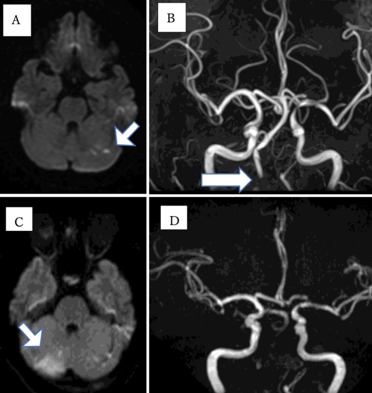
The clinical figure shows original MRI/MRA panels from a published vertebral-artery case. Panels A and C demonstrate progression of ischemic tissue abnormalities; B and D show a change in vascular visualization. Crucially, the later absent basilar MRA signal was not proof of a clot inside the basilar artery: subsequent catheter angiography, after crossing the obstructed right vertebral segment, showed a patent basilar artery. This example teaches the difference between absent flow-related signal and the exact site of obstruction. It is not presented as a confirmed basilar-thrombus photograph or as a treatment guideline. [15]
Nagasawa and colleagues, Figure 1, unchanged. Panels A and B are initial imaging; C and D are later imaging. The published workup ultimately identified right vertebral occlusion and demonstrated a patent basilar artery after the catheter crossed that lesion. Therefore this is not labeled as a photograph proving a basilar thrombus. Source [15]. Image: Junpei Nagasawa, Makiko Ogawa, Hiromi Konaka, Masaru Yanagihashi and Osamu Kano; original source; CC BY 4.0. [15].
Posterior circulation ASPECTS, abbreviated pc-ASPECTS, summarizes visible ischemic change on a 10-point scale. Lower scores indicate more extensive injury. The pons and midbrain each account for two points; each thalamus, cerebellar hemisphere and occipital territory accounts for one. The score must be interpreted with the actual lesion distribution, imaging modality and clinical situation. Severe examination findings with little established injury are different from the same findings with extensive bilateral brainstem destruction. [1][2]
Transfer the rule: choose CTA/MRA for major-vessel patency, DWI/ADC for tissue injury, and noncontrast CT for rapid hemorrhage assessment. Do not convert normal early tissue imaging into proof of normal arterial flow.
What makes reperfusion appropriate, and what remains after flow returns?
Stabilize breathing and circulation, check glucose, establish the last-known-well time, identify antithrombotic exposure and activate stroke expertise. Bulbar weakness or reduced consciousness can threaten airway protection. A swallow screen or specialist assessment must precede oral food, liquids and medications when indicated; preserved awareness does not imply safe swallowing. These steps occur alongside rapid imaging, not as a reason to postpone it. [1][14]
For eligible adults with disabling acute ischemic deficits, the standard intravenous thrombolysis window is within 4.5 hours. The 2026 AHA/ASA guideline supports alteplase or tenecteplase in that window. Selected unknown-onset or later presentations can qualify under advanced-imaging pathways. Severe deficits alone are not a reason to withhold treatment, and a low NIHSS does not make a disabling deficit nondisabling. Recent anticoagulant exposure, bleeding risk and other eligibility factors require explicit assessment. [1]
Thrombolysis and thrombectomy answer related but separate eligibility questions. When both are appropriate, intravenous treatment should not wait for the procedure, and referral should not wait to see whether intravenous treatment works. ESO/ESMINT also describes expert-consensus use of intravenous thrombolysis in selected basilar cases beyond the standard window, but this is not permission to give a lytic to every patient presenting within 24 hours. Use the applicable specialist pathway. [1][2]
The 2026 AHA/ASA adult algorithm recommends thrombectomy within 24 hours for selected basilar occlusions with NIHSS at least 10, limited established injury represented by pc-ASPECTS at least 6, and good baseline function, represented by prestroke modified Rankin score 0 or 1. The same algorithm gives a weaker, may-be-considered pathway for NIHSS 6 to 9 in otherwise eligible patients. These thresholds describe an evidence-based selection framework, not an instruction to delay treatment until 24 hours or to ignore deterioration below a score cutoff. [1]
ATTENTION studied selected patients within 12 hours, and BAOCHE studied selected patients 6 to 24 hours after onset. Both found better functional outcomes with thrombectomy in their study populations, with procedural and hemorrhagic risks. Much of the randomized evidence came from China, and benefit cannot be assumed identical for every severity, injury pattern or practice setting. The 2024 ESO/ESMINT guidance was more cautious for NIHSS below 10. Distinguish that earlier uncertainty from the newer AHA/ASA selection framework. [2][3][4]
Extensive established bilateral brainstem injury, substantial baseline disability and other adverse prognostic features can change the balance. A patent artery after a procedure does not guarantee recovery of already infarcted tissue. Conversely, coma or severe paralysis with little established injury is not itself proof of futility. The clock, examination, imaging and baseline function must be considered together. [1][2]
Predict the difference: two patients at 16 hours with the same severe examination
A previously independent patient with confirmed basilar occlusion and pc-ASPECTS 9 has a different evidence profile from a patient with extensive bilateral pontine and midbrain infarction. The first warrants urgent thrombectomy assessment; the second requires an individualized assessment rather than automatic treatment based only on being within 24 hours. Time remaining does not restore tissue already lost.
After reperfusion, reassess for hemorrhage and edema. Aspirin is commonly used for appropriate noncardioembolic ischemic stroke after hemorrhage exclusion, but it is not a substitute for indicated reperfusion. Following intravenous thrombolysis, antithrombotics are generally deferred until the 24-hour follow-up scan excludes hemorrhage, unless a specialist identifies a compelling exceptional indication. Thrombectomy alone does not create one universal antithrombotic regimen. Thrombolytic exposure, procedural stenting, hemorrhage and stroke mechanism all matter. Routine therapeutic heparin is not a substitute for acute reperfusion. [1][2][18]
For atrial-fibrillation-associated stroke, plan anticoagulation according to infarct size, hemorrhagic transformation, other bleeding risks and clinical stability. OPTIMAS found that starting a direct oral anticoagulant within four days was noninferior to starting at 7 to 14 days in its eligible population. This argues against an automatic one-to-two-week delay for everyone; it does not require immediate treatment in a patient with active bleeding or a large unstable infarct. Antiplatelet therapy, a selected short dual-antiplatelet course, treatment after a stent and anticoagulation for atrial fibrillation are different decisions, not interchangeable defaults. [1][2][13]
A large cerebellar infarct can swell, obstruct the fourth ventricle and compress the brainstem. New declining consciousness requires urgent neurocritical and neurosurgical evaluation. Deterioration from posterior fossa mass effect may require suboccipital decompression with dural expansion, with cerebrospinal fluid diversion for associated obstructive hydrocephalus as directed by the surgical team. A hemispheric hemicraniectomy is not the operation for this posterior fossa problem; drainage alone may not address the compressed brainstem. [14]
Transfer to recovery: identify the cause through vascular and cardiac evaluation, address hypertension, diabetes and smoking, assess communication and swallowing, and begin rehabilitation appropriate to deficits. The successful procedure is one part of care. Preventing aspiration, recognizing swelling and choosing cause-directed secondary prevention remain essential. [1][2][14]
Apply the examination, imaging and treatment distinctions
Case 1
Show answer and explanations for case 1
A. Left caudal lateral medulla (Why this does not fit)
It can combine ipsilateral facial and contralateral body sensory abnormalities with vestibular findings. The sixth-nerve deficit requires a pontine pathway, and the motor plus vibration deficits favor a medial rather than lateral lesion.
Reasoning steps for option A
Which findings can a lateral medullary lesion combine?
It can combine ipsilateral facial and contralateral body sensory abnormalities with vestibular findings.
Which findings require a different level and territory here?
The sixth-nerve deficit requires a pontine pathway, and the motor plus vibration deficits favor a medial rather than lateral lesion.
B. Left paramedian caudal pons (Best answer)
The left abducens fascicle is implicated because the other eye still adducts. Right motor and vibration deficits indicate left corticospinal and medial lemniscal involvement, fitting a left paramedian pontine lesion.
Reasoning steps for option B
Which side is indicated by isolated left abduction failure?
The left abducens fascicle is implicated because the other eye still adducts.
How do the body findings refine the site?
Right motor and vibration deficits indicate left corticospinal and medial lemniscal involvement, fitting a left paramedian pontine lesion.
C. Right paramedian caudal pons (Why this does not fit)
It would usually weaken the left limbs because the major motor crossing lies below the pons. No. Both the left sixth-nerve pathway and right limb deficits point to the opposite, left side of the pons.
Reasoning steps for option C
Which body side would a right pontine corticospinal lesion weaken?
It would usually weaken the left limbs because the major motor crossing lies below the pons.
Does that match the cranial nerve and limb findings?
No. Both the left sixth-nerve pathway and right limb deficits point to the opposite, left side of the pons.
D. Left ventral rostral midbrain (Why this does not fit)
A left cerebral peduncle lesion can cause right-sided weakness. The impaired left-eye abduction implicates the sixth-nerve pathway in the caudal pons, not the third-nerve pathway of the midbrain.
Reasoning steps for option D
What crossed motor pattern can a left midbrain lesion produce?
A left cerebral peduncle lesion can cause right-sided weakness.
Which ocular finding argues against that level?
The impaired left-eye abduction implicates the sixth-nerve pathway in the caudal pons, not the third-nerve pathway of the midbrain.
Takeaway: Combine the ipsilateral cranial nerve deficit with the side and modality of the long-tract deficit.
A. Right abducens nerve fascicle (Why this does not fit)
It carries motor output to the ipsilateral lateral rectus. No. It would impair right abduction but would not directly prevent left-eye adduction during rightward gaze.
Reasoning steps for option A
What does the abducens fascicle carry?
It carries motor output to the ipsilateral lateral rectus.
Would its isolated injury explain failure of both eyes to look right?
No. It would impair right abduction but would not directly prevent left-eye adduction during rightward gaze.
B. Left medial longitudinal fasciculus (Why this does not fit)
It can impair left-eye adduction during rightward gaze. The right eye also fails to abduct, indicating loss of the coordinated rightward gaze command rather than isolated internuclear conduction.
Reasoning steps for option B
What adduction problem can a left medial longitudinal fasciculus lesion cause?
It can impair left-eye adduction during rightward gaze.
Which additional finding is unexplained by that isolated lesion?
The right eye also fails to abduct, indicating loss of the coordinated rightward gaze command rather than isolated internuclear conduction.
C. Right oculomotor nerve fascicle (Why this does not fit)
It contributes to adduction, vertical gaze, eyelid function and pupillary constriction. They do not fit an isolated right third-nerve fascicular lesion, which also would not explain this paired rightward gaze failure.
Reasoning steps for option C
Which functions depend on the oculomotor nerve?
It contributes to adduction, vertical gaze, eyelid function and pupillary constriction.
How do intact convergence and a caudal pontine lesion compare?
They do not fit an isolated right third-nerve fascicular lesion, which also would not explain this paired rightward gaze failure.
D. Right abducens motor nucleus (Best answer)
Its motor neurons activate ipsilateral abduction and its internuclear neurons help drive contralateral adduction. Right nuclear injury explains loss of rightward conjugate gaze while separate convergence circuitry remains available; nearby facial fibers fit the facial weakness.
Reasoning steps for option D
How does the abducens nucleus coordinate two eyes?
Its motor neurons activate ipsilateral abduction and its internuclear neurons help drive contralateral adduction.
How does that organization fit this examination?
Right nuclear injury explains loss of rightward conjugate gaze while separate convergence circuitry remains available; nearby facial fibers fit the facial weakness.
Takeaway: A nuclear gaze lesion can affect both eyes; an isolated sixth-nerve fascicular lesion affects one lateral rectus.
A. Trigeminal pathway; left lateral pons (Best answer)
The trigeminal system carries facial somatic sensation; the facial nerve supplies facial expression. Together they fit a left lateral pontine region that can be affected in AICA territory ischemia.
Reasoning steps for option A
Which nerve conveys most facial somatic sensation?
The trigeminal system carries facial somatic sensation; the facial nerve supplies facial expression.
How do hearing loss, facial paresis and crossed body sensory loss refine localization?
Together they fit a left lateral pontine region that can be affected in AICA territory ischemia.
B. Facial motor pathway; left lateral pons (Why this does not fit)
It explains weakness of the left upper and lower face. No. The pontine region is appropriate, but facial pinprick depends on trigeminal sensory pathways rather than facial motor fibers.
Reasoning steps for option B
What does the facial motor pathway explain here?
It explains weakness of the left upper and lower face.
Does it explain reduced facial pinprick?
No. The pontine region is appropriate, but facial pinprick depends on trigeminal sensory pathways rather than facial motor fibers.
C. Trigeminal pathway; left lateral medulla (Why this does not fit)
It can affect trigeminal pain-temperature pathways on the same side. The combination of whole-face paresis and acute hearing loss favors caudal lateral pontine and cochleovestibular involvement.
Reasoning steps for option C
What facial finding can lateral medullary injury produce?
It can affect trigeminal pain-temperature pathways on the same side.
Which associated findings favor a more rostral site?
The combination of whole-face paresis and acute hearing loss favors caudal lateral pontine and cochleovestibular involvement.
D. Facial motor pathway; left medial pons (Why this does not fit)
It can affect descending motor fibers and nearby exiting cranial nerve fibers. Facial sensory loss is not facial motor dysfunction, and the auditory, vestibular and crossed pain-temperature findings favor a lateral distribution.
Reasoning steps for option D
What can a medial pontine lesion do to motor output?
It can affect descending motor fibers and nearby exiting cranial nerve fibers.
Which features make this pairing inadequate?
Facial sensory loss is not facial motor dysfunction, and the auditory, vestibular and crossed pain-temperature findings favor a lateral distribution.
Takeaway: Separate facial sensation from facial expression before combining the cranial findings into a vascular territory.
A. A: lateral medulla; B: caudal lateral pons (Why this does not fit)
Ataxia and crossed pain-temperature findings can occur in both lateral pontine and medullary lesions. Hearing and facial motor deficits favor the pons, whereas palatal weakness and hoarseness favor medullary nucleus ambiguus involvement.
Reasoning steps for option A
Which findings are shared across lateral brainstem levels?
Ataxia and crossed pain-temperature findings can occur in both lateral pontine and medullary lesions.
Why is the proposed order reversed?
Hearing and facial motor deficits favor the pons, whereas palatal weakness and hoarseness favor medullary nucleus ambiguus involvement.
B. A: medial pons; B: rostral midbrain (Why this does not fit)
Medial pontine disease can emphasize motor and vibration deficits; rostral midbrain disease can disturb vertical gaze and third-nerve function. The supplied auditory-facial and palatal-laryngeal findings instead distinguish two lateral brainstem levels.
Reasoning steps for option B
What do medial pontine and midbrain lesions often add?
Medial pontine disease can emphasize motor and vibration deficits; rostral midbrain disease can disturb vertical gaze and third-nerve function.
Do those features account for the differentiating findings?
The supplied auditory-facial and palatal-laryngeal findings instead distinguish two lateral brainstem levels.
C. A: caudal lateral pons; B: lateral medulla (Best answer)
The facial motor and auditory findings fit caudal lateral pontine and adjacent cochleovestibular involvement. Hoarseness and palatal weakness fit nucleus ambiguus dysfunction in the lateral medulla, often in vertebral or PICA territory.
Reasoning steps for option C
Which structures fit patient A?
The facial motor and auditory findings fit caudal lateral pontine and adjacent cochleovestibular involvement.
Which structures fit patient B?
Hoarseness and palatal weakness fit nucleus ambiguus dysfunction in the lateral medulla, often in vertebral or PICA territory.
D. A: rostral lateral pons; B: medial medulla (Why this does not fit)
Rostral lateral pontine disease may emphasize ataxia, while medial medullary disease can affect motor, vibration and tongue pathways. The caudal facial-auditory combination in A and nucleus ambiguus pattern in B favor different levels and compartments.
Reasoning steps for option D
What patterns can these proposed regions produce?
Rostral lateral pontine disease may emphasize ataxia, while medial medullary disease can affect motor, vibration and tongue pathways.
Which observations contradict that pairing?
The caudal facial-auditory combination in A and nucleus ambiguus pattern in B favor different levels and compartments.
Takeaway: Shared lateral brainstem signs require a second, level-specific cranial finding for useful localization.
A. Right posterior inferior cerebellar artery (Why this does not fit)
PICA supplies inferior cerebellar regions and can be associated with lateral medullary ischemia. The demonstrated superior cerebellar and rostral lateral pontine distribution is more consistent with SCA territory.
Reasoning steps for option A
What region commonly belongs to PICA territory?
PICA supplies inferior cerebellar regions and can be associated with lateral medullary ischemia.
What changes the territorial assignment here?
The demonstrated superior cerebellar and rostral lateral pontine distribution is more consistent with SCA territory.
B. Right anterior inferior cerebellar artery (Why this does not fit)
Caudal lateral pontine findings with auditory or facial motor involvement can suggest AICA ischemia. The rostral pontine and superior cerebellar location, with preserved hearing and facial function, favors the superior cerebellar artery.
Reasoning steps for option B
What clinical combination often suggests AICA territory?
Caudal lateral pontine findings with auditory or facial motor involvement can suggest AICA ischemia.
Why is another branch a better fit here?
The rostral pontine and superior cerebellar location, with preserved hearing and facial function, favors the superior cerebellar artery.
C. Right paramedian basilar perforators (Why this does not fit)
Descending motor and medial lemniscal deficits are common when medial pontine structures are injured. A superior cerebellar hemispheric infarct with a lateral pontine focus is outside an isolated paramedian perforator pattern.
Reasoning steps for option C
What deficits can paramedian pontine injury produce?
Descending motor and medial lemniscal deficits are common when medial pontine structures are injured.
Which demonstrated tissue distribution is not explained?
A superior cerebellar hemispheric infarct with a lateral pontine focus is outside an isolated paramedian perforator pattern.
D. Right superior cerebellar artery (Best answer)
Right dysmetria and rightward falling support right cerebellar or cerebellar-connection injury. The superior cerebellar hemisphere and rostral lateral pons fit the right SCA distribution; absent auditory and facial deficits are compatible.
Reasoning steps for option D
How does the cerebellar examination identify the side?
Right dysmetria and rightward falling support right cerebellar or cerebellar-connection injury.
How does MRI identify the branch territory?
The superior cerebellar hemisphere and rostral lateral pons fit the right SCA distribution; absent auditory and facial deficits are compatible.
Takeaway: Combine the cerebellar side with the rostrocaudal level and actual imaging distribution.
A. Left limb weakness with right vibration loss (Why this does not fit)
The left pontine medial lemniscus carries vibration and position information from the right body. No. A left pontine corticospinal lesion is above the motor crossing and therefore also predicts right-sided weakness.
Reasoning steps for option A
Which tract finding can explain right vibration loss?
The left pontine medial lemniscus carries vibration and position information from the right body.
Does the proposed motor side match the same lesion?
No. A left pontine corticospinal lesion is above the motor crossing and therefore also predicts right-sided weakness.
B. Right limb weakness with right vibration loss (Best answer)
The body vibration pathway has already crossed before reaching the left pontine medial lemniscus. The left corticospinal fibers cross farther caudally, so their injury produces right-sided weakness along with right vibration loss.
Reasoning steps for option B
Where have the affected sensory fibers crossed?
The body vibration pathway has already crossed before reaching the left pontine medial lemniscus.
Where will the affected motor fibers cross?
The left corticospinal fibers cross farther caudally, so their injury produces right-sided weakness along with right vibration loss.
C. Right limb weakness with left facial pinprick loss (Why this does not fit)
Right-sided weakness fits a left pontine corticospinal lesion. No. It would require trigeminal sensory pathway involvement not supplied here; lateral tegmental sparing makes the medial lemniscal deficit the supported sensory prediction.
Reasoning steps for option C
Which part of this option fits ventral motor injury?
Right-sided weakness fits a left pontine corticospinal lesion.
Is left facial pinprick loss required by the described boundaries?
No. It would require trigeminal sensory pathway involvement not supplied here; lateral tegmental sparing makes the medial lemniscal deficit the supported sensory prediction.
D. Left limb weakness with left body pinprick loss (Why this does not fit)
Body pain and temperature travel in the spinothalamic system rather than the medial lemniscus. The spared lateral compartment does not support the proposed pain-temperature deficit, and left motor weakness is opposite the expected motor side.
Reasoning steps for option D
What body sensory modality belongs to the lateral pathway?
Body pain and temperature travel in the spinothalamic system rather than the medial lemniscus.
How do both the modality and side conflict?
The spared lateral compartment does not support the proposed pain-temperature deficit, and left motor weakness is opposite the expected motor side.
Takeaway: Motor and dorsal-column pathways cross at different places, yet both can produce contralateral body deficits in the pons.
A. Bilateral ventral pontine injury with relative dorsal sparing (Best answer)
They provide reproducible behavioral evidence of comprehension and awareness despite absent limb and speech output. Bilateral ventral pontine injury can interrupt corticospinal and corticobulbar output while sufficiently sparing arousal networks and rostral vertical-gaze control.
Reasoning steps for option A
What do changing, accurate eye responses demonstrate?
They provide reproducible behavioral evidence of comprehension and awareness despite absent limb and speech output.
Which tissue pattern permits that dissociation?
Bilateral ventral pontine injury can interrupt corticospinal and corticobulbar output while sufficiently sparing arousal networks and rostral vertical-gaze control.
B. Bilateral thalamic injury with rostral arousal interruption (Why this does not fit)
It can impair wakefulness and awareness, sometimes with memory and ocular abnormalities. Consistent command-following with near-complete motor-output loss points more directly to a ventral pontine output lesion with awareness preserved.
Reasoning steps for option B
What can extensive bilateral thalamic arousal injury cause?
It can impair wakefulness and awareness, sometimes with memory and ocular abnormalities.
What makes it a weaker explanation for this pattern?
Consistent command-following with near-complete motor-output loss points more directly to a ventral pontine output lesion with awareness preserved.
C. Bilateral frontal injury with loss of behavioral initiation (Why this does not fit)
Akinetic mutism can reduce spontaneous action and speech despite available motor capacity. Reproducible eye responses, absent horizontal gaze and profound limb-output failure after a brainstem stroke support a specific motor-pathway interruption.
Reasoning steps for option C
What syndrome can severe medial frontal dysfunction produce?
Akinetic mutism can reduce spontaneous action and speech despite available motor capacity.
Why is the pontine output explanation stronger?
Reproducible eye responses, absent horizontal gaze and profound limb-output failure after a brainstem stroke support a specific motor-pathway interruption.
D. Bilateral occipital injury with preserved motor pathways (Why this does not fit)
Conscious visual processing is threatened, potentially causing cortical blindness. It would not explain quadriplegia, anarthria and absent horizontal gaze while the patient uses vertical gaze to communicate.
Reasoning steps for option D
What function is primarily threatened by bilateral occipital injury?
Conscious visual processing is threatened, potentially causing cortical blindness.
Would that explain this combination of motor and eye findings?
It would not explain quadriplegia, anarthria and absent horizontal gaze while the patient uses vertical gaze to communicate.
Takeaway: Demonstrated awareness must be separated from the ability to express it through limbs or speech.
A. Greater interruption of spinal motor output in patient B (Why this does not fit)
It explains weakness and inability to express a limb response. No. Patient A demonstrates that severe paralysis can coexist with awareness; the additional dorsal and rostral injury matters in B.
Reasoning steps for option A
What does interruption of descending motor output explain?
It explains weakness and inability to express a limb response.
Does additional motor paralysis by itself establish loss of awareness?
No. Patient A demonstrates that severe paralysis can coexist with awareness; the additional dorsal and rostral injury matters in B.
B. Greater interruption of visual cortex input in patient B (Why this does not fit)
It can impair conscious vision despite preserved pupillary responses. The demonstrated difference is dorsal and rostral brainstem injury with failure to awaken, not isolated cortical visual injury.
Reasoning steps for option B
What can visual cortex injury impair?
It can impair conscious vision despite preserved pupillary responses.
Why is that insufficient for the supplied difference?
The demonstrated difference is dorsal and rostral brainstem injury with failure to awaken, not isolated cortical visual injury.
C. Additional injury to arousal-related networks in patient B (Best answer)
Comprehension and awareness remain demonstrable through a non-limb response. Dorsal pontine and rostral brainstem extension can impair arousal networks as well as motor output, explaining the loss of wakefulness.
Reasoning steps for option C
Which function remains demonstrable in patient A?
Comprehension and awareness remain demonstrable through a non-limb response.
How does the additional lesion distribution change the interpretation in B?
Dorsal pontine and rostral brainstem extension can impair arousal networks as well as motor output, explaining the loss of wakefulness.
D. Additional peripheral neuromuscular transmission failure in patient B (Why this does not fit)
It could produce profound weakness with preserved central awareness. No. It does not account for the larger brainstem injury and the absence of arousal after confounders were assessed.
Reasoning steps for option D
What could peripheral transmission failure cause?
It could produce profound weakness with preserved central awareness.
Does it explain the imaging-linked loss of wakefulness?
No. It does not account for the larger brainstem injury and the absence of arousal after confounders were assessed.
Takeaway: Loss of motor output and impaired arousal are separate consequences of different lesion extents.
A. CT angiography of the head and neck (Why this does not fit)
CTA evaluates arterial anatomy and can identify a basilar occlusion. The immediate distinction is intracranial bleeding versus ischemia; noncontrast CT is the standard rapid study for that safety question, often acquired alongside CTA.
Reasoning steps for option A
Which urgent question does CTA address?
CTA evaluates arterial anatomy and can identify a basilar occlusion.
Which question is asked before reperfusion here?
The immediate distinction is intracranial bleeding versus ischemia; noncontrast CT is the standard rapid study for that safety question, often acquired alongside CTA.
B. Noncontrast CT of the head (Best answer)
No. Neither the preceding symptoms nor the motor-output phenotype reliably excludes a pontine hemorrhage. Noncontrast head CT evaluates acute intracranial hemorrhage before reperfusion decisions; vessel imaging remains necessary when occlusion is suspected.
Reasoning steps for option B
Does the preceding transient diplopia exclude hemorrhage now?
No. Neither the preceding symptoms nor the motor-output phenotype reliably excludes a pontine hemorrhage.
Which rapid study addresses that uncertainty?
Noncontrast head CT evaluates acute intracranial hemorrhage before reperfusion decisions; vessel imaging remains necessary when occlusion is suspected.
C. CT perfusion of the brain (Why this does not fit)
It can characterize regional perfusion and contribute to selected tissue-based decisions. No. Perfusion maps are not the primary study for excluding acute intracranial bleeding before reperfusion.
Reasoning steps for option C
What additional information can perfusion imaging provide?
It can characterize regional perfusion and contribute to selected tissue-based decisions.
Does it replace the initial hemorrhage assessment?
No. Perfusion maps are not the primary study for excluding acute intracranial bleeding before reperfusion.
D. Contrast echocardiography of the heart (Why this does not fit)
It can investigate potential embolic sources. A cardiac-source study cannot determine whether the current brainstem syndrome is caused by hemorrhage or ischemia.
Reasoning steps for option D
What cause of ischemic stroke can cardiac imaging investigate?
It can investigate potential embolic sources.
Why does that not answer the immediate safety question?
A cardiac-source study cannot determine whether the current brainstem syndrome is caused by hemorrhage or ischemia.
Takeaway: Localize the syndrome, but use brain imaging rather than its tempo to distinguish bleeding from ischemia.
A. Occlusion of an isolated left paramedian pontine branch (Why this does not fit)
It can produce crossed cranial nerve and contralateral motor or medial sensory deficits. The homonymous field loss and memory disturbance together with vertical-gaze and arousal abnormalities require a more rostral and cerebral distribution.
Reasoning steps for option A
What functional pattern can a paramedian pontine lesion produce?
It can produce crossed cranial nerve and contralateral motor or medial sensory deficits.
Which findings extend beyond that territory?
The homonymous field loss and memory disturbance together with vertical-gaze and arousal abnormalities require a more rostral and cerebral distribution.
B. Occlusion of the proximal left anterior inferior cerebellar artery (Why this does not fit)
Vestibular, auditory, facial and cerebellar findings can dominate. It does not unify the visual-field deficit, memory disturbance and rostral ocular-arousal abnormalities.
Reasoning steps for option B
Which findings commonly accompany AICA-region ischemia?
Vestibular, auditory, facial and cerebellar findings can dominate.
What is missing from that explanation?
It does not unify the visual-field deficit, memory disturbance and rostral ocular-arousal abnormalities.
C. Occlusion of the distal right middle cerebral artery (Why this does not fit)
It can cause left visual-field loss and cortical deficits. It does not directly account for the vertical-gaze deficit and rostral arousal pattern while limb strength is relatively preserved.
Reasoning steps for option C
What can right cerebral hemispheric ischemia cause?
It can cause left visual-field loss and cortical deficits.
Why is an isolated distal MCA lesion less coherent here?
It does not directly account for the vertical-gaze deficit and rostral arousal pattern while limb strength is relatively preserved.
D. Occlusion at the distal basilar artery bifurcation (Best answer)
Rostral midbrain and thalamic involvement explain ocular and arousal abnormalities, while PCA territories explain field and memory deficits. The ventral pontine motor pathways can be less affected than the distal distribution, so severe visual and behavioral findings need not accompany quadriplegia.
Reasoning steps for option D
Which destinations explain the different findings?
Rostral midbrain and thalamic involvement explain ocular and arousal abnormalities, while PCA territories explain field and memory deficits.
Why can limb power remain relatively preserved?
The ventral pontine motor pathways can be less affected than the distal distribution, so severe visual and behavioral findings need not accompany quadriplegia.
Takeaway: Relatively preserved strength does not exclude a dangerous distal posterior circulation event.
A. Occlusion of both superior cerebellar arteries (Why this does not fit)
Superior cerebellar and adjacent rostral lateral brainstem territories can be affected. It does not explain symmetric paramedian thalamic infarction with major distal arteries patent.
Reasoning steps for option A
Which tissue is usually threatened by SCA occlusion?
Superior cerebellar and adjacent rostral lateral brainstem territories can be affected.
Does that distribution account for the dominant MRI pattern?
It does not explain symmetric paramedian thalamic infarction with major distal arteries patent.
B. Occlusion of the middle basilar arterial trunk (Why this does not fit)
Pontine perforator ischemia and motor or cranial nerve findings are common. The basilar trunk is patent and the injury is centered on paired paramedian thalami rather than the pons.
Reasoning steps for option B
What tissue pattern often accompanies middle basilar compromise?
Pontine perforator ischemia and motor or cranial nerve findings are common.
How does the supplied imaging alter the explanation?
The basilar trunk is patent and the injury is centered on paired paramedian thalami rather than the pons.
C. Occlusion of a shared paramedian thalamic perforator (Best answer)
An artery of Percheron variant can supply bilateral paramedian thalami, sometimes with rostral midbrain supply. The obstruction can lie in that perforator, so normal basilar and proximal PCA patency does not contradict the bilateral infarct pattern.
Reasoning steps for option C
How can one small artery supply both thalami?
An artery of Percheron variant can supply bilateral paramedian thalami, sometimes with rostral midbrain supply.
Why can the large arteries remain patent?
The obstruction can lie in that perforator, so normal basilar and proximal PCA patency does not contradict the bilateral infarct pattern.
D. Occlusion of both distal posterior cerebral cortical branches (Why this does not fit)
They can affect occipital and medial temporal cortical territories. The occipital lobes are spared and the paired paramedian thalami dominate, favoring a shared perforator rather than two cortical branch occlusions.
Reasoning steps for option D
What tissue can distal PCA cortical branches affect?
They can affect occipital and medial temporal cortical territories.
Why does the supplied pattern favor a different branch level?
The occipital lobes are spared and the paired paramedian thalami dominate, favoring a shared perforator rather than two cortical branch occlusions.
Takeaway: Bilateral thalamic injury does not require occlusion of the basilar trunk.
A. Bilateral occipital cortical infarction (Best answer)
They support preserved alertness and language sufficient for the examination despite loss of reported vision. Bilateral occipital injury can impair conscious vision while retinal input to the midbrain light-reflex pathway remains intact.
Reasoning steps for option A
What do naming by touch and following spoken commands show?
They support preserved alertness and language sufficient for the examination despite loss of reported vision.
How can visual loss coexist with reactive pupils?
Bilateral occipital injury can impair conscious vision while retinal input to the midbrain light-reflex pathway remains intact.
B. Bilateral pretectal midbrain infarction (Why this does not fit)
It participates in the pupillary light-reflex circuitry. Brisk bilateral light responses are inconsistent with using bilateral pretectal interruption to explain the visual complaint.
Reasoning steps for option B
What function depends on the pretectal region?
It participates in the pupillary light-reflex circuitry.
Which finding argues against this as the primary explanation?
Brisk bilateral light responses are inconsistent with using bilateral pretectal interruption to explain the visual complaint.
C. Bilateral ventral pontine infarction (Why this does not fit)
Severe limb and bulbar motor-output loss can occur with awareness preserved. No. It does not explain bilateral visual loss with preserved tactile naming and without the expected severe motor-output syndrome.
Reasoning steps for option C
What deficit is characteristic of extensive ventral pontine injury?
Severe limb and bulbar motor-output loss can occur with awareness preserved.
Does that explain isolated loss of conscious vision here?
No. It does not explain bilateral visual loss with preserved tactile naming and without the expected severe motor-output syndrome.
D. Bilateral paramedian thalamic infarction (Why this does not fit)
Arousal and memory can be impaired, sometimes with ocular abnormalities. The patient is awake and engaged, with a predominantly visual deficit after a distal circulation event and intact ocular light-reflex circuitry.
Reasoning steps for option D
Which functions can bilateral paramedian thalamic injury disturb?
Arousal and memory can be impaired, sometimes with ocular abnormalities.
Why is another distribution a better fit?
The patient is awake and engaged, with a predominantly visual deficit after a distal circulation event and intact ocular light-reflex circuitry.
Takeaway: The pupillary light reflex and conscious visual perception follow different pathways.
A. Peripheral vestibular loss; treat as vestibular neuritis (Why this does not fit)
A peripheral vestibular deficit can produce persistent vertigo, nystagmus and an abnormal head impulse. The normal head impulse, direction-changing gaze nystagmus and skew are central warning findings in this appropriate examination setting.
Reasoning steps for option A
What pattern often accompanies vestibular neuritis?
A peripheral vestibular deficit can produce persistent vertigo, nystagmus and an abnormal head impulse.
Which supplied findings oppose that interpretation?
The normal head impulse, direction-changing gaze nystagmus and skew are central warning findings in this appropriate examination setting.
B. Triggered positional vertigo; proceed to canalith treatment (Why this does not fit)
Brief reproducible attacks triggered by position support a positional syndrome. No. Symptoms are continuous with spontaneous nystagmus and central ocular findings, so a positional-treatment pathway is inadequate.
Reasoning steps for option B
Which timing pattern fits positional vertigo?
Brief reproducible attacks triggered by position support a positional syndrome.
Does this patient have that timing and examination pattern?
No. Symptoms are continuous with spontaneous nystagmus and central ocular findings, so a positional-treatment pathway is inadequate.
C. Recurrent vestibular migraine; arrange routine follow-up (Why this does not fit)
It can cause episodic or prolonged vestibular symptoms and may be considered in an appropriate history. The acute central ocular pattern and inability to walk require urgent assessment for posterior circulation disease before a reassuring alternative is assigned.
Reasoning steps for option C
What can vestibular migraine cause?
It can cause episodic or prolonged vestibular symptoms and may be considered in an appropriate history.
Why is routine follow-up insufficient here?
The acute central ocular pattern and inability to walk require urgent assessment for posterior circulation disease before a reassuring alternative is assigned.
D. Central vestibular dysfunction; pursue urgent stroke assessment (Best answer)
The patient has a continuous acute vestibular syndrome with spontaneous nystagmus and is examined by a trained clinician. They support a central process, requiring urgent stroke assessment and appropriate brain and vascular imaging rather than peripheral reassurance.
Reasoning steps for option D
Why is HINTS interpretable in this presentation?
The patient has a continuous acute vestibular syndrome with spontaneous nystagmus and is examined by a trained clinician.
What do the combined ocular findings imply?
They support a central process, requiring urgent stroke assessment and appropriate brain and vascular imaging rather than peripheral reassurance.
Takeaway: The bedside test is useful only in the correct syndrome and with the necessary examination expertise.
A. Perform HINTS now and use its result for disposition (Why this does not fit)
It is intended for an appropriate ongoing acute vestibular syndrome, generally with spontaneous nystagmus, and a trained examiner. The episodes have resolved and nystagmus is absent, so the test cannot exclude a transient posterior circulation event.
Reasoning steps for option A
When is HINTS intended to be used?
It is intended for an appropriate ongoing acute vestibular syndrome, generally with spontaneous nystagmus, and a trained examiner.
Why would its use here be misleading?
The episodes have resolved and nystagmus is absent, so the test cannot exclude a transient posterior circulation event.
B. Obtain urgent stroke assessment and head-and-neck CTA or MRA (Best answer)
Transient diplopia, dysarthria and gait dysfunction combine brainstem and cerebellar functions without a positional trigger. Symptoms can resolve while a dangerous vertebrobasilar lesion remains, so urgent assessment including appropriate CTA or MRA is warranted.
Reasoning steps for option B
What makes the symptom combination concerning?
Transient diplopia, dysarthria and gait dysfunction combine brainstem and cerebellar functions without a positional trigger.
Why does a normal interval examination not settle the cause?
Symptoms can resolve while a dangerous vertebrobasilar lesion remains, so urgent assessment including appropriate CTA or MRA is warranted.
C. Start a vestibular suppressant and reassess after one week (Why this does not fit)
It can reduce symptoms in selected vestibular disorders. It would not explain the recurrent focal ocular and speech symptoms or identify a threatened posterior circulation.
Reasoning steps for option C
What can a vestibular suppressant address?
It can reduce symptoms in selected vestibular disorders.
Which important question would remain unanswered?
It would not explain the recurrent focal ocular and speech symptoms or identify a threatened posterior circulation.
D. Order ambulatory cardiac monitoring before brain imaging (Why this does not fit)
It can help identify atrial fibrillation or another relevant rhythm disturbance. It does not evaluate the responsible brain and neck vessels during a potentially unstable recurrent vascular syndrome.
Reasoning steps for option D
What role can cardiac monitoring have after suspected ischemia?
It can help identify atrial fibrillation or another relevant rhythm disturbance.
Why is that not the first sufficient approach here?
It does not evaluate the responsible brain and neck vessels during a potentially unstable recurrent vascular syndrome.
Takeaway: A normal interval examination does not cancel the urgency of a recurrent focal posterior circulation syndrome.
A. Isolated peripheral vestibular neuritis with an incidental hearing deficit (Why this does not fit)
It can indicate dysfunction in the peripheral vestibular apparatus or its pathway. AICA-related ischemia can affect the inner ear and central tissue, and the crossed sensory and facial motor findings remain unexplained by a benign interpretation.
Reasoning steps for option A
What does an abnormal head impulse suggest about vestibular function?
It can indicate dysfunction in the peripheral vestibular apparatus or its pathway.
Why does it not exclude vascular disease here?
AICA-related ischemia can affect the inner ear and central tissue, and the crossed sensory and facial motor findings remain unexplained by a benign interpretation.
B. Isolated cochlear ischemia without additional brainstem involvement (Why this does not fit)
It can explain hearing loss and may coexist with vestibular symptoms. Whole-face weakness, limb dysmetria and opposite-side body sensory loss indicate additional neural structures beyond the cochlea.
Reasoning steps for option B
What can isolated cochlear disease explain?
It can explain hearing loss and may coexist with vestibular symptoms.
Which associated findings require a broader lesion?
Whole-face weakness, limb dysmetria and opposite-side body sensory loss indicate additional neural structures beyond the cochlea.
C. Combined inner-ear and right lateral pontine ischemia (Best answer)
The labyrinthine artery commonly arises from AICA, allowing ischemia to affect inner-ear function. Right facial motor and cerebellar deficits with left body sensory loss fit additional right lateral pontine involvement, warranting urgent vascular evaluation.
Reasoning steps for option C
Which vascular relationship links the hearing and vestibular findings?
The labyrinthine artery commonly arises from AICA, allowing ischemia to affect inner-ear function.
How do the remaining findings complete the localization?
Right facial motor and cerebellar deficits with left body sensory loss fit additional right lateral pontine involvement, warranting urgent vascular evaluation.
D. Concurrent peripheral facial neuropathy and vestibular neuritis (Why this does not fit)
Separate peripheral disorders could account for some ear and facial symptoms. They do not readily unify abrupt ipsilateral ataxia and contralateral body sensory loss with the auditory-facial syndrome.
Reasoning steps for option D
What could multiple peripheral lesions explain in principle?
Separate peripheral disorders could account for some ear and facial symptoms.
Why is that less coherent than the vascular interpretation?
They do not readily unify abrupt ipsilateral ataxia and contralateral body sensory loss with the auditory-facial syndrome.
Takeaway: One peripheral-appearing vestibular result does not negate a central vascular pattern elsewhere in the examination.
A. CT angiography of the head and neck now (Best answer)
It has not shown hemorrhage but cannot establish vertebrobasilar patency or exclude early posterior fossa ischemia. Immediate CTA can identify a basilar or vertebral large-vessel lesion that changes acute reperfusion planning.
Reasoning steps for option A
What has the noncontrast CT established?
It has not shown hemorrhage but cannot establish vertebrobasilar patency or exclude early posterior fossa ischemia.
What information is needed promptly with these focal findings?
Immediate CTA can identify a basilar or vertebral large-vessel lesion that changes acute reperfusion planning.
B. Repeat noncontrast head CT in six hours (Why this does not fit)
Evolving infarction can become more visible with time. It delays identification of a treatable arterial obstruction during an active acute stroke assessment.
Reasoning steps for option B
What might later CT show that early CT does not?
Evolving infarction can become more visible with time.
Why is waiting for that change inappropriate here?
It delays identification of a treatable arterial obstruction during an active acute stroke assessment.
C. MRI of the internal auditory canals now (Why this does not fit)
It evaluates selected retrocochlear and local auditory pathologies. No. The immediate concern is posterior circulation stroke and major-vessel patency, not an isolated auditory lesion.
Reasoning steps for option C
What question does dedicated auditory-canal imaging address?
It evaluates selected retrocochlear and local auditory pathologies.
Does it answer the urgent question raised by crossed focal deficits?
No. The immediate concern is posterior circulation stroke and major-vessel patency, not an isolated auditory lesion.
D. CT perfusion without angiographic imaging (Why this does not fit)
It can show regional hemodynamic abnormalities and assist selected treatment decisions. It does not directly replace head-and-neck angiographic evaluation of the suspected large-vessel obstruction.
Reasoning steps for option D
What can perfusion imaging contribute?
It can show regional hemodynamic abnormalities and assist selected treatment decisions.
Why is it not a replacement for the proposed vessel study?
It does not directly replace head-and-neck angiographic evaluation of the suspected large-vessel obstruction.
Takeaway: A negative noncontrast CT does not answer the large-vessel patency question.
A. Treat the negative DWI as exclusion of an ischemic cause (Why this does not fit)
It is sensitive to acute ischemic tissue injury and can define small posterior fossa lesions. Small posterior circulation infarcts can initially be DWI-negative, so persistent focal findings cannot be dismissed on that result alone.
Reasoning steps for option A
What makes DWI valuable in acute stroke?
It is sensitive to acute ischemic tissue injury and can define small posterior fossa lesions.
Why is a negative early study not definitive here?
Small posterior circulation infarcts can initially be DWI-negative, so persistent focal findings cannot be dismissed on that result alone.
B. Continue stroke evaluation and reassess vascular and tissue imaging (Best answer)
It supports a focal brainstem process rather than a nonspecific isolated vestibular complaint. It reduces visible evidence of tissue injury but does not exclude ischemia; the unresolved vessel question and possible need for repeat MRI remain.
Reasoning steps for option B
What does the crossed sensory and ocular pattern imply?
It supports a focal brainstem process rather than a nonspecific isolated vestibular complaint.
How should early negative DWI affect the workup?
It reduces visible evidence of tissue injury but does not exclude ischemia; the unresolved vessel question and possible need for repeat MRI remain.
C. Use a normal carotid ultrasound to settle the vascular question (Why this does not fit)
It principally assesses extracranial carotid disease rather than providing a complete view of intracranial vertebrobasilar vessels. It would not explain or exclude the suspected posterior circulation lesion.
Reasoning steps for option C
What territory is routine carotid ultrasound designed to assess?
It principally assesses extracranial carotid disease rather than providing a complete view of intracranial vertebrobasilar vessels.
Why would a normal result be inadequate?
It would not explain or exclude the suspected posterior circulation lesion.
D. Reclassify the episode as peripheral vertigo without further imaging (Why this does not fit)
It can produce vertigo and imbalance, sometimes with auditory symptoms. Persistent crossed face-body sensory findings and diplopia indicate a focal central pattern despite the early MRI result.
Reasoning steps for option D
What can peripheral vestibular disease produce?
It can produce vertigo and imbalance, sometimes with auditory symptoms.
Which findings resist that reclassification?
Persistent crossed face-body sensory findings and diplopia indicate a focal central pattern despite the early MRI result.
Takeaway: Early negative tissue imaging must be interpreted against persistent focal findings and the missing vascular information.
A. Wait for perfusion maps before initiating the transfer (Why this does not fit)
It can add information about tissue perfusion and influence some reperfusion decisions. It is not a universal prerequisite for basilar thrombectomy selection, and the supplied severe syndrome, vascular lesion and limited injury already warrant urgent specialist treatment assessment.
Reasoning steps for option A
What can advanced perfusion imaging contribute in selected patients?
It can add information about tissue perfusion and influence some reperfusion decisions.
Why is waiting for it not justified in this case?
It is not a universal prerequisite for basilar thrombectomy selection, and the supplied severe syndrome, vascular lesion and limited injury already warrant urgent specialist treatment assessment.
B. Observe locally until the NIHSS changes further (Why this does not fit)
It can detect progression or improvement in neurologic status. A severe confirmed basilar occlusion already exists; waiting for additional deterioration can lose an opportunity for reperfusion.
Reasoning steps for option B
What can serial examination identify?
It can detect progression or improvement in neurologic status.
Why is observation alone inadequate here?
A severe confirmed basilar occlusion already exists; waiting for additional deterioration can lose an opportunity for reperfusion.
C. Transfer urgently with the available vascular and tissue studies (Best answer)
The patient has severe deficits, a confirmed basilar occlusion, good baseline function and limited visible injury within the relevant window. No. Transfer for thrombectomy assessment should proceed without treating unavailable perfusion software as a mandatory gate.
Reasoning steps for option C
Which eligibility features are already established?
The patient has severe deficits, a confirmed basilar occlusion, good baseline function and limited visible injury within the relevant window.
Is delayed local perfusion imaging necessary before referral?
No. Transfer for thrombectomy assessment should proceed without treating unavailable perfusion software as a mandatory gate.
D. Give aspirin locally and defer transfer until daytime (Why this does not fit)
It can contribute to appropriate antithrombotic management after relevant safety assessment. It does not replace timely evaluation for thrombectomy in a severe large-vessel basilar occlusion.
Reasoning steps for option D
What role can aspirin have in ischemic stroke?
It can contribute to appropriate antithrombotic management after relevant safety assessment.
Why is it not an adequate substitute here?
It does not replace timely evaluation for thrombectomy in a severe large-vessel basilar occlusion.
Takeaway: Do not convert an optional advanced imaging test into a delay before an indicated specialist pathway.
A. Start intravenous heparin as the acute reperfusion treatment (Why this does not fit)
It inhibits further coagulation but is not a reliable acute large-vessel recanalization procedure. Routine heparin does not replace thrombectomy assessment and adds bleeding concerns after acute stroke.
Reasoning steps for option A
What does therapeutic heparin change?
It inhibits further coagulation but is not a reliable acute large-vessel recanalization procedure.
Why is it the wrong substitute for the remaining option?
Routine heparin does not replace thrombectomy assessment and adds bleeding concerns after acute stroke.
B. Delay all reperfusion decisions until apixaban has cleared (Why this does not fit)
The local team found intravenous thrombolysis inappropriate under its exposure protocol. No. Catheter-based treatment has a separate eligibility assessment, and waiting for drug clearance can cause avoidable delay.
Reasoning steps for option B
What treatment decision was affected by recent apixaban?
The local team found intravenous thrombolysis inappropriate under its exposure protocol.
Does that establish the same exclusion for thrombectomy?
No. Catheter-based treatment has a separate eligibility assessment, and waiting for drug clearance can cause avoidable delay.
C. Use antiplatelet therapy instead of referring for intervention (Why this does not fit)
It can contribute to selected ischemic stroke prevention and early management decisions. The severe confirmed basilar occlusion with limited injury has a time-sensitive procedural treatment option independent of the IV lytic decision.
Reasoning steps for option C
What can antiplatelet therapy address?
It can contribute to selected ischemic stroke prevention and early management decisions.
Why is it insufficient as the only response here?
The severe confirmed basilar occlusion with limited injury has a time-sensitive procedural treatment option independent of the IV lytic decision.
D. Proceed with immediate endovascular thrombectomy assessment (Best answer)
Severe deficits, good baseline function, pc-ASPECTS 9 and confirmed basilar occlusion within two hours fit the strong-treatment selection profile. It does not automatically exclude thrombectomy; the endovascular team evaluates procedural eligibility separately.
Reasoning steps for option D
Which findings support urgent procedural assessment?
Severe deficits, good baseline function, pc-ASPECTS 9 and confirmed basilar occlusion within two hours fit the strong-treatment selection profile.
What does the IV thrombolysis exclusion mean for this assessment?
It does not automatically exclude thrombectomy; the endovascular team evaluates procedural eligibility separately.
Takeaway: An exclusion from intravenous thrombolysis is not automatically an exclusion from thrombectomy.
A. Give eligible IV thrombolysis and expedite thrombectomy transfer (Best answer)
The patient has disabling ischemic deficits within the standard window and no identified contraindication. Eligible IV thrombolysis should proceed while thrombectomy transfer is expedited, without waiting for one treatment to demonstrate failure before pursuing the other.
Reasoning steps for option A
What does the current assessment establish about IV treatment?
The patient has disabling ischemic deficits within the standard window and no identified contraindication.
How should that treatment relate to the endovascular pathway?
Eligible IV thrombolysis should proceed while thrombectomy transfer is expedited, without waiting for one treatment to demonstrate failure before pursuing the other.
B. Give IV thrombolysis and transfer only if paralysis persists (Why this does not fit)
It can restore perfusion in eligible acute ischemic stroke. Waiting to assess treatment response delays the separate indicated thrombectomy pathway for a confirmed severe basilar occlusion.
Reasoning steps for option B
What potential benefit does IV thrombolysis offer?
It can restore perfusion in eligible acute ischemic stroke.
Why is conditional delay before transfer not appropriate?
Waiting to assess treatment response delays the separate indicated thrombectomy pathway for a confirmed severe basilar occlusion.
C. Withhold IV thrombolysis because thrombectomy is planned (Why this does not fit)
A large-vessel obstruction may persist despite intravenous therapy. No. Planned endovascular therapy alone is not a reason to withhold eligible IV thrombolysis in the standard pathway.
Reasoning steps for option C
Why might an invasive treatment still be needed after a lytic?
A large-vessel obstruction may persist despite intravenous therapy.
Does planned thrombectomy negate otherwise established IV eligibility?
No. Planned endovascular therapy alone is not a reason to withhold eligible IV thrombolysis in the standard pathway.
D. Give aspirin first and reconsider reperfusion after transfer (Why this does not fit)
It can serve appropriate antithrombotic management after safety assessment. It would substitute a preventive antithrombotic for available time-sensitive reperfusion in an eligible patient.
Reasoning steps for option D
What clinical role can aspirin serve?
It can serve appropriate antithrombotic management after safety assessment.
Why is it not the best initial replacement here?
It would substitute a preventive antithrombotic for available time-sensitive reperfusion in an eligible patient.
Takeaway: When both pathways are indicated, neither should be delayed to wait for the other to fail.
A. Exclude thrombectomy because the standard IV window has elapsed (Why this does not fit)
The usual 4.5-hour IV thrombolysis window has elapsed. No. Selected basilar thrombectomy candidates can benefit within 24 hours, so the IV window is not the relevant exclusion.
Reasoning steps for option A
Which time window has definitely elapsed?
The usual 4.5-hour IV thrombolysis window has elapsed.
Does that eliminate the demonstrated procedural option?
No. Selected basilar thrombectomy candidates can benefit within 24 hours, so the IV window is not the relevant exclusion.
B. Use intravenous heparin instead of thrombectomy assessment (Why this does not fit)
It inhibits coagulation and may be used for other selected indications. Routine heparin is not a substitute for an indicated large-vessel thrombectomy assessment.
Reasoning steps for option B
What mechanism does heparin target?
It inhibits coagulation and may be used for other selected indications.
Why does that not fit the demonstrated acute lesion?
Routine heparin is not a substitute for an indicated large-vessel thrombectomy assessment.
C. Pursue urgent thrombectomy assessment on the current findings (Best answer)
NIHSS 23 and pc-ASPECTS 9 indicate severe deficits with relatively limited established injury, alongside good baseline function. Sixteen hours is within the relevant 24-hour window for selected basilar occlusion, supporting urgent procedural assessment rather than delay.
Reasoning steps for option C
How do severity and imaging compare with the adult selection framework?
NIHSS 23 and pc-ASPECTS 9 indicate severe deficits with relatively limited established injury, alongside good baseline function.
How does the timing affect the choice?
Sixteen hours is within the relevant 24-hour window for selected basilar occlusion, supporting urgent procedural assessment rather than delay.
D. Defer reperfusion until a new MRI shows further infarction (Why this does not fit)
It would indicate progression of tissue injury rather than a beneficial eligibility development. The current confirmed vessel lesion, severe deficits and limited injury already support urgent assessment; waiting can sacrifice tissue.
Reasoning steps for option D
What would increasing visible infarction represent?
It would indicate progression of tissue injury rather than a beneficial eligibility development.
Why should treatment not wait for that change?
The current confirmed vessel lesion, severe deficits and limited injury already support urgent assessment; waiting can sacrifice tissue.
Takeaway: Eligibility for late-window basilar thrombectomy combines time, deficit severity, established injury and baseline function.
A. A weaker consideration pathway than the NIHSS-at-least-10 group (Best answer)
The patient is within 24 hours with limited established injury and good baseline function. The 2026 AHA/ASA adult algorithm places NIHSS 6 to 9 in a weaker consideration pathway than NIHSS at least 10, requiring specialist assessment rather than automatic treatment or dismissal.
Reasoning steps for option A
Which favorable selection features are present?
The patient is within 24 hours with limited established injury and good baseline function.
How does NIHSS 7 change the evidence category?
The 2026 AHA/ASA adult algorithm places NIHSS 6 to 9 in a weaker consideration pathway than NIHSS at least 10, requiring specialist assessment rather than automatic treatment or dismissal.
B. The same strong recommendation as every severe basilar occlusion (Why this does not fit)
Time, imaging and baseline function are favorable in both groups. The lower NIHSS group has less certain procedural benefit, and the current algorithm assigns a weaker recommendation.
Reasoning steps for option B
What overlap exists with strongly supported candidates?
Time, imaging and baseline function are favorable in both groups.
What important distinction prevents identical evidence claims?
The lower NIHSS group has less certain procedural benefit, and the current algorithm assigns a weaker recommendation.
C. A mandatory exclusion because NIHSS is below ten (Why this does not fit)
The clearest randomized benefit and earlier guideline support concerned more severe presentations. The 2026 algorithm permits consideration for NIHSS 6 to 9 in otherwise eligible adults, while retaining uncertainty and specialist judgment.
Reasoning steps for option C
Why has this threshold been emphasized historically?
The clearest randomized benefit and earlier guideline support concerned more severe presentations.
Why is a mandatory exclusion too strong now?
The 2026 algorithm permits consideration for NIHSS 6 to 9 in otherwise eligible adults, while retaining uncertainty and specialist judgment.
D. A proven equivalent benefit regardless of symptom severity (Why this does not fit)
Reliable evidence would need to show comparable treatment effects across the relevant severity groups. No. It distinguishes severity categories, so evidence from severe cases cannot simply be assigned to this patient.
Reasoning steps for option D
What would be required to claim equivalent benefit?
Reliable evidence would need to show comparable treatment effects across the relevant severity groups.
Does the supplied guidance support that claim?
No. It distinguishes severity categories, so evidence from severe cases cannot simply be assigned to this patient.
Takeaway: A lower NIHSS changes certainty and selection; it does not justify ignoring a confirmed basilar occlusion.
A. The ten-hour interval is outside studied basilar treatment windows (Why this does not fit)
ATTENTION included selected patients within 12 hours and BAOCHE studied selected patients 6 to 24 hours after onset. No. It falls within studied windows; the extensive established injury is the more important mismatch.
Reasoning steps for option A
Which basilar trials included later presentations?
ATTENTION included selected patients within 12 hours and BAOCHE studied selected patients 6 to 24 hours after onset.
Does ten hours alone explain the limitation?
No. It falls within studied windows; the extensive established injury is the more important mismatch.
B. Extensive infarction differs from the favorable trial imaging profile (Best answer)
A severe examination can occur with either limited or extensive established tissue injury, and those situations are not equivalent. Extensive bilateral pontine and midbrain infarction with pc-ASPECTS 3 lies outside the favorable imaging profile supporting routine extrapolation; individualized specialist assessment is required.
Reasoning steps for option B
What distinguishes deficit severity from tissue loss?
A severe examination can occur with either limited or extensive established tissue injury, and those situations are not equivalent.
Which supplied feature changes applicability here?
Extensive bilateral pontine and midbrain infarction with pc-ASPECTS 3 lies outside the favorable imaging profile supporting routine extrapolation; individualized specialist assessment is required.
C. The presence of coma excludes every patient from reperfusion (Why this does not fit)
It indicates a severe clinical presentation and demands urgent assessment of cause, airway and tissue injury. Severe clinical impairment can still occur with limited established injury; the actual tissue extent, not coma by itself, drives the limitation described.
Reasoning steps for option C
What concern does coma raise?
It indicates a severe clinical presentation and demands urgent assessment of cause, airway and tissue injury.
Why is coma alone not the proper categorical exclusion?
Severe clinical impairment can still occur with limited established injury; the actual tissue extent, not coma by itself, drives the limitation described.
D. The prior independent walking status weakens treatment applicability (Why this does not fit)
It is generally a favorable baseline functional feature. It does not offset the much more adverse extensive established bilateral brainstem injury.
Reasoning steps for option D
What does prior independence usually contribute to selection?
It is generally a favorable baseline functional feature.
Why is it not the limiting feature here?
It does not offset the much more adverse extensive established bilateral brainstem injury.
Takeaway: Do not substitute the treatment clock or a consciousness label for the actual amount and location of injured tissue.
A. Give full-dose heparin now to maintain the reopened artery (Why this does not fit)
It attempts to prevent recurrent thrombosis after reperfusion. Therapeutic heparin is not a default treatment after successful thrombectomy and adds bleeding concern soon after thrombolysis.
Reasoning steps for option A
What benefit is being sought with this proposal?
It attempts to prevent recurrent thrombosis after reperfusion.
Why is it not routine care in this setting?
Therapeutic heparin is not a default treatment after successful thrombectomy and adds bleeding concern soon after thrombolysis.
B. Start dual antiplatelet therapy now because thrombectomy was performed (Why this does not fit)
It may be used for selected stroke mechanisms, qualifying minor events or procedural stenting. No stent or other urgent indication is supplied, and only six hours have elapsed since thrombolysis.
Reasoning steps for option B
When can dual antiplatelet therapy be relevant?
It may be used for selected stroke mechanisms, qualifying minor events or procedural stenting.
Which facts argue against this immediate default?
No stent or other urgent indication is supplied, and only six hours have elapsed since thrombolysis.
C. Give aspirin now because limb function has improved (Why this does not fit)
It may indicate benefit from restored perfusion. No. It does not replace the standard post-thrombolysis timing and follow-up imaging safety assessment.
Reasoning steps for option C
What does neurologic improvement indicate?
It may indicate benefit from restored perfusion.
Does improvement exclude hemorrhagic risk after thrombolysis?
No. It does not replace the standard post-thrombolysis timing and follow-up imaging safety assessment.
D. Defer routine antithrombotics until the post-lytic follow-up scan (Best answer)
Recent intravenous thrombolysis generally requires deferring routine antithrombotics until the 24-hour follow-up scan excludes hemorrhage. Not by itself. No stent or compelling separate indication is supplied, so the routine post-lytic safety pathway applies.
Reasoning steps for option D
Which treatment exposure controls the immediate safety interval?
Recent intravenous thrombolysis generally requires deferring routine antithrombotics until the 24-hour follow-up scan excludes hemorrhage.
Does successful thrombectomy create an exception here?
Not by itself. No stent or compelling separate indication is supplied, so the routine post-lytic safety pathway applies.
Takeaway: The antithrombotic safety interval depends on thrombolytic exposure, not simply whether a thrombectomy succeeded.
A. Plan DOAC initiation now after the completed bleeding-risk assessment (Best answer)
The infarct is small, the patient is stable and follow-up imaging shows no hemorrhagic transformation or another supplied bleeding concern. Early initiation within four days was noninferior to initiation at 7 to 14 days in eligible patients, supporting individualized early consideration rather than a universal delay.
Reasoning steps for option A
Which facts support considering an earlier start?
The infarct is small, the patient is stable and follow-up imaging shows no hemorrhagic transformation or another supplied bleeding concern.
What does OPTIMAS add to the timing decision?
Early initiation within four days was noninferior to initiation at 7 to 14 days in eligible patients, supporting individualized early consideration rather than a universal delay.
B. Wait until day fourteen because all ischemic infarcts require that interval (Why this does not fit)
Clinicians have sought to reduce hemorrhagic transformation risk after a recent infarct. Contemporary randomized evidence supports earlier initiation in eligible patients, and the current small stable infarct does not establish a mandatory two-week delay.
Reasoning steps for option B
Why has delayed initiation been used?
Clinicians have sought to reduce hemorrhagic transformation risk after a recent infarct.
Why is a fixed fourteen-day rule unsupported here?
Contemporary randomized evidence supports earlier initiation in eligible patients, and the current small stable infarct does not establish a mandatory two-week delay.
C. Use therapeutic heparin until day fourteen before starting a DOAC (Why this does not fit)
It provides immediate anticoagulation before later oral therapy. A routine heparin bridge adds a different bleeding exposure and is not required to implement the evidence for appropriately timed direct oral anticoagulation.
Reasoning steps for option C
What is this proposed bridge intended to provide?
It provides immediate anticoagulation before later oral therapy.
Why is it not the default supported strategy?
A routine heparin bridge adds a different bleeding exposure and is not required to implement the evidence for appropriately timed direct oral anticoagulation.
D. Substitute long-term dual antiplatelet therapy for anticoagulation (Why this does not fit)
They can be appropriate in selected noncardioembolic stroke and arterial disease contexts. Confirmed atrial fibrillation calls for an anticoagulation decision unless contraindicated; dual antiplatelet therapy is not an equivalent default replacement.
Reasoning steps for option D
What mechanisms can antiplatelet strategies target?
They can be appropriate in selected noncardioembolic stroke and arterial disease contexts.
Why is this substitution not justified by the supplied mechanism?
Confirmed atrial fibrillation calls for an anticoagulation decision unless contraindicated; dual antiplatelet therapy is not an equivalent default replacement.
Takeaway: Choose anticoagulant timing from the patient and imaging, not an automatic one-to-two-week rule.
A. Supratentorial hemicraniectomy with dural expansion (Why this does not fit)
It creates space for a swollen supratentorial cerebral hemisphere. The obstructing and compressive lesion is in the posterior fossa, not a swollen cerebral hemisphere.
Reasoning steps for option A
What problem is a hemispheric decompression designed to address?
It creates space for a swollen supratentorial cerebral hemisphere.
Why is it anatomically mismatched here?
The obstructing and compressive lesion is in the posterior fossa, not a swollen cerebral hemisphere.
B. Ventricular drainage alone as definitive treatment of the mass effect (Why this does not fit)
It can relieve obstructive hydrocephalus by diverting cerebrospinal fluid. The swollen cerebellum is also directly compressing the brainstem; drainage alone does not decompress that mass and requires careful neurosurgical planning.
Reasoning steps for option B
What component could ventricular drainage address?
It can relieve obstructive hydrocephalus by diverting cerebrospinal fluid.
Why might it be insufficient or hazardous as the sole plan?
The swollen cerebellum is also directly compressing the brainstem; drainage alone does not decompress that mass and requires careful neurosurgical planning.
C. Suboccipital decompression with hydrocephalus management (Best answer)
The posterior fossa contains the swollen cerebellum, compressed brainstem and obstructed fourth ventricle. Urgent suboccipital decompression with dural expansion addresses posterior fossa compression, with ventricular drainage or other hydrocephalus management as directed by neurosurgery.
Reasoning steps for option C
Which compartment contains the life-threatening mass effect?
The posterior fossa contains the swollen cerebellum, compressed brainstem and obstructed fourth ventricle.
Which treatment direction addresses both consequences?
Urgent suboccipital decompression with dural expansion addresses posterior fossa compression, with ventricular drainage or other hydrocephalus management as directed by neurosurgery.
D. Repeat thrombectomy to treat the established cerebellar edema (Why this does not fit)
It treats an eligible arterial obstruction rather than removing established edematous tissue. The scan demonstrates posterior fossa mass effect and hydrocephalus, which require urgent decompressive and neurocritical assessment.
Reasoning steps for option D
What problem does thrombectomy directly treat?
It treats an eligible arterial obstruction rather than removing established edematous tissue.
What is the demonstrated immediate cause of deterioration?
The scan demonstrates posterior fossa mass effect and hydrocephalus, which require urgent decompressive and neurocritical assessment.
Takeaway: Match the decompressive operation to the swollen compartment and assess hydrocephalus separately.
A. Permit oral intake because reliable answers demonstrate airway safety (Why this does not fit)
It demonstrates comprehension and awareness through an available response. No. Wet voice, weak cough and pooling secretions indicate a separate bulbar and swallowing concern.
Reasoning steps for option A
What does accurate communication establish?
It demonstrates comprehension and awareness through an available response.
Does it establish safe bolus handling and airway protection?
No. Wet voice, weak cough and pooling secretions indicate a separate bulbar and swallowing concern.
B. Give thickened liquids before assessment because they prevent aspiration (Why this does not fit)
It may be part of an individualized swallowing plan after appropriate evaluation. No swallowing assessment has established a suitable consistency, and the clinical signs raise concern even before food or liquid is offered.
Reasoning steps for option B
What role can texture modification have?
It may be part of an individualized swallowing plan after appropriate evaluation.
Why should it not be assumed safe here?
No swallowing assessment has established a suitable consistency, and the clinical signs raise concern even before food or liquid is offered.
C. Give medications with water and postpone the swallowing evaluation (Why this does not fit)
Cause-directed stroke treatment and supportive medications remain important. No. Medication timing does not establish swallowing safety; the team must select an appropriate route while evaluating airway protection.
Cause-directed stroke treatment and supportive medications remain important.
Does that justify an unassessed oral route?
No. Medication timing does not establish swallowing safety; the team must select an appropriate route while evaluating airway protection.
D. Withhold oral intake and assess swallowing and airway protection (Best answer)
Wet voice, weak cough and pooled secretions suggest impaired bulbar function and possible aspiration risk. Awareness is preserved but swallowing safety is unproven, so oral intake should wait for appropriate assessment while airway protection and alternative medication routes are addressed.
Reasoning steps for option D
Which findings identify the immediate hazard?
Wet voice, weak cough and pooled secretions suggest impaired bulbar function and possible aspiration risk.
What distinction determines the plan?
Awareness is preserved but swallowing safety is unproven, so oral intake should wait for appropriate assessment while airway protection and alternative medication routes are addressed.
Takeaway: Awareness, communication and swallowing safety are different functional assessments.
A. Recanalization determines an identical regimen in both patients (Why this does not fit)
Both had restoration of basilar flow without a stent or demonstrated hemorrhagic transformation. The underlying thromboembolic mechanisms differ, and the procedure alone does not select the secondary-prevention strategy.
Reasoning steps for option A
What common procedural result did both patients achieve?
Both had restoration of basilar flow without a stent or demonstrated hemorrhagic transformation.
Why does that not determine an identical long-term regimen?
The underlying thromboembolic mechanisms differ, and the procedure alone does not select the secondary-prevention strategy.
B. Stroke mechanism guides the anticoagulant or antiplatelet strategy (Best answer)
It raises the indication and timing for oral anticoagulation after individual bleeding assessment. It raises an antiplatelet-based and vascular risk-management pathway, including consideration of a selected dual-antiplatelet course when criteria are met rather than automatic anticoagulation.
Reasoning steps for option B
What prevention question is raised by atrial fibrillation in A?
It raises the indication and timing for oral anticoagulation after individual bleeding assessment.
How does symptomatic arterial atherosclerosis in B differ?
It raises an antiplatelet-based and vascular risk-management pathway, including consideration of a selected dual-antiplatelet course when criteria are met rather than automatic anticoagulation.
C. The absence of a stent eliminates antithrombotic indications in both (Why this does not fit)
A stent can create a specific antiplatelet-management requirement. No. Atrial fibrillation and symptomatic arterial disease still require appropriate secondary-prevention assessment.
Reasoning steps for option C
What decision is affected by procedural stenting?
A stent can create a specific antiplatelet-management requirement.
Does absence of a stent eliminate the original stroke mechanism?
No. Atrial fibrillation and symptomatic arterial disease still require appropriate secondary-prevention assessment.
D. The absence of hemorrhage mandates immediate therapeutic heparin in both (Why this does not fit)
It removes one important imaging concern at that time. It does not establish a heparin indication, erase other bleeding risks or replace cause-directed selection of longer-term therapy.
Reasoning steps for option D
What safety information does a negative hemorrhage study add?
It removes one important imaging concern at that time.
Why does it not mandate the same heparin regimen?
It does not establish a heparin indication, erase other bleeding risks or replace cause-directed selection of longer-term therapy.
Takeaway: A successful procedure does not erase the mechanism that caused the stroke.