Connect acute groin pain and quadriceps weakness to a concealed pelvic bleed, localize the nerve injury, and assess bleeding and compression separately.
How can sudden groin pain become a weak, buckling knee? Follow a concealed iliacus bleed to the femoral nerve, then decide whether blood loss, pressure on the nerve, or both require urgent treatment.
By the end, you should be able to localize the deficit, interpret the role and limits of CT, and justify escalation or monitored observation from serial findings. All short patient examples are constructed teaching scenarios.
Why can a pelvic bleed weaken the knee?
A 71-year-old taking warfarin develops deep right groin pain without a fall. Several hours later, the knee buckles. Before naming a nerve, connect the location of the pain to the new loss of function: could one concealed lesion explain both?
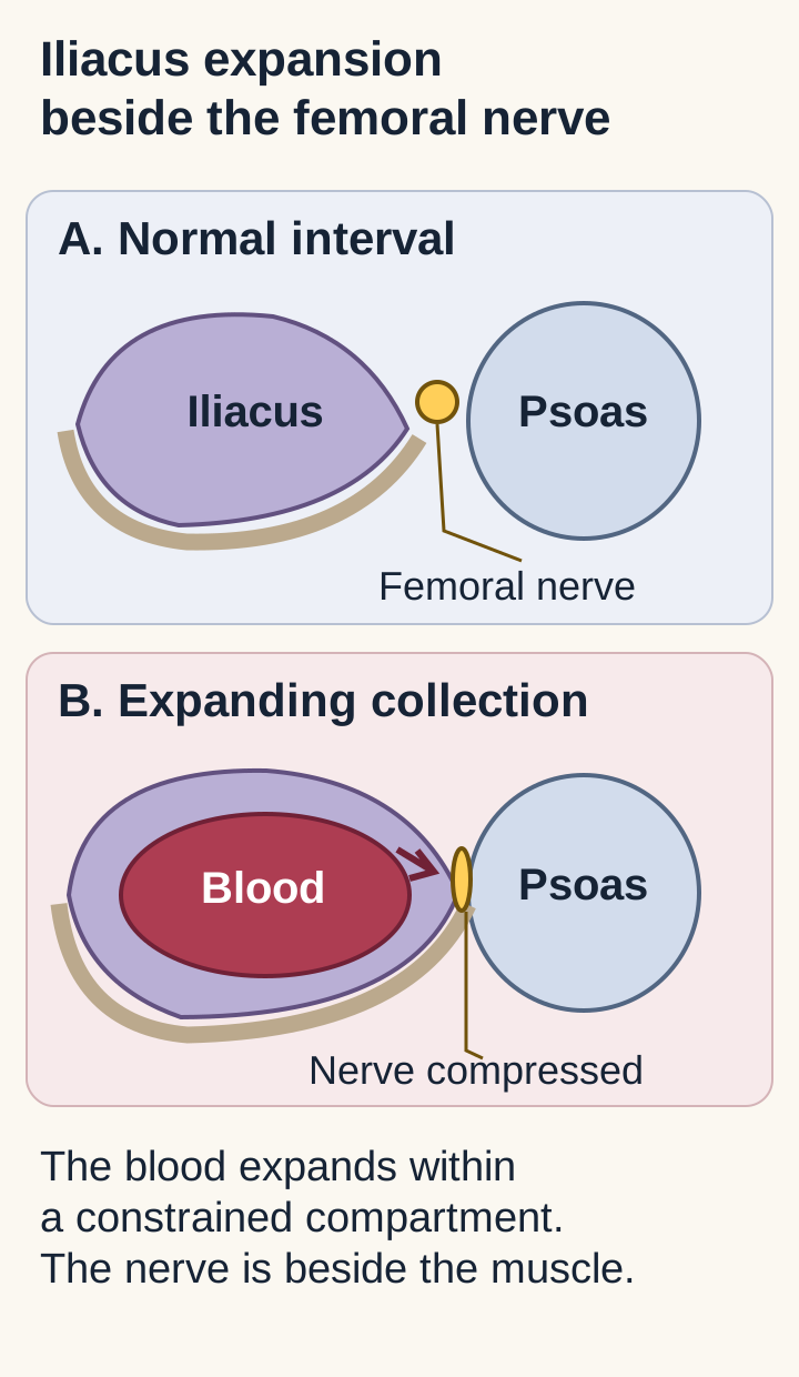
The iliacus occupies the inner surface of the ilium, behind the abdominal lining. Its fascia constrains expansion. The femoral nerve forms from L2-L4, emerges beside psoas major, travels in the interval between psoas and iliacus, and passes beneath the inguinal ligament. Blood within this region can compress or stretch the nerve along its course. A deep collection can therefore injure a nerve without producing a visible thigh bruise. [1][2]
The compartment diagram separates the muscle containing blood from the nerve beside it. Trace from the expanding iliacus compartment to the femoral nerve, then predict which action at the knee will fail. The consequence is reduced quadriceps activation: the patient cannot reliably straighten the knee or prevent it from collapsing during stance. This is not a torn quadriceps tendon simply because the knee is where the disability appears. [1][2]
Trace from blood inside the iliacus region to the nerve beside it. The tan arc represents adjacent iliac bone; muscle outlines emphasize a constrained region, not a measured fascial pressure or complete anatomic section. Predict the effect on knee extension before reading the section explanation. [1][2]
Anticoagulants, bleeding disorders, pelvic trauma and recent procedures raise concern for hemorrhage. Absence of substantial trauma does not exclude it. Pain in the groin, hip, flank or anterior thigh can precede measurable weakness; waiting for the complete neurologic pattern can delay assessment. A patient may hold the hip flexed because extension tensions the painful iliopsoas region. That posture identifies irritation, not its cause: infection can produce it too. [4][6][10]
Apply the relationship: in a patient with a new pelvic collection and knee buckling, assess knee extension, the patellar reflex and sensation before attributing the difficulty to pain alone. In a second patient with the same groin pain but normal strength, a falling hemoglobin still demands attention to bleeding. Nerve findings are useful, but their absence is not a hemorrhage exclusion test. [6][7]
Which examination findings belong together?
Does anterior thigh numbness establish femoral neuropathy? Not by itself. Build a localization from motor, reflex and sensory findings, then look for a function outside that nerve's territory. [2][8]
A focused comparison for proximal leg symptoms
Observation
Anatomic interpretation
ObservationWeak knee extension
Anatomic interpretationQuadriceps output through the femoral nerve
ObservationReduced patellar reflex
Anatomic interpretationImpaired L2-L4 reflex arc, especially L4; not nerve-specific alone
ObservationAnterior thigh and medial leg sensory loss
Anatomic interpretationFemoral cutaneous branches and its saphenous continuation
ObservationPreserved hip adduction
Anatomic interpretationSupports sparing of the obturator distribution
ObservationPreserved ankle dorsiflexion and plantar flexion
Anatomic interpretationSupports sparing of more distal, nonfemoral motor pathways
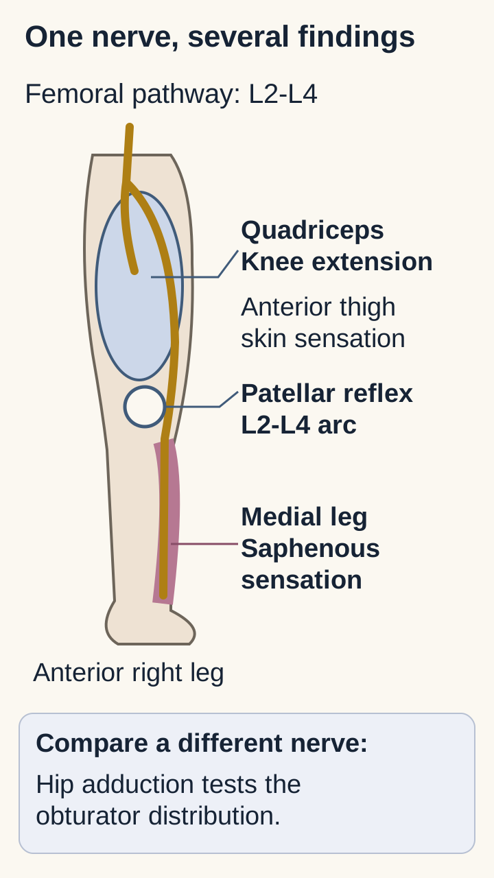
On the territory diagram, connect weak knee extension to the front of the thigh and medial leg, not the lateral foot. Now compare two examinations. Patient A has knee extension 2/5, hip adduction 5/5 and an absent patellar reflex. Patient B has the same quadriceps weakness but also weak adduction and lumbar paraspinal denervation. The additional abnormalities in B extend beyond an isolated femoral nerve lesion and favor root-level or broader involvement. Neither a single sensory boundary nor a normal back examination settles this alone. Sensory responses are usually preserved in a lesion proximal to the dorsal root ganglion, whereas a postganglionic lesion can reduce them. Ganglion involvement and technical factors create exceptions, so interpret these results with the sampled muscle pattern. [2][8][11]
Connect knee-extension weakness, reflex change and medial leg numbness to one pathway. Then compare hip adduction through the obturator distribution. Territories are approximate and overlap; this is not a full dermatome map or a rule that one normal test excludes a lesion. [2][3][8]
Hip flexion requires careful interpretation. Iliacus receives femoral innervation, whereas psoas major receives direct lumbar branches. A proximal femoral lesion can weaken iliacus while psoas still supplies some flexion. Pain can also limit the test. Preserved hip flexion therefore does not exclude femoral neuropathy, and reduced flexion does not prove that the entire lumbar plexus is injured. [2][3]
Try the localization: imagine the patient has medial leg numbness after knee surgery, but quadriceps power and the patellar reflex are normal. The affected territory could be an isolated saphenous branch, a sensory nerve, rather than the proximal femoral trunk. Conversely, isolated lateral thigh tingling with intact power and reflexes fits lateral femoral cutaneous neuropathy better than a femoral motor lesion. Use several concordant findings and allow for overlap. [2][8]
Compare your prediction for a proximal femoral lesion
Quadriceps weakness plus a reduced patellar reflex and saphenous sensory change supports the femoral trunk. Preserved adduction helps delimit the lesion. These findings locate dysfunction; imaging and clinical context establish whether the cause is hemorrhage. [2][8]
What must imaging explain?
An anticoagulated patient has new groin pain, a hemoglobin decline and a femoral-pattern examination. Which question should be answered first: whether the nerve conducts normally, or whether a deep hemorrhage needs urgent treatment?
CT of the abdomen and pelvis can establish a retroperitoneal collection and its extent. Contrast-enhanced imaging or CT angiography, selected with the treating team, can assess active extravasation and guide arterial treatment. A noncontrast study may demonstrate blood but cannot supply the same information about active contrast leakage. Imaging choice must account for clinical stability, kidney function and the immediate question; resuscitation should not be postponed for a routine diagnostic sequence. [6][7]
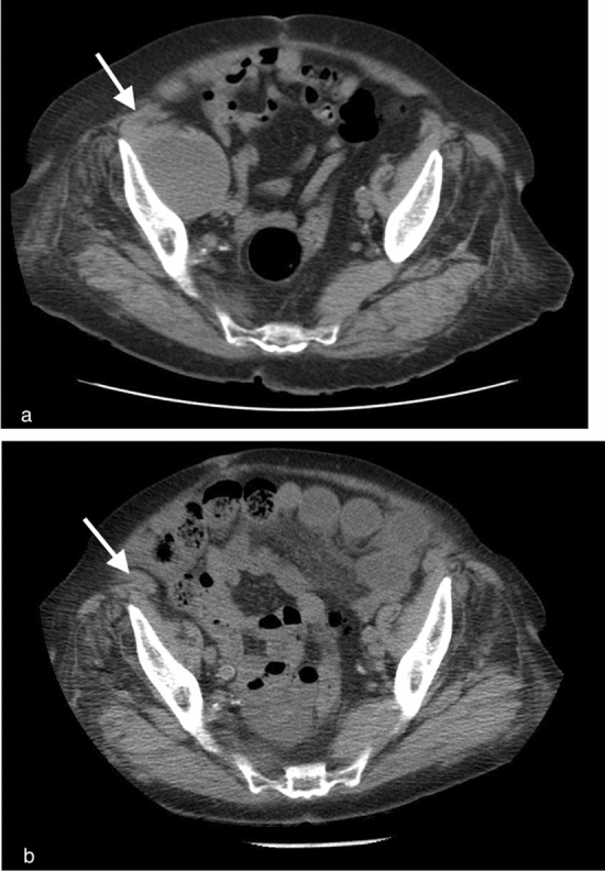
The clinical CT pair shows a right iliacus hematoma in panel A and resolution one year later in panel B. Compare the iliacus regions on the two sides before following the published white arrow. Identify the enlarged compartment, then predict which examination would establish functional femoral involvement. The image shows the collection, not nerve conduction or a guaranteed neurologic outcome. Its follow-up interval is a published observation, not a recommendation to wait a year. [6]
Compare the right iliacus region with the opposite side in the upper panel A, then with the one-year follow-up in the lower panel B. Lukies and colleagues report conservative treatment in this illustrated patient. The image demonstrates the collection and later radiographic resolution; it does not document a femoral nerve examination, prove nerve injury, or establish a recommended follow-up interval. Reproduced unchanged from Figure 1 of Lukies et al. (2023), CC BY 4.0. Image: Matthew Lukies, Jacob Gipson, Sia Yang Tan and Warren Clements; original source; Creative Commons Attribution 4.0 International (CC BY 4.0). [6].
Let an alternative explain the entire presentation
Alternative
Findings that change the assessment
AlternativeLumbar radiculopathy
Findings that change the assessmentRoot-compatible back or radiating pain, abnormalities beyond one peripheral nerve, or concordant spinal imaging
AlternativeDiabetic radiculoplexus neuropathy
Findings that change the assessmentPainful subacute progression, weight loss and weakness across several nerve territories after a structural lesion is excluded
AlternativeHip joint disease
Findings that change the assessmentJoint-centered pain and restricted passive rotation without a matching cutaneous, reflex and motor neuropathy
AlternativeIliopsoas abscess
Findings that change the assessmentInfectious context with a compatible collection, including central low attenuation, rim enhancement or gas
Fever and a rim-enhancing collection redirect care toward infection; absence of fever alone does not exclude an abscess. Aneurysmal and other vascular sources of retroperitoneal blood also remain in the differential. The compartment containing blood does not necessarily identify its original vessel. [5][6] Diabetes is not permission to ignore an acute hemoglobin decline or a visible compressive collection. A small incidental hematoma also does not explain every later neurologic finding. New bilateral weakness, saddle sensory loss or bladder dysfunction demands urgent consideration of spinal compression rather than anchoring on the iliacus. Urgent lumbar MRI is appropriate when cauda equina compression is suspected. [8][9][10][12]
Apply a changed finding: if CT identifies a collection but adduction and ankle power are also abnormal, broaden the localization. After urgent structural and bleeding questions are addressed, electrodiagnostic studies can characterize the distribution and severity of nerve injury. Their timing and technical limitations matter; they are not a reason to delay emergency imaging or consultation. [8]
Is the blood loss, the nerve compression, or both worsening?
Normal blood pressure and a stable hemoglobin can coexist with a worsening compressive neuropathy. Keep two assessments separate: the bleeding trajectory and the neurologic trajectory. These are bedside questions, not a validated numerical score. [5][6][7]
For bleeding, follow perfusion, pulse, blood pressure, serial hemoglobin, transfusion requirements and imaging when indicated. A single early hemoglobin may underrepresent acute blood loss; fluids and transfusion also affect later measurements. Active contrast extravasation, expanding blood, ongoing transfusion needs or deteriorating perfusion justify urgent bleeding-control assessment alongside resuscitation. [6][7]
For the nerve, compare documented knee-extension power, functional knee stability, sensation and pain over time. Severe or progressive weakness warrants urgent surgical and other relevant specialist assessment for pressure relief even without active contrast leakage. A collection need not still be filling to remain compressive. Hematoma dimensions inform the discussion, but the literature does not establish one diameter or a universal number of hours that determines treatment for every patient. [4][5]
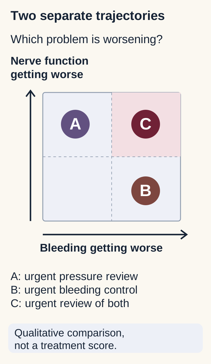
Use the two-trajectory exercise: patient A has blood pressure 118/74 mm Hg, hemoglobin 9.7 then 9.6 g/dL and knee extension declining from 4/5 to 1/5. Patient B has stable knee power but new hypotension, rising pulse and a continuing hemoglobin decline. Place each patient on the two-axis diagram before reading the consequence. A needs urgent reassessment of compression; B needs urgent hemorrhage control. Both require continued surveillance of the other problem. [5][6][7]
Place the two constructed patients from this section before comparing the explanations. A requires urgent assessment of compression, B urgent bleeding control, and C reassessment of both. Use the positions to compare urgency; choose intervention from the clinical assessment. [4][5][6][7]
Embolization targets a bleeding artery. Surgical evacuation or selected percutaneous drainage addresses a collection and its mass effect. These procedures answer different questions and are not interchangeable. An organized clot may not drain through a needle, and hemostasis after embolization does not establish that the nerve is decompressed. Procedural choice requires anatomy, clot characteristics, bleeding risk, deficit severity and local expertise. [4][5][6]
Transfer to a new situation: after successful arterial embolization, the hemoglobin stabilizes but quadriceps strength continues to decline. The conclusion is not that treatment failed to stop bleeding; it is that a second threat remains unresolved. Reassess the compartment and the need for decompression without waiting for shock to develop. [4][5][6]
Compare the two different reasons to escalate
Bleeding can be dangerous before palsy develops. Compression can be dangerous after bleeding stops. Improving one trajectory should never substitute for reassessing the other. [5][6][7]
What would make observation an active treatment plan?
Observation does not mean checking whether the pain is tolerable and sending the patient away. In selected patients with controlled bleeding, a limited collection and mild stable deficits, a monitored conservative plan can be appropriate. It requires a defined examination, review schedule, bleeding correction and a route to prompt escalation. [5][6]
Record knee extension using the same position and grading approach, hip adduction as a comparison, the patellar reflex and the sensory area. Grade 3/5 means full range against gravity; 4/5 adds resistance but is weaker than expected; 5/5 is normal power. Pain, sedation and effort can confound strength, so repeat an uncertain examination rather than labeling it either normal or definitively denervated. Document whether the knee buckles during safely supervised activity. [2][8]
Bedside record: compare serial strength, pain and hemodynamics
Measure
Initial
Reassessment
MeasureHemoglobin
Initial10.1 g/dL
Reassessment10.0 g/dL
MeasureKnee extension
Initial4/5
Reassessment2/5
MeasurePain after analgesia
Initial8/10
Reassessment3/10
MeasureMedial leg sensation
InitialReduced
ReassessmentReduced over a larger area
Choose the reassuring and concerning findings separately. The hemoglobin and pain are more reassuring, but power and sensation are deteriorating. The overall plan cannot remain unchanged just because analgesia works. This pattern warrants urgent specialist reassessment, with imaging and intervention considered according to the new findings. [5][6][7][8]
Once bleeding and compression are stable, rehabilitation addresses function: assisted transfers, gait assessment, quadriceps rehabilitation and appropriate knee support when buckling creates a fall risk. Progress activity within the treating team's restrictions. Forceful hip extension, aggressive massage and manual treatment of presumed psoas dysfunction are not substitutes for evaluating an acute hemorrhage. The contraindication here is the suspected or active bleeding lesion, not a claim that all later rehabilitation is unsafe. [1][4][8]
Apply to recovery: a patient can report less numbness before knee stability returns. Improvement may be prolonged and incomplete, so reassess objective function instead of promising a fixed recovery date. Electrodiagnostic follow-up can help when persistent weakness or an uncertain localization would change care; it does not replace serial bedside assessment. [8]
How do bleeding control and future anticoagulation fit together?
A patient with a major iliacus hemorrhage also has a mechanical valve. Does the valve mean anticoagulation must continue unchanged, or does the bleed mean it must be discontinued permanently? Neither conclusion follows automatically. Stabilize the immediate emergency and separately plan protection from future thrombosis. [7]
Urgent care includes circulation assessment, appropriate access and resuscitation, blood testing and blood-product support when indicated, identification of the bleeding source, and prompt discussion with the teams able to control it. Interrupt anticoagulation for major bleeding under the treating team's direction. For life-threatening warfarin-associated bleeding, four-factor prothrombin complex concentrate with intravenous vitamin K provides rapid factor replacement plus sustained correction; plasma is an alternative when the concentrate is unavailable. Agent, dose and monitoring belong to the current local reversal protocol. [7]
A normal INR does not reliably exclude clinically relevant factor Xa inhibitor activity. Establish the actual medication, dose timing, kidney function and available drug-calibrated testing. Do not wait for every specialized assay before treating a clinically evident life-threatening hemorrhage. This lesson does not prescribe a universal direct-acting anticoagulant antidote regimen. [7]
Compare two histories: after bleeding control, one patient has a mechanical valve and another completed treatment for a single remote, surgery-provoked venous thrombosis. Predict which history supplies an ongoing reason for therapeutic anticoagulation. The mechanical valve changes thrombotic risk and makes a coordinated resumption plan especially important; a resolved transient indication may lead to a different decision. In both, reassess hemostasis, imaging when relevant, neurologic stability and the original indication rather than selecting a fixed calendar day. [7]
Evidence for iliacus-related femoral palsy is largely observational. A descriptive review assembled 174 cases and found substantial persistent disability, but patients were not randomized to treatment and reports differed in severity and follow-up. Better outcomes in one reported treatment group cannot establish how that treatment would perform in every other patient. Use this uncertainty to seek timely expert assessment, not to justify delay or guarantee that every hematoma needs surgery. [5]
Before handing over care: state the compartment, the localized deficit, the direction of bleeding and nerve findings, and what would trigger reassessment. Identify who owns bleeding control, who reassesses compression, and who coordinates anticoagulation and rehabilitation. A complete explanation connects the groin pain to the knee deficit while keeping hemorrhage and nerve function independently visible. [4][5][6][7][8]
Apply the findings to a new patient
Answer the clinical question before considering the options. Then compare the explanations for the competing answers. The bank below contains original educational cases, not actual examination items.
Case 1
Show answer and explanations for case 1
A. Reduced sensation over the lateral foot (Why this does not fit)
Lateral foot sensation can be abnormal in a sural or S1 distribution. The supplied motor and reflex pattern instead centers on quadriceps and the femoral pathway. A distal lateral-foot territory does not match an isolated femoral lesion. [1] [2]
Reasoning steps for option A
Which territory suggests sural or S1 involvement?
Lateral foot sensation can be abnormal in a sural or S1 distribution. [1] [2]
Which motor and reflex pattern instead implicates femoral fibers?
The supplied motor and reflex pattern instead centers on quadriceps and the femoral pathway. [1] [2]
Why does lateral foot numbness not localize to isolated femoral injury?
A distal lateral-foot territory does not match an isolated femoral lesion. [1] [2]
B. Reduced sensation over the first dorsal web space (Why this does not fit)
The first dorsal web space belongs to the deep fibular sensory territory. Normal ankle dorsiflexion and a proximal quadriceps deficit do not support that nerve as the injured pathway. Localize sensation to the same nerve system as the motor deficit. [1] [2]
Reasoning steps for option B
Which nerve supplies the first dorsal web space?
The first dorsal web space belongs to the deep fibular sensory territory. [1] [2]
Why do normal dorsiflexion and quadriceps weakness disfavor this nerve?
Normal ankle dorsiflexion and a proximal quadriceps deficit do not support that nerve as the injured pathway. [1] [2]
How does first web-space sensation differ from the affected motor pathway?
The first dorsal web space is deep fibular territory, whereas the motor deficit is in the femoral pathway. [1] [2]
C. Reduced sensation along the medial lower leg (Best answer)
Weak quadriceps and loss of the patellar reflex support femoral dysfunction. The saphenous nerve carries femoral sensory fibers to the medial lower leg. A proximal femoral lesion can therefore affect both knee extension and this distal sensory territory. [1] [2]
Reasoning steps for option C
Which two examination deficits support femoral dysfunction?
Weak quadriceps and loss of the patellar reflex support femoral dysfunction. [1] [2]
Which femoral branch supplies the medial lower leg?
The saphenous nerve carries femoral sensory fibers to the medial lower leg. [1] [2]
How can proximal femoral injury link knee extension and distal sensation?
A proximal femoral lesion can therefore affect both knee extension and this distal sensory territory. [1] [2]
D. Reduced sensation across the plantar forefoot (Why this does not fit)
Plantar forefoot sensation travels through tibial nerve branches. Preserved plantar flexion and the quadriceps-centered findings favor a different pathway. A femoral lesion does not ordinarily produce an isolated plantar sensory deficit. [1] [2]
Reasoning steps for option D
Which branches supply plantar forefoot sensation?
Plantar forefoot sensation travels through tibial nerve branches. [1] [2]
How does preserved plantar flexion weigh against tibial localization?
Preserved plantar flexion and the quadriceps-centered findings favor a different pathway. [1] [2]
Would isolated femoral injury produce isolated plantar numbness?
A femoral lesion does not ordinarily produce an isolated plantar sensory deficit. [1] [2]
Takeaway: Quadriceps and reflex findings can locate the nerve before its distal sensory territory is predicted.
A. A preganglionic lumbar root lesion (Best answer)
Quadriceps and adductor longus are supplied through different peripheral nerves. Paraspinal involvement and preserved distal sensory response support a lesion proximal to the sensory ganglion. The distribution fits a lumbar root process better than an isolated femoral trunk injury. [2] [8] [11]
Reasoning steps for option A
Why do quadriceps and adductor longus findings cross nerve boundaries?
Quadriceps and adductor longus are supplied through different peripheral nerves. [2] [8] [11]
How do paraspinal findings and preserved sensory response localize relative to the ganglion?
Paraspinal involvement and preserved distal sensory response support a lesion proximal to the sensory ganglion. [2] [8] [11]
Which localization explains the combined distribution better than femoral trunk injury?
The distribution fits a lumbar root process better than an isolated femoral trunk injury. [2] [8] [11]
B. A femoral trunk lesion within the pelvis (Why this does not fit)
Femoral injury can impair quadriceps and the patellar reflex. It does not account for both obturator-innervated adductor longus and lumbar paraspinal abnormalities. Several concordant nonfemoral findings require a broader localization. [2] [8] [11]
Reasoning steps for option B
What deficits can femoral injury itself explain?
Femoral injury can impair quadriceps and the patellar reflex. [2] [8] [11]
Which additional muscles cannot be explained by femoral injury?
It does not account for both obturator-innervated adductor longus and lumbar paraspinal abnormalities. [2] [8] [11]
What broader localization is required by the nonfemoral abnormalities?
Adductor longus and lumbar paraspinal abnormalities alongside quadriceps involvement favor a lumbar root process over isolated femoral injury. [2] [8] [11]
C. An obturator nerve lesion within the pelvis (Why this does not fit)
Obturator injury can weaken hip adduction. It does not account for quadriceps or paraspinal denervation. One affected muscle should not outweigh the full distribution. [2] [8] [11]
Reasoning steps for option C
What action can obturator injury impair?
Obturator injury can weaken hip adduction. [2] [8] [11]
Which denervated muscles lie outside the obturator nerve?
It does not account for quadriceps or paraspinal denervation. [2] [8] [11]
Adductor weakness alone does not explain the concurrent quadriceps and paraspinal abnormalities. [2] [8] [11]
D. A saphenous nerve lesion below the knee (Why this does not fit)
The saphenous nerve supplies medial leg sensation. It is sensory and cannot produce the observed weakness in two proximal muscle groups. Motor and paraspinal abnormalities place this lesion outside an isolated saphenous branch. [2] [8] [11]
Reasoning steps for option D
Which region receives saphenous sensation?
The saphenous nerve supplies medial leg sensation. [2] [8] [11]
Why cannot this sensory branch explain two weak proximal muscle groups?
It is sensory and cannot produce the observed weakness in two proximal muscle groups. [2] [8] [11]
What do motor and paraspinal abnormalities exclude?
Motor and paraspinal abnormalities place this lesion outside an isolated saphenous branch. [2] [8] [11]
Takeaway: Abnormalities across peripheral nerve territories plus paraspinal involvement favor a root-level process.
A. An isolated L4 root lesion (Why this does not fit)
An L4 lesion can cause quadriceps weakness and medial leg symptoms. The asymmetric abnormal sensory response supports a postganglionic lesion, while sampled nonfemoral and paraspinal muscles are spared. Taken together, these findings favor the femoral nerve over an isolated root. [2] [8] [11]
Reasoning steps for option A
What symptoms could an L4 root injury also cause?
An L4 lesion can cause quadriceps weakness and medial leg symptoms. [2] [8] [11]
How do the sensory response and spared muscles challenge root localization?
The asymmetric abnormal sensory response supports a postganglionic lesion, while sampled nonfemoral and paraspinal muscles are spared. [2] [8] [11]
What lesion site best fits these combined findings?
Taken together, these findings favor the femoral nerve over an isolated root. [2] [8] [11]
B. An isolated obturator nerve lesion (Why this does not fit)
The obturator nerve can be affected by pelvic disease. Its sampled adductor muscle is normal and it does not supply quadriceps or the saphenous territory. Match the affected motor and sensory fibers, not just the pelvic location. [2] [8] [11]
Reasoning steps for option B
Why is obturator injury plausible with pelvic disease?
The obturator nerve can be affected by pelvic disease. [2] [8] [11]
Which normal muscle and abnormal territories contradict obturator injury?
Its sampled adductor muscle is normal and it does not supply quadriceps or the saphenous territory. [2] [8] [11]
Why do quadriceps and saphenous abnormalities favor femoral over obturator fibers?
The affected quadriceps and saphenous fibers favor a femoral pathway rather than the spared obturator distribution. [2] [8] [11]
C. An isolated saphenous branch lesion (Why this does not fit)
A saphenous lesion explains medial leg sensory loss and an abnormal sensory response. It cannot produce denervation in quadriceps muscles. Combined motor and sensory findings place the lesion proximal to this branch. [2] [8] [11]
Reasoning steps for option C
Which sensory findings could an isolated saphenous lesion explain?
A saphenous lesion explains medial leg sensory loss and an abnormal sensory response. [2] [8] [11]
Why does quadriceps denervation exclude an isolated saphenous lesion?
It cannot produce denervation in quadriceps muscles. [2] [8] [11]
Where must the lesion lie relative to the sensory branch?
Combined motor and sensory findings place the lesion proximal to this branch. [2] [8] [11]
D. A postganglionic femoral nerve lesion (Best answer)
The affected quadriceps muscles and saphenous sensory fibers belong to the femoral pathway. Preserved adductor and paraspinal sampling supports a restricted peripheral distribution. The entire pattern favors a femoral nerve lesion, interpreted with the clinical and imaging history. [2] [8] [11]
Reasoning steps for option D
Which nerve links quadriceps and saphenous fibers?
The affected quadriceps muscles and saphenous sensory fibers belong to the femoral pathway. [2] [8] [11]
What do normal adductor and paraspinal samples imply?
Preserved adductor and paraspinal sampling supports a restricted peripheral distribution. [2] [8] [11]
How should the full pattern be interpreted with clinical imaging?
The entire pattern favors a femoral nerve lesion, interpreted with the clinical and imaging history. [2] [8] [11]
Takeaway: A sensory branch abnormality plus motor findings in its parent nerve localizes more proximally than the branch itself.
A. The saphenous branch within the adductor canal (Why this does not fit)
A lesion in the adductor canal can affect medial leg sensation. The saphenous branch is distal and sensory, so it does not denervate iliacus or quadriceps. A sensory branch cannot explain proximal muscle involvement. [2] [3] [8]
Reasoning steps for option A
Which symptom could arise from an adductor canal lesion?
A lesion in the adductor canal can affect medial leg sensation. [2] [3] [8]
Why cannot the distal saphenous branch denervate iliacus?
The saphenous branch is distal and sensory, so it does not denervate iliacus or quadriceps. [2] [3] [8]
B. The femoral nerve before its iliacus branches (Best answer)
Iliacus is innervated by branches from the femoral nerve in the pelvis. Involvement of iliacus and quadriceps with spared psoas and adductors fits injury before those branches separate. The additional muscle finding places the lesion proximally rather than below the ligament. [2] [3] [8]
Reasoning steps for option B
Where do the iliacus branches leave the femoral nerve?
Iliacus is innervated by branches from the femoral nerve in the pelvis. [2] [3] [8]
How do affected iliacus and quadriceps but spared psoas and adductors constrain localization?
Involvement of iliacus and quadriceps with spared psoas and adductors fits injury before those branches separate. [2] [3] [8]
Why does iliacus denervation favor a site above the ligament?
The additional muscle finding places the lesion proximally rather than below the ligament. [2] [3] [8]
C. The obturator nerve near the pelvic brim (Why this does not fit)
The obturator nerve passes through the pelvis and supplies major adductors. Adductor longus is spared, and obturator injury does not explain quadriceps denervation. Regional proximity does not mean that the nerve supplies the affected muscles. [2] [3] [8]
Reasoning steps for option C
What muscle group is innervated by the pelvic obturator nerve?
The obturator nerve passes through the pelvis and supplies major adductors. [2] [3] [8]
Which findings contradict obturator nerve injury?
Adductor longus is spared, and obturator injury does not explain quadriceps denervation. [2] [3] [8]
Why does pelvic proximity not prove obturator innervation?
Pelvic proximity does not make obturator injury responsible for iliacus and quadriceps denervation when adductor longus is spared. [2] [3] [8]
D. The femoral nerve distal to the inguinal ligament (Why this does not fit)
A distal femoral lesion can affect quadriceps. It is beyond the pelvic branches supplying iliacus and cannot account for that added denervation. Use the most proximal affected branch to constrain lesion location. [2] [3] [8]
Reasoning steps for option D
Which deficit could distal femoral injury explain?
A distal femoral lesion can affect quadriceps. [2] [3] [8]
Why cannot a distal injury account for iliacus denervation?
It is beyond the pelvic branches supplying iliacus and cannot account for that added denervation. [2] [3] [8]
How does iliacus branch anatomy constrain the injury site?
Iliacus denervation requires injury proximal to its pelvic femoral branches, not a distal femoral lesion. [2] [3] [8]
Takeaway: Iliacus involvement places a femoral lesion proximal to its pelvic motor branches.
A. The sensory findings indicate an isolated saphenous lesion (Why this does not fit)
Saphenous dysfunction can account for medial leg numbness. It cannot explain profound quadriceps weakness and an absent patellar reflex. The motor findings require a lesion involving more than this sensory branch. [2] [3]
Reasoning steps for option A
Which symptom can saphenous dysfunction cause?
Saphenous dysfunction can account for medial leg numbness. [2] [3]
Which motor and reflex deficits exceed a sensory branch lesion?
It cannot explain profound quadriceps weakness and an absent patellar reflex. [2] [3]
What extent of lesion is required by these motor findings?
The motor findings require a lesion involving more than this sensory branch. [2] [3]
B. The deficit is primarily within the obturator distribution (Why this does not fit)
Obturator injury can leave some hip flexion intact. Normal adduction and marked knee-extension weakness instead support femoral involvement. The pattern of preserved and impaired actions is more informative than flexion alone. [2] [3]
Reasoning steps for option B
Can obturator injury leave some hip flexion intact?
Obturator injury can leave some hip flexion intact. [2] [3]
Which impaired and preserved actions point instead to femoral injury?
Normal adduction and marked knee-extension weakness instead support femoral involvement. [2] [3]
Why is hip-flexion strength alone insufficient to distinguish these nerves?
Normal hip adduction alongside marked knee-extension weakness favors femoral involvement despite partly preserved hip flexion. [2] [3]
C. Direct lumbar innervation can preserve psoas major function (Best answer)
Quadriceps, reflex and saphenous abnormalities localize to the femoral pathway. Psoas major receives direct lumbar branches and can contribute hip flexion despite injury to femoral-innervated iliacus. Relatively preserved hip flexion does not exclude proximal femoral neuropathy. [2] [3]
Reasoning steps for option C
Which deficits jointly implicate the femoral pathway?
Quadriceps, reflex and saphenous abnormalities localize to the femoral pathway. [2] [3]
How can psoas preserve hip flexion despite iliacus injury?
Psoas major receives direct lumbar branches and can contribute hip flexion despite injury to femoral-innervated iliacus. [2] [3]
Does relatively intact hip flexion rule out femoral neuropathy?
Relatively preserved hip flexion does not exclude proximal femoral neuropathy. [2] [3]
D. Pain inhibition accounts for the entire abnormal examination (Why this does not fit)
Pain can reduce effort during strength testing. The matching reflex and sensory abnormalities add evidence of neurologic dysfunction beyond pain-limited effort. Pain and neuropathy can coexist and require separate assessment. [2] [3]
Reasoning steps for option D
How can pain affect strength testing?
Pain can reduce effort during strength testing. [2] [3]
Which reflex and sensory findings argue against pain as the only cause?
The matching reflex and sensory abnormalities add evidence of neurologic dysfunction beyond pain-limited effort. [2] [3]
Can pain and neuropathy coexist in this presentation?
Pain and neuropathy can coexist and require separate assessment. [2] [3]
Takeaway: Do not use preserved hip flexion to veto an otherwise coherent femoral localization.
Femoral injury can produce proximal leg weakness and sensory symptoms. Preserved quadriceps and patellar reflex with selective adductor weakness do not fit its main motor distribution. Use the impaired action to distinguish neighboring pelvic nerves. [2]
Reasoning steps for option A
What symptoms could femoral injury produce generally?
Femoral injury can produce proximal leg weakness and sensory symptoms. [2]
Which preserved femoral actions contradict that localization here?
Preserved quadriceps and patellar reflex with selective adductor weakness do not fit its main motor distribution. [2]
Which movement distinguishes obturator from femoral dysfunction here?
Selective hip-adduction weakness favors obturator rather than femoral dysfunction when quadriceps and reflex are preserved. [2]
B. Obturator nerve (Best answer)
Adductor longus receives obturator innervation. Selective adduction weakness with preserved femoral function and a collection near the obturator canal fits that nerve. The examination and lesion location independently support the same localization. [2]
How do selective weakness and canal collection converge anatomically?
Selective adduction weakness with preserved femoral function and a collection near the obturator canal fits that nerve. [2]
What independent examination and imaging clues support obturator injury?
Selective adduction weakness and a collection near the obturator canal both support obturator neuropathy. [2]
C. Lateral femoral cutaneous nerve (Why this does not fit)
This nerve can be affected by pelvic or inguinal compression. It is sensory and supplies the lateral rather than medial thigh territory. It cannot account for the observed adductor weakness. [2]
Reasoning steps for option C
Can this sensory nerve be compressed in the inguinal area?
This nerve can be affected by pelvic or inguinal compression. [2]
What sensory territory and motor limitation characterize it?
It is sensory and supplies the lateral rather than medial thigh territory. [2]
Can a lateral thigh sensory nerve cause adductor weakness?
It cannot account for the observed adductor weakness. [2]
D. Common fibular nerve (Why this does not fit)
Common fibular injury can cause leg weakness after prolonged positioning. It affects ankle dorsiflexion or eversion rather than hip adduction, and ankle power is normal here. A distal leg nerve does not fit a selective proximal adductor deficit. [2]
Reasoning steps for option D
Which distal nerve injury can follow prolonged positioning?
Common fibular injury can cause leg weakness after prolonged positioning. [2]
Which ankle actions would be affected instead of adduction?
It affects ankle dorsiflexion or eversion rather than hip adduction, and ankle power is normal here. [2]
Why does normal ankle strength contradict distal fibular injury?
A distal leg nerve does not fit a selective proximal adductor deficit. [2]
Takeaway: Preserved quadriceps and reflexes help distinguish obturator from femoral involvement.
A. New objective weakness of knee extension (Best answer)
The initial sensory-only lateral thigh pattern favors lateral femoral cutaneous neuropathy. That nerve has no motor supply to quadriceps. New knee-extension weakness requires a broader or different localization rather than attribution to the same sensory nerve. [2] [8]
Reasoning steps for option A
Which nerve fits the original sensory-only lateral thigh complaint?
That nerve has no motor supply to quadriceps. [2] [8]
How does new knee-extension weakness alter localization?
New knee-extension weakness requires a broader or different localization rather than attribution to the same sensory nerve. [2] [8]
B. Symptoms restricted to the anterolateral thigh (Why this does not fit)
The lateral femoral cutaneous nerve supplies this sensory territory. The restricted distribution is compatible with the initial normal motor and reflex findings. Persistence in that area would not itself contradict the leading localization. [2] [8]
Reasoning steps for option B
What cutaneous nerve covers the affected lateral thigh?
The lateral femoral cutaneous nerve supplies this sensory territory. [2] [8]
Why did normal quadriceps function and patellar reflex fit the initial lateral thigh sensory complaint?
The restricted distribution is compatible with the initial normal motor and reflex findings. [2] [8]
Would persistent sensory symptoms confined to the lateral thigh contradict lateral femoral cutaneous neuropathy?
Persistence in that area would not itself contradict the leading localization. [2] [8]
C. Discomfort when a belt crosses the inguinal region (Why this does not fit)
Mechanical pressure near the inguinal ligament can irritate the lateral femoral cutaneous nerve. The history and sensory distribution already fit that possibility. This provocation supports rather than challenges the proposed localization. [2] [8]
Reasoning steps for option C
What pressure site can irritate the lateral thigh sensory nerve?
Mechanical pressure near the inguinal ligament can irritate the lateral femoral cutaneous nerve. [2] [8]
How did belt pressure and lateral thigh sensory distribution support lateral femoral cutaneous irritation?
Belt pressure near the inguinal ligament and the lateral thigh sensory distribution already fit lateral femoral cutaneous irritation. [2] [8]
Does reproduction of lateral thigh symptoms by belt pressure challenge lateral femoral cutaneous localization?
Reproduction of lateral thigh symptoms by belt pressure supports rather than challenges lateral femoral cutaneous localization. [2] [8]
D. Preserved sensation along the medial lower leg (Why this does not fit)
The medial lower leg is supplied through the saphenous branch of the femoral nerve. Its preservation is compatible with a separate lateral thigh sensory neuropathy. Normal function outside the affected territory helps delimit the lesion. [2] [8]
Reasoning steps for option D
Which femoral sensory branch supplies the spared medial lower leg?
The medial lower leg is supplied through the saphenous branch of the femoral nerve. [2] [8]
Why is preserved medial leg sensation compatible with lateral thigh neuropathy?
Its preservation is compatible with a separate lateral thigh sensory neuropathy. [2] [8]
What does preserved medial sensation imply about lesion extent?
Preserved medial lower-leg sensation limits the findings to the lateral thigh rather than the saphenous territory. [2] [8]
Takeaway: A sensory-only nerve cannot explain new objective quadriceps weakness.
A. Anticoagulant reversal and transfusion-directed observation (Why this does not fit)
A hemorrhagic collection with significant blood loss can require reversal and blood-product support. The bacteremia, fever and gas-containing rim-enhancing collection instead favor infection. A hemorrhage-focused plan alone would leave the likely infectious source untreated. [6] [10]
Reasoning steps for option A
When is reversal and transfusion appropriate for an iliacus collection?
A hemorrhagic collection with significant blood loss can require reversal and blood-product support. [6] [10]
Which findings instead suggest an infected collection?
The bacteremia, fever and gas-containing rim-enhancing collection instead favor infection. [6] [10]
What does hemorrhage-only care leave untreated?
A hemorrhage-focused plan alone would leave the likely infectious source untreated. [6] [10]
B. Arterial embolization directed at the iliolumbar circulation (Why this does not fit)
Embolization is useful for selected ongoing arterial hemorrhage. The supplied imaging identifies an abscess-like collection without demonstrated arterial extravasation. Treat the demonstrated disease rather than selecting a procedure from location alone. [6] [10]
Reasoning steps for option B
When is arterial embolization useful?
Embolization is useful for selected ongoing arterial hemorrhage. [6] [10]
What does abscess-like imaging without extravasation suggest instead?
The supplied imaging identifies an abscess-like collection without demonstrated arterial extravasation. [6] [10]
Why is collection location alone insufficient reason for embolization?
An abscess-like collection without arterial extravasation does not justify embolization merely because it lies in the iliopsoas region. [6] [10]
C. Analgesia and progressive rehabilitation without source evaluation (Why this does not fit)
Analgesia and rehabilitation may be appropriate after a noninfectious injury stabilizes. Current bacteremia, fever and the collection demand acute infection treatment and source assessment. Symptom management does not replace care of an active deep infection. [6] [10]
Reasoning steps for option C
When might analgesia and rehabilitation be appropriate?
Analgesia and rehabilitation may be appropriate after a noninfectious injury stabilizes. [6] [10]
Which current findings demand infection treatment and source assessment?
Current bacteremia, fever and the collection demand acute infection treatment and source assessment. [6] [10]
Why is symptom control insufficient with bacteremia?
Symptom management does not replace care of an active deep infection. [6] [10]
D. Antimicrobial treatment and assessment for drainage (Best answer)
Bacteremia plus the CT characteristics strongly supports an iliopsoas abscess. Antimicrobial therapy and specialist source-control assessment address that process. The drainage approach depends on the collection, patient condition and local expertise. [6] [10]
Reasoning steps for option D
Which CT and microbiologic clues support iliopsoas abscess?
Bacteremia with a rim-enhancing, gas-containing iliopsoas collection on CT strongly supports an iliopsoas abscess. [6] [10]
What two management components address infection?
Antimicrobial therapy and specialist source-control assessment address that process. [6] [10]
What factors determine how drainage is performed?
The drainage approach depends on the collection, patient condition and local expertise. [6] [10]
Takeaway: Hip-extension pain identifies an irritated region; clinical context and imaging distinguish blood from infection.
A. Pressure injury restricted to the femoral nerve in iliacus (Why this does not fit)
A femoral lesion could explain quadriceps weakness. It does not explain the additional adductor and ankle deficits, and imaging shows no iliacus collection. Do not narrow a multifocal examination to one nerve because the pain began in the thigh. [9]
Reasoning steps for option A
Which isolated deficit might femoral neuropathy explain?
A femoral lesion could explain quadriceps weakness. [9]
Which other deficits and imaging findings weaken femoral compression?
It does not explain the additional adductor and ankle deficits, and imaging shows no iliacus collection. [9]
Why is thigh pain onset insufficient to restrict localization?
Do not narrow a multifocal examination to one nerve because the pain began in the thigh. [9]
B. Ischemic injury involving multiple lumbosacral nerve territories (Best answer)
The painful subacute course with weight loss and spreading weakness fits radiculoplexus neuropathy. The abnormal actions span several peripheral nerves, and structural hemorrhage or spinal compression has been assessed. An ischemic inflammatory radiculoplexus process fits better than a single compressed femoral nerve. [9]
Reasoning steps for option B
What course and constitutional feature support radiculoplexus neuropathy?
The painful subacute course with weight loss and spreading weakness fits radiculoplexus neuropathy. [9]
How do cross-nerve weakness and negative structural assessment fit?
The abnormal actions span several peripheral nerves, and structural hemorrhage or spinal compression has been assessed. [9]
Which process better explains spreading weakness than a single nerve lesion?
An ischemic inflammatory radiculoplexus process fits better than a single compressed femoral nerve. [9]
C. Compression of the lateral femoral cutaneous nerve at the groin (Why this does not fit)
Lateral femoral cutaneous neuropathy can cause painful thigh paresthesia. It is sensory and cannot explain progressive weakness across these muscle groups. The motor distribution requires a broader process. [9]
Reasoning steps for option C
What symptoms might lateral femoral cutaneous neuropathy cause?
Lateral femoral cutaneous neuropathy can cause painful thigh paresthesia. [9]
Why cannot that nerve explain the progressive weakness?
It is sensory and cannot explain progressive weakness across these muscle groups. [9]
Which motor pattern demands a broader process?
Progressive weakness across several muscle groups exceeds a purely sensory lateral femoral cutaneous lesion. [9]
D. An isolated tear of the quadriceps tendon near the patella (Why this does not fit)
A quadriceps tendon tear can impair knee extension. It does not account for hip adduction and ankle weakness or the subacute systemic history. A focal extensor injury is insufficient to explain the full presentation. [9]
Reasoning steps for option D
What isolated weakness could quadriceps tendon tear cause?
A quadriceps tendon tear can impair knee extension. [9]
Which additional deficits and history refute tendon tear alone?
It does not account for hip adduction and ankle weakness or the subacute systemic history. [9]
Why does focal extensor damage not explain the full presentation?
A focal quadriceps extensor injury does not explain additional hip-adduction and ankle weakness or the subacute systemic history. [9]
Takeaway: Diabetes-associated proximal pain is a diagnosis to investigate, not a reason to overlook acute hemorrhage.
A. Loss of quadriceps motor axons from a femoral lesion (Why this does not fit)
Femoral neuropathy can make stairs difficult by weakening quadriceps. Normal controlled strength, reflexes and sensory testing do not support that explanation here. Functional difficulty should be separated from a demonstrated motor deficit. [2] [8]
Reasoning steps for option A
How could femoral weakness impair stair climbing?
Femoral neuropathy can make stairs difficult by weakening quadriceps. [2] [8]
Which normal neurologic findings challenge femoral neuropathy?
Normal controlled strength, reflexes and sensory testing do not support that explanation here. [2] [8]
Why is stair difficulty not proof of quadriceps weakness?
Difficulty climbing stairs without objective quadriceps weakness, reflex loss or sensory deficit does not establish femoral neuropathy. [2] [8]
B. Loss of adductor motor axons from an obturator lesion (Why this does not fit)
Obturator injury may impair proximal leg function. The dominant findings are painful restricted joint rotation, not documented adductor weakness. A joint-centered examination has a different explanation from selective denervation. [2] [8]
Reasoning steps for option B
What proximal function might obturator injury impair?
Obturator injury may impair hip adduction and proximal leg function. [2] [8]
What joint finding predominates instead of documented adductor weakness?
The dominant findings are painful restricted joint rotation, not documented adductor weakness. [2] [8]
How does joint-centered restriction differ from denervation?
Painful restricted passive hip rotation with radiographic hip disease favors a joint process rather than selective obturator denervation. [2] [8]
C. A sensory branch lesion within the adductor canal (Why this does not fit)
Saphenous injury can cause medial leg sensory symptoms. Sensation is normal and that branch would not explain restricted passive hip rotation. The distribution and type of abnormality do not match a sensory mononeuropathy. [2] [8]
Reasoning steps for option C
What sensory symptom could saphenous injury cause?
Saphenous injury can cause medial leg sensory symptoms. [2] [8]
Which normal sense and abnormal passive movement contradict it?
Sensation is normal and that branch would not explain restricted passive hip rotation. [2] [8]
Why does painful passive hip rotation not fit sensory mononeuropathy?
The distribution and type of abnormality do not match a sensory mononeuropathy. [2] [8]
D. Pain-limited function from degenerative hip disease (Best answer)
Chronic load-related groin pain and restricted passive rotation fit the radiographic hip disease. Preserved strength after analgesia with normal reflexes and sensation argues against a matching femoral neuropathy. The supplied findings favor joint pain as the cause of impaired function. [2] [8]
Reasoning steps for option D
Which pain and motion findings match the radiographic hip disease?
Chronic load-related groin pain and restricted passive rotation fit the radiographic hip disease. [2] [8]
What findings after analgesia weaken femoral neuropathy?
Preserved strength after analgesia with normal reflexes and sensation argues against a matching femoral neuropathy. [2] [8]
What explanation best integrates functional impairment and intact neurologic testing?
The supplied findings favor joint pain as the cause of impaired function. [2] [8]
Takeaway: Difficulty using a limb is not synonymous with denervation; document power, reflexes and sensation.
A. Contrast-enhanced CT of the abdomen and pelvis (Best answer)
Pain, falling hemoglobin and a femoral-pattern deficit suggest concealed retroperitoneal hemorrhage. Cross-sectional imaging can locate the collection and assess features relevant to bleeding control. The immediate structural question takes priority over delayed nerve characterization. [4] [6] [8]
Reasoning steps for option A
What triad makes concealed retroperitoneal bleeding the immediate concern?
Pain, falling hemoglobin and a femoral-pattern deficit suggest concealed retroperitoneal hemorrhage. [4] [6] [8]
What can cross-sectional imaging establish about the suspected collection?
Cross-sectional imaging can locate the collection and assess features relevant to bleeding control. [4] [6] [8]
Why should imaging precede detailed nerve characterization?
The immediate structural question takes priority over delayed nerve characterization. [4] [6] [8]
B. MRI of the lumbar spine with nerve-root assessment (Why this does not fit)
Spinal disease can cause quadriceps weakness and a reduced reflex. The abrupt hemoglobin decline and flank-groin pain during heparin treatment favor a bleeding source outside the spinal canal. Select imaging that addresses the leading cause rather than the reflex finding alone. [4] [6] [8]
Reasoning steps for option B
Which spinal finding could initially mimic this femoral presentation?
Spinal disease can cause quadriceps weakness and a reduced reflex. [4] [6] [8]
Why do heparin exposure, flank-groin pain and falling hemoglobin shift localization away from the spine?
The abrupt hemoglobin decline and flank-groin pain during heparin treatment favor a bleeding source outside the spinal canal. [4] [6] [8]
Why is imaging based solely on the diminished reflex insufficient?
Select imaging for suspected heparin-associated retroperitoneal bleeding rather than investigating the reduced patellar reflex alone. [4] [6] [8]
C. Nerve conduction studies with needle electromyography (Why this does not fit)
Electrodiagnostic studies can later define nerve distribution and injury severity. They do not identify or control an acutely bleeding retroperitoneal lesion. A suspected hemorrhage should be assessed before waiting for detailed nerve testing. [4] [6] [8]
Reasoning steps for option C
What useful information can electrodiagnostic testing supply later?
Electrodiagnostic studies can later define nerve distribution and injury severity. [4] [6] [8]
Why cannot nerve conduction and needle studies answer the urgent hemorrhage question?
They do not identify or control an acutely bleeding retroperitoneal lesion. [4] [6] [8]
What must be investigated before scheduling detailed nerve testing?
A suspected hemorrhage should be assessed before waiting for detailed nerve testing. [4] [6] [8]
D. Ultrasonography confined to the symptomatic hip joint (Why this does not fit)
Hip ultrasound can help evaluate an effusion. It does not comprehensively evaluate the retroperitoneum or explain the combined blood-loss and nerve findings. The examination requires assessment beyond the joint. [4] [6] [8]
Reasoning steps for option D
What hip-joint abnormality might ultrasound detect?
Hip ultrasound can help evaluate an effusion. [4] [6] [8]
Why does joint ultrasound miss the principal blood-loss and nerve concern?
It does not comprehensively evaluate the retroperitoneum or explain the combined blood-loss and nerve findings. [4] [6] [8]
How far beyond the hip joint must the initial examination extend?
The examination must assess the retroperitoneum beyond the hip joint. [4] [6] [8]
Takeaway: A bleeding trajectory changes the first diagnostic question from nerve physiology to an urgent structural cause.
A. The similar hemoglobin makes significant acute blood loss unlikely enough to defer imaging (Why this does not fit)
A stable hemoglobin can be reassuring when interpreted in a stable serial clinical course. This is an early isolated measurement with new pain, tachycardia and a focal deficit. Hemoglobin concentration alone cannot exclude recent concealed hemorrhage. [4] [6] [7]
Reasoning steps for option A
Under what clinical circumstances would an unchanged hemoglobin be reassuring?
A stable hemoglobin can be reassuring when interpreted in a stable serial clinical course. [4] [6] [7]
Why is this early hemoglobin value not reassuring in this presentation?
This is an early isolated measurement with new pain, tachycardia and a focal deficit. [4] [6] [7]
What cannot a single hemoglobin concentration exclude immediately after blood loss?
B. The absence of bruising favors a superficial muscle strain over internal bleeding (Why this does not fit)
Visible bruising can support a traumatic injury. A retroperitoneal collection is deep and may be present without a visible surface mark. The lack of external bleeding does not resolve the internal bleeding concern. [4] [6] [7]
Reasoning steps for option B
What might visible bruising indicate after trauma?
Visible bruising can support a traumatic injury. [4] [6] [7]
Why may an iliacus or other retroperitoneal bleed leave no surface bruise?
A retroperitoneal collection is deep and may be present without a visible surface mark. [4] [6] [7]
Does absence of external bleeding settle the internal hemorrhage question?
The lack of external bleeding does not resolve the internal bleeding concern. [4] [6] [7]
C. The early hemoglobin does not exclude a concealed bleed needing urgent evaluation (Best answer)
Acute blood loss may not immediately produce a proportional fall in measured hemoglobin concentration. The new pain, pulse change and knee instability remain concerning despite that value. Assess circulation and the possible retroperitoneal source without waiting for serial anemia to prove the diagnosis. [4] [6] [7]
Reasoning steps for option C
Why can measured hemoglobin initially lag behind acute hemorrhage?
Acute blood loss may not immediately produce a proportional fall in measured hemoglobin concentration. [4] [6] [7]
Which new symptoms and vital-sign change still support concern despite the laboratory value?
The new pain, pulse change and knee instability remain concerning despite that value. [4] [6] [7]
What should clinicians assess before serial anemia becomes apparent?
Assess circulation and the possible retroperitoneal source without waiting for serial anemia to prove the diagnosis. [4] [6] [7]
D. The low-energy mechanism favors nerve irritation without a bleeding lesion (Why this does not fit)
Major trauma is a recognized hemorrhage risk. Anticoagulation allows clinically important bleeding after minor trauma or without a clear injury. The apparent size of the trigger should not override the evolving examination. [4] [6] [7]
Reasoning steps for option D
What does major trauma contribute to hemorrhage risk?
Major trauma is a recognized hemorrhage risk. [4] [6] [7]
Why is a major injury not required for bleeding during anticoagulation?
Anticoagulation allows clinically important bleeding after minor trauma or without a clear injury. [4] [6] [7]
Why should a minor or absent trigger not override the evolving neurologic examination?
The apparent size of the trigger should not override the evolving examination. [4] [6] [7]
Takeaway: An early normal hemoglobin cannot veto a concerning acute hemorrhagic presentation.
A. The collection explains the anemia, so observation is sufficient (Why this does not fit)
A demonstrated hematoma provides an anatomic source for blood loss. Continued hemoglobin decline and transfusion need show that locating the source has not established stability. The bleeding trajectory still requires urgent reassessment. [6] [7]
Reasoning steps for option A
What does detection of the hematoma establish about the source of anemia?
A demonstrated hematoma provides an anatomic source for blood loss. [6] [7]
Why do continued hemoglobin decline and transfusions contradict a claim of stability?
Continued hemoglobin decline and transfusion need show that locating the source has not established stability. [6] [7]
What does the persistent bleeding trajectory demand despite source identification?
The bleeding trajectory still requires urgent reassessment. [6] [7]
B. The contrast risk requires delaying source assessment until renal recovery (Why this does not fit)
Severe kidney impairment affects the risk-benefit discussion for contrast imaging. It does not eliminate the need to assess potentially life-threatening ongoing bleeding. Select an urgent diagnostic and procedural plan with the team while resuscitation continues. [6] [7]
Reasoning steps for option B
How does severe renal impairment complicate a contrast-imaging decision?
Severe kidney impairment affects the risk-benefit discussion for contrast imaging. [6] [7]
It does not eliminate the need to assess potentially life-threatening ongoing bleeding. [6] [7]
What urgent collaborative plan is needed while resuscitation continues?
Select an urgent diagnostic and procedural plan with the team while resuscitation continues. [6] [7]
C. The absent contrast leak favors a completed bleed over an active source (Why this does not fit)
On a suitable enhanced study, a leak can support active bleeding. No contrast was administered in this study, so the lack of a described leak cannot establish hemostasis. Match a conclusion to what the actual imaging protocol can demonstrate. [6] [7]
Reasoning steps for option C
What would contrast leakage signify on an appropriately enhanced scan?
On a suitable enhanced study, a leak can support active bleeding. [6] [7]
Why does the noncontrast protocol make absence of reported leakage uninformative?
No contrast was administered in this study, so the lack of a described leak cannot establish hemostasis. [6] [7]
What principle limits inferences about hemostasis from this particular scan?
Do not infer hemostasis from absent extravasation on a scan performed without contrast. [6] [7]
D. It locates the collection without resolving current arterial bleeding activity (Best answer)
The noncontrast study supplies an anatomic explanation for the pain and anemia. It cannot provide the same active-extravasation information as a contrast-enhanced examination. Further bleeding assessment should be selected urgently with the team while resuscitation continues. [6] [7]
Reasoning steps for option D
What can this noncontrast scan explain anatomically?
The noncontrast study supplies an anatomic explanation for the pain and anemia. [6] [7]
Which active-bleeding finding cannot this protocol assess as an enhanced scan could?
It cannot provide the same active-extravasation information as a contrast-enhanced examination. [6] [7]
How should the team pursue further assessment while supporting circulation?
Further bleeding assessment should be selected urgently with the team while resuscitation continues. [6] [7]
Takeaway: A study that establishes a collection may leave bleeding activity unresolved.
A. Continue the same observation plan until hemoglobin declines (Why this does not fit)
Serial hemoglobin helps assess bleeding. It does not measure the pressure effect on the femoral nerve, which is deteriorating objectively. A stable bleeding trajectory should not preserve an unchanged plan for progressive palsy. [4] [5] [8]
Reasoning steps for option A
Which aspect of hemorrhage can serial hemoglobin track?
Serial hemoglobin helps assess bleeding. [4] [5] [8]
Why does stable hemoglobin fail to exclude worsening femoral nerve compression?
It does not measure the pressure effect on the femoral nerve, which is deteriorating objectively. [4] [5] [8]
What change in management does objectively progressive palsy require despite stable bleeding?
Progressive palsy despite stable bleeding requires urgent reassessment of femoral nerve compression, not an unchanged observation plan. [4] [5] [8]
B. Use arterial angiography as the primary treatment for the weakness (Why this does not fit)
Angiography can identify and treat an ongoing arterial bleeding source. The supplied problem is progressive neurologic dysfunction despite stable bleeding indicators and no extravasation. The residual collection may need pressure-relief assessment rather than an arterial procedure directed at an unproven current source. [4] [5] [8]
Reasoning steps for option B
What type of ongoing source can angiography identify and treat?
Angiography can identify and treat an ongoing arterial bleeding source. [4] [5] [8]
Why does absent extravasation with progressive weakness not primarily point to new arterial treatment?
The supplied problem is progressive neurologic dysfunction despite stable bleeding indicators and no extravasation. [4] [5] [8]
Which unresolved problem may require intervention instead of targeting unproven arterial inflow?
The residual collection may need pressure-relief assessment rather than an arterial procedure directed at an unproven current source. [4] [5] [8]
C. Obtain urgent specialist assessment for decompression (Best answer)
Progressive quadriceps weakness and expanding sensory loss indicate worsening nerve dysfunction. The absence of active leakage does not exclude continued pressure from the existing hematoma. Urgent multidisciplinary assessment should address whether pressure relief is required. [4] [5] [8]
Reasoning steps for option C
Which changes in knee extension and sensation demonstrate progression?
Progressive quadriceps weakness and expanding sensory loss indicate worsening nerve dysfunction. [4] [5] [8]
Why does lack of active leakage not mean the existing hematoma is neurologically harmless?
The absence of active leakage does not exclude continued pressure from the existing hematoma. [4] [5] [8]
What question should urgent multidisciplinary review address about the collection?
Urgent multidisciplinary assessment should address whether pressure relief is required. [4] [5] [8]
D. Obtain electrodiagnostic testing before requesting procedural review (Why this does not fit)
Electrodiagnostic testing can characterize a persistent neuropathy. A documented rapidly progressive deficit beside a known compressive collection already warrants urgent review. Do not delay the structural treatment discussion for a test that cannot relieve pressure. [4] [5] [8]
Reasoning steps for option D
When can electrodiagnostic testing help characterize a femoral neuropathy?
Electrodiagnostic testing can characterize a persistent neuropathy. [4] [5] [8]
Why is it inappropriate to wait for that test when a known collection accompanies rapidly progressive palsy?
A documented rapidly progressive deficit beside a known compressive collection already warrants urgent review. [4] [5] [8]
What urgent discussion must not be postponed for testing that cannot decompress the nerve?
Do not delay the structural treatment discussion for a test that cannot relieve pressure. [4] [5] [8]
Takeaway: Neurologic deterioration can justify urgent decompression assessment despite stable blood pressure and hemoglobin.
A. Continue reversal and transfusion with serial reassessment before intervention (Why this does not fit)
Resuscitation and correction of the bleeding driver are essential initial measures. They have not controlled shock and blood loss, while a target arterial source is demonstrated. Persistent failure of initial measures supports urgent procedural source-control assessment. [6] [7]
Reasoning steps for option A
Which immediate supportive and reversal measures are essential in hemorrhagic shock?
Resuscitation and correction of the bleeding driver are essential initial measures. [6] [7]
What shows those measures have not achieved source control here?
They have not controlled shock and blood loss, while a target arterial source is demonstrated. [6] [7]
Why does failure of initial stabilization prompt a procedural assessment?
B. Request urgent angiography and arterial embolization assessment (Best answer)
Resuscitation and reversal have not controlled the continuing hemorrhagic physiology. The angiographic CT finding identifies an arterial source amenable to urgent interventional evaluation. Bleeding control should proceed without requiring new nerve weakness. [6] [7]
Reasoning steps for option B
What physiologic evidence shows ongoing hemorrhage despite resuscitation and reversal?
Blood pressure of 82/48 and continued hemoglobin decline despite resuscitation and reversal indicate ongoing hemorrhagic physiology. [6] [7]
How does the angiographic CT finding guide an interventional approach?
The angiographic CT finding identifies an arterial source amenable to urgent interventional evaluation. [6] [7]
Is new motor weakness necessary before controlling this arterial bleed?
Bleeding control should proceed without requiring new nerve weakness. [6] [7]
C. Perform open evacuation as the initial source-control procedure (Why this does not fit)
Open surgery can be needed for selected bleeding or compressive lesions. An arterial source is identified, interventional radiology is immediately available and no progressive motor deficit is supplied. Urgent targeted embolization assessment better matches this persistent arterial hemorrhage; surgery remains a possible alternative if required. [6] [7]
Reasoning steps for option C
When might open surgery have a role for bleeding or compression?
Open surgery can be needed for selected bleeding or compressive lesions. [6] [7]
Which case features favor immediate interventional radiology evaluation over initial open surgery?
An arterial source is identified, interventional radiology is immediately available and no progressive motor deficit is supplied. [6] [7]
What targeted option best fits persistent arterial bleeding while retaining surgery as a fallback?
Urgent targeted embolization assessment better matches this persistent arterial hemorrhage; surgery remains a possible alternative if required. [6] [7]
D. Drain the hematoma percutaneously before addressing the arterial source (Why this does not fit)
Percutaneous drainage can reduce selected accessible, drainable collections. Draining a collection does not control the demonstrated continuing arterial inflow in a patient with shock. Choose a hemostatic approach for the current life-threatening problem. [6] [7]
Reasoning steps for option D
What can percutaneous drainage accomplish in a suitable collection?
Percutaneous drainage can reduce selected accessible, drainable collections. [6] [7]
Why will draining the hematoma not stop demonstrated arterial inflow during shock?
Draining a collection does not control the demonstrated continuing arterial inflow in a patient with shock. [6] [7]
Which immediate treatment goal should determine the procedure chosen?
Choose arterial hemostasis rather than collection drainage for the current life-threatening hemorrhage. [6] [7]
Takeaway: An unchanged nerve examination does not make uncontrolled arterial bleeding safe to observe.
A. Persistent pressure may remain; request urgent decompression assessment (Best answer)
Embolization treats arterial inflow rather than physically evacuating the collection. The worsening motor and sensory findings suggest an unresolved compressive threat despite hemostasis. Urgent reassessment should address pressure relief rather than assuming successful embolization completed all treatment. [4] [5] [6] [8]
Reasoning steps for option A
What does embolization control, and what does it leave in place?
Embolization treats arterial inflow rather than physically evacuating the collection. [4] [5] [6] [8]
Why do worsening strength and sensation remain concerning after hemostasis?
The worsening motor and sensory findings suggest an unresolved compressive threat despite hemostasis. [4] [5] [6] [8]
What must urgent review consider now that embolization has succeeded?
Urgent reassessment should address pressure relief rather than assuming successful embolization completed all treatment. [4] [5] [6] [8]
B. The embolization has failed; repeat the same arterial procedure without reassessment (Why this does not fit)
Recurrent arterial bleeding can require repeat angiography. The supplied hemodynamics, hemoglobin and imaging support bleeding control rather than demonstrate recurrence. Determine which threat remains before choosing another procedure. [4] [5] [6] [8]
Reasoning steps for option B
What finding would make repeat angiography relevant?
Which hemodynamic, laboratory and imaging observations argue against recurrent bleeding?
Stable blood pressure and hemoglobin with no new extravasation support bleeding control rather than recurrence. [4] [5] [6] [8]
What distinction should guide selection between repeat source control and decompression?
Distinguish recurrent arterial hemorrhage from persistent hematoma compression before choosing repeat angiography. [4] [5] [6] [8]
C. Begin long-term rehabilitation for presumed completed axonal injury (Why this does not fit)
Rehabilitation is important after an acute nerve lesion is stabilized. The current deficit is progressing beside a residual compressive collection, so the injury cannot be assumed complete or untreatable. Urgent structural reassessment should precede an exclusively rehabilitative plan. [4] [5] [6] [8]
Reasoning steps for option C
When is rehabilitation appropriate after a femoral nerve injury?
Rehabilitation is important after an acute nerve lesion is stabilized. [4] [5] [6] [8]
Why should evolving deficits adjacent to the collection not be deemed irreversible?
The current deficit is progressing beside a residual compressive collection, so the injury cannot be assumed complete or untreatable. [4] [5] [6] [8]
What assessment must take priority over rehabilitation alone?
Urgent structural reassessment should precede an exclusively rehabilitative plan. [4] [5] [6] [8]
D. The stable collection supports waiting until electrodiagnostic changes mature (Why this does not fit)
Later testing can provide information about denervation and recovery. Stable collection size does not establish safe compartment pressure, and the deficit is worsening. A timing preference for electrodiagnostic testing should not delay urgent compression assessment. [4] [5] [6] [8]
Reasoning steps for option D
What can later electrodiagnostic studies reveal about injury and recovery?
Later testing can provide information about denervation and recovery. [4] [5] [6] [8]
Why does unchanged collection size not rule out dangerous compression as function deteriorates?
Stable collection size does not establish safe compartment pressure, and the deficit is worsening. [4] [5] [6] [8]
Why must compression assessment not await an optimal electrodiagnostic interval?
A timing preference for electrodiagnostic testing should not delay urgent compression assessment. [4] [5] [6] [8]
Takeaway: Successful hemostasis is not the same outcome as relief of femoral compression.
A. Immediate open evacuation because a femoral deficit is present (Why this does not fit)
Evacuation can be considered for severe or progressive compressive neuropathy. This patient has mild stable findings and controlled bleeding under close supervision. The presence of any deficit alone does not establish that procedural benefit outweighs risk. [5] [6]
Reasoning steps for option A
What neurologic course can justify hematoma evacuation?
Evacuation can be considered for severe or progressive compressive neuropathy. [5] [6]
Which stable clinical findings make immediate evacuation less compelling here?
This patient has mild stable findings and controlled bleeding under close supervision. [5] [6]
Why is any femoral deficit alone an inadequate procedural indication?
The presence of any deficit alone does not establish that procedural benefit outweighs risk. [5] [6]
B. Prophylactic arterial embolization before further observation (Why this does not fit)
Embolization can control an identified or strongly suspected ongoing arterial source. The supplied serial and imaging findings do not demonstrate continuing hemorrhage. An invasive bleeding procedure should address a defined current indication. [5] [6]
Reasoning steps for option B
What active bleeding problem can embolization treat?
Embolization can control an identified or strongly suspected ongoing arterial source. [5] [6]
Which serial and imaging observations fail to establish that problem here?
Stable serial hemoglobin and vital signs with no imaging evidence of extravasation do not demonstrate continuing hemorrhage. [5] [6]
What present indication is missing for invasive arterial source control?
No ongoing arterial bleeding or strong suspicion of it is supplied to justify embolization now. [5] [6]
C. Monitored conservative care with explicit escalation criteria (Best answer)
Bleeding is controlled and the neurologic deficit is mild and stable. Close serial assessment permits prompt recognition of worsening power, sensation or hemorrhage. Conservative management is an active plan for this selected presentation, not a guarantee of recovery. [5] [6]
Reasoning steps for option C
Which two clinical trajectories make observation reasonable?
Bleeding is controlled and the neurologic deficit is mild and stable. [5] [6]
How can serial checks detect failure of conservative management?
Close serial assessment permits prompt recognition of worsening power, sensation or hemorrhage. [5] [6]
Why is conservative care here active surveillance rather than a promise of recovery?
Conservative management is an active plan for this selected presentation, not a guarantee of recovery. [5] [6]
D. Percutaneous drainage to prevent any later nerve deterioration (Why this does not fit)
Drainage can be appropriate for selected symptomatic collections. Its routine preventive benefit is not established for a limited collection with stable mild deficits. Collection characteristics and the clinical trajectory should determine whether drainage is justified. [5] [6]
Reasoning steps for option D
When might drainage of a hematoma be reasonable?
Drainage can be appropriate for selected symptomatic collections. [5] [6]
Why is routine preventive drainage unproven for this limited stable collection?
Its routine preventive benefit is not established for a limited collection with stable mild deficits. [5] [6]
What collection and trajectory factors should determine whether drainage is warranted?
The limited collection and stable mild neurologic deficit do not currently justify routine drainage. [5] [6]
Takeaway: Mild stable deficits and controlled bleeding can support observation when surveillance and escalation are reliable.
A. Major arterial patency does not exclude local femoral nerve compression (Best answer)
Pedal pulses describe distal arterial inflow. Femoral nerve compression can impair function while the major arteries remain patent. The objective motor-sensory decline still warrants urgent assessment of the compressive lesion. [1] [2] [5]
Reasoning steps for option A
What do palpable pedal pulses tell us about arterial inflow?
Why can femoral neuropathy worsen despite patent major arteries?
Femoral nerve compression can impair function while the major arteries remain patent. [1] [2] [5]
Which deteriorating neurologic findings still require assessment of the hematoma?
The objective motor-sensory decline still warrants urgent assessment of the compressive lesion. [1] [2] [5]
B. The patent arteries favor an isolated lateral femoral cutaneous neuropathy (Why this does not fit)
A lateral femoral cutaneous lesion can occur without arterial compromise. It cannot explain quadriceps weakness or medial leg sensory loss. Normal vascular findings do not select a sensory-only nerve diagnosis. [1] [2] [5]
Reasoning steps for option B
Why can a lateral femoral cutaneous neuropathy coexist with normal perfusion?
A lateral femoral cutaneous lesion can occur without arterial compromise. [1] [2] [5]
Which motor and sensory deficits fall outside that sensory-only nerve territory?
It cannot explain quadriceps weakness or medial leg sensory loss. [1] [2] [5]
Why do normal pulses not justify choosing a lateral femoral cutaneous diagnosis?
Normal vascular findings do not select a sensory-only nerve diagnosis. [1] [2] [5]
C. The warm foot favors an uncomplicated muscle strain as the cause of weakness (Why this does not fit)
A strain can be painful while distal perfusion is normal. The coherent quadriceps and sensory deficits beside a hematoma indicate a neurologic concern beyond muscle pain alone. Perfusion and neural function need independent assessment. [1] [2] [5]
Reasoning steps for option C
Why could a muscle strain cause pain without disturbing pulses?
A strain can be painful while distal perfusion is normal. [1] [2] [5]
What makes the deficits beside a hematoma more than muscular pain?
The coherent quadriceps and sensory deficits beside a hematoma indicate a neurologic concern beyond muscle pain alone. [1] [2] [5]
Why must vascular perfusion and nerve function be evaluated separately?
Normal distal perfusion cannot dismiss femoral nerve dysfunction beside the hematoma. [1] [2] [5]
D. The sensory deficit implies an arterial occlusion missed by the pulse examination (Why this does not fit)
Arterial ischemia can cause sensory and motor loss. Warmth, symmetric pulses and documented major-vessel patency favor the known local compressive explanation in this distribution. An arterial cause should be considered when supported, not inferred from every sensory deficit. [1] [2] [5]
Reasoning steps for option D
Can arterial ischemia itself produce motor and sensory impairment?
Arterial ischemia can cause sensory and motor loss. [1] [2] [5]
Which perfusion findings and lesion pattern favor local compression instead?
Warmth, symmetric pulses and documented major-vessel patency favor the known local compressive explanation in this distribution. [1] [2] [5]
When should an arterial cause be favored over the demonstrated neural pattern?
Favor arterial ischemia only when vascular findings support it, not merely because sensory loss is present. [1] [2] [5]
Takeaway: A perfused foot can coexist with a threatened femoral nerve.
A. Degenerative hip arthritis with pain-limited quadriceps effort (Why this does not fit)
Hip arthritis can produce groin pain and difficulty using the leg. It does not account for the abrupt substantial hemoglobin decline during anticoagulation. A joint diagnosis should not obscure evidence of concealed blood loss. [4] [6] [7]
Reasoning steps for option A
Which symptoms could hip arthritis explain in isolation?
Hip arthritis can produce groin pain and difficulty using the leg. [4] [6] [7]
Which abrupt laboratory change during anticoagulation does arthritis leave unexplained?
It does not account for the abrupt substantial hemoglobin decline during anticoagulation. [4] [6] [7]
What danger is missed by anchoring on a joint diagnosis?
A joint diagnosis should not obscure evidence of concealed blood loss. [4] [6] [7]
B. Lumbar radiculopathy with referred anterior thigh pain (Why this does not fit)
A lumbar root lesion can explain anterior thigh symptoms and weakness. The falling hemoglobin requires a separate explanation that a root lesion does not provide. A structural bleeding cause can account for both the pain and neurologic change. [4] [6] [7]
Reasoning steps for option B
Which thigh and motor findings might arise from lumbar root disease?
A lumbar root lesion can explain anterior thigh symptoms and weakness. [4] [6] [7]
What finding requires an explanation beyond radiculopathy?
The falling hemoglobin requires a separate explanation that a root lesion does not provide. [4] [6] [7]
Which alternative process unifies pain with the laboratory and neurologic changes?
A structural bleeding cause can account for both the pain and neurologic change. [4] [6] [7]
C. Diabetic radiculoplexus neuropathy with proximal weakness (Why this does not fit)
Radiculoplexus disease can cause painful proximal weakness. The acute blood-loss pattern and anticoagulant exposure prioritize a concealed hemorrhage, regardless of whether diabetes is present. Use the time course and laboratory trajectory before accepting an inflammatory neuropathy. [4] [6] [7]
Radiculoplexus disease can cause painful proximal weakness. [4] [6] [7]
Which time-course, blood-loss and drug clues prioritize hemorrhage instead?
The acute blood-loss pattern and anticoagulant exposure prioritize a concealed hemorrhage, regardless of whether diabetes is present. [4] [6] [7]
What evidence should be weighed before diagnosing inflammatory neuropathy?
Weigh abrupt anemia and anticoagulant exposure against an inflammatory neuropathy explanation. [4] [6] [7]
D. Iliacus hemorrhage with femoral nerve compression (Best answer)
The pain, new knee instability and hemoglobin decline fit retroperitoneal blood with possible femoral involvement. A therapeutic INR does not prevent every anticoagulant-associated hemorrhage. Urgent imaging should assess the suspected collection and bleeding rather than dismiss the diagnosis on the INR alone. [4] [6] [7]
Reasoning steps for option D
Which concurrent symptoms and laboratory findings suggest retroperitoneal bleeding with femoral involvement?
The pain, new knee instability and hemoglobin decline fit retroperitoneal blood with possible femoral involvement. [4] [6] [7]
Why does a therapeutic INR not exclude anticoagulant-associated bleeding?
A therapeutic INR does not prevent every anticoagulant-associated hemorrhage. [4] [6] [7]
What urgent diagnostic action follows despite the INR?
Urgent imaging should assess the suspected collection and bleeding rather than dismiss the diagnosis on the INR alone. [4] [6] [7]
Takeaway: Therapeutic anticoagulation is not immunity from bleeding.
A. Intravenous vitamin K without factor replacement (Why this does not fit)
Intravenous vitamin K supports renewed production of functional coagulation factors. It does not immediately replace circulating factors in a life-threatening hemorrhage. Rapid factor replacement should accompany it in this setting. [7]
Reasoning steps for option A
What does intravenous vitamin K restore in warfarin-associated bleeding?
Intravenous vitamin K supports renewed production of functional coagulation factors. [7]
Why is vitamin K alone too slow for shock with INR 6.1?
It does not immediately replace circulating factors in a life-threatening hemorrhage. [7]
What must accompany vitamin K to correct the immediate factor deficit?
Rapid factor replacement should accompany it in this setting. [7]
B. Four-factor concentrate with intravenous vitamin K (Best answer)
The concentrate rapidly replaces deficient vitamin K-dependent coagulation factors. Intravenous vitamin K supports sustained correction as the infused factors are consumed or cleared. The combination addresses immediate and continuing warfarin-related impairment within the local protocol. [7]
Reasoning steps for option B
What does four-factor concentrate supply immediately in this warfarin hemorrhage?
The concentrate rapidly replaces deficient vitamin K-dependent coagulation factors. [7]
Why add intravenous vitamin K after giving the concentrate?
Intravenous vitamin K supports sustained correction as the infused factors are consumed or cleared. [7]
How does combining concentrate and vitamin K cover both phases of reversal?
The combination addresses immediate and continuing warfarin-related impairment within the local protocol. [7]
C. Four-factor concentrate without vitamin K treatment (Why this does not fit)
The concentrate can rapidly correct factor deficiency. Without vitamin K, ongoing warfarin effect can outlast the infused factors. A sustained reversal plan requires more than the immediate replacement alone. [7]
Reasoning steps for option C
What immediate benefit can four-factor concentrate provide without vitamin K?
The concentrate can rapidly correct factor deficiency. [7]
Why can anticoagulant impairment recur after infused factors clear?
Without vitamin K, ongoing warfarin effect can outlast the infused factors. [7]
What is missing from a concentrate-only reversal strategy?
Intravenous vitamin K is needed alongside the concentrate to sustain correction after the infused factors are cleared. [7]
D. Plasma with intravenous vitamin K as the preferred available treatment (Why this does not fit)
Plasma can replace factors when four-factor concentrate is unavailable. The concentrate is available here and is preferred for rapid warfarin reversal in this major bleeding context. Plasma remains an alternative, not the preferred option under the supplied availability. [7]
Reasoning steps for option D
When can plasma provide coagulation factors instead of four-factor concentrate?
Plasma can replace factors when four-factor concentrate is unavailable. [7]
How does availability of concentrate change the preferred rapid reversal choice here?
The concentrate is available here and is preferred for rapid warfarin reversal in this major bleeding context. [7]
What role remains for plasma when concentrate is on hand?
Plasma remains an alternative, not the preferred option under the supplied availability. [7]
Takeaway: Rapid factor replacement and sustained correction address different parts of warfarin reversal.
A. Apixaban-calibrated anti-factor Xa assay (Best answer)
Apixaban inhibits factor Xa, and a drug-calibrated assay addresses its residual activity. A normal INR does not reliably exclude clinically relevant apixaban exposure. Interpret testing with dose timing and renal function while necessary hemorrhage care continues. [7]
Reasoning steps for option A
Which assay directly addresses residual activity of this factor Xa inhibitor?
The apixaban-calibrated anti-factor Xa assay assesses residual activity of apixaban, a factor Xa inhibitor. [7]
Why does the INR of 1.1 fail to rule out apixaban activity?
A normal INR does not reliably exclude clinically relevant apixaban exposure. [7]
Which dosing and clearance details must accompany assay interpretation during urgent care?
Interpret testing with dose timing and renal function while necessary hemorrhage care continues. [7]
B. A repeat INR using the routine laboratory reagent (Why this does not fit)
INR is useful for monitoring warfarin effect. Repeating a normal routine INR does not make it a reliable apixaban activity assay. Use a test suited to the actual anticoagulant. [7]
Reasoning steps for option B
For which anticoagulant is the INR an appropriate monitoring measure?
INR is useful for monitoring warfarin effect. [7]
Why would repeating this normal INR not quantify apixaban?
Repeating a normal routine INR does not make it a reliable apixaban activity assay. [7]
What property should the selected laboratory assay match?
Select an assay calibrated for apixaban activity rather than a routine INR suited to warfarin monitoring. [7]
C. A thrombin time using a dabigatran-sensitive reagent (Why this does not fit)
Thrombin-based assays can help assess a direct thrombin inhibitor. Apixaban is a factor Xa inhibitor rather than a direct thrombin inhibitor. Matching the assay to the drug target is necessary. [7]
Reasoning steps for option C
Which drug class is assessed by a sensitive thrombin-time method?
Thrombin-based assays can help assess a direct thrombin inhibitor. [7]
Why does a dabigatran-sensitive thrombin test miss the relevant apixaban target?
Apixaban is a factor Xa inhibitor rather than a direct thrombin inhibitor. [7]
Which distinction between thrombin and factor Xa inhibition determines assay selection?
Apixaban inhibits factor Xa, so a drug-calibrated anti-Xa assay addresses its activity; a thrombin-based assay targets direct thrombin inhibition instead. [7]
D. A platelet aggregation study using standard agonists (Why this does not fit)
Platelet testing evaluates a different component of hemostasis. It does not quantify the anticoagulant effect of apixaban. A normal platelet-function result would not resolve this drug-activity question. [7]
Reasoning steps for option D
Which part of hemostasis is tested by standard platelet aggregation?
Standard platelet aggregation evaluates platelet function rather than apixaban-mediated factor Xa inhibition. [7]
Why does platelet aggregation fail to quantify apixaban exposure?
It does not quantify the anticoagulant effect of apixaban. [7]
Could normal platelet aggregation settle whether apixaban remains active?
A normal platelet-function result would not resolve this drug-activity question. [7]
Takeaway: Normal INR is not a reliable exclusion test for apixaban activity.
A. The higher hemoglobin is sufficient evidence that arterial bleeding has stopped (Why this does not fit)
Transfusion can increase measured hemoglobin concentration. That increase does not demonstrate hemostasis when perfusion is deteriorating. Blood loss can continue while transfused cells temporarily raise the laboratory value. [6] [7]
Reasoning steps for option A
What direct effect can two red-cell units have on measured hemoglobin?
Transfusion can increase measured hemoglobin concentration. [6] [7]
Why does a rise to 8.6 g/dL not prove hemostasis as vasopressor needs rise?
That increase does not demonstrate hemostasis when perfusion is deteriorating. [6] [7]
How can bleeding persist despite a temporarily higher hemoglobin?
Blood loss can continue while transfused cells temporarily raise the laboratory value. [6] [7]
B. Persistent or recurrent hemorrhage remains a concern despite the higher hemoglobin (Best answer)
Transfusion changes the concentration being measured. Worsening circulatory support needs indicate unresolved physiology requiring urgent reassessment, including for ongoing bleeding. Interpret hemoglobin with transfusion timing, perfusion and source-control findings. [6] [7]
Reasoning steps for option B
How does transfusion affect the hemoglobin measurement independently of bleeding control?
Transfusion changes the concentration being measured. [6] [7]
What does worsening hypotension and tachycardia demand despite the higher value?
Worsening circulatory support needs indicate unresolved physiology requiring urgent reassessment, including for ongoing bleeding. [6] [7]
Which clinical and procedural context must be integrated with post-transfusion hemoglobin?
Interpret hemoglobin with transfusion timing, perfusion and source-control findings. [6] [7]
C. The hemoglobin response establishes that the remaining problem is nerve compression (Why this does not fit)
Nerve compression can persist after hemostasis. A rise in hemoglobin after transfusion does not establish hemostasis or explain worsening shock as neuropathy. The circulatory deterioration needs its own urgent evaluation. [6] [7]
Reasoning steps for option C
Can femoral compression remain after a hemorrhage is controlled?
Nerve compression can persist after hemostasis. [6] [7]
Why does a transfusion-related hemoglobin rise not isolate neuropathy as the cause of shock?
A rise in hemoglobin after transfusion does not establish hemostasis or explain worsening shock as neuropathy. [6] [7]
Which deterioration requires separate urgent attention rather than attributing it to compression?
The circulatory deterioration needs its own urgent evaluation. [6] [7]
D. The change reflects uncomplicated neurologic recovery after correction of anemia (Why this does not fit)
Restoring oxygen-carrying capacity can support tissue perfusion. Increasing pulse and vasopressor need are not evidence of an uncomplicated recovery. Clinical trajectory should determine whether treatment goals have actually been achieved. [6] [7]
Reasoning steps for option D
What can red-cell replacement improve physiologically?
Restoring oxygen-carrying capacity can support tissue perfusion. [6] [7]
Do rising pulse and pressor requirements indicate straightforward recovery?
Increasing pulse and vasopressor need are not evidence of an uncomplicated recovery. [6] [7]
What evidence should determine whether the hemorrhage treatment has succeeded?
Rising pulse, falling blood pressure and increasing vasopressor support show unresolved circulatory deterioration despite the post-transfusion hemoglobin rise. [6] [7]
Takeaway: A post-transfusion hemoglobin increase cannot substitute for evidence of hemostasis and adequate perfusion.
A. Persistent numbness in the same small medial leg area (Why this does not fit)
A stable sensory deficit may persist after a nerve injury. Without expansion or another deterioration, persistence alone does not demonstrate a new compressive trajectory. Compare change in function rather than equating every residual symptom with treatment failure. [2] [5] [8]
Reasoning steps for option A
Can a fixed medial-leg sensory deficit persist after femoral injury?
A stable sensory deficit may persist after a nerve injury. [2] [5] [8]
Does unchanged numbness alone signal newly progressive compression here?
Without expansion or another deterioration, persistence alone does not demonstrate a new compressive trajectory. [2] [5] [8]
What functional change matters more than a residual sensory symptom?
New loss of knee-extension power or expansion of sensory loss is more concerning for progression than numbness confined to the same small medial-leg area. [2] [5] [8]
B. Pain falling from 8/10 to 3/10 after analgesia (Why this does not fit)
Analgesia can improve comfort while the structural lesion remains. Pain improvement alone does not establish motor recovery, but it is not itself evidence of neurologic deterioration. Continue objective examinations even when pain improves. [2] [5] [8]
Reasoning steps for option B
How can analgesia alter pain while the iliacus lesion persists?
Analgesia can improve comfort while the structural lesion remains. [2] [5] [8]
Does a pain score falling to 3/10 signify new neurologic decline?
Pain improvement alone does not establish motor recovery, but it is not itself evidence of neurologic deterioration. [2] [5] [8]
What examination remains necessary despite improved comfort?
Repeat objective knee-extension and sensory examinations even when analgesia lowers the pain score. [2] [5] [8]
C. Reproducible knee extension now limited to 2/5 (Best answer)
The same muscle has lost the ability to complete its action against gravity. The patient is alert and cooperative, making a reproducible decline more concerning than sedation-limited effort. Progressive motor dysfunction can require urgent compression assessment despite stable bleeding indicators. [2] [5] [8]
Reasoning steps for option C
What does a fall in knee extension from 4/5 to 2/5 indicate mechanically?
The same muscle has lost the ability to complete its action against gravity. [2] [5] [8]
Why is the now reproducible weakness credible in this alert patient?
The patient is alert and cooperative, making a reproducible decline more concerning than sedation-limited effort. [2] [5] [8]
Why reconsider conservative care despite unchanged hemoglobin and hematoma size?
Progressive motor dysfunction can require urgent compression assessment despite stable bleeding indicators. [2] [5] [8]
D. An unchanged absent patellar reflex on the affected side (Why this does not fit)
The reflex was already impaired as part of the femoral-pattern injury. An unchanged reflex is compatible with persistent dysfunction rather than a newly worsening course. The direction of objective findings matters when choosing whether to escalate. [2] [5] [8]
Reasoning steps for option D
How does the absent patellar reflex relate to the previously identified injury?
The reflex was already impaired as part of the femoral-pattern injury. [2] [5] [8]
What does its unchanged status indicate about interval deterioration?
An unchanged reflex is compatible with persistent dysfunction rather than a newly worsening course. [2] [5] [8]
Which aspect of serial objective examination drives escalation decisions?
A new decline in knee-extension strength or expanding sensory loss supports escalation; an unchanged absent reflex alone does not show new deterioration. [2] [5] [8]
Takeaway: Serial objective strength can reveal deterioration that stable hemoglobin and improved comfort miss.
A. Independent walking with increased analgesia before activity (Why this does not fit)
Pain control can make activity more tolerable. The demonstrated problem is mechanical knee instability from quadriceps weakness, which analgesia does not correct. Less discomfort should not be treated as proof that independent gait is safe. [2] [8]
Reasoning steps for option A
What benefit might more analgesia bring before walking?
Pain control can make activity more tolerable. [2] [8]
Why does pain relief not stabilize a knee buckling with 2/5 extension?
The demonstrated problem is mechanical knee instability from quadriceps weakness, which analgesia does not correct. [2] [8]
Why is comfort inadequate evidence for unsupervised walking safety?
Less discomfort should not be treated as proof that independent gait is safe. [2] [8]
B. Prolonged bed rest until the patellar reflex returns (Why this does not fit)
Temporary activity restriction can be required during active bleeding. The acute lesion is now stable, and reflex recovery is not an established prerequisite for appropriately supported rehabilitation. Match restrictions to current safety and function rather than an isolated reflex endpoint. [2] [8]
Reasoning steps for option B
When might temporary immobility be required during an iliacus bleed?
Temporary activity restriction can be required during active bleeding. [2] [8]
Why is waiting for reflex return inappropriate after hematoma stabilization?
The acute lesion is now stable, and reflex recovery is not an established prerequisite for appropriately supported rehabilitation. [2] [8]
What current findings should determine activity restriction instead of reflex amplitude?
The current 2/5 knee extension and witnessed buckling require supported mobility based on safety and function, not bed rest until an isolated reflex returns. [2] [8]
C. An ankle-foot orthosis with unsupervised gait practice (Why this does not fit)
An ankle-foot orthosis can help selected distal motor deficits and gait problems. Ankle strength is intact and the observed failure is knee buckling, so a distal device alone does not establish safety. Support should be selected from a supervised assessment of the actual instability. [2] [8]
Reasoning steps for option C
For what kind of motor deficit might an ankle-foot orthosis help?
An ankle-foot orthosis can help selected distal motor deficits and gait problems. [2] [8]
Why is ankle support alone mismatched to intact ankles and knee buckling?
Ankle strength is intact and the observed failure is knee buckling, so a distal device alone does not establish safety. [2] [8]
How should the target for mobility support be established?
Supervised assessment should target the demonstrated knee buckling and need for knee stabilization rather than assuming ankle support makes gait safe. [2] [8]
D. Assisted transfers with assessment for knee stabilization (Best answer)
The patient has a documented risk of falling because the weak quadriceps cannot stabilize the knee. Supervised gait and transfer training with individualized knee support addresses that risk while allowing rehabilitation. Progress independence from observed function, not improving sensation alone. [2] [8]
Reasoning steps for option D
Why do repeated buckles during transfers create a fall risk?
The patient has a documented risk of falling because the weak quadriceps cannot stabilize the knee. [2] [8]
How can assisted training and individualized knee support address that risk?
Supervised gait and transfer training with individualized knee support addresses that risk while allowing rehabilitation. [2] [8]
What should govern progression to independent walking despite sensory improvement?
Progress toward independence according to observed knee stability during supervised transfers and gait, not improving sensation alone. [2] [8]
Takeaway: Recovery of sensation and preserved ankle power do not establish safe knee control during gait.
A. Whether the patellar reflex has returned to its previous amplitude (Why this does not fit)
A recovering reflex may provide neurologic follow-up information. It does not by itself establish hemostasis or quantify the mechanical valve-related thrombotic risk. A neurologic recovery milestone is not a universal anticoagulation endpoint. [7]
Reasoning steps for option A
What follow-up information can patellar reflex recovery supply?
A recovering reflex may provide neurologic follow-up information. [7]
Why cannot reflex amplitude determine valve thrombosis risk or hemostasis?
It does not by itself establish hemostasis or quantify the mechanical valve-related thrombotic risk. [7]
Is complete neurologic recovery a prerequisite to resuming anticoagulation?
A neurologic recovery milestone is not a universal anticoagulation endpoint. [7]
B. Current hemostatic stability and the continuing valve-related thrombotic risk (Best answer)
The bleeding course and the indication for anticoagulation supply competing risks. A mechanical mitral valve remains an important ongoing indication even while the nerve is still recovering. Coordinate an individualized resumption plan rather than waiting for complete neurologic recovery or using a fixed day. [7]
Reasoning steps for option B
Which competing risks must be evaluated after this major bleed?
Current bleeding stability and rebleeding risk must be balanced against the continuing thrombotic risk from the mechanical mitral valve. [7]
Why does a mechanical mitral valve still matter despite incomplete quadriceps recovery?
A mechanical mitral valve remains an important ongoing indication even while the nerve is still recovering. [7]
Why should resumption be individualized rather than tied to a fixed day or full strength?
Individualize resumption with the treating teams using current hemostasis and valve-related thrombotic risk, rather than waiting for full quadriceps recovery or a fixed day. [7]
C. Whether the hematoma has completely disappeared from repeat imaging (Why this does not fit)
Residual blood may remain after active bleeding has stopped. Requiring complete radiographic disappearance can postpone needed anticoagulation without demonstrating that such delay is necessary. Interpret imaging as part of hemostatic assessment rather than demanding anatomical normalization. [7]
Reasoning steps for option C
Can a residual hematoma remain once bleeding has stopped?
Residual blood may remain after active bleeding has stopped. [7]
Requiring complete radiographic disappearance can postpone needed anticoagulation without demonstrating that such delay is necessary. [7]
What should repeat imaging inform instead of demanding disappearance?
Interpret imaging as part of hemostatic assessment rather than demanding anatomical normalization. [7]
D. The number of days since the first report of groin pain (Why this does not fit)
Elapsed time contributes to context and follow-up. It cannot alone account for current bleeding control and the high-risk underlying anticoagulation indication. A calendar rule should not replace individualized reassessment. [7]
Reasoning steps for option D
How does time since groin-pain onset contribute to the discussion?
Elapsed time contributes to context and follow-up. [7]
Why cannot elapsed days alone represent hemostasis and valve-related risk?
It cannot alone account for current bleeding control and the high-risk underlying anticoagulation indication. [7]
What assessment should supersede a fixed calendar rule?
Reassess present bleeding control and the mechanical valve-related thrombotic risk rather than using the number of days since groin pain as a restart rule. [7]
Takeaway: An ongoing anticoagulation indication must be weighed against present hemostasis, not a fixed neurologic or calendar milestone.
A. Repeat arterial embolization without a new bleeding assessment (Why this does not fit)
Embolization can control persistent or recurrent arterial hemorrhage. The current concern is mass effect from organized blood, with no supplied evidence of ongoing arterial filling. Choose the procedure for the unresolved pressure problem rather than repeating a hemostatic intervention. [4] [5]
Reasoning steps for option A
What problem does arterial embolization address in an iliacus hemorrhage?
Embolization can control persistent or recurrent arterial hemorrhage. [4] [5]
Why is repeat embolization mismatched to organized clot without new bleeding evidence?
The current concern is mass effect from organized blood, with no supplied evidence of ongoing arterial filling. [4] [5]
Which unresolved mechanism should dictate the next procedure?
Choose the procedure for the unresolved pressure problem rather than repeating a hemostatic intervention. [4] [5]
B. Wait for spontaneous liquefaction before reconsidering treatment (Why this does not fit)
Some collections evolve over time and may later be more amenable to drainage. Progressive severe motor loss makes an open-ended wait unsafe without urgent specialist reassessment. The neurologic trajectory can outweigh the convenience of waiting for a more drainable collection. [4] [5]
Reasoning steps for option B
Why might clot become easier to drain over time?
Some collections evolve over time and may later be more amenable to drainage. [4] [5]
Why is open-ended observation hazardous when knee extension continues to decline?
Progressive severe motor loss makes an open-ended wait unsafe without urgent specialist reassessment. [4] [5]
What clinical trend outweighs waiting for clot liquefaction?
The neurologic trajectory can outweigh the convenience of waiting for a more drainable collection. [4] [5]
C. Request surgical reassessment for possible evacuation (Best answer)
An organized clot may not be removable through a percutaneous needle. Failure to aspirate does not establish absence of a compressive lesion, particularly with continuing motor deterioration. Urgent surgical assessment can determine whether another route of pressure relief is appropriate. [4] [5]
Reasoning steps for option C
Why might a needle yield almost nothing from this collection?
An organized clot may not be removable through a percutaneous needle. [4] [5]
Why does failed aspiration not exclude persisting femoral compression?
Failure to aspirate does not establish absence of a compressive lesion, particularly with continuing motor deterioration. [4] [5]
What can surgical reassessment determine about pressure relief?
Urgent surgical assessment can determine whether another route of pressure relief is appropriate. [4] [5]
D. Increase needle-aspiration attempts as the default treatment (Why this does not fit)
Image-guided drainage can help when the collection is suitably accessible and drainable. The assessment and failed attempt identify a predominantly organized clot rather than free fluid. The collection characteristics should prompt reassessment of the method rather than automatic repetition. [4] [5]
Reasoning steps for option D
When can image-guided drainage be effective for a collection?
Image-guided drainage can help when the collection is suitably accessible and drainable. [4] [5]
What do organized material and negligible aspirate say about repeating needles?
The assessment and failed attempt identify a predominantly organized clot rather than free fluid. [4] [5]
How should clot characteristics alter the intervention plan?
The collection characteristics should prompt reassessment of the method rather than automatic repetition. [4] [5]
Takeaway: A failed aspiration can reflect organized clot, not absence of clinically important compression.
A. Collection dimensions can determine the treatment threshold from these group results (Why this does not fit)
A size-based rule would need validated patient-level relationships between dimensions, treatment and outcomes. The supplied descriptive group percentages do not establish such a threshold. Do not infer an anatomical treatment rule from an aggregate outcome comparison. [5]
Reasoning steps for option A
What evidence would be required to derive a size-based treatment threshold?
A size-based rule would need validated patient-level relationships between dimensions, treatment and outcomes. [5]
Why do 34% and 66% residual deficit rates not define a hematoma-size cutoff?
The supplied descriptive group percentages do not establish such a threshold. [5]
What treatment rule cannot be inferred from the aggregate percentages?
Do not infer an anatomical treatment rule from an aggregate outcome comparison. [5]
B. The lower surgical percentage establishes a 32-point benefit for each patient (Why this does not fit)
Subtracting the observed percentages gives a descriptive group difference. Nonrandomized selection, heterogeneous severity and follow-up prevent interpreting that difference as an individual causal treatment effect. Arithmetic alone does not remove bias from a comparison. [5]
Reasoning steps for option B
What does subtraction of the two reported percentages actually measure?
Subtracting the observed percentages gives a descriptive group difference. [5]
Why is the observed 32-point difference not an individual surgery effect?
Nonrandomized selection, heterogeneous severity and follow-up prevent interpreting that difference as an individual causal treatment effect. [5]
Which limitation remains after calculating the percentage difference?
Calculating a 32-point difference does not remove bias from nonrandomized treatment selection, variable severity or unequal follow-up. [5]
C. Adjustment for follow-up alone would establish the causal effect of surgery (Why this does not fit)
Differences in follow-up can bias reported recovery rates. Treatment selection and baseline severity also differ, so addressing follow-up alone does not establish comparability. Several sources of confounding must be considered before making a causal treatment claim. [5]
Reasoning steps for option C
How might unequal follow-up distort reported motor outcomes?
Differences in follow-up can bias reported recovery rates. [5]
Why would follow-up adjustment alone leave treatment groups incomparable?
Treatment selection and baseline severity also differ, so addressing follow-up alone does not establish comparability. [5]
Which other differences preclude a causal surgery estimate?
Differences in treatment selection and baseline severity remain after follow-up adjustment and prevent that adjustment alone from establishing a causal surgical effect. [5]
D. The comparison does not establish a patient-level causal treatment benefit (Best answer)
Persistent motor impairment remains important in both reported groups. The groups and follow-up were not comparable enough to establish a universal causal benefit from surgery. Choose timely individualized assessment from severity and trajectory while explaining the evidence limits. [5]
Reasoning steps for option D
What outcome is clinically important in both treatment groups?
Persistent motor impairment remains important in both reported groups. [5]
Why cannot these nonrandomized group rates prove a universal surgical benefit?
The groups and follow-up were not comparable enough to establish a universal causal benefit from surgery. [5]
How should the team use this limited evidence for an individual patient?
Choose timely individualized assessment from severity and trajectory while explaining the evidence limits. [5]
Takeaway: A descriptive outcome difference is not a randomized estimate of treatment benefit.
A. Urgent lumbar MRI to assess possible cauda equina compression (Best answer)
The new bilateral, saddle and bladder findings extend beyond an isolated right femoral nerve. The stable iliacus lesion does not adequately explain the new distribution. Urgent spinal imaging addresses a potentially time-sensitive alternative rather than anchoring on the known hematoma. [2] [12]
Reasoning steps for option A
Which newly developed findings fall outside a unilateral femoral neuropathy?
The new bilateral, saddle and bladder findings extend beyond an isolated right femoral nerve. [2] [12]
Why is a stable right iliacus hematoma insufficient to explain saddle and bilateral deficits?
The stable iliacus lesion does not adequately explain the new distribution. [2] [12]
What time-sensitive diagnosis should urgent lumbar imaging investigate?
Urgent lumbar MRI should assess possible cauda equina compression rather than attributing bilateral weakness, saddle loss and urinary retention to the stable unilateral hematoma. [2] [12]
B. Repeat pelvic CT to measure the known iliacus collection first (Why this does not fit)
Repeat CT can help when a retroperitoneal lesion is suspected to change. The new bilateral and sacral findings localize beyond the unilateral femoral distribution, while bleeding indicators are unchanged. Prioritize the unresolved spinal question in this presentation. [2] [12]
Reasoning steps for option B
When might repeat pelvic CT be useful during hematoma observation?
Repeat CT can help when a retroperitoneal lesion is suspected to change. [2] [12]
Why do bilateral weakness and saddle loss take precedence over stable abdominal findings?
The new bilateral and sacral findings localize beyond the unilateral femoral distribution, while bleeding indicators are unchanged. [2] [12]
Which diagnostic question must be answered before remeasuring the pelvic collection?
Possible cauda equina compression should be assessed urgently with lumbar MRI before prioritizing remeasurement of the stable pelvic collection. [2] [12]
C. Nerve conduction studies to grade bilateral femoral nerve injury (Why this does not fit)
Electrodiagnostic testing can characterize selected persistent peripheral deficits. It is not the initial study for acute saddle sensory change and urinary retention with bilateral weakness. Do not delay evaluation of possible spinal compression for peripheral nerve testing. [2] [12]
Reasoning steps for option C
What can nerve conduction studies characterize in peripheral nerve disease?
Electrodiagnostic testing can characterize selected persistent peripheral deficits. [2] [12]
Why are these studies poorly suited as the first test for acute retention and saddle loss?
It is not the initial study for acute saddle sensory change and urinary retention with bilateral weakness. [2] [12]
Which emergency evaluation must not await electrodiagnostic grading?
Do not delay evaluation of possible spinal compression for peripheral nerve testing. [2] [12]
D. Repeat bladder scanning after catheterization and analgesic adjustment (Why this does not fit)
Bladder support and medication review may be needed during care. They do not explain away the accompanying saddle and bilateral motor abnormalities. Treat the bladder problem while urgently evaluating its possible neurologic cause. [2] [12]
Reasoning steps for option D
What supportive bladder and medication measures may accompany assessment?
Bladder support and medication review may be needed during care. [2] [12]
Why do catheterization or analgesic changes not resolve the neurologic red flags?
They do not explain away the accompanying saddle and bilateral motor abnormalities. [2] [12]
How should urinary retention be managed alongside investigation of its cause?
Treat the bladder problem while urgently evaluating its possible neurologic cause. [2] [12]
Takeaway: A known peripheral lesion must not obscure new bilateral or sacral neurologic abnormalities.