Idiopathic Intracranial Hypertension: pressure without a mass
🧠
Idiopathic Intracranial Hypertension: pressure without a mass
CSF pressure climbs, the optic discs swell, vision flickers out for seconds at a time. No tumor on imaging. The body is doing it to itself, and the patient is almost always an obese woman of reproductive age.
A 28-year-old woman with a BMI of 34 has been on isotretinoin for six weeks for cystic acne. She arrives with daily headaches that are worst on waking, brief blackouts of vision when she stands up, and a new whooshing sound in her ears that matches her pulse. Fundoscopy shows bilateral disc swelling with blurred margins and dilated vessels. Brain MRI and MRV are unremarkable.
The clue stack: obese reproductive-age woman, vitamin A derivative (Accutane), positional headache, transient visual obscurations, pulsatile tinnitus, and bilateral papilledema with a clean MRI. That is the textbook IIH fingerprint.
The chain: something (vitamin A excess in this case) jams up CSF absorption at the arachnoid granulations. CSF keeps being made at the same rate but it cannot drain. Pressure builds. The optic nerve sheaths swell from behind, the discs balloon forward into the retina (papilledema), and brief drops in optic nerve perfusion show up as seconds-long vision blackouts.
Confirm it with a lumbar puncture. Opening pressure will be high; the CSF itself will look completely normal. That normal CSF is the "idiopathic" part.
The Clue Stack
Five fingerprints. See three in one stem, IIH is on the board. See all five, it is the answer.
👩
Demographic
Obese woman, reproductive age. The classic stem opens with BMI and an age between 20 and 45.
👀
Fundoscopy
Bilateral papilledema. Swollen optic discs with blurred margins. Both eyes, every time.
🌚
Vision
Transient visual obscurations. Seconds-long blackouts when she stands, bends, or coughs.
🔔
Hearing
Pulsatile tinnitus. Whoosh in the ear that beats with her pulse. Very specific for raised ICP.
💊
Drug history
Vitamin A or a vitamin A derivative. Isotretinoin for acne, retinoids, doxycycline or minocycline.
Drug Triggers
Tap a card to see why it pushes ICP up. Vitamin A and the tetracyclines are the two the clinical medicine will hand you.
🥕
Vitamin A (high dose)
Retinoid load
💉
Isotretinoin (Accutane)
Acne retinoid
😀
All-trans retinoic acid
APL therapy
🦴
Tetracyclines
Doxy · Mino
💪
Growth hormone
Recombinant GH
🌹
Oral contraceptives
Estrogen pill
⚡
Lithium
Mood stabilizer
💧
Recent steroid taper
Withdrawal trigger
Tap any card
Tap a drug above to see why it raises CSF pressure.
Workup Ladder
You cannot diagnose IIH on the clue stack alone. The board wants imaging first, tap second. Skip the imaging and you miss a venous sinus thrombosis dressed up as IIH.
1
Rule out a mass
MRI brain with contrast
IIH is a diagnosis of exclusion. Before you call the pressure idiopathic you have to prove nothing is causing it. MRI rules out a tumor, abscess, or hydrocephalus. In IIH the MRI is usually normal, but it can show subtle pressure signs: flattened posterior globe, partially empty sella, optic nerve sheath distension, transverse sinus stenosis.
2
Rule out a clot
MR venogram (MRV)
Cerebral venous sinus thrombosis mimics IIH exactly. Same headache, same papilledema, same demographic if she is also on an OCP or peripartum. The MRV looks at the dural sinuses for a clot. If the sinuses are clean, the obstruction is at the absorption level (arachnoid granulations), not the venous outflow.
3
Measure the pressure
Lumbar puncture with opening pressure
Imaging negative, papilledema confirmed, now you tap. Patient lateral decubitus, legs straight (knees flexed inflates the reading). Record the opening pressure with a manometer. Send CSF for cell count, glucose, protein, and culture to confirm normal composition.
Lumbar Puncture: the diagnostic card
The number on the manometer plus a clean CSF panel locks the diagnosis.
Opening pressure (adult)
> 25 cm H2O with the patient flat on her side, legs extended. This adult threshold holds regardless of BMI.
Opening pressure (pediatric)
> 28 cm H2O threshold. The higher cutoff is the pediatric one, not a rule that slim adults need a higher number.
CSF composition
Normal across the board. Normal cells, normal protein, normal glucose, no blood. The pressure is the only abnormal value.
Position matters
Lateral decubitus, legs straight. Knees flexed compresses the abdomen and falsely raises the pressure.
Bonus: the tap can treat
Draining enough CSF to bring the closing pressure down to roughly 15 cm H2O often relieves the headache and is sometimes used as a serial temporizing measure while you wait for weight loss and acetazolamide to take hold.
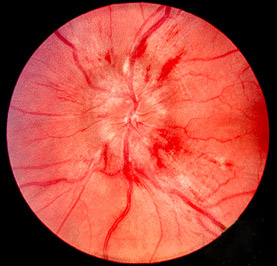
Papilledema: real disc, schematic disc
Left: actual fundus photo. Right: schematic so you can see what the textbook is pointing at. Tap the photo to enlarge.
Fundus photograph · papilledema
Swollen optic disc from raised intracranial pressure. The disc margins are obscured, the cup is filled in, and the surrounding nerve fiber layer looks hazy.
Wikimedia Commons · The Eyes Have It, Kellogg Eye Center · tap to enlarge
What you are looking for:
Disc margins blurred (you cannot trace the edge cleanly)
Optic cup obliterated (disc looks heaped up, not scooped)
Retinal veins engorged and tortuous
Flame-shaped hemorrhages at the disc margin
Bilateral. One-sided swelling is something else.
Treatment Ladder
Climb in order. Weight loss is the cure; everything else buys time or saves vision.
1
Cornerstone
Weight loss
Even 5 to 10 percent body weight reduction can resolve papilledema and headaches. This is not an adjunct, it is the actual fix. Bariatric surgery is on the table for severe disease. Drugs and procedures keep her vision safe while the weight comes off.
2
First-line drug
Acetazolamide
Carbonic anhydrase inhibitor. Blocks the enzyme the choroid plexus uses to pump bicarbonate (and water with it) into the ventricles. Less CSF made, less pressure. Side effects: paresthesias, metallic taste, kidney stones, metabolic acidosis. Topiramate is the backup (also CAI activity, plus appetite suppression which helps weight loss).
3
Stop the trigger
Discontinue offending drug
If she is on isotretinoin, tetracyclines, growth hormone, or high-dose vitamin A, stop it. Coordinate with the prescribing service. Symptoms often regress over weeks once the retinoid load drops.
4
Temporizing
Serial therapeutic LPs
Drain CSF down to a normal closing pressure (around 15 cm H2O). Each tap buys days to weeks of relief. Used while weight loss and acetazolamide take effect, or as a bridge to surgery if vision is at risk.
5
Save the optic nerve
Optic nerve sheath fenestration (ONSF)
Surgeon cuts a window in the dural sheath around the optic nerve so CSF can vent locally. Indicated when vision is failing (worsening visual fields, progressive papilledema, no response to medical therapy). Protects the nerve without addressing the headache.
6
Last resort
VP or LP shunt
Ventriculoperitoneal or lumboperitoneal shunt for refractory disease with headache as the dominant symptom. Addresses both pressure and headache, but carries shunt-failure risk over time. Venous sinus stenting is an emerging alternative when MRV shows a stenotic transverse sinus.
Vision Threatened: act now
IIH does not kill her, but it can blind her. Untreated chronic papilledema leads to optic atrophy and permanent vision loss. These three signs together change the conversation from clinic management to urgent ophthalmology.
🚨
Surgical referral triad
Worsening papilledema on serial fundoscopy + shrinking visual fields on perimetry + MRI showing optic nerve sheath distension or flattened posterior globe.
This is urgent ophthalmology and discussion of ONSF. Acetazolamide alone is not enough once the nerve is on the clock. Chronic papilledema leads to optic atrophy. Optic atrophy is permanent.
Test Yourself
Five clinical vignettes. Every wrong answer comes with a discriminator rule so you do not pick it twice.
Papilledema · fundus photographBilateral papilledema is the fundoscopic signature of raised intracranial pressure. Disc margins are obscured, the cup is filled in, and surrounding nerve fibers look hazy from edema.Wikimedia Commons · The Eyes Have It, Kellogg Eye Center, University of Michigan · CC BY 3.0
Medically reviewed by Kaitlyn Cocuzzo, MD and Fatima Ali, DO · Last updated June 30, 2026 at 2:05 AM ET
Bone Wizardry is an independent educational resource for visual learning in the medical sciences. It is not affiliated with, endorsed by, or sponsored by any licensing or examination board, contains no real or recalled examination questions, and does not guarantee any educational or examination outcome.