Radial Head: Rotation, Examination, and Safe Decisions
Understand radial-head rotation, interpret the examination, compare treatment principles, and distinguish pulled elbow from fractures and other elbow conditions.
Two patients cannot turn a palm upward. One has a reproducible rotation restriction without injury; the other has a swollen elbow after a fall. The motion complaint is similar, but the first decision is different. Learn the radial-head examination, then decide whether manipulation belongs in the plan at all.
How can the palm rotate while the elbow stays beside the body?
Imagine carrying a bowl and then turning a key. Elbow bending and forearm rotation are related but distinct functions. The humeroulnar articulation supplies the main hinge. The humeroradial articulation participates in bending and permits the radial head to rotate against the capitellum. At the proximal radioulnar articulation, the radial head pivots beside the radial notch of the ulna. These three articulations share the elbow capsule. [1]
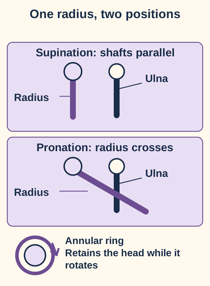
The proximal head rotates in a retaining ring while the distal radius changes its relationship to the ulna.
In anatomical position, the radius is on the thumb side and the ulna on the little-finger side. During pronation the distal radius crosses the ulna; during supination they become more nearly parallel. The ulna is relatively stable during an isolated forearm examination, not immobile during every real-world task. The annular ligament retains the radial head at the proximal radioulnar joint while allowing rotation. Do not confuse that ring with the collateral ligaments supporting the sides of the elbow. [1]
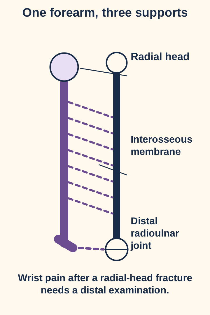
The radial collateral ligament blends with the annular ligament laterally. The ulnar collateral ligament supports the medial elbow, where throwing can create a different injury pattern. Between the forearm shafts, the interosseous membrane helps link the radius and ulna; distally, the distal radioulnar joint also participates in rotation. A proximal complaint therefore warrants a distal examination rather than an assumption that wrist pain is merely referred. [1][8]
Try the comparison. On the drawing, follow the radius from its proximal head to its distal end in both positions. Which end has the larger sweep? The distal end crosses around the ulna while the proximal head primarily rotates. Now imagine an injured retaining ring: rotation and stability can both be affected without a primary humeroulnar hinge problem.
The biceps brachii contributes to supination as well as elbow flexion. The supinator also turns the radius toward supination; pronator teres and pronator quadratus produce pronation. Muscles supply rotational force, while the annular ligament retains the proximal pivot. That distinction explains why an active resisted test and a passive glide test ask different questions. [9]
In a comfortable, uninjured arm, a fingertip just distal to the lateral epicondyle can feel the head rotate during gentle pronation and supination. This is an anatomy observation, not an invitation to press on a painful post-traumatic elbow. Clinicians compare landmarks, active and passive motion, tenderness, swelling, and distal neurovascular findings before assigning a somatic dysfunction. [2][4]
Name the examination pattern, not the accident
Does a fall on a palm-up hand establish an anterior radial head? No. A mechanism can suggest what to examine, but it does not establish either a somatic dysfunction or freedom from fracture. For the conventional OMM radial-head model, supination accompanies anterior glide and pronation accompanies posterior glide. The name describes the direction of relative ease; the restriction is the opposite glide and its associated rotation. This is a clinical teaching convention, not proof that the bone is dislocated. [2][4]
Translate a concordant examination in both directions
Pattern
Relative ease
Restricted direction
PatternAnterior radial head
Relative easeAnterior glide; supination
Restricted directionPosterior glide; pronation
PatternPosterior radial head
Relative easePosterior glide; pronation
Restricted directionAnterior glide; supination
Test the directional model
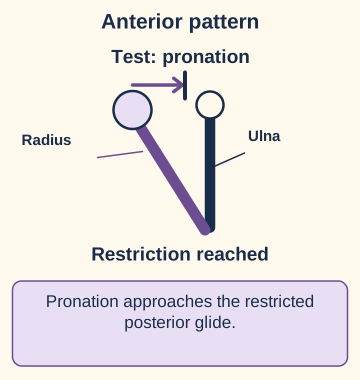
Choose a recorded pattern and a rotation to test. Compare the resulting forearm geometry with the permitted glide. This simplified model does not simulate tissue forces or a reduction.
Anterior pattern plus pronation: the tested rotation approaches the restricted posterior glide.
Pronation is the restricted direction. A direct treatment would first approach this barrier gently, not force beyond it.
Static worked example: Restricted supination plus restricted anterior glide supports a posterior pattern. Testing pronation explores its relative ease. The table above supplies all four comparisons when the interactive model is unavailable.
Separate a true directional end-feel from protective guarding. A patient may stop active rotation because it hurts even when passive excursion is relatively preserved. Painful limitation in both directions can accompany effusion, synovitis, fracture, or guarding; it does not by itself establish either named radial-head pattern. A prominent-looking head without matching motion findings is insufficient. If the recorded label and repeat examination disagree, resolve the disagreement before selecting a technique. [2][4]
Apply it elsewhere. A left forearm supinates easily but has restricted pronation and posterior glide. First identify the deficient glide; then name its opposite ease. The result is anterior, exactly as on the right. Changing sides does not reverse anterior and posterior. Changing the current examination can reverse the diagnosis, regardless of the remembered injury position.
Try it here · Checkpoint 1 of 3
Make your prediction before reading the choices. A first attempt is just a starting point.
Case 5
Show answer and explanations for case 5
A. Use a supination barrier based on the recorded positional name (Why this does not fit)
A confirmed posterior pattern would justify a supination barrier. No. The label rests on prominence while the motion description and guarding undermine it. Resolve an uncertain diagnosis before applying its treatment rule.
Reasoning steps for option A
What finding would support this option?
A confirmed posterior pattern would justify a supination barrier.
Was that pattern established by concordant findings?
No. The label rests on prominence while the motion description and guarding undermine it.
What does that distinction imply here?
Resolve an uncertain diagnosis before applying its treatment rule.
B. Repeat a relaxed comparative rotation and glide examination before choosing a setup (Best answer)
A reproducible paired examination is needed to select a directional technique. Guarding and disagreement between the prominence-based label and rotation findings prevent it. Re-examination is the appropriate response to an unreliable record.
Reasoning steps for option B
Do the observations support one consistent directional pattern?
No. The positional label and motion description conflict.
What limits the reliability of the motion assessment?
Forearm guarding interferes with translation testing.
What resolves the treatment uncertainty?
A relaxed comparative examination before assigning the setup.
What principle transfers to the next patient?
When prominence, rotation, and glide disagree, repeat the examination rather than choosing whichever datum fits a memorized treatment.
C. Use a pronation barrier based only on the painful active motion (Why this does not fit)
Restricted pronation can support an anterior pattern when passive glide findings agree. No. The patient is tensing and translation has not been characterized reliably. Do not replace one unsupported label with another based on a single finding.
Reasoning steps for option C
What finding would support this option?
Restricted pronation can support an anterior pattern when passive glide findings agree.
Does painful active motion alone establish that articular restriction?
No. The patient is tensing and translation has not been characterized reliably.
What does that distinction imply here?
Do not replace one unsupported label with another based on a single finding.
D. Treat both directions sequentially because the findings establish two dysfunctions (Why this does not fit)
Several somatic findings can coexist in one region. No. The examination is inconsistent, not demonstrably bidirectionally restricted. A contradictory chart is a reason to clarify, not to prescribe opposing techniques.
Reasoning steps for option D
What finding would support this option?
Several somatic findings can coexist in one region.
Are independent restrictions in both directions documented reliably?
No. The examination is inconsistent, not demonstrably bidirectionally restricted.
What does that distinction imply here?
A contradictory chart is a reason to clarify, not to prescribe opposing techniques.
Takeaway: When prominence, rotation, and glide disagree, repeat the examination rather than choosing whichever datum fits a memorized treatment.
Separate clinician positioning from patient effort
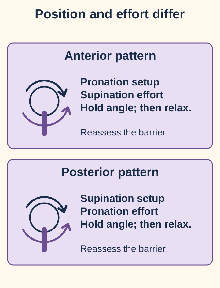
Why does the patient try to supinate when the clinician is treating restricted pronation? In the post-isometric muscle-energy variant taught here, the clinician first approaches the restricted direction. The patient then contracts gently toward relative ease while the clinician prevents rotation. After relaxation, the clinician takes up any newly available range. The setup, contraction, and reassessment are different events. Consent, a safe structural examination, and the ability to cooperate come first. [2]
The patient's effort opposes the setup direction; balanced resistance keeps the contraction isometric. Original treatment-principle diagram.
For an anterior pattern, the setup is pronation and the patient's effort is supination. For a posterior pattern, reverse those directions. A commonly taught example uses brief contractions of about 3 to 5 seconds and several repetitions, with reassessment after each relaxation. These are technique conventions, not a mandatory dose or a promise of correction. Stop for new pain, neurologic symptoms, or an unexpected end-feel. Do not replace a gentle barrier with a painful forced endpoint. [2]
Predict the consequence. During a posterior-pattern treatment, the forearm visibly rotates farther into pronation during the contraction. Is that the described isometric phase? No. The contraction should be balanced by resistance so the joint angle stays essentially unchanged. After relaxation, the clinician reassesses available supination; the patient does not twist farther through a painful limit.
High-velocity, low-amplitude treatment uses a brief clinician-applied thrust rather than a patient contraction. In a direct approach, the intended corrective direction is toward the restricted glide: posterior for an anterior pattern, anterior for a posterior pattern. That principle is not a universal thumb-contact recipe. Position, stabilization, contraindications, and supervised procedural training determine whether a particular method is appropriate. A thrust is not a substitute for fracture reduction or an acceptable way to overcome unexplained resistance. [2][4]
Counterstrain instead supports a point-specific position of comfort. A traditional target is at least 70% less tenderness, followed by a roughly 90-second passive hold, slow return, and reassessment. Those timings do not prove a particular physiologic mechanism. Wong describes a supinator point near the anterior radial head, often relieved with comfortable extension and supination, and a pronator point on the anteromedial proximal forearm, often relieved with flexion and pronation. Position is adjusted to actual tenderness, never forced. These point names are not interchangeable with articular anterior/posterior diagnoses. [7]
Compare participation rather than memorizing a hierarchy: muscle energy requires a voluntary contraction; counterstrain requires tolerable passive positioning and useful patient feedback; thrust techniques add a rapid impulse. None is automatically appropriate because another is unsuitable. In particular, a healing fracture or unstable joint does not become safe to manipulate merely by selecting a gentler label. Reassess symptoms, function, motion, and safety after any appropriate treatment; partial relief does not establish complete recovery. [2][4][7]
Try it here · Checkpoint 2 of 3
Make your prediction before reading the choices. A first attempt is just a starting point.
Case 20
Show answer and explanations for case 20
A. Ask for repeated brief contractions because the pain reduction was only 25% (Why this does not fit)
Brief voluntary contractions are characteristic of muscle energy rather than counterstrain. The reduction is 6 divided by 8, or 75%, not 25%. Calculate the change correctly and retain the selected passive method.
Reasoning steps for option A
What finding would support this option?
Brief voluntary contractions are characteristic of muscle energy rather than counterstrain.
What is the actual percentage reduction from 8 to 2?
The reduction is 6 divided by 8, or 75%, not 25%.
What does that distinction imply here?
Calculate the change correctly and retain the selected passive method.
B. Maintain the comfortable passive position, then return slowly and reassess (Best answer)
A reduction from 8 to 2 meets the conventional target of at least 70% less tenderness. A conventional passive hold of about 90 seconds is followed by slow return and reassessment. A conventional positioning target is not a guarantee of clinical efficacy.
Reasoning steps for option B
What proportion of the original tenderness remains?
Two divided by eight is 25%.
What proportion has decreased?
Tenderness has decreased by 75%.
Which action matches the selected method?
Maintain the tolerable passive position, then return slowly and reassess.
What principle transfers to the next patient?
For conventional counterstrain, a 75% tenderness reduction can support a comfortable passive hold followed by slow return and reassessment.
C. Advance to the painful endpoint because tenderness has fallen by 75% (Why this does not fit)
Reduced tenderness helps identify a tolerable position of comfort. No. Counterstrain maintains the comfortable position rather than advancing into pain. Do not convert a passive comfort technique into a forced stretch.
Reasoning steps for option C
What finding would support this option?
Reduced tenderness helps identify a tolerable position of comfort.
Does that authorize forcing the restrictive endpoint?
No. Counterstrain maintains the comfortable position rather than advancing into pain.
What does that distinction imply here?
Do not convert a passive comfort technique into a forced stretch.
D. Abandon this position because conventional counterstrain requires complete absence of tenderness (Why this does not fit)
Rechecking comfort during positioning is important. No. A 75% reduction meets the commonly described threshold. Use the specified response criterion without inventing a stricter universal requirement.
Reasoning steps for option D
What finding would support this option?
Rechecking comfort during positioning is important.
Does the conventional target require tenderness to become zero?
No. A 75% reduction meets the commonly described threshold.
What does that distinction imply here?
Use the specified response criterion without inventing a stricter universal requirement.
Takeaway: For conventional counterstrain, a 75% tenderness reduction can support a comfortable passive hold followed by slow return and reassessment.
A toddler who stops using an arm needs a different framework
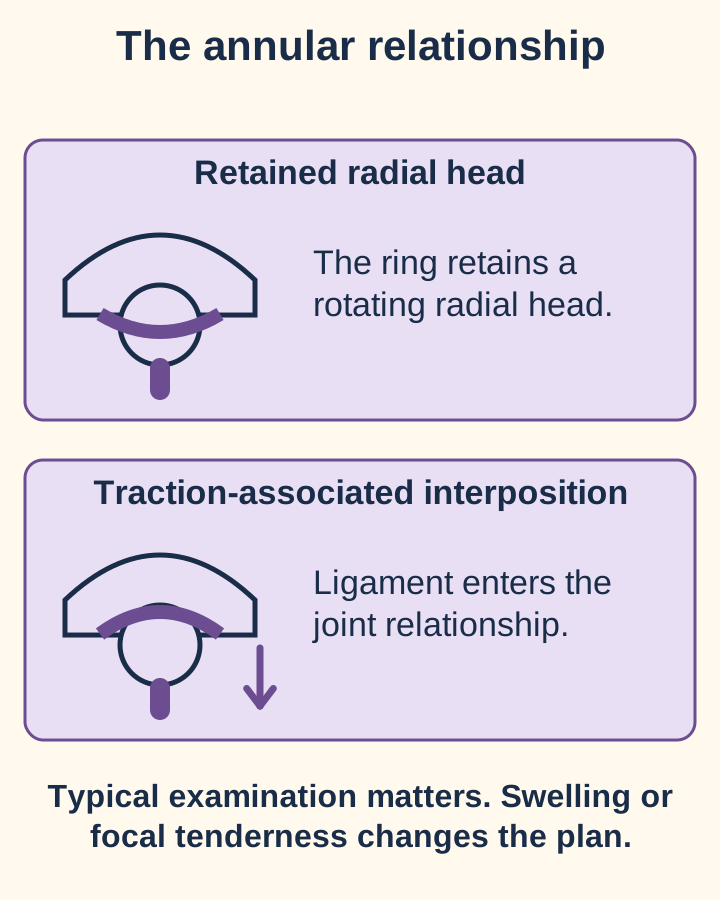
A two-year-old cries after being lifted by one hand and then holds that forearm pronated. Is this simply the adult posterior-pattern model? No. Pulled elbow, also called nursemaid's elbow or radial-head subluxation, is a pediatric annular-ligament injury. Traction can disturb the ligament's relationship to the radial head and allow interposition. It is a structural reduction problem, not an invitation to apply an adult OMT naming rule. [3][5]
Ligament interposition explains why a traction history and refusal to use the arm can fit pulled elbow. The injury drawing is simplified, not an image of a specific child.
The usual age is 1 to 4 years. The arm is often held extended or slightly flexed with the forearm pronated. A classic examination lacks substantial swelling, bruising, deformity, and focal bony tenderness. Absence of a witnessed pull does not exclude the diagnosis. Examine the whole limb, including the clavicle and wrist, before deciding that an apparently painful elbow is the source. [3]
Compare before treating: one child has the classic posture without swelling; another has the same posture after a fall but also has distal humeral tenderness and bruising. The second child's focal findings change the action. Imaging and injury assessment take priority. A classic presentation can be diagnosed clinically without routine radiographs, but atypical findings or failed reduction warrant reassessment and imaging as indicated. [3]
A trained clinician may use hyperpronation or supination followed by elbow flexion after assessment, explanation, and appropriate analgesia. Hyperpronation may succeed more often on the first attempt, but the cited review judged that evidence low certainty and could not establish a pain advantage. Neither technique should become repeated force against unexpected resistance. Reduction of a suspected fracture-dislocation is a separate problem. [3][6]
A palpable click is neither required nor sufficient to demonstrate success. Observe return of comfortable spontaneous function, such as reaching for a toy. This commonly occurs within 5 to 30 minutes. Persistent non-use requires a fresh examination; after an unsuccessful attempt, seek experienced assistance and consider an alternative technique only when the diagnosis remains convincing. Repeated failure calls for imaging and escalation, not increasingly forceful repetition. New focal tenderness justifies imaging earlier. [3]
Test the discharge decision. A child now reaches and plays normally after reduction. Routine immobilization is generally unnecessary for that uncomplicated recovery. Teach caregivers to lift under the arms and avoid pulling or swinging by the hands. In a presentation delayed more than 12 hours, arm use may return more slowly even after a satisfactory reduction; RCH describes temporary support and planned review in selected cases. Do not use that exception to dismiss new swelling, worsening pain, or an uncertain diagnosis. [3][5]
Try it here · Checkpoint 3 of 3
Make your prediction before reading the choices. A first attempt is just a starting point.
Case 25
Show answer and explanations for case 25
A. Repeat the previous reduction because the posture is similar (Why this does not fit)
A prior pulled elbow can recur in a susceptible child. No. A fall with bruising, swelling, and distal humeral tenderness raises concern for fracture. A familiar previous diagnosis should not override a different current examination.
Reasoning steps for option A
What finding would support this option?
A prior pulled elbow can recur in a susceptible child.
Does recurrence explain away the new focal injury findings?
No. A fall with bruising, swelling, and distal humeral tenderness raises concern for fracture.
What does that distinction imply here?
A familiar previous diagnosis should not override a different current examination.
B. Try both reduction methods before considering radiographs (Why this does not fit)
An alternative reduction may be appropriate after an unsuccessful attempt in a still-typical presentation. No. The focal bony and visible injury findings justify imaging before attempting reduction. Imaging is not reserved for failure of two maneuvers.
Reasoning steps for option B
What finding would support this option?
An alternative reduction may be appropriate after an unsuccessful attempt in a still-typical presentation.
Is this a typical presentation suitable for that sequence?
No. The focal bony and visible injury findings justify imaging before attempting reduction.
What does that distinction imply here?
Imaging is not reserved for failure of two maneuvers.
C. Use comfortable passive positioning as treatment for a recurrent radial-head pattern (Why this does not fit)
Comfortable support can help protect a painful limb during assessment. No. The examination requires fracture assessment rather than a positional diagnosis. Use comfort as supportive care, not as a substitute for injury evaluation.
Reasoning steps for option C
What finding would support this option?
Comfortable support can help protect a painful limb during assessment.
Would treating a presumed somatic pattern address the suspected injury?
No. The examination requires fracture assessment rather than a positional diagnosis.
What does that distinction imply here?
Use comfort as supportive care, not as a substitute for injury evaluation.
D. Provide analgesia and assess the injury with indicated radiographs before reduction (Best answer)
Focal bony tenderness and visible injury findings after a fall warrant fracture assessment. No. It does not exclude a distal humeral injury. Analgesia and appropriate imaging take priority over repeating a familiar reduction.
Reasoning steps for option D
Which features differ from a typical uncomplicated pulled elbow?
The fall, bruising, swelling, and focal distal humeral tenderness differ.
What alternative must be assessed?
A fracture or other structural injury must be assessed.
When should that assessment occur?
Before attempting the presumed pulled-elbow reduction.
What principle transfers to the next patient?
Atypical injury findings justify imaging before a pulled-elbow reduction; a prior successful reduction does not establish the present diagnosis.
Takeaway: Atypical injury findings justify imaging before a pulled-elbow reduction; a prior successful reduction does not establish the present diagnosis.
When does an elbow complaint stop being a manipulation problem?
A patient falls, cannot rotate the forearm comfortably, and has focal radial-head tenderness. A normal initial radiograph does not make manipulation the next diagnostic test: a nondisplaced fracture may be occult. Protect the limb and arrange appropriate injury reassessment and imaging rather than forcing rotation. Severe pain, deformity, suspected infection, instability, or a new neurovascular deficit also changes the priority. A warm, perfused hand does not cancel the significance of new motor or sensory findings. [4]
Fracture management depends on displacement, stability, mechanical obstruction, and associated injury. A stable, isolated nondisplaced fracture often permits early controlled motion after brief support under a fracture-care plan. This is different from forceful manual treatment. A displaced or comminuted fracture with a mechanical block needs orthopaedic assessment; fixation, replacement, or selected fragment treatment depends on the injury rather than a single fragment-count rule. Persistent stiffness does not justify attempting to push a fragment out of the way. [4]
Examine the full forearm after radial-head trauma. Distal symptoms can identify an associated injury rather than a second benign complaint.
After a radial-head fracture, wrist pain or distal radioulnar instability raises concern for longitudinal forearm injury, including an Essex-Lopresti pattern involving the interosseous membrane and distal radioulnar joint. Assess the wrist and forearm, and obtain specialist-directed imaging. With longitudinal instability, simply excising the radial head can worsen proximal migration of the radius; restoring the proximal support and addressing distal instability matter together. [8]
At the lateral epicondyle
Localized pain with gripping or resisted wrist extension favors lateral elbow tendinopathy when the rest of the examination fits. It is not identical to a restricted proximal radial glide.
At the medial elbow
Throwing pain with valgus laxity suggests a collateral-ligament problem. A radial-head label does not explain objective medial instability.
Over the olecranon
A superficial posterior swelling suggests bursitis. Redness, warmth, a wound, or systemic illness requires assessment for infection rather than routine manual treatment.
Throughout the joint
Effusion, prolonged stiffness, and restriction in several planes suggest an articular process. Rheumatoid synovitis and erosions are inflammatory disease, not merely a positional dysfunction. [10]
These are localization patterns, not stand-alone diagnostic tests. Check the history and the entire examination; more than one condition can coexist. [1][2][4]
Transfer the safety rule. After treatment, an adult reports less elbow pain but still has a hard rotational block and wrist tenderness. The pain score improved, but the unexplained structural findings remain. Reassess the diagnosis and associated injury instead of declaring success or automatically scheduling the same technique again. A safe plan follows the patient's findings, not a fixed number of repetitions or a universal return interval.
Apply the lesson
Case 1
Show answer and explanations for case 1
A. Anterior radial head; supination barrier (Why this does not fit)
An anterior pattern is supported by restricted posterior glide. Pronation, not supination, has the reduced excursion. Name the ease, but set up a direct technique toward the restriction.
Reasoning steps for option A
What finding would support this option?
An anterior pattern is supported by restricted posterior glide.
Which rotation is actually restricted?
Pronation, not supination, has the reduced excursion.
What does that distinction imply here?
Name the ease, but set up a direct technique toward the restriction.
B. Anterior radial head; pronation barrier (Best answer)
Restricted posterior glide with reduced pronation supports an anterior pattern. The clinician gently approaches pronation, the deficient rotation. The diagnostic name and the setup direction describe opposite sides of the same restriction.
Reasoning steps for option B
Which paired motion is deficient?
Posterior glide and pronation are deficient.
Which relative ease gives the diagnosis?
Anterior glide and supination correspond to an anterior pattern.
Where does direct positioning begin?
At the gentle pronation barrier, not at the supination ease.
What principle transfers to the next patient?
Use the paired rotation and glide findings to name relative ease, then approach the opposite restriction for direct treatment.
C. Posterior radial head; supination barrier (Why this does not fit)
A posterior pattern would restrict anterior glide and supination. Supination is preserved and posterior glide is restricted instead. Do not name a dysfunction for the direction it cannot comfortably reach.
Reasoning steps for option C
What finding would support this option?
A posterior pattern would restrict anterior glide and supination.
Does the measured supination deficit support that pattern?
Supination is preserved and posterior glide is restricted instead.
What does that distinction imply here?
Do not name a dysfunction for the direction it cannot comfortably reach.
D. Posterior radial head; pronation barrier (Why this does not fit)
Pronation is the appropriate barrier for the supplied rotation deficit. No. Restricted posterior glide corresponds to relative anterior ease. A correct setup cannot rescue an incorrect diagnosis.
Reasoning steps for option D
What finding would support this option?
Pronation is the appropriate barrier for the supplied rotation deficit.
Does a posterior name match restricted posterior glide?
No. Restricted posterior glide corresponds to relative anterior ease.
What does that distinction imply here?
A correct setup cannot rescue an incorrect diagnosis.
Takeaway: Use the paired rotation and glide findings to name relative ease, then approach the opposite restriction for direct treatment.
A. Pronation setup; effort toward supination (Why this does not fit)
A pronation barrier with supination effort treats the opposite directional pattern. Posterior glide would be restricted, but this patient lacks anterior glide. Reverse both directions when the examination changes from anterior to posterior pattern.
Reasoning steps for option A
What finding would support this option?
A pronation barrier with supination effort treats the opposite directional pattern.
Which glide would need to be restricted for this pairing?
Posterior glide would be restricted, but this patient lacks anterior glide.
What does that distinction imply here?
Reverse both directions when the examination changes from anterior to posterior pattern.
B. Supination setup; effort toward supination (Why this does not fit)
Supination is the appropriate setup for this restriction. Effort is toward relative ease, which is pronation here. Do not confuse the direction held by the clinician with the direction attempted by the patient.
Reasoning steps for option B
What finding would support this option?
Supination is the appropriate setup for this restriction.
In the specified post-isometric variant, which direction should the effort target?
Effort is toward relative ease, which is pronation here.
What does that distinction imply here?
Do not confuse the direction held by the clinician with the direction attempted by the patient.
C. Pronation setup; effort toward pronation (Why this does not fit)
Pronation is the patient's relative ease in this examination. No. The deficient direction is supination. An appropriate effort still requires the correct initial barrier.
Reasoning steps for option C
What finding would support this option?
Pronation is the patient's relative ease in this examination.
Does setting up in that ease engage the direct barrier?
No. The deficient direction is supination.
What does that distinction imply here?
An appropriate effort still requires the correct initial barrier.
D. Supination setup; effort toward pronation (Best answer)
Restricted anterior glide and supination indicate a posterior pattern. The patient attempts pronation against matched resistance while the angle remains stable. Relaxation precedes reassessment of any newly available supination.
Reasoning steps for option D
What articular pattern fits the measurements?
A posterior pattern fits restricted anterior glide and supination.
Which direction engages its barrier?
The clinician approaches supination.
Which effort opposes that position?
The patient attempts pronation without actual rotation.
What principle transfers to the next patient?
For a posterior radial-head pattern, this muscle-energy variant pairs a supination barrier with isometric pronation.
Takeaway: For a posterior radial-head pattern, this muscle-energy variant pairs a supination barrier with isometric pronation.
A. Posterior pattern based on the current concordant examination (Best answer)
Concordant current rotation and glide findings support a directional diagnosis. Posterior glide and pronation are the relative ease. Use the present examination after appropriate injury assessment, not a deterministic mechanism rule.
Reasoning steps for option A
Is an unevaluated acute injury driving this decision?
No. Injury assessment is complete and current red flags are absent.
Which current findings agree?
Restricted supination and anterior glide both support posterior ease.
Which record should guide positioning?
The corrected current posterior-pattern examination should guide it.
What principle transfers to the next patient?
Mechanism suggests possibilities; reproducible current findings determine the directional examination diagnosis.
B. Anterior pattern based on the original loading direction (Why this does not fit)
Forced supination can guide what to examine after an injury. No. The current paired findings support posterior ease. A loading history does not establish the persistent articular diagnosis.
Reasoning steps for option B
What finding would support this option?
Forced supination can guide what to examine after an injury.
Does that history override the current anterior-glide restriction?
No. The current paired findings support posterior ease.
What does that distinction imply here?
A loading history does not establish the persistent articular diagnosis.
C. Combined anterior and posterior patterns based on both descriptions (Why this does not fit)
Multiple limitations may coexist and warrant a broader examination. No. Only the posterior-pattern examination is reproducible now. Conflicting historical labels are not proof of two simultaneous positional diagnoses.
Reasoning steps for option C
What finding would support this option?
Multiple limitations may coexist and warrant a broader examination.
Are two independent, current opposite restrictions demonstrated?
No. Only the posterior-pattern examination is reproducible now.
What does that distinction imply here?
Conflicting historical labels are not proof of two simultaneous positional diagnoses.
D. No directional pattern because the mechanism and examination differ (Why this does not fit)
An internally inconsistent examination would require clarification. They agree with each other; only the historical label differs. Do not discard a concordant current examination merely because an old inference differs.
Reasoning steps for option D
What finding would support this option?
An internally inconsistent examination would require clarification.
Are the current rotation and glide results internally inconsistent?
They agree with each other; only the historical label differs.
What does that distinction imply here?
Do not discard a concordant current examination merely because an old inference differs.
Takeaway: Mechanism suggests possibilities; reproducible current findings determine the directional examination diagnosis.
A. Reduced anterior glide with a new posterior pattern (Why this does not fit)
A new posterior pattern would be accompanied by restricted supination. No. It remains 85 degrees while pronation improves. Improvement in one restriction does not imply conversion to its opposite.
Reasoning steps for option A
What finding would support this option?
A new posterior pattern would be accompanied by restricted supination.
Did supination become limited after treatment?
No. It remains 85 degrees while pronation improves.
What does that distinction imply here?
Improvement in one restriction does not imply conversion to its opposite.
B. Symmetric glide with proof of complete recovery (Why this does not fit)
Symmetric motion and function could support recovery on reassessment. Pronation remains below the opposite arm, and repeat glide has not yet been measured. Do not infer complete correction or structural safety from partial range improvement.
Reasoning steps for option B
What finding would support this option?
Symmetric motion and function could support recovery on reassessment.
Do the supplied measurements establish symmetry?
Pronation remains below the opposite arm, and repeat glide has not yet been measured.
What does that distinction imply here?
Do not infer complete correction or structural safety from partial range improvement.
C. Greater posterior glide with possible residual anterior-pattern restriction (Best answer)
Pronation is coupled to posterior glide in this examination model. Posterior glide may improve while some anterior-pattern restriction persists. Confirm the predicted palpatory change rather than treating it as an unmeasured fact.
Reasoning steps for option C
Which motion improved?
Pronation increased by 25 degrees.
Which coupled glide would be expected to improve?
Posterior glide would be expected to improve.
What remains uncertain?
Residual restriction and functional recovery still require reassessment.
What principle transfers to the next patient?
A treatment response supports a direction-specific prediction; it does not replace repeat examination or prove full recovery.
D. Greater anterior glide with persistent posterior-pattern restriction (Why this does not fit)
Improved anterior glide would more directly correspond to improved supination. Pronation increased; supination was unchanged. Predict the glide linked to the measured change, not the glide named by the diagnosis.
Reasoning steps for option D
What finding would support this option?
Improved anterior glide would more directly correspond to improved supination.
Which rotation actually changed?
Pronation increased; supination was unchanged.
What does that distinction imply here?
Predict the glide linked to the measured change, not the glide named by the diagnosis.
Takeaway: A treatment response supports a direction-specific prediction; it does not replace repeat examination or prove full recovery.
A. Isolated anterior radial-head dysfunction producing a pronation deficit (Why this does not fit)
An anterior pattern can reduce pronation with posterior-glide restriction. It does not account for the effusion, erosions, and limitation across several planes. A directional finding does not replace assessment of inflammatory joint disease.
Reasoning steps for option A
What finding would support this option?
An anterior pattern can reduce pronation with posterior-glide restriction.
Does that single pattern explain the other supplied abnormalities?
It does not account for the effusion, erosions, and limitation across several planes.
What does that distinction imply here?
A directional finding does not replace assessment of inflammatory joint disease.
B. Isolated posterior radial-head dysfunction producing a supination deficit (Why this does not fit)
A posterior pattern can reduce supination with anterior-glide restriction. Both rotation directions and hinge motion are affected in an erosive, swollen joint. Integrate the distribution of restriction with the articular findings.
Reasoning steps for option B
What finding would support this option?
A posterior pattern can reduce supination with anterior-glide restriction.
Why is that insufficient as the overall explanation?
Both rotation directions and hinge motion are affected in an erosive, swollen joint.
What does that distinction imply here?
Integrate the distribution of restriction with the articular findings.
C. Olecranon bursitis mechanically blocking the entire joint (Why this does not fit)
Olecranon bursitis can produce a posterior superficial swelling. No. The findings are intra-articular, with erosions and effusion rather than a focal bursal swelling. Localize the pathology before attributing global stiffness to a superficial structure.
Reasoning steps for option C
What finding would support this option?
Olecranon bursitis can produce a posterior superficial swelling.
Is the abnormality localized to that bursa?
No. The findings are intra-articular, with erosions and effusion rather than a focal bursal swelling.
What does that distinction imply here?
Localize the pathology before attributing global stiffness to a superficial structure.
D. Inflammatory articular disease producing multiaxial restriction (Best answer)
Synovitis and erosive rheumatoid disease can restrict several elbow motions. Effusion, marginal erosions, and multiaxial limitation support an articular process. Assess the inflammatory joint rather than forcing a directional manipulation.
Reasoning steps for option D
Where is the pathology localized?
Effusion and erosions localize it to the joint.
Does one radial-glide direction explain the entire examination?
No. Several independent planes are limited.
What should guide the evaluation?
Inflammatory articular disease should guide it.
What principle transfers to the next patient?
Multiaxial restriction with effusion or erosions requires an articular differential; rheumatoid disease is not simply a positional or degenerative label.
Takeaway: Multiaxial restriction with effusion or erosions requires an articular differential; rheumatoid disease is not simply a positional or degenerative label.
A. Protect the elbow and arrange reassessment for an occult fracture or associated injury (Best answer)
A nondisplaced radial-head fracture may not be visible on initial radiographs. No. Trauma, swelling, focal bony tenderness, and painful rotation sustain that concern. Protect and reassess a suspected injury instead of using manipulation as a diagnostic test.
Reasoning steps for option A
What structural injury remains plausible?
An occult nondisplaced radial-head fracture remains plausible.
Why does the distal examination not settle that question?
Normal perfusion does not assess proximal bone integrity.
Which action protects against the unresolved risk?
Support the limb and arrange appropriate injury reassessment.
What principle transfers to the next patient?
Persistent clinical suspicion after trauma outweighs a nondiagnostic first radiograph when deciding whether to manipulate.
B. Use muscle energy now because the initial radiographs show no definite fracture (Why this does not fit)
Muscle energy can be considered for an appropriately assessed somatic dysfunction. No. The clinical suspicion remains high despite nondiagnostic films. A negative initial radiograph is not clearance for local manipulation.
Reasoning steps for option B
What finding would support this option?
Muscle energy can be considered for an appropriately assessed somatic dysfunction.
Has a fracture been adequately excluded by the first study alone?
No. The clinical suspicion remains high despite nondiagnostic films.
What does that distinction imply here?
A negative initial radiograph is not clearance for local manipulation.
C. Use counterstrain now because it does not require a thrust (Why this does not fit)
Counterstrain avoids a rapid thrust and uses a comfortable position. No. The same unresolved structural concern remains. Changing technique labels does not remove an injury contraindication.
Reasoning steps for option C
What finding would support this option?
Counterstrain avoids a rapid thrust and uses a comfortable position.
Does its gentler classification establish safety over a suspected fracture?
No. The same unresolved structural concern remains.
What does that distinction imply here?
Changing technique labels does not remove an injury contraindication.
D. Reassure and resume normal loading because distal perfusion is preserved (Why this does not fit)
Normal distal perfusion is reassuring about one component of the examination. No. A radial-head fracture can occur with intact pulses and sensation. Assess bone and joint injury independently of distal vascular status.
Reasoning steps for option D
What finding would support this option?
Normal distal perfusion is reassuring about one component of the examination.
Does it rule out a painful proximal fracture?
No. A radial-head fracture can occur with intact pulses and sensation.
What does that distinction imply here?
Assess bone and joint injury independently of distal vascular status.
Takeaway: Persistent clinical suspicion after trauma outweighs a nondiagnostic first radiograph when deciding whether to manipulate.
A. Common extensor tendon injury at the lateral epicondyle (Why this does not fit)
A common extensor tendon injury can cause lateral elbow pain with loading. No. It does not adequately explain objective distal radioulnar instability after a proximal radial fracture. Assess the connected forearm stabilizers when symptoms extend beyond the elbow.
Reasoning steps for option A
What finding would support this option?
A common extensor tendon injury can cause lateral elbow pain with loading.
Does it account for the distal translation and forearm findings?
No. It does not adequately explain objective distal radioulnar instability after a proximal radial fracture.
What does that distinction imply here?
Assess the connected forearm stabilizers when symptoms extend beyond the elbow.
B. Isolated annular ligament laxity at the proximal radioulnar joint (Why this does not fit)
The annular ligament stabilizes the proximal radial head. No. Abnormal translation is measured distally, with tenderness along the forearm. An isolated proximal explanation is insufficient for a longitudinal injury pattern.
Reasoning steps for option B
What finding would support this option?
The annular ligament stabilizes the proximal radial head.
Is the demonstrated instability confined to that proximal articulation?
No. Abnormal translation is measured distally, with tenderness along the forearm.
What does that distinction imply here?
An isolated proximal explanation is insufficient for a longitudinal injury pattern.
C. Interosseous membrane injury with distal radioulnar instability (Best answer)
The radial head, interosseous membrane, and distal radioulnar joint cooperate in forearm stability. The proximal fracture with distal instability raises concern for an Essex-Lopresti injury pattern. Evaluate the entire forearm and obtain specialist assessment rather than assuming referred wrist pain.
Reasoning steps for option C
What finding prevents an isolated-elbow interpretation?
Objective distal radioulnar instability accompanies the proximal fracture.
Which intervening structure may also be injured?
The interosseous membrane may be disrupted.
What broader pattern requires assessment?
An Essex-Lopresti pattern requires whole-forearm specialist assessment.
What principle transfers to the next patient?
Wrist symptoms with distal radioulnar instability after a radial-head fracture require assessment for longitudinal forearm injury.
D. Isolated olecranon bursal inflammation (Why this does not fit)
Olecranon bursitis can explain a superficial posterior elbow swelling. No. The supplied abnormality is a linked proximal and distal forearm injury. Do not substitute a superficial pain source for demonstrated instability.
Reasoning steps for option D
What finding would support this option?
Olecranon bursitis can explain a superficial posterior elbow swelling.
Would that local process explain distal radioulnar translation?
No. The supplied abnormality is a linked proximal and distal forearm injury.
What does that distinction imply here?
Do not substitute a superficial pain source for demonstrated instability.
Takeaway: Wrist symptoms with distal radioulnar instability after a radial-head fracture require assessment for longitudinal forearm injury.
A. Loss of proximal radial support could worsen longitudinal instability (Best answer)
The radial head is an important proximal support in a longitudinally unstable forearm. The disrupted interosseous membrane and unstable distal radioulnar joint cannot provide normal longitudinal support. Reconstruct or replace proximal support as appropriate and address distal instability together.
Reasoning steps for option A
Which stabilizing system is deficient?
Longitudinal forearm stability is deficient at several levels.
What additional support would excision sacrifice?
It would sacrifice the proximal radial support.
How does that change planning?
Proximal reconstruction or replacement and distal stabilization require coordinated consideration.
What principle transfers to the next patient?
In an Essex-Lopresti injury, radial-head treatment must preserve or restore longitudinal support rather than relying on isolated excision.
B. Excision would selectively prevent humeroulnar flexion despite stable forearm length (Why this does not fit)
Elbow procedures can affect motion and require careful planning. No. The supplied associated injuries specifically threaten longitudinal forearm stability. Match the predicted consequence to the structures demonstrated to be deficient.
Reasoning steps for option B
What finding would support this option?
Elbow procedures can affect motion and require careful planning.
Is an isolated hinge restriction the main consequence identified by these injuries?
No. The supplied associated injuries specifically threaten longitudinal forearm stability.
What does that distinction imply here?
Match the predicted consequence to the structures demonstrated to be deficient.
C. Excision would correct the distal instability but leave a tendon pain generator (Why this does not fit)
Pain generators can coexist after a complex injury. No. It can worsen the longitudinal problem rather than correcting the distal instability. Do not assume a proximal excision repairs an injured interosseous membrane or distal joint.
Reasoning steps for option C
What finding would support this option?
Pain generators can coexist after a complex injury.
Would proximal excision restore the disrupted distal stabilizers?
No. It can worsen the longitudinal problem rather than correcting the distal instability.
What does that distinction imply here?
Do not assume a proximal excision repairs an injured interosseous membrane or distal joint.
D. Loss of the radial head would primarily create a pediatric-type annular subluxation (Why this does not fit)
Pulled elbow involves an annular ligament problem in a young child. No. The concern is loss of longitudinal radial support after a complex fracture. Distinguish pediatric annular interposition from adult longitudinal instability.
Reasoning steps for option D
What finding would support this option?
Pulled elbow involves an annular ligament problem in a young child.
Does that explain the principal risk in this adult injury?
No. The concern is loss of longitudinal radial support after a complex fracture.
What does that distinction imply here?
Distinguish pediatric annular interposition from adult longitudinal instability.
Takeaway: In an Essex-Lopresti injury, radial-head treatment must preserve or restore longitudinal support rather than relying on isolated excision.
A. Rigid immobilization for six weeks before any elbow motion (Why this does not fit)
Immobilization can protect selected fractures during early care. The supplied fracture pattern supports early controlled motion rather than prolonged routine immobilization. Protect healing tissue while avoiding unnecessary elbow stiffness.
Reasoning steps for option A
What finding would support this option?
Immobilization can protect selected fractures during early care.
Does this stable, unblocked injury require six weeks without motion?
The supplied fracture pattern supports early controlled motion rather than prolonged routine immobilization.
What does that distinction imply here?
Protect healing tissue while avoiding unnecessary elbow stiffness.
B. Forceful end-range stretching to regain full rotation immediately (Why this does not fit)
Restoring motion is an important part of elbow recovery. No. The healing fracture still limits what loading and motion are appropriate. Graded motion is not equivalent to forceful manipulation.
Reasoning steps for option B
What finding would support this option?
Restoring motion is an important part of elbow recovery.
Does early controlled motion mean forcing a painful endpoint?
No. The healing fracture still limits what loading and motion are appropriate.
What does that distinction imply here?
Graded motion is not equivalent to forceful manipulation.
C. Unrestricted lifting as soon as resting pain subsides (Why this does not fit)
Improving pain is a favorable clinical finding. No. Bone healing and the agreed fracture-care restrictions still matter. Advance loading according to the injury plan, not resting pain alone.
Reasoning steps for option C
What finding would support this option?
Improving pain is a favorable clinical finding.
Does absence of resting pain establish readiness for unrestricted lifting?
No. Bone healing and the agreed fracture-care restrictions still matter.
What does that distinction imply here?
Advance loading according to the injury plan, not resting pain alone.
D. Brief support followed by pain-limited motion under the fracture-care plan (Best answer)
Stable nondisplaced radial-head fractures often permit early gradual motion after brief support. Stability, absence of a mechanical block, and improving pain support controlled rehabilitation. Individualize motion and loading under the fracture-care plan.
Reasoning steps for option D
Which findings permit a conservative motion plan?
The fracture is stable, isolated, and not mechanically obstructing rotation.
What competing harm does prolonged immobilization create?
Unnecessary immobilization can contribute to elbow stiffness.
How should motion be introduced?
With pain-limited controlled activity within the fracture-care plan.
What principle transfers to the next patient?
A stable unblocked radial-head fracture can call for early controlled motion, not prolonged routine immobilization or forceful local treatment.
Takeaway: A stable unblocked radial-head fracture can call for early controlled motion, not prolonged routine immobilization or forceful local treatment.
A. A prolonged sling-only plan with reassessment after six weeks (Why this does not fit)
Sling-based care can be appropriate for stable fractures without mechanical obstruction. No. A displaced fragment continues to block rotation after analgesia. Treat a demonstrated mechanical block as an injury problem, not routine stiffness.
Reasoning steps for option A
What finding would support this option?
Sling-based care can be appropriate for stable fractures without mechanical obstruction.
Does this injury have that unobstructed pattern?
No. A displaced fragment continues to block rotation after analgesia.
What does that distinction imply here?
Treat a demonstrated mechanical block as an injury problem, not routine stiffness.
B. Orthopaedic evaluation for operative reconstruction or replacement as appropriate (Best answer)
A displaced comminuted fracture with a mechanical block needs specialist operative assessment. The fragment physically obstructs the articulation. The operative choice depends on reconstructability, stability, and associated injuries.
Reasoning steps for option B
Did analgesia eliminate the endpoint?
No. The endpoint persists at the same angle.
What explains that persistence?
A demonstrated fracture fragment blocks the articulation.
Which treatment category addresses the cause?
Orthopaedic operative assessment addresses the structural obstruction.
What principle transfers to the next patient?
A demonstrated fracture fragment blocking rotation warrants orthopaedic management, even when distal neurovascular findings are normal.
C. Muscle energy directed at the restricted rotation (Why this does not fit)
Muscle energy can address an appropriately diagnosed functional restriction. Imaging demonstrates a fragment obstructing the joint. Voluntary contraction does not correct an intra-articular fracture fragment.
Reasoning steps for option C
What finding would support this option?
Muscle energy can address an appropriately diagnosed functional restriction.
What accounts for the endpoint in this patient?
Imaging demonstrates a fragment obstructing the joint.
What does that distinction imply here?
Voluntary contraction does not correct an intra-articular fracture fragment.
D. Repeated passive rotation to determine whether the fragment can clear (Why this does not fit)
Repeat examination can clarify uncertain restriction after pain is controlled. No. The fragment and reproducible mechanical endpoint are demonstrated. Do not repeatedly force a known bony obstruction.
Reasoning steps for option D
What finding would support this option?
Repeat examination can clarify uncertain restriction after pain is controlled.
Is the source of obstruction still uncertain here?
No. The fragment and reproducible mechanical endpoint are demonstrated.
What does that distinction imply here?
Do not repeatedly force a known bony obstruction.
Takeaway: A demonstrated fracture fragment blocking rotation warrants orthopaedic management, even when distal neurovascular findings are normal.
A. Use counterstrain over the fracture because indirect methods do not stress bone (Why this does not fit)
Counterstrain is generally gentler than a thrust technique. No. Positioning and local pressure still require assessment within the fracture-care restrictions. A gentler label does not provide automatic clearance for local treatment.
Reasoning steps for option A
What finding would support this option?
Counterstrain is generally gentler than a thrust technique.
Does that distinction establish safety at an incompletely healed fracture?
No. Positioning and local pressure still require assessment within the fracture-care restrictions.
What does that distinction imply here?
A gentler label does not provide automatic clearance for local treatment.
B. Use a small-amplitude thrust because the fracture has remained nondisplaced (Why this does not fit)
A nondisplaced fracture may be managed without surgery in selected patients. No. Healing is incomplete and focal pain persists. Fracture stability during protection is not permission to apply a rapid impulse.
Reasoning steps for option B
What finding would support this option?
A nondisplaced fracture may be managed without surgery in selected patients.
Does remaining nondisplaced mean it is ready for thrust treatment?
No. Healing is incomplete and focal pain persists.
What does that distinction imply here?
Fracture stability during protection is not permission to apply a rapid impulse.
C. Defer local manipulation and coordinate care with the fracture clinician (Best answer)
Incomplete healing and persistent focal pain require respect for the injury plan. The fracture clinician should reassess healing and clarify permitted activity. Defer treatment of the painful fracture region rather than substituting another technique.
Reasoning steps for option C
What remains unresolved?
The local fracture has not completed healing and remains painful.
Does technique classification resolve that risk?
No. Both positioning and loading must respect fracture restrictions.
Who should guide the next local intervention?
The fracture-care clinician should reassess healing and permitted rehabilitation.
What principle transfers to the next patient?
Persistent fracture pain and incomplete healing take priority over choosing between direct and indirect technique labels.
D. Use resisted pronation because an active contraction proves tolerable loading (Why this does not fit)
Active exercise may be part of an approved fracture rehabilitation plan. No. The existing restrictions and incomplete healing still govern loading. Do not use painful resistance as a safety test for a healing fracture.
Reasoning steps for option D
What finding would support this option?
Active exercise may be part of an approved fracture rehabilitation plan.
Does the ability to attempt a contraction establish that it is safe now?
No. The existing restrictions and incomplete healing still govern loading.
What does that distinction imply here?
Do not use painful resistance as a safety test for a healing fracture.
Takeaway: Persistent fracture pain and incomplete healing take priority over choosing between direct and indirect technique labels.
A. The annular ligament at the proximal radial head (Why this does not fit)
An annular or proximal radial-head problem can cause lateral elbow symptoms. The head glides and rotates comfortably while focal epicondylar loading reproduces pain. Localize the symptomatic tissue rather than assigning every lateral complaint to the radial head.
Reasoning steps for option A
What finding would support this option?
An annular or proximal radial-head problem can cause lateral elbow symptoms.
Do the supplied motion and tenderness findings localize there?
The head glides and rotates comfortably while focal epicondylar loading reproduces pain.
What does that distinction imply here?
Localize the symptomatic tissue rather than assigning every lateral complaint to the radial head.
B. The common extensor origin at the lateral epicondyle (Best answer)
The common extensor origin is loaded by resisted wrist extension and gripping. Focal epicondylar tenderness and pain during resisted wrist extension agree. Preserved comfortable passive rotation makes a primary rotational restriction less explanatory.
Reasoning steps for option B
Which tissue is loaded by the painful task?
The common extensor origin is loaded by resisted wrist extension.
Does the passive joint examination offer a better explanation?
No. Rotation and radial-head glide are comfortable and symmetric.
What principle transfers to the next patient?
Focal epicondylar tenderness with painful extensor loading favors a tendon-origin assessment over a radial-glide diagnosis.
C. The ulnar collateral ligament at the medial elbow (Why this does not fit)
The ulnar collateral ligament supports the medial elbow under valgus loading. The demonstrated pain is lateral and linked to wrist extensor loading. Use the anatomic site and provocative task together.
Reasoning steps for option C
What finding would support this option?
The ulnar collateral ligament supports the medial elbow under valgus loading.
Does this examination identify a medial instability problem?
The demonstrated pain is lateral and linked to wrist extensor loading.
What does that distinction imply here?
Use the anatomic site and provocative task together.
D. The superficial bursa over the olecranon (Why this does not fit)
Olecranon bursitis produces a superficial posterior elbow problem. It is at the lateral epicondyle during extensor loading, not over the posterior bursa. A neighboring structure is not the same localization.
Reasoning steps for option D
What finding would support this option?
Olecranon bursitis produces a superficial posterior elbow problem.
Where is the demonstrated pain generator?
It is at the lateral epicondyle during extensor loading, not over the posterior bursa.
What does that distinction imply here?
A neighboring structure is not the same localization.
Takeaway: Focal epicondylar tenderness with painful extensor loading favors a tendon-origin assessment over a radial-glide diagnosis.
A. Assess an anterior radial-head pattern from the throwing history (Why this does not fit)
A history can direct a clinician toward a focused motion examination. No. Rotation is full and the abnormal stress response is medial. History alone cannot supply a missing directional deficit.
Reasoning steps for option A
What finding would support this option?
A history can direct a clinician toward a focused motion examination.
Does the current examination demonstrate the expected pronation restriction?
No. Rotation is full and the abnormal stress response is medial.
What does that distinction imply here?
History alone cannot supply a missing directional deficit.
B. Assess extensor-origin tendinopathy from repetitive use (Why this does not fit)
Repetitive use can contribute to tendon-origin pain. Asymmetric medial opening during valgus stress demonstrates an instability concern. Objective instability needs evaluation beyond an overuse label.
Reasoning steps for option B
What finding would support this option?
Repetitive use can contribute to tendon-origin pain.
What supplied finding makes a ligament assessment more urgent?
Asymmetric medial opening during valgus stress demonstrates an instability concern.
What does that distinction imply here?
Objective instability needs evaluation beyond an overuse label.
C. Assess an isolated posterior radial-head pattern from the throwing history (Why this does not fit)
A posterior radial-head pattern can restrict supination. Neither is demonstrated; the positive findings are medial under valgus stress. Do not infer a radial-head pattern from repetitive throwing alone.
Reasoning steps for option C
What finding would support this option?
A posterior radial-head pattern can restrict supination.
Is supination restricted or the radial head tender?
Neither is demonstrated; the positive findings are medial under valgus stress.
What does that distinction imply here?
Do not infer a radial-head pattern from repetitive throwing alone.
D. Assess injury of the medial collateral ligament complex (Best answer)
The medial collateral ligament complex resists valgus stress. Asymmetric opening accompanies medial pain during the relevant stress test. Evaluate the demonstrated ligament instability rather than prescribing a rotational technique.
Reasoning steps for option D
Where is the abnormal loading response?
At the medial elbow during valgus stress.
What changes the priority from symptom treatment to structural assessment?
Asymmetric opening raises concern for ligament instability.
What principle transfers to the next patient?
Medial pain plus valgus laxity directs assessment toward collateral-ligament injury, not a radial-head naming rule.
Takeaway: Medial pain plus valgus laxity directs assessment toward collateral-ligament injury, not a radial-head naming rule.
A. Evaluate an infected olecranon bursal process before manual treatment (Best answer)
A superficial olecranon swelling can represent bursitis. Warmth, erythema, a preceding abrasion, and fever raise concern for infection. Assess infection before considering local manual care.
Reasoning steps for option A
Where is the swelling localized?
Over the superficial olecranon bursa rather than the proximal radial articulation.
What additional findings change the action?
Fever and local inflammatory findings after a skin break raise concern for infection.
What comes before manual treatment?
Evaluation of the possible infected bursa.
What principle transfers to the next patient?
A warm superficial elbow swelling with skin disruption and fever warrants an infection assessment even when rotation is preserved.
B. Use a supination barrier because posterior elbow swelling implies a posterior radial head (Why this does not fit)
A posterior radial-head pattern is a specific rotation and glide finding. No. The swelling is superficial and posterior, with comfortable rotation and systemic symptoms. Anatomic posterior swelling is not a posterior radial-head diagnosis.
Reasoning steps for option B
What finding would support this option?
A posterior radial-head pattern is a specific rotation and glide finding.
Does a swelling over the olecranon establish that pattern?
No. The swelling is superficial and posterior, with comfortable rotation and systemic symptoms.
What does that distinction imply here?
Anatomic posterior swelling is not a posterior radial-head diagnosis.
C. Begin counterstrain because preserved rotation excludes infection (Why this does not fit)
Comfortable rotation can help distinguish a superficial process from a painful joint. No. Bursal infection can coexist with relatively preserved joint motion. Preserved rotation does not make a febrile inflamed bursa suitable for routine treatment.
Reasoning steps for option C
What finding would support this option?
Comfortable rotation can help distinguish a superficial process from a painful joint.
Does it exclude infection in a superficial bursa?
No. Bursal infection can coexist with relatively preserved joint motion.
What does that distinction imply here?
Preserved rotation does not make a febrile inflamed bursa suitable for routine treatment.
D. Treat extensor tendinopathy because gardening repeatedly loads the forearm (Why this does not fit)
Gardening can provoke overuse symptoms. A focal hot bursal swelling, skin break, and fever indicate a different concern. An overuse history should not displace signs of infection.
Reasoning steps for option D
What finding would support this option?
Gardening can provoke overuse symptoms.
Which supplied findings are not adequately explained by tendinopathy?
A focal hot bursal swelling, skin break, and fever indicate a different concern.
What does that distinction imply here?
An overuse history should not displace signs of infection.
Takeaway: A warm superficial elbow swelling with skin disruption and fever warrants an infection assessment even when rotation is preserved.
A. The improved pain score confirms adequate recovery despite the motor finding (Why this does not fit)
Pain improvement is one useful outcome to record. No. New motor and sensory findings require a separate safety assessment. A lower pain score does not establish an uncomplicated recovery.
Reasoning steps for option A
What finding would support this option?
Pain improvement is one useful outcome to record.
Does it outweigh a newly demonstrated functional deficit?
No. New motor and sensory findings require a separate safety assessment.
What does that distinction imply here?
A lower pain score does not establish an uncomplicated recovery.
B. A palpable pulse permits completion of the planned technique repetitions (Why this does not fit)
A palpable pulse supports preserved arterial flow at that site. No. Vascular and neurologic findings assess different components of limb safety. Do not use a pulse to dismiss a new motor deficit.
Reasoning steps for option B
What finding would support this option?
A palpable pulse supports preserved arterial flow at that site.
Does it establish normal nerve function?
No. Vascular and neurologic findings assess different components of limb safety.
What does that distinction imply here?
Do not use a pulse to dismiss a new motor deficit.
C. The new neurologic findings require stopping treatment and prompt reassessment (Best answer)
New neurologic symptoms or weakness during care require prompt reassessment. The deficit is new and was absent before the intervention. Stop local treatment and assess the new problem instead of completing a fixed repetition count.
Reasoning steps for option C
Which outcome improved?
Reported elbow pain improved.
Which separate outcome worsened?
New neurologic symptoms and motor weakness appeared.
Which outcome determines the immediate priority?
The new deficit requires stopping and promptly reassessing.
What principle transfers to the next patient?
Reassessment includes neurologic function; new deficits take priority over partial pain relief or an intact pulse.
D. The new symptoms identify a posterior radial-head pattern requiring the opposite technique (Why this does not fit)
A directional radial-head pattern requires matching rotation and glide findings. No. They signal a neurologic concern rather than identifying anterior or posterior ease. Do not respond to an unexplained deficit by reversing a manipulation.
Reasoning steps for option D
What finding would support this option?
A directional radial-head pattern requires matching rotation and glide findings.
Do finger weakness and tingling provide those findings?
No. They signal a neurologic concern rather than identifying anterior or posterior ease.
What does that distinction imply here?
Do not respond to an unexplained deficit by reversing a manipulation.
Takeaway: Reassessment includes neurologic function; new deficits take priority over partial pain relief or an intact pulse.
A. Ask the patient to contract toward supination instead (Why this does not fit)
Other treatment variants can use different contraction strategies. No. In this variant the patient attempts the relative ease, which is pronation. Distinguish an incorrect force balance from an incorrect direction.
Reasoning steps for option A
What finding would support this option?
Other treatment variants can use different contraction strategies.
Would reversing the patient effort match the specified post-isometric variant?
No. In this variant the patient attempts the relative ease, which is pronation.
What does that distinction imply here?
Distinguish an incorrect force balance from an incorrect direction.
B. Return the forearm to unrestricted pronation before each contraction (Why this does not fit)
Pronation is the relative ease in this examination. No. The intended setup remains the gentle supination barrier. Correct the contraction without abandoning the required barrier.
Reasoning steps for option B
What finding would support this option?
Pronation is the relative ease in this examination.
Is that the direct setup specified for this treatment?
No. The intended setup remains the gentle supination barrier.
What does that distinction imply here?
Correct the contraction without abandoning the required barrier.
C. Continue allowing rotation because it demonstrates successful isometric resistance (Why this does not fit)
Rotation can occur during a dynamic contraction. No. The measured joint angle should remain essentially constant. Visible excursion means the resistance and patient effort are not balanced as intended.
Reasoning steps for option C
What finding would support this option?
Rotation can occur during a dynamic contraction.
Is actual rotation consistent with an isometric phase?
No. The measured joint angle should remain essentially constant.
What does that distinction imply here?
Visible excursion means the resistance and patient effort are not balanced as intended.
D. Reduce and match the effort and resistance so the joint angle remains stable (Best answer)
An isometric contraction uses matched forces without appreciable joint rotation. The patient effort was not adequately balanced by the clinician resistance. Use gentle matched forces, then relax before reassessing available range.
Reasoning steps for option D
Are the setup and effort directions correct for the stated variant?
Yes. Supination setup and pronation effort match the posterior pattern.
What observation shows the execution problem?
The forearm rotates during the intended isometric phase.
What corrects that problem without reversing the technique?
Gentler matched effort and resistance maintain the joint angle.
What principle transfers to the next patient?
Correct directions are not enough: the specified isometric phase also requires balanced force and an essentially stable joint angle.
Takeaway: Correct directions are not enough: the specified isometric phase also requires balanced force and an essentially stable joint angle.
A. Approach the newly available pronation barrier gently and reassess before another contraction (Best answer)
The relaxation phase permits reassessment of newly available range. Pronation improved, so the clinician can gently take up available range toward that barrier. Reassess between brief contractions rather than forcing a predetermined target.
Reasoning steps for option A
Which directional pattern was treated?
The anterior pattern was treated at its pronation barrier.
What changed after relaxation?
Additional pronation became available without pain.
What should the clinician do with that finding?
Gently reassess the current pronation barrier before deciding on another contraction.
What principle transfers to the next patient?
In this muscle-energy variant, relaxation is followed by gentle reassessment and use of available range, not forced progression or automatic diagnostic reversal.
B. Ask for stronger pronation effort to push directly through the old endpoint (Why this does not fit)
Active effort is part of muscle energy. No. The patient effort remains toward relative supination ease, with balanced resistance. Do not substitute force through an endpoint for reassessment after relaxation.
Reasoning steps for option B
What finding would support this option?
Active effort is part of muscle energy.
Does stronger effort toward the restriction fit this specified variant?
No. The patient effort remains toward relative supination ease, with balanced resistance.
What does that distinction imply here?
Do not substitute force through an endpoint for reassessment after relaxation.
C. Repeat from the original angle without reassessing available range (Why this does not fit)
A consistent starting position can help standardize measurement. No. The clinician reassesses and approaches the current barrier after relaxation. The barrier is a present examination finding, not an immutable angle.
Reasoning steps for option C
What finding would support this option?
A consistent starting position can help standardize measurement.
Is the treatment meant to ignore newly available range?
No. The clinician reassesses and approaches the current barrier after relaxation.
What does that distinction imply here?
The barrier is a present examination finding, not an immutable angle.
D. Change immediately to supination positioning because the diagnosis is now posterior (Why this does not fit)
A changed diagnosis requires new concordant examination findings. No. Improvement in the original deficit does not establish a posterior pattern. Do not reverse a diagnosis solely because the original restriction improved.
Reasoning steps for option D
What finding would support this option?
A changed diagnosis requires new concordant examination findings.
Does improved pronation alone demonstrate restricted supination and anterior glide?
No. Improvement in the original deficit does not establish a posterior pattern.
What does that distinction imply here?
Do not reverse a diagnosis solely because the original restriction improved.
Takeaway: In this muscle-energy variant, relaxation is followed by gentle reassessment and use of available range, not forced progression or automatic diagnostic reversal.
A. Anterior glide with further relative supination ease (Why this does not fit)
Anterior glide accompanies supination in the conventional model. No. Both correspond to the current relative ease. A direct corrective aim is toward the deficient motion, not simply farther into ease.
Reasoning steps for option A
What finding would support this option?
Anterior glide accompanies supination in the conventional model.
Does that pairing target the demonstrated restriction?
No. Both correspond to the current relative ease.
What does that distinction imply here?
A direct corrective aim is toward the deficient motion, not simply farther into ease.
B. Posterior glide with further relative supination ease (Why this does not fit)
Posterior glide is the deficient translation in this patient. Pronation, rather than further supination, is the corresponding restricted rotation. Keep the glide and rotation components internally consistent.
Reasoning steps for option B
What finding would support this option?
Posterior glide is the deficient translation in this patient.
Which rotation is coupled to that glide in this model?
Pronation, rather than further supination, is the corresponding restricted rotation.
What does that distinction imply here?
Keep the glide and rotation components internally consistent.
C. Posterior glide with restoration of restricted pronation (Best answer)
A direct approach targets the restricted glide rather than the direction named as ease. Posterior glide and pronation are deficient together. This directional principle does not establish a universal hand-contact or thrust recipe.
Reasoning steps for option C
What does the anterior name describe?
It describes relative anterior ease in this model.
Which opposite glide is deficient?
Posterior glide is deficient.
Which rotational function corresponds to that glide?
Pronation is the corresponding restricted function.
What principle transfers to the next patient?
The intended direct correction opposes the named ease; procedural contacts require a separately taught, appropriately supervised method.
D. Anterior glide with restoration of restricted supination (Why this does not fit)
Anterior glide with supination would address the opposite directional restriction. No. Supination is already the relative ease and posterior glide is restricted. Do not choose a corrective vector merely because it shares the diagnostic name.
Reasoning steps for option D
What finding would support this option?
Anterior glide with supination would address the opposite directional restriction.
Is that the examination supplied here?
No. Supination is already the relative ease and posterior glide is restricted.
What does that distinction imply here?
Do not choose a corrective vector merely because it shares the diagnostic name.
Takeaway: The intended direct correction opposes the named ease; procedural contacts require a separately taught, appropriately supervised method.
A. An anterior point establishes an anterior articular dysfunction despite the normal glide examination (Why this does not fit)
An anterior radial-head articular pattern can have local tenderness. No. Passive rotation and glide do not demonstrate that pattern here. Do not equate a soft-tissue point label with a joint diagnosis.
Reasoning steps for option A
What finding would support this option?
An anterior radial-head articular pattern can have local tenderness.
Does point location alone establish its expected motion deficit?
No. Passive rotation and glide do not demonstrate that pattern here.
What does that distinction imply here?
Do not equate a soft-tissue point label with a joint diagnosis.
B. The positional name requires flexion even if tenderness increases (Why this does not fit)
Technique descriptions can offer initial positioning suggestions. No. The current position reduces tenderness without provoking symptoms. Use tolerable point-specific response rather than a rigid anterior-equals-flexion rule.
Reasoning steps for option B
What finding would support this option?
Technique descriptions can offer initial positioning suggestions.
Should a naming shortcut override the actual comfortable response?
No. The current position reduces tenderness without provoking symptoms.
What does that distinction imply here?
Use tolerable point-specific response rather than a rigid anterior-equals-flexion rule.
C. Full passive rotation excludes a clinically relevant tender point (Why this does not fit)
A normal passive rotation examination makes a major directional joint restriction less likely. No. The patient has a reproducible point with positional relief. Articular range and point tenderness are different findings.
Reasoning steps for option C
What finding would support this option?
A normal passive rotation examination makes a major directional joint restriction less likely.
Does it exclude a focal soft-tissue tender point?
No. The patient has a reproducible point with positional relief.
What does that distinction imply here?
Articular range and point tenderness are different findings.
D. Tender-point response guides counterstrain positioning independently of an articular naming rule (Best answer)
Counterstrain positioning is guided by a tolerable reduction in point tenderness. It supports keeping the articular diagnosis separate from the responsive tender point. A supinator-point comfort position is not determined by the anterior/posterior articular label.
Reasoning steps for option D
Does the examination establish an anterior articular restriction?
No. Passive rotation and glide are not restricted.
What finding should guide the selected point technique?
The reproducible, tolerable reduction in point tenderness should guide it.
What principle transfers to the next patient?
A tender point and an articular radial-head diagnosis are different examination findings; position counterstrain according to the point response.
Takeaway: A tender point and an articular radial-head diagnosis are different examination findings; position counterstrain according to the point response.
A. A monitored passive counterstrain approach (Best answer)
Counterstrain can use passive positioning with reliable symptom feedback. The patient permits comfortable positioning and can report the tender-point response. Choose an agreed, appropriately assessed method rather than substituting coercive or higher-force treatment.
Reasoning steps for option A
Which requirement of the proposed muscle-energy protocol is absent?
Agreement to voluntary resisted contraction is absent.
Which requirements of a passive point technique are present?
Comfortable positioning, reliable feedback, and an appropriate safety examination are present.
What follows for the treatment choice?
A monitored passive approach fits the stated agreement and examination.
What principle transfers to the next patient?
Select a technique using examination, contraindications, patient feedback, and agreement; participation in one method cannot be assumed for another.
B. A post-isometric muscle-energy approach with brief resisted contractions (Why this does not fit)
The stated muscle-energy protocol requires voluntary participation. No. The patient specifically declines contractions that aggravate another injury. A passive consent does not authorize an active resisted technique.
Reasoning steps for option B
What finding would support this option?
The stated muscle-energy protocol requires voluntary participation.
Has the patient agreed to its contraction phase?
No. The patient specifically declines contractions that aggravate another injury.
What does that distinction imply here?
A passive consent does not authorize an active resisted technique.
C. A direct thrust approach instead of the declined contraction protocol (Why this does not fit)
A thrust does not require the same active contraction as muscle energy. No. No separate indication, assessment, or agreement for a thrust is supplied. Inability or unwillingness to perform one method does not automatically justify another.
Reasoning steps for option C
What finding would support this option?
A thrust does not require the same active contraction as muscle energy.
Does that absence of effort establish appropriateness or consent?
No. No separate indication, assessment, or agreement for a thrust is supplied.
What does that distinction imply here?
Inability or unwillingness to perform one method does not automatically justify another.
D. A passive end-range stretching approach followed by resisted exercise (Why this does not fit)
Graded activity may be part of a broader rehabilitation plan. No. The tolerated and agreed intervention is gentle supported positioning. Respect the demonstrated response and the patient's stated limits.
Reasoning steps for option D
What finding would support this option?
Graded activity may be part of a broader rehabilitation plan.
Does the present agreement include painful end-range stretching?
No. The tolerated and agreed intervention is gentle supported positioning.
What does that distinction imply here?
Respect the demonstrated response and the patient's stated limits.
Takeaway: Select a technique using examination, contraindications, patient feedback, and agreement; participation in one method cannot be assumed for another.
A. Posterior radial-head somatic dysfunction; resisted pronation at a supination barrier (Why this does not fit)
A posterior articular pattern can also be associated with limited supination. The classic toddler traction presentation instead supports a pediatric annular ligament injury. Distinguish a pulled elbow from an adult somatic-dysfunction treatment model.
Reasoning steps for option A
What finding would support this option?
A posterior articular pattern can also be associated with limited supination.
Does an adult directional label explain this age and acute traction event?
The classic toddler traction presentation instead supports a pediatric annular ligament injury.
What does that distinction imply here?
Distinguish a pulled elbow from an adult somatic-dysfunction treatment model.
B. Supracondylar fracture; routine casting without additional evaluation (Why this does not fit)
A child who stops using an arm may have a fracture. The traction history and absence of focal bony or visible injury findings support pulled elbow; casting without assessment does not resolve it. Evaluate the actual presentation rather than casting every painful pediatric elbow.
Reasoning steps for option B
What finding would support this option?
A child who stops using an arm may have a fracture.
Which features make the proposed immediate fracture plan less fitting here?
The traction history and absence of focal bony or visible injury findings support pulled elbow; casting without assessment does not resolve it.
What does that distinction imply here?
Evaluate the actual presentation rather than casting every painful pediatric elbow.
C. Pulled elbow involving annular ligament interposition; reduction by a trained clinician (Best answer)
A typical toddler traction event with pronated non-use and no focal injury findings supports pulled elbow. An appropriate reduction by a trained clinician addresses the structural problem. Typical clinical findings can justify reduction without routine preliminary radiographs.
Reasoning steps for option C
Which diagnosis best integrates the age, event, and examination?
Pulled elbow best integrates the findings.
Which structure is involved?
The annular ligament relationship to the proximal radial head is disturbed.
What addresses that mechanism?
A trained clinician performs an appropriate reduction after assessment.
What principle transfers to the next patient?
A typical pulled elbow is a pediatric annular-ligament reduction problem, not an adult radial-head muscle-energy problem.
D. Olecranon bursitis; local anti-inflammatory treatment and routine activity (Why this does not fit)
Olecranon bursitis can produce posterior elbow pain and swelling. No. The sudden traction-related non-use has a different mechanism and examination pattern. Match the proposed condition to both mechanism and anatomic findings.
Reasoning steps for option D
What finding would support this option?
Olecranon bursitis can produce posterior elbow pain and swelling.
Is that localized swelling demonstrated?
No. The sudden traction-related non-use has a different mechanism and examination pattern.
What does that distinction imply here?
Match the proposed condition to both mechanism and anatomic findings.
Takeaway: A typical pulled elbow is a pediatric annular-ligament reduction problem, not an adult radial-head muscle-energy problem.
A. A missing traction history excludes pulled elbow (Why this does not fit)
A traction history can strongly support pulled elbow. No. Some children present without a witnessed pull. Use the complete examination instead of treating an absent witness as an exclusion criterion.
Reasoning steps for option A
What finding would support this option?
A traction history can strongly support pulled elbow.
Is such a history present in every clinically diagnosed case?
No. Some children present without a witnessed pull.
What does that distinction imply here?
Use the complete examination instead of treating an absent witness as an exclusion criterion.
B. The absence of a witnessed pull does not exclude pulled elbow when the examination is typical (Best answer)
Pulled elbow can present without a witnessed traction event. The characteristic non-use and otherwise typical whole-limb examination remain supportive. An unwitnessed event should prompt careful assessment, not automatic rejection of the diagnosis.
Reasoning steps for option B
Does a witnessed pull have to be present?
No. Its absence does not exclude pulled elbow.
What governs whether the diagnosis remains reasonable?
The complete limb examination and clinical context govern it.
What principle transfers to the next patient?
A typical examination can support pulled elbow without a witnessed traction event; atypical findings still require a broader injury assessment.
C. Normal distal perfusion excludes both fracture and pulled elbow (Why this does not fit)
Normal perfusion is reassuring about the distal vascular examination. No. Those conditions can occur with preserved distal perfusion. Vascular findings cannot replace the musculoskeletal assessment.
Reasoning steps for option C
What finding would support this option?
Normal perfusion is reassuring about the distal vascular examination.
Does it determine whether the annular relationship or bone is injured?
No. Those conditions can occur with preserved distal perfusion.
What does that distinction imply here?
Vascular findings cannot replace the musculoskeletal assessment.
D. An unwitnessed event establishes an adult-type radial-head restriction instead (Why this does not fit)
An adult articular pattern requires a directional rotation and glide examination. No. The child's age and examination still require the pediatric injury framework. A missing mechanism does not justify substituting a different diagnostic system.
Reasoning steps for option D
What finding would support this option?
An adult articular pattern requires a directional rotation and glide examination.
Does an unwitnessed event supply that diagnosis in a toddler?
No. The child's age and examination still require the pediatric injury framework.
What does that distinction imply here?
A missing mechanism does not justify substituting a different diagnostic system.
Takeaway: A typical examination can support pulled elbow without a witnessed traction event; atypical findings still require a broader injury assessment.
A. Consider a careful hyperpronation attempt with the experienced clinician and reassess function (Best answer)
An alternative reduction technique can be considered after initial failure when reassessment remains typical. The experienced reassessment still supports pulled elbow and identifies no new injury features. Use a reassessed diagnosis and careful technique, not escalating force.
Reasoning steps for option A
What shows that the first attempt has not established success?
The child continues to avoid using the arm.
What supports a careful alternative rather than immediate forceful repetition?
A fresh experienced examination still supports pulled elbow without atypical injury findings.
What must follow any additional attempt?
Reassessment of comfortable spontaneous arm function.
What principle transfers to the next patient?
After one failed attempt, reassess the diagnosis and seek experienced help; an alternate technique may be reasonable when the presentation remains typical.
B. Repeat supination-flexion with substantially greater force (Why this does not fit)
A first unsuccessful attempt may lead to reconsideration of technique. No. It calls for reassessment and an appropriate alternative rather than forceful repetition. Unexpected resistance or ongoing failure should prompt evaluation, not stronger loading.
Reasoning steps for option B
What finding would support this option?
A first unsuccessful attempt may lead to reconsideration of technique.
Does failure justify substantially greater force?
No. It calls for reassessment and an appropriate alternative rather than forceful repetition.
What does that distinction imply here?
Unexpected resistance or ongoing failure should prompt evaluation, not stronger loading.
C. Apply a long-arm cast and defer reassessment for a week (Why this does not fit)
Support may be needed for selected delayed or uncertain presentations. No. The child still needs appropriate reduction reassessment and a timely plan. Do not substitute prolonged immobilization for evaluating persistent non-use.
Reasoning steps for option C
What finding would support this option?
Support may be needed for selected delayed or uncertain presentations.
Does routine casting with a week-long delay address this unresolved episode?
No. The child still needs appropriate reduction reassessment and a timely plan.
What does that distinction imply here?
Do not substitute prolonged immobilization for evaluating persistent non-use.
D. Declare the reduction successful if a click was felt despite continued non-use (Why this does not fit)
A click can occur during reduction. No. Continued non-use requires reassessment regardless of a click. Observe clinical function rather than treating a sound as the endpoint.
Reasoning steps for option D
What finding would support this option?
A click can occur during reduction.
Does it establish recovery when function has not returned?
No. Continued non-use requires reassessment regardless of a click.
What does that distinction imply here?
Observe clinical function rather than treating a sound as the endpoint.
Takeaway: After one failed attempt, reassess the diagnosis and seek experienced help; an alternate technique may be reasonable when the presentation remains typical.
A. Continue alternating maneuvers until a palpable click occurs (Why this does not fit)
Different techniques may be tried in a carefully reassessed typical presentation. Persistent non-use and uncertain localization make repeated manipulation without further assessment inappropriate. Unresolved failure is a reason to reassess the diagnosis, not pursue a click indefinitely.
Reasoning steps for option A
What finding would support this option?
Different techniques may be tried in a carefully reassessed typical presentation.
What has changed after two unsuccessful attempts?
Persistent non-use and uncertain localization make repeated manipulation without further assessment inappropriate.
What does that distinction imply here?
Unresolved failure is a reason to reassess the diagnosis, not pursue a click indefinitely.
B. Discharge with reassurance because a traction history establishes the diagnosis (Why this does not fit)
A traction history can support an initial suspicion of pulled elbow. No. Persistent non-use warrants further assessment for missed or persistent injury. An initial history is not a substitute for demonstrated recovery.
Reasoning steps for option B
What finding would support this option?
A traction history can support an initial suspicion of pulled elbow.
Does it establish that diagnosis despite unresolved treatment failure?
No. Persistent non-use warrants further assessment for missed or persistent injury.
What does that distinction imply here?
An initial history is not a substitute for demonstrated recovery.
C. Obtain indicated imaging and further clinical assessment for an alternative or persistent injury (Best answer)
Repeated unsuccessful reduction warrants reassessment and consideration of radiographs or specialist help. The cause of continued non-use and the adequacy of the initial diagnosis remain uncertain. Escalate evaluation rather than relying on increasingly repetitive maneuvers.
Reasoning steps for option C
What evidence weakens confidence in an uncomplicated resolved pulled elbow?
Two attempts have not restored use and localization is now uncertain.
What question must be reopened?
Whether the diagnosis or persistence of injury has been adequately assessed.
What action addresses that uncertainty?
Appropriate imaging and further clinical assessment address it.
What principle transfers to the next patient?
Persistent non-use after repeated reduction attempts requires further diagnostic assessment; normal perfusion alone does not justify continued manipulation.
D. Treat the child using the adult muscle-energy approach instead (Why this does not fit)
Adult muscle energy treats a different clinical problem. No. It would apply an unsupported treatment before the injury is clarified. A failed pediatric reduction does not establish an adult articular dysfunction.
Reasoning steps for option D
What finding would support this option?
Adult muscle energy treats a different clinical problem.
Would changing the technique category resolve uncertainty about a child's structural injury?
No. It would apply an unsupported treatment before the injury is clarified.
What does that distinction imply here?
A failed pediatric reduction does not establish an adult articular dysfunction.
Takeaway: Persistent non-use after repeated reduction attempts requires further diagnostic assessment; normal perfusion alone does not justify continued manipulation.
A. Repeat elbow reduction because a click indicates the correct diagnosis (Why this does not fit)
A click can accompany a successful pulled-elbow reduction. No. Those findings point to an alternative or associated injury. Reopen the diagnosis when the repeat examination localizes pain elsewhere.
Reasoning steps for option A
What finding would support this option?
A click can accompany a successful pulled-elbow reduction.
Does it explain persistent hand avoidance and newly localized wrist findings?
No. Those findings point to an alternative or associated injury.
What does that distinction imply here?
Reopen the diagnosis when the repeat examination localizes pain elsewhere.
B. Reassess for a distal forearm injury and obtain indicated wrist or forearm imaging (Best answer)
Focal distal radial tenderness and swelling warrant an injury assessment at that site. Comfortable spontaneous function has not returned. Assess the whole limb and image the clinically implicated region as indicated.
Reasoning steps for option B
What clinical endpoint has not been achieved?
Comfortable spontaneous arm and hand use has not returned.
Where does the repeat examination localize the problem?
At the distal radius and wrist.
What should change in the assessment?
The distal forearm injury pathway should take priority over another elbow maneuver.
What principle transfers to the next patient?
A procedural click does not replace whole-limb reassessment; persistent non-use with wrist findings warrants evaluation of the wrist or forearm.
C. Discharge because a palpable click is the accepted marker of recovery (Why this does not fit)
A click is a possible procedural observation. No. Recovery is judged by clinical reassessment and function, not sound alone. Do not discharge an unresolved focal injury on the basis of a click.
Reasoning steps for option C
What finding would support this option?
A click is a possible procedural observation.
Is it sufficient evidence of a resolved episode?
No. Recovery is judged by clinical reassessment and function, not sound alone.
What does that distinction imply here?
Do not discharge an unresolved focal injury on the basis of a click.
D. Use a different elbow reduction technique without re-examining the wrist (Why this does not fit)
An alternate technique can be useful when the diagnosis remains convincing after reassessment. No. The newly identified distal radial findings require attention first. Do not repeat a local treatment while ignoring a different demonstrated pain source.
Reasoning steps for option D
What finding would support this option?
An alternate technique can be useful when the diagnosis remains convincing after reassessment.
Does the new examination preserve an isolated elbow explanation?
No. The newly identified distal radial findings require attention first.
What does that distinction imply here?
Do not repeat a local treatment while ignoring a different demonstrated pain source.
Takeaway: A procedural click does not replace whole-limb reassessment; persistent non-use with wrist findings warrants evaluation of the wrist or forearm.
A. Use a long-arm splint for two weeks because this is a recurrence (Why this does not fit)
Immobilization can be appropriate for selected injuries or delayed uncertain recovery. No. Comfortable normal function has returned after an otherwise uncomplicated episode. Do not routinely immobilize a recovered pulled elbow solely because it has recurred.
Reasoning steps for option A
What finding would support this option?
Immobilization can be appropriate for selected injuries or delayed uncertain recovery.
Does this child have persistent pain or non-use that requires that exception?
No. Comfortable normal function has returned after an otherwise uncomplicated episode.
What does that distinction imply here?
Do not routinely immobilize a recovered pulled elbow solely because it has recurred.
B. Arrange annular ligament repair because a second episode indicates permanent instability (Why this does not fit)
Persistent structural instability can require specialist evaluation in other injuries. No. Recurrence can occur with developmental susceptibility in young children. Recurrence alone does not establish a need for operative ligament repair.
Reasoning steps for option B
What finding would support this option?
Persistent structural instability can require specialist evaluation in other injuries.
Does a second typical recovered pulled elbow establish that indication?
No. Recurrence can occur with developmental susceptibility in young children.
What does that distinction imply here?
Recurrence alone does not establish a need for operative ligament repair.
C. Start a strengthening program before permitting ordinary play (Why this does not fit)
Rehabilitation can help patients recover from selected musculoskeletal injuries. No. The child is already reaching and playing normally. Avoid prescribing a formal recovery program for an absent functional deficit.
Reasoning steps for option C
What finding would support this option?
Rehabilitation can help patients recover from selected musculoskeletal injuries.
Is residual weakness or functional loss demonstrated here?
No. The child is already reaching and playing normally.
What does that distinction imply here?
Avoid prescribing a formal recovery program for an absent functional deficit.
D. Allow normal comfortable use and avoid lifting or swinging the child by the hands (Best answer)
Avoiding traction by the hands or wrists reduces exposure to the typical injury mechanism. Support the child under the arms rather than pulling through the hand. Normal comfortable activity is appropriate after uncomplicated restoration of function.
Reasoning steps for option D
Has the child regained the expected clinical function?
Yes. Reaching and play are normal and comfortable.
Which practical advice addresses recurrence risk?
Avoid lifting or swinging by the hands or wrists; lift under the arms.
What principle transfers to the next patient?
After uncomplicated functional recovery, routine immobilization is generally unnecessary; caregiver education should focus on avoiding hand or wrist traction.
Takeaway: After uncomplicated functional recovery, routine immobilization is generally unnecessary; caregiver education should focus on avoiding hand or wrist traction.
A. Consider temporary support with a planned review within 48 hours and clear earlier-return precautions (Best answer)
Function can return more slowly after a pulled elbow has been present for more than 12 hours. An experienced reassessment is satisfactory and no atypical injury findings are present. RCH describes temporary support and planned review in selected delayed presentations; worsening or uncertain findings require earlier reassessment.
Reasoning steps for option A
Which feature can delay return of arm use?
The injury has been present for more than 12 hours.
What findings permit consideration of a selected supportive plan?
Satisfactory experienced reassessment and absence of atypical injury findings permit it.
What must accompany that plan?
Planned review within 48 hours and earlier reassessment for worsening or uncertain symptoms.
What principle transfers to the next patient?
Selected delayed pulled-elbow presentations may recover function slowly; temporary support requires planned review and clear precautions, not automatic reassurance.
B. Repeat increasingly forceful reductions until full use returns during this visit (Why this does not fit)
Persistent non-use after reduction normally prompts a fresh assessment. No. Delayed return of use can occur and does not call for forceful repetition. Use reassessment and an explicit plan rather than chasing immediate full motion with greater force.
Reasoning steps for option B
What finding would support this option?
Persistent non-use after reduction normally prompts a fresh assessment.
Does a satisfactory delayed-presentation assessment justify stronger repeated force?
No. Delayed return of use can occur and does not call for forceful repetition.
What does that distinction imply here?
Use reassessment and an explicit plan rather than chasing immediate full motion with greater force.
C. Discharge without a review plan because delayed presentation proves the reduction succeeded (Why this does not fit)
Delay can explain slower functional recovery in a selected case. No. Ongoing incomplete use still needs a planned review and safety advice. An exception to immediate recovery is not an exception to follow-up.
Reasoning steps for option C
What finding would support this option?
Delay can explain slower functional recovery in a selected case.
Does the delay prove success or eliminate follow-up needs?
No. Ongoing incomplete use still needs a planned review and safety advice.
What does that distinction imply here?
An exception to immediate recovery is not an exception to follow-up.
D. Apply a rigid cast for several weeks because slow recovery establishes a fracture (Why this does not fit)
A fracture should be considered when the examination or course is atypical. No. The stated reassessment remains satisfactory without focal injury findings. Do not convert slower recovery into an unsupported structural diagnosis.
Reasoning steps for option D
What finding would support this option?
A fracture should be considered when the examination or course is atypical.
Does delayed return of use alone establish a fracture needing prolonged casting?
No. The stated reassessment remains satisfactory without focal injury findings.
What does that distinction imply here?
Do not convert slower recovery into an unsupported structural diagnosis.
Takeaway: Selected delayed pulled-elbow presentations may recover function slowly; temporary support requires planned review and clear precautions, not automatic reassurance.
A. Hyperpronation is proven to prevent recurrence because it fails less often initially (Why this does not fit)
First-attempt success is a clinically useful procedural outcome. No. Immediate reduction success and recurrence are different outcomes. Do not extend a measured short-term benefit to an unmeasured long-term claim.
Reasoning steps for option A
What finding would support this option?
First-attempt success is a clinically useful procedural outcome.
Does that outcome establish later recurrence risk?
No. Immediate reduction success and recurrence are different outcomes.
What does that distinction imply here?
Do not extend a measured short-term benefit to an unmeasured long-term claim.
B. Supination-flexion should be abandoned because any first-attempt failure indicates tissue injury (Why this does not fit)
A method with a higher initial failure rate may be less attractive for a first attempt. No. The review compares success, not a rule that every failure is harmful. Preserve reasonable alternatives and reassessment rather than inventing a harm mechanism.
Reasoning steps for option B
What finding would support this option?
A method with a higher initial failure rate may be less attractive for a first attempt.
Does failure itself prove tissue injury or eliminate the method's role?
No. The review compares success, not a rule that every failure is harmful.
What does that distinction imply here?
Preserve reasonable alternatives and reassessment rather than inventing a harm mechanism.
C. Hyperpronation may improve first-attempt success, but a pain advantage is not established by this evidence (Best answer)
The reported failure rates favor hyperpronation on the first attempt. The certainty is low and the comparative pain evidence is insufficient to establish an advantage. State the supported outcome and its uncertainty rather than promising a painless procedure.
Reasoning steps for option C
Which outcome favors hyperpronation in the reported data?
Which separate claim remains unsupported by these pain data?
A reliable comparative pain advantage remains unestablished.
How should the clinician communicate the result?
Describe the possible first-attempt advantage together with the outcome-specific uncertainty.
What principle transfers to the next patient?
Evidence favoring first-attempt success does not by itself establish less pain, greater safety, or reduced recurrence.
D. The methods are equally successful because low-certainty evidence means no difference can exist (Why this does not fit)
Low certainty requires caution about an effect estimate. No. Uncertainty about an observed difference is not evidence of equivalence. Distinguish an uncertain advantage from demonstrated equal effectiveness.
Reasoning steps for option D
What finding would support this option?
Low certainty requires caution about an effect estimate.
Does it prove that the methods have identical effects?
No. Uncertainty about an observed difference is not evidence of equivalence.
What does that distinction imply here?
Distinguish an uncertain advantage from demonstrated equal effectiveness.
Takeaway: Evidence favoring first-attempt success does not by itself establish less pain, greater safety, or reduced recurrence.
A. The interosseous membrane between the forearm shafts (Why this does not fit)
The interosseous membrane connects the forearm shafts and contributes to longitudinal stability. No. Its shaft-spanning location differs from the retaining ring at the proximal joint. Distinguish shaft linkage from proximal radial-head containment.
Reasoning steps for option A
What finding would support this option?
The interosseous membrane connects the forearm shafts and contributes to longitudinal stability.
Is it the ring surrounding the proximal radial head?
No. Its shaft-spanning location differs from the retaining ring at the proximal joint.
What does that distinction imply here?
Distinguish shaft linkage from proximal radial-head containment.
B. The annular ligament attached around the radial notch (Best answer)
The annular ligament retains the radial head against the radial notch while permitting rotation. A stable proximal pivot coexists with rotation and distal radial crossover. Containment and rotation can coexist at a ring-supported pivot.
Reasoning steps for option B
Where is the observed retaining structure?
Around the proximal radial head at the radial notch.
Which ligament has that ring-like role?
The annular ligament has that role.
What principle transfers to the next patient?
The annular ligament permits a retained proximal radial pivot while the distal radius changes position around the ulna.
C. The biceps tendon inserting on the radius (Why this does not fit)
The biceps tendon applies muscular force to the radius and assists supination. No. A force-transmitting tendon is not the encircling annular restraint. Separate an active rotator from the ligament retaining its joint.
Reasoning steps for option C
What finding would support this option?
The biceps tendon applies muscular force to the radius and assists supination.
Does it form the passive ring around the proximal radial head?
No. A force-transmitting tendon is not the encircling annular restraint.
What does that distinction imply here?
Separate an active rotator from the ligament retaining its joint.
D. The medial collateral ligament of the elbow (Why this does not fit)
The medial collateral ligament contributes to stability of the medial elbow. No. The described retaining ring belongs to the proximal radioulnar articulation. Identify the actual articulation before selecting its ligament.
Reasoning steps for option D
What finding would support this option?
The medial collateral ligament contributes to stability of the medial elbow.
Does it surround the lateral proximal radial pivot?
No. The described retaining ring belongs to the proximal radioulnar articulation.
What does that distinction imply here?
Identify the actual articulation before selecting its ligament.
Takeaway: The annular ligament permits a retained proximal radial pivot while the distal radius changes position around the ulna.