Posterior Innominate Rotation and Hamstring Dysfunction
Pair pelvic landmarks, distinguish rotation from shear, select the direct muscle-energy directions, and separate hamstring mechanics from diagnostic certainty.
A high anterior pelvic landmark does not, by itself, tell you which way the innominate is rotated. Read its posterior partner, decide which side is relevant, and then predict the restricted direction. By the end, you should be able to distinguish rotation from translation, explain the hamstring attachment, and choose a safe direct muscle-energy setup.
These are osteopathic examination and technique concepts, not instructions for self-manipulation. Major trauma, inability to bear weight after injury, systemic illness, or new neurologic or bladder symptoms require medical assessment before elective manual care. A familiar landmark pattern does not clear a fracture or exclude a serious alternative cause. [13][15][16]
Read two landmarks, not one
Does a higher anterior superior iliac spine (ASIS) establish posterior rotation? No. First locate the posterior superior iliac spine (PSIS) on the same innominate. Compare right ASIS with left ASIS, and right PSIS with left PSIS. Do not compare the absolute height of one person's ASIS with that person's PSIS: these are different anatomical landmarks.
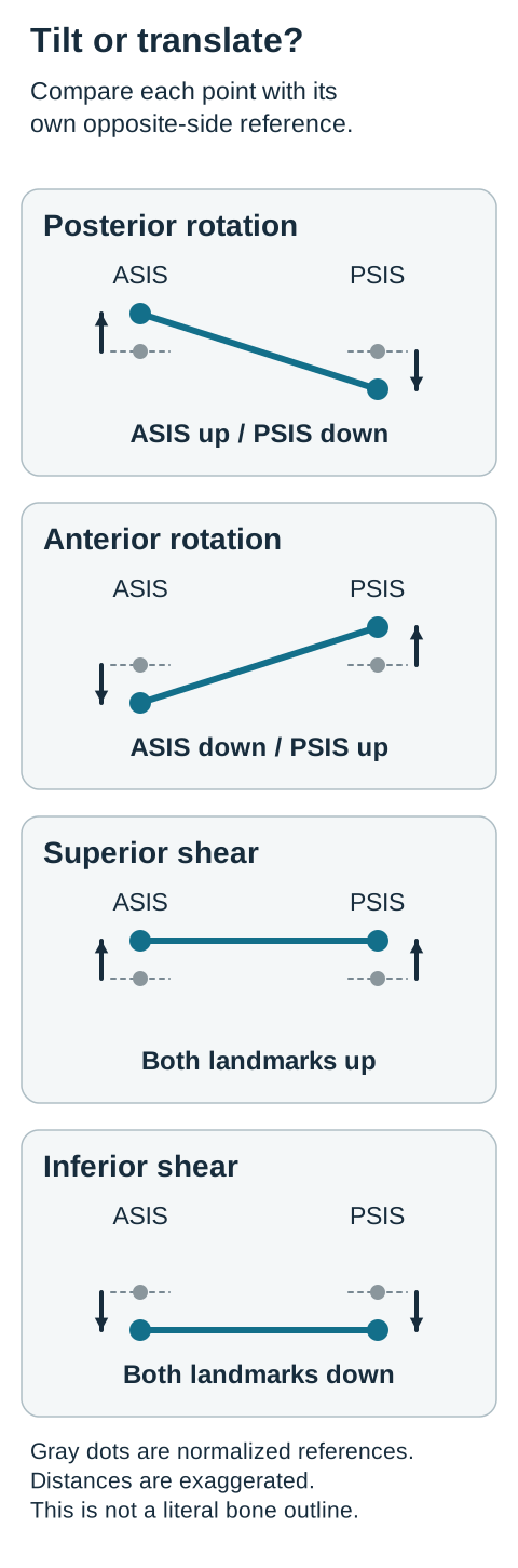
In the conventional posterior-rotation model, the ASIS is superior and the PSIS inferior relative to their opposite-side partners. The innominate is described as having greater ease toward posterior rotation and a restriction toward anterior rotation. In the anterior pattern, the paired vertical relationship is reversed. These labels describe an examination pattern; they do not imply a dislocated pelvis. [1][2]
Think of two labeled points on a rigid segment viewed from the side. Tilting the segment sends one point upward and the other downward. Translating the segment vertically sends both points in the same direction. The accompanying diagram exaggerates the distances to make this relationship visible; it is not a measurement of sacroiliac motion.
Same-side findings compared with the opposite side
ASIS
PSIS
Conventional pattern
ASISSuperior
PSISInferior
Conventional patternPosterior rotation
ASISInferior
PSISSuperior
Conventional patternAnterior rotation
ASISSuperior
PSISSuperior
Conventional patternSuperior shear
ASISInferior
PSISInferior
Conventional patternInferior shear
The two translation patterns are also terminology-based descriptions. Their names specify the easier direction; a superior shear is restricted toward inferior translation, and an inferior shear toward superior translation. [3][4]
Trace the pair: A runner has concordant right-sided restriction, a higher right ASIS, and a lower right PSIS. Point to the two corresponding symbols in the diagram, then predict which way each landmark would tend toward its contralateral level if the described rotation decreased.
Each gray reference is the corresponding opposite-side landmark normalized for comparison. Compare relative landmark heights using the gray reference lines. Predict the second landmark before comparing the four patterns. [1][2][3][4]Compare your predicted directions
The right ASIS would tend downward and the right PSIS upward, consistent with increased anterior rotation. This is a prediction within the model, not proof that a treatment has altered bone position.
The reason both points matter is now visible: a downward adjustment of the front point alone does not specify what happened behind it. In a new patient whose two right landmarks are both high, do not reuse the runner's label. The second observation changes the classification. [1][3]
Separate a static height from a motion test
How can a PSIS be low at rest and still have a positive standing-flexion finding? Static height and excursion during forward bending are different observations. In the traditional standing-flexion test, greater superior excursion of one PSIS is used as a laterality finding. It does not, by itself, name the rotation or establish the cause of pain.
Imagine two marks on a ruler. The right mark starts below the left, but travels farther upward during the test. The initial offset and the distance traveled can both be recorded without contradiction. A question may supply a positive right test together with a low resting right PSIS because it is describing both observations, not two competing diagnoses.
Sort this examination: A patient has left posterior pelvic discomfort, repeated restriction localized to the left, a superior left ASIS, an inferior left PSIS, and less left hip extension when the pelvis is supported. Separate the side, paired height pattern, and restricted motion before naming the finding.
The side is left; the opposite landmark directions fit posterior rotation; restricted extension may support the chosen treatment barrier. Pain location alone did not establish the side, and hip extension alone did not name an innominate diagnosis. A hip, lumbar, or soft-tissue limitation can also reduce measured extension. [1][5][16]
Use the examination convention without overstating its accuracy. Ribeiro and colleagues found poor agreement between different examiners for standing and sitting flexion testing. Dreyfuss and colleagues found positive sacroiliac screening findings in some asymptomatic adults. These studies caution against treating one positive result as a definitive structural lesion or pain generator. Repeating a test does not remove all measurement uncertainty. [6][7]
Now change one fact: two examiners obtain opposite laterality findings after inconsistent foot placement, while the resting landmark comparisons are not reproducible. The sensible consequence is to standardize position, repeat the relevant examination, and reconsider the working explanation. It is not to average the two sides, diagnose both sides from disagreement, or apply a stronger technique.
For a new symptom-free athlete with a positive screening test, ask whether there is a clinical problem requiring treatment. An isolated test result does not create an indication for correction. For a patient with pain, continue the broader history, neurologic assessment when indicated, and functional examination rather than treating the screening result as the endpoint. [7][16]
Change one landmark and test your explanation
A high ASIS can occur in more than one pattern. Which second observation would make you abandon posterior rotation? A high PSIS on the same side changes the pair from opposite directions to the same direction. That favors the superior-shear description instead. [1][3]
The following comparison holds the affected side and the ASIS observation constant. Only the PSIS changes. This makes the posterior landmark causally useful to the classification rather than another item to memorize.
Controlled comparison: right-sided findings
Examination
Right ASIS
Right PSIS
Interpretation
ExaminationA
Right ASISSuperior
Right PSISInferior
InterpretationPosterior rotation pattern
ExaminationB
Right ASISSuperior
Right PSISSuperior
InterpretationSuperior shear pattern
ExaminationC
Right ASISSuperior
Right PSISNot established
InterpretationInsufficient paired data
Try the substitution: Cover the last column. For each row, say whether the two landmarks describe a tilt, a vertical translation, or missing information. Then compare your interpretation with the visible column. Row C is not permission to guess the missing PSIS from the ASIS.
For a posterior pattern, the missing direction is anterior rotation. For a superior-shear pattern, the missing direction is inferior translation. Those are different mechanical descriptions, so selecting the posterior-rotation setup simply because the ASIS is high would skip a necessary diagnostic step. This lesson does not teach a shear-treatment procedure. [1][3]
Reverse the entire translation example: both left landmarks are inferior with concordant left-sided restriction. The conventional label is left inferior shear, not left anterior rotation. An anterior rotation requires an opposite-direction pair, not merely a low ASIS. [2][4]
Vertical information also does not establish an inflare or outflare, which concerns a different spatial relationship. Similarly, innominate landmark heights do not supply the sacral examination needed to name a sacral torsion. Avoid creating a second diagnosis from data the case has not provided.
In a new patient after a high-energy collision, both landmarks may look high for a dangerous reason rather than a benign descriptive shear. Do not request standing flexion or repeated pelvic stressing in a person who cannot bear weight. The injury mechanism changes the priority from classification to trauma assessment. [13]
Keep the setup direction separate from the effort
If the innominate rests in posterior rotation, why ask the patient to flex the hip? The direct setup and the patient's isometric effort have different jobs. The setup engages the restriction toward anterior rotation using supported hip extension. The brief patient effort is gentle hip flexion against matching resistance, so the thigh stays still. [1][5]
First check eligibility: explain the procedure, obtain consent, and confirm comfortable participation. Suspected fracture or instability, severe pain with positioning, possible acute infection or inflammatory disease, and new neurologic findings require assessment rather than forceful testing. A patient unable to follow instructions or produce the requested contraction cannot perform this active technique as described. [5][13][15][16]
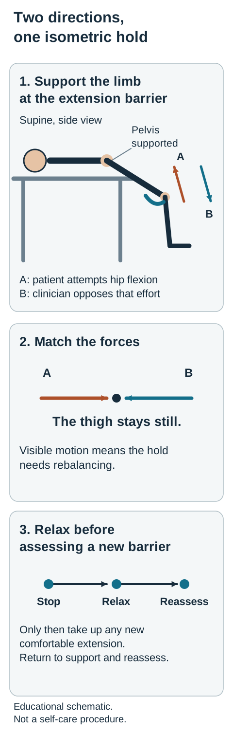
For the supine approach illustrated here, the involved leg is supported near the table edge. The clinician stabilizes the pelvis and prevents rolling, then guides the hip into a comfortable extension barrier. The leg is not simply dropped by gravity. Monitor the pelvic relationship so lumbar arching or rolling does not substitute for the intended direction. This is one described setup, not a claim that it is the only valid osteopathic approach. [5]
The setup, patient effort and counterforce have different jobs. The isometric phase has no visible joint excursion. The figure omits other body parts for clarity and is not a self-treatment procedure. [1][5]
The patient then attempts a small hip-flexion effort while the clinician supplies an equal opposing force. With a true isometric contraction, the hip angle does not visibly change. After the patient stops and fully relaxes, the clinician takes up any available slack toward a new comfortable extension barrier. A published description uses several brief contractions with reassessment afterward; the objective is controlled participation, not maximal effort or a mandatory angular target. [5]
Trace three arrows: Using the figure, label the initial hip-extension direction, the attempted hip-flexion direction, and the opposing clinician force. Then predict what you would see if the patient's force exceeded the clinician's resistance.
Check the force comparison
The thigh would rise toward hip flexion instead of remaining still. That would not match the intended isometric phase. Re-establish a comfortable position and a lower, matched effort rather than describing visible motion as a successful isometric hold.
The visible consequence of correctly matched forces is no joint excursion during the effort. Additional extension, when available, is introduced after relaxation, not while the patient is still contracting. The final step is a return to a supported neutral position followed by reassessment, not another stronger contraction until landmarks appear equal. [5]
Apply this to a new patient with a reproducible left posterior pattern: all the described directions apply to the left side. Conversely, a high right ASIS with a high right PSIS does not justify this rotation-specific setup. Side selection and pattern selection both precede technique selection. [1][3]
Fix one attachment before predicting hamstring action
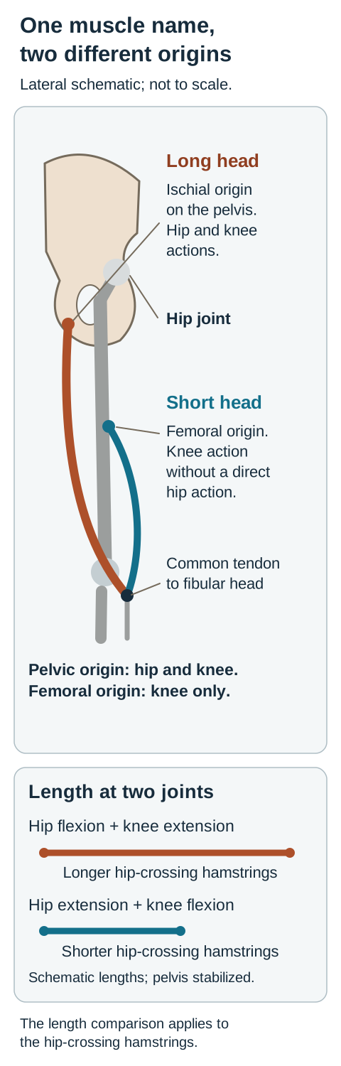
How can the hamstrings extend the hip yet be associated with posterior pelvic tilt? First specify which attachment is relatively fixed. The long head of biceps femoris, semitendinosus, and semimembranosus connect the ischial region to the proximal lower leg and cross both the hip and knee. With the pelvis stabilized, their contraction can extend the hip and flex the knee. [8][9][11]
With the lower limb relatively fixed, tension acts back on the pelvic attachment and can influence pelvic tilt. This is a mechanical relationship, not evidence that every tight hamstring has caused a unilateral sacroiliac lesion. A cadaver study found that anterior pelvic tilt over a fixed limb increased hamstring elongation. Its controlled preparation supports the length relationship, but not the diagnostic accuracy of a palpation test or a clinical claim of cause and cure. [12]
The figure deliberately separates the two attachments. Trace from the ischial tuberosity to the knee-crossing tendons, then compare the short head of biceps femoris. That head begins on the femur, below the hip joint, so it contributes to knee flexion but does not itself span the hip. It is the important exception to an unqualified statement that every posterior-thigh muscle arises from the pelvis. [10]
The attachment location determines which joints a muscle can directly act across. The length comparison applies to the hip-crossing hamstrings, not the short head at the hip. [8][9][10][11][12]
Predict length, not just an action word: In an uninjured person whose pelvis is stabilized, which combination lengthens the hip-crossing hamstrings at both joints: hip flexion with knee extension, or hip extension with knee flexion?
Compare the attachment distances
Hip flexion with knee extension lengthens the two-joint hamstrings. Hip extension with knee flexion shortens them. Therefore, the hip-extension setup in the preceding section should not be called a hamstring stretch. Its purpose is to engage a pelvic restriction, and its active phase uses a hip-flexion effort. [5][8][9][11]
The distinction prevents two common errors. A tight hamstring does not prove posterior innominate rotation, and asking for hamstring contraction is not interchangeable with the flexion effort in the described posterior-rotation technique. Evaluate the muscle finding and the pelvic finding separately, then explain how they may interact. [1][5][7][12]
Now apply the anatomy to a different problem. An adolescent sprinter feels a sudden pop with focal pain over the ischial tuberosity and pain during resisted knee flexion. The traction site raises concern for an apophyseal avulsion, not merely a need to stretch a tight muscle. Protect the painful region and arrange injury assessment; an AP pelvic radiograph is the usual initial study when this injury is suspected. A normal film does not settle persistent high suspicion for a subtle injury. [14]
Do not convert the cadaveric length finding into a rule that all posterior pelvic pain is caused by hamstring shortening. New severe symptoms, focal injury signs, or a pattern inconsistent with the examination require a different explanation. [12][14][16]
Reassess what changed, then decide what it means
Does less pain prove that the innominate has been corrected? No. A symptom report is valuable, but it is a different outcome from a repeated landmark or motion finding. Reassess the same side-to-side landmarks, the original laterality test, hip extension with the same positioning, and relevant tissue texture. Also ask about the function that originally mattered to the patient. [5][6][16]
Interpret this follow-up: A runner reports pain falling from 6/10 to 2/10. Supported right hip extension increases from 5 to 10 degrees. The right ASIS remains relatively high and the right PSIS relatively low, with the same standing-flexion laterality. These numbers describe this example; they are not treatment targets or validated thresholds.
Different observations support different conclusions
Observation
What it supports
ObservationLess reported pain
What it supportsSymptomatic improvement
ObservationMore extension with the same stabilization
What it supportsImproved measured range
ObservationPersistent paired asymmetry
What it supportsThe recorded landmark pattern persists
ObservationBetter walking or sitting tolerance
What it supportsImprovement in the selected function
Choose a documentation statement before reading on. The defensible statement is that symptoms and measured range improved while the landmark pattern persisted. Neither pain relief alone nor unchanged asymmetry alone tells the complete story. A change in examiner or positioning also weakens direct comparison, because test reliability is limited. [6][7]
The consequence is a measured next decision: document findings, reassess the working diagnosis if progress is discordant, and avoid repeated force to pursue perfect symmetry. Persistent uncomplicated back pain is not automatically an indication for radiographs. Imaging is guided by suspected pathology and whether the result would change management. For appropriate nonspecific back pain, manual care belongs within a broader plan that includes activity and exercise rather than a promise of permanent alignment correction. [16]
Transfer the reasoning to new information. Severe radiating back pain with newly impaired bladder function or perineal sensation requires immediate evaluation for possible cauda equina syndrome, even if the patient also has a recognizable pelvic landmark pattern. Following major trauma, suspected pelvic instability likewise takes priority over the osteopathic label. [13][15]
A complete explanation connects the paired landmarks, the restricted direction, the fixed attachment, and the patient's safety and function. Keep those observations related, but do not let one stand in for all the others.
Apply the relationships to new cases
Identify the relevant finding before comparing the options. Several cases require changing the working explanation rather than repeating the same technique.
Case 1
Show answer and explanations for case 1
A. ASIS superior; PSIS inferior (Why this does not fit)
This pair represents additional posterior rotation. It follows the described position of ease instead of the restricted anterior direction. [1] [2] [3] [4]
Reasoning steps for option A
What resting right-sided landmark pair makes further posterior rotation tempting?
The right ASIS is high and PSIS low, the posterior-rotation pair.
If the right innominate rotated farther posteriorly, where would its ASIS and PSIS move?
Further posterior rotation raises the right ASIS and lowers the right PSIS.
Why would that motion miss the runner's restricted barrier?
That follows posterior ease rather than testing the restricted anterior direction.
B. ASIS inferior; PSIS superior (Best answer)
Anterior rotation lowers the anterior point and raises the posterior point in the simplified model. The right-sided posterior pattern is restricted toward that opposite rotation. [1] [2] [3] [4]
Reasoning steps for option B
Which right landmark must descend as this posterior pattern moves toward its barrier?
The right ASIS must descend toward the left ASIS level.
How does anterior rotation move the right PSIS relative to its left partner?
Anterior rotation raises the right PSIS toward its opposite-side partner.
Why is the ASIS-down, PSIS-up pair the relevant restricted motion?
The opposite movements reverse the posterior resting pair and test anterior restriction.
C. ASIS inferior; PSIS inferior (Why this does not fit)
Both landmarks descending represents inferior translation. The original opposite-direction pair indicates rotation rather than a superior shear needing inferior translation. [1] [2] [3] [4]
Reasoning steps for option C
What would simultaneous descent of the right ASIS and PSIS signify?
Both descending represents inferior translation.
Would inferior translation create the opposing motion of the two landmarks?
No. Inferior translation lowers both landmarks, rather than lowering ASIS while raising PSIS.
Why does the runner's high-ASIS, low-PSIS pair not establish superior shear?
A superior shear would start with both right landmarks high; here only the ASIS is high.
D. ASIS superior; PSIS superior (Why this does not fit)
Both landmarks rising represents superior translation. That does not reverse the opposite-direction rotational pattern described here. [1] [2] [3] [4]
Reasoning steps for option D
What relationship between the two right landmarks indicates tilt rather than translation?
Opposite height deviations, high ASIS and low PSIS, indicate tilt.
What mechanical movement raises both right landmarks together?
Superior translation raises both the right ASIS and PSIS.
Why does upward translation fail to reverse the runner's posterior tilt?
Raising both landmarks cannot reverse the high-ASIS, low-PSIS rotational pair.
Takeaway: Infer the paired pattern first, then predict its restricted direction.
A. Anterior rotation of the right innominate (Why this does not fit)
Anterior rotation is the restricted direction for a posterior-rotation pattern. A posterior pattern requires an inferior, not superior, right PSIS. [1] [2] [3] [4]
Reasoning steps for option A
Which posterior landmark contradicts the student's posterior-rotation assumption?
The right PSIS is high; a posterior pattern would have a low PSIS.
For which paired pattern is anterior rotation the restricted direction?
Anterior rotation is restricted in a posterior-rotation pattern with high ASIS and low PSIS.
Why does the observed high right PSIS rule out that pair?
A high right PSIS makes the observed pair high-high, not posterior rotation.
B. Posterior rotation of the right innominate (Why this does not fit)
Posterior rotation is restricted in an anterior-rotation pattern. The ASIS would be relatively inferior in that pattern, unlike this examination. [1] [2] [3] [4]
Reasoning steps for option B
What ASIS height would support a right anterior-rotation pattern?
The ASIS would be inferior to the left ASIS.
When is posterior rotation a restricted rather than an easy direction?
Posterior rotation is restricted when the innominate rests anteriorly rotated.
Why does the actual superior right ASIS oppose this interpretation?
The right ASIS is high rather than low, so the anterior pattern is absent.
C. Superior translation of the right innominate (Why this does not fit)
Superior translation is restricted in an inferior-shear pattern. Both observed landmarks are superior rather than inferior. [1] [2] [3] [4]
Reasoning steps for option C
What two landmark heights would justify an inferior-shear diagnosis?
Both right ASIS and PSIS would have to be low.
Which translation is restricted when an innominate rests inferiorly?
An inferior shear is restricted toward superior translation.
How do this patient's two high right landmarks conflict with that premise?
Both observed right landmarks are high, the reverse of an inferior shear.
D. Inferior translation of the right innominate (Best answer)
A superior-shear description combines both landmarks being high with restriction toward inferior translation. The paired observations support that description; bilateral hip-extension limitation does not convert it to a rotation. [1] [2] [3] [4]
Reasoning steps for option D
What does the simultaneous elevation of right ASIS and PSIS indicate?
Both elevated landmarks describe a superior shear.
Which direction is restricted by a superior-shear pattern?
The restricted movement is inferior translation of the right innominate.
Why does bilateral hip-extension loss not override the paired heights?
Bilateral hip-extension limitation cannot distinguish right rotation from the high-high shear pair.
Takeaway: A high ASIS needs its PSIS partner before a rotation-specific barrier is selected.
A. Posterior rotation of the left innominate (Best answer)
The left anterior-rotation pattern is expected to be restricted toward posterior rotation. The affected side and both landmark directions agree with this target. [1] [2] [6]
Reasoning steps for option A
Which side has reproducible restriction despite the bilateral comparison?
Repeated testing localizes restriction to the left.
Which rotation has a low left ASIS and high left PSIS as its resting pair?
The low left ASIS and high left PSIS describe left anterior rotation.
What opposite rotational direction should be tested on that left side?
The expected restricted direction is left posterior rotation.
B. Anterior rotation of the left innominate (Why this does not fit)
This is the restricted direction of a left posterior-rotation pattern. That pattern would have a superior left ASIS and inferior left PSIS, the reverse of this case. [1] [2] [6]
Reasoning steps for option B
What left PSIS position would a posterior-rotation pattern require?
The left PSIS would have to lie below the right PSIS.
For which resting rotation is anterior rotation restricted?
Anterior rotation is restricted in a posterior-rotation pattern.
Why do the patient's actual left landmark heights reverse that premise?
Here the left ASIS is low and PSIS high, the reverse of that posterior pair.
C. Posterior rotation of the right innominate (Why this does not fit)
This direction would address a right anterior-rotation pattern. The case localizes restriction to the left rather than assigning dysfunction from the mirror-image right landmarks. [1] [2] [6]
Reasoning steps for option C
What does the examination say about the side requiring a direct technique?
The left side needs assessment for a direct technique.
What right-sided pattern would be treated by posterior rotation?
Right anterior rotation would be restricted toward right posterior rotation.
Why cannot the mirror-image right comparison substitute for left laterality?
Relative right heights do not outweigh the repeated left-sided restriction.
D. Anterior rotation of the right innominate (Why this does not fit)
This direction would address a right posterior-rotation pattern. The supplied laterality is left, and a relative comparison does not independently diagnose the opposite side. [1] [2] [6]
Reasoning steps for option D
Which side is selected by the repeated motion examination?
The repeated examination selects the left side.
Which right-sided resting pattern would make anterior rotation its barrier?
A right posterior-rotation pattern would be restricted toward right anterior rotation.
Why does the relative right landmark pair not establish a right lesion here?
The contralateral comparison does not independently establish a right lesion.
Takeaway: Use the localized side and the paired pattern before reversing the direction to identify a direct barrier.
A. Compare right and left posterior-thigh tenderness (Why this does not fit)
Muscle tenderness can identify a symptomatic soft-tissue region. Both candidate pelvic patterns can coexist with tenderness, so this does not supply the missing posterior landmark. [1] [3] [6] [7]
Reasoning steps for option A
What anatomical structure does posterior-thigh tenderness assess?
It assesses a symptomatic soft-tissue region in the posterior thigh.
Can tenderness determine whether the left PSIS rests above or below the right?
No. Tenderness does not specify resting left PSIS height.
Why could either proposed pelvic pattern coexist with tender hamstrings?
Either a posterior rotation or superior shear can coexist with tender posterior-thigh muscles.
B. Compare right and left hip-extension range (Why this does not fit)
Hip extension can help characterize the available treatment range. Its restriction is not specific enough to separate these two patterns when the PSIS relationship is unknown. [1] [3] [6] [7]
Reasoning steps for option B
What can comparing left and right hip extension characterize?
It characterizes available hip-extension motion.
Does reduced extension identify the resting left PSIS height?
No. Extension loss does not establish the resting PSIS relationship.
Why would this range comparison leave posterior rotation and superior shear unresolved?
Both patterns may have limited extension; the missing PSIS comparison remains unresolved.
C. Compare bilateral resting PSIS heights (Best answer)
A low ipsilateral PSIS would complete a posterior pair, whereas a high one would complete a superior-shear pair. This directly supplies the observation on which the two proposed descriptions differ. [1] [3] [6] [7]
Reasoning steps for option C
With the left ASIS high, which missing landmark separates tilt from translation?
The left PSIS resting height relative to the right is missing.
What diagnosis follows if the resting left PSIS is low versus high?
A low left PSIS completes posterior rotation; a high one completes superior shear.
Why does comparing bilateral PSIS heights directly resolve the competing patterns?
With left ASIS already high, PSIS height decides whether the points oppose or rise together.
D. Compare repeated standing-flexion laterality (Why this does not fit)
Repeating laterality can improve consistency of the examination. It still does not establish whether the posterior landmark is relatively high or low at rest. [1] [3] [6] [7]
Reasoning steps for option D
What information does the left standing-flexion finding already provide?
It suggests left-sided laterality on the motion screen.
Would repeating the flexion test establish the resting PSIS relationship?
No. Repetition can improve consistency but does not measure resting PSIS heights.
Why is reliable laterality still insufficient to distinguish these two left-sided patterns?
Both candidate patterns could be left-sided, so laterality cannot classify their landmark geometry.
Takeaway: Choose the next observation for its ability to distinguish the actual competitors.
A. Anterior rotation; restricted posterior rotation (Why this does not fit)
A low ASIS can be part of an anterior-rotation pattern. Its PSIS should be relatively superior, so the second landmark contradicts this description. [1] [2] [3] [4]
Reasoning steps for option A
Which single low left landmark could falsely suggest anterior rotation?
The low left ASIS alone could suggest anterior rotation.
Where should the left PSIS lie in an anterior-rotation pair?
Anterior rotation requires the left PSIS to be superior, not inferior.
Why does the actual low PSIS defeat the trainee's interpretation?
The observed low PSIS makes the pair low-low rather than an opposing rotational pair.
B. Posterior rotation; restricted anterior rotation (Why this does not fit)
Posterior rotation is restricted in the opposite rotational direction. It requires a high ASIS and low PSIS, not two low landmarks. [1] [2] [3] [4]
Reasoning steps for option B
What ASIS position would accompany the low PSIS in posterior rotation?
The left ASIS would have to be high, opposite the low PSIS.
Which direction is restricted when the innominate rests posteriorly rotated?
A posterior resting rotation is restricted toward anterior rotation.
Why do two low left landmarks not form that opposing-height pair?
Both left landmarks are low, so this is translation rather than posterior tilt.
C. Superior shear; restricted inferior translation (Why this does not fit)
A superior shear is limited toward inferior translation. Its two landmarks are relatively high rather than low. [1] [2] [3] [4]
Reasoning steps for option C
How would both left landmarks appear in a superior shear?
Both left landmarks would be high in superior shear.
What direction is restricted in that high-high translation pattern?
Superior shear is restricted toward inferior translation.
Why is the observed low-low pair incompatible with superior shear?
Both observed landmarks are low, so superior shear is the wrong translation direction.
D. Inferior shear; restricted superior translation (Best answer)
Two low ipsilateral landmarks fit the inferior-shear description. The expected restricted direction is superior translation rather than a rotational direction. [1] [2] [3] [4]
Reasoning steps for option D
What common direction do the left ASIS and PSIS share at rest?
Both left landmarks are below their right-sided counterparts.
Which shear label corresponds to both landmarks lying below the right?
This low-low pair is called left inferior shear.
Why is upward translation, rather than rotation, the expected barrier?
Its barrier is superior translation, the reverse of the inferior resting displacement.
Takeaway: A translation label requires the pair to shift together; its restriction is in the opposite vertical direction.
A. Use the first examiner's side for treatment (Why this does not fit)
The initial result can appear to offer a definite starting point. It is not privileged when positioning and the paired landmarks were inconsistent. [6] [7] [16]
Reasoning steps for option A
Which change between examinations undermines privileging the first examiner?
Foot position and examiner changed; even resting landmarks disagreed.
What apparent certainty does the initial right flexion result offer?
The first right-sided finding may appear to identify an immediate treatment side.
Why should a right-sided technique wait for reproducible landmarks and laterality?
No first-result preference is justified when laterality and paired heights are not reproducible.
B. Standardize position and repeat the focused examination (Best answer)
Comparable positioning permits a more interpretable repeat assessment. Discordance should prompt reassessment rather than an unsupported choice of side. [6] [7] [16]
Reasoning steps for option B
What positioning difference could explain opposite standing-flexion results?
Foot position differed, and a different examiner retested the patient.
How does standardizing stance improve comparison of the two examinations?
Comparable stance reduces a positional source of variation in the flexion finding.
What should discordant ASIS and PSIS observations prompt before selecting a side?
Repeat the focused assessment before attributing discordant findings to either side.
C. Treat both innominates during the same session (Why this does not fit)
Opposite results could be mistaken for evidence of bilateral dysfunction. Disagreement between tests does not establish two reproducible lesions. [6] [7] [16]
Reasoning steps for option C
Do alternating right and left flexion results demonstrate two lesions?
No. Alternating test sides are not two confirmed lesions.
How could examiner disagreement be mistaken for bilateral dysfunction?
One might mistake a right result plus a later left result for bilateral involvement.
What reproducible findings are absent before treating both innominates?
There are no two reproducible side-specific landmark and motion patterns to justify bilateral treatment.
D. Use the side of pain to select treatment (Why this does not fit)
Pain location is relevant to the clinical assessment. It does not replace the missing reproducible side-specific mechanical findings. [6] [7] [16]
Reasoning steps for option D
What role does posterior pelvic pain have in this assessment?
Pain location remains relevant to symptom assessment.
Can its location replace inconsistent side-specific motion testing?
No. Pain location cannot determine the innominate motion barrier.
Why is pain alone an unsound selector for a side-specific technique?
The examinations disagree on side and landmarks, so pain cannot select the technique side.
Takeaway: Inconsistent examination data require a better comparison, not a more confident label.
A. Schedule periodic muscle energy to normalize the screening test (Why this does not fit)
Repeated treatment could seem preventive when a screening result is abnormal. No symptomatic or functional problem has been established, and a positive test alone does not demonstrate progressive damage. [6] [7] [16]
Reasoning steps for option A
What symptom or functional deficit would preventive muscle energy address here?
There is no pain or functional deficit for preventive muscle energy to address.
What does an isolated left flexion finding establish about future damage?
A positive flexion screen alone does not demonstrate future pelvic damage.
Why is periodic correction unjustified without a clinical problem?
Repeated correction lacks an indication when the athlete functions normally and paired landmarks are inconsistent.
B. Obtain pelvic radiographs before permitting usual exercise (Why this does not fit)
Imaging might appear to confirm a suspected structural abnormality. The history and examination do not supply an injury or other specific indication for radiographs. [6] [7] [16]
Reasoning steps for option B
What injury history would raise a reason to image this athlete's pelvis?
There is no reported injury or focal concern suggesting a pelvic lesion requiring imaging.
What structural question might radiographs otherwise investigate?
Radiographs might be used to investigate a suspected structural injury.
Why do comfortable activity and inconsistent landmarks not warrant radiographs?
No injury, symptom, or other specific pathology justifies delaying usual exercise for radiographs.
C. Prescribe a unilateral shoe lift to correct the test asymmetry (Why this does not fit)
A shoe lift changes loading and may seem to address an apparent asymmetry. The case has not established a structural limb-length difference or a symptomatic indication for correction. [6] [7] [16]
Reasoning steps for option C
What measured limb-length difference supports considering a shoe lift?
No structural limb-length discrepancy has been measured.
Could the isolated flexion finding establish that structural discrepancy?
No. A flexion screen is not a limb-length measurement.
Why might changing unilateral loading be inappropriate in this asymptomatic athlete?
A lift changes loading without an established discrepancy or symptomatic need.
D. Continue usual activity without treating the isolated test result (Best answer)
Positive screening findings can occur in asymptomatic individuals. Without pain, functional limitation, or a specific pathologic concern, the test alone does not establish a need for corrective treatment. [6] [7] [16]
Reasoning steps for option D
What does the athlete report about running and daily function?
Running and daily activities are pain-free and unrestricted.
Can a positive flexion screen occur without symptomatic disease?
Yes. Screening findings can occur in asymptomatic people.
Why is continuing activity preferable to treating the screen alone?
Without a symptomatic or pathologic indication, continue usual activity rather than treating the test.
Takeaway: An isolated screening finding is not, by itself, a treatment indication.
A. Repeat hip extension while controlling pelvic and lumbar compensation (Best answer)
Fixing the comparison conditions isolates the intended assessment more closely. The additional excursion occurred only after pelvic rolling and lumbar arching were allowed. [5] [6]
Reasoning steps for option A
Under which position was the original right extension limitation measured?
The baseline right extension assessment had the pelvis supported.
Which compensations must be controlled on a repeat measurement?
Control pelvic rolling and lumbar arching during the repeat assessment.
Why does comparing stabilized hip motion test improvement more directly?
The same stabilization isolates actual hip extension rather than added spine or pelvis movement.
B. Use the greatest thigh excursion from either position as the result (Why this does not fit)
The largest visible excursion can appear to represent the best available hip range. It combines different contributions and cannot be compared with the stabilized baseline. [5] [6]
Reasoning steps for option B
What changed when the thigh descended farther?
Pelvic rolling and lumbar arching were newly allowed.
Does maximum thigh excursion isolate right hip extension?
No. Maximum thigh descent includes pelvis and lumbar contributions.
Why cannot the rolled-pelvis angle be compared with the supported baseline?
The unsupported angle cannot be compared with the supported baseline as pure hip motion.
C. Reclassify the innominate as anteriorly rotated from the larger angle (Why this does not fit)
A larger extension angle might be mistaken for a reversal of the pelvic diagnosis. The changed setup does not establish a new paired landmark pattern. [5] [6]
Reasoning steps for option C
What paired landmarks originally supported the posterior pattern?
High right ASIS and low right PSIS support a posterior-rotation pattern.
Can a larger angle with lumbar arching reverse those landmark observations?
No. The larger angle reflects changed positioning, not reversed landmark heights.
Why is a new anterior-rotation label unsupported by compensated motion?
No low-ASIS, high-PSIS pair was found to justify anterior reclassification.
D. Use posterior-thigh tenderness to estimate the corrected extension angle (Why this does not fit)
Tenderness can provide information about a symptomatic muscle region. It does not measure the hip range or distinguish true change from the observed compensation. [5] [6]
Reasoning steps for option D
What does palpated posterior-thigh tenderness measure?
Tenderness measures sensitivity of posterior-thigh tissues, not an angle.
Can tenderness quantify degrees of true hip extension?
No. Palpation cannot measure degrees of hip extension.
Why would it fail to separate hip improvement from pelvic rolling?
It cannot separate true hip range change from the observed pelvic and lumbar compensation.
Takeaway: Compare range under the same stabilization before interpreting a change.
A. MRI of the lumbar spine (Why this does not fit)
Lumbar imaging evaluates suspected spinal causes of referred pain. The acute traction event and focal ischial tenderness instead localize the immediate concern to the hamstring origin. [8] [9] [11] [14]
Reasoning steps for option A
Which event localized this sprinter's new pain to the lower buttock?
An acceleration-related pop with focal ischial pain localizes the new concern to the hamstring origin.
What suspected pain source would lumbar MRI investigate instead?
Lumbar MRI investigates a spinal source of pain or referred symptoms.
Why does focal ischial tenderness take priority over spinal imaging?
The acute ischial traction findings call for assessment of that attachment instead.
B. Ultrasonography of the inguinal canal (Why this does not fit)
An inguinal study can evaluate a local groin process. The symptomatic site is posterior and directly related to hamstring loading, not an inguinal presentation. [8] [9] [11] [14]
Reasoning steps for option B
Is the painful attachment posterior or in the inguinal canal?
The painful attachment is posterior at the ischial tuberosity.
What condition would inguinal ultrasound be suited to assess?
Inguinal ultrasound assesses a groin or inguinal process.
Why does pain with hamstring loading not point to a groin study?
Pain at the ischium with hamstring loading does not localize to the inguinal canal.
C. Anteroposterior radiograph of the pelvis (Best answer)
An AP pelvic film can detect an apophyseal avulsion at a pelvic tendon attachment. An adolescent with a sudden traction event and focal ischial findings warrants this injury assessment before stretching or manual care. [8] [9] [11] [14]
Reasoning steps for option C
Why does the patient's age matter after an acceleration-related pop?
An adolescent ischial apophysis can avulse under sudden hamstring traction.
Which initial image can show an ischial apophyseal avulsion?
An AP pelvic radiograph is the usual initial image for suspected ischial apophyseal avulsion.
Why should focal traction injury be assessed before stretching or manual treatment?
Assess possible avulsion before stretching or manual care that could stress the injured attachment.
D. Standing radiograph of lower-limb alignment (Why this does not fit)
An alignment study can address selected structural loading questions. It does not prioritize the acute focal traction injury suggested by the new history. [8] [9] [11] [14]
Reasoning steps for option D
What acute mechanism demands attention despite old pelvic asymmetry?
The new sprinting pop and focal ischial tenderness supersede the old minor asymmetry.
What question does a standing alignment film primarily address?
A standing alignment film addresses structural lower-limb alignment or loading.
Why would that film miss the priority of assessing the ischial attachment?
It does not prioritize imaging the suspected acute ischial traction injury.
Takeaway: Adolescent traction pain at the ischial origin requires an injury assessment, not an assumption of muscle tightness.
A. Hip extension without a direct knee action (Why this does not fit)
A muscle crossing the hip but not the knee can extend the hip. The described portion starts on the femur below the hip and crosses the knee instead. [9] [10]
Reasoning steps for option A
Does an isolated hip extensor match the reported femoral origin?
No. The injured portion starts on the lateral linea aspera, distal to the hip joint.
What joint must a femur-to-fibular-head portion cross?
It crosses the knee, not the hip, so isolated hip extension is not its direct action.
Why does the intact ischial-origin portion matter here?
It separates the hip-crossing long head from the injured short head, ruling out hip extension as the injured portion’s action.
B. Knee flexion without a direct hip action (Best answer)
The short head of biceps femoris crosses the knee but not the hip. Its femoral origin and fibular attachment fit the report and explain its contribution to knee flexion. [9] [10]
Reasoning steps for option B
Which biceps femoris head begins on the lateral linea aspera?
The short head arises from the femur and joins the tendon inserting at the fibular head.
Does that short head span the hip joint?
No. Its femoral origin lies below the hip, so it has no direct hip action.
What action follows from its posterolateral course across the knee?
The short head assists knee flexion, matching the localized injury and the knee-only action.
C. Hip flexion together with knee extension (Why this does not fit)
A two-joint anterior-thigh muscle can combine these actions. The described posterolateral femoral origin and fibular attachment do not match that anatomy. [9] [10]
Reasoning steps for option C
Would a hip flexor and knee extensor attach to the fibular head?
No. That paired action suggests an anterior two-joint thigh muscle, not this posterolateral insertion.
How does the reported femoral origin constrain a proposed hip-flexion action?
An origin on the lateral linea aspera leaves the injured portion below the hip joint.
What knee action contradicts the anterior-thigh alternative?
The injured biceps femoris short head passes behind the knee to assist flexion, not extension.
D. Knee extension without a direct hip action (Why this does not fit)
A femoral-origin muscle on the anterior thigh can extend the knee. The injured portion reaches the posterior-lateral knee region as biceps femoris, which assists flexion. [9] [10]
Reasoning steps for option D
Can a femoral origin alone establish knee extension?
No. The muscle’s course relative to the knee also determines its action.
Where does the fibular-head tendon run relative to the knee?
The biceps femoris tendon runs posterolaterally, consistent with knee flexion.
Which feature distinguishes this injury from a vastus injury?
The fibular-head insertion identifies the short head of biceps femoris rather than an anterior knee extensor.
Takeaway: The short head of biceps femoris is the femoral-origin exception and does not span the hip.
These muscles cross both the hip and knee, so hip flexion lengthens them before knee extension adds further length. The increased knee range when the hip is less flexed fits a two-joint posterior-thigh limitation. [8] [9] [10] [11]
Reasoning steps for option A
Why does flexing the dancer’s hip reduce available knee extension?
Hip flexion first lengthens hamstrings that span the hip; knee extension adds tension at their other joint.
What does the posterior-thigh stretch localize?
It points to posterior two-joint tissue rather than an anterior muscle or a neurologic complaint.
Why does bringing the hip toward neutral restore knee range?
It slackens the hip-crossing hamstrings, allowing more knee extension before they limit motion.
B. Short head of biceps femoris (Why this does not fit)
This muscle portion crosses the knee and can limit knee extension when short. Because it does not cross the hip, it does not directly explain the marked dependence on hip position. [8] [9] [10] [11]
Reasoning steps for option B
Can the short head of biceps femoris oppose knee extension?
Yes. As a knee flexor it can resist extension if shortened.
Would moving the hip from 90 degrees toward neutral change its length directly?
No. The short head begins on the femur and does not span the hip.
What observation argues against the short head as the primary limiter?
The large gain in knee extension with hip repositioning requires a hip-crossing structure.
C. Vastus muscle group (Why this does not fit)
The vasti cross the knee and provide knee extension. They do not cross the hip and are not the posterior two-joint tissues lengthened in this comparison. [8] [9] [10] [11]
Reasoning steps for option C
What is the vasti’s primary knee action?
The vasti extend the knee rather than provide the posterior flexor stretch described.
Does hip flexion directly lengthen the vasti?
No. They cross the knee but not the hip.
Why does the dancer’s positional comparison disfavor vastus limitation?
Knee extension improves as hip flexion is reduced, implicating posterior tissue spanning both joints.
D. Iliopsoas muscle group (Why this does not fit)
Iliopsoas spans the hip and can influence hip-extension range. It does not cross the knee and does not explain this posterior-thigh response to knee extension. [8] [9] [10] [11]
Reasoning steps for option D
Which joint does iliopsoas span in this comparison?
It spans the hip, not the knee.
Would knee extension with a fixed flexed hip directly lengthen iliopsoas?
No. Changing the knee angle does not lengthen this hip flexor across a knee joint.
What finding instead selects posterior two-joint tissue?
A localized posterior-thigh stretch that varies strongly with hip position during knee extension fits hip-crossing hamstrings.
Takeaway: A restriction that changes with position at a second joint suggests a structure spanning both joints.
A. Left hip flexion against an extension counterforce (Why this does not fit)
This is an isometric hip-flexion arrangement on the left. The examination and supported barrier are right-sided, so it does not recruit the intended limb. [1] [5]
Reasoning steps for option A
Which side do the high ASIS and low PSIS identify?
The paired findings and restriction identify the right posterior innominate pattern.
What contraction would the left-flexion choice recruit?
It would recruit left hip flexors while the supported extension barrier is on the right.
Why can matching the flexion direction still be wrong?
The extension counterforce must oppose the involved right hip, not an uninvolved left effort.
B. Right hip extension against a flexion counterforce (Why this does not fit)
This arrangement recruits right hip extensors against opposing resistance. The described posterior-rotation approach instead uses gentle hip flexion from the extension barrier. [1] [5]
Reasoning steps for option B
What motion would right hip extension effort attempt at the barrier?
It would recruit right hip extensors against a flexion counterforce.
Which contraction is specified for the right posterior pattern?
The patient should attempt right hip flexion from the supported extension barrier.
Why is the right-extension alternative insufficient despite correct laterality?
It reverses the prescribed active force direction and does not perform the selected isometric phase.
C. Right hip flexion against an extension counterforce (Best answer)
The patient attempts flexion while the clinician balances that effort to keep the thigh still. It uses the affected side and the appropriate effort for this extension-barrier technique. [1] [5]
Reasoning steps for option C
What direction is restricted by the right posterior landmark pair?
Anterior rotation is restricted, and the supported right hip is placed at its comfortable extension barrier.
Which effort should the patient make at that right-sided barrier?
The patient gently attempts right hip flexion.
How should the clinician keep that effort isometric?
An equal extension counterforce balances right hip flexion so the thigh stays still.
D. Left hip extension against a flexion counterforce (Why this does not fit)
This arrangement recruits left hip extensors. It differs both in side and in contraction direction from the described right-sided approach. [1] [5]
Reasoning steps for option D
Does left hip extension address the side found restricted?
No. The right ASIS and PSIS findings and localized restriction identify the right side.
What does a flexion counterforce oppose in this option?
It opposes left hip extension, not the desired right hip-flexion effort.
How does this proposal differ from the intended active phase?
Both the limb and the patient’s contraction direction are reversed.
Takeaway: At the supported extension barrier, the involved limb attempts gentle hip flexion against matching resistance.
A. Position in flexion; attempt flexion against resistance (Why this does not fit)
Both parts of this combination favor a flexed hip position or flexion effort. The direct setup must first engage the restricted anterior-rotation direction using supported hip extension. [1] [5]
Reasoning steps for option A
What rotation do a high left ASIS and low left PSIS suggest?
They support a left posterior-rotation pattern.
Where should the left hip be placed to engage its direct barrier?
Supported hip extension engages the restricted anterior-rotation direction, not initial hip flexion.
Why does attempting flexion from a flexed setup miss the combination?
Although flexion is the correct effort, the flexed starting position does not engage the prescribed extension barrier.
B. Position in extension; attempt extension against resistance (Why this does not fit)
The extension position can engage the relevant barrier. The patient effort in this described approach is flexion rather than additional extension. [1] [5]
Reasoning steps for option B
What part of the extension-plus-extension proposal is appropriate?
Positioning the left hip in extension can engage the direct barrier.
What active effort is called for once the extension barrier is reached?
The patient should attempt left hip flexion against resistance.
Why not ask for further left hip extension during the hold?
That substitutes extensor recruitment for the flexor effort specified in this posterior-rotation technique.
C. Position in flexion; attempt extension against resistance (Why this does not fit)
This reverses both the setup and effort used in the described posterior-rotation approach. It does not place this posterior pattern at the intended direct barrier. [1] [5]
Reasoning steps for option C
Does initial left hip flexion engage the missing anterior-rotation direction?
No. The direct setup uses supported extension for the left posterior pattern.
What contraction does this alternative pair with the flexed position?
It asks for resisted left hip extension, the opposite of the intended flexion effort.
Which two corrections would align this choice with the technique?
Change the position to extension and the resisted patient effort to flexion.
D. Position in extension; attempt flexion against resistance (Best answer)
Extension establishes the direct barrier and the flexion effort is resisted isometrically. The left paired findings support applying those distinct directions to the left limb. [1] [5]
Reasoning steps for option D
Which side and pattern do the paired landmarks establish?
The superior left ASIS and inferior left PSIS support left posterior rotation.
What positioning direction engages the restricted barrier?
Supported left hip extension approaches the restricted anterior-rotation direction.
What distinct patient effort follows the extension setup?
Gentle left hip flexion is resisted isometrically, rather than repeating the extension positioning direction.
Takeaway: Positioning and patient effort are different variables; specify both rather than memorizing one action word.
A. Let the thigh rise and return against steady resistance (Why this does not fit)
This creates visible joint excursion against resistance. It changes the exercise to a dynamic contraction rather than restoring the intended fixed-angle hold. [5]
Reasoning steps for option A
What does visible thigh rise reveal about the current hold?
Hip-flexion effort exceeds the opposing force, so the joint angle is changing.
Would letting the thigh rise and return maintain an isometric phase?
No. The proposed excursion turns the hold into a dynamic contraction.
Why is steady resistance alone insufficient here?
Resistance must balance the patient’s force closely enough to prevent thigh movement.
B. Reduce the patient effort and match the opposing force (Best answer)
A smaller voluntary effort is easier to balance while maintaining the supported position. Matched forces should prevent the observed thigh excursion during the intended isometric phase. [5]
Reasoning steps for option B
What force imbalance is indicated by upward thigh excursion?
The patient’s flexion force is greater than the clinician’s extension counterforce.
How does reducing patient effort help at the supported barrier?
A gentler flexion effort can be matched without the thigh lifting.
What observable sign confirms that matched resistance restores the goal?
The thigh remains at a fixed angle throughout the isometric contraction.
C. Advance hip extension while the patient continues lifting (Why this does not fit)
Advancing the hip introduces passive excursion during the contraction. Taking up a new barrier belongs after relaxation, not during the effort that is already overpowering resistance. [5]
Reasoning steps for option C
When should the clinician take up more hip extension?
Only after the patient stops contracting and relaxes, when a new barrier can be assessed.
What would extension advancement during the lift do to the joint angle?
It would add passive motion while the active flexion force is already unbalanced.
Does moving the barrier solve the underlying force mismatch?
No. The isometric phase requires a stationary thigh with matched opposing force.
D. Reverse the patient effort to active hip extension (Why this does not fit)
Reversing effort recruits a different action. It does not correct the mismatched flexion counterforce within the selected posterior-rotation technique. [5]
Reasoning steps for option D
What action is the patient currently trying to perform?
The patient is attempting hip flexion, as intended for this posterior-rotation method.
Would changing the effort to extension preserve the selected technique?
No. It would recruit hip extensors instead of correcting flexion resistance.
What adjustment addresses the actual cause of thigh rise?
Reduce the flexion effort and balance it with extension counterforce while maintaining the angle.
Takeaway: Visible excursion during an intended isometric hold signals that the forces or instructions need adjustment.
A. Wait for full relaxation before taking up a new barrier (Best answer)
The new passive barrier is assessed after the voluntary contraction has ended. This separates the isometric effort from the subsequent supported extension phase. [5]
Reasoning steps for option A
What is wrong with advancing extension before flexion effort ends?
It moves the supported hip during the intended fixed-angle contraction.
What must happen immediately after the resisted flexion hold?
The patient stops the effort and fully relaxes.
When is the next extension barrier assessed?
Only after relaxation should the clinician gently take up the new passive barrier.
B. Take up the new barrier during the strongest part of the effort (Why this does not fit)
This combines passive repositioning with active contraction. It reproduces the sequencing error rather than allowing relaxation before reassessing the barrier. [5]
Reasoning steps for option B
At what point does this choice advance the new barrier?
It moves farther into extension at the strongest part of active flexion.
Why does that timing violate the isometric hold?
The clinician changes the joint angle while the patient is still contracting.
Which interval is missing before passive repositioning?
Full relaxation must separate the resisted effort from reassessment of the extension barrier.
C. Release the counterforce while the patient keeps flexing (Why this does not fit)
The unopposed effort allows active hip flexion. It does not create the relaxed state needed before a new passive extension barrier is assessed. [5]
Reasoning steps for option C
What happens if counterforce ends while the patient continues flexing?
The patient’s effort becomes unopposed and can move the thigh into flexion.
Has the voluntary contraction ended in this proposal?
No. The patient is still actively flexing, so relaxation has not occurred.
Why can the next extension barrier not yet be assessed?
Passive reassessment follows cessation of effort and relaxation, not unresisted motion.
D. Omit the relaxation interval when the contraction is gentle (Why this does not fit)
A low-force effort can still be an ongoing contraction. Gentleness does not remove the distinct relaxation step in the described sequence. [5]
Reasoning steps for option D
Does a gentle hip-flexion effort count as a contraction?
Yes. Even low-force flexion remains active until the patient stops.
What timing step would omitting relaxation erase?
It removes the pause between the fixed-angle effort and passive extension reassessment.
What should determine when extension is advanced, rather than effort intensity?
The patient must fully relax before the clinician takes up a new comfortable barrier.
Takeaway: The next comfortable barrier is assessed after the patient fully relaxes.
A. Lengthens the hamstrings at the hip; engages anterior rotation (Why this does not fit)
Anterior rotation is the intended pelvic restriction addressed by this setup. However, hip extension does not lengthen the hip-crossing hamstrings at the hip when knee position is unchanged. [1] [5] [8] [9] [11]
Reasoning steps for option A
What pelvic direction is targeted by extension in this posterior pattern?
The setup approaches the restricted anterior-rotation direction.
With knee angle fixed, does hip extension lengthen the long hamstrings?
No. Hip extension brings their hip-level attachments closer, shortening them there.
Which half of this otherwise correct pelvic description fails?
Calling the setup a hamstring-lengthening maneuver reverses its hip-level length effect.
B. Lengthens the hamstrings at the hip; engages posterior rotation (Why this does not fit)
Hip flexion, rather than extension, lengthens the long hamstrings at the hip. This option also selects the position-of-ease direction instead of the restricted anterior direction. [1] [5] [8] [9] [11]
Reasoning steps for option B
Which hip motion would lengthen the hip-crossing hamstrings?
Hip flexion, not the hip extension described in setup.
Is posterior rotation the restricted direction in this patient’s pattern?
No. Posterior rotation describes the existing pattern; anterior rotation is the direct target.
How many components of this alternative conflict with the setup?
Both its hamstring-lengthening claim and its posterior-rotation target are wrong.
C. Shortens the hamstrings at the hip; engages anterior rotation (Best answer)
Hip extension shortens the hip-crossing hamstrings at the hip. The supported setup serves the restricted anterior-rotation direction and should not be mislabeled a hamstring stretch. [1] [5] [8] [9] [11]
Reasoning steps for option C
How does increasing hip extension affect long hamstrings at the hip?
It shortens their hip-spanning portion while the knee angle remains unchanged.
What direction is restricted in a posterior-rotation pattern?
Anterior rotation is the restricted direction approached by this direct setup.
Why is the extended position not a direct hamstring stretch?
Its pelvic purpose is to engage the anterior-rotation barrier, and extension does not lengthen hamstrings at the hip.
D. Shortens the hamstrings at the hip; engages posterior rotation (Why this does not fit)
The stated hip-level shortening is consistent with extension. The proposed pelvic target is reversed: the direct setup addresses the restricted anterior direction. [1] [5] [8] [9] [11]
Reasoning steps for option D
Which muscle-length claim does this choice get right?
Hip extension shortens the long hamstrings at the hip with knee position fixed.
What rotation does the setup seek in a posterior innominate pattern?
It seeks the restricted anterior-rotation direction.
Why is posterior rotation the wrong target despite correct muscle mechanics?
Posterior rotation is the described resting pattern, not the direct barrier being engaged.
Takeaway: A correct anatomical length prediction and a correct treatment purpose must both fit the setup.
A. Have an assistant cycle the leg through the contraction phase (Why this does not fit)
An assistant can generate passive joint excursion. That does not replace the patient's controlled voluntary isometric effort required by this active method. [5]
Reasoning steps for option A
What can an assistant provide by cycling the leg?
An assistant can create passive movement of the hip.
Can passive movement substitute for the patient’s voluntary isometric contraction?
No. This muscle-energy phase depends on the patient initiating and controlling effort.
Which missing ability remains after adding an assistant?
The patient still cannot reliably start and stop the required contraction.
B. Defer this active method and reassess suitable treatment options (Best answer)
The selected method depends on controlled patient contraction and relaxation. Inability to perform those steps makes a different, individually assessed approach preferable to forcing the protocol. [5]
Reasoning steps for option B
Does comfortable passive positioning establish readiness for this active method?
No. Comfortable positioning does not demonstrate voluntary contraction control.
What active requirements does the patient fail despite demonstration?
The patient cannot reliably initiate and terminate the requested hip contraction.
What decision follows from that unmet requirement?
Defer this muscle-energy method and individually assess other suitable options rather than forcing it.
C. Use longer resisted holds to compensate for inconsistent control (Why this does not fit)
Longer holds change the duration of an effort. They do not establish the ability to start, grade, and stop the required contraction. [5]
Reasoning steps for option C
Would a longer hold teach reliable initiation of hip effort?
No. Duration does not provide the missing ability to initiate a controlled contraction.
How might extra hold time affect a patient who cannot stop on request?
It prolongs an inconsistently controlled effort instead of restoring a reliable stop.
Why does the proposed duration change not make the method appropriate?
The method still requires graded voluntary effort and relaxation that the patient cannot reliably perform.
D. Add passive hip extension during each attempted contraction (Why this does not fit)
Passive extension can alter the joint position. It neither supplies voluntary participation nor preserves the intended fixed-angle effort and relaxation sequence. [5]
Reasoning steps for option D
What does adding passive extension change?
It moves the hip while the patient is attempting a contraction.
Would that motion supply the missing voluntary control?
No. Passive clinician movement cannot make the patient reliably start and stop effort.
Which technique feature is also lost during passive extension?
The intended contraction is fixed-angle and must be followed by relaxation before repositioning.
Takeaway: Comfortable passive positioning does not establish eligibility for an active technique requiring controlled participation.
A. Repeat the assessment in sitting before deciding on pelvic imaging (Why this does not fit)
A seated examination might appear less demanding than standing. It still prioritizes provocative mechanical classification over evaluation of a potentially serious pelvic injury. [13]
Reasoning steps for option A
Why is a seated flexion test still inappropriate after this collision?
Severe pelvic and groin pain with inability to bear weight raises concern for traumatic pelvic injury even without standing.
Would sitting remove the need to stress the injured pelvis?
No. A seated laterality maneuver still prioritizes mechanical testing before injury assessment.
What must precede any elective positional examination?
Protect the pelvis and obtain urgent trauma evaluation for fracture or instability.
B. Arrange outpatient lumbar MRI while restricting sports activity (Why this does not fit)
Lumbar MRI can address selected spinal concerns. This plan does not match the urgency or pelvic localization of the high-energy injury with inability to bear weight. [13]
Reasoning steps for option B
Where is the chief post-collision concern localized?
The severe pain is pelvic and groin-centered after high-energy impact, with inability to bear weight.
What question would an outpatient lumbar MRI primarily address?
It would investigate selected spinal pathology rather than promptly assess a potentially unstable pelvis.
Why is sports restriction plus outpatient imaging inadequate?
Neither the delayed setting nor lumbar focus meets the immediate need for pelvic trauma assessment.
C. Reassess the old rotation after analgesia improves weight bearing (Why this does not fit)
Analgesia may reduce pain and facilitate parts of an examination. Improved comfort would not exclude a fracture or justify loading a potentially unstable pelvis first. [13]
Reasoning steps for option C
Does the old right posterior-rotation note explain the new inability to bear weight?
No. It predates the high-speed collision and cannot exclude a new pelvic fracture.
Could analgesia make laterality testing safe by restoring comfort?
Pain relief may improve tolerance but does not establish pelvic stability.
What should happen before reconsidering the old rotation?
Urgent injury assessment must come before loading or provocative testing of the pelvis.
D. Protect the pelvis and arrange urgent trauma assessment (Best answer)
High-energy pelvic trauma requires assessment for fracture, instability and associated injury. An initially normal blood pressure and an old osteopathic label do not clear those concerns or justify standing testing. [13]
Reasoning steps for option D
Which collision findings demand immediate protection?
High-energy impact, severe pelvic and groin pain, and inability to bear weight suggest possible fracture or instability.
Does an initially normal blood pressure clear traumatic pelvic injury?
No. Hemodynamic stability at one time point cannot rule out fracture or associated injury.
Why choose trauma assessment over standing flexion?
The new injury risk takes precedence over classifying an old osteopathic finding.
Takeaway: After high-energy trauma, do not use a prior pelvic pattern to explain away a new inability to bear weight.
A. Arrange immediate assessment for possible cauda equina syndrome (Best answer)
New bladder dysfunction and perineal sensory change with severe radiating back pain are emergency warning features. The prior response to manual care and a recurring landmark pattern do not explain away the new neurologic presentation. [15]
Reasoning steps for option A
Which new symptoms make the recurrent pelvic landmark pattern secondary?
Severe bilateral radiating back pain, difficulty initiating urination, and reduced perineal sensation are neurologic warning features.
What serious syndrome must this combination raise?
Possible cauda equina syndrome requires urgent evaluation, not a mechanical explanation inferred from landmarks.
Does previous improvement after manual care change the timing?
No. A past response does not diminish the need for immediate assessment of new bladder and sensory deficits.
B. Repeat the previous muscle-energy method and recheck bladder symptoms (Why this does not fit)
A previous symptomatic response may make the old treatment seem attractive. Waiting for another response risks delaying evaluation of new bladder and perineal symptoms. [15]
Reasoning steps for option B
Why might repeating muscle energy seem reasonable at first?
Manual care previously eased posterior pelvic discomfort and the right landmark pattern has recurred.
Which current features cannot safely wait for a treatment response?
New urinary difficulty and perineal sensory loss with severe bilateral leg radiation.
What is the risk of checking bladder symptoms only afterward?
It delays emergency assessment for possible cauda equina syndrome.
C. Arrange routine electromyography to localize the leg symptoms (Why this does not fit)
Electrophysiology can help with selected peripheral neurologic questions. It is not the immediate priority for a presentation concerning for cauda equina syndrome. [15]
Reasoning steps for option C
What clinical question can routine electromyography help answer?
It can help localize selected peripheral nerve or muscle disorders in a nonemergency workup.
Do urinary difficulty and perineal numbness permit a routine leg-symptom workup?
No. Together with severe radiating back pain they raise an urgent central neurologic concern.
What should replace routine electrophysiology as the next step?
Immediate assessment for possible cauda equina syndrome.
D. Obtain outpatient pelvic imaging before another manual-care visit (Why this does not fit)
Pelvic imaging may be relevant to selected structural concerns. An outpatient pelvic workup does not address the urgent neurologic warning features described here. [15]
Reasoning steps for option D
Why might pelvic imaging appear relevant to the old right-sided finding?
The recurrent landmark asymmetry could invite investigation of a structural pelvic explanation.
Would outpatient pelvic imaging address the new bladder and saddle-area symptoms?
No. Those features require urgent neurologic evaluation rather than delayed pelvic characterization.
Which concern determines the disposition today?
Possible cauda equina syndrome overrides the previous musculoskeletal label.
Takeaway: New bladder or perineal symptoms change the urgency even when an old musculoskeletal pattern recurs.
A. Begin a graded hamstring exercise program and review in a week (Why this does not fit)
Exercise can be part of care for appropriate nonspecific musculoskeletal pain. Acute fever and systemic illness require assessment for another cause before a routine exercise-based explanation is adopted. [1] [16]
Reasoning steps for option A
What makes a routine hamstring exercise plan premature?
New deep pain at rest accompanies a temperature of 38.9 degrees C and systemic illness, without an exercise trigger.
Does a high ASIS establish that the hamstrings cause this pain?
No. It is a single landmark and does not diagnose a rotation or explain fever.
What should occur before graded exercise and a one-week review?
Prompt medical assessment for a potentially infectious or other serious cause.
B. Treat the provisional posterior pattern and repeat the temperature afterward (Why this does not fit)
The high ASIS could tempt a provisional mechanical label. A single landmark does not establish that label, and a treatment response would not exclude infection. [1] [16]
Reasoning steps for option B
Can an isolated high ipsilateral ASIS identify posterior rotation?
No. A paired PSIS comparison and reproducible examination are missing.
Would symptom improvement after manual treatment exclude infection?
No. A transient treatment response cannot account for fever and systemic illness.
Why not defer the temperature check until after treatment?
Acute febrile rest pain warrants medical assessment before elective manual care.
C. Arrange urgent medical assessment for a possible infectious cause (Best answer)
Fever, systemic illness and new rest pain are not explained by an isolated landmark asymmetry. Investigation of serious alternatives should precede elective manual treatment. [1] [16]
Reasoning steps for option C
Which findings point beyond a mechanical buttock-pain label?
Rapidly worsening rest pain, 38.9-degree fever, and feeling systemically unwell suggest a serious alternative.
How much diagnostic weight can the high ASIS carry here?
Very little: the paired landmark and comfortable reproducible motion findings are absent.
What immediate action fits this risk balance?
Arrange urgent medical evaluation for a possible infectious cause before manual treatment.
D. Reassess chronic inflammatory back disease at a routine follow-up (Why this does not fit)
Inflammatory disease is among the alternatives to nonspecific back pain. The acute febrile illness requires prompt evaluation rather than deferral to a routine chronic-pain review. [1] [16]
Reasoning steps for option D
Is chronic inflammatory back disease the only possible explanation for this presentation?
No. An acute febrile illness with new worsening rest pain also raises concern for infection.
Does a routine follow-up match the two-day deterioration and fever?
No. Deferral would leave a potentially serious acute cause unassessed.
What takes priority over chronic-disease review?
Prompt medical evaluation of the systemic illness and pain.
Takeaway: An incomplete mechanical examination must not distract from acute systemic warning features.
A. The pelvic pattern resolved because pain and extension improved (Why this does not fit)
Pain reduction and increased measured range are meaningful outcomes. They do not establish resolution of the paired landmark finding, which remains present. [1] [3] [5] [6]
Reasoning steps for option A
Which recorded outcomes improved after muscle energy?
Pain decreased from 6/10 to 2/10 and stabilized right hip extension increased from 5 to 10 degrees.
What findings would contradict a claim of resolved pelvic pattern?
The right ASIS remains high, right PSIS low, and standing-flexion laterality unchanged.
How should the improvement be described without claiming correction?
Record symptom and range improvement separately from the persistent landmark pattern.
B. The treatment produced no response because landmark heights persisted (Why this does not fit)
Persistent landmark findings mean that observation has not changed. They do not erase the recorded improvement in symptoms and measured range. [1] [3] [5] [6]
Reasoning steps for option B
What has not changed in the pelvic examination?
The high right ASIS, low right PSIS, and standing-flexion laterality persist.
Does persistent asymmetry mean the patient had no response?
No. Pain and measured supported hip extension both improved.
Why is a blanket no-response label inaccurate?
It discards two documented outcomes simply because a separate landmark observation stayed the same.
C. The innominate changed from posterior rotation to superior shear (Why this does not fit)
A superior-shear pattern would have both ipsilateral landmarks relatively high. The PSIS remains low, so the observed pair has not changed to the proposed translation pattern. [1] [3] [5] [6]
Reasoning steps for option C
Which landmark pair would support a right superior shear?
Both the right ASIS and right PSIS would be relatively high.
What posterior landmark is actually recorded after treatment?
The right PSIS remains relatively low while the right ASIS stays high.
Can improved extension alone establish a switch from rotation to shear?
No. The unchanged opposite-direction landmark pair does not support superior translation.
D. Symptoms and range improved; the landmark pattern persisted (Best answer)
Each outcome can be documented separately without forcing a single success-or-failure label. This statement matches the symptom, range and paired-landmark observations without inferring unmeasured correction. [1] [3] [5] [6]
Reasoning steps for option D
What changed under the same examiner and stabilization?
Reported pain fell four points and measured right hip extension increased five degrees.
What remained on reassessment?
The right high-ASIS, low-PSIS pair and standing-flexion laterality remained.
What is the appropriately limited documentation?
Symptoms and range improved while the recorded pelvic landmark pattern persisted; no structural correction is inferred.
Takeaway: Document symptoms, standardized range and landmark observations as distinct outcomes.
A. Continue graded activity and track meaningful function and symptoms (Best answer)
Function and symptoms are improving within an active care plan. Persistent asymmetry alone does not justify escalating force or making perfect landmark symmetry the treatment endpoint. [6] [7] [16]
Reasoning steps for option A
Which patient-centered change should guide follow-up?
Walking tolerance rose from 5 to 30 minutes and pain became less intrusive during active care.
Does mild residual asymmetry negate those gains?
No. A landmark difference alone is an uncertain endpoint, especially with a normal neurologic examination.
What plan preserves the demonstrated progress?
Continue graded activity and monitor function and symptoms rather than pursue forced symmetry.
B. Increase manual-treatment force until landmark heights are equal (Why this does not fit)
A symmetry target can seem to provide an objective endpoint. It does not account for the functional gains or the uncertainty of the landmark measurements, and it is not a reason to escalate force. [6] [7] [16]
Reasoning steps for option B
Why might equal landmark heights seem like a treatment target?
The mild persistent asymmetry offers an apparently measurable goal despite functional improvement.
What does escalating force fail to address?
Walking has improved substantially, pain is less intrusive, and landmark measurements are uncertain.
Is there an indication here for stronger manual treatment?
No. Persistent asymmetry alone does not justify force escalation.
C. Suspend activity until the standing-flexion test becomes negative (Why this does not fit)
A screening result could be mistaken for a clearance test for all activity. The patient is improving functionally, and the supplied findings do not justify restricting activity until test normalization. [6] [7] [16]
Reasoning steps for option C
What evidence shows the patient is tolerating more activity?
Walking duration increased sixfold, from 5 to 30 minutes, while pain improved.
Is standing-flexion normalization a required activity-clearance test?
No. A residual screening finding does not override improved function and a normal neurologic exam.
What is the consequence of suspending activity until a negative test?
It unnecessarily interrupts graded recovery to chase an uncertain examination endpoint.
D. Obtain serial pelvic radiographs to track the remaining asymmetry (Why this does not fit)
Repeated imaging might appear to document a structural treatment endpoint. There is no new injury or other specific indication for serial imaging in this improving uncomplicated presentation. [6] [7] [16]
Reasoning steps for option D
What would serial pelvic radiographs be intended to track here?
They would be used to follow mild landmark asymmetry rather than a suspected new injury.
Does this improving uncomplicated case supply an imaging indication?
No. There is no new trauma or neurologic concern, and function and pain are improving.
What is more useful to track than repeated films?
Walking tolerance, symptoms, and other meaningful clinical changes during graded activity.
Takeaway: An improving patient needs a functional care plan, not repeated force to pursue perfect symmetry.
A. Begin end-range stretching for a routine muscle strain (Why this does not fit)
A strain can cause posterior-thigh pain and often receives rehabilitation. Persistent focal ischial tenderness after a traction event leaves an attachment injury unresolved and argues against assuming routine tightness. [8] [9] [11] [14]
Reasoning steps for option A
Which event raises concern beyond routine posterior-thigh tightness?
A 16-year-old felt a sudden pop during a powerful kick, with focal pain at the ischial attachment.
Does a normal initial AP pelvic film settle the persistent focal tenderness?
No. A subtle apophyseal injury can remain occult on the first radiograph.
Why avoid end-range stretching now?
Persistent bony tenderness and loading pain after 12 days warrant injury reassessment before strain rehabilitation.
B. Arrange injury reassessment with consideration of pelvic MRI (Best answer)
A subtle apophyseal injury may remain occult on the initial film. Persistent focal symptoms after protection justify reassessment and consideration of additional imaging rather than immediate stretching. [8] [9] [11] [14]
Reasoning steps for option B
What injury mechanism remains plausible despite the initial normal film?
Forceful hamstring traction in an adolescent can injure the ischial apophysis.
What finding sustains suspicion twelve days later?
Focal bony tenderness and pain on gentle hamstring loading persist despite sport protection.
Why consider pelvic MRI during reassessment?
Further imaging may reveal a subtle attachment injury not apparent on the initial AP radiograph.
C. Arrange lumbar electrophysiology for a referred pain syndrome (Why this does not fit)
Electrophysiology can investigate selected neurologic causes of leg symptoms. The history remains an acute traction event with focal bony attachment pain rather than a supplied neurologic pattern. [8] [9] [11] [14]
Reasoning steps for option C
What pattern would support a lumbar electrophysiology workup?
A suspected neurologic or referred leg-pain syndrome would make nerve localization relevant.
What pattern is supplied instead?
A sudden kick-related pop and persistent focal ischial bony tenderness localize concern to the attachment.
Why does electrophysiology miss the priority?
It does not resolve concern for an occult apophyseal injury after the traction event.
D. Treat the pelvic pattern before reconsidering an attachment injury (Why this does not fit)
A pelvic asymmetry may coexist with the symptoms. Its presence and a normal initial film do not exclude a subtle avulsion that still fits the mechanism and focal findings. [8] [9] [11] [14]
Reasoning steps for option D
Could a minor posterior pelvic asymmetry coexist with this injury?
Yes, but coexistence does not make it the cause of focal pain after a sudden kick.
Does a normal initial radiograph rule out ischial apophyseal injury?
No. Persistent focal tenderness after protection leaves a subtle injury possible.
What must precede treating the asymmetry or restarting stretching?
Reassess the attachment injury and consider additional imaging such as pelvic MRI.
Takeaway: A normal initial film does not settle persistent clinical suspicion for a subtle adolescent attachment injury.
A. Standing-flexion laterality identifies the symptomatic innominate in living patients (Why this does not fit)
This is a clinical diagnostic-accuracy claim about a palpation test. The experiment measured tissue length in specimens and did not evaluate a screening test or symptomatic patients. [12]
Reasoning steps for option A
What did the seven-specimen experiment actually measure?
It measured hamstring length changes as anterior pelvic tilt varied with the femur and tibia fixed.
Was standing-flexion laterality tested against symptoms in living people?
No. There were no living participants, palpation tests, or pain outcomes.
Can the experiment validate identification of a symptomatic innominate?
No. That diagnostic-accuracy conclusion exceeds its mechanical length measurements.
B. Reducing hamstring tone corrects unilateral innominate rotation in athletes (Why this does not fit)
This is a clinical claim about altering muscle tone and correcting a unilateral finding. The experiment varied pelvic orientation and did not test tone reduction or unilateral clinical correction. [12]
Reasoning steps for option B
Which variable did investigators manipulate?
They changed anterior pelvic tilt, not hamstring tone or a unilateral rotation treatment.
What outcome did they observe?
Elongation of three hip-crossing hamstrings in fresh-frozen specimens.
Does that prove tone reduction corrects unilateral rotation in athletes?
No. Neither tone reduction nor clinical correction was tested.
C. The supine muscle-energy technique provides superior long-term pain relief (Why this does not fit)
This is a comparative treatment-efficacy claim. No treatment groups or living pain outcomes were studied. [12]
Reasoning steps for option C
Was supine muscle energy assigned to a treatment group?
No. This specimen study did not compare manual treatments.
Were long-term pain outcomes measured in participants?
No. Only tissue elongation under changed pelvic tilt was observed.
Why is superior long-term pain relief unsupported?
There are no clinical outcomes or comparator groups from which to infer treatment efficacy.
D. Pelvic orientation alters hamstring length with distal attachments fixed (Best answer)
This describes the manipulated variable, measured outcome and controlled attachment condition. It fits the experiment without extending its result to diagnosis or clinical treatment efficacy. [12]
Reasoning steps for option D
What stayed fixed when anterior pelvic tilt changed?
The femur and tibia were fixed, constraining the distal attachments of the hip-crossing hamstrings.
What happened to the measured muscle tissues with greater anterior tilt?
The long head of biceps femoris, semitendinosus, and semimembranosus elongated.
How far does that observation support a conclusion?
It supports a pelvic-orientation and hamstring-length relationship under specimen conditions, not diagnosis or treatment benefit.
Takeaway: A controlled anatomical experiment supports a mechanical relationship, not an unmeasured diagnostic or treatment claim.
A. Right ASIS high, right PSIS high; right-sided restriction (Why this does not fit)
The concordant right-sided pair places both landmarks in the same vertical direction. That fits a superior-shear description rather than a posterior-rotation pattern. [1] [2] [3] [6] [12]
Reasoning steps for option A
What does a high right ASIS plus high right PSIS depict?
Both same-side landmarks are displaced superiorly relative to their opposite-side counterparts.
Does the right-sided restriction turn that pair into a rotation pattern?
No. Concordant upward landmarks describe superior shear rather than opposite-direction tilt.
Why is this not the requested right posterior rotation?
Posterior rotation requires a high right ASIS paired with a low right PSIS.
B. Right ASIS low, right PSIS high; right-sided restriction (Why this does not fit)
The opposite-direction right pair fits an anterior-rotation description. It is the reverse of the paired directions required for posterior rotation. [1] [2] [3] [6] [12]
Reasoning steps for option B
Which way do the right landmarks point in this option?
The right ASIS is low and right PSIS high, an opposite-direction pair.
Which right-sided rotational label fits that pair?
It is the conventional right anterior-rotation pattern when restriction localizes right.
How does it differ from the sought posterior pattern?
The ASIS and PSIS directions are both reversed from high ASIS and low PSIS.
C. Right ASIS high, right PSIS low; right-sided restriction (Best answer)
This supplies a localized right restriction and the corresponding opposite-direction landmark pair. Those findings support the conventional right posterior-rotation description, unlike bilateral posture alone. [1] [2] [3] [6] [12]
Reasoning steps for option C
Can bilateral posterior tilt alone localize a right innominate?
No. Global posture and bilateral thigh tightness do not establish unilateral laterality.
Which additional right-sided comparison supplies the rotational pattern?
A high right ASIS and low right PSIS form the opposite-direction pair of posterior rotation.
What motion finding localizes this high-ASIS, low-PSIS pair to the right?
Localized right-sided restriction supports naming the right posterior-rotation pattern.
D. Left ASIS high, left PSIS low; left-sided restriction (Why this does not fit)
This supplies a localized left posterior-rotation pattern. It does not support naming the right side as the dysfunctional side merely from global posterior pelvic tilt. [1] [2] [3] [6] [12]
Reasoning steps for option D
What side does the localized restriction identify here?
It identifies the left, not the requested right, as the side of the finding.
What does high left ASIS with low left PSIS describe?
The left-sided opposite-direction pair supports a left posterior-rotation description.
Can bilateral thigh tightness convert that left pattern into a right diagnosis?
No. Global posture cannot override the left-specific landmark and restriction findings.
Takeaway: Global pelvic posture and unilateral innominate findings are related concepts, not interchangeable diagnoses.