Interpret thoracic motion tests, distinguish vertebral and rib patterns, choose safe treatment priorities, and evaluate visceral symptoms without overclaiming.
A posterior transverse process tells you about rotation. It does not tell you whether the segment prefers flexion, whether a thrust is appropriate, or whether the patient's pain comes from an internal organ. Those are three separate decisions.
By the end, you should be able to turn a thoracic examination into a defensible positional diagnosis, predict the restrictive directions, identify a respiratory rib pattern, and recognize when medical evaluation takes priority over manipulation. The central question is: what does each finding establish, and what remains unproved?
Establish the pattern before naming it
Does tenderness identify a complete dysfunction? No. TART organizes tissue texture change, asymmetry, restriction of motion and tenderness. Warm or boggy tissue can accompany an acute process; firmer or fibrotic texture can accompany a longer-standing process. These descriptions are not a reliable clock and do not identify the cause. Tenderness deserves attention but, by itself, does not specify rotation, sidebending or a sagittal preference. Document the observed findings rather than filling in an untested diagnosis. [4][7]
Compare neighboring levels, both transverse processes and the response to controlled testing. A transverse process is a lateral bony projection; the spinous process is the midline projection. A process that is posterior relative to its partner is not the same finding as a raised rib or a tender muscle.
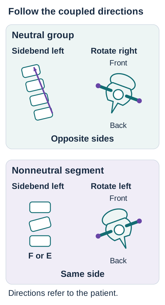
Separate views distinguish sidebending from rotation. Neutral groups couple opposite sides; nonneutral segments couple the same side. [1]Open full-size diagram
Fryette's first principle describes a neutral group: sidebending and rotation are in opposite directions. T4-T8 neutral, sidebent left and rotated right can be written T4-T8 N Sl Rr. Neutral here means no defining flexion or extension preference of that pattern, not a completely flat thoracic spine. The normal thoracic curve remains. [1]
The second principle describes a nonneutral segment with a flexion or extension preference: rotation and sidebending are to the same side. T6 extended, rotated right and sidebent right is T6 E Rr Sr. The compact form ERS right means the same thing. A focal pattern can coexist with a surrounding neutral group; do not make one erase the other.
Trace the two arrows in the diagram before reading the labels. A neutral group sidebent right pairs with left rotation. A flexed segment with left rotation pairs with left sidebending in this model. Neutral: opposite. Nonneutral: same. These are useful osteopathic conventions, not proof that every measured spine must behave identically. Contradictory findings require reassessment, not a declaration that the patient's anatomy is impossible. [1]
A related third principle states that establishing motion in one plane alters the available motion in the others. Prepositioning commonly reduces remaining freedom. It explains why testing and localization depend on position; it does not justify forcing a segment or automatically treating the most restricted level first. A Type I pattern need not be chronic, and a Type II pattern need not be caused by trauma. History and examination establish those claims separately.
Transfer: a patient has a longstanding focal flexion preference with right rotation and right sidebending. Duration does not convert the finding into a neutral group. Its measured pattern still supports F Rr Sr.
Name the position of ease, not the failed test
For a conventional positional diagnosis, a more posterior right transverse process indicates right rotation of the vertebra. Determine the sagittal preference by comparing the same segment during flexion and extension. Improved symmetry in flexion supports a flexed dysfunction; improved symmetry in extension supports an extended dysfunction. Do not substitute the direction that hurts for the measured restriction. [2][4]
Two findings that must agree
Observed response
Named preference
Restricted direction
Observed responseAsymmetry decreases in flexion, increases in extension
Named preferenceFlexed, F
Restricted directionExtension
Observed responseAsymmetry decreases in extension, increases in flexion
Named preferenceExtended, E
Restricted directionFlexion
Observed responseA multilevel pattern has no defining sagittal preference
Named preferenceAssess a neutral group, N
Restricted directionDetermine rotation and sidebending separately
Unchanged asymmetry alone does not prove a neutral somatic dysfunction: structural anatomy, positioning and measurement error may also affect the examination. Check the distribution, tissue findings and actual range before assigning a group diagnosis.
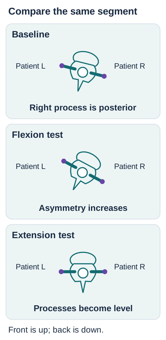
The same modeled segment becomes more asymmetric in flexion and symmetric in extension. [2][4]Open full-size diagram
Test the same segment in different positions
In this simplified model, the right transverse process begins posterior to the left. Predict whether the difference will shrink or grow during each test, then open that test. Each optional panel shows the same segment under one test. Closing it returns attention to the baseline comparison. No force is being prescribed.
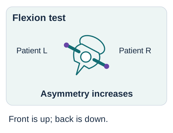
What appears when the model is tested in flexion?Same model, flexion test.
The right-left depth difference increases. Flexion exposes the restriction rather than naming the preferred position. This is evidence against an F diagnosis in the model.
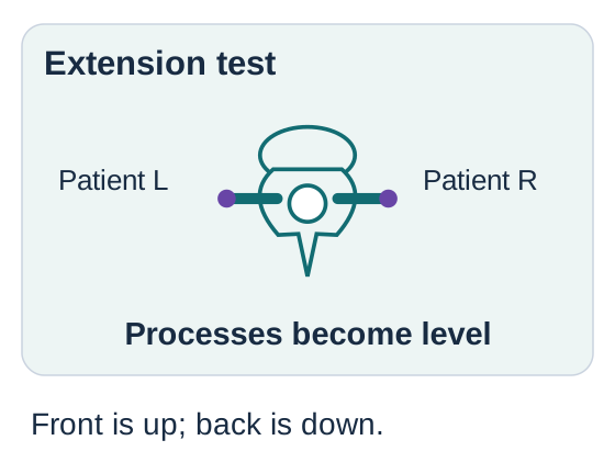
What appears when the model is tested in extension?Same model, extension test.
The two processes become level. Extension is the position of ease. Combining that response with the original posterior right process gives E Rr Sr under conventional Type II coupling.
Complete comparison: the same right-sided asymmetry becomes larger in flexion and smaller in extension. Therefore the model prefers extension and is restricted toward flexion. Its diagnosis is E Rr Sr, not F Rr Sr. The diagram records relative depth, not measured degrees or millimeters. It is not an anatomic simulation.
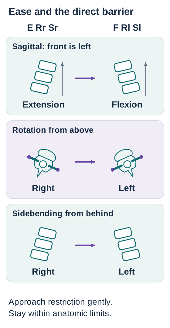
A direct technique approaches the restrictive directions. For E Rr Sr those directions are flexion, left rotation and left sidebending. For F Rl Sl they are extension, right rotation and right sidebending. An indirect technique initially positions toward ease instead. These are positioning concepts; they do not specify a universal thrust vector. [5][6]
Transfer: keep the right process posterior but reverse the test response, so symmetry now improves in flexion. Only the sagittal letter changes: F Rr Sr. The direct sagittal barrier is now extension.
Try it here · Checkpoint 1 of 3
Make your prediction before reading the choices. A first attempt is just a starting point.
Case 1
Show answer and explanations for case 1
A. Extension, left rotation and left sidebending (Why this does not fit)
Left rotation and left sidebending oppose the measured right-sided preferences. Extension reduces this segment's asymmetry, so it is ease rather than the sagittal barrier. Check each axis rather than accepting a partly correct setup.
Reasoning steps for option A
Why do left rotation and left sidebending in this T8 setup seem plausible?
The posterior right transverse process and easier right sidebending indicate rightward ease, so leftward motion approaches both transverse barriers.
Why does adding extension defeat this direct setup?
T8 asymmetry nearly disappears in extension and increases in flexion; extension follows ease instead of the flexion barrier.
Which axis must change to make this option fully direct?
Replace extension with flexion while retaining left rotation and left sidebending, opposing all three T8 preferences.
B. Flexion, right rotation and right sidebending (Why this does not fit)
Flexion approaches the tested sagittal restriction. Right rotation and right sidebending retain the other preferences instead of opposing them. A direct setup addresses all relevant restrictive directions.
Reasoning steps for option B
Which part of this position addresses the T8 sagittal barrier?
Flexion increases the T8 asymmetry, so flexion approaches its sagittal restriction.
Why are right rotation and right sidebending not direct barriers here?
The posterior right transverse process indicates right rotation and testing finds right sidebending easier; both rightward directions follow ease.
What correction would make this a three-axis direct setup?
Keep flexion but use left rotation and left sidebending to oppose the two rightward preferences.
C. Flexion, left rotation and left sidebending (Best answer)
A direct setup approaches the directions opposite the documented ease. Improvement in extension plus posterior right process and easier right sidebending indicates E Rr Sr. Name the preference first, then identify its restrictive directions.
Reasoning steps for option C
Which T8 positional diagnosis leads to this three-direction setup?
Asymmetry improving in extension, a posterior right transverse process, and easier right sidebending indicate E Rr Sr.
How does each proposed direction relate to that diagnosis?
Flexion opposes extension ease, while left rotation and left sidebending oppose the rightward preferences.
Why does the direct nonthrust request favor this option?
A direct setup approaches the restrictive direction on each of the three tested axes, rather than positioning into ease.
D. Extension, right rotation and right sidebending (Why this does not fit)
Positioning into all directions of ease is an indirect concept. The question specifies a direct technique for a segment that prefers extension and the right. Distinguish technique category before selecting its setup.
Reasoning steps for option D
What does extension with both rightward directions reproduce at T8?
Extension reduces asymmetry, the right process is posterior, and right sidebending is easier: these are the E Rr Sr directions of ease.
Which treatment category could start in this complete ease position?
An indirect approach could follow extension, right rotation and right sidebending initially.
Why is this option not the requested initial position?
The clinician selected a direct nonthrust approach, which instead approaches flexion, left rotation and left sidebending barriers.
Takeaway: Improvement in extension supports E; direct positioning approaches flexion and the opposite rotation and sidebending.
Separate respiratory position from vertebral position
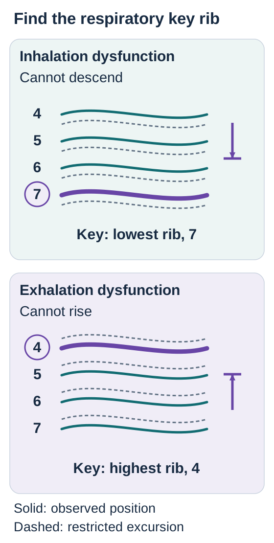
Does failure to rise mean an inhalation dysfunction? No: name the easier phase. A rib that remains relatively high during exhalation has an inhalation dysfunction: inhalation is its easier direction, and downward excursion during exhalation is restricted. An exhalation dysfunction remains relatively low and lacks normal upward excursion during inhalation. Observe both phases rather than naming the pattern from the first position seen. [4][6]
Trace the restricted phase, then identify the matching group boundary. Solid curves show observed positions; dashed curves show restricted excursion. [4][6]Open full-size diagram
For a respiratory group, identify the limiting rib in the appropriate phase. In an inhaled group, the lowest affected rib is the conventional key rib. In an exhaled group, the highest affected rib is the key rib. Thus ribs 4-7 that remain high on exhalation point to rib 7; ribs 4-7 that fail to rise on inhalation point to rib 4. This selects a treatment focus after diagnosis and safety assessment, not permission to apply a thrust. [6]
Use the diagram as a tracing task: follow the exhalation arrow downward and locate the bottom of the persistently high group. Then follow the inhalation arrow upward and locate the top of the persistently low group. Changing the restricted phase changes which end of the group matters. Transfer this to ribs 6-9: inhalation dysfunction identifies rib 9; exhalation dysfunction identifies rib 6.
Find the structure before interpreting its depth
The posterior rib angle is a useful palpatory region lateral to the transverse processes. Follow the rib contour and compare its respiratory excursion. The sympathetic trunk lies deeper near the rib heads; the posterior rib angle is not a direct surface marker of the ganglia. Pressure at an angle does not demonstrate direct ganglion contact or selective control of a particular organ. [7][8]
The traditional rule of threes estimates where a spinous-process tip lies relative to its own transverse processes. T1-T3 and T12 are taught as approximately level; T4-T6 and T11 as half a segment below; T7-T10 as one segment below. Starting from a spinous tip, the corresponding transverse processes are therefore sought superiorly by the taught offset, not further inferiorly.
For example, the traditional estimate places T8 transverse processes about one segment above the T8 spinous tip. This is a mnemonic, not an accurate coordinate system for every person. A cadaveric study found substantial disagreement with the rule. Confirm anatomy rather than assigning a precise level from that shortcut alone, especially before a procedure. T1 and T12 are transitional regions, not interchangeable copies of a typical midthoracic vertebra. [3]
Try it here · Checkpoint 2 of 3
Make your prediction before reading the choices. A first attempt is just a starting point.
Case 9
Show answer and explanations for case 9
A. Rib 4 (Best answer)
An exhaled group is localized at its current uppermost affected rib. Rib 3 is no longer restricted, and the remaining group begins at rib 4. Reassess the actual group boundaries after treatment rather than retaining the original key.
Reasoning steps for option A
What pattern persists after rib 3 moves normally?
Left ribs 4-6 remain low and fail to rise with inhalation, an exhaled group.
Where is the upper boundary of the remaining restriction?
Rib 4 is now the highest rib still failing to rise.
Why does that make rib 4 the reassessment key?
An exhaled group's key is its current uppermost affected rib, not the original rib 3.
B. Rib 3 (Why this does not fit)
Rib 3 was the original upper boundary of the exhaled group. Its excursion is now normal in both phases. An original treatment target is not automatically the target at reassessment.
Reasoning steps for option B
Why was rib 3 initially a reasonable key?
It was the highest affected rib when the exhaled group included ribs 3-6.
What changed at rib 3 following intervention?
It now has normal excursion through both respiratory phases.
Why not retain rib 3 as the key?
The residual exhaled group starts at rib 4, so its upper boundary has moved.
C. Rib 6 (Why this does not fit)
The lower boundary is the key in an inhaled group. The remaining restriction is still failure to rise on inhalation, not failure to descend. Changing the size of a group does not necessarily change its respiratory pattern.
Reasoning steps for option C
Which group rule could point to rib 6?
Rib 6 is the lowest remaining affected rib, the key location for an inhaled group.
Do ribs 4-6 show an inhaled restriction?
No. They fail to rise during inhalation rather than remain high during exhalation.
Which end is keyed for their actual pattern?
Their exhaled pattern calls for the upper affected rib, rib 4, not rib 6.
D. Rib 7 (Why this does not fit)
A nearby normal rib can be useful for comparison. Rib 7 was not part of either documented restricted group. Follow the observed residual restriction instead of extending the diagnosis beyond the data.
Reasoning steps for option D
Why might an examiner palpate rib 7?
It can provide comparison with the nearby residual ribs 4-6.
Is rib 7 documented as unable to rise?
No. Neither the original restricted ribs 3-6 nor the remaining ribs 4-6 include rib 7.
Why cannot rib 7 guide this group's reassessment?
A respiratory key must be within the currently documented restricted group, at rib 4 here.
Takeaway: After one rib normalizes, identify the boundaries and key of the remaining respiratory group.
A correct label does not automatically justify a thrust
Would every E Rr Sr finding receive the same treatment? No. First establish consent, a meaningful indication, the patient's ability to cooperate and the absence of a concerning alternative diagnosis. High-velocity, low-amplitude treatment, or HVLA, uses a brief, small-amplitude force at a localized restrictive barrier. It must remain within anatomic limits. The shorthand does not encode force magnitude, contact choice or patient suitability. Hands-on training and supervision are required. [5]
Each arrow points from ease toward a restrictive direction. This describes positioning, not a thrust vector. [4][5]Open full-size diagram
Trace each axis from the position of ease to the restrictive direction in the diagram. The opposite directions describe a direct setup, not an instruction to push maximally in three directions. A patient with an unstable fracture has the same directional vocabulary as anyone else, but that does not make a manipulative setup appropriate.
Recognize the position, not a rigid regional recipe
In a supine crossed-arm approach, the patient's thorax is supported while a trained clinician localizes the target. The cited procedure describes this as the Kirksville Crunch; it is also commonly called a supine thoracic technique. Seated methods use a different patient position and clinician support. Prone variants are another option in appropriately selected patients. Technique names vary, so describe the position and contact rather than treating a nickname as universal. Do not assign a compulsory vertebral range or a fixed 45-degree thrust to every patient. Exhalation can facilitate relaxation, but respiratory timing does not make an unsafe treatment safe. [5]
Suspected fracture, local spinal infection, destructive malignancy, instability or acute cord compromise requires evaluation rather than local thrust treatment. Significant osteoporosis and prolonged glucocorticoid exposure increase concern for fragility injury. Anticoagulation raises bleeding risk and requires individualized assessment; it is not interchangeable with uncomplicated stiffness. Neurologic deficits, recent trauma and progressive pain change the decision. An incidental degenerative image finding alone does not determine suitability. Screen the whole setup, including any cervical positioning. [5][14]
Judge the response without chasing a sound
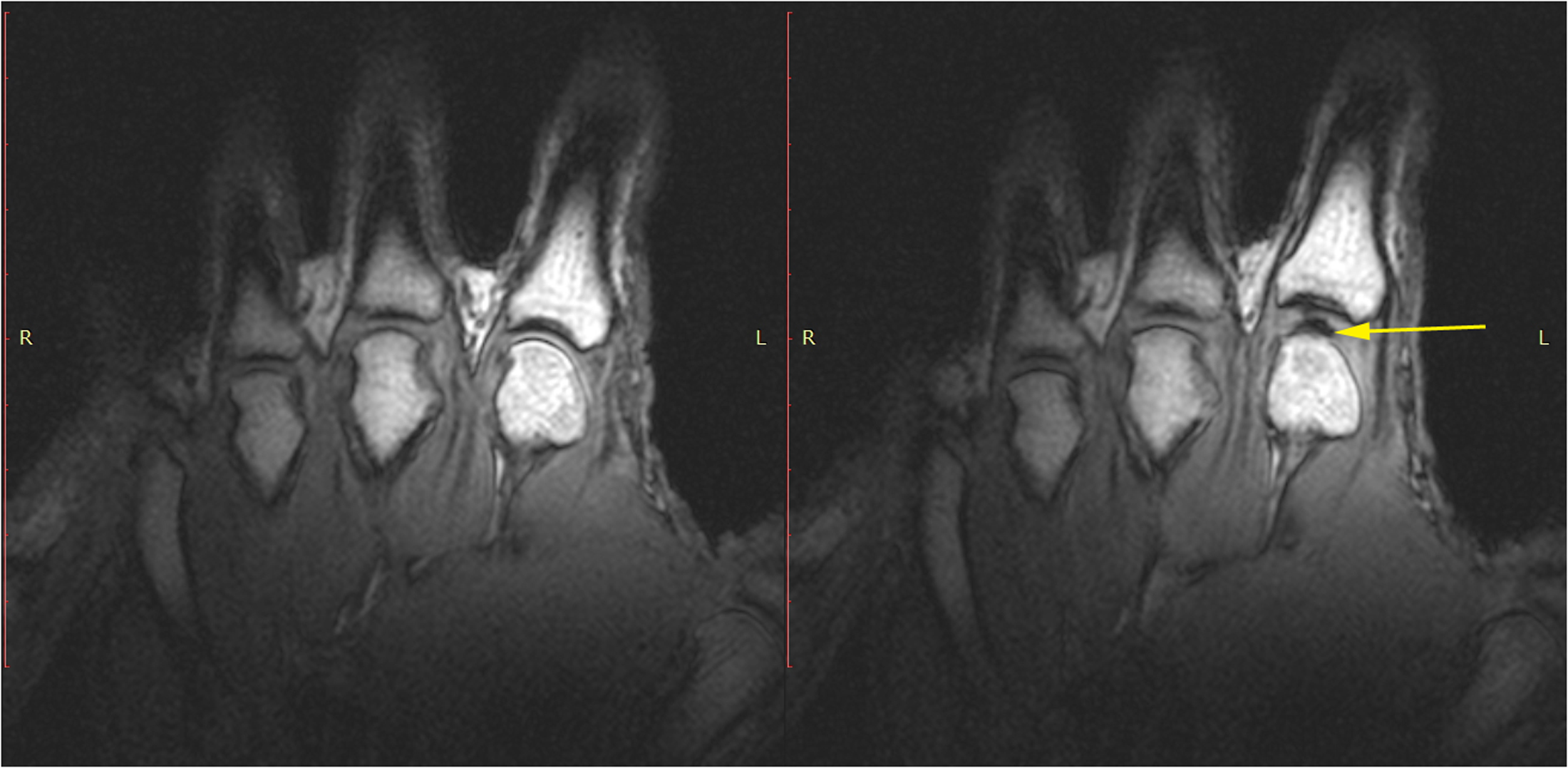
Joint distraction can produce cavitation, a gas-containing cavity associated with rapid separation of joint surfaces. Real-time MRI demonstrated cavity formation in finger joints. That study does not establish the exact acoustic mechanism in every thoracic joint, identify nitrogen as the only gas, or show that an audible event means a vertebra has been corrected. [10]
Compare the finger joint-space MRI before and after cracking with additional distraction. Locate the void marked by the arrow; this finger-joint demonstration does not map a thoracic segment. Image: Kawchuk and colleagues, PLOS ONE (2015); source figure; CC BY 4.0. [10]
Reassess the original restriction, function, symptoms and neurologic status. Improvement without a pop can be useful; a pop without improvement is not proof of success. Mild transient soreness can occur, but new severe or progressive pain, weakness, sensory change or breathing difficulty calls for prompt assessment rather than reassurance based on the sound. Never repeat a thrust merely to obtain cavitation. [5]
Transfer: a patient develops focal pain after coughing and has known osteoporosis. A plausible positional diagnosis does not exclude a compression fracture. Evaluate the fragility-injury possibility before selecting a manual technique.
Use visceral connections to widen assessment, not to make a diagnosis
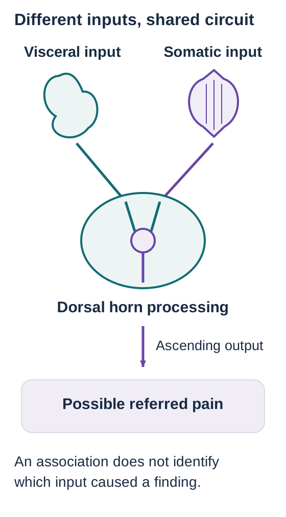
Can a tender T6 diagnose gallbladder disease? No. Visceral and somatic sensory input can converge on spinal circuits, helping explain referred pain and associated muscular responses. Experiments in rat spinal cord demonstrate convergence of visceral and somatic C-fiber input. This is a plausible neural substrate, not a validated organ-disease test based on palpation. Sensory afferents that accompany sympathetic pathways are not the same thing as sympathetic motor output. [15]
A shared circuit supports referred-pain physiology. It cannot identify the source of a clinical finding. [15]Open full-size diagram
Trace each input toward the shared spinal circuit. The overlap explains why the perceived or muscular response need not sit directly over the diseased organ. Now reverse the inference: finding a somatic response does not identify which input caused it. A musculoskeletal problem and a visceral illness can also coexist.
Commonly taught, overlapping sympathetic regions
Region
Examples and interpretation
RegionT1-T5
Examples and interpretationHeart. Cardiac assessment depends on symptoms and clinical testing, not the side of a thoracic finding.
RegionUpper to middle thoracic
Examples and interpretationLungs are commonly taught as T2-T7; some sources use T1-T6. Esophagus is often listed as T2-T8.
RegionT5-T9
Examples and interpretationForegut structures, including stomach, liver and gallbladder. Right-sided findings can accompany biliary disease but are not diagnostic.
RegionT10-T11
Examples and interpretationMidgut, including small bowel and proximal colon. Gonadal pathways are also commonly mapped here.
RegionT12-L2
Examples and interpretationHindgut, including distal colon and rectum, not the entire colon.
RegionT10-L1 region
Examples and interpretationKidney and ureter associations overlap; the cited chart places kidney at T10-T11 and ureter at T10-L1. Adrenal association is commonly listed near T10.
RegionLower thoracic to upper lumbar
Examples and interpretationBladder commonly T11-L2; uterus and cervix T10-L2. These sympathetic maps are not a complete account of pelvic sensory or parasympathetic pathways.
These are educational autonomic maps with variable boundaries. Spinal cord segment labels, palpated vertebral levels and dermatomes are not interchangeable coordinates. An isolated T9 finding cannot be called a match merely by claiming it lies within T10-L1. Likewise, C8-T1 findings in the hand or forearm concern a somatic neurologic localization, not a cardiac sympathetic level. Use a full neurologic examination when limb symptoms are present. [7]
Chest pressure, exertional upper abdominal discomfort, dyspnea or diaphoresis requires an appropriate cardiac assessment even when pain is reproducible or thoracic asymmetry is present. A thoracic finding neither rules in ischemia nor rules it out. Acute concerning symptoms take priority over manipulation. [11]
Early appendicitis can cause poorly localized periumbilical discomfort, classically associated with the T10 region. Later localization to the right lower abdomen reflects a different pattern of involvement, including the parietal peritoneum; the disease has not traveled from one vertebra to another. Fever, worsening abdominal pain and guarding require urgent assessment. [16][18]
Fever, flank pain, costovertebral angle tenderness and inflammatory urine findings support suspected pyelonephritis. Obtain a urine culture before antibiotics when feasible without an unsafe delay, start appropriate antimicrobial treatment and assess severity. Neither a positive dipstick alone nor a thoracic level proves the diagnosis. Right upper abdominal attacks after meals instead warrant biliary assessment, often including ultrasound. Persistent pain, fever or jaundice demands prompt evaluation for complications. [12][13][17]
Peptic disease can also cause epigastric symptoms; heartburn and indigestion are not interchangeable, and dysphagia warrants prompt assessment. [19] Acute testicular pain, particularly in adolescents, requires urgent evaluation for torsion rather than a spinal treatment trial. [20]
Keep proposed physiology separate from proven benefit
A somatovisceral reflex describes influence in the other direction, from somatic input toward visceral responses. It does not establish that thoracic dysfunction causes an arrhythmia or that its treatment cures gastric disease. A small rib-raising study found short-term changes in a salivary sympathetic biomarker in healthy volunteers; this is not evidence of reliable clinical control of an organ. [4][8]
Rib raising and thoracic lymphatic pumping are distinct techniques. The former addresses rib and paraspinal mechanics; the latter uses rhythmic thoracic compression and release with a proposed fluid-flow effect. Neither mechanically drains alveolar exudate into the thoracic duct or replaces antibiotics. In a randomized pneumonia trial, the primary intention-to-treat comparisons did not show significant differences. Favorable per-protocol findings do not establish benefit for every patient or isolate the effect of one component. [9]
Transfer: if an adjunct improves rib excursion while fever and oxygen requirements persist, the infection still needs medical treatment and reassessment. Better mechanics is not evidence of pathogen clearance.
Try it here · Checkpoint 3 of 3
Make your prediction before reading the choices. A first attempt is just a starting point.
Case 23
Show answer and explanations for case 23
A. Somatic and visceral afferents synapse with one another inside a dorsal root ganglion (Why this does not fit)
Both types of sensory neuron have cell bodies associated with spinal sensory pathways. A dorsal root ganglion is not the usual synaptic relay described by the spinal convergence model. Distinguish sensory neuron cell-body location from the central site of processing.
Reasoning steps for option A
Where are spinal sensory neuron cell bodies located?
Visceral and somatic primary sensory neurons have cell bodies in dorsal root ganglia.
Is the dorsal root ganglion the proposed site where these inputs synapse together?
No. The convergence model concerns shared central processing in the spinal dorsal horn, not a synapse between afferents in the ganglion.
How should the right midback discomfort be explained without overreading palpation?
Shared spinal processing can account for referred discomfort, but nonspecific right-sided tissue changes cannot identify a gallbladder diagnosis.
B. Preganglionic sympathetic motor fibers carry the perceived pain directly from the gallbladder to skeletal muscle (Why this does not fit)
Sympathetic pathways participate in visceral regulation and sensory fibers can travel alongside them. Preganglionic motor output is not the sensory afferent route establishing referred pain. Separate afferent sensation from autonomic efferent control.
Reasoning steps for option B
Why could sympathetic anatomy seem relevant to gallbladder-related pain?
Visceral sensory fibers may travel alongside sympathetic pathways from the organ toward spinal levels.
Which direction and function does a preganglionic sympathetic motor fiber have?
It carries autonomic efferent output away from the spinal cord, not perceived pain from gallbladder to skeletal muscle.
What neural signal is needed for the referred-pain explanation?
Visceral afferent input converging centrally with somatic sensory input, rather than sympathetic motor output.
C. Somatic and visceral sensory inputs converge on shared dorsal horn circuits (Best answer)
Convergence permits different peripheral sources to influence shared spinal processing. The visceral symptoms and somatic region can be associated without proving that a palpated segment caused the illness. A neural substrate explains plausibility; it does not turn palpation into an organ-specific test.
Reasoning steps for option C
What inputs can produce a shared spinal perception?
Visceral input associated with the meal-related right upper abdominal pain and somatic input from the right midback can reach shared dorsal horn circuits.
Why does dorsal horn convergence fit the question’s processing level?
Central convergence offers a substrate for referred pain without requiring direct communication between peripheral tissues.
What does right-sided thoracic palpation not prove?
It does not show that a particular segment caused the gallstones or independently establish an organ-specific diagnosis.
D. All gallbladder pain is carried by vagal motor fibers terminating in the thoracic dorsal horn (Why this does not fit)
The vagus contributes to visceral innervation and has sensory as well as motor components. Vagal motor fibers do not terminate as the proposed thoracic spinal sensory relay. Keep spinal visceral afferent pathways distinct from vagal autonomic output.
Reasoning steps for option D
Does the vagus have a role in visceral innervation?
Yes. It carries visceral sensory and autonomic components, making it superficially relevant to gallbladder physiology.
Can vagal motor fibers provide the proposed thoracic dorsal horn relay?
No. Motor output is not a somatic-visceral sensory convergence pathway in the spinal dorsal horn.
Which distinction matters for this patient’s referred midback pain?
A spinal afferent processing model is different from vagal efferent regulation of the gallbladder.
Takeaway: Spinal sensory convergence supports referred-pain physiology without proving the cause of a palpatory finding.
Keep the structural and medical questions separate
A 52-year-old reports three weeks of midback stiffness and intermittent epigastric discomfort. The right T6 transverse process is posterior. Its asymmetry decreases in extension and increases in flexion; right sidebending is easier than left. There has been no injury. Before selecting a technique, what can already be concluded?
Structural conclusion: the measured pattern supports T6 E Rr Sr. The sagittal response, not the duration or abdominal symptom, determines E. A direct setup would approach flexion, left rotation and left sidebending, provided treatment is otherwise appropriate. [1][2][5]
Now add one piece of history: the epigastric discomfort reliably begins while climbing stairs and is accompanied by sweating. Pause and predict which decision changes. The structural notation does not change, but the care priority does. A possible cardiac equivalent requires prompt medical assessment rather than a trial of manipulation to decide whether the symptom was muscular. [11]
In a different patient, similar discomfort might instead follow heavy meals, localize to the right upper abdomen and coincide with gallstones on ultrasound. Those findings support a biliary explanation; T6 alone would not. Neither story permits a diagnosis of organ disease from palpation, and neither permits reassurance merely because the segment becomes less tender. [13][17]
Carry two statements forward: name the mechanical findings accurately, and investigate the medical problem on its own evidence. Do not let either statement substitute for the other.
Independent clinical practice
These are original educational cases, not patient records or recalled examination items. Read the entire presentation, decide what the question asks and formulate an answer before comparing the options. Explanations remain available without a timed or compulsory sequence.
Case 2
Show answer and explanations for case 2
A. Flexion, left rotation and left sidebending (Best answer)
An indirect technique initially follows the directions of ease. Flexion reduces the asymmetry and both remaining preferences are leftward. The same diagnosis produces different starting positions for direct and indirect care.
Reasoning steps for option A
What sagittal response supports flexion at T6?
The left transverse-process asymmetry decreases in flexion and increases in extension, making flexion the easier direction.
Why are both leftward components appropriate for an indirect start?
The posterior left process supports left rotational preference, and left sidebending is easier than right.
How does this position implement the chosen low-force strategy?
It follows all three F Rl Sl directions of ease rather than approaching the extension and rightward barriers.
B. Extension, right rotation and right sidebending (Why this does not fit)
This position opposes an F Rl Sl pattern. The clinician has selected an indirect rather than a direct approach. Read the technique category as well as the positional diagnosis.
Reasoning steps for option B
What does extension with right rotation and right sidebending represent for T6?
Each direction opposes the flexion, left rotation and left sidebending preferences, forming the direct-barrier setup.
Which detail of the clinical request excludes that setup?
The patient prefers an indirect low-force approach, which initially follows ease, not all three restrictions.
Which setup follows the actual T6 ease instead?
Flexion, left rotation and left sidebending matches the improving flexion test and leftward findings.
C. Flexion, right rotation and right sidebending (Why this does not fit)
Flexion fits the measured sagittal preference. Right rotation and right sidebending oppose the other two preferences. A partly matching position does not describe complete ease.
Reasoning steps for option C
Why is flexion a reasonable component of this proposed T6 position?
The asymmetry becomes smaller in flexion, consistent with sagittal ease.
What makes the rightward components inconsistent with complete ease?
The posterior left process and easier left sidebending indicate leftward, not rightward, rotational and sidebending preferences.
What changes yield the requested indirect position?
Keep flexion and switch both rightward components to left rotation and left sidebending.
D. Extension, left rotation and left sidebending (Why this does not fit)
The leftward directions agree with the measured rotational and sidebending ease. Extension increases the asymmetry and is the restricted sagittal direction. Do not name a dysfunction after the test that worsens it.
Reasoning steps for option D
Which elements of this T6 position follow measured ease?
The posterior left transverse process and easier left sidebending support the two leftward components.
What does extension do to the T6 asymmetry?
It enlarges the asymmetry, so extension is the sagittal restriction rather than the indirect starting direction.
Which sagittal component should replace extension?
Flexion decreases the asymmetry and completes the F Rl Sl ease position.
Takeaway: F Rl Sl is placed into flexion and leftward ease for an indirect approach.
A. Right rotation and right sidebending (Why this does not fit)
Right sidebending opposes the observed left sidebending. Right rotation is the observed rotational ease, not its opposite. Evaluate the rotational and sidebending axes independently.
Reasoning steps for option A
Which half of this pair opposes the T4-T7 group preference?
Right sidebending opposes the observed left sidebending.
Why is right rotation not a restrictive direction?
The group already rotates right; its rotational barrier is left rotation.
What pair opposes both observed group directions?
Left rotation and right sidebending oppose the right rotation and left sidebending pattern independently.
B. Left rotation and left sidebending (Why this does not fit)
Left rotation opposes the observed right rotation. Left sidebending repeats the observed sidebending preference. Opposite coupling does not mean both barriers are leftward.
Reasoning steps for option B
Why does left rotation belong among the restrictive directions?
It opposes the group’s observed right rotational preference.
Why does left sidebending fail the restriction test?
The group already sidebends left, so right sidebending is the sidebending barrier.
Does opposite-sided neutral coupling make both barriers leftward?
No. The T4-T7 group is right-rotated and left-sidebent, making its barriers left rotation and right sidebending.
C. Right rotation and left sidebending (Why this does not fit)
These directions describe the supplied neutral group position. The question asks for restriction rather than the position used to name the group. A diagnosis and its restrictive directions are complementary descriptions.
Reasoning steps for option C
What does right rotation with left sidebending describe in this stem?
It restates the observed T4-T7 neutral group position without a defining flexion or extension preference.
Why does that pair not answer the question?
The question asks for relatively restricted directions, not the directions of positional ease.
Which paired directions are restricted instead?
Oppose each observed axis: left rotation and right sidebending.
D. Left rotation and right sidebending (Best answer)
Restrictive directions oppose the observed positional preferences. The neutral group is rotated right and sidebent left. A neutral group retains opposite coupling in its paired restrictive directions.
Reasoning steps for option D
How is the T4-T7 neutral group positioned on the two tested axes?
It rotates right and sidebends left, with no defining sagittal preference.
Which directions oppose each of those preferences?
Left rotation opposes right rotation; right sidebending opposes left sidebending.
Why is this pair the restrictive answer?
Both components are barriers relative to the observed group position, while the alternatives repeat at least one direction of ease.
Takeaway: T4-T7 N Sl Rr implies relative restriction toward right sidebending and left rotation.
A change in sagittal test response can change the first letter. The first response improves in flexion, not extension. Assign F or E from ease at each examination.
Reasoning steps for option A
Why might the sagittal letter change between these T7 examinations?
The asymmetry improves in flexion first but in extension after the findings change.
What is reversed in the proposed E-to-F sequence?
The first examination favors flexion, so it begins F; the second favors extension, so it ends E.
Which letters remain stable through both examinations?
The posterior right process and easier right sidebending support Rr Sr at both times.
B. F Rr Sr to E Rr Sr (Best answer)
The sagittal letter follows the position that reduces asymmetry. The first examination favors flexion and the second favors extension while the other preferences stay rightward. Change only the diagnostic components supported by new findings.
Reasoning steps for option B
What establishes F in the first T7 notation?
The first examination shows decreasing asymmetry in flexion and increasing asymmetry in extension.
What establishes E while retaining Rr Sr in the second?
The second examination improves in extension, while the right posterior process and easier right sidebending persist.
Why change only the first letter?
Only the sagittal ease reverses; the supplied rotation and sidebending findings remain rightward.
C. F Rl Sl to E Rl Sl (Why this does not fit)
The F-to-E sequence matches the changed sagittal response. Both examinations show a posterior right process and easier right sidebending. A changed sagittal response does not reverse the measured rotational side.
Reasoning steps for option C
Which portion of this proposed transition matches the tests?
F to E matches the first flexion-improving and second extension-improving responses.
Why are Rl Sl unsupported at either time?
Both examinations retain a posterior right transverse process and easier right sidebending, supporting Rr Sr.
What should the complete transition read?
F Rr Sr to E Rr Sr changes the documented sagittal preference without inventing a leftward shift.
D. N Sl Rr to N Sr Rl (Why this does not fit)
Neutral groups have opposite rotation and sidebending. Both examinations show a focal sagittal preference with same-sided rotation and sidebending. Do not relabel a nonneutral segment as neutral merely because findings change.
Reasoning steps for option D
What feature might superficially suggest a neutral label?
Neutral group notation pairs opposite rotation and sidebending, as in Sl Rr or Sr Rl.
Why does neutral notation not describe these T7 findings?
Each examination has focal flexion or extension ease and same-sided right rotation and sidebending.
What diagnostic change actually occurred?
The focal T7 sagittal preference changed from F to E; right rotation and sidebending did not reverse.
Takeaway: Repeat examinations can change the sagittal label without changing the rotation or sidebending label.
A. Search approximately one segment superiorly, then verify the actual anatomy (Best answer)
The traditional rule places the T8 spinous tip about one segment below its own transverse processes. Cadaveric evaluation shows that this rule is not consistently accurate in individual specimens. Use a landmark mnemonic as a provisional estimate rather than a procedural coordinate.
Reasoning steps for option A
Where does the traditional rule place the T8 transverse processes relative to its spinous tip?
Approximately one segment superior to the T8 spinous-process tip.
Why must this superior search remain provisional?
Cadaveric evaluation finds the rule inconsistent across individual specimens; a mnemonic cannot fix an exact contact point.
What should happen before choosing the palpatory contact?
Verify the patient’s actual anatomy after using the one-segment-superior estimate.
B. Search approximately one segment inferiorly, then verify the actual anatomy (Why this does not fit)
Independent confirmation is appropriate because surface landmarks vary. The traditional relationship places the spinous tip inferior to its own transverse processes, so this search direction is reversed. Separate the direction of an estimate from confidence in its precision.
Reasoning steps for option B
What is appropriate about verifying anatomy after a T8 landmark estimate?
Individual surface anatomy varies, so an estimated level requires confirmation.
Why is one segment inferior the wrong search direction?
In the traditional T8 rule, the spinous tip lies below its own transverse processes, placing the transverse processes superiorly.
How should direction and certainty be combined here?
Begin roughly one segment superior, then verify; uncertainty does not reverse the traditional direction.
C. Search approximately half a segment superiorly and accept the level without further confirmation (Why this does not fit)
A half-segment offset belongs to other levels in the traditional scheme. T8 is taught in the full-segment group, and the rule is not exact enough to dispense with confirmation. Correct recall does not replace anatomic verification.
Reasoning steps for option C
Why might a half-segment superior estimate sound familiar?
The traditional rule assigns half-segment offsets at other thoracic levels.
Why does it fail specifically for the T8 spinous tip?
T8 belongs to the traditional full-segment offset group, not the half-segment group.
Why is accepting any unverified T8 level inappropriate?
The rule is imperfect in individual specimens, so even a correctly recalled offset needs anatomic confirmation.
D. Search at the same horizontal level and accept the level without further confirmation (Why this does not fit)
Same-level spinous and transverse processes are part of the traditional scheme for selected levels. T8 is not assigned that pattern, and individual variability remains substantial. Do not generalize one thoracic region's mnemonic to every level.
Reasoning steps for option D
When could a same-level spinous-tip estimate sound plausible?
The traditional scheme uses same-level relationships at selected thoracic levels.
Why is that estimate misplaced at T8?
The traditional T8 transverse-process estimate is about one segment superior to its spinous tip.
What additional problem does accepting that level create?
It treats a variable surface landmark as a confirmed procedural coordinate without checking the patient’s anatomy.
Takeaway: The traditional T8 estimate is superior from the spinous tip, but the actual contact requires confirmation.
A. A single T6 E Rr Sr finding accounts for the entire region (Why this does not fit)
A focal nonneutral restriction can coexist with regional findings. The focal asymmetry improves in flexion, and the surrounding neutral group remains separately demonstrable. Do not erase a regional pattern or reverse sagittal ease.
Reasoning steps for option A
Which aspect of this option recognizes a real T6 finding?
The focal right-sided asymmetry with easier right sidebending supports a segmental rightward pattern.
Why is E at T6 inconsistent with testing?
T6 asymmetry nearly disappears in flexion and worsens in extension, indicating F rather than E.
What regional finding would this single-segment label omit?
The T4-T8 group remains left-sidebent and right-rotated without a defining sagittal preference, requiring separate neutral-group documentation.
B. A T4-T8 neutral group alone accounts for all findings (Why this does not fit)
The multilevel opposite-sided pattern supports a neutral group. T6 also has a focal, reproducible sagittal preference with right sidebending. Assess focal findings within a broader pattern rather than averaging them away.
Reasoning steps for option B
Why is a T4-T8 neutral group supported?
The multilevel pattern has left sidebending and right rotation and remains similar in flexion and extension.
What focal evidence is lost if the group stands alone?
T6 has extra right asymmetry that eases in flexion and focal right sidebending preference.
How should T6 be documented alongside the group?
Record focal T6 F Rr Sr in addition to the T4-T8 N Sl Rr group.
C. T4-T8 N Sl Rr with a focal T6 F Rr Sr finding (Best answer)
Neutral group and focal nonneutral patterns can be described separately. The surrounding pattern is opposite-sided, while the focal T6 asymmetry improves in flexion. Preserve distinct regional and segmental findings when both are demonstrated.
Reasoning steps for option C
Which tests support the T4-T8 N Sl Rr component?
The group sidebends left and rotates right, with no defining flexion-extension preference.
Which tests support the separate T6 F Rr Sr component?
Focal T6 right-sided asymmetry nearly vanishes in flexion, grows in extension, and favors right sidebending.
Why preserve both entries rather than merge them?
The focal nonneutral T6 response and the broader neutral group are distinct simultaneously demonstrated findings.
D. T4-T8 F Rr Sr without a separate T6 finding (Why this does not fit)
F Rr Sr fits the focal T6 findings. The surrounding segments retain left sidebending and no defining sagittal preference. A focal result should not be imposed on every segment in a group.
Reasoning steps for option D
Which part of this regional label resembles the focal T6 finding?
T6 shows flexion ease and right-sided rotational and sidebending preferences, consistent with focal F Rr Sr.
Why can that label not be extended to all T4-T8?
The surrounding group instead sidebends left, rotates right, and has no defining flexion or extension preference.
What documentation avoids imposing the T6 pattern on the group?
Keep T4-T8 N Sl Rr and add T6 F Rr Sr as a separate focal finding.
Takeaway: A neutral group does not exclude a superimposed focal nonneutral finding.
A rib immediately above an affected group may be examined as a comparison. The documented restricted group begins at rib 4, not rib 3. Select the key from the demonstrated group rather than an adjacent normal level.
Reasoning steps for option A
Why might rib 3 be checked during examination?
It lies just above the affected ribs 4-7 and may provide a comparison level.
Why can rib 3 not be this group’s key?
The restricted group starts at rib 4; rib 3 is not documented as failing to rise on inhalation.
Which boundary determines the key instead?
Failure to rise defines an exhaled group, for which the uppermost affected rib, rib 4, is the key.
B. Rib 4 (Best answer)
An exhaled group is conventionally addressed at its uppermost affected rib. Ribs 4-7 fail to rise during inhalation, making rib 4 the upper limiting level. Use the restricted respiratory phase and group boundary, not maximum tenderness.
Reasoning steps for option B
Which respiratory motion is restricted in ribs 4-7?
They stay relatively low and fail to rise normally during inhalation while exhalation remains preserved.
Why does that make rib 4 the key?
For an exhaled group restricted in inhalation, the conventional key is the uppermost affected rib, here rib 4.
Why not choose the tender rib 6 instead?
Maximum tenderness identifies a symptom site but does not supersede the group’s respiratory boundary rule.
C. Rib 6 (Why this does not fit)
The most tender site can be important for symptom assessment. Tenderness does not replace the respiratory key-rib rule for this exhaled group. The symptom maximum and mechanical group boundary need not coincide.
Reasoning steps for option C
What specific finding draws attention to rib 6?
Its angle is the most tender point in the examined group.
Why does tenderness not establish the exhaled group’s key?
The key is based on restricted inhalation and the upper affected boundary, not on which rib is most tender.
Which rib does the group rule select?
Rib 4 is the uppermost member of ribs 4-7 that fail to rise normally.
D. Rib 7 (Why this does not fit)
The lowest affected rib is the key in an inhaled group. This group fails to rise on inhalation rather than failing to descend on exhalation. Determine which respiratory phase is restricted before choosing the group end.
Reasoning steps for option D
Under what different respiratory pattern would rib 7 be the key?
The lowermost affected rib is the key for an inhaled group restricted in exhalation.
Which observed motion contradicts that pattern?
These ribs descend during exhalation but fail to rise during inhalation, indicating an exhaled group.
Which end of this exhaled group is the key?
The upper end of ribs 4-7, rib 4, rather than the lower end at rib 7.
Takeaway: An exhaled group has restricted inhalation; its uppermost affected rib is the key.
An adjacent rib can provide a normal respiratory comparison. Rib 5 is outside the demonstrated group. Keep the treatment focus within the established mechanical finding.
Reasoning steps for option A
Why might rib 5 be checked after the coughing episode?
It could serve as an adjacent comparison for right rib motion, not a member of the restricted ribs 6-9.
Does rib 5 meet the observed group boundary?
No. Only ribs 6-9 are reported to remain high on exhalation.
Why does that exclude rib 5 as the key?
The key must lie in the demonstrated inhaled group, whose lowest member is rib 9.
B. Rib 6 (Why this does not fit)
The uppermost affected rib is the key for an exhaled group. These ribs fail to descend, so the group is inhaled despite tenderness at its upper end. Respiratory restriction is more informative for key selection than pain intensity.
Reasoning steps for option B
When would the upper rib 6 be the key?
The uppermost affected rib keys an exhaled group that fails to rise with inhalation.
What direction is restricted in ribs 6-9 here?
They rise but do not descend normally on exhalation, indicating an inhaled group.
Does maximal tenderness at rib 6 override the group rule?
No. Tenderness identifies a painful site, whereas exhalation restriction makes rib 9 the lower key.
C. Rib 8 (Why this does not fit)
A middle rib may be palpably restricted within a group. The conventional key for an inhaled group is the lowest affected member, which is rib 9. A group midpoint is not the respiratory key by default.
Reasoning steps for option C
Why is rib 8 a plausible mechanical candidate?
Rib 8 lies within the restricted right ribs 6-9 and may share their failure to descend.
Is rib 8 the lower boundary of this inhaled group?
No. Rib 9 is also affected and lies below rib 8.
What distinguishes the key from another involved rib?
For an inhaled group, choose its lowest restricted rib, not an interior rib such as 8.
D. Rib 9 (Best answer)
An inhaled group is conventionally addressed at its lowest affected rib. Ribs 6-9 remain high during exhalation, placing the lower boundary at rib 9. Failure to descend identifies inhalation dysfunction and the lower key.
Reasoning steps for option D
What respiratory pattern follows from ribs 6-9 staying high?
Failure to descend during exhalation identifies inhalation dysfunction.
Which involved rib lies at the lower boundary?
Rib 9 is the lowest of the documented affected ribs 6-9.
Why is rib 9 the conventional key despite rib 6 tenderness?
The lower affected rib keys an inhaled group; pain at the upper rib does not change its respiratory boundary.
Takeaway: Ribs that fail to descend have inhalation dysfunction; select the lowest affected rib.
A. The remaining ribs form an exhaled group keyed at rib 7 (Why this does not fit)
The uppermost affected rib is the key in an exhaled group. These ribs fail to descend during exhalation rather than failing to rise during inhalation. Name the respiratory preference from the restricted phase, not the phase being tested.
Reasoning steps for option A
What would make rib 7 the key for an exhaled group?
As uppermost of ribs 7-10, it would key a group unable to rise on inhalation.
Which respiratory movement actually remains restricted?
Right ribs 7-10 stay high and fail to descend on exhalation.
What key follows from this observed pattern?
This is an inhaled group, keyed at its lowest affected member, rib 10.
B. The vertebral response establishes that the rib dysfunction has resolved (Why this does not fit)
Vertebral and rib mechanics can influence one another. Direct rib testing still shows the original respiratory restriction. Improvement in one structure does not replace reassessment of its neighbors.
Reasoning steps for option B
Why might normalized T8 testing seem to settle the rib finding?
Vertebral and neighboring rib mechanics can be related, and the T8 asymmetry improved after treatment.
What does direct testing show after T8 becomes symmetric?
The same right ribs 7-10 still fail to descend on exhalation.
Why is vertebral improvement insufficient evidence of rib resolution?
The independently measured rib restriction persists despite a symmetric T8 test.
C. The remaining ribs form an inhaled group keyed at rib 10 (Best answer)
Failure to descend indicates an inhaled group with a lower key rib. The residual group is ribs 7-10 despite normalized T8 testing. Track vertebral and respiratory findings separately at reassessment.
Reasoning steps for option C
What pattern does failure of ribs 7-10 to descend identify?
Remaining high during exhalation identifies an inhaled rib group.
Which rib is its lower affected boundary?
Rib 10 is the lowest of the still-restricted right ribs 7-10.
How does the normalized T8 finding affect this key selection?
It does not erase the separate rib finding; the inhaled group's key remains rib 10.
D. The remaining ribs form an inhaled group keyed at rib 7 (Why this does not fit)
The respiratory classification is correctly identified as inhaled. Its key is the lowest affected member, not the uppermost. After identifying the phase, apply the matching group-end rule.
Reasoning steps for option D
Which part of the rib 7 inhaled-group label is supported?
The inhaled classification fits ribs 7-10 remaining high during exhalation.
Where is rib 7 relative to the affected group?
Rib 7 is its upper boundary, not its lowest affected member.
Why is rib 7 the wrong key even with the right pattern?
An inhaled group keys at the lower boundary, rib 10; the upper boundary applies to exhaled groups.
Takeaway: A resolved vertebral finding does not establish resolution of separately measured rib restriction.
A. Use the neutral diagnosis because group mechanics have priority over focal mechanics (Why this does not fit)
Neutral groups and focal nonneutral findings can coexist. The examiners have not established that they tested the same structure under comparable conditions. Resolve examination inconsistency before selecting one pattern by a supposed hierarchy.
Reasoning steps for option A
Why can the neutral report not automatically outrank the focal report?
A neutral regional pattern can coexist with a separate nonneutral focal finding; neither has automatic priority.
What weakens a direct comparison of the two T6 reports?
The trainees contacted different heights and did not document pelvic stability.
What must precede choosing neutral right rotation and left sidebending?
Relocalize the level and repeat controlled motion tests under comparable conditions.
B. Repeat level localization and controlled motion testing with comparable positioning (Best answer)
A reproducible diagnosis depends on matching the structure and testing conditions. Different contact heights and unrecorded stabilization can explain the apparent disagreement. Reassess discordant observations rather than forcing them into a preferred convention.
Reasoning steps for option B
What could produce the apparent conflict between T6 descriptions?
Different contact heights may sample different structures, and unrecorded pelvic movement can affect testing.
What should be standardized during the repeat examination?
Match the T6 contact level and positioning while controlling the pelvis during neutral and flexion testing.
What result is needed before naming the pattern?
Reproducible motion preferences at the same localized structure, rather than preference for either trainee's account.
C. Use the flexed diagnosis because a sagittal preference excludes any neutral regional pattern (Why this does not fit)
A focal flexion preference supports a nonneutral segment when reliably measured. It neither excludes a surrounding group nor resolves the different contacts in this examination. A focal finding should be distinguished from the region rather than treated as its replacement.
Reasoning steps for option C
What supports a focal nonneutral interpretation if reproducible?
Easier flexion with right rotation and right sidebending can describe a focal flexed preference.
Why does that not invalidate the neutral observation here?
A surrounding neutral pattern may coexist, and the contacts were made at different heights.
How should the flexed T6 report be handled?
Repeat localization and controlled tests before using it to replace a regional observation.
D. Record both descriptions as two confirmed diagnoses of the same segment (Why this does not fit)
Multiple findings can be documented when separately demonstrated. These reports conflict under nonstandardized conditions and do not yet establish two reliable diagnoses. Document uncertainty explicitly until the repeated examination resolves it.
Reasoning steps for option D
When could two findings be recorded separately?
When repeated, comparable testing confirms distinct focal and regional findings at their respective levels.
Do these two trainee reports establish that now?
No. Contact heights differ, pelvic stabilization is undocumented, and their T6 descriptions conflict.
What should the record say until testing is repeated?
Describe the discrepancy as unresolved, not two confirmed diagnoses of the same segment.
Takeaway: Inconsistent contacts and positioning require a better examination, not a forced diagnostic label.
A. A louder sound during the next treatment attempt (Why this does not fit)
Cavitation can accompany joint distraction. Sound does not measure the remaining T6 restriction. Do not use an acoustic event as a substitute for mechanical reassessment.
Reasoning steps for option A
Why might a treatment sound attract attention?
Cavitation can occur with joint distraction during manipulation.
Does loudness measure the T6 flexion barrier?
No. The right transverse process remains posterior and asymmetry still increases in flexion.
What should replace sound as evidence of improvement?
Compare T6 asymmetry during the same segmental flexion test before and after treatment.
B. Normal excursion of ribs several levels below T6 (Why this does not fit)
Rib mechanics are relevant to a thoracic examination. A distant rib response does not directly assess the original T6 sagittal asymmetry. Match the reassessment measure to the structure and restriction originally identified.
Reasoning steps for option B
Why examine ribs in a thoracic patient?
Rib excursion can be relevant to adjacent thoracic mechanics.
Would normal motion several rib levels below T6 retest the original finding?
No. The documented restriction was right T6 posterior prominence increasing in flexion.
What measure directly addresses the original T6 deficit?
Less focal T6 asymmetry during comparable flexion testing, not distant rib excursion.
C. Further pain reduction without repeating the motion test (Why this does not fit)
Symptom improvement is clinically meaningful. It cannot by itself establish that the originally measured segmental restriction has changed. Track symptoms and mechanical findings as related but distinct outcomes.
Reasoning steps for option C
What does additional pain relief demonstrate?
It demonstrates symptomatic benefit beyond the pain decrease already reported.
What does it leave unanswered at T6?
Whether the posterior right transverse process and increased asymmetry on flexion have changed.
Why repeat the segmental test despite improved pain?
Pain and the measured T6 motion restriction are different outcomes and can diverge.
D. Less T6 asymmetry during comparable flexion testing (Best answer)
Improvement should be assessed with the same targeted test under comparable conditions. Flexion originally exposed the T6 restriction and still does so despite global improvement. Use matched pre- and post-treatment observations to judge the specific mechanical response.
Reasoning steps for option D
Which original maneuver exposed the T6 restriction?
Flexion increased the right posterior T6 transverse-process asymmetry in this E Rr Sr finding.
What would a matched follow-up show if that restriction improved?
Less right T6 asymmetry during comparable segmental flexion testing.
Why is global trunk flexion alone insufficient?
The trunk moves better, but repeat focal testing still shows the T6 flexion asymmetry.
Takeaway: Global range and pain can improve while a focal measured restriction persists.
A. Obtain thoracic MRI before any conservative care because focal restriction is present (Why this does not fit)
MRI is useful when neurologic compromise or serious spinal pathology is suspected. This acute presentation has no supplied red flags or neurologic abnormality. A positional finding alone does not establish an imaging indication.
Reasoning steps for option A
What concern might make thoracic MRI appropriate?
Suspected neurologic compromise or serious spinal disease, rather than palpatory restriction alone.
Which findings argue against immediate MRI in this 27-year-old?
Four days of activity-related pain, normal strength, sensation, reflexes and gait, and no supplied red flags.
Why does focal paraspinal restriction not change the imaging decision?
It describes a mechanical exam finding without establishing a serious lesion requiring MRI.
B. Obtain a routine thoracic radiograph to confirm the somatic dysfunction label (Why this does not fit)
Radiographs can evaluate selected suspected structural injuries. The supplied uncomplicated acute pattern does not require an image to validate a palpatory label. Select imaging for a clinical question it can answer.
Reasoning steps for option B
What clinical question can a thoracic radiograph address?
It can evaluate a suspected structural injury in an appropriately selected patient.
Is such an injury suggested by this history?
There is no significant trauma, and pain varies with trunk use and eases at rest.
Why not image to confirm the somatic label?
A radiograph does not need to validate palpatory restriction in this uncomplicated acute presentation.
C. Begin conservative care with follow-up and instructions to report concerning changes (Best answer)
Uncomplicated acute thoracic pain is commonly managed without initial imaging. Mechanical symptoms, a normal neurologic examination and absence of red flags support that pathway. Reassess persistence, progression or new warning features rather than ordering routine confirmation images.
Reasoning steps for option C
What supports a conservative initial approach?
This four-day pain follows carrying furniture, varies with trunk use and improves at rest.
Which examination and history findings lower immediate concern?
Mild paraspinal restriction with normal neurologic testing and no trauma, fever, cancer history or immunosuppression.
What safety net accompanies conservative care?
Arrange follow-up and ask the patient to report persistent, progressive or newly concerning features.
D. Refer directly for a spinal injection because the pain is reproducible on examination (Why this does not fit)
An intervention may be considered for selected persistent, well-characterized pain disorders. A four-day uncomplicated presentation has not established failure of conservative care or an injection target. Match treatment intensity to the demonstrated problem and its course.
Reasoning steps for option D
When could an injection become a consideration?
For a selected persistent, characterized pain disorder with an appropriate intervention target.
What is missing in this four-day presentation?
No failure of conservative care or specific injection target has been established.
Why is reproducible paraspinal pain not enough?
Provocation on examination supports a mechanical presentation, not immediate invasive treatment.
Takeaway: An uncomplicated acute mechanical presentation does not require routine imaging to confirm a palpatory diagnosis.
A. Assess for a fragility fracture and obtain appropriate imaging (Best answer)
A compression fracture can occur after low-energy loading in a fragile spine. Osteoporosis, glucocorticoid exposure and abrupt focal pain outweigh reassurance from a normal neurologic examination. A plausible somatic finding does not exclude a structural injury.
Reasoning steps for option A
Why can coughing precipitate a vertebral fracture here?
Low-energy loading can injure an osteoporotic spine, especially with long-term glucocorticoid exposure.
Which findings warrant assessment before local manipulation?
Sudden focal thoracic pain and marked midline tenderness in this 72-year-old with bone fragility.
Does the normal neurologic exam exclude injury?
No. A painful compression fracture may lack weakness, sensory loss or gait change; obtain appropriate imaging.
B. Treat the positional finding with a lower-amplitude thrust and image only if it persists (Why this does not fit)
Force can be tailored in suitable patients. Reducing amplitude does not establish safety when a fracture is reasonably suspected. Evaluate a possible fragility injury before selecting a local thrust.
Reasoning steps for option B
Why might a smaller thrust seem attractive?
Lower amplitude sounds less forceful than a standard local thrust.
What unresolved risk makes thrust inappropriate now?
Osteoporosis, chronic glucocorticoids, abrupt cough-related pain and midline tenderness raise concern for fracture.
What must happen before selecting local thrust?
Assess and appropriately image a possible fragility fracture; amplitude reduction does not rule it out.
C. Begin forceful rib pumping because the pain began during coughing (Why this does not fit)
Respiratory techniques may be considered for selected chest-wall restrictions. The trigger does not exclude a vertebral fracture, and compression could aggravate an unassessed injury. Do not infer the safe treatment target from the precipitating activity alone.
Reasoning steps for option C
Why might rib pumping be proposed after coughing?
The respiratory trigger can draw attention to chest-wall mechanics.
What alternative source of pain remains concerning?
Abrupt midline thoracic pain in an osteoporotic patient on glucocorticoids may reflect vertebral fracture.
Why avoid forceful pumping before evaluation?
Compression near an unassessed fragile spine could aggravate an injury; coughing does not establish a safe rib target.
D. Defer fracture assessment unless a neurologic deficit appears (Why this does not fit)
Neurologic changes can signal complications of spinal injury. Many compression fractures do not initially cause a neurologic deficit. Normal strength and sensation do not rule out a painful vertebral fracture.
Reasoning steps for option D
What would a new neurologic deficit indicate?
It could signal a complication requiring urgent assessment, but it is not required for fracture suspicion.
Which existing clues justify assessment without a deficit?
A. Treat the restriction as a postural Type I pattern and reassess in two weeks (Why this does not fit)
Postural patterns can cause regional discomfort and restriction. Recent bacteremia, fever and worsening focal midline pain raise concern for a deeper infection. Systemic findings can change the significance of a familiar structural examination.
Reasoning steps for option A
Why might the marked restriction suggest a postural Type I pattern?
Postural dysfunction can cause focal thoracic discomfort and restricted motion.
Which findings argue against waiting two weeks after postural treatment?
Recent bloodstream infection, 38.5 C fever, worsening midline pain and percussion tenderness raise concern for spinal infection.
Why does a postural explanation not settle this presentation?
A palpable restriction can coexist with infection and cannot explain away systemic red flags.
B. Arrange prompt evaluation for spinal infection, including appropriate imaging and laboratory assessment (Best answer)
Hematogenous infection can involve vertebral structures before neurologic deficits develop. The recent bloodstream infection and focal febrile pain make this a red-flag presentation. Assess possible spinal infection rather than treating the local restriction in isolation.
Reasoning steps for option B
How could the recent bloodstream infection relate to focal thoracic pain?
Bloodborne infection can seed vertebral structures and cause focal pain before neurologic deficits appear.
Which findings make prompt infection evaluation appropriate?
The 38.5 C fever, worsening midline pain and percussion tenderness after bacteremia warrant imaging and laboratory assessment.
Does preserved strength and sensation justify delaying this evaluation?
No; normal current neurologic function does not rule out vertebral infection.
C. Use local HVLA because normal sensation excludes an infectious contraindication (Why this does not fit)
A normal neurologic examination is reassuring about current detectable deficits. It does not exclude vertebral infection or establish that local force is safe. Screen for local structural disease independently of the neurologic examination.
Reasoning steps for option C
What does normal strength and sensation actually establish here?
No deficit is detectable on those portions of the current examination.
Why does that not make local HVLA safe?
Fever and focal percussion tenderness after bacteremia still suggest possible infected vertebral structures, regardless of neurologic findings.
What must be assessed before applying a thoracic thrust?
Potential local spinal infection and its structural implications, rather than relying on sensation alone.
D. Obtain a routine radiograph and consider infection excluded if it is normal (Why this does not fit)
Radiographs can provide structural information in selected pain presentations. A normal early radiograph does not adequately settle this red-flag infection concern. Choose an investigation suited to the suspected pathology, not merely the painful region.
Reasoning steps for option D
Why could a thoracic radiograph seem useful for this focal pain?
It can show some structural abnormalities at the painful level.
Would a normal routine radiograph exclude early spinal infection here?
No; early infection may not be apparent on plain radiographs despite fever and pain after bacteremia.
What should determine the investigation instead?
The suspected spinal infection calls for prompt appropriate imaging and laboratory assessment, not reassurance from one normal film.
Takeaway: Fever and focal spinal pain after bacteremia require evaluation even before neurologic deficits appear.
A. Treat the T7 restriction and repeat the neurologic examination after a routine follow-up interval (Why this does not fit)
A localized restriction can coexist with pain. Bilateral upper motor neuron findings and a sensory level indicate a potentially time-sensitive cord process. A structural treatment trial should not delay assessment of new myelopathy.
Reasoning steps for option A
Why is the palpable T7 restriction not the priority?
It may contribute to pain but does not account for progressive difficulty walking and bilateral neurologic deficits.
Which examination pattern makes routine follow-up unsafe?
Bilateral leg weakness, increased leg reflexes and reduced pinprick below the lower chest suggest cord involvement.
Why avoid a treatment trial before neurologic assessment?
Delaying evaluation of possible thoracic myelopathy risks missing a time-sensitive cord process.
B. Investigate an isolated lower thoracic nerve-root disorder with outpatient electrodiagnostic testing first (Why this does not fit)
A nerve-root disorder can cause bandlike sensory symptoms. Bilateral leg weakness and increased reflexes below a sensory level localize more centrally than one thoracic root. Integrate the distribution and reflex pattern before selecting a neurologic localization.
Reasoning steps for option B
What sensory symptom could initially suggest a thoracic root problem?
A thoracic root can produce segmental bandlike sensory symptoms.
Why is an isolated root localization inadequate here?
Bilateral leg weakness and brisk reflexes below a lower-chest sensory level are not explained by one thoracic root.
What localization should guide the next step?
The combined bilateral motor, reflex and sensory findings require cord-focused evaluation and imaging.
C. Assess for a primary respiratory rib dysfunction before investigating the neurologic findings (Why this does not fit)
Rib restriction can accompany thoracic discomfort. It does not explain the combined leg weakness, brisk reflexes and sensory level. A musculoskeletal finding must account for the important abnormalities before it becomes the leading explanation.
Reasoning steps for option C
Why might a rib-focused examination appear relevant?
Thoracic pain can coexist with altered rib motion or chest-wall restriction.
Which findings cannot be explained by primary respiratory rib dysfunction?
Bilateral leg weakness, hyperreflexia and reduced pinprick below the lower chest indicate neurologic involvement.
What takes precedence over treating a rib finding?
Urgent investigation of the possible spinal cord process.
D. Arrange urgent evaluation and imaging for thoracic cord pathology (Best answer)
A spinal cord process can cause bilateral upper motor neuron deficits below a sensory level. The examination supplies that pattern together with progressive thoracic pain. Prioritize possible cord compromise over treatment of a concurrent palpatory restriction.
Reasoning steps for option D
How does a cord lesion explain this patient's examination?
Cord dysfunction can produce bilateral weakness and increased reflexes below a sensory level.
Which details support urgent thoracic cord assessment?
Progressive thoracic pain, gait difficulty, bilateral leg weakness, brisk reflexes and a lower-chest pinprick level occur together.
Does the painful T7 restriction change that priority?
No; a concurrent palpatory finding does not replace urgent evaluation and imaging for cord pathology.
Takeaway: Bilateral upper motor neuron findings and a sensory level require a cord-focused assessment.
A. Proceed with a smaller thrust because a normal neurologic examination excludes procedural bleeding risk (Why this does not fit)
The neurologic examination helps identify existing deficits. It does not neutralize an INR above the prescribed range or the risk of traumatic bleeding. Evaluate treatment risk separately from the absence of a current complication.
Reasoning steps for option A
What does the normal neurologic examination tell us about a thrust today?
It identifies no current neurologic deficit, not the risk of bleeding from a procedure.
How does today's INR alter the decision?
An INR of 4.8 exceeds the documented 2.5-3.5 target and raises bleeding concern with forceful treatment.
Would a smaller thrust resolve this problem?
No; reducing thrust size does not establish procedural safety with excess anticoagulation.
B. Classify the discomfort as a neutral group and defer anticoagulation review until after treatment (Why this does not fit)
The structural examination may guide later care for a mechanical component. A positional category does not resolve the current excess anticoagulation. A mechanical label should not postpone assessment of a separate safety problem.
Reasoning steps for option B
Could a mechanical thoracic pattern still be present?
Yes; paraspinal tenderness and discomfort may have a mechanical component.
Why should neutral-group classification not precede anticoagulation review?
The positional label cannot change the INR of 4.8, above this patient's 2.5-3.5 target.
Which issue must be addressed before elective thrust care?
The excess anticoagulation should be assessed with the prescribing team rather than deferred for structural treatment.
C. Defer thrust treatment and assess the excess anticoagulation with the prescribing team (Best answer)
Anticoagulation requires individualized bleeding-risk assessment before a forceful procedure. The INR exceeds this patient's supplied therapeutic target. Address the modifiable safety concern before considering the suitability of manual care.
Reasoning steps for option C
Why is forceful thoracic treatment deferred in this patient?
Warfarin anticoagulation with INR 4.8, above the documented target, increases procedural bleeding concern.
What comparison establishes that this INR is excessive?
The patient's stated therapeutic range is 2.5-3.5; 4.8 lies above its upper limit.
Who should assess the anticoagulation concern?
The prescribing team should evaluate it before reconsidering thrust treatment; the stem does not support unilateral cessation.
D. Treat the INR as therapeutic because mechanical valves can have higher anticoagulation targets (Why this does not fit)
Some mechanical-valve indications use a higher target than other anticoagulation indications. This patient's target is explicitly 2.5-3.5, and 4.8 exceeds it. Use the documented patient-specific range rather than assuming that any higher value is acceptable.
Reasoning steps for option D
Why might the mechanical valve tempt someone to accept a higher INR?
Anticoagulation targets can vary by valve indication and patient.
What supplied number defeats that assumption here?
The explicitly documented target ends at 3.5, whereas today's INR is 4.8.
What range should govern the thrust decision?
Use this patient's prescribed 2.5-3.5 range, not a hypothetical higher valve target.
Takeaway: An excessive INR changes procedural risk; it does not justify unilaterally stopping indicated anticoagulation.
A. Assess the measured response; an audible event is not required to establish improvement (Best answer)
Symptoms and matched examination findings can document a useful response. Those outcomes improved despite absence of a pop. Do not repeat force solely to obtain a sound.
Reasoning steps for option A
Which post-intervention outcomes show a response without a pop?
Pain during ordinary activity decreased and repeat range testing improved.
Does the absence of an audible event negate those changes?
No; sound is not required to demonstrate the observed symptomatic and examination improvement.
Should the technique be repeated solely to elicit cavitation?
No; judge benefit by the measured response rather than pursuing an acoustic event.
B. Repeat the technique because benefit cannot occur without an audible cavity event (Why this does not fit)
Cavitation can accompany joint distraction. It is not a required measurement of symptom or functional improvement. Acoustic events and clinical response are different outcomes.
Reasoning steps for option B
What physical event could produce an audible pop?
Cavitation may accompany joint distraction during a manual technique.
What contradicts the claim that benefit requires that event?
The patient has less activity-related pain and improved range despite no audible pop.
Which outcome should guide any repeat-treatment decision?
Clinical symptoms and repeat examination, not whether a sound was heard.
C. Interpret the absent sound as proof that only muscle tension, rather than joint mechanics, changed (Why this does not fit)
Changes in muscle tension can contribute to a treatment response. The absence of an acoustic event cannot identify which tissue mechanisms produced the measured improvement. Describe the observed response without assigning an unmeasured mechanism.
Reasoning steps for option C
Can reduced muscle tension contribute to improved motion?
It may contribute, but the case does not measure the responsible tissue mechanism.
What does the absent pop prove about joint mechanics?
Nothing specific; silence cannot distinguish muscle effects from joint-related effects.
What can be stated confidently about this intervention?
Pain and range improved without an audible event, without assigning an unmeasured mechanism.
D. Order routine imaging because absence of a pop indicates an occult complication (Why this does not fit)
Imaging can assess suspected complications when clinically indicated. No new warning features are present, and absent cavitation is not such a feature. Investigate symptoms and examination findings rather than the absence of a sound.
Reasoning steps for option D
When might imaging after manual treatment be relevant?
New symptoms or examination signs suggesting a complication could warrant investigation.
Which post-treatment findings argue against routine imaging here?
There is no new pain or neurologic change, while activity pain and range have improved.
Does lack of a pop itself signal occult injury?
No; absence of cavitation is not a warning finding or an imaging indication.
Takeaway: Clinical improvement is assessed from symptoms and examination, not from whether a pop occurred.
A. Observe at home for 48 hours because soreness commonly follows cavitation (Why this does not fit)
Mild transient soreness may occur after manual treatment. Hypoxemia, dyspnea and unilateral breath-sound changes do not fit isolated uncomplicated soreness. New respiratory abnormalities require assessment regardless of whether a pop occurred.
Reasoning steps for option A
What common effect might be mistaken for this new pain?
Mild transient soreness can follow manual treatment.
Why does home observation for 48 hours not fit these findings?
New dyspnea, oxygen saturation 89% and diminished right breath sounds are not isolated soreness.
What matters more than whether a pop occurred?
The acute respiratory and oxygenation changes require emergency assessment.
B. Arrange emergency assessment for an acute thoracic complication, including pneumothorax (Best answer)
A pleural complication can cause unilateral respiratory findings and impaired oxygenation. The acute pleuritic pain, dyspnea and hypoxemia make immediate clinical assessment necessary. Treat post-procedure warning findings as a new medical problem, not evidence of successful cavitation.
Reasoning steps for option B
What acute complication could link pleuritic pain and diminished right breath sounds?
A pleural problem such as pneumothorax can impair ventilation on one side.
Which measurements and symptoms make this urgent?
Twenty minutes after treatment, right-sided pleuritic pain, dyspnea and 89% room-air saturation accompany diminished right breath sounds.
Does the preceding pop confirm the diagnosis?
No; it does not establish pneumothorax, but the new respiratory findings warrant emergency evaluation for it and other complications.
C. Repeat rib raising to determine whether the breath sounds normalize (Why this does not fit)
Respiratory mechanics can be reassessed during selected manual care. A treatment trial would delay investigation of new hypoxemia and unilateral respiratory signs. Exclude an acute complication before further chest-wall intervention.
Reasoning steps for option C
Why might someone consider another rib maneuver?
Rib techniques can target selected chest-wall motion problems.
Why is rib raising inappropriate as a diagnostic trial now?
It could delay assessment of dyspnea, 89% oxygen saturation and reduced right breath sounds after manipulation.
What must happen before further chest-wall treatment?
Urgently assess the acute respiratory abnormality and possible thoracic complication.
D. Schedule routine outpatient spine imaging before deciding whether respiratory assessment is needed (Why this does not fit)
Spine imaging may be useful for suspected bony injury. The immediate abnormalities concern breathing and oxygenation and require urgent bedside evaluation. Choose the urgency and investigation according to the current physiological problem.
Reasoning steps for option D
When might spine imaging be relevant after manipulation?
It can be considered if a spinal or bony injury is clinically suspected.
Why is routine outpatient spine imaging the wrong first step?
The immediate problem is pleuritic breathing pain, dyspnea, hypoxemia and diminished right breath sounds.
What should direct the first evaluation?
Emergency respiratory assessment, with investigations selected for the acute thoracic findings.
Takeaway: Post-treatment dyspnea, hypoxemia and unilateral respiratory signs are not routine soreness.
A. Use improvement after thoracic treatment to decide whether further cardiac testing is necessary (Why this does not fit)
A mechanical component can respond to manual care. A response would not exclude a simultaneous cardiac cause of exertional pressure and sweating. Do not use manipulation as a diagnostic exclusion test for ischemia.
Reasoning steps for option A
What does reproduction of some back pain at T6 suggest?
A concurrent musculoskeletal component may contribute to back discomfort.
Why cannot improvement after thoracic treatment rule out ischemia?
It would not explain away exertional epigastric pressure with sweating and relief at rest.
What should determine whether cardiac testing continues?
A structured assessment of the concerning symptoms and timed cardiac testing, not response to manipulation.
B. End the cardiac assessment because tenderness and an initially normal troponin identify a musculoskeletal cause (Why this does not fit)
Reproducible discomfort and reassuring initial tests can contribute to assessment. They do not independently exclude an early cardiac event in this concerning symptom pattern. Interpret tests in relation to symptom timing and the complete risk assessment.
Reasoning steps for option B
Why might the initial ECG and troponin seem reassuring?
The ECG has no diagnostic ischemic change and the first high-sensitivity troponin is below threshold.
Why do those results not end evaluation of this episode?
The new episode began only 30 minutes ago; one early sample and nondiagnostic ECG cannot alone exclude an acute cardiac event.
How should reproducible back tenderness be interpreted?
It may identify coexisting somatic pain but does not negate exertional pressure and diaphoresis.
C. Investigate foregut disease alone because T6 lies in a taught foregut region (Why this does not fit)
Foregut disorders can cause upper abdominal discomfort. A regional spinal association does not outweigh the exertional pattern, diaphoresis and new acute episode. Clinical presentation has priority over a nonspecific autonomic map.
Reasoning steps for option C
What feature makes a foregut explanation superficially plausible?
Epigastric discomfort and a T6 finding may suggest a regional association.
Which pattern argues against investigating foregut disease alone?
Pressure while walking uphill, sweating and relief at rest are concerning for cardiac ischemia.
Can the T6 autonomic map diagnose the source of this acute episode?
No; a regional spinal association is nonspecific and cannot override the symptom pattern.
D. Continue prompt cardiac evaluation using a structured pathway and serial assessment as indicated (Best answer)
Acute chest-pain equivalents require clinical risk assessment and appropriately timed testing. The exertional pressure and sweating remain concerning despite a concurrent somatic finding and early nondiagnostic tests. A thoracic label does not replace evaluation of a potentially ischemic syndrome.
Reasoning steps for option D
Why does epigastric pressure require cardiac consideration?
Its exertional onset, accompanying sweat and relief at rest can represent a cardiac chest-pain equivalent.
What fails to exclude that concern in the first 30 minutes?
T6 E Rr Sr, reproducible back pain, a nondiagnostic ECG and one below-threshold troponin do not settle the new episode.
What is the appropriate next pathway?
Continue prompt structured cardiac evaluation with serial assessment as indicated.
Takeaway: A somatic finding and one early nondiagnostic test do not settle a concerning cardiac presentation.
A. Obtain urine culture when feasible, start appropriate treatment for suspected pyelonephritis and assess severity (Best answer)
Fever with flank findings and inflammatory urine results supports an upper urinary tract infection. Normal creatinine and a concurrent T9 restriction do not remove that concern. Treat and investigate the medical syndrome rather than using a spinal level as its diagnostic test.
Reasoning steps for option A
Which findings favor an upper urinary tract infection?
Fever, nausea, right flank pain, right costovertebral tenderness and positive leukocyte esterase and nitrites support suspected pyelonephritis.
Do normal creatinine and T9 E Rr Sr change that inference?
No; creatinine 0.9 mg/dL and a thoracic restriction do not exclude the febrile urinary syndrome.
What initial actions follow in a stable patient?
Obtain urine culture when feasible, start appropriate treatment and assess illness severity.
B. Treat the T9 restriction first and prescribe antibiotics only if the back pain fails to improve (Why this does not fit)
The patient may have a treatable mechanical component. A reduction in back pain would not exclude the febrile urinary infection suggested by the other findings. A manual-treatment response should not determine whether suspected pyelonephritis receives antibiotics.
Reasoning steps for option B
Why might the T9 finding merit later attention?
A mechanical restriction can coexist with right-sided back discomfort.
Why must antibiotics not depend on response to T9 treatment?
Even if pain improves, fever, flank tenderness and positive urinary markers still support infection.
What should guide initial management instead?
The suspected pyelonephritis requires timely treatment and severity assessment independently of manual response.
C. Manage as uncomplicated lower urinary symptoms because renal function is normal (Why this does not fit)
Lower urinary infection can produce positive dipstick findings. Fever, flank pain and costovertebral tenderness support upper-tract involvement despite normal creatinine. Localize a urinary syndrome from the clinical presentation rather than renal function alone.
Reasoning steps for option C
What feature overlaps with uncomplicated lower urinary infection?
Positive leukocyte esterase and nitrites can occur with lower-tract infection.
What findings favor upper rather than lower tract disease?
Fever, nausea, right flank pain and costovertebral angle tenderness indicate possible renal involvement.
Why does creatinine 0.9 mg/dL not settle localization?
Normal renal function does not exclude pyelonephritis; symptoms and examination localize the syndrome.
D. Collect a urine culture and withhold antibiotics until susceptibilities return because she is stable (Why this does not fit)
Culture results help refine antibiotic selection. Clinical stability does not justify leaving suspected pyelonephritis untreated while awaiting those results. Obtain a culture when feasible, begin appropriate treatment and revise it as evidence becomes available.
Reasoning steps for option D
Why obtain a urine culture in suspected pyelonephritis?
It can identify the organism and guide refinement of antibiotic selection.
Why not wait for susceptibilities despite stable vital status?
Fever, flank pain and costovertebral tenderness with positive urinary markers support an infection needing timely treatment.
What sequence balances culture and treatment?
Collect culture when feasible, initiate appropriate antibiotics and adjust treatment when results return.
Takeaway: Fever, flank tenderness and inflammatory urine findings need an infection-focused response regardless of a thoracic label.
A. The persistent T9 finding demonstrates that antibiotics have failed (Why this does not fit)
Persistent or recurrent clinical infection would require reassessment. An isolated palpatory finding after resolution of the infectious syndrome is not evidence of bacterial persistence. Assess treatment response using the disease's clinical and laboratory context, not a single spinal observation.
Reasoning steps for option A
After culture-directed treatment, what would actually raise concern for antibiotic failure?
Persistent or recurrent fever, flank tenderness, urinary symptoms, or relevant laboratory evidence would prompt reassessment.
What happened to this patient’s infectious findings?
Fever, nausea, and flank tenderness resolved, with no new urinary or systemic symptoms; only T9 restriction remains.
Can a persistent T9 restriction establish ongoing bacteria?
No. Palpation does not test bacterial clearance, and trunk-reproducible discomfort warrants its own assessment.
B. The T9 finding confirms ongoing renal reflex activity because T9 is within T10-L1 (Why this does not fit)
Lower thoracic regions are commonly associated with renal and ureteral pathways. T9 is not within the stated T10-L1 interval, and a level match would not prove active infection anyway. Check both the factual range and the limits of what a regional association can establish.
Reasoning steps for option B
Which stated spinal interval is associated with renal and ureteral pathways?
The referenced lower thoracic to upper lumbar interval is T10-L1, not T9.
Does this patient’s persistent finding even fall in that interval?
No. The documented restriction is at T9, one level above the stated range.
Would a matching level confirm ongoing renal infection anyway?
No. A regional association cannot establish active infection after fever and flank symptoms resolve.
C. Assess the residual mechanical complaint without diagnosing persistent infection from the spinal finding alone (Best answer)
A mechanical finding can coexist with or persist after a treated visceral illness. The infectious symptoms resolved while local activity-related discomfort remains. Reassess new or recurrent medical symptoms, but do not infer bacterial persistence from palpation alone.
Reasoning steps for option C
What remains after the pyelonephritis treatment?
A T9 restriction and local discomfort provoked by trunk use remain despite resolution of fever, nausea, and flank tenderness.
Why assess the residual complaint separately?
Reproduction with trunk movement supports evaluating a mechanical source; it does not demonstrate persistent bacteria.
What change would alter the follow-up approach?
New or recurrent urinary or systemic symptoms would warrant medical reassessment rather than relying on T9 palpation.
D. The resolution of fever proves that no independent mechanical disorder is present (Why this does not fit)
Resolution of fever supports improvement in the systemic illness. It does not account for every persistent musculoskeletal finding. Medical and mechanical outcomes should be evaluated separately.
Reasoning steps for option D
What does the disappearance of fever indicate here?
It supports improvement of the treated systemic illness, along with resolved nausea and flank tenderness.
Does afebrile status explain the remaining trunk-provoked pain?
No. The T9 finding and movement-related discomfort can persist independently of infection.
Why is the claim of no mechanical disorder too strong?
Resolution of one infectious symptom neither diagnoses nor excludes a separate musculoskeletal problem.
Takeaway: A persistent spinal finding neither proves persistent infection nor identifies its origin.
A. The T7 finding establishes that the gallstones are producing symptomatic biliary disease (Why this does not fit)
Biliary illness can coexist with midthoracic symptoms. The patient lacks a biliary symptom pattern, and palpation is not a validated test of symptomatic stones. Do not convert incidental imaging into a clinical syndrome by adding a nonspecific segment.
Reasoning steps for option A
What evidence initially makes the gallstones relevant?
Prior ultrasound showed stones, and biliary disorders may coexist with thoracic complaints.
What features favor a mechanical source of this soreness instead?
It follows prolonged desk work and is reproduced by trunk rotation, without meal-related pain, nausea, fever, or jaundice.
Can tenderness at T7 establish symptomatic gallstones?
No. A nonspecific palpatory level cannot turn incidental imaging into a biliary clinical syndrome.
B. The stones may be incidental, and the mechanical complaint should be assessed on its own findings (Best answer)
Asymptomatic gallstones can be incidental findings. The supplied discomfort is activity-related without biliary or systemic symptoms. Match a diagnosis to its clinical syndrome rather than to an image and a regional association alone.
Reasoning steps for option B
What is the most cautious reading of the prior ultrasound?
Gallstones were found incidentally during an unrelated assessment and may not explain current symptoms.
Which present findings should guide evaluation of the midback complaint?
Activity-related soreness reproduced by trunk rotation and a tender T7 segment support assessing the mechanical complaint on its own merits.
Why not assign the pain to the stones automatically?
There is no meal-related or systemic biliary symptom pattern, and imaging alone does not establish symptomatic disease.
C. The absence of jaundice excludes the possibility of any future biliary symptoms (Why this does not fit)
Jaundice can signal a complication of biliary obstruction. Its absence now does not predict that stones will never become symptomatic. Avoid turning a present negative finding into a permanent disease exclusion.
Reasoning steps for option C
What does absence of jaundice tell us today?
There is no reported current jaundice, a finding relevant to possible biliary obstruction.
Does this finding guarantee that stones will stay asymptomatic?
No. A present negative sign cannot exclude future biliary pain or complications.
What would justify revisiting the incidental finding?
A later biliary symptom pattern or concerning signs would call for new clinical assessment, not a permanent exclusion now.
D. The incidental stones establish that the palpated T7 restriction cannot be musculoskeletal (Why this does not fit)
Visceral and musculoskeletal conditions can occur together. No finding supplied excludes an independent mechanical source of the activity-related discomfort. An incidental visceral abnormality does not cancel the rest of the examination.
Reasoning steps for option D
Can gallstones and a musculoskeletal T7 complaint coexist?
Yes. An incidental visceral finding does not prevent a separate mechanical source of pain.
What in this stem supports examining the back independently?
Desk-related onset and pain reproduced by trunk rotation remain relevant despite the prior ultrasound.
Does an ultrasound finding invalidate a palpated mechanical restriction?
No. Neither incidental stones nor T7 tenderness alone establishes or rules out the other condition.
Takeaway: An incidental abnormality and a matching educational region do not establish a symptomatic visceral disorder.
A. The appendix changes its spinal autonomic origin as inflammation progresses (Why this does not fit)
Visceral sensory pathways help explain the initial referred region. The changing pain pattern does not require the appendix to acquire a new spinal innervation. A shift in the involved tissue can change localization without changing the organ's origin.
Reasoning steps for option A
What explains the early vague periumbilical discomfort?
Visceral afferent pain from appendiceal inflammation can be perceived diffusely around the umbilicus.
Must the appendix change its spinal origin when pain becomes focal?
No. Its neural origin does not switch as inflammation progresses; the involved pain-sensitive tissues change.
Which new finding points beyond a change in visceral referral alone?
Focal right lower abdominal pain with guarding suggests additional local somatic involvement.
B. Increasing visceral distension alone converts diffuse referred pain into precise right lower abdominal tenderness (Why this does not fit)
Visceral distension can contribute to abdominal pain. New localized guarding and the classic progression favor additional parietal involvement rather than a purely visceral explanation. Consider which tissue now accounts for the localization and accompanying examination findings.
Reasoning steps for option B
Can visceral distension contribute to early abdominal discomfort?
Yes. Visceral stimulation can produce diffuse, poorly localized pain.
Why is distension alone inadequate for the later phase?
Precise right lower abdominal pain and guarding developing over hours favor irritation of the parietal peritoneum.
Which mechanism better accounts for the new localization?
Somatic innervation of the involved parietal peritoneum localizes pain more precisely than visceral referral alone.
C. Early visceral referral is followed by more localized somatic pain as the parietal peritoneum becomes involved (Best answer)
Visceral pain is often diffuse, whereas parietal peritoneal irritation can localize more precisely. The sequence from periumbilical discomfort to right lower pain with guarding fits that change in tissue involvement. Recognize the urgent abdominal syndrome rather than attributing its evolution to a spinal finding.
Reasoning steps for option C
How does the initial periumbilical phase differ from the later right lower pain?
Early visceral referral is relatively vague; subsequent parietal peritoneal involvement produces localized somatic pain.
Which findings support that progression in this teenager?
Over several hours, discomfort becomes focal in the right lower abdomen with fever and guarding.
Should lower thoracic tenderness replace assessment of this abdominal syndrome?
No. The evolving pain with guarding and fever requires urgent evaluation rather than attribution to a spinal finding.
D. The focal abdominal phase represents resolution of inflammation because less of the abdomen hurts (Why this does not fit)
A smaller perceived pain area can sometimes accompany improvement. Here localization accompanies fever and guarding, indicating a concerning progression rather than reassurance. Interpret localization together with time course and accompanying signs.
Reasoning steps for option D
Could a smaller pain area ever seem reassuring?
Localization might be mistaken for reduced extent of discomfort if the accompanying signs are ignored.
What makes that interpretation unsafe here?
Fever, guarding, and progression from vague to focal right lower pain signal worsening concern, not documented resolution.
What determines the meaning of focality in this case?
The time course and peritoneal signs, not the size of the painful region alone.
Takeaway: Changing visceral and parietal involvement explains the classic pain sequence; urgent abdominal assessment is still required.
A. They are two names for rib raising, so response to either establishes clearance of alveolar infection (Why this does not fit)
Both procedures involve the thoracic region and may affect chest-wall mechanics. The described actions are distinct, and a mechanical response does not establish infection clearance. Identify the actual procedure and keep its proposed physiology separate from a clinical cure.
Reasoning steps for option A
What action defines the first proposed procedure?
Contacts near posterior rib angles articulate the ribs, describing rib raising.
What action defines the second procedure?
Rhythmic thoracic compression and release describes a thoracic lymphatic pump, not another name for rib raising.
Would a mechanical response document clearance of pneumonia?
No. Chest-wall movement does not establish elimination of alveolar infection; antibiotics remain indicated.
B. They describe rib raising and a thoracic lymphatic pump, respectively, considered only as adjuncts to indicated medical care (Best answer)
Rib articulation and rhythmic thoracic compression are different procedures. The patient already has an infectious illness requiring medical treatment; neither procedure replaces that care. Use the technique's actual action rather than conflating names or promising antimicrobial effects.
Reasoning steps for option B
How do the two proposed maneuvers differ physically?
Rib raising articulates ribs through posterior-angle contacts; the thoracic lymphatic pump uses rhythmic compression and release.
What role can either have while this patient receives antibiotics?
After an appropriate safety assessment, they may be considered adjunctive manual care, not replacement antimicrobial treatment.
What claim should be avoided if chest mechanics change?
Improved motion does not prove that pneumonia has cleared or establish a clinical cure.
C. They are direct treatments of alveolar exudate, so an improved cough proves that consolidated lung has drained into lymph (Why this does not fit)
Cough and chest-wall mechanics can influence respiratory function. Neither described procedure directly empties alveolar contents through the thoracic duct. Do not equate a change in mechanics with a demonstrated route of alveolar clearance.
Reasoning steps for option C
Why might improved cough seem related to treatment?
Chest-wall mechanics can affect respiratory effort and the ability to cough.
Does either described maneuver directly empty alveolar exudate into the thoracic duct?
No. Rib articulation and external compression are not demonstrated direct drainage of consolidated alveoli.
What can an improved cough not establish in this pneumonia case?
It cannot prove alveolar infection has resolved or that exudate drained through a particular lymphatic route.
D. They differ only in patient position, so either can be selected without reviewing respiratory or skeletal risk (Why this does not fit)
Patient positioning matters to manual treatment selection. These procedures also differ in action, and both require an appropriate safety assessment. An adjunctive label does not dispense with contraindication screening.
Reasoning steps for option D
Does patient position alone distinguish these procedures?
No. Posterior rib articulation differs in action from rhythmic thoracic compression and release.
What must be reviewed before adding either maneuver for this 70-year-old?
Respiratory and skeletal risks and other relevant contraindications require an appropriate safety assessment.
Does adjunctive use waive that review or replace antibiotics?
No. Safety screening remains necessary, and indicated pneumonia treatment continues.
Takeaway: Rib raising and thoracic lymphatic pumping are distinct adjunctive procedures, not alternatives to pneumonia treatment.
A. The biomarker result establishes a clinically important improvement in asthma control (Why this does not fit)
Salivary alpha-amylase can be used as a noninvasive autonomic-associated biomarker. Asthma symptoms, exacerbations and treatment outcomes were not measured in the described study. Do not substitute a physiological marker for an unmeasured patient-centered outcome.
Reasoning steps for option A
What did the seven-per-group pilot actually measure?
It measured a short-term change in salivary alpha-amylase after rib raising in healthy volunteers.
Did it measure a clinically important asthma outcome?
No. Asthma control, symptoms, and exacerbations were not assessed.
Why can the biomarker decline not answer the patient’s question?
An autonomic-associated salivary signal is not proof of normalized organ function or improved asthma control.
B. The study establishes that rib raising cannot have any clinical effect in asthma (Why this does not fit)
The small healthy-volunteer design does not establish asthma efficacy. Lack of an asthma outcome in this study is not evidence that all possible clinical effects are absent. Distinguish insufficient evidence from proof of no effect.
Reasoning steps for option B
What limitation correctly follows from testing healthy volunteers?
The sample of 14, with seven in each group, cannot establish an asthma treatment effect from an unmeasured asthma endpoint.
Does failure to study asthma prove rib raising has no possible clinical effect?
No. Missing clinical outcomes mean efficacy remains unestablished, not that every potential effect has been disproved.
What distinction should frame counseling?
Insufficient direct evidence for asthma benefit is different from definitive evidence of no benefit.
C. The study proves that each thoracic level can selectively regulate its associated organ (Why this does not fit)
Segmental associations are part of an osteopathic physiological framework. A short-term salivary marker does not measure organ selectivity or validate exact thoracic targeting. A regional theory needs direct testing before it supports a selective clinical claim.
Reasoning steps for option C
Why could thoracic-level organ targeting sound plausible?
Segmental visceral associations supply a physiological rationale for investigating regional effects.
What did the pilot test instead of selective organ regulation?
It recorded short-term salivary alpha-amylase in healthy volunteers after rib raising.
Can that endpoint prove each thoracic level selectively controls its associated organ?
No. Neither organ-specific outcomes nor level-by-level selectivity were demonstrated.
D. The study supports a limited short-term biomarker observation, not established asthma benefit (Best answer)
The conclusion should stay within the population and endpoint actually studied. Healthy volunteers and a salivary marker do not provide a demonstrated asthma-control outcome. Evaluate population, endpoint and duration before applying a physiological result to treatment claims.
Reasoning steps for option D
What limited observation did the pilot report?
A short-term reduction in salivary alpha-amylase after rib raising among healthy participants who completed testing.
Which patient-relevant outcomes are absent?
Asthma control and exacerbations were not measured, and the participants were not the patient’s asthma population.
What conclusion can be shared without overclaiming?
The small study supports a biomarker observation, not proven asthma benefit or normalization of organ function.
Takeaway: A short-term autonomic-associated marker is not proof of organ-specific therapeutic benefit.
A. The findings do not establish a universal benefit; selected per-protocol results should not replace the primary randomized comparison (Best answer)
Intention-to-treat and per-protocol analyses answer different questions and can include different patients. The primary comparison was not positive, and the favorable selected comparison did not distinguish treatment from light touch. Describe uncertainty and continue indicated pneumonia care rather than promising a guaranteed outcome.
Reasoning steps for option A
What did the randomized trial’s primary intention-to-treat analysis find?
It did not detect significant differences in its primary outcomes.
How should the favorable per-protocol comparison be qualified?
Some outcomes favored the osteopathic group versus conventional care among selected participants, but not versus light touch.
What should the hospitalized patient be told about length of stay?
The evidence does not guarantee shorter admission for every patient; continue indicated pneumonia care and describe uncertainty.
B. The per-protocol result proves that one thoracic pump component caused the shorter admissions (Why this does not fit)
A favorable selected analysis can generate a hypothesis about benefit. A multicomponent protocol and the comparison results do not isolate the effect of one technique. Do not assign a protocol-level result to a single component without evidence.
Reasoning steps for option B
What could a favorable per-protocol result suggest?
It can raise a hypothesis about benefit in the analyzed adherent subset.
Did the trial isolate a thoracic pump component?
No. The osteopathic intervention was a multicomponent protocol, so its component-specific effect was not established.
What else limits the claimed cause of shorter admissions?
The favorable selected comparison was not superior to light touch, and the primary intention-to-treat outcomes were not significantly different.
C. The absence of a significant intention-to-treat difference establishes equivalence between every treatment option (Why this does not fit)
A nonsignificant difference can occur when evidence is insufficient to establish superiority. It is not automatically an equivalence result with a prespecified acceptable margin. Distinguish no demonstrated difference from demonstrated equivalence.
Reasoning steps for option C
What does a nonsignificant intention-to-treat finding actually indicate?
This primary analysis did not demonstrate a difference on the measured outcomes.
Was equivalence between all treatment options established?
No. Equivalence requires an appropriate prespecified margin and analysis, not merely failure to find significance.
Why does the per-protocol finding not rescue the equivalence claim?
It differs by comparator and selected population, and does not establish equality across every treatment option.
D. Restricting analysis to adherent patients eliminates the possibility of selection bias in the favorable comparison (Why this does not fit)
A per-protocol analysis examines patients who met specified treatment or adherence requirements. Excluding patients after randomization can disrupt the comparability established by random assignment. Interpret a favorable selected analysis alongside the primary comparison and the reasons for exclusion.
Reasoning steps for option D
Who is represented in a per-protocol analysis?
Patients meeting specified protocol or adherence criteria, rather than necessarily everyone randomized.
What risk arises from restricting analysis after randomization?
Exclusions can weaken baseline comparability and introduce selection bias; adherence does not eliminate it.
How should its favorable conventional-care comparison be weighed?
Read it beside the nonsignificant primary intention-to-treat results and the absence of advantage over light touch.
Takeaway: An adjunctive multicomponent trial does not justify a universal benefit claim or replacement of medical care.