Interpret thoracic landmarks and motion preferences, translate Type II diagnoses into safe muscle-energy barriers, and reassess the original findings.
A posterior right transverse process does not tell you whether a thoracic segment is flexed or extended. The central question is: which directions are easier at this segment, and which directions are restricted? Use that distinction to name the dysfunction, select a direct muscle-energy setup, and decide whether treatment is appropriate.
By the end, you should be able to interpret a paired transverse-process examination, distinguish a nonneutral segment from a neutral group, separate treatment positioning from patient effort, and reassess the original findings. The examples are educational, not instructions for unsupervised manipulation.
Single segment or neutral group?
Does every posterior transverse process establish a Type II dysfunction? No. A positional asymmetry is one observation. Interpret it with segmental motion testing and the pattern across neighboring levels. Somatic dysfunction describes impaired function, not proof of fracture, dislocation, or a vertebra displaced out of place. Record tissue texture, asymmetry, restricted motion, and tenderness in their clinical context. [2][4]
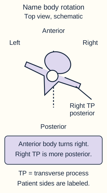
The anterior body and posterior spinous process point in different directions during rotation. Name vertebral rotation from the body convention and confirm with paired transverse-process testing. This is a simplified top view, not clinical imaging. [2]
In the conventional thoracic and lumbar model, Type II is nonneutral: a segment has a flexion or extension preference, with rotation and sidebending to the same side. Type I is a neutral pattern, typically involving a group, with rotation and sidebending to opposite sides. These are examination and naming conventions. Do not turn a coupling rule into a substitute for actual testing. [1][2]
Compare the entire pattern
Observation
Type I pattern
Type II pattern
ObservationDistribution
Type I patternCommonly a group
Type II patternCommonly one segment
ObservationSagittal testing
Type I patternNo distinct flexion or extension preference
Type II patternFlexion or extension preference
ObservationCoupling
Type I patternRotation and sidebending opposite
Type II patternRotation and sidebending same side
Compare two examinations: T4 through T8 share right rotation and left sidebending without a distinct sagittal preference; T3 separately prefers extension, right rotation, and right sidebending. Before reading further, assign each pattern to its category. The group fits Type I; the focal T3 pattern fits Type II. The posterior right landmarks occur in both, so they cannot distinguish the categories alone.
Now apply the comparison to adjacent T6 and T7 findings that each have a different nonneutral preference. More than one Type II dysfunction can coexist. Assess each segment rather than labeling every multilevel finding Type I. Likewise, chronic symptoms do not automatically establish Type I, and acute tenderness does not by itself establish Type II. [4]
Name rotation and sagittal preference
If asymmetry is worse in flexion, should the diagnosis begin with F? No. The diagnosis names relative ease, not the direction that makes the restriction most apparent. First establish the vertebral level and compare its paired transverse processes. A posterior right transverse process supports right vertebral rotation; a posterior left transverse process supports left rotation. Confirm the preference with motion testing. Do not name rotation from the spinous process pointing the other way. [2]
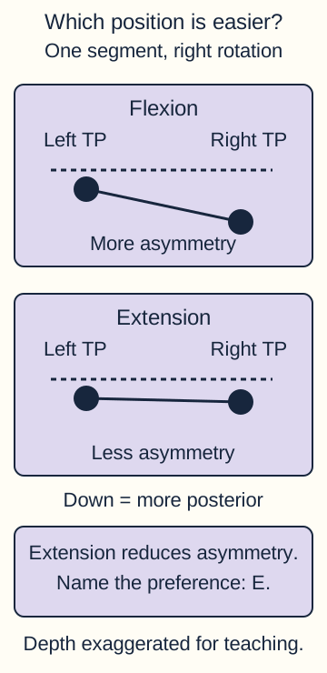
Keep the same level and contact points while comparing flexion and extension. In the standard examination pattern, asymmetry that decreases in flexion and becomes more apparent in extension supports a flexed diagnosis. The reverse supports an extended diagnosis. Test sidebending and rotation as well; same-side coupling should agree with the proposed Type II pattern. If guarding, uncertain landmarks, or inconsistent findings prevent that conclusion, repeat the assessment rather than inventing a complete diagnosis. [2]
Same posterior right transverse process, different sagittal results
Test
Examination A
Examination B
TestFlexion
Examination AAsymmetry decreases
Examination BAsymmetry increases
TestExtension
Examination AAsymmetry increases
Examination BAsymmetry decreases
TestConfirmed lateral preferences
Examination ARight rotation and sidebending
Examination BRight rotation and sidebending
TestDiagnosis
Examination AF Rr Sr
Examination BE Rr Sr
Trace Examination B from flexion to extension. Decide which position reduces the asymmetry before naming it. Extension is relatively easier, so the diagnosis is E Rr Sr, not F Rr Sr. F and E refer to sagittal preference; Rr means rotated right, and Sr means sidebent right. Rl and Sl indicate left.
Track the right landmark across both tests. Less asymmetry in extension supports an extended preference when the complete examination agrees. [2]Predict the result if only the posterior side changes
If the left transverse process is posterior, extension still reduces asymmetry, and testing confirms left rotation and left sidebending ease, the result is E Rl Sl. Changing the rotational side does not change the independently tested sagittal preference.
A resting photograph cannot establish this dynamic diagnosis. Nor does a spinous-process tip reliably identify the paired transverse processes at every thoracic level: their vertical relationships vary. Localize before comparing, and avoid interpreting neighboring levels as opposite sides of one vertebra. [1]
Translate ease into a direct barrier
Should direct treatment reproduce the named position? No. A diagnosis records the easier directions. A direct technique engages the restrictive barrier, opposite those directions. An indirect setup instead approaches ease. Neither phrase describes the strength of the force; a direct setup can and should be gentle. [1][4]
Use the three-axis comparison below as a mental rehearsal. Start with T3 F Rr Sr. Reverse one component at a time without changing the others. This is a reasoning activity, not a request to position your own neck.
Three-axis translation for T3 F Rr Sr
Axis
Named ease
Direct barrier
AxisSagittal
Named easeFlexion
Direct barrierExtension
AxisRotation
Named easeRight
Direct barrierLeft
AxisSidebending
Named easeRight
Direct barrierLeft
Read down the final column: extension, left rotation, left sidebending. If only extension is reversed, two components remain in ease. If rotation alone is reversed, the setup is also incomplete. The actual barrier must be felt at T3 rather than inferred from the visible orientation of the head.
Check one axis: what if T3 is E Rr Sr instead?
Only the sagittal component changes. The direct setup becomes flexion, left rotation, and left sidebending. The opposite sagittal preference does not reverse the lateral preferences.
Check the effort: which direction does the patient attempt?
From that barrier, the patient gently attempts a return toward ease against equal resistance. The clinician prevents actual segmental motion during the contraction. Positioning into restriction and effort toward freedom are different phases.
For a new diagnosis, T8 F Rl Sl, the direct setup is extension, right rotation, and right sidebending. Predict those directions before looking back at the table. Head and neck leverage is commonly used for upper thoracic levels; trunk positioning is used for lower levels. Both require local monitoring by a trained clinician, not maximal regional positioning. [1]
Sequence the isometric treatment
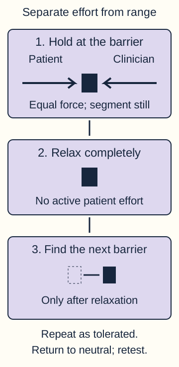
If the patient visibly returns toward ease during contraction, is the intended hold still isometric? No. In the described post-isometric relaxation technique, patient force and clinician counterforce are balanced so the segment stays at its localized barrier during the effort. The patient participates actively, but the hold does not become a resisted range-of-motion exercise. [1]
Trace the three states in order. Only the final state changes position; it follows relaxation rather than overlapping active resistance. Force arrows show balance, not a prescribed force magnitude. [1][4]
Explain the plan, obtain consent, screen safety, and establish reproducible segmental findings.
Gently engage the restrictive barrier in the relevant planes while monitoring the target segment.
Ask for a small, controlled effort toward freedom while providing equal counterforce.
Allow complete relaxation before gently taking up the newly available range to the next barrier.
Repeat a limited series as tolerated, return to neutral, and reassess.
The cited thoracic procedure describes a 3 to 5 second effort, 3 to 5 seconds of relaxation, and 3 to 5 repetitions. These are procedural examples, not permission to continue despite pain or loss of localization. Use only enough effort to engage the intended segment. More force is not the purpose of the next cycle. [1]
Compare two attempts. In the first, the segment remains still during the effort; after relaxation, additional comfortable range becomes available. In the second, the clinician pushes farther while the patient is still contracting. Identify which attempt preserves the intended sequence. The first separates contraction from the subsequent barrier adjustment; the second introduces loading during active resistance.
What should change if the patient's effort exceeds the counterforce?
Pause and reset with a smaller, better coordinated effort. Reestablish localization and an isometric hold instead of simply competing with a stronger contraction.
Post-isometric relaxation describes the treatment sequence and its intended reduction in restriction. Proposed neuromuscular explanations are not proof that one reflex fully explains every clinical response. Do not describe the technique as putting a displaced vertebra back into place. For another patient whose barrier does not change, reassess positioning, cooperation, diagnosis, and safety rather than repeating indefinitely. [4]
Decide whether loading is safe
Does a convincing somatic examination explain away a new warning sign? No. A named segmental pattern and a serious underlying disorder can coexist. The safety decision precedes the treatment setup, even though it is taught here after the directional logic. A gentle technique is still inappropriate when the tissue or patient cannot safely tolerate loading. [1]
Consider a patient with previously documented T5 dysfunction who now has focal midline pain after a fall. Compare that history with uncomplicated focal discomfort after prolonged sitting, normal neurologic findings, and no concerning systemic symptoms. Decide whether the same treatment pathway fits both. Trauma and bony tenderness require evaluation for fracture or instability before direct treatment; the prior somatic label does not lower that priority. ACR guidance specifically distinguishes uncomplicated acute thoracic pain from trauma, osteoporosis, neurologic findings, and suspected infection or cancer. [3]
Acute fracture, dislocation, instability, suspected local infection, or bony malignancy: withhold loading of the involved region and evaluate the underlying disorder.
New neurologic deficit or unstable vital signs: prioritize medical assessment and stabilization, not repeated contractions.
Substantial muscle, tendon, or ligament injury; severe osteoporosis; or restricted postoperative activity: reassess candidacy and loading limits rather than treating routine muscle energy as automatically safe.
Inability to understand, cooperate, relax, or tolerate positioning: this active technique is not presently suitable. A communication barrier should first prompt appropriate assistance, not an assumption that the patient is incapable.
Centrally mediated spasm or structural guarding is not an instruction to apply stronger force. New pain during positioning is a reason to stop and reassess. Excessive force can cause injury, including rib fracture in a susceptible patient. [1][4]
Apply the distinction to an older patient without a major fall
New focal thoracic pain after a minor load in a patient with osteoporosis still raises concern for fracture. Low-energy trauma and reduced bone strength matter together; a dramatic accident is not required. [3]
Conversely, routine imaging is not automatically needed for every acute thoracic ache without red flags, myelopathy, or radiculopathy. Safety assessment is selective clinical reasoning, not either universal imaging or universal manipulation. [3]
Initial imaging follows the clinical concern, not the somatic label [3]
Clinical pattern
ACR imaging guidance
Clinical patternAcute pain without red flags or neurologic features
ACR imaging guidanceRoutine initial imaging is usually not appropriate.
Clinical patternLow-energy trauma or substantial bone-fragility risk
ACR imaging guidanceRadiographs, noncontrast CT, or noncontrast MRI are appropriate initial alternatives; they are not a mandatory sequence.
Clinical patternMyelopathy or radiculopathy
ACR imaging guidanceNoncontrast thoracic MRI is usually appropriate.
Clinical patternSuspected infection or cancer
ACR imaging guidanceMRI without contrast or without and with contrast is usually appropriate; choose according to the clinical question.
A sensory level with bilateral leg weakness, abnormal reflexes, and bladder dysfunction suggests a spinal cord process, not merely a painful thoracic segment. New or progressive deficits require urgent spinal assessment and imaging; a palpable asymmetry does not determine the neurologic localization. [5]
Recheck the same findings
Does less pain establish that the segmental dysfunction has resolved? No. Symptom change and examination change are separate outcomes. Return the patient to neutral, then repeat the comparison that established the diagnosis: tissue texture, tenderness, paired transverse-process asymmetry, and motion preferences in flexion, extension, rotation, and sidebending. Use the same level and comparable positioning. [1][2]
Suppose discomfort decreases after treatment of T3 F Rr Sr, but the right transverse process remains posterior and asymmetry still increases in extension. Decide whether to document complete resolution. The residual examination still supports restriction; record symptom improvement alongside persistent findings. Conversely, improved symmetry without a meaningful symptom change should not be described as proven pain relief.
What if the retest appears to reverse the sagittal diagnosis?
Confirm neutral return, landmark level, and reproducibility before assigning a new preference. If repeat testing consistently supports a different pattern, document those findings rather than carrying forward the original label automatically.
For a new patient with persistent pain and newly developing weakness, the next assessment is broader than another transverse-process comparison. Reconsider the clinical differential and safety priority. The goal is accurate reassessment, not a predetermined declaration of correction. [3]
Independent practice
For each case, decide what the supplied findings establish and what they do not. Form an answer before reading the choices if useful. These original educational scenarios are not patient records.
Case 1
Show answer and explanations for case 1
A. T4 F Rl Sl; extension-right-right barrier (Best answer)
Improvement in flexion establishes flexion ease. The posterior left process and freer left coupled motions identify leftward ease. Direct positioning reverses all three planes.
Reasoning steps for option A
At T4, what does near symmetry in flexion imply about the sagittal preference?
Near symmetry in flexion indicates flexion ease at T4.
How do the posterior left process and freer left lateral motions name the coupled ease?
The posterior left T4 process and freer left rotation and sidebending support Rl Sl.
Which three restrictive directions oppose this rower's F Rl Sl pattern?
Oppose F Rl Sl with extension, right rotation, and right sidebending.
B. T4 E Rl Sl; flexion-right-right barrier (Why this does not fit)
Extension provokes asymmetry rather than relieving it. Thus extension is the restrictive sagittal motion, not the named ease. The proposed flexion barrier misses that restriction.
Reasoning steps for option B
Why does extension accentuating the T4 asymmetry undermine an E diagnosis?
Extension accentuates T4 asymmetry, so E cannot name ease.
Is extension the named sagittal ease or the restriction in this rower?
Extension is T4 sagittal restriction, not its ease.
Why would a flexion-right-right setup miss the sagittal barrier?
A flexion barrier moves toward ease; extension is the required sagittal barrier.
C. T4 F Rr Sr; extension-left-left barrier (Why this does not fit)
Sagittal preference is correctly identified, but a posterior left process identifies left rotation. Freer left sidebending supports left coupling. The proposed leftward barrier reinforces ease.
Reasoning steps for option C
Which side does a posterior left T4 transverse process name despite the proposed Rr?
A posterior left T4 process supports left, not right, rotation.
What does easier left sidebending say about the proposed Sr component?
Freer left sidebending supports Sl rather than Sr.
Why are left rotation and left sidebending unsuitable direct barriers here?
Left rotation and sidebending follow ease; direct barriers are rightward.
D. T4 N Rr Sl; extension-left-left barrier (Why this does not fit)
Neutral Type I typically has a group pattern and opposite coupling. This finding is focal and sagittal-dependent with leftward ease. The proposed diagnosis reverses the observed rotation.
Reasoning steps for option D
Would a focal T4 asymmetry that improves in flexion fit a neutral group?
A focal T4 asymmetry that improves in flexion favors nonneutral Type II over neutral group mechanics.
Which lateral ease is actually observed rather than N Rr Sl?
T4 eases into left rotation and left sidebending, not Rr Sl.
Which rotational component of this Type I label contradicts the left posterior landmark?
The Rr label contradicts the posterior left process and freer left rotation.
Takeaway: Reverse all planes to extension-right-right.
A. Flexion-right-right ease; extension-left-left barrier (Why this does not fit)
Flexion increases asymmetry and is restrictive. Extension reduces it, making extension the sagittal ease. The proposed barrier reverses the actual sagittal preference.
Reasoning steps for option A
Which T7 sagittal test increases the pianist's right prominence?
Flexion increases the pianist's right T7 prominence.
Which sagittal position reduces that prominence and thus represents ease?
Extension reduces it and therefore represents sagittal ease.
Why is extension not the direct sagittal barrier in this pairing?
Extension is ease; flexion is the direct sagittal barrier, contrary to this option.
B. Extension-right-right ease; flexion-left-left barrier (Best answer)
Extension reduces prominence, and posterior right process names right rotation. Freer right sidebending completes same-side coupling. Flexion-left-left is the opposite direct barrier.
Reasoning steps for option B
What does extension symmetry plus a posterior right T7 process establish?
Extension reduces T7 prominence, and its posterior right process supports right rotation.
Which tested sidebending direction completes the right-sided ease?
Freer right sidebending completes same-side right lateral ease.
What are the three directions opposite this T7 ease for direct treatment?
The direct T7 barrier is flexion, left rotation, and left sidebending.
C. Extension-left-left ease; flexion-right-right barrier (Why this does not fit)
Extension preference is correctly identified. Yet posterior right prominence identifies right rather than left rotation. A rightward rotational barrier is therefore not restrictive.
Reasoning steps for option C
Which part of extension-left-left is supported by the T7 sagittal comparison?
Extension ease is supported, but the proposed leftward lateral ease is not.
What rotational side is indicated by the posterior right T7 landmark?
The posterior right T7 landmark supports right rotation.
Why would a right rotational barrier follow ease instead of restriction?
A right rotational barrier follows the demonstrated right ease; the barrier is left rotation.
D. Neutral-right-left ease; flexion-only barrier (Why this does not fit)
The focal segment has a reproducible sagittal preference. Rightward rotation and sidebending are concordant rather than opposite. A single-plane barrier omits two components.
Reasoning steps for option D
What feature of focal T7 testing argues against a neutral diagnosis?
Reproducible extension preference at focal T7 contradicts neutral mechanics.
How do the pianist's freer right rotation and right sidebending couple?
Both rotation and sidebending are freer rightward, consistent with Type II same-side coupling.
Why does a flexion-only setup leave T7 lateral restrictions unaddressed?
A flexion-only barrier omits left rotation and left sidebending restriction.
A. Five independent extended right Type II lesions (Why this does not fit)
Five isolated lesions would require focal nonneutral testing at each segment. No level stands out and sagittal motion does not change the pattern. Calling each palpable process Type II discards the group mechanics.
Reasoning steps for option A
What focal sagittal evidence would five separate extended Type II diagnoses need?
Five separate extended lesions would require reproducible focal extension ease at each level.
Does the persistent T5-T9 ridge identify any one nonneutral level?
No single T5-T9 level stands out or changes specifically with sagittal testing.
Why should the five posterior landmarks be assessed as a group rather than five Type II lesions?
A shared neutral, oppositely coupled group better explains the five-level ridge.
B. A flexed right Type II lesion at T7 (Why this does not fit)
A flexed segment should improve in flexion and worsen in extension. The group asymmetry instead persists similarly in both positions. T7 is not uniquely localized.
Reasoning steps for option B
If T7 were flexed, what should flexion do to its asymmetry?
A flexed T7 should become more symmetric in flexion and less symmetric in extension.
Does this T5-T9 ridge vary between flexion and extension as a flexed lesion should?
No: the T5-T9 ridge persists similarly in both positions.
What prevents selecting T7 specifically from the supplied examination?
No examination finding uniquely localizes T7 within the T5-T9 group.
C. A neutral right-rotated, left-sidebent Type I group (Best answer)
Several contiguous levels share a pattern without sagittal preference. Right rotation and left sidebending are opposite coupled motions. This fits a neutral Type I group rather than a focal Type II segment.
Reasoning steps for option C
What does the contiguous T5-T9 distribution without sagittal preference suggest?
Contiguous T5-T9 findings without sagittal preference support a neutral Type I group.
Which opposing lateral preferences define this group's neutral coupling?
Right rotation and left sidebending are the observed opposite lateral preferences.
Which classification and directions best localize this ridge?
Localize the ridge as a neutral, right-rotated, left-sidebent Type I group.
D. A neutral left-rotated, right-sidebent Type I group (Why this does not fit)
Neutral opposite coupling is the appropriate class. Yet the supplied posterior ridge and free rotation are rightward, not leftward. The orientation is reversed in this option.
Reasoning steps for option D
Which part of a neutral, opposite-coupled diagnosis fits T5-T9?
Neutral opposite coupling fits the group's category, but not these proposed sides.
Which side is actually posterior and easier in group rotation?
The right ridge and freer right rotation support right rather than left rotation.
Why must left-rotated right-sidebent be rejected despite the correct Type I category?
The actual group is right-rotated and left-sidebent, opposite this option.
Takeaway: Use opposite coupling to classify Type I.
A. Left rotation with a flexion barrier (Why this does not fit)
Flexion is the correct sagittal barrier because extension reduces asymmetry. The posterior right transverse process supports right, not left, vertebral rotation. Spinous direction does not reverse that convention.
Reasoning steps for option A
Which part of left rotation with flexion barrier matches T6 sagittal testing?
Flexion is the direct sagittal barrier because T6 is more symmetric in extension.
What does the posterior right transverse process say about vertebral rotation?
The posterior right T6 transverse process supports right vertebral rotation.
Can the left-pointing spinous process override paired transverse-process naming?
No: a left-pointing spinous tip does not override the paired transverse-process convention.
B. Right rotation with an extension barrier (Why this does not fit)
Right rotation is correctly named from the transverse process. Extension is relatively easier, not the restrictive sagittal direction. The direct sagittal barrier is flexion.
Reasoning steps for option B
Which T6 landmark supports the right-rotation portion of this choice?
The posterior right T6 transverse process supports right rotation.
What does symmetry in extension indicate about sagittal ease?
Extension reduces asymmetry and therefore represents relative sagittal ease.
Which sagittal direction actually engages restriction at T6?
Flexion engages the T6 sagittal restriction.
C. Left rotation with an extension barrier (Why this does not fit)
The posterior right process supports right rotation. Symmetry in extension supports extension ease, so the sagittal barrier is flexion. Both proposed components are reversed.
Reasoning steps for option C
Which rotation is supported by the right posterior process rather than the spinous tip?
The right posterior T6 transverse process supports right rotation.
Does extension symmetry indicate an extension barrier or ease?
Extension is ease; flexion is the direct sagittal barrier.
How many of this choice's two proposed components contradict testing?
Both the left rotational label and the extension barrier contradict T6 testing.
D. Right rotation with a flexion barrier (Best answer)
The posterior right transverse process identifies right rotation despite the leftward spinous direction. Extension reduces the asymmetry and is therefore the relative sagittal ease. Flexion is the direct sagittal barrier; lateral-plane testing is still needed.
Reasoning steps for option D
Why does the student's spinous-process inference fail at T6?
The left-pointing spinous tip does not establish left vertebral rotation; the right transverse process establishes right.
Which sagittal preference follows from symmetry in extension?
Symmetry in extension indicates extension ease at T6.
What sagittal barrier can be named without assuming an untested sidebending direction?
Flexion is the sagittal barrier; sidebending remains untested.
Takeaway: Name rotation from the body convention and name the sagittal preference from the test response.
A. Change flexion to extension (Why this does not fit)
Improvement in extension identifies an extended preference. Flexion therefore already engages the sagittal restriction. Changing it would abandon that barrier.
Reasoning steps for option A
Which T8 sagittal position decreases asymmetry?
Extension decreases T8 asymmetry.
Does the clinician's current flexion already oppose that sagittal ease?
Yes: flexion opposes extension ease and already engages restriction.
Why would switching to extension worsen this direct setup?
Switching to extension would abandon the correct flexion barrier.
B. Change right rotation to left rotation (Why this does not fit)
The posterior left landmark and easier left rotation identify left rotational ease. Right rotation already engages restriction. Changing rotation would reproduce ease.
Reasoning steps for option B
Which side is posterior and easier in T8 rotation?
The left T8 transverse process is posterior and left rotation is easier.
Is the existing right rotation already opposite the tested preference?
Yes: existing right rotation opposes left rotational ease.
What would changing rotation to left do to the barrier?
Switching to left rotation would reproduce ease instead of restriction.
C. Change both lateral components to neutral (Why this does not fit)
Both lateral preferences are leftward. Neutral lateral positioning does not establish the rightward barriers. The incorrect sidebending component needs reversal rather than eliminating both components.
Reasoning steps for option C
What two lateral preferences are reported at T8?
Both rotation and sidebending are easier leftward at T8.
Does neutral rotation and sidebending engage rightward restriction?
No: neutral positioning does not reach either rightward lateral barrier.
Which lateral component alone needs correction rather than neutralizing both?
Only the currently leftward sidebending needs changing to right; keep right rotation.
D. Change left sidebending to right sidebending (Best answer)
The findings establish extended, rotated left and sidebent left ease. Flexion and right rotation are already restrictive. Right sidebending completes the three-plane direct setup.
Reasoning steps for option D
What full three-axis ease is established at T8?
T8 favors extension, left rotation, and left sidebending.
Which two directions of the current setup already oppose ease?
The present flexion and right rotation already oppose ease.
Which replacement for left sidebending supplies the missing barrier?
Replace left sidebending with right sidebending to complete the barrier.
Takeaway: Derive ease from the examination before judging which treatment axis is incorrect.
A. Attempt extension-right-right against equal resistance (Why this does not fit)
The examination supports flexion and leftward ease. Extension and rightward directions are the restrictive setup, not the intended return toward freedom. Equal resistance does not correct the wrong effort directions.
Reasoning steps for option A
Which three directions of ease are indicated by T3 testing?
T3 favors flexion, left rotation, and left sidebending.
Are extension and rightward movements the barrier or the return toward ease?
Extension, right rotation, and right sidebending form the restrictive setup.
Why does equal counterforce not rescue effort aimed toward restriction?
Equal counterforce holds the segment still but cannot make barrier-directed effort into ease-directed effort.
B. Attempt flexion-right-right against equal resistance (Why this does not fit)
Flexion matches sagittal ease. Right rotation and right sidebending remain directed toward the restrictive components. Both lateral directions must be leftward for the described effort.
Reasoning steps for option B
Which sagittal effort agrees with improvement in T3 flexion?
Flexion matches the T3 sagittal ease.
How do the proposed rightward efforts compare with tested left lateral ease?
Rightward rotation and sidebending approach the barriers, not leftward ease.
Which lateral effort directions are needed while the clinician resists motion?
The patient should attempt left rotation and left sidebending along with flexion.
C. Attempt flexion-left-left against equal resistance (Best answer)
The examination supports a flexed, rotated left and sidebent left diagnosis. The clinician holds extension and rightward restriction while the patient attempts toward the named ease. Equal counterforce prevents actual motion.
Reasoning steps for option C
How do flexion symmetry and left lateral freedom define T3 ease?
Flexion decreases T3 prominence, while left rotation and left sidebending are freer.
Which opposite three-plane barrier does the clinician maintain?
The clinician holds extension, right rotation, and right sidebending restriction.
What does equal resistance accomplish when the patient attempts flexion-left-left?
Equal resistance prevents actual motion during the flexion-left-left effort.
D. Attempt extension-left-right against equal resistance (Why this does not fit)
Left rotation matches ease, but extension and right sidebending do not. These directions mix restriction with freedom. The proposed effort therefore fails to follow the diagnosed pattern.
Reasoning steps for option D
Which component of extension-left-right follows observed T3 ease?
Left rotation alone matches the T3 ease.
Which proposed sagittal and sidebending efforts instead approach restriction?
Extension and right sidebending approach restriction rather than flexion and left sidebending ease.
Why is this mixed-direction effort inconsistent with an isometric return toward freedom?
Effort should aim toward flexion-left-left; this extension-left-right mixture does not.
Takeaway: Infer the ease pattern first, then distinguish patient effort from the held direct barrier.
A. E Rl Sl; stop loading and repeat the clinical assessment (Why this does not fit)
Stopping for the new pain is appropriate. The original asymmetry improves in flexion, supporting F rather than E. Pain during setup does not reverse that prior sagittal finding.
Reasoning steps for option A
Does T9 asymmetry diminish in extension or flexion before the painful setup?
T9 asymmetry diminishes in flexion, not extension.
Which sagittal letter follows from that original improvement?
Improvement in flexion supports F rather than E.
Does new sharp midline pain justify revising the old sagittal finding to E?
No: new pain changes loading safety, not the prior sagittal test result.
B. F Rl Sl; stop loading and repeat the clinical assessment (Best answer)
The original findings support F Rl Sl. The new sharp midline pain changes tolerance and requires reassessment before loading. Preserve the supported examination while reconsidering safety.
Reasoning steps for option B
Which diagnosis follows from T9 flexion improvement and left lateral ease?
Flexion improvement and easier left rotation and sidebending support T9 F Rl Sl.
What changed when right sidebending caused sharp midline pain?
New sharp midline pain during right sidebending raises a new safety concern.
What should happen before any further loading of this T9 segment?
Stop loading and reassess T9 before attempting another cycle.
C. F Rl Sl; complete a smaller-amplitude cycle before reassessment (Why this does not fit)
The original F Rl Sl interpretation is correct. Smaller amplitude does not establish that the new sharp pain is safe to load. Reassess before an additional cycle rather than afterward.
Reasoning steps for option C
Which portion of this option correctly interprets the original T9 examination?
The F Rl Sl interpretation of the initial T9 examination is correct.
Does smaller amplitude establish that newly sharp midline pain is safe?
No: smaller amplitude does not establish safety after new sharp midline pain.
Should reassessment occur before or after another contraction cycle?
Reassess before, not after, another contraction.
D. E Rl Sl; complete a smaller-amplitude cycle before reassessment (Why this does not fit)
The original flexion improvement supports F rather than E. The newly painful setup also requires reassessment before another cycle. Reducing amplitude does not resolve either uncertainty.
Reasoning steps for option D
Which sagittal designation conflicts with improved symmetry in T9 flexion?
E conflicts with the observed improvement in flexion; F was supported.
What safety action does new sharp pain during right sidebending demand?
New sharp midline pain requires stopping loading and reassessment.
Why can neither an E label nor a smaller cycle settle this case?
Neither reducing amplitude nor relabeling the sagittal preference resolves the safety concern.
Takeaway: A valid mechanical interpretation and a reason to stop treatment can coexist.
A. Take up the new flexion range while retaining the tested leftward barriers (Best answer)
Extension-right-right ease requires a flexion-left-left barrier. Only the sagittal endpoint gained comfortable range after relaxation. Engage that new flexion endpoint without assuming gains in the other planes.
Reasoning steps for option A
Which direct three-plane barrier opposes T5 extension-right-right ease?
T5 extension-right-right ease is opposed by flexion-left-left restriction.
Which endpoint changed after complete relaxation?
Only comfortable flexion increased; left rotation and sidebending endpoints stayed unchanged.
How should the next setup incorporate only the newly comfortable flexion range?
Take up the new flexion range while retaining the measured left lateral barriers.
B. Return to the original flexion angle and add more left rotation (Why this does not fit)
The examination found a sagittal gain, not a rotational gain. Adding unobserved left rotation does not follow the tested endpoints. The next barrier should reflect the actual available range.
Reasoning steps for option B
Did T5 gain flexion or left rotation after this contraction?
T5 gained flexion range, not left rotational range.
Why is adding left rotation unsupported by the unchanged endpoint?
Its left rotation endpoint is unchanged, so pushing farther left is unsupported.
Which tested direction should advance for the next barrier?
Advance flexion to its newly available comfortable barrier.
C. Take up the new flexion range and add more left sidebending (Why this does not fit)
Flexion gain is supported, but sidebending gain is not. Additional left sidebending would exceed the newly tested endpoint. Reassess each component rather than advancing them together automatically.
Reasoning steps for option C
Which T5 plane gained new comfortable range?
Comfortable flexion range increased at T5.
What was observed at the left sidebending endpoint?
The left sidebending endpoint did not change.
Why should flexion advance without automatically increasing sidebending?
Advance flexion but retain the previously measured left sidebending barrier.
D. Return toward extension while retaining the tested leftward barriers (Why this does not fit)
Extension is the named ease rather than the direct sagittal barrier. Returning toward it abandons the newly available flexion restriction. Retaining the lateral barriers alone does not complete the setup.
Reasoning steps for option D
Is extension at T5 a named ease or a restrictive sagittal barrier?
Extension is T5 ease; flexion is the direct sagittal barrier.
What happens to the new flexion range if the clinician returns toward extension?
Returning toward extension abandons the newly available restrictive flexion range.
Can unchanged left lateral barriers alone constitute the full direct setup?
No: left lateral barriers alone omit the needed flexion barrier.
Takeaway: After relaxation, adjust only the barrier components whose comfortable range has actually changed.
A. The original flexed-right-right diagnosis is fully confirmed (Why this does not fit)
The original examination supported all three components. The current sagittal response differs and sidebending is not retested. Carrying forward the entire label ignores changed and missing information.
Reasoning steps for option A
Which three preferences were supported by the original T4 tests?
Before treatment T4 favored flexion, right rotation, and right sidebending.
Which sagittal and sidebending components have not been confirmed on retest?
The sagittal difference has disappeared, and right sidebending has not yet been retested.
Why is retaining the entire F Rr Sr label premature despite reduced pain?
Pain relief cannot confirm the full original F Rr Sr label in the face of changed and missing motion findings.
B. Right rotational preference persists; the sagittal label needs reassessment (Best answer)
Right rotational ease persists on direct testing, whereas the original flexion-versus-extension distinction is no longer reproduced. Reduced pain does not settle the remaining motion diagnosis. Reassess the sagittal and sidebending components rather than automatically retaining or reversing them.
Reasoning steps for option B
Which T4 rotational finding remains demonstrable after treatment?
Easier right rotation persists at T4.
What happened to the original flexion-versus-extension asymmetry difference?
Flexion and extension now produce no distinguishing change in landmark asymmetry.
Which missing or changed components require retesting before a full new diagnosis?
Repeat sagittal and sidebending testing before naming a complete post-treatment pattern.
C. The segment is now extended-right-right because flexion no longer helps (Why this does not fit)
Failure to reproduce the former flexion response does not prove extension ease. Both sagittal tests now have similar effects. An extended diagnosis would require a reproducible new preference.
Reasoning steps for option C
Does loss of the former flexion response establish extension ease?
No: loss of flexion preference does not by itself demonstrate extension preference.
How do flexion and extension now affect the right landmark?
Neither flexion nor extension now changes the T4 landmark asymmetry.
What reproducible sagittal finding would be needed to support the E component of E Rr Sr?
Extension would need to reproducibly reduce asymmetry relative to flexion; sidebending would still require retesting for a complete label.
D. The rotation is normalized; only the flexed preference persists (Why this does not fit)
Right rotation remains easier, so rotational normalization is not demonstrated. The former flexion preference is also no longer reproduced. This option assigns improvement to the wrong component.
Reasoning steps for option D
What does persistent easier right rotation say about rotational normalization?
Persistent easier right rotation does not demonstrate rotational normalization.
Is the former flexion preference still reproducible?
No: flexion no longer uniquely reduces the T4 asymmetry.
Which two claims in this option invert the actual retest findings?
Both claimed normalized rotation and persistent flexion preference contradict retesting.
Takeaway: When retest findings change selectively, retain only supported components and reassess the rest.
A. Use the neck as a lever until its own barrier is reached (Why this does not fit)
Neck leverage can be useful for upper thoracic segments. Here it creates regional tension without engaging T8. The neck endpoint does not establish a T8 barrier.
Reasoning steps for option A
Why might neck leverage seem plausible for a thoracic target?
Neck leverage can be useful for upper thoracic segments.
What does neck tension without a monitored T8 change reveal?
Here it creates regional tension without engaging T8.
Why is reaching the neck endpoint insufficient for this lower thoracic target?
A neck endpoint does not establish a barrier at T8; the clinician must detect the barrier locally at T8.
B. Keep the head position and increase the patient effort (Why this does not fit)
Patient effort can activate a correctly localized setup. Here the monitoring hand has not detected the intended segmental barrier. Greater effort cannot substitute for localization.
Reasoning steps for option B
When can patient contraction help a muscle-energy setup?
Patient effort can activate a correctly localized setup.
What prerequisite has the T8 monitoring hand failed to confirm?
Here the monitoring hand has not detected the intended segmental barrier.
Would stronger effort correct an unlocalized T8 barrier?
Increase in effort cannot replace localization; first establish the restrictive barrier at T8.
C. Use the trunk to engage extension-right-right at T8 (Best answer)
Lower thoracic localization commonly uses the trunk rather than the head and neck. A flexed left-coupled diagnosis requires extension and rightward restrictions. Monitor T8 while gently positioning those components.
Reasoning steps for option C
Which body region offers leverage better suited to T8 than the neck?
Lower thoracic localization commonly uses the trunk rather than the head and neck.
Which restrictive directions oppose T8 flexion with left rotation and sidebending?
A flexed left-coupled diagnosis requires extension and rightward restrictions.
Where must extension and rightward positioning be confirmed?
Monitor T8 while gently engaging extension, right rotation, and right sidebending.
D. Use trunk positioning to reproduce flexion and leftward ease at T8 (Why this does not fit)
Trunk leverage is suitable for this lower thoracic target. Flexion and leftward positioning reproduce the named ease, however. The planned direct technique requires the opposite directions.
Reasoning steps for option D
Why is trunk positioning reasonable for a T8 target?
Trunk leverage is suitable for this lower thoracic target.
Does flexing and turning left place this F Rl Sl segment at ease or restriction?
Flexion and leftward positioning reproduce the named ease, however.
Which directional change makes the trunk setup direct rather than indirect?
For direct MET at T8 F Rl Sl, reverse ease to extension, right rotation, and right sidebending.
Takeaway: Choose leverage that reaches the target, then confirm every barrier locally.
A. Request MRI without contrast first for suspected infection (Why this does not fit)
MRI is also an appropriate initial fracture study in this risk setting. This option instead proposes infection as the reason, which is less supported than the trauma-related fracture concern. Reject the stated diagnostic target, not MRI as a modality.
Reasoning steps for option A
Can noncontrast MRI evaluate a possible fracture in an osteoporotic patient?
MRI is also an appropriate initial fracture study in this risk setting.
Does a low-energy fall with focal T7 tenderness chiefly suggest infection?
This option instead proposes infection as the reason, which is less supported than the trauma-related fracture concern.
Is the objection to MRI itself or to the infection-centered indication?
Reject the stated diagnostic target, not MRI as a modality.
B. Treat the segment because neurologic testing is intact (Why this does not fit)
Intact neurologic testing does not exclude vertebral fracture. Focal bony tenderness after a fall still warrants structural assessment. Defer loading until clarified.
Reasoning steps for option B
Does intact motor and sensory testing rule out an osteoporotic T7 fracture?
Intact neurologic testing does not exclude vertebral fracture.
Which post-fall finding still calls for assessment before segmental loading?
Focal bony tenderness after a fall still warrants structural assessment.
When should MET be deferred despite a posterior transverse process?
Defer MET and segmental loading until the possible T7 fracture has been assessed.
C. Request CT angiography of the chest for vascular injury (Why this does not fit)
CT angiography addresses a different vascular question. No vascular symptoms or high-energy mechanism are described. Target the suspected vertebral injury.
Reasoning steps for option C
What diagnostic question does chest CT angiography primarily answer?
CT angiography addresses a different vascular question.
Are vascular symptoms or a high-energy injury given in this T7 scenario?
No vascular symptoms or high-energy mechanism are described.
What structural concern should guide the imaging choice instead?
Investigate possible vertebral fracture at T7 rather than an unsupported vascular injury.
D. Obtain thoracic radiographs for possible fracture (Best answer)
Low-energy trauma, osteoporosis and focal midline tenderness raise concern for fracture despite intact neurologic testing. Radiographs are one appropriate initial study among the offered plans. ACR also accepts noncontrast CT or MRI as initial alternatives; radiographs are not uniquely mandatory.
Reasoning steps for option D
Which combination makes fracture the priority despite a normal neurologic examination?
Low-energy trauma, osteoporosis and focal midline tenderness raise concern for fracture despite intact neurologic testing.
Why are thoracic radiographs an appropriate offered first step?
Radiographs are one appropriate initial study among the offered plans.
Are radiographs the only acceptable initial imaging choice under ACR guidance?
ACR also accepts noncontrast CT or MRI as initial alternatives; radiographs are not uniquely mandatory.
A. MRI of the thoracic spine to assess possible infection (Best answer)
Bacteremia and fever with new focal spinal pain raise concern for infection. MRI is the appropriate targeted imaging assessment. A somatic finding may coexist but cannot exclude infection.
Reasoning steps for option A
What possibility links recent bacteremia, fever, and focal T5 pain?
Bacteremia and fever with new focal spinal pain raise concern for infection.
Which imaging modality evaluates the suspected spinal infectious focus?
MRI is the appropriate targeted imaging assessment.
Can a right posterior process exclude infection in this patient?
A somatic finding may coexist but cannot exclude infection.
B. Thoracic radiographs as the initial definitive assessment (Why this does not fit)
Radiographs can assess bony trauma but are not the preferred evaluation of suspected spinal infection. Bacteremia, fever and focal night pain favor an infectious process. A negative plain film would not settle it.
Reasoning steps for option B
For what concern are plain thoracic radiographs more directly suited?
Radiographs can assess bony trauma but are not the preferred evaluation of suspected spinal infection.
How do bacteremia and new night pain change the leading differential?
Bacteremia, fever and focal night pain favor an infectious process.
Would normal radiographs definitively exclude this suspected infection?
A negative plain film would not settle it.
C. Inflammatory markers alone before deciding whether to image (Why this does not fit)
Inflammatory markers may contribute to an infection evaluation. They do not define the spinal structures or exclude a local infectious focus in this presentation. MRI directly addresses the concern raised by bacteremia and focal pain.
Reasoning steps for option C
What useful role might inflammatory markers play in this febrile presentation?
Inflammatory markers may contribute to an infection evaluation.
Can blood markers alone locate or exclude a spinal infectious focus?
They do not define the spinal structures or exclude a local infectious focus in this presentation.
Which study addresses the focal spinal concern without waiting on markers alone?
MRI directly addresses the concern raised by bacteremia and focal pain.
D. Noncontrast thoracic CT as the sole spinal assessment (Why this does not fit)
CT can identify osseous abnormalities. The suspected infectious process also raises concern for marrow and soft-tissue involvement. MRI is the more appropriate offered study for that clinical question.
Reasoning steps for option D
What type of T5 pathology can noncontrast CT show well?
CT can identify osseous abnormalities.
What additional tissues matter when fever and bacteremia suggest infection?
The suspected infectious process also raises concern for marrow and soft-tissue involvement.
Which offered modality better assesses marrow and soft tissue involvement?
MRI is the more appropriate offered study for that clinical question.
A. Lumbar MRI for an isolated cauda equina process (Why this does not fit)
Bladder dysfunction and leg weakness can occur with cauda equina disease. The supplied upper motor neuron signs and thoracic sensory level instead favor a cord process. An isolated cauda equina explanation does not account for that pattern.
Reasoning steps for option A
Which two symptoms could initially prompt consideration of cauda equina disease?
Bladder dysfunction and leg weakness can occur with cauda equina disease.
How do bilateral extensor plantar signs and a mid-thoracic sensory level alter localization?
The supplied upper motor neuron signs and thoracic sensory level instead favor a cord process.
Why would an isolated lumbar cauda equina process fail to explain the full examination?
An isolated cauda equina explanation does not account for that pattern.
B. Peripheral nerve studies for a length-dependent neuropathy (Why this does not fit)
Peripheral neuropathy can cause leg symptoms. Brisk reflexes, extensor plantar responses and a truncal sensory level favor central rather than length-dependent peripheral disease. Nerve studies do not address the urgent spinal concern.
Reasoning steps for option B
Why might a peripheral neuropathy enter the differential for leg complaints?
Peripheral neuropathy can cause leg symptoms.
Do brisk reflexes, extensor plantar responses, and a truncal level fit a length-dependent neuropathy?
Brisk reflexes, extensor plantar responses and a truncal sensory level favor central rather than length-dependent peripheral disease.
Would peripheral nerve studies address the urgent localization here?
Peripheral nerve studies do not address the urgent possible thoracic spinal cord lesion.
C. Urgent thoracic MRI within emergency spinal assessment (Best answer)
Bilateral upper motor neuron signs with a thoracic sensory level localize concern to the spinal cord. Urinary retention and progression make evaluation urgent. Thoracic MRI is part of emergency assessment, not a reason to delay it or perform MET first.
Reasoning steps for option C
Where do bilateral upper motor neuron signs plus a thoracic sensory level point?
Bilateral upper motor neuron signs with a thoracic sensory level localize concern to the spinal cord.
How do two days of progression and urinary retention affect urgency?
Urinary retention and progression make evaluation urgent.
What role should thoracic MRI play relative to emergency assessment and MET?
Thoracic MRI is part of emergency assessment, not a reason to delay it or perform MET first.
D. Brain MRI for a cerebral hemispheric lesion (Why this does not fit)
Cerebral disease can produce upper motor neuron findings. The bilateral leg syndrome and truncal sensory level favor the spinal cord over a hemispheric lesion. Prioritize the anatomical region supported by the whole examination.
Reasoning steps for option D
Which finding might make brain imaging appear relevant?
Cerebral disease can produce upper motor neuron findings.
Why does the truncal sensory level with bilateral leg weakness favor the cord over one cerebral hemisphere?
The bilateral leg syndrome and truncal sensory level favor the spinal cord over a hemispheric lesion.
Which anatomical region should the first urgent workup target?
Prioritize urgent spinal assessment focused on the thoracic cord, rather than a hemispheric lesion.
Takeaway: Localize neurologic findings independently of a palpable segmental asymmetry.
A. Extended and right-rotated; repeat sidebending after relaxation (Why this does not fit)
Right rotation is supported by both landmark and motion testing. Extension exaggerates rather than reduces asymmetry, so the sagittal interpretation is reversed. Repeating sidebending cannot repair that naming error.
Reasoning steps for option A
Which T10 findings support rightward rotation in this proposal?
Right rotation is supported by both landmark and motion testing.
Does greater asymmetry in extension support an extended or flexed preference?
Extension exaggerates rather than reduces asymmetry, so the sagittal interpretation is reversed.
Can repeating guarded sidebending rescue an incorrect sagittal label?
No; retesting sidebending cannot change the flexion preference supported by reduced asymmetry in flexion.
B. Flexed and left-rotated; repeat sidebending after relaxation (Why this does not fit)
Flexion ease is correctly inferred. The posterior right process and easier right rotation contradict left rotation. Sidebending remains the unconfirmed component.
Reasoning steps for option B
Which sagittal component of flexed-left-rotated is supported?
Flexion ease is correctly inferred.
What do the right posterior process and freer right rotation say about the proposed left rotation?
The posterior right process and easier right rotation contradict left rotation.
Which motion axis remains uncertain because the patient braces?
Sidebending at T10 remains unconfirmed because bracing makes the test inconsistent.
C. Flexed and right-sidebent; repeat rotation after relaxation (Why this does not fit)
Flexion is supported, but right sidebending has not been established. Rotation has already been tested as easier rightward. Repeat the unreliable sidebending assessment rather than asserting its result.
Reasoning steps for option C
Has right sidebending been demonstrated despite the expected Type II coupling?
Flexion is supported, but right sidebending has not been established.
Which lateral motion has actually been tested as easier at T10?
Rotation has already been tested as easier rightward.
Should the clinician repeat rotation or the guarded sidebending test?
Repeat the unreliable sidebending assessment rather than asserting its result.
D. Flexed and right-rotated; repeat sidebending after relaxation (Best answer)
Flexion reduces asymmetry and right rotation is supported directly. Guarding prevents a reliable sidebending result. Preserve the supported components and retest the missing one after relaxation.
Reasoning steps for option D
What do reduced asymmetry in flexion and easier right rotation establish?
Flexion reduces asymmetry and right rotation is supported directly.
Why can the clinician not yet assign a sidebending direction?
Guarding prevents a reliable sidebending result.
What should be retested after relaxation to complete the diagnosis?
Preserve the supported components and retest the missing one after relaxation.
Takeaway: A coupling expectation does not supply a motion-test result that could not be obtained.
A. Neutral disagreement proves a Type I group (Why this does not fit)
A group requires several segments with opposite coupling. A single T4 dynamic finding does not establish that pattern. Neutral disagreement is not group evidence.
Reasoning steps for option A
What multisegment and coupling evidence would support a Type I group?
A group requires several segments with opposite coupling.
Does disagreement about one neutral T4 landmark supply that evidence?
A single T4 dynamic finding does not establish that pattern.
Can variable neutral palpation alone establish a neutral group?
No; discordant neutral T4 palpation does not establish a multisegment Type I group.
B. Flexion ease with right rotation is supported (Best answer)
Repeated flexion improvement supports flexion ease despite a disputed static snapshot. Right posterior prominence identifies right rotation. Sidebending remains to be established.
Reasoning steps for option B
Which reproducible T4 change identifies sagittal ease despite the disputed neutral view?
What rotation does the right posterior transverse-process prominence support?
Right posterior prominence identifies right rotation.
Which component still needs direct testing before a full Type II label?
Sidebending must still be tested to complete the proposed T4 Type II diagnosis.
C. The spinous direction must settle rotation (Why this does not fit)
Spinous direction is not provided. The right transverse prominence supplies the relevant rotation finding. An absent landmark cannot supersede repeatable motion testing.
Reasoning steps for option C
Was the T4 spinous-process direction actually reported?
Spinous direction is not provided.
Which paired landmark observation already informs rotation?
The right transverse prominence supplies the relevant rotation finding.
Why should an unreported spinous direction not displace repeatable dynamic evidence?
An unreported spinous direction cannot override the reproducible flexion improvement at T4.
D. No sagittal preference exists because neutral findings differ (Why this does not fit)
Disputed neutral findings can reflect variable posture or palpation. Reproducible flexion and extension differences establish a sagittal preference. Dynamic findings still require interpretation.
Reasoning steps for option D
What might account for two examiners disagreeing in neutral?
Disputed neutral findings can reflect variable posture or palpation.
What does repeatably improved symmetry in flexion versus extension establish?
Reproducible flexion and extension differences establish a sagittal preference.
Does neutral disagreement justify ignoring the sagittal motion comparison?
No; the repeatable flexion improvement supports a flexed preference despite neutral disagreement.
Takeaway: Flexion preference and right rotation are supported; confirm sidebending.
A. Stop and reassess pain; if appropriate, reset for gentle rightward effort held still (Best answer)
The examination supports rightward ease, so rotational effort should be rightward from a held leftward barrier. Actual motion is not the intended isometric hold. First stop and reassess pain; a corrected attempt is conditional on safe, comfortable participation.
Reasoning steps for option A
From a held leftward rotational barrier, which effort direction returns T6 toward its easier rotation?
The examination supports rightward ease, so rotational effort should be rightward from a held leftward barrier.
Does the observed leftward rotation during rising counterforce remain isometric?
Actual motion is not the intended isometric hold.
What must happen before any corrected low-force attempt after increasing pain?
Stop and reassess the increasing pain; only if safe, reset for a gentle rightward effort toward ease without actual T6 rotation.
B. Stop and reassess pain; if appropriate, reset for gentle leftward effort held still (Why this does not fit)
Stopping for pain is appropriate and holding still matches isometric technique. Leftward effort remains toward rotational restriction rather than rightward ease. The direction error would persist.
Reasoning steps for option B
Which parts of stopping and holding still address pain and isometric control?
Stopping for pain is appropriate and holding still matches isometric technique.
Why would asking for leftward effort retain the original direction mistake?
Leftward effort remains toward rotational restriction rather than rightward ease.
What rotational effort direction would be required if resuming is safe?
If safe to resume, effort should be rightward toward T6 rotational ease while the segment is held still.
C. Stop and reassess pain; if appropriate, permit rightward rotation during effort (Why this does not fit)
Rightward effort matches ease and pain must first be assessed. Permitting visible rotation changes the contraction from the intended isometric hold. Equal counterforce should maintain the segmental position.
Reasoning steps for option C
Which proposed effort direction matches T6 rightward ease?
Rightward effort matches ease and pain must first be assessed.
Would allowing visible right rotation preserve the intended isometric contraction?
Permitting visible rotation changes the contraction from the intended isometric hold.
What should equal counterforce prevent during any safe renewed effort?
Equal counterforce should prevent actual T6 rotation while the patient gently attempts rightward effort.
D. Stop and reassess pain; if appropriate, permit leftward rotation during effort (Why this does not fit)
The initial pause is appropriate. Leftward effort is toward restriction, and allowing rotation also violates the isometric plan. Both direction and contraction control remain incorrect.
Reasoning steps for option D
Is pausing in response to increasing pain appropriate?
The initial pause is appropriate.
What two errors remain if leftward effort produces visible rotation?
Leftward effort is toward restriction, and allowing rotation also violates the isometric plan.
Can correcting only the pain response make this direct isometric attempt valid?
No; even after pausing for pain, leftward effort and visible movement remain incompatible with the intended isometric hold.
Takeaway: First reassess pain, then correct both effort direction and motion control only if further treatment is appropriate.
Extension reduces prominence, indicating extension ease. Left posterior prominence and freer left motions identify left coupling. Flexion-right-right reverses all components.
Reasoning steps for option A
Which sagittal preference follows when T2 prominence disappears in extension?
Which side is favored by left posterior prominence and easier left motions?
Left posterior prominence and freer left motions identify left coupling.
What position opposes all three preferred directions for a direct setup?
Flexion, right rotation, and right sidebending reverse the T2 E Rl Sl ease.
B. Extension-right-right (Why this does not fit)
Rightward motions are restrictive, but extension is the sagittal ease. The setup neglects flexion restriction. Reversal must include every component.
Reasoning steps for option B
Which two lateral restrictions does extension-right-right correctly engage?
Rightward motions are restrictive, but extension is the sagittal ease.
Why does extension fail to engage the sagittal restriction at T2?
The setup neglects flexion restriction.
How must the sagittal component change to complete the direct barrier?
Change extension to flexion while retaining right rotation and right sidebending to engage all three T2 barriers.
C. Extension-left-left (Why this does not fit)
Extension and leftward motions all recreate ease. That position does not engage a direct barrier. Distinguish comfortable positioning from treatment setup.
Reasoning steps for option C
Which directions constitute the observed T2 ease?
Extension and leftward motions all recreate ease.
Does extension-left-left engage any of the three restrictive barriers?
That position does not engage a direct barrier.
Why is reproducing comfort not equivalent to a direct treatment position?
Extension-left-left reproduces T2 ease; direct treatment requires flexion-right-right.
D. Flexion-left-right (Why this does not fit)
Flexion is restrictive, but left rotation remains easy. Right sidebending alone cannot compensate for the wrong rotation. The barrier is incomplete.
Reasoning steps for option D
Which sagittal component in flexion-left-right reaches restriction?
Flexion is restrictive, but left rotation remains easy.
Does left rotation engage the rotational barrier identified by easier left motion?
Right sidebending alone cannot compensate for the wrong rotation.
Can correct right sidebending compensate for leaving rotation at ease?
No; flexion and right sidebending do not compensate for left rotation remaining in ease; use right rotation.
A. Accept the firmer endpoint as the new barrier and start the next effort (Why this does not fit)
A firmer endpoint can reflect ongoing activation rather than a new passive restriction. The observed bracing means relaxation has not been confirmed. Starting another effort would use an uncertain endpoint.
Reasoning steps for option A
Why is the firmer T8 endpoint suspect after the patient says the effort ended?
The T8 endpoint feels firmer while shoulder and trunk muscles remain active, so it may reflect bracing rather than a passive barrier.
What does palpable shoulder and trunk bracing contradict?
Palpable bracing contradicts the claim that the effort has fully stopped.
What would another effort at this firmer endpoint risk treating as passive restriction?
Another effort would treat an activation-dependent endpoint as if it were the newly available passive barrier.
B. Return fully to neutral and retest while the patient keeps bracing (Why this does not fit)
Neutral return is useful for final reassessment. Continued bracing still prevents a comparable relaxed motion test. The immediate need is to establish relaxation before interpreting the endpoint.
Reasoning steps for option B
When is returning T8 to neutral useful in this treatment sequence?
Returning to neutral is useful during final reassessment of the original T8 findings.
Can motion retesting from neutral be comparable while trunk bracing persists?
No. Persistent trunk bracing changes resistance even when the patient returns to neutral.
What must happen before interpreting a neutral retest as passive motion?
First confirm that shoulder and trunk muscles have relaxed, then compare motion from a stable baseline.
C. Wait the usual interval, then advance even if the bracing persists (Why this does not fit)
A typical relaxation interval is not proof of actual relaxation. The clinician still detects active bracing. Let the patient release before assessing additional passive range.
Reasoning steps for option C
Does waiting a standard number of seconds establish release of the T8 contraction?
No. The usual interval does not prove that muscle activation has ended.
Which observed sign overrides the elapsed relaxation interval?
The clinician still feels sustained shoulder and trunk bracing after the interval.
What should precede advancing the barrier despite persistent bracing?
Wait for and verify full relaxation before testing or advancing to a new passive endpoint.
D. Confirm full relaxation before testing the next endpoint (Best answer)
The verbal report and palpated activation disagree. Ongoing bracing can distort the passive endpoint. Confirm full relaxation rather than infer it from words or elapsed time.
Reasoning steps for option D
What discrepancy must be resolved between the verbal report and palpation at T8?
The patient reports stopping, but palpation detects continued shoulder and trunk contraction.
How could residual shoulder and trunk contraction alter endpoint resistance?
Active bracing can make T8 resistance seem firmer without a genuine change in passive range.
Which confirmation should precede a test of newly available passive range?
Confirm full relaxation by examination before testing the next endpoint.
Takeaway: Elapsed time and verbal cessation do not substitute for a relaxed endpoint.
A. Sagittal gain with rotational restriction; proceed with a rotation-focused cycle (Why this does not fit)
The selective outcome is described correctly. The newly painful rotation means another cycle should not proceed before reassessment. A persistent restriction alone does not establish that further loading is safe.
Reasoning steps for option A
Which plane improved when comfortable extension increased at T3?
Comfortable extension improved in the sagittal plane; left rotation did not improve.
Why does newly painful left rotation change the plan despite accurate selective documentation?
New pain with left rotation requires pausing and reassessing before another rotational cycle.
Does remaining rotational restriction justify another rotation-focused contraction now?
No. Persistent restriction does not establish that further loading is safe when rotation is newly painful.
B. Sagittal and rotational gain; finish treatment and record partial correction (Why this does not fit)
Additional comfortable extension supports sagittal gain. Left rotation remains restricted and painful, so rotational improvement is not demonstrated. Ending with a claim of both gains misrepresents the examination.
Reasoning steps for option B
What does increased comfortable T3 extension actually demonstrate?
Additional comfortable extension demonstrates a sagittal gain at T3.
Did left rotation become freer or less painful after relaxation?
No. Left rotation remains painful and no freer, with the right transverse process still posterior.
Why would recording gains in both planes overstate the retest?
Only extension improved; recording rotational gain would contradict the retest.
C. Sagittal gain with rotational restriction; pause to reassess the new pain (Best answer)
Extension gain is a sagittal improvement, while unchanged left rotation and right prominence indicate residual rotational restriction. New pain is a separate reason to pause. Document the selective response and reassess safety before more loading.
Reasoning steps for option C
How do extension gain and persistent posterior right prominence separate the T3 outcomes?
Extension is more comfortable, but left rotation remains restricted and the right T3 transverse process remains posterior.
What independent warning comes from newly painful left rotation?
New pain during left rotation independently warrants stopping further loading.
What should be documented and checked before further T3 loading?
Document sagittal gain and residual rotational restriction, then reassess the new pain before further MET.
D. Rotational gain with sagittal restriction; pause to reassess the new pain (Why this does not fit)
Pausing for new pain is appropriate. The supplied improvement is in extension, not rotation. This record assigns gain to the wrong plane.
Reasoning steps for option D
Is pausing for the new left-rotation pain the problematic part of this choice?
No. Pausing to assess new pain is appropriate.
Which T3 motion improved while left rotation stayed restricted?
Comfortable extension improved; left rotation stayed painful and no freer.
Why does this option invert the reported sagittal and rotational results?
It calls the unchanged rotation a gain and the improved sagittal motion a restriction.
Takeaway: Identify which plane improved, then separately decide whether new symptoms permit more loading.
A. Repeat flexion only from the braced baseline, then name the segment (Why this does not fit)
Flexion alone cannot distinguish its effect from the similar extension response. The braced baseline is itself variable. Repeating one test from that baseline preserves the ambiguity.
Reasoning steps for option A
Why cannot repeating T5 flexion alone settle a comparison in which extension also reduces asymmetry?
Extension also reduces T5 asymmetry, so flexion alone cannot establish which sagittal direction is easier.
How does the braced, shifting neutral position impair this one-direction retest?
Bracing changes the neutral comparison point, making the apparent flexion response unreliable.
What ambiguity remains if a flexion result is compared with that unstable baseline?
A flexion response from a variable baseline cannot distinguish true flexion preference from the similar extension response.
B. Repeat both sagittal tests after establishing a relaxed, reproducible baseline (Best answer)
Right rotation is supported, but both sagittal tests improve a changing baseline. Establish comparable relaxed positioning before comparing them again. Sidebending will still need reliable testing before a complete label is assigned.
Reasoning steps for option B
What does right rotational ease establish independently of the T5 sagittal tests?
Right rotational ease and the posterior right transverse process support right rotation, not a sagittal diagnosis.
How should the changing braced baseline be corrected before repeating flexion and extension?
Relax the patient and establish the same reproducible neutral baseline before comparing flexion with extension again.
What additional lateral test remains necessary before naming all components?
Sidebending must also be tested reproducibly before assigning a complete Type II label.
C. Infer sagittal preference from the posterior right landmark, then test sidebending (Why this does not fit)
The posterior right landmark supports rotation, not flexion versus extension. Testing sidebending does not supply a missing sagittal comparison. The unstable baseline needs attention first.
Reasoning steps for option C
What diagnostic axis is supported by the posterior right T5 transverse process?
The posterior right T5 transverse process supports right vertebral rotation.
Can a sidebending result decide between flexion and extension preference?
No. Sidebending is a separate axis and cannot determine flexion versus extension preference.
Which unreliable condition must be corrected before a sagittal label is inferred?
The shifting, braced neutral baseline must be stabilized before repeating the sagittal comparison.
D. Test sidebending only and infer sagittal preference from its direction (Why this does not fit)
Sidebending is a separate diagnostic component. Its direction cannot establish whether the segment prefers flexion or extension. Repeat the ambiguous sagittal comparison under comparable conditions.
Reasoning steps for option D
Does T5 sidebending measure the same axis as flexion versus extension?
No. Sidebending is lateral motion; flexion and extension are sagittal motion.
Could even a reproducible sidebending direction distinguish the two similarly improving sagittal positions?
No. Even reliable sidebending would not distinguish flexion from extension when each reduces asymmetry similarly.
What paired comparison is still required from a stable neutral position?
Repeat both flexion and extension from the same relaxed, reproducible neutral baseline.
Takeaway: Compare sagittal tests from a reproducible baseline before assigning a nonneutral preference.
A. Continue after lengthening the scheduled pause between efforts (Why this does not fit)
A longer interval may help a patient who can stop contracting. Here repeated demonstrations still fail to produce that control. Elapsed time does not resolve persistent uncontrolled effort.
Reasoning steps for option A
When might a longer post-effort pause help a patient performing MET?
A longer pause can help someone who can stop an effort but needs time to relax fully.
What persisted after two demonstrations and a lower-effort trial here?
Pushing and twisting persisted despite two demonstrations and a lower-effort trial.
Can extra waiting alone turn ongoing twisting into a controlled isometric hold?
No. Time alone does not establish a controlled isometric hold or release.
B. Continue after simplifying the directions once more without retesting control (Why this does not fit)
Clear instructions and demonstration have already been tried and their effect assessed. Continuing without confirming control repeats the unresolved limitation. Reliable participation, not the number of explanations, determines candidacy.
Reasoning steps for option B
Which communication obstacles have already been checked or addressed?
Hearing and language comprehension are confirmed, and instructions, two demonstrations, and a lower-effort trial have already been tried.
Why is another simplified instruction without a control retest inadequate?
Another instruction without checking the response cannot show that pushing and twisting have stopped.
What patient ability, rather than another explanation, determines active MET suitability?
The patient must demonstrate a controlled stationary effort followed by relaxation to participate in active MET.
C. Continue at the same barrier using the absence of pain as the safety endpoint (Why this does not fit)
Absence of pain is useful but does not establish controlled participation. Twisting and inability to relax defeat the intended isometric sequence. Safety requires more than a painless attempt.
Reasoning steps for option C
Does the absence of pain verify the required hold and release?
No. The patient is painless but still cannot maintain a controlled hold and release.
What do continued pushing and twisting do to the intended isometric sequence?
Continued pushing and twisting create movement rather than the intended balanced isometric contraction.
Why can a painless barrier still be inappropriate for further active efforts?
A painless but uncontrolled contraction cannot be used safely as the intended active MET cycle.
D. Defer active MET and reconsider an approach the patient can participate in (Best answer)
The patient remains unable to provide a controlled effort and relaxation despite appropriate communication attempts. Active MET therefore cannot be reliably performed now. Reconsider a tolerable plan rather than increasing restraint or assuming more attempts will correct it.
Reasoning steps for option D
What did the repeated coached trials fail to establish?
Coached trials failed to produce a stationary controlled effort followed by relaxation.
Can active MET be performed reliably without controlled contraction and relaxation?
No. Without that sequence, the active technique cannot be executed reliably.
What treatment decision follows despite no reported pain and intact comprehension?
Defer active MET now and reconsider an approach the patient can tolerate and participate in.
Takeaway: Comfort alone does not establish candidacy for a technique requiring controlled active participation.
A. Retain flexion and change both lateral barriers to left (Why this does not fit)
The new sagittal response no longer supports extension ease. The lateral preferences have not changed and still require rightward barriers. This option changes the wrong components.
Reasoning steps for option A
Which sagittal preference follows when T4 asymmetry now decreases in flexion?
Decreased asymmetry in flexion supports a new flexed T4 preference, not the former extended preference.
Have left rotational and sidebending ease changed on the later visit?
No. Left rotation and left sidebending remain easier, so rightward barriers remain appropriate.
Why would retaining flexion while switching the lateral barriers to left reverse the wrong axes?
It retains the obsolete flexion barrier and shifts the lateral setup into ease rather than restriction.
B. Change flexion to extension and retain both rightward barriers (Best answer)
The new response supports flexion rather than extension ease. The direct sagittal barrier therefore changes from flexion to extension. Unchanged leftward ease still requires right rotation and right sidebending.
Reasoning steps for option B
What does the new flexion-improved, extension-worsened T4 asymmetry name?
The new findings support T4 flexed, rotated left and sidebent left.
How does the direct sagittal barrier change from the former extended diagnosis?
The direct sagittal barrier changes from flexion for the old extended pattern to extension for the new flexed pattern.
Which barriers still oppose unchanged left rotation and left sidebending ease?
Right rotation and right sidebending still oppose the unchanged leftward ease.
C. Retain flexion and both rightward barriers (Why this does not fit)
The former flexion barrier matched the earlier extended preference. The newly reproducible flexion preference requires the opposite sagittal setup. Retaining all components disregards the retest.
Reasoning steps for option C
Why was flexion the former direct barrier for extended T4?
Flexion opposed the earlier T4 extension preference, making it the former direct sagittal barrier.
What new sagittal finding makes that flexion barrier obsolete?
The asymmetry now decreases in flexion and increases in extension, indicating flexion ease and extension restriction.
Why can the old three-plane setup not simply be reused?
The prior flexion barrier now follows ease rather than the new sagittal restriction.
D. Change flexion to extension and change both lateral barriers to left (Why this does not fit)
Changing to extension matches the new sagittal restriction. Changing lateral barriers to left would reproduce the unchanged leftward ease. A sagittal change does not imply that every diagnostic component has reversed.
Reasoning steps for option D
Which part of this revised setup correctly opposes the new T4 flexion ease?
Extension correctly opposes the newly demonstrated flexion ease.
Would leftward rotational and sidebending positioning oppose or reproduce current lateral ease?
Left rotation and left sidebending reproduce the unchanged leftward ease rather than oppose it.
Why should a sagittal switch leave the rightward lateral barriers intact?
Only the sagittal barrier switches to extension; both lateral barriers remain rightward.
Takeaway: Update only the treatment components whose diagnostic preferences have changed.
A. A regional neutral right-left group without a focal nonneutral finding (Why this does not fit)
The regional neutral pattern is supported. The separate T7 examination also establishes a nonneutral preference with same-side coupling. Omitting it discards a distinct finding.
Reasoning steps for option A
Which T5-T8 group directions does the neutral regional test support?
In neutral, the T5-T8 group rotates right and sidebends left.
What separate extension response and same-side ease were found at T7?
At focused T7 testing, asymmetry decreases in extension, while right rotation and right sidebending are easier.
What clinically distinct focal finding would this regional-only record omit?
A regional-only record omits the distinct focal T7 extended-right-right nonneutral finding.
B. Independent flexed-right-right dysfunctions at every level from T5 to T8 (Why this does not fit)
Only T7 has the supplied focused nonneutral test result. The regional curve does not establish independent sagittal preferences at every level. T7 improves in extension rather than flexion.
Reasoning steps for option B
At which level was focused nonneutral testing actually supplied?
Only T7 has focused sagittal and same-side lateral testing.
Does a regional right-left curve prove each T5-T8 level is flexed-right-right?
No. The regional neutral right-left pattern does not prove separate flexed-right-right dysfunctions at T5, T6, T7, and T8.
Does reduced T7 asymmetry in extension support a flexed T7 label?
No. Reduced T7 asymmetry in extension supports an extended, not flexed, T7 preference.
C. A regional neutral right-left pattern with a separate T7 extended-right-right finding (Best answer)
Regional neutral testing and isolated nonneutral testing answer different questions. The group has opposite coupling, while the focused T7 findings support extended-right-right ease. Record the testing conditions rather than assigning both patterns to identical conditions.
Reasoning steps for option C
How does opposite coupling characterize the neutral T5-T8 region?
Neutral T5-T8 testing shows right rotation coupled with opposite, left sidebending.
Which sagittal and lateral directions characterize isolated T7 ease?
The focused T7 examination supports extension, right rotation, and right sidebending ease.
Why can both findings be recorded without conflating regional and focused conditions?
The neutral regional right-left pattern and focal nonneutral T7 extended-right-right pattern describe different tests and can both be recorded.
D. A regional neutral left-right pattern with a separate T7 flexed-right-right finding (Why this does not fit)
The regional rotation and sidebending directions are reversed in this option. The focused T7 sagittal response also supports extension rather than flexion. Both records should match their respective examinations.
Reasoning steps for option D
What actual rotation and sidebending directions were seen in the neutral group?
The neutral T5-T8 group rotates right and sidebends left, not left and right.
Which sagittal position reduces T7 asymmetry in the focused examination?
T7 asymmetry decreases in extension, supporting an extended rather than flexed preference.
Which two directional errors appear in this proposed combined record?
The proposed record reverses the regional lateral directions and mislabels the focal T7 sagittal preference.
Takeaway: Record regional neutral behavior separately from focused nonneutral segmental testing.
Radiographs can show some vertebral structural changes. They are less suitable than MRI for possible marrow or epidural involvement suggested by this history. Positional asymmetry does not lower the concern raised by the cancer history and progressive pain.
Reasoning steps for option A
Can plain thoracic radiographs detect some vertebral structural abnormalities?
Yes. Radiographs can reveal some structural vertebral abnormalities.
Why does treated lung cancer with progressive T8 rest pain raise a question beyond radiographic bone changes?
Past lung cancer plus progressive focal T8 pain at rest raises concern for marrow and possible epidural involvement.
Does reduced right prominence in flexion make radiographs adequate to exclude marrow or epidural disease?
No. A positional landmark response cannot exclude malignancy or make radiographs as informative as MRI for that concern.
B. Thoracic CT without contrast (Why this does not fit)
CT can characterize cortical bone and fractures. This history raises concern for possible marrow or epidural involvement rather than only bony detail. MRI better addresses that combined concern.
Reasoning steps for option B
What type of thoracic pathology does noncontrast CT characterize well?
Noncontrast CT characterizes cortical bone and fracture anatomy well.
What marrow or epidural concern is raised by this cancer history and focal rest pain?
Progressive focal rest pain after treated lung cancer raises concern for marrow or epidural spinal disease, not merely cortical injury.
Which modality better evaluates that concern before T8 loading?
Thoracic MRI better evaluates marrow and epidural involvement before manual loading.
C. Whole-body bone scintigraphy (Why this does not fit)
Bone scintigraphy can survey skeletal turnover. It does not define local epidural and neural structures as directly as MRI. The immediate question concerns the symptomatic thoracic region.
Reasoning steps for option C
What does whole-body bone scintigraphy survey?
Scintigraphy surveys skeletal turnover across the body.
Can that survey delineate the local T8 epidural space and neural structures as directly as MRI?
No. It does not directly characterize the local epidural space or neural structures as well as MRI.
Why is characterization of the painful thoracic level the immediate priority?
The immediate issue is whether the symptomatic T8 region has spinal involvement relevant to safe loading.
D. Thoracic MRI (Best answer)
A history of treated cancer with new progressive focal rest pain raises concern for spinal involvement. MRI assesses marrow and epidural structures. Defer manual loading while this concern is evaluated.
Reasoning steps for option D
What combination of past malignancy and new symptoms raises spinal concern at T8?
Treated lung cancer combined with new progressive focal T8 pain, including at rest, raises concern for spinal malignancy.
Which tissues relevant to tumor extension can thoracic MRI evaluate?
Thoracic MRI evaluates vertebral marrow and the epidural space at the symptomatic level.
What should happen to planned manual loading pending evaluation?
Defer manual loading while the possible spinal disease is evaluated.
Takeaway: A mechanical examination response does not exclude cancer-related spinal disease.
A. No routine imaging at this initial visit (Best answer)
This is acute thoracic pain without the supplied red flags or neurologic features. ACR rates routine initial imaging as usually not appropriate in this setting. A focal somatic finding does not independently create an imaging indication.
Reasoning steps for option A
Which red flags or neurologic abnormalities accompany this two-day desk-work pain?
No trauma, fever, cancer history, immunosuppression, osteoporosis, steroid use, radicular symptoms, or abnormal neurologic findings are supplied.
How does that acute uncomplicated pattern affect routine initial imaging appropriateness?
For uncomplicated acute thoracic pain, routine initial imaging is usually not appropriate.
Does a reproducible segmental restriction independently override the no-imaging plan?
No. A reproducible segmental restriction alone does not establish an imaging indication.
B. Thoracic radiographs at this initial visit (Why this does not fit)
Radiographs are appropriate in selected fracture-risk settings. This patient lacks trauma and the described red flags. A reproducible mechanical restriction alone does not require radiographs before conservative care.
Reasoning steps for option B
When might initial thoracic radiographs be warranted for fracture risk?
Radiographs may be appropriate when trauma or bone fragility raises fracture concern.
What trauma or bone-fragility history is absent in this 30-year-old?
This patient has no trauma, osteoporosis, or chronic glucocorticoid use.
Does a palpable motion restriction by itself require radiographs before conservative care?
No. The focal restriction alone does not require radiographs before conservative care.
C. Thoracic MRI at this initial visit (Why this does not fit)
MRI is useful when cord, root, infection or cancer concerns are present. Focal restriction without those features does not establish such a concern. It is not a routine initial investigation here.
Reasoning steps for option C
Which cord, root, infectious, or malignant concern would motivate thoracic MRI?
Cord or root deficits, suspected infection, or suspected malignancy could justify thoracic MRI.
Are such concerns supplied by this history or neurologic examination?
No such neurologic, infectious, or cancer concern is present in the supplied history and examination.
Why is MRI not a routine initial test for this focal mechanical discomfort?
MRI is not routinely indicated at this initial visit for uncomplicated two-day thoracic discomfort.
D. Thoracic CT at this initial visit (Why this does not fit)
CT can define bony injury in an appropriate risk setting. A normal neurologic examination does not create a CT indication. The history lacks the supplied risk features that would change the imaging pathway.
Reasoning steps for option D
What bony injury question could justify initial thoracic CT?
A suspected vertebral fracture or other bony injury could warrant CT in an appropriate risk setting.
Which trauma or fragility risk in this history supports that question?
No trauma, osteoporosis, or chronic glucocorticoid exposure supports a bony injury concern here.
Why does the normal neurologic exam not itself call for CT?
A normal neurologic examination supplies no CT indication; this uncomplicated presentation does not warrant routine initial CT.
Takeaway: Imaging follows the clinical risk pattern, not the mere presence of a segmental diagnosis.