Build digoxin's pump mechanism, apply it to diagnosis and management, then extend the same logic to dosing, monitoring, interactions and counseling.
A familiar electrocardiogram pattern and new vomiting with atrioventricular block can require very different responses to the same drug. Begin with the Step 1 pump mechanism, use it for Step 2 and Step 3 decisions about treatment, rhythm and toxicity, then extend the same logic Beyond the boards to dosing, monitoring, interactions, consultation and counseling. A digoxin concentration informs a decision; it cannot replace the patient, rhythm, sampling time, kidney function or electrolyte history.
One drug has two clinically different effects
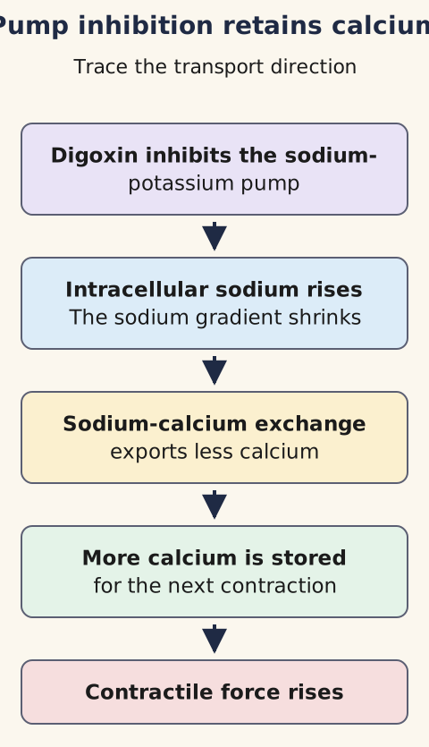
Why can one medicine strengthen contraction while slowing the ventricular response to atrial fibrillation? Begin with the sodium-potassium adenosine triphosphatase (Na+/K+-ATPase), an energy-consuming membrane pump. Each cycle normally exports three sodium ions and imports two potassium ions. Digoxin, a cardiac glycoside historically associated with foxglove (Digitalis), inhibits this pump. Sodium consequently accumulates inside cardiac muscle cells; that smaller inward sodium gradient provides less driving force for the sodium-calcium exchanger (NCX) to extrude calcium.
Calcium is not necessarily completely blocked from leaving: NCX transport also depends on membrane voltage and ion gradients. Less net calcium extrusion allows greater calcium storage in the sarcoplasmic reticulum (SR), the intracellular calcium reservoir, and greater release with the next contraction. This is positive inotropy, not direct beta-1 stimulation, phosphodiesterase-3 inhibition, or opening of L-type calcium channels.
This foundation later guides treatment selection, rhythm interpretation, exposure monitoring and long-term safety. [1]
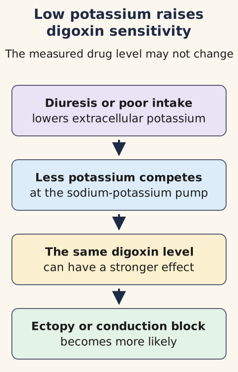
Use the pump figure associated with this section: read from pump inhibition through intracellular sodium accumulation and reduced exchanger-mediated calcium extrusion to greater sarcoplasmic reticulum release. Cover the final box and predict contraction. The answer is greater contractile force. Then compare the separate potassium figure: when a loop diuretic lowers extracellular potassium, digoxin can occupy the pump more effectively and provoke adverse effects without increasing the measured concentration. These figures show transport direction, not a heart cross-section. [1][5]
Predict contractility by following sodium first, then calcium. [1]
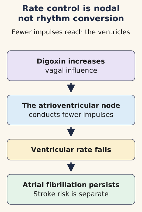
Now separate the electrical effect. Digoxin increases parasympathetic (vagal) influence on the atrioventricular (AV) node, slowing its conduction and lengthening refractoriness. In atrial fibrillation (AF), fewer chaotic atrial impulses reach the ventricles: the ventricular rate may fall although the atria remain fibrillatory. This neither reliably restores sinus rhythm nor prevents embolic stroke. Imagine a resting AF rate falling from 126 to 82 beats per minute while fibrillatory activity persists: attribute the rate change to nodal filtering, not a newly organized atrial rhythm. Anticoagulation is a separate risk-based decision. [2]
A slower ventricular response does not mean atrial fibrillation or stroke risk has disappeared. [2]
At excessive tissue exposure, calcium overload can cause spontaneous SR release and delayed afterdepolarizations, voltage disturbances after a normal action potential that trigger ectopic impulses. This increased automaticity can coexist with impaired AV transmission. If an atrial focus fires rapidly but only every third impulse reaches the ventricles, the atria are fast and the ventricular pulse is comparatively slow: there is no contradiction. Do not confuse this toxicity mechanism with beneficial vagal rate control. [1][5]
Foundation: why sodium affects calcium
NCX normally uses inward movement of sodium to help export calcium from a cardiac cell. When pump inhibition leaves more sodium inside, the sodium gradient is smaller; under usual conditions calcium export through NCX is reduced. The downstream rise in stored calcium supports contraction, but surplus calcium can initiate abnormal electrical impulses.
Checkpoint 1 of 3: Predict what changes if atrial fibrillation persists but the ventricular rate falls after digoxin. The ventricular slowing reflects increased AV nodal refractoriness; do not infer rhythm conversion or stroke protection. Apply this to a patient whose pulse rises again while walking: vagal control is less reliable during exertion, so reassess rate control rather than escalating a level blindly. [2]
Try it here · Checkpoint 1 of 3
Make your prediction before reading the choices. A first attempt is just a starting point.
Case 2
Show answer and explanations for case 2
A. Increased parasympathetic atrioventricular nodal inhibition (Best answer)
The atria still fibrillate while fewer impulses reach the ventricles through the atrioventricular (AV) node.
Reasoning steps for option A
What tells us the atria have not converted to sinus rhythm?
P waves remain absent and the ventricular intervals remain irregularly irregular.
Which site can slow the ventricular rate from 128 to 79/min while atrial fibrillation persists?
Greater vagal effect on atrioventricular nodal conduction allows fewer fibrillatory impulses to reach the ventricles.
B. Restoration of organized sinus-node pacing (Why this does not fit)
Sinus conversion would produce organized P waves and usually regular R-R intervals.
Reasoning steps for option B
What electrocardiographic sign would organized sinus-node pacing restore?
Discrete P waves preceding the ventricular complexes would return.
Does the follow-up tracing support sinus conversion?
No; absent P waves and persistent irregularity show continued atrial fibrillation despite the lower ventricular rate.
C. Suppression of accessory-pathway conduction (Why this does not fit)
There is no preexcitation or evidence that an accessory pathway mediates ventricular activation.
Reasoning steps for option C
What finding would raise concern for accessory-pathway conduction in atrial fibrillation?
Preexcitation or an unusually rapid, often wide and variably shaped ventricular response would be concerning.
Why is pathway blockade not the mechanism of this response?
No preexcitation is described, and digoxin slows atrioventricular nodal transmission rather than reliably blocking an accessory pathway.
D. Suppression of fibrillatory atrial impulse generation (Why this does not fit)
Continued absent P waves and irregular intervals show the atria remain fibrillatory; nodal filtering explains the reduced ventricular response.
Reasoning steps for option D
If digoxin had directly terminated fibrillation, would P waves remain absent?
No; organized atrial activation would replace the persistent irregular fibrillatory pattern.
What changed while the atrial rhythm persisted over three days?
Ventricular transmission fell from 128 to 79/min, compatible with atrioventricular (AV) nodal filtering rather than atrial suppression.
E. Reduced conduction velocity in ventricular myocardium (Why this does not fit)
Slower intraventricular conduction would widen QRS complexes rather than chiefly reduce ventricular response.
Reasoning steps for option E
What tracing feature would suggest primarily slowed ventricular myocardial conduction?
Widening of the QRS complex would suggest delayed spread through ventricular tissue.
Which observation instead identifies altered atrioventricular impulse delivery?
The fibrillating atria persist while the average ventricular response drops from 128 to 79/min.
Takeaway: Digoxin increases vagal influence on AV nodal conduction without necessarily restoring sinus rhythm.
Which outcome are you seeking: fewer heart failure admissions, a slower ventricular rate, or improved survival? In symptomatic heart failure with reduced ejection fraction (HFrEF), digoxin may be considered to reduce hospitalizations when symptoms persist despite guideline-directed treatment or that treatment cannot be tolerated. Contemporary foundational classes are a renin-angiotensin system inhibitor, preferably an angiotensin receptor-neprilysin inhibitor when appropriate; an evidence-based beta blocker; a mineralocorticoid receptor antagonist; and a sodium-glucose cotransporter-2 inhibitor. Diuretics treat congestion. Digoxin supplements rather than replaces these therapies and has not demonstrated an overall survival benefit. [3]
Consider two otherwise similar patients, both with reduced ejection fraction: one has not yet received tolerable foundational therapy; the other remains symptomatic despite it. Before considering adjunct digoxin in the second patient, identify and address opportunities to implement foundational drugs in the first. The Digitalis Investigation Group (DIG) randomized trial found fewer heart failure hospitalizations without reduced overall mortality. Its participants were in sinus rhythm and were treated in an era of diuretics and angiotensin-converting enzyme inhibitors, not today's four-class background regimen. Do not translate that older trial into proof of an added modern-era survival benefit. [3][4]
For AF rate control, beta blockers and, if left ventricular function allows, diltiazem or verapamil are common options. Long-term diltiazem or verapamil should be avoided when left ventricular ejection fraction is below 40% because negative inotropy can worsen heart failure. Digoxin may be useful in selected patients with heart failure symptoms as an adjunct or when other agents are poorly tolerated, including selected stable patients whose blood pressure limits alternatives.
Ask whether the patient is stable before choosing a slower-onset nodal drug: shock or other hemodynamic instability caused by AF requires urgent synchronized cardioversion rather than waiting for digoxin. A satisfactory resting rate may conceal fast ventricular rates during activity because sympathetic stimulation weakens vagal control. Assess symptoms and activity rates rather than titrating only to a resting pulse. [2]
Try changing the route of conduction in your mental model. In pre-excited AF, an accessory pathway bypasses the AV node; digoxin and other AV nodal blockers are contraindicated because slowing the node can favor dangerous conduction through that pathway. An irregular wide-complex tachycardia suggesting pre-excitation is not ordinary narrow-complex AF to treat with digoxin. Likewise, existing symptomatic bradycardia, sinus-node disease, or AV block warrants caution: digoxin can worsen conduction disease. Check the baseline electrocardiogram (ECG), renal function, medicines, and symptoms. Even if rate control succeeds, evaluate anticoagulation independently. [1][2]
What does comparative trial evidence allow? A small randomized rate-control comparison, RATE-AF, did not show a significant difference in its primary six-month quality-of-life endpoint between low-dose digoxin and bisoprolol. It does not establish universal superiority of digoxin or justify use in unstable or pre-excited AF. Observational associations between digoxin and mortality can reflect who received the medicine and how ill they were; they cannot on their own establish a causal survival penalty or advantage. Use clinical indication and randomized endpoints rather than choosing a treatment from an unadjusted association. [12][16]
Read ST shape and rhythm separately
Is a scooped ST segment itself poisoning? A therapeutic digitalis effect can include scooped ST depression (a depression of the interval after ventricular activation during recovery), T-wave flattening or inversion, shorter QT interval (the interval from ventricular activation through recovery), and modest PR prolongation (the interval from atrial activation to ventricular activation). Stable repolarization changes alone do not diagnose toxicity or require antibody treatment.
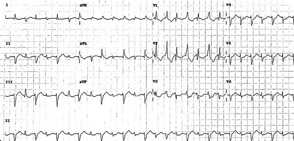
Compare a real patient's current tracing with their own prior ECG, symptoms, rate, and conduction, not with a generic drawing. The associated licensed clinical rhythm image shows bidirectional ventricular tachycardia: inspect the lead III tracing and compare adjacent QRS complexes, the waveform of ventricular activation. One complex points predominantly upward and the next predominantly downward, reflecting alternating electrical axes.
Cover the caption and decide whether that alternating direction alone proves digoxin toxicity: it does not; interpret exposure and clinical context. This image establishes morphology, not a measurable rate, amplitude, or timing because its calibration cannot be read reliably. [14] A patient with new chest pressure still needs appropriate acute ischemia evaluation including ECG assessment and cardiac troponin when indicated, even if some ST appearance resembles a familiar drug effect. [1][10][13]
Compare adjacent QRS complexes in lead III: their dominant deflections alternate direction. This supports bidirectional ventricular tachycardia, not a uniquely diagnostic cause. Use exposure, kidney function and symptoms to assess digoxin toxicity; exercise-related inherited arrhythmia is another setting. Original Figure 3 is reproduced without alteration. Do not calculate intervals or amplitudes from this reproduction. [9 [9],14 [14]] Jose Martin Alanis Naranjo, Kevin David Aragon Ontiveros, Julio Cesar Rivera Hermosillo and Virginia Campos Garcilazo; original source; CC BY 4.0.
Picture two tracings: a comfortable patient with unchanged scoop and normal perfusion; another with new anorexia, vomiting, dizziness, and new high-grade AV block. Cover the symptoms and ask whether the ST shape separates them. It does not. Reveal the history: the second patient requires an exposure, electrolyte, renal, and rhythm assessment, even if the laboratory level lies within an older reference interval. Typical chronic toxicity can also present as fatigue, confusion, poor intake or falls, especially in an older adult. Yellow-green visual disturbance or halos support toxicity if present, but absence does not exclude it. [1][5]
Inspect atrial and ventricular activity separately. Digoxin toxicity can generate atrial tachycardia with AV block: calcium-mediated triggered activity increases atrial impulses while nodal impairment prevents many from reaching the ventricles. Sinus bradycardia, junctional rhythms, varying AV block, ventricular ectopy and ventricular tachycardia also occur. Predict what an atrial rate of 180 with 3:1 AV conduction means: approximately 60 ventricular activations per minute, not a normal atrial rate. This paired fast-source/slow-gate pattern is a particularly useful discriminator, not an obligatory diagnostic criterion. [5]
Bidirectional ventricular tachycardia has alternating QRS axes and raises concern for digoxin toxicity in an exposed patient, but it is not unique: catecholaminergic polymorphic ventricular tachycardia (CPVT), an inherited exertion- or emotion-triggered arrhythmia syndrome, can also cause it. Compare the contexts: an older patient with renal deterioration taking digoxin and an adolescent with exertional syncope and no digoxin exposure require different etiologic investigations despite a superficially similar rhythm description. An ECG pattern should launch, not end, causal reasoning. [5][9]
Checkpoint 2 of 3: With rapid atrial activity and only one ventricular response for every three atrial impulses, predict two simultaneous effects. The pattern reflects increased atrial automaticity plus impaired AV conduction. Apply that prediction to a patient whose ventricular rate looks deceptively reassuring while nausea and new block are developing: inspect the atrial rhythm and prior tracing instead of equating a slow pulse with safety. [1][5]
Try it here · Checkpoint 2 of 3
Make your prediction before reading the choices. A first attempt is just a starting point.
Case 20
Show answer and explanations for case 20
A. Reduced atrial automaticity with faster atrioventricular conduction (Why this does not fit)
That predicts fewer atrial impulses and more ventricular capture, opposite to this tracing.
Reasoning steps for option A
Does atrial activity near 180/min indicate suppressed automaticity?
No. Newly rapid organized atrial depolarizations are compatible with enhanced atrial impulse generation.
Would faster atrioventricular conduction yield only one ventricular response per three atrial impulses?
No. The slow regular ventricular rhythm reflects impaired conduction, not acceleration through the node.
B. Sinus bradycardia with preserved atrioventricular conduction (Why this does not fit)
Atrial rate 180/min with only one of three beats conducted is neither sinus bradycardia nor complete capture.
Reasoning steps for option B
Is an atrial rate near 180/min sinus bradycardia?
No. The atria are depolarizing rapidly while the ventricles respond slowly.
Are all atrial impulses capturing the ventricles?
No. Only every third atrial impulse conducts, contradicting complete ventricular capture.
C. Enhanced atrial automaticity with impaired atrioventricular conduction (Best answer)
Discrete ectopic P waves separated by an isoelectric baseline identify atrial tachycardia rather than flutter; 3:1 conduction fits enhanced atrial automaticity with impaired atrioventricular (AV) nodal conduction.
Reasoning steps for option C
What explains the newly rapid organized atrial impulses?
Digoxin-associated enhanced atrial automaticity can produce atrial tachycardia, especially with new kidney dysfunction and toxicity symptoms.
Why is the ventricular rate slow despite rapid atrial activity?
Concurrent atrioventricular nodal conduction block permits only every third atrial impulse to reach the ventricles.
D. Accessory-pathway conduction with rapid ventricular activation (Why this does not fit)
A slow ventricular response with dropped atrial beats contradicts 1:1 accessory conduction.
Reasoning steps for option D
What ventricular response would 1:1 accessory-pathway conduction predict?
Each atrial impulse near 180/min would activate the ventricles, not a slow rhythm with two of every three impulses dropped.
What observed feature contradicts accessory-pathway acceleration?
The tracing describes regular slow ventricular responses after only every third organized atrial depolarization.
E. Ventricular automaticity with atrioventricular dissociation (Why this does not fit)
Organized rapid atrial activity is visible and central to interpreting this rhythm.
Reasoning steps for option E
Are atrial impulses absent from this tracing?
No. Organized atrial depolarizations are explicitly seen at approximately 180/min.
Can isolated ventricular automaticity account for the atrial-to-ventricular ratio?
No. The recurring one-in-three atrial conduction pattern requires an explanation involving atrial activity and conduction block.
Takeaway: Atrial tachycardia with AV block can reflect digoxin-related increased atrial automaticity and depressed AV nodal conduction; distinguish it from flutter by the atrial waveform and baseline.
Why can yesterday's unchanged prescription become today's toxic exposure? Digoxin has a narrow therapeutic margin, distributes extensively into tissues, and depends substantially on renal elimination. Its usual elimination half-life is about 36 to 48 hours with normal kidney function and lengthens when renal function falls. [1][11] Acute kidney injury (AKI), dehydration, declining renal clearance, older age and low lean body mass can turn a once-tolerated dose into excess exposure.
Base dosing and reassessment on kidney function and lean body weight rather than total weight alone. A seemingly modest creatinine in a frail person with little muscle need not indicate reassuring clearance. Ask what changed since the last dose decision, not just whether the pill strength changed. [1]
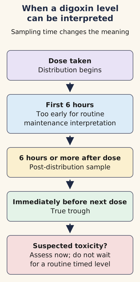
Use the concentration-versus-time schematic linked to this section. Cover the late sample and compare one taken an hour after dosing with one obtained at least six hours after dosing or immediately before the next dose. Early blood still reflects distribution and may overstate the equilibrated concentration; document both dose and sample times. A later correctly timed value is more useful for routine dose adjustment, but post-distribution is not steady state.
A new maintenance regimen generally takes about 7 to 10 days to approach steady state with normal renal function (roughly five elimination half-lives) and longer with renal impairment; assess suspected toxicity immediately rather than waiting for steady state. [1][11] During a dangerous suspected overdose, an initial level can inform toxicology consultation, but do not delay stabilization or antidote decisions until a later trough becomes available.
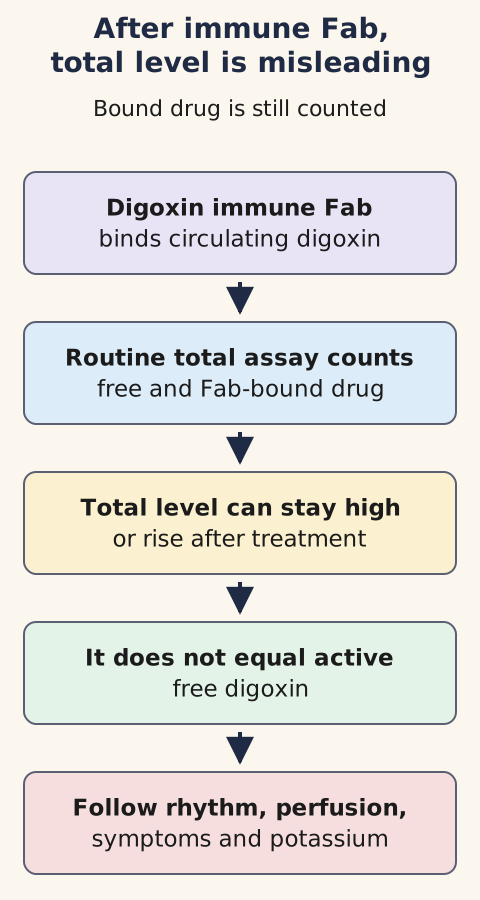
After digoxin immune Fab (antigen-binding antibody fragments), the ordinary total assay behaves differently, as the final section explains. [1][5]
Interpret sampling time before interpreting the number; suspected toxicity is assessed immediately. [1][5]
For HFrEF, the guideline's intended serum range is 0.5 to less than 0.9 ng/mL; for AF, the guideline recommends a target below 1.2 ng/mL if a level is measured. These are treatment targets, not boundaries below which toxicity is impossible. Suppose a symptomatic patient has new block, AKI, and a level of 0.8 ng/mL collected at an appropriate time: assess for toxicity and competing causes rather than declaring the result protective.
A modest isolated elevation or an early post-dose sample does not automatically require Fab. However, a potentially fatal ingestion or a very high, appropriately interpreted concentration may warrant Fab before dangerous rhythms develop; seek urgent toxicology advice using product-label and consensus criteria. [5][6] The concentration answers an exposure question only when timing and patient susceptibility are known. [2][3][5]
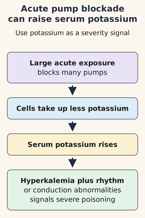
The potassium schematic poses two different predictions. Low potassium increases sensitivity to a given digoxin exposure because extracellular potassium competes with digoxin at the pump. A loop or thiazide diuretic can therefore precipitate toxicity without making the measured drug concentration higher. High potassium in severe acute poisoning can instead reflect extensive pump inhibition: cells take up less potassium and serum potassium rises.
Chronic toxicity may show low, normal, or high potassium depending on renal status, diuretics and concurrent drugs. Do not infer a dose or serum concentration from potassium alone, and never administer potassium merely because digoxin appears on the medicine list. Check the actual electrolyte and trend. [1][5]
Low potassium can increase susceptibility without increasing the digoxin level. [1][5]Predict potassium sensitivity and potassium release in two patients
Patient A has a correctly timed digoxin level of 0.8 ng/mL and potassium falls from 4.2 to 2.8 mmol/L after diuresis. Predict whether the measured digoxin concentration must rise before toxicity becomes more likely. It need not rise: reduced potassium competition at the pump increases sensitivity at the same measured drug level. [1][5]
Patient B has a severe acute ingestion and potassium rises to 6.2 mmol/L. Predict the direction of cellular potassium uptake. It falls because extensive pump inhibition prevents usual potassium entry into cells; assess this patient's rhythm and perfusion urgently rather than assuming potassium itself measures ingested dose. [5][6]
Low magnesium and high calcium increase susceptibility to dysrhythmia; measure and correct electrolyte disturbances with appropriate monitoring. Digoxin is a P-glycoprotein (P-gp) substrate, meaning this transport protein helps control its absorption and elimination. Amiodarone, quinidine, verapamil and some macrolide antibiotics may increase exposure through transporter interactions. The prescribing information recommends an approximately 30 to 50 percent digoxin dose reduction or altered dosing frequency when starting amiodarone, followed by clinical and concentration monitoring.
Both drugs can slow AV conduction even if the digoxin level changes little. In a patient newly started on amiodarone who develops nausea and bradycardia, check dosing history, ECG, renal function, potassium and magnesium together. [1]
Treat the patient before the drug level
Which problem needs treatment first: the number or the perfusion-threatening rhythm? Stop digoxin when toxicity is suspected; assess airway, breathing, perfusion, continuous ECG, electrolytes (especially potassium and magnesium), renal function, and appropriately timed concentration. Identify interacting drugs and ask poison control or a medical toxicologist about severity and antidote dosing. In mild, stable chronic toxicity without dangerous arrhythmia, withholding drug, addressing AKI or electrolyte depletion, and observing with repeat assessment may suffice. Neither a modest elevation alone nor an isolated familiar ST scoop automatically warrants Fab. [5]
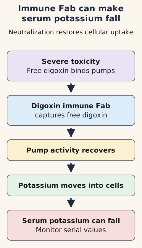
The Fab schematic shows digoxin immune Fab binding free digoxin so less reaches the pump. Promptly give Fab for life-threatening ventricular arrhythmia, severe symptomatic conduction block, shock or similarly severe toxicity; do not wait for a laboratory level in a deteriorating patient. Hyperkalemia in acute poisoning strengthens concern. The adult DigiFab label includes potassium above 5.5 mEq/L with rapidly progressive toxicity, while a 2023 expert consensus lists potassium above 6.5 mmol/L among immediate-treatment triggers.
These different thresholds have different evidentiary and clinical contexts: neither is a mandatory waiting point for a patient with shock or dangerous rhythm, and a number alone never replaces clinical judgment. The 2023 consensus is an expert narrative review, not a universally binding rule. [5][6] A separate expert consensus abstract also emphasizes exposure pattern and severity in selecting Fab; its abstract alone does not establish either numeric potassium cutoff. [15]
In acute poisoning, hyperkalemia reflects extensive pump inhibition and helps signal severity. [1][5]
After effective Fab, serum potassium can fall quickly and requires serial monitoring. [5][6]
Predict management for an older patient with nausea, AKI, stable blood pressure, no new block, and a moderately elevated trough: stop the drug, review precipitants and monitor. Now change only the rhythm to high-grade block with poor perfusion: stabilize and obtain Fab urgently, rather than waiting for a confirmatory concentration. Atropine can temporarily support symptomatic bradycardia while Fab is prepared. Pacing or cardioversion for digoxin-associated arrhythmia needs expert judgment because the myocardium can be irritable; a pulseless patient still receives indicated advanced cardiac life support (ACLS) resuscitation. [5][6]
The old assertion that intravenous calcium inevitably produces a fatal 'stone heart' is not supported by human evidence. One retrospective cohort observed no malignant dysrhythmias within an hour in 23 calcium-treated patients with digoxin toxicity, but a small nonrandomized sample cannot establish universal safety or a mortality benefit. In severe poisoning, Fab remains definitive; individualize additional hyperkalemia care with toxicology input rather than treating calcium as either an absolute taboo or an automatic replacement for Fab. [6][7]
Follow the Fab diagram through the next few hours. As pump function recovers, cellular potassium uptake can resume and serum potassium can fall; follow ECG, perfusion and serial electrolytes. Watch for recurrent toxicity, especially with severe renal dysfunction, and for lost therapeutic benefit: AF rate may rise or heart failure symptoms worsen. Routine total digoxin assays after Fab detect bound as well as free drug and can appear high despite effective neutralization.
Do not redose antidote merely because a total level remains high; selected free-drug measurement may help where available. Digoxin's tissue distribution makes hemodialysis ineffective for clearing it; renal failure still prolongs the observation problem, and EXTRIP recommends against extracorporeal removal. [6][8]
After Fab, follow the patient, rhythm and electrolytes rather than the total digoxin assay. [5][6]
Compare digoxin findings and actions
Clinical setting
Key discriminator
Response
Clinical settingExpected drug effect
Key discriminatorOld scooped ST contour without new symptoms
ResponseAssess clinical context; do not diagnose poisoning from contour alone.
Clinical settingElectrolyte susceptibility
Key discriminatorNew ectopy with low potassium or magnesium despite an ordinary concentration
ResponseAssess possible toxicity, measure and correct the deficit.
Clinical settingExposure increase
Key discriminatorRenal decline or a new interacting medication
ResponseReassess dose, symptoms, rhythm and a correctly timed level.
Clinical settingSevere toxicity
Key discriminatorDangerous ventricular rhythm, high-grade block with poor perfusion, or shock
ResponseResuscitate and promptly give digoxin immune Fab.
Clinical settingAfter Fab
Key discriminatorClinical recovery despite high total digoxin; falling potassium
ResponseFollow rhythm, perfusion and serial electrolytes.
Severity determines urgency; serum concentration, sampling time and electrolyte sensitivity refine rather than replace the bedside assessment. [1][5][6]
Checkpoint 3 of 3: Predict the potassium direction after effective Fab in a severely poisoned patient with initial hyperkalemia. Potassium moves downward as cellular uptake recovers; verify it with serial measurements, not a single assumption. Apply the same principle to a rising post-Fab total digoxin assay: assess clinical status, ECG and potassium rather than interpreting antibody-bound drug as new free poisoning. [6]
Try it here · Checkpoint 3 of 3
Make your prediction before reading the choices. A first attempt is just a starting point.
Case 16
Show answer and explanations for case 16
A. Fab directly promotes urinary potassium loss (Why this does not fit)
The antibody binds digoxin; the immediate shift does not require enhanced renal potassium excretion.
Reasoning steps for option A
What does digoxin immune Fab (antigen-binding antibody fragments) bind in this acute ingestion?
It binds circulating digoxin, not potassium; the potassium fall does not demonstrate direct renal potassium removal by Fab.
Does the four-hour decline establish increased urinary potassium excretion?
No urine output or urinary potassium measurement is supplied, while recovering atrioventricular (AV) conduction and pressure support reversal of digoxin's pump effect.
B. Insulin treatment caused intracellular uptake (Why this does not fit)
No insulin was administered during the observed fall.
Reasoning steps for option B
Was an insulin-mediated potassium shift induced?
No insulin was administered between the potassium measurements of 6.0 and 3.3 mmol/L.
What intervention coincided with the change instead?
Immune digoxin immune Fab (antigen-binding antibody fragments) was given, followed by improving conduction and a rapid fall in extracellular potassium.
C. Dialysis removed the extracellular potassium (Why this does not fit)
No dialysis occurred and the improvement tracked reversal of pump inhibition.
Reasoning steps for option C
Was there extracorporeal potassium clearance during observation?
No dialysis was performed in the four hours during which potassium fell to 3.3 mmol/L.
What concurrent cardiac response argues for reversal of digoxin action?
Bradycardia and impaired atrioventricular conduction improved after digoxin immune Fab (antigen-binding antibody fragments), consistent with relieved sodium-potassium pump inhibition.
D. Continued pump inhibition enhances potassium uptake (Why this does not fit)
Continuing inhibition would impair uptake rather than produce this decrease.
Reasoning steps for option D
What does persistent sodium-potassium pump blockade do to cellular potassium uptake?
It reduces uptake, tending to leave more potassium outside cells rather than explaining a fall from 6.0 to 3.3 mmol/L.
Does the recovery of atrioventricular conduction support persistent blockade?
No. Improving conduction after digoxin immune Fab (antigen-binding antibody fragments) points toward less active digoxin at its target.
E. Restored pump transport moves potassium into cells (Best answer)
digoxin immune Fab (antigen-binding antibody fragments) reverses digoxin-mediated pump inhibition, so extracellular potassium can fall quickly.
Reasoning steps for option E
Why was potassium initially elevated after severe ingestion?
Acute digoxin inhibition of sodium-potassium adenosine triphosphatase impairs movement of potassium into cells.
Why monitor potassium frequently after digoxin immune Fab (antigen-binding antibody fragments)?
Neutralizing digoxin restores pump-mediated uptake, so extracellular potassium may rapidly fall, as it did here to 3.3 mmol/L.
Takeaway: Neutralizing digoxin restores sodium-potassium ATPase transport and shifts potassium into cells.
Long-term care starts by confirming the continuing indication. For heart failure with reduced ejection fraction, digoxin is an adjunct for persistent symptoms or hospitalization burden, not a substitute for disease-modifying therapy. For atrial fibrillation, it slows the ventricular response but does not restore sinus rhythm or replace a separate stroke-prevention assessment. Reassess the indication whenever kidney function, rhythm strategy, functional status or goals change. [2][3][4]
At baseline and after meaningful change, document pulse and rhythm, kidney function, potassium, magnesium, calcium and the full medication list. Repeat this review after dehydration, acute illness, reduced urine output, a dose change or an interacting drug; an old normal value does not protect a patient whose clearance or electrolyte state has changed. A concentration is useful only when sampling time and the clinical question are known. [1][5]
After suspected toxicity, correct reversible causes and decide whether digoxin still has a net benefit before restarting it. New syncope, sustained palpitations, chest pain, major dyspnea, severe dizziness or confusion warrants urgent evaluation. A dangerous rhythm, high-grade atrioventricular block, hyperkalemia after an acute ingestion or shock warrants resuscitation and toxicology-directed antidote care, not routine outpatient adjustment. [5][6]
Beyond the boards: dosing, consultation and counseling
Past-board details support learning, not an individualized prescription. A common adult maintenance orientation is 0.125 to 0.25 mg by mouth once daily; older adults, people with low lean body mass or impaired renal clearance may need 0.0625 to 0.125 mg daily or a longer interval. Individualize by lean body size, kidney function, indication and interacting medicines, and verify against current labeling before selecting a dose. Routine loading is not required for every stable outpatient. [1]
For maintenance assessment, obtain a post-distribution concentration at least six hours after a dose; a sample immediately before the next dose is the true trough. After a new stable regimen, allow roughly seven to ten days for steady state with normal renal function and longer with renal impairment, unless suspected toxicity requires immediate clinical assessment. Interpret the number with symptoms, electrocardiogram, kidney function and electrolytes rather than chasing a universal laboratory target. [1][5]
Amiodarone can substantially increase digoxin exposure; current labeling advises a 30 to 50 percent dose reduction or a frequency change with continued monitoring. Quinidine, verapamil, some macrolide antibiotics and other P-glycoprotein inhibitors can also raise exposure, while beta blockers and other atrioventricular nodal drugs can add conduction slowing. Confirm the exact product interaction and current labeling rather than applying one reduction to every combination. [1]
Contact a poison center or medical toxicologist for a significant ingestion, dangerous rhythm, high-grade block, hyperkalemia, shock, uncertain digoxin immune Fab dosing or recurrent toxicity after Fab. Seek cardiology input for pre-excited atrial fibrillation, refractory rate control, important conduction disease, device questions or an uncertain long-term indication. Consultation supports, but does not delay, immediate stabilization. [2][5][6]
Counsel patients to take the medicine at the same time each day, avoid doubling a missed dose, and keep an updated medication list. Ask them to call before starting a new prescription, over-the-counter product or herbal supplement and to report nausea, vomiting, poor appetite, new visual disturbance, palpitations, fainting, confusion, diarrhea, dehydration or reduced urine output. The practical rule is simple: a new symptom plus a new exposure risk deserves early review. [1][5]
Practice the distinctions
Use the cases to choose a clinical action or interpretation before reading each rationale. Revisit pump effects, treatment purpose, ECG context, concentration timing, electrolyte direction and severity-based antidote decisions in a new patient setting.
Case 1
Show answer and explanations for case 1
A. Increased calcium extrusion through sodium-calcium exchange (Why this does not fit)
That would lower cytosolic calcium and weaken, not strengthen, systole.
Reasoning steps for option A
What does increased sodium-calcium exchange export from these ventricular cells?
It exports more calcium, leaving less intracellular calcium for systole.
Would that direction explain her improved stroke volume?
No; more calcium removal predicts weaker contraction despite the observed improvement.
B. Increased calcium influx through beta-1 stimulation (Why this does not fit)
Beta-1 stimulation can raise calcium but is not the action of digoxin when pump activity falls.
Reasoning steps for option B
What pathway would increase calcium entry through beta-1 signaling?
Adrenergic receptor stimulation raises cyclic adenosine monophosphate and calcium-channel activity.
Which target is actually inhibited in her myocyte preparation?
Sodium-potassium adenosine triphosphatase, not a β₁ receptor, is inhibited.
C. Reduced calcium extrusion through sodium-calcium exchange (Best answer)
Higher intracellular sodium weakens the gradient driving forward sodium-calcium exchange, retaining calcium for contraction despite unchanged extracellular calcium.
Reasoning steps for option C
What happens to intracellular sodium when digoxin inhibits the measured pump?
Less sodium leaves the ventricular myocyte, so intracellular sodium rises.
How does this sodium change improve her stroke volume?
A smaller sodium gradient reduces sodium-calcium exchange-mediated calcium extrusion, retaining calcium for stronger systole.
D. Reduced calcium release from the sarcoplasmic reticulum (Why this does not fit)
Depleting stored calcium would oppose the observed improvement in stroke volume.
Reasoning steps for option D
What would less calcium in the sarcoplasmic reticulum do to this patient's contraction?
It would reduce calcium released during systole and diminish ventricular force.
Does that match the echocardiographic finding after digoxin?
No; her stroke volume improved, requiring more available contractile calcium, not less.
E. Increased sodium export by the membrane pump (Why this does not fit)
The measured pump inhibition predicts less, not more, sodium export.
Reasoning steps for option E
In which direction does sodium transport change when sodium-potassium adenosine triphosphatase is inhibited?
Sodium extrusion falls rather than rises.
Why cannot increased pump-mediated sodium export explain her contractility?
The preparation directly demonstrates inhibited pump activity, which instead increases intracellular sodium and retains calcium.
Takeaway: Pump inhibition raises intracellular sodium, reducing calcium extrusion by sodium-calcium exchange and increasing calcium available for contraction.
A. Lower all-cause mortality replacing sacubitril-valsartan therapy (Why this does not fit)
Neither Digitalis Investigation Group (DIG) mortality results nor current practice supports replacing disease-modifying angiotensin receptor-neprilysin inhibitor (ARNI) with digoxin.
Reasoning steps for option A
What mortality result did the historical Digitalis Investigation Group trial establish?
It did not establish reduced all-cause mortality with digoxin.
Why is replacing her angiotensin receptor-neprilysin inhibitor particularly unsupported?
Her foundational therapy is disease modifying, whereas digoxin is considered an adjunct for persistent symptoms or admissions.
B. Fewer admissions solely through slower atrial fibrillation ventricular rates (Why this does not fit)
This patient is in sinus rhythm; the historical hospitalization result does not depend on rate control for atrial fibrillation.
Reasoning steps for option B
Is this woman hospitalized with rapid atrial fibrillation?
No; she is in sinus rhythm during recurrent congestion admissions.
Does the historic reduction in heart-failure admissions require ventricular-rate slowing in atrial fibrillation (AF)?
No; that trial largely enrolled sinus-rhythm patients, so an AF rate mechanism does not explain its endpoint.
C. Improved survival established alongside empagliflozin (Why this does not fit)
The historical trial predates sodium-glucose cotransporter-2 (SGLT2) inhibitors and did not establish overall survival improvement.
Reasoning steps for option C
Were sodium-glucose cotransporter-2 inhibitors part of the historical trial's background therapy?
No; that drug class entered heart-failure treatment much later.
Did that trial demonstrate an overall survival advantage even on its original background regimen?
No; it cannot establish improved survival alongside her present regimen.
D. Fewer heart failure admissions, without proven survival gain (Best answer)
Digitalis Investigation Group (DIG) studied predominantly sinus-rhythm heart failure with reduced ejection fraction (HFrEF) on older background therapy; its admission signal is not a mortality claim on modern therapy.
Reasoning steps for option D
Which outcome is pertinent to her two congestion admissions this year?
The Digitalis Investigation Group trial found fewer heart-failure hospitalizations with digoxin.
What limitation should accompany that expectation in her modern regimen?
No overall mortality benefit was demonstrated, and trial background therapy differed from her current four-drug foundation.
E. No plausible role once foundational therapy is optimized (Why this does not fit)
Selected persistently symptomatic patients with recurrent heart failure (HF) admissions may still be considered for adjunctive digoxin.
Reasoning steps for option E
Does her optimized foundational regimen eliminate every potential adjunctive goal?
No; she remains symptomatic with recurrent congestion admissions despite tolerated therapy.
How narrow is the evidence-based role of digoxin in this situation?
It may be considered to reduce hospitalization burden, not to replace her foundational agents or promise longer survival.
Takeaway: The historical DIG trial supports fewer heart-failure hospitalizations, not a demonstrated overall survival benefit; modern background therapy differs.
A. A normal digoxin level excludes drug-associated ectopy (Why this does not fit)
Electrolyte depletion can make usual concentrations arrhythmogenic.
Reasoning steps for option A
Does a concentration of 0.8 ng/mL exclude digoxin-associated ectopy?
No. Her new ectopy followed potassium falling to 2.8 mmol/L, which can increase sensitivity at an ordinary measured concentration.
What changed despite an unchanged digoxin prescription?
Doubling the loop diuretic preceded marked potassium loss; the assay value alone cannot dismiss the temporal clinical signal.
B. Potassium depletion heightens digoxin sensitivity (Best answer)
Low extracellular potassium favors digoxin interaction with the sodium-potassium pump despite the measured level.
Reasoning steps for option B
What does potassium of 2.8 mmol/L imply for digoxin binding?
Less extracellular potassium competes at the sodium-potassium pump, increasing susceptibility to digoxin effects.
Why is this explanation better than an overdose explanation?
The correctly timed level is 0.8 ng/mL and the dose stayed stable, whereas substantial potassium depletion coincided with ventricular ectopy.
C. Stable creatinine proves no cardiac drug effect (Why this does not fit)
Renal stability does not remove the potassium-dependent sensitivity caused by her diuretic.
Reasoning steps for option C
Does creatinine of 1.0 mg/dL rule out a new digoxin effect?
No. Renal function can remain stable while hypokalemia independently increases cardiac sensitivity.
Which laboratory change is more informative than creatinine here?
Potassium is 2.8 mmol/L after intensified diuresis, a plausible trigger for premature ventricular beats during digoxin treatment.
D. Loop diuretics directly raise the digoxin assay result (Why this does not fit)
Her level is not elevated; the clinically important change is potassium depletion.
Reasoning steps for option D
Would direct interference by a loop diuretic explain this reported concentration?
No. The correctly timed assay measures 0.8 ng/mL rather than a spurious high value needing explanation.
What indirect loop-diuretic effect matters clinically?
Increased urinary potassium loss can amplify digoxin-associated arrhythmia risk even if the digoxin dose and measured concentration do not rise.
E. Serum potassium loss accounts for the ectopy without any digoxin effect (Why this does not fit)
Potassium depletion itself promotes ectopy, but it also increases digoxin sensitivity; the unchanged timed concentration does not exclude a digoxin contribution.
Reasoning steps for option E
Can a potassium level of 2.8 mmol/L independently predispose to ventricular premature complexes?
Yes, but this does not make potassium depletion the only contributor to the new ectopy.
What else becomes more potent after the loop dose is doubled?
Digoxin can inhibit the pump more effectively at the same timed concentration when extracellular potassium is depleted.
Takeaway: Potassium depletion potentiates digoxin binding and arrhythmic susceptibility independent of a high assay value.
A. Diagnose severe toxicity from the one-hour value (Why this does not fit)
The timing prevents direct trough-like interpretation and she has no clinical toxicity.
Reasoning steps for option A
Can the 09:00 result diagnose severe toxicity after an 08:00 dose?
No. A one-hour sample is drawn before digoxin finishes distributing into tissues and cannot be interpreted like a later equilibrated level.
Which bedside findings also argue against acting on the number alone?
She has no nausea, visual disturbance, bradycardia or ectopy, and kidney function and potassium are unchanged.
B. Give immune Fab on the laboratory number alone (Why this does not fit)
An early isolated value without dangerous clinical findings does not justify antidote.
Reasoning steps for option B
Does a single early concentration of 2.1 ng/mL justify digoxin immune Fab (antigen-binding antibody fragments)?
No. Sampling one hour after dosing can transiently inflate the blood concentration relative to postdistribution values.
What severe finding would be needed to support urgent antidote evaluation instead?
Dangerous arrhythmia or another serious toxicity manifestation would shift urgency; none is described in this stable patient.
C. Repeat a sample at least 6-8 hours after a dose (Best answer)
The regimen is already established, but a one-hour postdose specimen is still in the distribution phase; obtain a consistently timed postdistribution sample and interpret it alongside symptoms and electrocardiogram (ECG).
Reasoning steps for option C
When should her digoxin concentration be rechecked?
Obtain a sample at least six hours after dosing, once the early distribution phase has passed.
Why postpone a dose change until that result is available?
The 09:00 value followed the 08:00 dose by only one hour, so it may misrepresent the concentration relevant to maintenance dosing.
D. Double the dose because the patient is asymptomatic (Why this does not fit)
Absence of symptoms does not mean the early assay demonstrates underexposure.
Reasoning steps for option D
Does being asymptomatic show that digoxin exposure is too low?
No. Absence of toxicity does not establish inadequate treatment or justify doubling her dose.
What specific uncertainty makes escalation unsafe?
The only reported value, 2.1 ng/mL, came during the postdose distribution phase and has not been confirmed with properly timed sampling.
E. Stop checking timing because levels never matter (Why this does not fit)
Correctly timed concentrations can assist dosing when interpreted with the clinical picture.
Reasoning steps for option E
Are digoxin concentrations categorically useless for management?
No. A properly timed level can help evaluate exposure when combined with symptoms, rhythm, kidney function and electrolytes.
Which sampling detail must the clinic document before using this result?
The interval since the last dose matters: the existing specimen was collected just one hour after ingestion.
Takeaway: Steady-state dosing does not make a one-hour postdose level interpretable as an equilibrated concentration; sample at least 6-8 hours after dosing and assess clinically.
A. Increase digoxin because amiodarone speeds elimination (Why this does not fit)
Amiodarone tends to increase, not reduce, digoxin exposure.
Reasoning steps for option A
What happens to digoxin exposure when amiodarone is introduced?
Amiodarone inhibits P-glycoprotein-mediated digoxin transport, so exposure tends to rise rather than fall.
Why is an increase from her current 0.125 mg daily particularly hazardous?
Her ventricular rate is already controlled and her estimated filtration rate is 48 mL/min/1.73 m2, leaving less margin for accumulation.
B. Reduce digoxin and monitor for accumulation (Best answer)
Amiodarone inhibits P-glycoprotein transport and can increase digoxin exposure on top of reduced renal clearance; both drugs can slow atrioventricular (AV) nodal conduction. Reduce digoxin and monitor timed levels, heart rate, electrocardiogram (ECG) and symptoms without assuming a universal reduction percentage.
Reasoning steps for option B
Which new prescription changes her digoxin dosing requirement?
Starting amiodarone can increase digoxin concentrations by inhibiting its transport and clearance.
How should the clinician act before prolonged combined exposure?
Reduce the digoxin dose and follow pulse, symptoms, kidney function, electrolytes and appropriately timed concentrations.
C. Keep both unchanged because potassium is normal (Why this does not fit)
Normal potassium does not negate increased exposure or additive atrioventricular (AV) nodal slowing with the combination.
Reasoning steps for option C
Does potassium of 4.2 mmol/L exclude an amiodarone-digoxin interaction?
No. Potassium is currently normal, but it does not prevent amiodarone from raising digoxin exposure.
What could happen if both doses remain unchanged despite her reduced kidney function?
Digoxin may accumulate over subsequent days, producing gastrointestinal symptoms or new conduction abnormalities despite a normal baseline potassium.
D. Check a digoxin level first, then leave both doses unchanged if it is normal (Why this does not fit)
A baseline level does not prevent amiodarone from increasing subsequent digoxin exposure; adjust and monitor proactively.
Reasoning steps for option D
Can a normal digoxin concentration before the first amiodarone dose exclude later accumulation?
No. The interaction develops with combined exposure, so an initially acceptable value does not guarantee subsequent safety.
What should change even if her baseline rate and electrolytes are stable?
Reduce digoxin and monitor after amiodarone begins, especially with estimated filtration rate 48 mL/min/1.73 m2.
E. Reduce amiodarone instead and continue the digoxin dose (Why this does not fit)
Amiodarone is being started for recurrent symptomatic episodes; reducing it does not reliably avert the digoxin interaction and undermines its intended regimen.
Reasoning steps for option E
Why was amiodarone prescribed for this woman despite her controlled ventricular rate?
She continues to have recurrent symptomatic rhythm episodes requiring a new rhythm-directed treatment.
Why is cutting amiodarone instead of adjusting digoxin not the best interaction plan?
It may compromise the intended rhythm treatment while leaving her digoxin dose vulnerable to increased exposure from the combination.
Takeaway: Anticipate amiodarone-related digoxin accumulation and additive AV nodal slowing, especially with impaired renal function; adjust dosing and follow timed levels, ECG and symptoms.
A. Chronic potassium depletion from diuresis (Why this does not fit)
Diuretic potassium loss generally causes hypokalemia and is not described here.
Reasoning steps for option A
Would chronic loop-diuretic potassium loss predict a potassium of 6.2 mmol/L?
No. Loop diuretics generally lower potassium, whereas this confirmed elevation arose shortly after a large acute ingestion.
Which findings connect the potassium result to the ingestion instead?
Vomiting, junctional bradycardia and worsening conduction two hours after digoxin ingestion point to severe pump blockade.
B. Potassium release caused by sample hemolysis (Why this does not fit)
The repeat nonhemolyzed specimen confirms a genuine elevation.
Reasoning steps for option B
What specimen detail makes pseudohyperkalemia unlikely?
The potassium elevation persisted in a repeat sample specifically reported as nonhemolyzed.
Why does attributing 6.2 mmol/L to sampling error miss the clinical emergency?
He also has acute vomiting and marked conduction abnormalities after a large digoxin ingestion, consistent with true severe toxicity.
C. Expected potassium rise during ordinary therapy (Why this does not fit)
This degree of hyperkalemia with vomiting and conduction delay indicates severe toxicity.
Reasoning steps for option C
Is potassium of 6.2 mmol/L an expected harmless effect of an ordinary therapeutic dose?
No. This follows intentional ingestion of many tablets, not routine treatment.
What accompanying abnormalities argue against calling it safe?
Junctional bradycardia and widening conduction delay accompany the hyperkalemia, signaling dangerous intoxication.
D. Impaired potassium excretion from aldosterone excess (Why this does not fit)
Aldosterone excess usually lowers potassium and does not explain the acute ingestion sequence.
Reasoning steps for option D
How does excess aldosterone ordinarily affect serum potassium?
It increases renal potassium excretion, tending toward hypokalemia rather than the confirmed hyperkalemia here.
Why does an endocrine explanation fit the timeline poorly?
The abrupt electrolyte and conduction changes appeared two hours after massive digoxin ingestion in a person without known chronic kidney disease.
E. Severe pump inhibition reduces cellular potassium uptake (Best answer)
Severe acute sodium-potassium adenosine triphosphatase (ATPase) inhibition reduces cellular potassium uptake, raising serum potassium; this is distinct from hypokalemia increasing susceptibility to digoxin.
Reasoning steps for option E
What cellular process does toxic digoxin suppress in this acute overdose?
Marked inhibition of the sodium-potassium adenosine triphosphatase pump reduces potassium movement into cells.
Why is this patient's potassium result a severity signal?
A confirmed value of 6.2 mmol/L alongside bradycardia and conduction delay indicates major pump inhibition and requires urgent poisoning management.
Takeaway: Acute poisoning can cause hyperkalemia through profound pump inhibition; this differs from hypokalemia increasing digoxin sensitivity and warrants urgent antidote assessment.
A. Administer digoxin immune Fab promptly (Best answer)
Unstable ventricular arrhythmia in this exposure context is an antidote indication independent of a pending assay.
Reasoning steps for option A
What makes this an emergency before the assay returns?
Sustained ventricular tachycardia is impairing perfusion, with blood pressure 68/38 mm Hg after syncope.
Why is digoxin immune Fab (antigen-binding antibody fragments) appropriate in addition to resuscitation?
Chronic digoxin exposure, reduced kidney function and recent anorexia support digoxin-associated life-threatening arrhythmia; Fab need not await a concentration.
B. Wait for the exact serum level before antidote (Why this does not fit)
The delay is unsafe in a shocked patient with a plausible digoxin-related ventricular arrhythmia.
Reasoning steps for option B
What clinical finding already supplies an antidote indication?
Her poorly perfusing sustained ventricular tachycardia is life-threatening in the setting of suspected digoxin toxicity.
What harm follows waiting an hour for a numerical result?
It postpones specific treatment while she remains in shock and at risk of further malignant arrhythmia.
C. Give another digoxin dose to slow the ventricle (Why this does not fit)
Additional digoxin would worsen suspected toxicity and is not treatment for ventricular tachycardia.
Reasoning steps for option C
Is a ventricular rate slowed by digoxin the immediate problem?
No. She has sustained ventricular tachycardia and severe hypotension, not uncomplicated rapid atrial fibrillation.
How would another dose affect a patient with falling urine output and suspected accumulation?
It could add to toxic tissue exposure and worsen the rhythm disturbance.
D. Use dialysis as the sole emergency antidote (Why this does not fit)
Dialysis removes little tissue-distributed digoxin and cannot replace digoxin immune Fab (antigen-binding antibody fragments) for severe poisoning.
Reasoning steps for option D
Would conventional dialysis rapidly remove most digoxin in this setting?
No. Digoxin distributes extensively into tissues, limiting clearance by dialysis.
What treatment does dialysis fail to replace in her shock-producing arrhythmia?
Digoxin digoxin immune Fab (antigen-binding antibody fragments) is the specific reversal treatment needed alongside immediate resuscitative care.
E. Observe until spontaneous rhythm recovery (Why this does not fit)
Sustained ventricular tachycardia with shock requires immediate active resuscitation and specific therapy.
Reasoning steps for option E
Is her ventricular tachycardia a benign telemetry finding to watch?
No. Syncope and blood pressure 68/38 mm Hg document inadequate perfusion.
What does the history add to the need for action?
Days of anorexia and reduced urine output while taking digoxin make accumulation plausible, so spontaneous recovery cannot be assumed.
Takeaway: Life-threatening suspected toxicity warrants immediate digoxin immune Fab alongside standard resuscitation.
A. Give Fab solely because the level is 2.0 (Why this does not fit)
A number alone without dangerous clinical findings is not an automatic antidote indication.
Reasoning steps for option A
What does the nine-hour concentration of 2.0 ng/mL contribute?
It supports concern about excess exposure after a creatinine rise, but must be interpreted with clinical severity.
Why does that number alone not require digoxin immune Fab (antigen-binding antibody fragments) in this man?
He is perfusing normally, has controlled atrial fibrillation, normal potassium and no new block or ventricular ectopy; this is not a blanket rule against Fab for sufficiently high concentrations or other serious indications.
B. Increase digoxin to improve his atrial fibrillation rate (Why this does not fit)
His rate is controlled and added exposure would worsen the suspected adverse effect.
Reasoning steps for option B
Does an atrial fibrillation rate of 78/min call for additional rate slowing?
No. His current ventricular response is controlled.
What makes a higher digoxin dose especially unsafe today?
Nausea, poor intake and creatinine rising from 1.1 to 1.5 mg/dL raise concern for accumulation, not undertreatment.
C. Ignore symptoms because potassium is normal (Why this does not fit)
Normal potassium does not exclude chronic digoxin toxicity, particularly after renal decline.
Reasoning steps for option C
Can potassium of 4.1 mmol/L rule out chronic digoxin adverse effects?
No. Chronic toxicity can present with anorexia and nausea without hyperkalemia.
Which change still demands reassessment despite normal potassium?
Renal function has deteriorated while he continues digoxin, increasing the risk of sustained exposure.
D. Perform urgent cardioversion for controlled atrial fibrillation (Why this does not fit)
Stable atrial fibrillation (AF) at 78/min is not an emergency cardioversion indication.
Reasoning steps for option D
What rhythm is actually documented?
Atrial fibrillation with a controlled ventricular rate of 78/min, without new block or ventricular ectopy.
Why would urgent cardioversion not address the immediate digoxin concern?
His blood pressure is 120/74 mm Hg and there is no unstable tachyarrhythmia; the relevant issue is possible medication accumulation.
E. Withhold digoxin and monitor while correcting contributors (Best answer)
Stable suspected mild chronic toxicity calls for stopping exposure and reassessing renal function, symptoms and rhythm.
Reasoning steps for option E
What initial exposure change addresses his symptoms and kidney decline?
Withhold digoxin rather than administering more while evaluating possible mild chronic toxicity.
What should be followed while reversible contributors are corrected?
Recheck symptoms, pulse and electrocardiographic rhythm, kidney function and electrolytes as hydration and intercurrent illness are addressed; escalate if severe toxicity develops.
Takeaway: Hold digoxin and address reversible factors with repeat clinical, ECG, electrolyte and renal assessment.
A. Repeat total digoxin until it reaches zero (Why this does not fit)
Total assays include bound drug after digoxin immune Fab (antigen-binding antibody fragments) and cannot be used as a short-term clearance target.
Reasoning steps for option A
What does a total digoxin assay measure after digoxin immune Fab (antigen-binding antibody fragments) administration?
It can detect digoxin bound to antibody fragments as well as unbound drug, so the reported total can rise.
Why is reaching zero on that assay a poor short-term treatment target?
Her ventricular rhythm, blood pressure and potassium have normalized; a high post-Fab total does not measure ongoing free-drug toxicity.
B. Follow rhythm, perfusion, symptoms and electrolytes (Best answer)
Clinical course and electrocardiogram (ECG) remain useful whereas total digoxin rises because assays detect digoxin immune Fab (antigen-binding antibody fragments)-bound drug.
Reasoning steps for option B
What objective changes show whether reversal is holding in this patient?
Watch for recurrent ventricular arrhythmia or conduction problems, hypotension, symptoms and changes in serum potassium.
Why prefer these findings over the dramatic total concentration at twelve hours?
digoxin immune Fab (antigen-binding antibody fragments)-bound digoxin contributes to the total assay, making it misleading after antidote treatment despite her clinical stabilization.
C. Give more Fab solely because total digoxin rose (Why this does not fit)
The post-digoxin immune Fab (antigen-binding antibody fragments) number alone is not evidence of recurrent active toxicity in a stable patient.
Reasoning steps for option C
Does a rising total digoxin value prove that active unbound drug is rising after digoxin immune Fab (antigen-binding antibody fragments)?
No. The antibody-drug complex can account for the increased measured total.
What evidence would instead raise concern about recurrent toxicity requiring reassessment?
Return of ventricular arrhythmia, poor perfusion or concerning electrolyte changes would matter; none is present twelve hours after treatment.
D. Stop monitoring potassium after the rhythm improves (Why this does not fit)
Potassium may shift rapidly after reversal and still requires serial measurement.
Reasoning steps for option D
Can potassium still change after successful digoxin reversal?
Yes. Restored sodium-potassium pump activity can shift potassium back into cells, potentially causing hypokalemia.
Does a single current value of 4.0 mmol/L justify ending electrolyte checks?
No. Serial potassium assessment remains important after digoxin immune Fab (antigen-binding antibody fragments) even with an improved rhythm.
E. Restart digoxin immediately based on stable blood pressure (Why this does not fit)
Recovery from severe poisoning does not establish safe immediate re-exposure.
Reasoning steps for option E
What event prompted digoxin immune Fab (antigen-binding antibody fragments) treatment twelve hours earlier?
Digoxin-associated ventricular tachycardia with shock, a life-threatening toxicity episode.
Why does restored blood pressure not justify immediate digoxin restart?
Hemodynamic recovery shows the acute episode has improved, not that renewed exposure is safe before the indication and precipitating factors are reassessed.
Takeaway: Conventional total assays detect Fab-bound digoxin and do not measure active free drug reliably after treatment.
A. Digoxin is confined almost entirely to plasma (Why this does not fit)
A plasma-confined drug would generally be more accessible to extracorporeal removal.
Reasoning steps for option A
Would a predominantly plasma-confined poison be accessible to hemodialysis?
Yes; dialysis acts on blood, so confinement to plasma would favor rather than limit extracorporeal clearance.
Does digoxin remain mostly in plasma in this patient?
No. Extensive distribution into tissues leaves relatively little of the total drug burden in the circulating compartment.
B. The drug is destroyed within minutes in blood (Why this does not fit)
Digoxin persists and its elimination can be prolonged in kidney failure.
Reasoning steps for option B
Does digoxin disappear from blood within minutes?
No. Digoxin persists, and advanced kidney failure can prolong its elimination.
Would rapid spontaneous destruction explain the need for digoxin immune Fab (antigen-binding antibody fragments) during recurrent unstable arrhythmias?
No. Ongoing severe toxicity calls for specific binding of digoxin, not reliance on immediate chemical breakdown.
C. Extensive tissue distribution limits dialytic removal (Best answer)
Only a small fraction is accessible in plasma; digoxin immune Fab (antigen-binding antibody fragments) is the specific therapy for life-threatening poisoning, although dialysis may treat separate renal or metabolic indications.
Reasoning steps for option C
What portion of digoxin can a dialyzer directly access?
Primarily the small circulating fraction, not the extensive tissue-distributed drug pool.
Why is digoxin immune Fab (antigen-binding antibody fragments) prioritized with recurrent unstable ventricular arrhythmias?
It binds digoxin in life-threatening poisoning; dialysis cannot efficiently remove the large distributed burden even with kidney failure.
D. Dialysis prevents Fab from binding digoxin (Why this does not fit)
The central problem is low dialytic access to tissue drug, not inability of digoxin immune Fab (antigen-binding antibody fragments) to bind.
Reasoning steps for option D
Is digoxin immune Fab (antigen-binding antibody fragments)'s ability to bind digoxin the reason dialysis is ineffective?
No. The stated pharmacokinetic obstacle is limited blood access to tissue-distributed digoxin.
What is already arranged for these unstable arrhythmias?
Resuscitation and immune Fab, the specific antidote, are underway rather than awaiting hemodialysis for drug clearance.
E. Normal kidney filtration makes dialysis unnecessary (Why this does not fit)
This patient has advanced kidney failure; poor dialysis removal reflects distribution, not normal filtration.
Reasoning steps for option E
Does this man have preserved kidney filtration?
No. The stem specifies advanced kidney failure following a digoxin dosing error.
Why does dialysis still fail as the principal removal strategy despite renal failure?
Digoxin's extensive tissue distribution, rather than normal kidney function, leaves little circulating drug available for dialysis.
Takeaway: Extensive tissue distribution limits dialysis as a digoxin-removal method, even when dialysis is needed for independent renal or metabolic reasons.
atrioventricular (AV) nodal blockade can favor rapid accessory-pathway conduction and precipitate ventricular fibrillation (VF).
Reasoning steps for option A
What rhythm is suggested by irregular wide complexes with changing morphology in Wolff-Parkinson-White pattern?
Preexcited atrial fibrillation is likely, with ventricular activation variably using an accessory pathway.
Why is intravenous digoxin hazardous here?
Slowing atrioventricular nodal conduction can favor dangerously rapid accessory-pathway conduction and ventricular fibrillation.
B. Intravenous diltiazem (Why this does not fit)
atrioventricular (AV) nodal blockade is unsafe in this irregular preexcited wide-complex rhythm.
Reasoning steps for option B
Would slowing only the atrioventricular node control this preexcited rhythm safely?
No. The accessory pathway can continue to deliver rapid atrial impulses to the ventricles.
What makes diltiazem a poor choice despite blood pressure 118/72 mm Hg?
Her irregular wide-complex tachycardia in Wolff-Parkinson-White pattern suggests preexcited atrial fibrillation, for which atrioventricular nodal blockers are avoided.
C. Intravenous adenosine (Why this does not fit)
The rhythm is irregular with variable wide QRS complexes, not regular atrioventricular (AV)-node-dependent supraventricular tachycardia (SVT).
Reasoning steps for option C
Does this electrocardiogram describe a regular node-dependent supraventricular tachycardia?
No. It is irregular at 210/min with successive-complex changes in QRS width and morphology.
Why not give adenosine on the assumption of regular supraventricular tachycardia?
Blocking the atrioventricular node in suspected preexcited atrial fibrillation risks preferential accessory-pathway conduction.
D. Intravenous procainamide (Best answer)
For stable preexcited atrial fibrillation (AF) without severe structural disease, monitored intravenous (IV) procainamide targets accessory-pathway conduction; watch blood pressure and QRS width and perform immediate synchronized cardioversion if unstable.
Reasoning steps for option D
Does her present blood pressure mandate immediate shock rather than any drug option?
At 118/72 mm Hg while alert, she is currently stable enough for monitored pharmacological treatment while cardioversion readiness is maintained.
Why choose procainamide over an isolated nodal blocker?
It is a pathway-safe antiarrhythmic option for stable preexcited atrial fibrillation; deterioration would prompt urgent electrical cardioversion.
E. Intravenous metoprolol (Why this does not fit)
Beta blockade of the atrioventricular (AV) node is not the safe approach in preexcited atrial fibrillation (AF).
Reasoning steps for option E
Which pathway would metoprolol primarily slow?
It would slow atrioventricular nodal conduction, not reliably prevent rapid accessory-pathway transmission.
Which two findings warn against empiric beta blockade?
Known Wolff-Parkinson-White pattern and an irregular, variable-width wide-complex tachycardia point to preexcited atrial fibrillation.
Takeaway: Stable preexcited AF calls for monitored pathway-safe therapy rather than AV nodal blockers; immediately cardiovert if unstable.
A. Titrate oral digoxin and review the rate tomorrow (Why this does not fit)
Oral therapy has delayed onset and cannot address current shock.
Reasoning steps for option A
Can an oral digoxin adjustment reverse pressure 74/42 mm Hg immediately?
No. Oral rate control takes time, whereas she is already confused and clammy with shock.
What current rhythm accompanies the instability?
Atrial fibrillation with a ventricular rate of 172/min is present, requiring an immediate rhythm intervention.
B. Perform immediate synchronized cardioversion (Best answer)
Severe hypotension and altered mentation make electrical rhythm restoration urgent.
Reasoning steps for option B
Which features establish hemodynamic instability?
Blood pressure 74/42 mm Hg, confusion, and clamminess accompany atrial fibrillation at 172/min.
Why synchronize the electrical shock?
An organized rapid ventricular rhythm is present, so synchronization targets cardioversion while avoiding shock delivery during vulnerable repolarization.
C. Wait for a digoxin concentration before intervening (Why this does not fit)
A laboratory assay must not delay treatment of unstable tachyarrhythmia.
Reasoning steps for option C
Would a digoxin assay change the immediate response to shock?
No. Her severe hypotension and altered mentation already require urgent treatment of the tachyarrhythmia.
What harm follows waiting for the result?
The ventricular rate remains 172/min while perfusion is inadequate, delaying potentially lifesaving cardioversion.
D. Use elective ambulatory rate monitoring first (Why this does not fit)
Monitoring is for stable assessment, not a patient with pressure 74/42 and confusion.
Reasoning steps for option D
What is ambulatory monitoring designed to assess?
It can characterize rhythm or rate over time in a stable outpatient, not rescue acute shock.
Why is monitoring alone inadequate here?
She is confused with blood pressure 74/42 mm Hg and atrial fibrillation at 172/min despite initial positioning and intravenous access.
E. Give prophylactic anticoagulation before any shock (Why this does not fit)
Anticoagulation matters after stabilization but cannot delay emergency cardioversion in shock.
Reasoning steps for option E
Does anticoagulation consideration disappear after emergency cardioversion?
No. Stroke prevention and subsequent anticoagulation assessment remain important once immediate stabilization is addressed.
Should prophylactic anticoagulation delay the shock in this case?
No. Marked hypotension and altered consciousness require immediate synchronized cardioversion, with sedation only if feasible without delay.
Takeaway: Hemodynamic instability attributed to AF requires prompt synchronized electrical cardioversion.
A. Catecholaminergic polymorphic ventricular tachycardia (Why this does not fit)
The morphology is a strong finding, not proof of a single etiology.
Reasoning steps for option A
What finding might favor an inherited exercise-triggered rhythm disorder?
Bidirectional ventricular tachycardia can occur in catecholaminergic polymorphic ventricular tachycardia.
Which details favor another cause in this patient?
He takes digoxin, has new oliguria and a creatinine rise to 2.4 mg/dL, and fainted after a gastrointestinal illness.
B. Digoxin-associated ventricular tachycardia (Best answer)
New renal impairment can increase digoxin exposure and low potassium increases sensitivity. Digoxin-associated bidirectional ventricular tachycardia is the leading explanation in this older treated patient, although the morphology alone is not specific.
Reasoning steps for option B
What changed about his digoxin clearance?
Oliguria and a creatinine increase from 1.0 to 2.4 mg/dL can increase digoxin exposure.
How does potassium 3.4 mmol/L affect the inference from the bidirectional rhythm?
Low potassium raises sensitivity to digoxin; taken together, the drug history and renal decline make digoxin-associated ventricular tachycardia most likely, though the pattern is not specific.
C. Acute coronary ischemia causing ventricular tachycardia (Why this does not fit)
This is ventricular tachycardia with alternating axes, not sinus rhythm.
Reasoning steps for option C
Can coronary ischemia provoke ventricular arrhythmia?
Yes, but the case gives no acute chest pain or ischemic tracing findings.
Which competing explanation matches the new renal decline and drug exposure?
Accumulation of renally eliminated digoxin with low potassium better accounts for his setting and rhythm.
D. Electrolyte-mediated ventricular tachycardia without digoxin contribution (Why this does not fit)
Ischemia can cause ventricular arrhythmias, but the distinctive pattern and exposure merit toxicity assessment.
Reasoning steps for option D
Could low potassium itself contribute to ventricular ectopy?
Why does an isolated electrolyte diagnosis fit less well?
The same potassium deficit also increases digoxin sensitivity in a patient with impaired renal digoxin clearance and a characteristic ventricular rhythm.
E. Congenital long-QT-associated ventricular tachycardia (Why this does not fit)
A potassium of 3.4 does not rule out severe arrhythmia or digoxin-associated toxicity.
Reasoning steps for option E
What additional history would support congenital long-QT-associated arrhythmia?
Recurrent episodes, family history and a prolonged QT interval would favor that possibility; none is described.
What positive findings favor drug-associated toxicity?
Digoxin exposure, renal deterioration, low potassium and bidirectional ventricular tachycardia cluster in this patient.
Takeaway: Bidirectional VT strongly raises concern for digoxin toxicity in context but has other recognized causes.
A. Stop stroke assessment because pulse is now 70 (Why this does not fit)
Rate control changes ventricular response, not the risk factors or persistent atrial fibrillation.
Reasoning steps for option A
What does the ventricular rate of 70/min demonstrate?
Digoxin and beta blockade have slowed ventricular response, not restored organized atrial activity.
Why is stopping stroke assessment inappropriate?
The electrocardiogram still shows atrial fibrillation, and age, hypertension, and diabetes sustain thromboembolic risk.
B. Use controlled resting rate to defer anticoagulation (Why this does not fit)
Ventricular rate control does not eliminate embolic risk from persistent atrial fibrillation in a woman with age, hypertension, and diabetes as risk factors.
Reasoning steps for option B
What does resting rate 70/min show?
Treatment has controlled ventricular response, but absent organized P waves and irregular R-R intervals confirm ongoing atrial fibrillation.
Why not defer anticoagulation discussion?
Age 79, hypertension, and diabetes sustain substantial stroke risk despite the improved resting pulse.
C. Prescribe aspirin as equivalent to indicated anticoagulation (Why this does not fit)
Aspirin is not an equivalent substitute for appropriate oral anticoagulation in high-risk atrial fibrillation (AF).
Reasoning steps for option C
Does aspirin provide equivalent protection to indicated anticoagulation here?
No. Her age, hypertension, and diabetes make embolic risk substantial, and aspirin is not an equivalent substitute.
Does a resting rate of 70/min change that comparison?
No. The atria remain in fibrillation despite ventricular rate control.
D. Evaluate anticoagulation using stroke and bleeding risk (Best answer)
Controlled ventricular rate does not remove atrial fibrillation (AF)-related atrial thromboembolic risk.
Reasoning steps for option D
Has atrial fibrillation ended after rate control?
No. Absent organized P waves and irregular R-R intervals show persistent atrial fibrillation.
What separate decision follows for this 79-year-old with hypertension and diabetes?
Assess stroke risk and bleeding considerations and discuss indicated oral anticoagulation; controlled pulse alone is not stroke prevention.
E. Wait for spontaneous sinus rhythm before discussing prevention (Why this does not fit)
Her ongoing atrial fibrillation (AF) and risk factors warrant assessment now, not after hypothetical conversion.
Reasoning steps for option E
Is sinus conversion required before stroke prevention can be discussed?
No. She has persistent atrial fibrillation now, with multiple stroke-risk factors.
What does waiting leave unaddressed?
Her current thromboembolic risk persists even though the ventricular response has slowed to 70/min.
Takeaway: Assess anticoagulation based on thromboembolic and bleeding risk independently of ventricular rate.
A. Evaluate suspected toxicity despite the level (Best answer)
Renal decline and low potassium can increase susceptibility even at 0.9 ng/mL; hold digoxin, correct potassium and assess magnesium, monitor electrocardiogram (ECG) and perfusion, and reassess for digoxin immune Fab (antigen-binding antibody fragments) if serious or progressive toxicity develops.
Reasoning steps for option A
Does a postdose digoxin concentration of 0.9 ng/mL settle the diagnosis?
No. A concentration drawn eight hours after dosing must be interpreted with the patient's symptoms and susceptibility, not as a toxicity exclusion test.
What findings support clinical toxicity assessment?
New atrioventricular block and confusion after vomiting, potassium 2.9 mmol/L, and creatinine rising to 1.7 mg/dL warrant holding and evaluating digoxin.
B. Exclude toxicity because concentration is below 1 (Why this does not fit)
The level does not override compatible symptoms, new conduction disease, and electrolyte depletion.
Reasoning steps for option B
Why is a concentration below 1 ng/mL insufficient reassurance?
Hypokalemia can heighten sensitivity to digoxin, and worsening kidney function increases accumulation risk.
Which actual findings contradict dismissal?
New second-degree atrioventricular block, anorexia, and confusion occurred during vomiting and renal decline.
C. Assume isolated hypokalemia explains every symptom (Why this does not fit)
Low potassium contributes susceptibility but does not negate a digoxin-related conduction effect.
Reasoning steps for option C
Can potassium 2.9 mmol/L contribute to her presentation?
Yes. Hypokalemia can promote conduction disturbances and increases susceptibility to digoxin's effects.
Why should it not be assumed to explain everything alone?
She also has new atrioventricular block and gastrointestinal and mental-status symptoms while taking renally cleared digoxin during acute kidney dysfunction.
D. Increase digoxin to overcome the slow ventricular rate (Why this does not fit)
New atrioventricular (AV) block is a reason to hold and assess the drug, not increase its dose.
Reasoning steps for option D
Would more digoxin correct her second-degree atrioventricular block?
No. Digoxin slows atrioventricular nodal conduction; an increased dose could worsen the new block.
What circumstances favor withholding rather than escalation?
Vomiting, potassium 2.9 mmol/L, and creatinine 1.7 mg/dL all increase concern for clinically important digoxin effect.
E. Give Fab automatically for every therapeutic-level block (Why this does not fit)
This stable Mobitz I block at 65/min without pauses, worsening electrocardiogram (ECG) or poor perfusion warrants close monitoring and severity assessment, not automatic digoxin immune Fab (antigen-binding antibody fragments) solely because block occurs at a conventional-range level.
Reasoning steps for option E
Does a measured level of 0.9 ng/mL automatically require digoxin immune Fab (antigen-binding antibody fragments)?
No. The assay value alone neither excludes toxicity nor establishes an antidote indication.
What severity information matters here?
She has stable blood pressure and no sustained ventricular arrhythmia; hold the drug, evaluate the block and correct contributing abnormalities while monitoring for deterioration.
Takeaway: A conventional-range concentration does not exclude toxicity; hold and evaluate digoxin, correct electrolyte deficits and monitor for progression or Fab indications.
A. Start verapamil for its favorable effect on reduced ejection fraction (Why this does not fit)
Non-dihydropyridine calcium-channel blockers have negative inotropy and are generally avoided in heart failure with reduced ejection fraction (HFrEF).
Reasoning steps for option A
What is the effect of verapamil on cardiac contraction?
Its negative inotropic effect can worsen systolic dysfunction when ejection fraction is 28%.
Why does a rate-control indication not make it a good substitute here?
This man has heart failure with reduced ejection fraction and limited blood pressure reserve, making verapamil generally unsuitable.
B. Escalate metoprolol despite symptomatic hypotension (Why this does not fit)
The pressure limits safe titration, so forcing more beta blocker is not the best next choice.
Reasoning steps for option B
Why has metoprolol not simply been titrated further?
His blood pressure is 94/60 mm Hg on a small tolerated dose, limiting additional beta blockade.
What is the safer direction for rate control?
Consider another compatible agent with careful monitoring rather than force a higher beta-blocker dose through hypotension.
C. Use digoxin alone as definitive stroke prevention (Why this does not fit)
Rate control does not prevent atrial fibrillation (AF)-related embolism; anticoagulation needs separate evaluation.
Reasoning steps for option C
What can digoxin achieve for this man's persistent atrial fibrillation?
It can slow resting ventricular response, potentially helping when additional beta blockade is limited.
What does it not replace?
It does not prevent atrial fibrillation-related embolism; stroke-risk assessment and anticoagulation decisions remain separate.
D. Consider cautious low-dose digoxin with monitoring (Best answer)
Cautious low-dose digoxin can supplement resting atrioventricular (AV) nodal rate control when symptomatic hypotension limits beta blockade; monitor renal function, electrolytes, electrocardiogram (ECG) and exertional rate.
Reasoning steps for option D
Why might digoxin help with rate 124/min and ejection fraction 28%?
It can slow atrioventricular nodal transmission at rest without verapamil's negative inotropy or forcing more metoprolol at pressure 94/60 mm Hg.
What precautions matter before and after starting it?
Use a cautious low dose and monitor response, kidney function with estimated filtration 54 mL/min/1.73 m2, potassium, and toxicity symptoms.
E. Give urgent synchronized cardioversion for stable pressure (Why this does not fit)
At 94/60 without shock or severe instability, immediate emergency cardioversion is not required solely for this rate.
Reasoning steps for option E
Does 94/60 mm Hg alone prove shock requiring immediate cardioversion?
No. The stem specifically excludes acute decompensated shock and reports no other severe instability.
What decision better matches his current presentation?
Assess a monitored rate-control addition for persistent atrial fibrillation rather than label every low pressure an emergency shock indication.
Takeaway: When symptomatic hypotension limits beta-blocker uptitration in HFrEF, consider cautious adjunctive digoxin with renal, electrolyte and exertional-rate monitoring.