Foscarnet | Activation, resistance and electrolyte safety
Test how foscarnet bypasses viral activation, then connect polymerase resistance, free calcium and renal clearance through diagrams and original cases.
If the virus stops activating a drug, can DNA copying still be stopped? Separate the activation enzyme from the polymerase target, then follow the calcium and kidney consequences.
First separate activation from the target
A virus can stop activating a drug without changing the enzyme that copies its DNA. Would a drug that arrives active still work? Start with that distinction, then test what happens when the target itself changes.
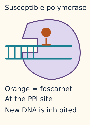
DNA polymerase is the enzyme that builds a new DNA strand from nucleotide building blocks. Adding a nucleotide releases pyrophosphate (PPi), a pair of linked phosphate groups. Foscarnet resembles pyrophosphate and inhibits the pyrophosphate-binding site of viral DNA polymerase. It does not need phosphorylation, the addition of phosphate groups by kinase enzymes, to become active. It is not incorporated as a replacement DNA nucleotide. [1]
Enlarge the whole diagramFoscarnet at viral polymerase. Qualitative schematic; shapes and particle counts do not specify molecular scale or patient values. [1]
Original Bone Wizardry teaching artwork. All rights reserved.
HSV means herpes simplex virus; CMV means cytomegalovirus. Acyclovir needs an initial phosphate added by HSV thymidine kinase, abbreviated TK. Host enzymes then produce its active triphosphate. In CMV, ganciclovir receives its initial phosphorylation principally through the viral UL97 kinase, followed by host enzymes. A nucleoside contains a base and sugar or a sugar-like scaffold without a phosphate group; a nucleotide includes phosphate. The parent drugs acyclovir and ganciclovir are nucleoside analogues; their active triphosphates are phosphorylated products. TK and UL97 are different proteins in different viruses. They perform related activation roles in these drug comparisons. [7][8]
Missing activation can prevent a nucleoside analogue from reaching its effective form. Foscarnet bypasses that requirement, but it still needs a susceptible polymerase. The experiment below isolates those two variables. It is a qualitative mechanism model, not a clinical susceptibility assay or a claim that acyclovir and ganciclovir are interchangeable across viruses.
A resistance gene is a starting point
An antiviral EC50 is the concentration reducing measured viral growth by 50% under the stated assay conditions. Compare an isolate with a matched reference tested with the same drug. A ratio describes that experiment; this lesson does not supply universal clinical resistance cutoffs. A purified-polymerase assay tests the target directly, while an infected-cell assay also includes entry and activation. A rescue experiment restores one missing component; a matched untreated control checks whether altered growth is distorting the drug comparison. [1][4]
Predict this before reading the genotype. If activation is intact but the polymerase no longer responds to foscarnet, will supplying more activation enzyme restore the drug effect?
UL97 encodes the CMV activation kinase. UL54 encodes CMV DNA polymerase, the downstream drug target. Some UL97 mutations reduce ganciclovir activation. Some UL54 mutations alter susceptibility to ganciclovir, cidofovir, foscarnet, or combinations of these drugs. Other sequence changes have little measurable effect. The gene name alone does not establish the resistance pattern. [2][4]
Recombinant phenotyping means placing a particular genetic change into a controlled viral background and measuring its drug response. It helps separate a causal resistance substitution from a coincidental variant. Chou reported ganciclovir/cidofovir resistance with F412L, F412S and L545W, while Q578H had a different pattern that included foscarnet resistance. These examples illustrate specificity; memorizing their numbers is not the task. [4]
A rising viral load is a reason to investigate treatment failure, not proof of one mutation. Check the disease assessment, exposure, adherence, absorption when relevant, immune status and the resistance interpretation. Retinal progression requires ophthalmologic assessment; blood CMV viral load is not recommended for monitoring retinitis relapse in people with HIV because its predictive value is poor. A plasma result does not replace examining the eye. [2]
Case 2
Show answer and explanations for case 2
A. Ganciclovir failure includes impaired UL97-dependent activation; the foscarnet result warns against assuming activation bypass will restore activity (Best answer)
Reason through this option
Which defect does isolated UL97 rescue support?
The patient virus has impaired UL97-dependent ganciclovir activation.
Where is the foscarnet inhibition shift demonstrated?
The foscarnet inhibition shift is present at the isolated polymerase target.
An activation defect can coexist with a second drug-specific target defect.[1][4][8]
Read all reasoning together
Which defect does isolated UL97 rescue support?
The patient virus has impaired UL97-dependent ganciclovir activation.
Where is the foscarnet inhibition shift demonstrated?
The foscarnet inhibition shift is present at the isolated polymerase target.
An activation defect can coexist with a second drug-specific target defect.[1][4][8]
Full authored source rationale
Rescue of both triphosphate formation and suppression after isolated UL97 replacement supports an activation defect rather than inadequate parent-drug delivery. The eightfold shift occurs in isolated polymerase, so bypassing UL97 does not eliminate the separate foscarnet target problem. This laboratory result informs the discussion without guaranteeing a clinical outcome.
B. Ganciclovir failure is primarily reduced entry of parent drug; the foscarnet result warns against assuming activation bypass will restore activity (Why this does not fit)
Reason through this option
What supplied result makes poor parent-drug entry insufficient?
Parent ganciclovir is equal before UL97 replacement.
Where is the foscarnet inhibition shift demonstrated?
The foscarnet inhibition shift is present at the isolated polymerase target.
A selective activation rescue can localize one defect while a purified-target assay identifies a second, coexisting limitation.[1][4][8]
Read all reasoning together
What supplied result makes poor parent-drug entry insufficient?
Parent ganciclovir is equal before UL97 replacement.
Where is the foscarnet inhibition shift demonstrated?
The foscarnet inhibition shift is present at the isolated polymerase target.
A selective activation rescue can localize one defect while a purified-target assay identifies a second, coexisting limitation.[1][4][8]
Full authored source rationale
Reduced uptake could lower active metabolite concentrations, but parent ganciclovir is already matched and changing UL97 restores the response. The eightfold shift occurs in isolated polymerase, so bypassing UL97 does not eliminate the separate foscarnet target problem. This laboratory result informs the discussion without guaranteeing a clinical outcome.
C. Ganciclovir failure reflects resistance despite normal activation; the foscarnet result points mainly to inadequate cell entry (Why this does not fit)
Reason through this option
What measured product is reduced before rescue?
Ganciclovir triphosphate is only 8% of the reference value.
What experimental feature excludes cell entry as the explanation for this shift?
Foscarnet was tested directly against purified polymerase.
Equal parent exposure and kinase rescue localize activation, while a cell-free target defect cannot be assigned to poor cell entry.[1][4][8]
Read all reasoning together
What measured product is reduced before rescue?
Ganciclovir triphosphate is only 8% of the reference value.
What experimental feature excludes cell entry as the explanation for this shift?
Foscarnet was tested directly against purified polymerase.
Equal parent exposure and kinase rescue localize activation, while a cell-free target defect cannot be assigned to poor cell entry.[1][4][8]
Full authored source rationale
A target defect can impair ganciclovir action, but the low triphosphate and UL97 rescue directly demonstrate abnormal activation in this experiment. Cell entry can confound whole-cell assays, but the foscarnet comparison used purified polymerase rather than intact cells.
D. Ganciclovir failure includes impaired UL97-dependent activation; the foscarnet result supports an unchanged target response after activation bypass (Why this does not fit)
Reason through this option
Which defect does isolated UL97 rescue support?
The patient virus has impaired UL97-dependent ganciclovir activation.
What conflicts with an unchanged foscarnet target response?
Patient polymerase requires eightfold more foscarnet for the same inhibition.
Activation bypass is insufficient when an independent target assay demonstrates reduced susceptibility to the proposed bypass drug.[1][4][8]
Read all reasoning together
Which defect does isolated UL97 rescue support?
The patient virus has impaired UL97-dependent ganciclovir activation.
What conflicts with an unchanged foscarnet target response?
Patient polymerase requires eightfold more foscarnet for the same inhibition.
Activation bypass is insufficient when an independent target assay demonstrates reduced susceptibility to the proposed bypass drug.[1][4][8]
Full authored source rationale
Rescue of both triphosphate formation and suppression after isolated UL97 replacement supports an activation defect rather than inadequate parent-drug delivery. UL97 bypass would be attractive for isolated activation failure, but the foscarnet assay independently demonstrates reduced target susceptibility.
E. Ganciclovir failure is primarily reduced entry of parent drug; the foscarnet result supports an unchanged target response after activation bypass (Why this does not fit)
Reason through this option
What supplied result makes poor parent-drug entry insufficient?
Parent ganciclovir is equal before UL97 replacement.
What conflicts with an unchanged foscarnet target response?
Patient polymerase requires eightfold more foscarnet for the same inhibition.
Use rescue and purified-target controls separately; neither impaired delivery nor activation bypass can explain every resistance result.[1][4][8]
Read all reasoning together
What supplied result makes poor parent-drug entry insufficient?
Parent ganciclovir is equal before UL97 replacement.
What conflicts with an unchanged foscarnet target response?
Patient polymerase requires eightfold more foscarnet for the same inhibition.
Use rescue and purified-target controls separately; neither impaired delivery nor activation bypass can explain every resistance result.[1][4][8]
Full authored source rationale
Reduced uptake could lower active metabolite concentrations, but parent ganciclovir is already matched and changing UL97 restores the response. UL97 bypass would be attractive for isolated activation failure, but the foscarnet assay independently demonstrates reduced target susceptibility.
Takeaway: An activation defect can coexist with a second drug-specific target defect.
CMV means cytomegalovirus. HSV means herpes simplex virus. Both are herpesviruses, but their treatment pathways are not interchangeable. Retinitis is inflammation and injury of the retina; CMV retinitis can threaten vision in people with advanced immunosuppression. Mucocutaneous HSV affects skin and mucosal surfaces. [2][3]
For CMV disease, ganciclovir or its oral prodrug valganciclovir is often preferred. A prodrug is converted into the active drug after administration. Foscarnet is an important alternative when ganciclovir toxicity or resistance limits treatment. NIH guidance supports foscarnet in selected CMV retinitis and gastrointestinal disease contexts; that is broader than the product label indication for CMV retinitis in AIDS. Site, severity, susceptibility and toxicities all matter. [1][2]
Ganciclovir can suppress bone marrow production of blood cells. Neutropenia means too few neutrophils; pancytopenia means reductions across white cells, red cells and platelets. Changing to oral valganciclovir does not eliminate the active drug's marrow toxicity. Foscarnet offers a different toxicity balance, not a promise of zero hematologic adverse effects. [1][2]
For suspected acyclovir-resistant HSV, persistent lesions despite adequate treatment warrant culture and susceptibility testing when possible, with expert involvement. NIH identifies IV foscarnet as the treatment of choice for acyclovir-resistant HSV. Valacyclovir becomes acyclovir, so improved oral delivery does not bypass TK resistance. [2][3] Cidofovir undergoes host-enzyme phosphorylation to its active diphosphate, unlike foscarnet, which needs no phosphorylation. [10][11] Cidofovir has its own renal precautions, including probenecid and hydration; do not transfer that protection regimen to foscarnet. [10]
Free calcium can change before creatinine
Ionized calcium is the unbound, electrically active calcium fraction. Total calcium includes ionized, protein-bound and other complexed calcium. A near-normal total concentration does not guarantee a normal ionized concentration. Paresthesia means tingling or altered sensation; tetany means involuntary muscle spasms from increased neuromuscular excitability. [1]
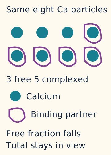
A divalent cation has two positive charges. Foscarnet can bind divalent ions such as calcium and magnesium into coordination complexes. This direct complexing, also called chelation, can lower ionized calcium during an infusion without requiring a prior rise in creatinine. The label notes that the ionized change may not be reflected in total calcium and can depend on infusion rate. [1]
Enlarge the whole diagramFree versus complexed calcium. Qualitative schematic; shapes and particle counts do not specify molecular scale or patient values. Compartment volume is held constant. [1]
Original Bone Wizardry teaching artwork. All rights reserved.
The particle comparison below distinguishes binding within blood from loss through an injured kidney. Binding changes the free fraction even if the total number of calcium particles in the pictured compartment stays constant. The comparison also holds compartment volume constant; changing volume would change concentrations even at the same amount. Renal electrolyte disturbances alter handling and may change the total amount in that compartment. Neither illustration is a patient calculation. [1]
Perioral tingling, hand spasms or a seizure during treatment require prompt assessment, including ionized calcium and other electrolytes. Low magnesium can contribute to neuromuscular excitability. Magnesium deficiency can also favor renal potassium loss and make potassium repletion less effective. Experimental channel and renal-cell studies support this potassium-handling relationship. [1][5]
Parathyroid hormone, or PTH, helps maintain blood calcium. Human depletion studies show that magnesium deficiency can impair PTH secretion and contribute to hypocalcemia. That is another reason a persistent calcium problem may require attention to magnesium. This physiological mechanism is separate from direct foscarnet binding of calcium. Do not let a reassuring total calcium end the evaluation of new symptoms. [6]
Case 8
Show answer and explanations for case 8
A. A: reduced total calcium from net depletion; B: progressive depletion causing symptomatic ionized hypocalcemia (Why this does not fit)
Reason through this option
Which measured serum concentration remains stable while A ionized calcium declines?
A has an unchanged measured serum total calcium concentration during the ionized decline.
What B result prevents inferring a low active calcium fraction from poor intake alone?
B's active calcium fraction remains within its reference range.
A stable measured serum total calcium concentration with a lower ionized fraction supports altered free availability; that concentration does not establish a conserved whole-body calcium amount.[1]
Read all reasoning together
Which measured serum concentration remains stable while A ionized calcium declines?
A has an unchanged measured serum total calcium concentration during the ionized decline.
What B result prevents inferring a low active calcium fraction from poor intake alone?
B's active calcium fraction remains within its reference range.
A stable measured serum total calcium concentration with a lower ionized fraction supports altered free availability; that concentration does not establish a conserved whole-body calcium amount.[1]
Full authored source rationale
Loss of calcium can cause hypocalcemia, but an abrupt fraction change with unchanged total calcium is a poor fit for total-pool depletion. Poor intake can raise concern for depletion, but B has neither measured ionized hypocalcemia nor the proposed neuromuscular symptoms.
B. A: increased nonprotein complexing of calcium; B: increased nonprotein complexing with ionized hypocalcemia (Why this does not fit)
Reason through this option
What does A's discordant total and ionized trend indicate?
A's calcium has shifted out of the free fraction without a comparable total decline.
What B observation defeats the proposed ionized hypocalcemia?
B has normal measured ionized calcium.
Directly normal ionized calcium should prevent a low total calcium result from being mislabeled as an active-fraction deficit.[1]
Read all reasoning together
What does A's discordant total and ionized trend indicate?
A's calcium has shifted out of the free fraction without a comparable total decline.
What B observation defeats the proposed ionized hypocalcemia?
B has normal measured ionized calcium.
Directly normal ionized calcium should prevent a low total calcium result from being mislabeled as an active-fraction deficit.[1]
Full authored source rationale
A's stable total calcium with an abrupt ionized decline supports redistribution into complexes during exposure. Stable albumin and pH reduce competing protein-binding explanations. Complexing is plausible during foscarnet therapy, but the B sample directly shows a normal ionized fraction despite the low total result.
C. A: reduced protein-bound calcium from lower albumin; B: increased nonprotein complexing with ionized hypocalcemia (Why this does not fit)
Reason through this option
What supplied finding argues against albumin loss in A?
A's albumin remains unchanged.
What B observation defeats the proposed ionized hypocalcemia?
B has normal measured ionized calcium.
Interpret calcium fractions against contemporaneous albumin, pH and symptoms instead of assigning one mechanism to every low calcium result.[1]
Read all reasoning together
What supplied finding argues against albumin loss in A?
A's albumin remains unchanged.
What B observation defeats the proposed ionized hypocalcemia?
B has normal measured ionized calcium.
Interpret calcium fractions against contemporaneous albumin, pH and symptoms instead of assigning one mechanism to every low calcium result.[1]
Full authored source rationale
Albumin loss can reduce total calcium, but A's albumin and total calcium stay unchanged while the ionized fraction falls. Complexing is plausible during foscarnet therapy, but the B sample directly shows a normal ionized fraction despite the low total result.
D. A: reduced protein-bound calcium from lower albumin; B: reduced protein-bound calcium from lower albumin (Why this does not fit)
Reason through this option
What supplied finding argues against albumin loss in A?
A's albumin remains unchanged.
How does low albumin connect B's lower total result with its normal ionized result?
Low albumin reduces the protein-bound contribution while B's ionized calcium remains normal.
Stable albumin and measured serum total calcium concentration with an abrupt ionized decline favor altered free availability rather than an albumin-related decrease in protein-bound calcium.[1]
Read all reasoning together
What supplied finding argues against albumin loss in A?
A's albumin remains unchanged.
How does low albumin connect B's lower total result with its normal ionized result?
Low albumin reduces the protein-bound contribution while B's ionized calcium remains normal.
Stable albumin and measured serum total calcium concentration with an abrupt ionized decline favor altered free availability rather than an albumin-related decrease in protein-bound calcium.[1]
Full authored source rationale
Albumin loss can reduce total calcium, but A's albumin and total calcium stay unchanged while the ionized fraction falls. B's total result follows albumin downward while the active fraction stays normal, favoring reduced protein-bound calcium rather than symptomatic ionized hypocalcemia.
E. A: increased nonprotein complexing of calcium; B: reduced protein-bound calcium from lower albumin (Best answer)
Reason through this option
What does A's discordant total and ionized trend indicate?
A's calcium has shifted out of the free fraction without a comparable total decline.
How does low albumin connect B's lower total result with its normal ionized result?
Low albumin reduces the protein-bound contribution while B's ionized calcium remains normal.
A total calcium result describes a different pool from the physiologically active free fraction.[1]
Read all reasoning together
What does A's discordant total and ionized trend indicate?
A's calcium has shifted out of the free fraction without a comparable total decline.
How does low albumin connect B's lower total result with its normal ionized result?
Low albumin reduces the protein-bound contribution while B's ionized calcium remains normal.
A total calcium result describes a different pool from the physiologically active free fraction.[1]
Full authored source rationale
A's stable total calcium with an abrupt ionized decline supports redistribution into complexes during exposure. Stable albumin and pH reduce competing protein-binding explanations. B's total result follows albumin downward while the active fraction stays normal, favoring reduced protein-bound calcium rather than symptomatic ionized hypocalcemia.
Takeaway: A total calcium result describes a different pool from the physiologically active free fraction.
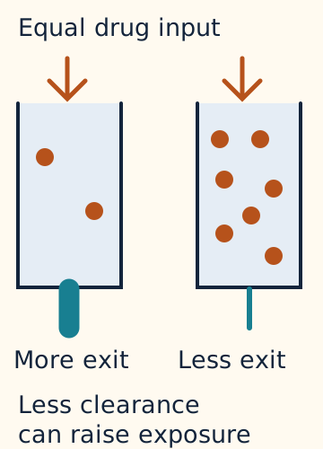
Renal clearance describes the kidney's removal of a substance from blood. Foscarnet is eliminated predominantly through the kidneys. When renal function declines, drug clearance falls and its half-life can lengthen. Half-life is the time for a drug concentration to decrease by half in a specified elimination phase. The same administered amount can then produce greater exposure. The clearance comparison assumes equal dosing rate, distribution volume and observation time; clinical concentrations also depend on sampling time. [1]
Nephrotoxicity means drug-related kidney injury. Renal impairment is a major foscarnet toxicity. Tubular injury also disrupts electrolyte handling. These are connected to, but distinct from, direct calcium complexing in blood. Creatinine is a marker used to assess renal function, not a direct measurement of ionized calcium or a guarantee that tubular function is normal. [1]
Enlarge the whole diagramClearance and exposure. Qualitative schematic; shapes and particle counts do not specify molecular scale or patient values. Dosing rate, distribution volume and observation time are comparable. [1]
Original Bone Wizardry teaching artwork. All rights reserved.
The label supports hydration and dosage adjustment for renal function. It provides a renal dosing scheme; this lesson does not calculate a dose. Hydration must account for fluid tolerance. The label advises avoiding foscarnet when its sodium or water load cannot be tolerated, including relevant cardiomyopathy, and in patients on a controlled sodium diet. A reduced hydration order does not establish that the drug itself is feasible. Review other nephrotoxic drugs because combined renal injury can change the balance of benefit and harm. [1]
Hypo means low and hyper means high. Hypokalemia is low potassium; hyperkalemia is high potassium. Calcium, magnesium, potassium and phosphate deserve surveillance, but not every value always falls. Both low and high phosphate are reported. Phosphate is not a positively charged divalent ion, and potassium carries one positive charge; direct divalent-cation complexing cannot explain the entire panel. Interpret the time course, kidney function and actual measurements. [1]
Case 11
Show answer and explanations for case 11
A. Restrict additional fluid as the principal volume response; retain the previous regimen while awaiting another clearance measurement (Why this does not fit)
Reason through this option
Which supplied findings favor volume repletion rather than restriction?
Weight loss, dry mucosa and orthostasis support volume depletion without supplied congestion.
What renal result is already available before another measurement?
Current creatinine clearance is in a lower label dosing band.
What should guide further dosing while hydration is assessed?
Recalculate the regimen using current renal function before further dosing.
Do not defer correction of a demonstrated volume deficit or ignore a current renal-band change while waiting for repeat measurements.[1]
Read all reasoning together
Which supplied findings favor volume repletion rather than restriction?
Weight loss, dry mucosa and orthostasis support volume depletion without supplied congestion.
What renal result is already available before another measurement?
Current creatinine clearance is in a lower label dosing band.
What should guide further dosing while hydration is assessed?
Recalculate the regimen using current renal function before further dosing.
Do not defer correction of a demonstrated volume deficit or ignore a current renal-band change while waiting for repeat measurements.[1]
Full authored source rationale
Fluid restriction can be useful for congestion, and repeating a questionable renal result can sometimes clarify a dosing decision. This patient instead has measured volume-loss findings without supplied overload, and the current clearance already lies in a lower label band. The plan therefore misses both an actionable volume deficit and a current dosing change. Clinically monitored hydration and reassessment from the present renal value address the two demonstrated limitations.
B. Replete volume with clinically monitored hydration; retain the previous regimen while observing the response to hydration (Why this does not fit)
Reason through this option
Which state is supported by weight loss, dry mucosa and orthostasis?
The patient has findings consistent with volume depletion.
What present measurement argues against simply retaining the old regimen?
Current creatinine clearance no longer matches the old dosing band.
Hydration may improve renal function, but current measured clearance must guide further dosing rather than an anticipated recovery.[1]
Read all reasoning together
Which state is supported by weight loss, dry mucosa and orthostasis?
The patient has findings consistent with volume depletion.
What present measurement argues against simply retaining the old regimen?
Current creatinine clearance no longer matches the old dosing band.
Hydration may improve renal function, but current measured clearance must guide further dosing rather than an anticipated recovery.[1]
Full authored source rationale
The examination supports volume depletion and supplies no evidence of congestion, so assessed hydration addresses a current modifiable risk. Fluid tolerance still requires observation during treatment. Hydration may improve renal perfusion, but waiting on that possibility while using the old renal band ignores the clearance already measured.
C. Restrict additional fluid as the principal volume response; recalculate the foscarnet regimen from current renal function before further dosing (Why this does not fit)
Reason through this option
Which supplied findings oppose a congestion-driven fluid restriction?
The patient has weight loss and dry mucosa.
Why is the previous dosing calculation no longer sufficient?
The patient's current clearance is in a lower label dosing band.
Use current volume findings to choose hydration or restriction, and assess renal dosing eligibility as a separate decision.[1]
Read all reasoning together
Which supplied findings oppose a congestion-driven fluid restriction?
The patient has weight loss and dry mucosa.
Why is the previous dosing calculation no longer sufficient?
The patient's current clearance is in a lower label dosing band.
Use current volume findings to choose hydration or restriction, and assess renal dosing eligibility as a separate decision.[1]
Full authored source rationale
Fluid restriction can be appropriate for congestion, but this patient has volume-loss findings and clear lungs rather than a supplied overload pattern. The old regimen used a different clearance band, so hydration alone does not make the previous dose appropriate. The current label scheme must guide reassessment.
D. Use probenecid as the principal renal-protection intervention; base further dosing primarily on the original baseline creatinine (Why this does not fit)
Reason through this option
Which present risk would probenecid leave uncorrected?
Probenecid would leave the patient's volume depletion uncorrected.
Which renal assessment should inform the next regimen?
The next regimen should use the current renal-function assessment.
Match renal precautions to the specific drug and current physiology; another drug's protective regimen cannot replace hydration and renal reassessment.[1][2]
Read all reasoning together
Which present risk would probenecid leave uncorrected?
Probenecid would leave the patient's volume depletion uncorrected.
Which renal assessment should inform the next regimen?
The next regimen should use the current renal-function assessment.
Match renal precautions to the specific drug and current physiology; another drug's protective regimen cannot replace hydration and renal reassessment.[1][2]
Full authored source rationale
Probenecid is familiar from cidofovir treatment, but it does not correct the demonstrated volume depletion or replace foscarnet precautions. A baseline value documents the starting state but cannot represent the demonstrated change in current clearance.
E. Replete volume with clinically monitored hydration; recalculate the foscarnet regimen from current renal function before further dosing (Best answer)
Reason through this option
Which state is supported by weight loss, dry mucosa and orthostasis?
The patient has findings consistent with volume depletion.
Why is the previous dosing calculation no longer sufficient?
The patient's current clearance is in a lower label dosing band.
A volume deficit and a changed renal dosing band require separate responses.[1]
Read all reasoning together
Which state is supported by weight loss, dry mucosa and orthostasis?
The patient has findings consistent with volume depletion.
Why is the previous dosing calculation no longer sufficient?
The patient's current clearance is in a lower label dosing band.
A volume deficit and a changed renal dosing band require separate responses.[1]
Full authored source rationale
The examination supports volume depletion and supplies no evidence of congestion, so assessed hydration addresses a current modifiable risk. Fluid tolerance still requires observation during treatment. The old regimen used a different clearance band, so hydration alone does not make the previous dose appropriate. The current label scheme must guide reassessment.
Takeaway: A volume deficit and a changed renal dosing band require separate responses.
Before and during IV foscarnet, assess renal function, calcium, magnesium, potassium and phosphate. NIH CMV guidance also calls for close complete blood count (CBC) surveillance; foscarnet is not free of hematologic toxicity. Stable outpatient maintenance requires at least weekly electrolytes, renal function and CBC under that guidance. [2] The prescribing information recommends creatinine clearance at baseline, two to three times weekly during induction and weekly during maintenance, with more frequent checks when clinically indicated. Electrolyte monitoring follows a similar schedule; symptoms or changing renal function can require earlier reassessment. [1]
An electrocardiogram, or ECG, records the heart's electrical activity. QT spans ventricular depolarization and repolarization, from the start of QRS to the end of the T wave. QRS covers depolarization; the subsequent JT interval covers repolarization. [9] QT prolongation can increase the risk of torsades de pointes, a dangerous ventricular rhythm. The label reports QT prolongation and torsades and advises ECG and electrolyte assessment before treatment and periodically during treatment, with attention to additional QT risks. Calcium, magnesium and potassium disturbances can all matter to electrical stability. [1]
A new seizure requires emergency assessment and treatment while investigating metabolic, infectious and other causes. In a patient receiving foscarnet, low ionized calcium and magnesium are actionable findings, especially if renal function has worsened. New tingling during an infusion also deserves action; the label advises stopping the infusion, obtaining electrolyte samples and consulting the treating clinician before resuming. [1]
Use three questions when transferring this lesson. Is the virus failing to activate a drug, or is its target altered? Is the electrolyte change a free-fraction effect, a renal handling effect, or both? Has the safety assessment kept up with the current symptoms and kidney function? These are educational decisions, not a personal prescribing plan. [1]
Practice across the mechanisms
All 18 original cases are part of this continuous lesson. Three appear beside their teaching sections; the rest follow here. Explore any option and retry without a score.
These are hypothetical educational cases and experiments, not published patient or study measurements. Numerical calculations use the values and controls supplied in each case. References support background mechanisms and clinical guidance, not the invented measurements or guaranteed treatment outcomes.
Case 1
Show answer and explanations for case 1
A. Investigate viral activation in the isolate; advance X as a host-kinase-activated polymerase inhibitor (Why this does not fit)
Reason through this option
What does restoration of inhibition by TK localize in this isolate?
The isolate has a defect at the initial viral activation step.
Which missing assay component defeats the host-activation interpretation?
The purified reaction contains no host kinases.
Activity in a kinase-free reaction rules out an obligatory phosphorylation requirement for that tested drug effect.[1][7]
Read all reasoning together
What does restoration of inhibition by TK localize in this isolate?
The isolate has a defect at the initial viral activation step.
Which missing assay component defeats the host-activation interpretation?
The purified reaction contains no host kinases.
Activity in a kinase-free reaction rules out an obligatory phosphorylation requirement for that tested drug effect.[1][7]
Full authored source rationale
The TK rescue localizes the acyclovir defect to initial activation: intracellular parent drug was already available, and restoring TK restored inhibition. Host activation is a plausible route for some antivirals, but X already inhibits a reaction that contains no kinases.
B. Investigate viral activation in the isolate; advance X as a direct, activation-independent polymerase inhibitor (Best answer)
Reason through this option
What does restoration of inhibition by TK localize in this isolate?
The isolate has a defect at the initial viral activation step.
What requirement does X avoid in the purified reaction?
Compound X does not require kinase activation to inhibit the polymerase reaction.
An activation rescue localizes one defect; a kinase-free assay tests whether another drug avoids that defect.[1][7]
Read all reasoning together
What does restoration of inhibition by TK localize in this isolate?
The isolate has a defect at the initial viral activation step.
What requirement does X avoid in the purified reaction?
Compound X does not require kinase activation to inhibit the polymerase reaction.
An activation rescue localizes one defect; a kinase-free assay tests whether another drug avoids that defect.[1][7]
Full authored source rationale
The TK rescue localizes the acyclovir defect to initial activation: intracellular parent drug was already available, and restoring TK restored inhibition. The kinase-free reaction establishes activity of unmodified X at the polymerase system, consistent with the defining activation-independent property of foscarnet.
C. Investigate intracellular drug entry in the isolate; advance X as a host-kinase-activated polymerase inhibitor (Why this does not fit)
Reason through this option
What finding argues against inadequate acyclovir entry?
Intracellular parent acyclovir is equal in patient and reference cultures.
Which missing assay component defeats the host-activation interpretation?
The purified reaction contains no host kinases.
Separate cellular delivery from enzymatic activation, then test whether the comparator actually needs the proposed activation machinery.[1][7]
Read all reasoning together
What finding argues against inadequate acyclovir entry?
Intracellular parent acyclovir is equal in patient and reference cultures.
Which missing assay component defeats the host-activation interpretation?
The purified reaction contains no host kinases.
Separate cellular delivery from enzymatic activation, then test whether the comparator actually needs the proposed activation machinery.[1][7]
Full authored source rationale
Poor uptake could reduce an antiviral effect, but equal intracellular parent acyclovir and rescue by TK argue against uptake as the limiting step here. Host activation is a plausible route for some antivirals, but X already inhibits a reaction that contains no kinases.
D. Investigate resistance to acyclovir triphosphate at the target; advance X as a viral-kinase-activated polymerase inhibitor (Why this does not fit)
Reason through this option
What does recovered inhibition after TK addition show about the target?
The target remains inhibitable after acyclovir activation is restored.
Which missing assay component defeats the viral-activation interpretation?
The purified reaction contains no viral kinases.
Recovery after activation rescue argues against a fixed active-metabolite target defect; a kinase-free comparator tests activation bypass.[1][7]
Read all reasoning together
What does recovered inhibition after TK addition show about the target?
The target remains inhibitable after acyclovir activation is restored.
Which missing assay component defeats the viral-activation interpretation?
The purified reaction contains no viral kinases.
Recovery after activation rescue argues against a fixed active-metabolite target defect; a kinase-free comparator tests activation bypass.[1][7]
Full authored source rationale
A resistant polymerase could explain treatment failure, but recovery of inhibition after TK addition shows that the activated acyclovir can inhibit this isolate. Viral activation explains acyclovir behavior, but it cannot account for X activity in a kinase-free reaction.
E. Investigate intracellular drug entry in the isolate; advance X as a direct, activation-independent polymerase inhibitor (Why this does not fit)
Reason through this option
What finding argues against inadequate acyclovir entry?
Intracellular parent acyclovir is equal in patient and reference cultures.
What requirement does X avoid in the purified reaction?
Compound X does not require kinase activation to inhibit the polymerase reaction.
Matched intracellular parent exposure plus selective enzyme rescue directs investigation to activation rather than drug entry.[1][7]
Read all reasoning together
What finding argues against inadequate acyclovir entry?
Intracellular parent acyclovir is equal in patient and reference cultures.
What requirement does X avoid in the purified reaction?
Compound X does not require kinase activation to inhibit the polymerase reaction.
Matched intracellular parent exposure plus selective enzyme rescue directs investigation to activation rather than drug entry.[1][7]
Full authored source rationale
Poor uptake could reduce an antiviral effect, but equal intracellular parent acyclovir and rescue by TK argue against uptake as the limiting step here. The kinase-free reaction establishes activity of unmodified X at the polymerase system, consistent with the defining activation-independent property of foscarnet.
Takeaway: An activation rescue localizes one defect; a kinase-free assay tests whether another drug avoids that defect.
A. Annotate V as causing reduced susceptibility in this assay; annotate W as causing reduced susceptibility in this assay (Why this does not fit)
Reason through this option
What does V reversion add to the sixfold shift?
V reversion ties the susceptibility shift to the introduced substitution.
What finding defeats a causal resistance annotation for W?
The W EC50 range overlaps the parental control range.
A treatment-associated sequence change requires a reproducible effect beyond matched control variability before receiving a resistance annotation.[4]
Read all reasoning together
What does V reversion add to the sixfold shift?
V reversion ties the susceptibility shift to the introduced substitution.
What finding defeats a causal resistance annotation for W?
The W EC50 range overlaps the parental control range.
A treatment-associated sequence change requires a reproducible effect beyond matched control variability before receiving a resistance annotation.[4]
Full authored source rationale
V changes susceptibility in a matched background, and reversing that single change restores the control response. This supports assay-level causation rather than a patient-level association alone. Treatment-associated sequence changes deserve investigation, but W produces no reproducible shift beyond control variability.
B. Leave V without a demonstrated susceptibility effect; annotate W as causing reduced susceptibility in this assay (Why this does not fit)
Reason through this option
Which control makes V more than an incidental sequence finding?
Reverting V restores the parental foscarnet response.
What finding defeats a causal resistance annotation for W?
The W EC50 range overlaps the parental control range.
Apply the same introduction, reversion and variability criteria to each variant instead of inferring causation from treatment history.[4]
Read all reasoning together
Which control makes V more than an incidental sequence finding?
Reverting V restores the parental foscarnet response.
What finding defeats a causal resistance annotation for W?
The W EC50 range overlaps the parental control range.
Apply the same introduction, reversion and variability criteria to each variant instead of inferring causation from treatment history.[4]
Full authored source rationale
Withholding a causal annotation is sensible for a sequence report alone, but V has a reproducible introduction-and-reversion effect under controlled conditions. Treatment-associated sequence changes deserve investigation, but W produces no reproducible shift beyond control variability.
C. Annotate V as causing reduced susceptibility in this assay; leave W without a demonstrated susceptibility effect (Best answer)
Reason through this option
What does V reversion add to the sixfold shift?
V reversion ties the susceptibility shift to the introduced substitution.
How should overlap between W and control affect annotation?
The W assay does not demonstrate reduced foscarnet susceptibility.
A matched intervention can establish an effect for one variant without establishing an effect for another.[4]
Read all reasoning together
What does V reversion add to the sixfold shift?
V reversion ties the susceptibility shift to the introduced substitution.
How should overlap between W and control affect annotation?
The W assay does not demonstrate reduced foscarnet susceptibility.
A matched intervention can establish an effect for one variant without establishing an effect for another.[4]
Full authored source rationale
V changes susceptibility in a matched background, and reversing that single change restores the control response. This supports assay-level causation rather than a patient-level association alone. W remains within control variability after introduction, so these results do not support a resistance annotation for W despite its occurrence after treatment.
D. Leave V without a demonstrated susceptibility effect; leave W without a demonstrated susceptibility effect (Why this does not fit)
Reason through this option
Which control makes V more than an incidental sequence finding?
Reverting V restores the parental foscarnet response.
How should overlap between W and control affect annotation?
The W assay does not demonstrate reduced foscarnet susceptibility.
Controlled introduction followed by phenotypic reversion supplies causal evidence that an isolated sequence association does not.[4]
Read all reasoning together
Which control makes V more than an incidental sequence finding?
Reverting V restores the parental foscarnet response.
How should overlap between W and control affect annotation?
The W assay does not demonstrate reduced foscarnet susceptibility.
Controlled introduction followed by phenotypic reversion supplies causal evidence that an isolated sequence association does not.[4]
Full authored source rationale
Withholding a causal annotation is sensible for a sequence report alone, but V has a reproducible introduction-and-reversion effect under controlled conditions. W remains within control variability after introduction, so these results do not support a resistance annotation for W despite its occurrence after treatment.
E. Annotate V as causing increased susceptibility in this assay; leave W without a demonstrated susceptibility effect (Why this does not fit)
Reason through this option
What does EC50 hold constant when two concentrations are compared?
Each concentration produces the same 50% inhibition endpoint. [4]
What does requiring sixfold more drug imply about V?
V is less susceptible in this assay, not more susceptible.
How do the introduction and reversion refine that conclusion?
They support V causing the measured decrease in susceptibility.
Does W show a comparably demonstrated effect?
Its repeated values overlap the matched control variability.
At a fixed inhibition endpoint, a higher required drug concentration indicates reduced susceptibility; assess causal evidence and direction separately.[4]
Read all reasoning together
What does EC50 hold constant when two concentrations are compared?
Each concentration produces the same 50% inhibition endpoint. [4]
What does requiring sixfold more drug imply about V?
V is less susceptible in this assay, not more susceptible.
How do the introduction and reversion refine that conclusion?
They support V causing the measured decrease in susceptibility.
Does W show a comparably demonstrated effect?
Its repeated values overlap the matched control variability.
At a fixed inhibition endpoint, a higher required drug concentration indicates reduced susceptibility; assess causal evidence and direction separately.[4]
Full authored source rationale
The matched introduction and reversion support a causal effect of V, but the direction of that effect is reversed in this annotation. A higher EC50 means that more drug is required to produce the same 50% inhibition, which is reduced rather than increased susceptibility. The sixfold V shift is therefore not evidence of improved drug sensitivity. Leaving W unclassified is appropriate because its repeated values overlap the supplied control variability. The mistake is the direction assigned to the demonstrated V phenotype, not a demand for evidence from untested genetic backgrounds.
Takeaway: A matched intervention can establish an effect for one variant without establishing an effect for another.
A. Advance foscarnet for exposure-controlled testing; complement UL97 and use restoration of intracellular phosphorylation as the primary endpoint (Why this does not fit)
Reason through this option
Why is foscarnet still a reasonable assay candidate?
Its 1.1-fold comparison remains inside the supplied variability range.
What would a successful UL97 rescue need to explain?
It would need an activation deficit that accounts for the impaired drug response.
Which control prevents an activation-only explanation here?
Reduced inhibition persists after active metabolites are supplied to purified polymerase.
Which experiment addresses the unresolved target variant?
A single-variant UL54 reconstruction with reversion tests V specifically.
When active drug is already available and the isolated target remains impaired, investigate the target rather than repeating activation rescue.[1][4][8][10][11]
Read all reasoning together
Why is foscarnet still a reasonable assay candidate?
Its 1.1-fold comparison remains inside the supplied variability range.
What would a successful UL97 rescue need to explain?
It would need an activation deficit that accounts for the impaired drug response.
Which control prevents an activation-only explanation here?
Reduced inhibition persists after active metabolites are supplied to purified polymerase.
Which experiment addresses the unresolved target variant?
A single-variant UL54 reconstruction with reversion tests V specifically.
When active drug is already available and the isolated target remains impaired, investigate the target rather than repeating activation rescue.[1][4][8][10][11]
Full authored source rationale
Foscarnet is the appropriate preserved-response candidate after comparison with its own reference. UL97 complementation would be informative if deficient phosphorylation were the unresolved limitation. Here, however, the active metabolites are already matched and the target remains less responsive when they are supplied directly. A kinase-rescue endpoint therefore does not test the target-level explanation suggested by these controls or establish the role of UL54 V.
B. Advance foscarnet for exposure-controlled testing; introduce UL54 V alone into a reference background and compare its matched revertant (Best answer)
Reason through this option
Which comparison identifies the preserved drug response?
Foscarnet has a P/reference ratio of 55/50 = 1.1.
Does matched active-metabolite exposure resolve the resistance phenotype?
Impaired inhibition persists at purified P polymerase despite supplied active metabolites.
Which mechanistic site does that persistence implicate?
The polymerase target, rather than an activation-only defect, needs investigation.
How can V be separated from the other sequence differences?
Introduce V alone into a matched reference background and test a corresponding revertant.
Normalize the phenotype, localize the defect with controlled exposure, then isolate a variant before assigning causation.[1][4][8][10][11]
Read all reasoning together
Which comparison identifies the preserved drug response?
Foscarnet has a P/reference ratio of 55/50 = 1.1.
Does matched active-metabolite exposure resolve the resistance phenotype?
Impaired inhibition persists at purified P polymerase despite supplied active metabolites.
Which mechanistic site does that persistence implicate?
The polymerase target, rather than an activation-only defect, needs investigation.
How can V be separated from the other sequence differences?
Introduce V alone into a matched reference background and test a corresponding revertant.
Normalize the phenotype, localize the defect with controlled exposure, then isolate a variant before assigning causation.[1][4][8][10][11]
Full authored source rationale
The calculated fold changes are 6 for ganciclovir, 8 for cidofovir and 1.1 for foscarnet, so the largest raw EC50 does not identify the least preserved response. Normal active-metabolite concentrations and impaired inhibition of purified P polymerase place the remaining problem at the target rather than solely at activation. V is still a candidate, not an established cause: introducing it alone and then reverting it tests whether that one change reproduces and reverses the phenotype. These are assay-development decisions, not a prediction of clinical cure.
C. Advance foscarnet for exposure-controlled testing; compare P with an unrelated V-negative clinical isolate and attribute any difference to V (Why this does not fit)
Reason through this option
Which drug retains the reference-range response?
Foscarnet remains within the stipulated variability range.
What differs besides V in an unrelated clinical isolate?
Its genetic background can contain other susceptibility-relevant differences.
Which comparison isolates the effect of V?
A matched single-variant recombinant and its revertant isolate that effect.
An associated sequence difference becomes a causal candidate to test, not a cause established by comparing unrelated isolates.[1][4][8][10][11]
Read all reasoning together
Which drug retains the reference-range response?
Foscarnet remains within the stipulated variability range.
What differs besides V in an unrelated clinical isolate?
Its genetic background can contain other susceptibility-relevant differences.
Which comparison isolates the effect of V?
A matched single-variant recombinant and its revertant isolate that effect.
An associated sequence difference becomes a causal candidate to test, not a cause established by comparing unrelated isolates.[1][4][8][10][11]
Full authored source rationale
The foscarnet selection is consistent with the matched susceptibility data. Comparing unrelated isolates could identify an association worth studying, but those isolates may differ at many sites besides V. The present isolate already contains more than one sequence difference. A causal assignment requires a matched reconstruction and appropriate reversion rather than assigning every between-isolate difference to the selected substitution.
D. Advance cidofovir for exposure-controlled testing; complement UL97 and use restoration of intracellular phosphorylation as the primary endpoint (Why this does not fit)
Reason through this option
What happens when cidofovir is compared with its own reference?
Its eightfold shift exceeds the stated reduced-susceptibility threshold.
Where does inhibition remain impaired after activation is bypassed experimentally?
The impairment remains in the purified polymerase reaction.
What should the next causal test vary?
It should vary the candidate polymerase substitution in a matched background.
Drug-specific normalization and target-localizing controls must agree before choosing an activation-directed rescue experiment.[1][4][8][10][11]
Read all reasoning together
What happens when cidofovir is compared with its own reference?
Its eightfold shift exceeds the stated reduced-susceptibility threshold.
Where does inhibition remain impaired after activation is bypassed experimentally?
The impairment remains in the purified polymerase reaction.
What should the next causal test vary?
It should vary the candidate polymerase substitution in a matched background.
Drug-specific normalization and target-localizing controls must agree before choosing an activation-directed rescue experiment.[1][4][8][10][11]
Full authored source rationale
This plan combines a raw-concentration comparison with an activation explanation that the controls do not support. Cidofovir is eightfold above its reference, even though its absolute EC50 is below the foscarnet number. The active-metabolite and purified-enzyme experiments place the residual limitation downstream of activation. UL97 complementation is therefore not the focused test of the proposed polymerase mechanism, and cidofovir is not the preserved-response candidate in this assay.
E. Advance cidofovir for exposure-controlled testing; introduce UL54 V alone into a reference background and compare its matched revertant (Why this does not fit)
Reason through this option
Does the smallest absolute EC50 identify the most preserved response?
No; cidofovir is eightfold above its own reference.
Can host activation alone overcome the observed cidofovir result?
The supplied active cidofovir metabolite still encounters an impaired polymerase response.
What part of this proposed plan remains informative?
Single-variant introduction and reversion can test whether V causes the target phenotype.
Compare each drug with its own control; an appropriate causal experiment cannot rescue an incorrectly classified susceptibility result.[1][4][8][10][11]
Read all reasoning together
Does the smallest absolute EC50 identify the most preserved response?
No; cidofovir is eightfold above its own reference.
Can host activation alone overcome the observed cidofovir result?
The supplied active cidofovir metabolite still encounters an impaired polymerase response.
What part of this proposed plan remains informative?
Single-variant introduction and reversion can test whether V causes the target phenotype.
Compare each drug with its own control; an appropriate causal experiment cannot rescue an incorrectly classified susceptibility result.[1][4][8][10][11]
Full authored source rationale
The recombinant design is appropriate for a candidate target substitution, but cidofovir is selected by its absolute concentration rather than its matched fold change. Its EC50 rises from 1 to 8, outside the stated susceptibility range, while foscarnet changes from 50 to 55. Host activation of cidofovir does not restore a measured downstream target response. The reconstruction can investigate V, but it does not turn the existing cidofovir phenotype into a preserved one.
Takeaway: Normalize the phenotype, localize the defect with controlled exposure, then isolate a variant before assigning causation.
A. A: use higher ganciclovir exposure as the principal response; B: use foscarnet after adding probenecid (Why this does not fit)
Reason through this option
Which A observation makes a delivery-only explanation less likely?
A's retinitis progressed during adequate observed ganciclovir exposure.
What patient-specific obstacle would probenecid leave unchanged?
Probenecid would leave B's infusion-load intolerance unchanged.
Consider resistance severity when weighing exposure escalation, and do not substitute another antiviral regimen's precautions for demonstrated infusion intolerance.[1][2][12]
Read all reasoning together
Which A observation makes a delivery-only explanation less likely?
A's retinitis progressed during adequate observed ganciclovir exposure.
What patient-specific obstacle would probenecid leave unchanged?
Probenecid would leave B's infusion-load intolerance unchanged.
Consider resistance severity when weighing exposure escalation, and do not substitute another antiviral regimen's precautions for demonstrated infusion intolerance.[1][2][12]
Full authored source rationale
Higher exposure can matter in selected low-level resistance contexts, but the supplied decision favors an active susceptible alternative after progression during adequate observed therapy. Probenecid belongs to cidofovir precautions and does not resolve B's foscarnet sodium or water intolerance.
B. A: propose monitored IV foscarnet; B: use renal-adjusted foscarnet with reduced hydration (Why this does not fit)
Reason through this option
Why does the A resistance result favor changing the active drug?
A has an activation defect despite adequate ganciclovir exposure.
Which risk remains after renal adjustment in B?
B's sodium and water intolerance remains present.
A susceptible alternative remains infeasible when the patient cannot tolerate the drug infusion load itself, even with individualized hydration.[1][2][12]
Read all reasoning together
Why does the A resistance result favor changing the active drug?
A has an activation defect despite adequate ganciclovir exposure.
Which risk remains after renal adjustment in B?
B's sodium and water intolerance remains present.
A susceptible alternative remains infeasible when the patient cannot tolerate the drug infusion load itself, even with individualized hydration.[1][2][12]
Full authored source rationale
Higher ganciclovir exposure can be useful for selected low-level UL97 resistance, but A has progression during adequate observed therapy. An active susceptible alternative with feasible renal and volume support favors a specialist-directed foscarnet switch in this case. Renal adjustment and individualized hydration are useful when treatment is feasible; they do not override B's demonstrated inability to tolerate the infusion load.
C. A: continue ganciclovir with an oral-prodrug substitution; B: avoid foscarnet and obtain a specialist alternative plan (Why this does not fit)
Reason through this option
What limitation persists after an oral-prodrug substitution in A?
The active ganciclovir still faces A's UL97 activation defect.
What B finding prevents assuming that a smaller hydration order makes foscarnet feasible?
B cannot tolerate the sodium and water burden of foscarnet itself.
An oral prodrug changes delivery rather than the active-drug resistance mechanism; compare present susceptibility with patient-specific feasibility.[1][2][8][12]
Read all reasoning together
What limitation persists after an oral-prodrug substitution in A?
The active ganciclovir still faces A's UL97 activation defect.
What B finding prevents assuming that a smaller hydration order makes foscarnet feasible?
B cannot tolerate the sodium and water burden of foscarnet itself.
An oral prodrug changes delivery rather than the active-drug resistance mechanism; compare present susceptibility with patient-specific feasibility.[1][2][8][12]
Full authored source rationale
An oral prodrug can improve delivery, but A already has adequate exposure and the resulting ganciclovir still faces the characterized activation defect. B has a potential antiviral benefit, but the label advises avoiding foscarnet when its sodium or water load cannot be tolerated. Reducing a hydration order does not establish feasibility.
D. A: propose monitored IV foscarnet; B: avoid foscarnet and obtain a specialist alternative plan (Best answer)
Reason through this option
Why does the A resistance result favor changing the active drug?
A has an activation defect despite adequate ganciclovir exposure.
What B finding prevents assuming that a smaller hydration order makes foscarnet feasible?
B cannot tolerate the sodium and water burden of foscarnet itself.
A susceptible virus does not make an intolerable treatment burden acceptable.[1][2][12]
Read all reasoning together
Why does the A resistance result favor changing the active drug?
A has an activation defect despite adequate ganciclovir exposure.
What B finding prevents assuming that a smaller hydration order makes foscarnet feasible?
B cannot tolerate the sodium and water burden of foscarnet itself.
A susceptible virus does not make an intolerable treatment burden acceptable.[1][2][12]
Full authored source rationale
Higher ganciclovir exposure can be useful for selected low-level UL97 resistance, but A has progression during adequate observed therapy. An active susceptible alternative with feasible renal and volume support favors a specialist-directed foscarnet switch in this case. B has a potential antiviral benefit, but the label advises avoiding foscarnet when its sodium or water load cannot be tolerated. Reducing a hydration order does not establish feasibility.
E. A: continue ganciclovir with an oral-prodrug substitution; B: use renal-adjusted foscarnet with reduced hydration (Why this does not fit)
Reason through this option
What limitation persists after an oral-prodrug substitution in A?
The active ganciclovir still faces A's UL97 activation defect.
Which risk remains after renal adjustment in B?
B's sodium and water intolerance remains present.
Neither a delivery change nor a smaller hydration order automatically resolves the distinct resistance and infusion-tolerance limitations.[1][2][8][12]
Read all reasoning together
What limitation persists after an oral-prodrug substitution in A?
The active ganciclovir still faces A's UL97 activation defect.
Which risk remains after renal adjustment in B?
B's sodium and water intolerance remains present.
Neither a delivery change nor a smaller hydration order automatically resolves the distinct resistance and infusion-tolerance limitations.[1][2][8][12]
Full authored source rationale
An oral prodrug can improve delivery, but A already has adequate exposure and the resulting ganciclovir still faces the characterized activation defect. Renal adjustment and individualized hydration are useful when treatment is feasible; they do not override B's demonstrated inability to tolerate the infusion load.
Takeaway: A susceptible virus does not make an intolerable treatment burden acceptable.
A. First person: oral valacyclovir; second person: an acyclovir-based regimen (Why this does not fit)
Reason through this option
What drug does valacyclovir deliver to the first isolate?
Valacyclovir delivers acyclovir, to which the first isolate is resistant.
Why does past resistance not determine the second person's present regimen?
The second person's newly tested isolate is acyclovir-susceptible.
Improving delivery of the same active drug does not bypass demonstrated resistance; reassess the current isolate before choosing therapy.[1][3]
Read all reasoning together
What drug does valacyclovir deliver to the first isolate?
Valacyclovir delivers acyclovir, to which the first isolate is resistant.
Why does past resistance not determine the second person's present regimen?
The second person's newly tested isolate is acyclovir-susceptible.
Improving delivery of the same active drug does not bypass demonstrated resistance; reassess the current isolate before choosing therapy.[1][3]
Full authored source rationale
Valacyclovir improves oral acyclovir delivery, but the first person already has observed adequate therapy and an acyclovir-resistant isolate. The second episode has a susceptible isolate, so historical resistance does not by itself require another nephrotoxic foscarnet course. Acyclovir-based therapy is a reasonable current-episode proposal.
B. First person: IV foscarnet; second person: an acyclovir-based regimen (Best answer)
Reason through this option
Which current result argues against simply improving acyclovir delivery?
The first person's current isolate is acyclovir-resistant.
Why does past resistance not determine the second person's present regimen?
The second person's newly tested isolate is acyclovir-susceptible.
Current susceptibility can differ from the susceptibility of an earlier episode.[1][3]
Read all reasoning together
Which current result argues against simply improving acyclovir delivery?
The first person's current isolate is acyclovir-resistant.
Why does past resistance not determine the second person's present regimen?
The second person's newly tested isolate is acyclovir-susceptible.
Current susceptibility can differ from the susceptibility of an earlier episode.[1][3]
Full authored source rationale
The first isolate remains foscarnet-susceptible after documented acyclovir failure; NIH identifies IV foscarnet as the preferred treatment for this resistance context. The second episode has a susceptible isolate, so historical resistance does not by itself require another nephrotoxic foscarnet course. Acyclovir-based therapy is a reasonable current-episode proposal.
C. First person: higher-dose acyclovir; second person: IV cidofovir (Why this does not fit)
Reason through this option
Which evidence distinguishes the first episode from simple underexposure?
Current susceptibility testing demonstrates acyclovir resistance.
What current evidence removes the need to assume resistant-HSV salvage in the second person?
The current second isolate is susceptible to acyclovir.
After confirmed resistance, consider an active alternative; when a later isolate is susceptible, avoid unnecessary return to nephrotoxic salvage therapy.[3]
Read all reasoning together
Which evidence distinguishes the first episode from simple underexposure?
Current susceptibility testing demonstrates acyclovir resistance.
What current evidence removes the need to assume resistant-HSV salvage in the second person?
The current second isolate is susceptible to acyclovir.
After confirmed resistance, consider an active alternative; when a later isolate is susceptible, avoid unnecessary return to nephrotoxic salvage therapy.[3]
Full authored source rationale
Underexposure is a competing explanation for persistent ulcers, but observed treatment and current susceptibility testing establish resistance here. Cidofovir is a possible resistant-HSV alternative, but the second isolate is acyclovir-susceptible and the history makes another nephrotoxic salvage drug less attractive.
D. First person: oral valacyclovir; second person: another foscarnet course (Why this does not fit)
Reason through this option
What drug does valacyclovir deliver to the first isolate?
Valacyclovir delivers acyclovir, to which the first isolate is resistant.
Which new finding weakens the reason to repeat foscarnet in the second person?
The recurrent isolate is now acyclovir-susceptible.
Use each episode's susceptibility result: neither a prodrug switch nor historical resistance should override the currently demonstrated phenotype.[1][3]
Read all reasoning together
What drug does valacyclovir deliver to the first isolate?
Valacyclovir delivers acyclovir, to which the first isolate is resistant.
Which new finding weakens the reason to repeat foscarnet in the second person?
The recurrent isolate is now acyclovir-susceptible.
Use each episode's susceptibility result: neither a prodrug switch nor historical resistance should override the currently demonstrated phenotype.[1][3]
Full authored source rationale
Valacyclovir improves oral acyclovir delivery, but the first person already has observed adequate therapy and an acyclovir-resistant isolate. Past resistant infection makes foscarnet familiar, but current acyclovir susceptibility supplies an alternative that avoids repeating the documented foscarnet renal toxicity.
E. First person: IV foscarnet; second person: another foscarnet course (Why this does not fit)
Reason through this option
Which current result argues against simply improving acyclovir delivery?
The first person's current isolate is acyclovir-resistant.
Which new finding weakens the reason to repeat foscarnet in the second person?
The recurrent isolate is now acyclovir-susceptible.
Base recurrent-infection treatment on current susceptibility and prior toxicity rather than automatically repeating the last salvage drug.[1][3]
Read all reasoning together
Which current result argues against simply improving acyclovir delivery?
The first person's current isolate is acyclovir-resistant.
Which new finding weakens the reason to repeat foscarnet in the second person?
The recurrent isolate is now acyclovir-susceptible.
Base recurrent-infection treatment on current susceptibility and prior toxicity rather than automatically repeating the last salvage drug.[1][3]
Full authored source rationale
The first isolate remains foscarnet-susceptible after documented acyclovir failure; NIH identifies IV foscarnet as the preferred treatment for this resistance context. Past resistant infection makes foscarnet familiar, but current acyclovir susceptibility supplies an alternative that avoids repeating the documented foscarnet renal toxicity.
Takeaway: Current susceptibility can differ from the susceptibility of an earlier episode.
A. A: change to oral valganciclovir; B: continue IV ganciclovir (Why this does not fit)
Reason through this option
Why is A's restored oral intake insufficient to justify valganciclovir?
A still has treatment-limiting toxicity from the resulting active drug.
Which finding prevents a reliable oral switch for B?
B continues to vomit oral medicines.
An oral prodrug retains toxicity from its active product; change the active drug when that toxicity remains treatment-limiting.[2]
Read all reasoning together
Why is A's restored oral intake insufficient to justify valganciclovir?
A still has treatment-limiting toxicity from the resulting active drug.
Which finding prevents a reliable oral switch for B?
B continues to vomit oral medicines.
An oral prodrug retains toxicity from its active product; change the active drug when that toxicity remains treatment-limiting.[2]
Full authored source rationale
Oral absorption makes valganciclovir feasible as a route, but A cannot tolerate the active ganciclovir that the prodrug produces. B's drug is effective and tolerated, while vomiting makes an oral switch unreliable. Continuing the IV route addresses that limitation without creating a new toxicity tradeoff.
B. A: continue IV ganciclovir without changing the active drug; B: change to IV foscarnet (Why this does not fit)
Reason through this option
Which qualifier defeats unchanged IV ganciclovir for A?
What supplied finding argues against a toxicity-driven change to foscarnet in B?
B has stable blood counts during effective ganciclovir treatment.
Change an effective drug for a demonstrated toxicity or resistance limitation, not merely because another patient needs that alternative.[1][2]
Full authored source rationale
Supportive treatment can sometimes permit ganciclovir continuation, but the stem specifies that neutropenia remains treatment-limiting despite that management. Foscarnet remains an alternative when toxicity or resistance limits ganciclovir; B has neither limitation and can continue the effective IV drug.
C. A: consider IV foscarnet; B: continue IV ganciclovir (Best answer)
Reason through this option
What problem in A persists if IV ganciclovir becomes oral valganciclovir?
Ganciclovir-associated marrow toxicity persists after the route change.
Which finding prevents a reliable oral switch for B?
B continues to vomit oral medicines.
Absorption decides whether an oral route works; active-drug toxicity decides whether that drug remains usable.[1][2]
Read all reasoning together
What problem in A persists if IV ganciclovir becomes oral valganciclovir?
Ganciclovir-associated marrow toxicity persists after the route change.
Which finding prevents a reliable oral switch for B?
B continues to vomit oral medicines.
Absorption decides whether an oral route works; active-drug toxicity decides whether that drug remains usable.[1][2]
Full authored source rationale
A's limiting problem is toxicity of ganciclovir itself, so an oral prodrug would not solve it. NIH supports foscarnet as an alternative for treatment-limiting toxicity in this GI disease context. B's drug is effective and tolerated, while vomiting makes an oral switch unreliable. Continuing the IV route addresses that limitation without creating a new toxicity tradeoff.
D. A: change to oral valganciclovir; B: change to oral valganciclovir (Why this does not fit)
Reason through this option
Why is A's restored oral intake insufficient to justify valganciclovir?
A still has treatment-limiting toxicity from the resulting active drug.
What requirement for B's oral transition remains unmet?
B cannot reliably retain the oral medicine.
Before oral conversion, separately verify that the active drug is tolerated and the patient can reliably absorb the oral formulation.[2]
Read all reasoning together
Why is A's restored oral intake insufficient to justify valganciclovir?
A still has treatment-limiting toxicity from the resulting active drug.
What requirement for B's oral transition remains unmet?
B cannot reliably retain the oral medicine.
Before oral conversion, separately verify that the active drug is tolerated and the patient can reliably absorb the oral formulation.[2]
Full authored source rationale
Oral absorption makes valganciclovir feasible as a route, but A cannot tolerate the active ganciclovir that the prodrug produces. Improving lesions can support an oral transition when medicines can be absorbed, but B cannot retain oral doses.
E. A: consider IV foscarnet; B: change to oral valganciclovir (Why this does not fit)
Reason through this option
What problem in A persists if IV ganciclovir becomes oral valganciclovir?
Ganciclovir-associated marrow toxicity persists after the route change.
What requirement for B's oral transition remains unmet?
B cannot reliably retain the oral medicine.
An oral step-down requires reliable retention and absorption, not just improving disease or an attractive formulation.[1][2]
Read all reasoning together
What problem in A persists if IV ganciclovir becomes oral valganciclovir?
Ganciclovir-associated marrow toxicity persists after the route change.
What requirement for B's oral transition remains unmet?
B cannot reliably retain the oral medicine.
An oral step-down requires reliable retention and absorption, not just improving disease or an attractive formulation.[1][2]
Full authored source rationale
A's limiting problem is toxicity of ganciclovir itself, so an oral prodrug would not solve it. NIH supports foscarnet as an alternative for treatment-limiting toxicity in this GI disease context. Improving lesions can support an oral transition when medicines can be absorbed, but B cannot retain oral doses.
Takeaway: Absorption decides whether an oral route works; active-drug toxicity decides whether that drug remains usable.
A. X: use the quencher to restore free availability; Y: use replacement to restore the missing pool (Best answer)
Reason through this option
What process can lower X free calcium while conserving its complete pool?
The model requires additional reversible binding rather than external loss.
Which intervention reverses that limitation without adding calcium?
The quencher disables the new binding sites in X.
What limits free availability in Y despite its unchanged background fraction?
Y has lost half its calcium pool into the external collector.
Which intervention addresses that different limitation?
Replacement restores the missing 50 calcium units in Y.
Do those selected rescues satisfy both outcome constraints?
Each restored 100-unit pool supplies 60 free units, opening both sensors without exceeding the original totals.
Identify whether availability is limited by binding or by loss before choosing reversal or replacement; then check the functional result.[1]
Read all reasoning together
What process can lower X free calcium while conserving its complete pool?
The model requires additional reversible binding rather than external loss.
Which intervention reverses that limitation without adding calcium?
The quencher disables the new binding sites in X.
What limits free availability in Y despite its unchanged background fraction?
Y has lost half its calcium pool into the external collector.
Which intervention addresses that different limitation?
Replacement restores the missing 50 calcium units in Y.
Do those selected rescues satisfy both outcome constraints?
Each restored 100-unit pool supplies 60 free units, opening both sensors without exceeding the original totals.
Identify whether availability is limited by binding or by loss before choosing reversal or replacement; then check the functional result.[1]
Full authored source rationale
The two low free-calcium amounts have different limiting processes. X has retained its full pool, so adding calcium is not required; disabling the new reversible sites restores the background free fraction without raising its total. Y has lost calcium, and its remaining free fraction is already the background 60%, so a binding-only intervention cannot restore what left the chamber. Replacement returns Y to its original total. After these different rescues, each chamber has 60 free units and its sensor opens. The numbers and device responses are hypothetical model premises, not patient dosing data.
B. X: use the quencher to restore free availability; Y: use the quencher to release calcium from binding (Why this does not fit)
Reason through this option
What makes the quencher appropriate for X?
X retains the calcium that can be released from new reversible binding.
What finding distinguishes the limitation in Y?
The missing 50 units are recovered in the external collector.
Can the quencher restore those externally lost units?
The quencher does not return calcium from the collector.
What functional limitation remains after that Y intervention?
Y retains only 30 free units and its sensor stays closed.
Do not apply a binding reversal to a depleted pool simply because both conditions lower a free concentration.[1]
Read all reasoning together
What makes the quencher appropriate for X?
X retains the calcium that can be released from new reversible binding.
What finding distinguishes the limitation in Y?
The missing 50 units are recovered in the external collector.
Can the quencher restore those externally lost units?
The quencher does not return calcium from the collector.
What functional limitation remains after that Y intervention?
Y retains only 30 free units and its sensor stays closed.
Do not apply a binding reversal to a depleted pool simply because both conditions lower a free concentration.[1]
Full authored source rationale
The X intervention matches its conserved-pool binding problem. In Y, however, the missing calcium has been recovered outside the chamber and the retained free fraction is already 60%. Disabling new binding cannot return the missing pool. Y therefore remains at 30 free units, below the sensor threshold, even though the same intervention restores X. Similar low free measurements do not establish the same correct rescue.
C. X: use replacement to increase the available pool; Y: use the quencher to release calcium from binding (Why this does not fit)
Reason through this option
What process must be reversed in X before considering extra calcium?
The new reversible binding within its conserved pool must be addressed.
What must be restored in Y to recover its original availability?
Its calcium lost into the external collector must be replaced.
Which outcome constraints would this reversed plan violate?
X would exceed its original total, while Y would remain below the sensor threshold.
Use mass conservation to select the intervention before predicting its effect; swapping release and replacement can fail in opposite directions.[1]
Read all reasoning together
What process must be reversed in X before considering extra calcium?
The new reversible binding within its conserved pool must be addressed.
What must be restored in Y to recover its original availability?
Its calcium lost into the external collector must be replaced.
Which outcome constraints would this reversed plan violate?
X would exceed its original total, while Y would remain below the sensor threshold.
Use mass conservation to select the intervention before predicting its effect; swapping release and replacement can fail in opposite directions.[1]
Full authored source rationale
This assigns each rescue to the wrong limiting process. X has a conserved pool with reduced free availability, so replacement adds to an already complete pool rather than selectively reversing binding. Y has an external deficit rather than an excess bound fraction, so the quencher cannot replace its missing calcium. The plan both exceeds the X total limit and leaves Y below the required free-calcium threshold.
D. X: use bound-calcium extraction to remove new binding; Y: use replacement to restore the missing pool (Why this does not fit)
Reason through this option
Does this extractor return the bound calcium to the free pool?
No; it exports the currently nonfree calcium with its binding partners.
What free amount is left for the X sensor immediately after extraction?
The unchanged 25 free units remain below the opening threshold.
Which intervention removes binding without exporting its calcium?
The nonextractive quencher releases calcium within the retained pool.
Distinguish releasing a bound solute from removing the solute with its binding partner when choosing a mechanism-based rescue.[1]
Read all reasoning together
Does this extractor return the bound calcium to the free pool?
No; it exports the currently nonfree calcium with its binding partners.
What free amount is left for the X sensor immediately after extraction?
The unchanged 25 free units remain below the opening threshold.
Which intervention removes binding without exporting its calcium?
The nonextractive quencher releases calcium within the retained pool.
Distinguish releasing a bound solute from removing the solute with its binding partner when choosing a mechanism-based rescue.[1]
Full authored source rationale
Removing a calcium-binding partner may seem similar to disabling it, but this extractor takes its currently bound calcium out as well. X has 25 free units before extraction; the procedure explicitly leaves that amount unchanged while exporting its nonfree calcium. Its sensor therefore remains closed. The correct nonextractive quencher releases calcium back into the conserved pool instead. Y replacement is appropriate, but it cannot compensate for the separate X extraction error.
E. X: use replacement to increase the available pool; Y: use replacement to restore the missing pool (Why this does not fit)
Reason through this option
Is the total calcium pool reduced in X?
No; X retains all 100 original calcium units.
What would the proposed replacement do to that total?
It would raise the X total to 150 units.
Why is replacement appropriate in Y but not the targeted first rescue in X?
Y needs lost calcium restored, whereas X needs calcium released from new binding.
Before replacing a low free fraction, establish whether the total pool is depleted; replacement and release solve different limitations.[1]
Read all reasoning together
Is the total calcium pool reduced in X?
No; X retains all 100 original calcium units.
What would the proposed replacement do to that total?
It would raise the X total to 150 units.
Why is replacement appropriate in Y but not the targeted first rescue in X?
Y needs lost calcium restored, whereas X needs calcium released from new binding.
Before replacing a low free fraction, establish whether the total pool is depleted; replacement and release solve different limitations.[1]
Full authored source rationale
Replacement addresses Y because its calcium total is truly reduced. X instead retains all 100 original units. Adding 50 would raise its total to 150, violating the stated constraint without directly correcting the new binding process. The model does not require guessing the resulting free fraction to reject that plan: pool conservation already shows why replacement is unnecessary in X. A release intervention addresses X without overfilling its pool.
Takeaway: Identify whether availability is limited by binding or by loss before choosing reversal or replacement; then check the functional result.
A. A: increased dosing frequency is the leading explanation; B: increased dosing frequency is established by the concentration increase (Why this does not fit)
Reason through this option
Which A condition defeats increased dosing frequency?
A's dosing interval is unchanged.
How does the accompanying renal change affect interpretation of A?
The higher matched predose concentration and worsening renal function support reduced elimination.
What documented B change affects the comparison?
B's sampling time changed.
A higher concentration does not identify a dosing-frequency change; check the documented regimen and sample timing before choosing a cause.[1]
Read all reasoning together
Which A condition defeats increased dosing frequency?
A's dosing interval is unchanged.
How does the accompanying renal change affect interpretation of A?
The higher matched predose concentration and worsening renal function support reduced elimination.
What documented B change affects the comparison?
B's sampling time changed.
A higher concentration does not identify a dosing-frequency change; check the documented regimen and sample timing before choosing a cause.[1]
Full authored source rationale
A shorter interval could raise predose concentrations, but the dose interval and number of preceding doses are explicitly comparable. The records establish a sampling difference, not a change in dosing frequency. A higher concentration alone cannot identify a shorter interval.
B. A: reduced elimination is supported; B: reduced elimination is established by the concentration increase (Why this does not fit)
Reason through this option
What is directly observed under comparable dose and sampling conditions?
Foscarnet predose concentration is higher in A.
How does the accompanying renal change affect interpretation of A?
The higher matched predose concentration and worsening renal function support reduced elimination.
What alternative explains B's higher concentration without a clearance change?
B was sampled closer to the completed infusion.
Match the position within the dosing cycle before using a concentration increase as evidence of reduced elimination.[1]
Read all reasoning together
What is directly observed under comparable dose and sampling conditions?
Foscarnet predose concentration is higher in A.
How does the accompanying renal change affect interpretation of A?
The higher matched predose concentration and worsening renal function support reduced elimination.
What alternative explains B's higher concentration without a clearance change?
B was sampled closer to the completed infusion.
Match the position within the dosing cycle before using a concentration increase as evidence of reduced elimination.[1]
Full authored source rationale
A has both worsening renal function and a higher predose concentration under comparable dosing and sampling. Reduced renal elimination therefore explains the increased predose concentration better than a timing change. A postinfusion value may exceed a trough even without altered clearance; the B timing difference prevents that conclusion from these data. These are concentration measurements supporting a clearance interpretation, not a direct measurement of total body drug amount.
C. A: altered sampling time is the leading explanation; B: reduced elimination is established by the concentration increase (Why this does not fit)
Reason through this option
Which A condition defeats the sampling-time explanation?
Both A samples were obtained immediately before the next comparable dose.
How does the accompanying renal change affect interpretation of A?
The higher matched predose concentration and worsening renal function support reduced elimination.
What alternative explains B's higher concentration without a clearance change?
B was sampled closer to the completed infusion.
Evaluate sampling comparability for each patient before assigning concentration changes to clearance rather than measurement timing.[1]
Read all reasoning together
Which A condition defeats the sampling-time explanation?
Both A samples were obtained immediately before the next comparable dose.
How does the accompanying renal change affect interpretation of A?
The higher matched predose concentration and worsening renal function support reduced elimination.
What alternative explains B's higher concentration without a clearance change?
B was sampled closer to the completed infusion.
Evaluate sampling comparability for each patient before assigning concentration changes to clearance rather than measurement timing.[1]
Full authored source rationale
A later or earlier sample could change the measured concentration, but A's two samples were obtained at the same predose point. A postinfusion value may exceed a trough even without altered clearance; the B timing difference prevents that conclusion from these data.
D. A: altered sampling time is the leading explanation; B: obtain comparable sampling before inferring reduced elimination (Why this does not fit)
Reason through this option
Which A condition defeats the sampling-time explanation?
Both A samples were obtained immediately before the next comparable dose.
How does the accompanying renal change affect interpretation of A?
The higher matched predose concentration and worsening renal function support reduced elimination.
Why can B's two concentrations not establish accumulation?
B's second concentration was sampled after infusion instead of at the predose point.
When sampling and dosing are matched, a concurrent renal decline supports reduced elimination; unmatched samples require a different interpretation.[1]
Read all reasoning together
Which A condition defeats the sampling-time explanation?
Both A samples were obtained immediately before the next comparable dose.
How does the accompanying renal change affect interpretation of A?
The higher matched predose concentration and worsening renal function support reduced elimination.
Why can B's two concentrations not establish accumulation?
B's second concentration was sampled after infusion instead of at the predose point.
When sampling and dosing are matched, a concurrent renal decline supports reduced elimination; unmatched samples require a different interpretation.[1]
Full authored source rationale
A later or earlier sample could change the measured concentration, but A's two samples were obtained at the same predose point. B's higher result comes from a different point in the dosing cycle, so the two numbers do not establish accumulation. Comparable samples are needed even though stable creatinine alone cannot exclude every handling change.
E. A: reduced elimination is supported; B: obtain comparable sampling before inferring reduced elimination (Best answer)
Reason through this option
What is directly observed under comparable dose and sampling conditions?
Foscarnet predose concentration is higher in A.
How does the accompanying renal change affect interpretation of A?
The higher matched predose concentration and worsening renal function support reduced elimination.
Why can B's two concentrations not establish accumulation?
B's second concentration was sampled after infusion instead of at the predose point.
A drug level means little without its dose and sampling context.[1]
Read all reasoning together
What is directly observed under comparable dose and sampling conditions?
Foscarnet predose concentration is higher in A.
How does the accompanying renal change affect interpretation of A?
The higher matched predose concentration and worsening renal function support reduced elimination.
Why can B's two concentrations not establish accumulation?
B's second concentration was sampled after infusion instead of at the predose point.
A drug level means little without its dose and sampling context.[1]
Full authored source rationale
A has both worsening renal function and a higher predose concentration under comparable dosing and sampling. Reduced renal elimination therefore explains the increased predose concentration better than a timing change. B's higher result comes from a different point in the dosing cycle, so the two numbers do not establish accumulation. Comparable samples are needed even though stable creatinine alone cannot exclude every handling change. These are concentration measurements supporting a clearance interpretation, not a direct measurement of total body drug amount.
Takeaway: A drug level means little without its dose and sampling context.
A. Phosphate output returns to reference; magnesium loss increases and remains above the matched reference (Best answer)
Reason through this option
What competing phosphate mechanism is tested by equal delivery directly to the tubules?
Excessive tubular phosphate reabsorption would still lower phosphate outflow at matched delivery.
What does the observed equal phosphate outflow instead support?
The original output limitation is reduced filtered delivery rather than excess tubular phosphate uptake.
What does greater magnesium outflow at the same input localize?
Less of the delivered magnesium is reabsorbed by the exposed tubules.
Which of these two limitations does restoring filtration actually remove?
It restores phosphate delivery but leaves the magnesium reabsorption defect unchanged.
What happens to magnesium loss when more is delivered through that unchanged excreted fraction?
Absolute loss increases and remains above the matched reference.
Localize delivery and tubular-handling limitations separately before predicting a selective renal rescue; filtration recovery can coexist with greater electrolyte wasting.[1]
Read all reasoning together
What competing phosphate mechanism is tested by equal delivery directly to the tubules?
Excessive tubular phosphate reabsorption would still lower phosphate outflow at matched delivery.
What does the observed equal phosphate outflow instead support?
The original output limitation is reduced filtered delivery rather than excess tubular phosphate uptake.
What does greater magnesium outflow at the same input localize?
Less of the delivered magnesium is reabsorbed by the exposed tubules.
Which of these two limitations does restoring filtration actually remove?
It restores phosphate delivery but leaves the magnesium reabsorption defect unchanged.
What happens to magnesium loss when more is delivered through that unchanged excreted fraction?
Absolute loss increases and remains above the matched reference.
Localize delivery and tubular-handling limitations separately before predicting a selective renal rescue; filtration recovery can coexist with greater electrolyte wasting.[1]
Full authored source rationale
The bypass experiment removes filtered delivery as a difference. Equal phosphate outflow at equal input therefore argues against excessive phosphate reabsorption in the exposed tubules; the original phosphate-output limitation lies upstream at filtration. Restoring that delivery returns phosphate output to reference in this controlled model. Magnesium is different: greater outflow at the same delivered amount establishes a smaller reabsorbed fraction. The filter rescue leaves that tubular defect in place and delivers more magnesium through it. With the excreted fraction unchanged, absolute magnesium loss increases and remains above reference. Improved filtration therefore does not establish recovered magnesium conservation. These are predictions for the supplied model, not guaranteed serum trends in a patient.
B. Phosphate output returns to reference; magnesium loss increases only to the matched reference (Why this does not fit)
Reason through this option
Which part of the prediction fits the matched-delivery phosphate result?
Phosphate output can return to reference when its filtered delivery is restored.
Does the magnesium difference disappear when the filters are bypassed?
No; exposed tubules still have greater magnesium outflow at matched input.
Does the filter-only rescue repair that downstream difference?
No; the tubular reabsorbed fraction is held unchanged.
What distinguishes the two predictions even though both increase absolute magnesium output?
The measured reabsorption defect makes the new output exceed reference rather than merely return to it.
An upstream renal improvement does not repair a downstream transport defect demonstrated after upstream delivery has already been controlled.[1]
Read all reasoning together
Which part of the prediction fits the matched-delivery phosphate result?
Phosphate output can return to reference when its filtered delivery is restored.
Does the magnesium difference disappear when the filters are bypassed?
No; exposed tubules still have greater magnesium outflow at matched input.
Does the filter-only rescue repair that downstream difference?
No; the tubular reabsorbed fraction is held unchanged.
What distinguishes the two predictions even though both increase absolute magnesium output?
The measured reabsorption defect makes the new output exceed reference rather than merely return to it.
An upstream renal improvement does not repair a downstream transport defect demonstrated after upstream delivery has already been controlled.[1]
Full authored source rationale
The phosphate prediction uses the bypass control appropriately: the tubules handle matched phosphate delivery like the reference, so restored filtration removes that limitation. An increase in magnesium output is also expected from the larger filtered input. The error is assuming that this increase stops at the reference output. That would follow if magnesium handling, like phosphate handling, were normal at matched delivery. Instead, the exposed tubules lose more magnesium even when input is identical. Restoring the reference filtered load through those unchanged tubules therefore produces more magnesium output than the reference, not merely a return to it. The magnesium bypass result is needed to distinguish these predictions; improved filtration by itself cannot do so.
C. Phosphate output remains below reference; magnesium loss increases and remains above the matched reference (Why this does not fit)
Reason through this option
Could low phosphate output initially reflect either low delivery or excess tubular uptake?
Yes; the initial output alone does not separate those mechanisms.
What does the matched-input phosphate control show?
The exposed and reference tubules have equal phosphate outflow.
What follows when the selectively reduced upstream delivery is restored?
Phosphate output returns to reference under the supplied fixed-handling controls.
Why can magnesium loss nevertheless increase?
The separate magnesium reabsorption defect persists as its filtered delivery rises.
Use a delivery bypass to test a proposed tubular cause before assuming that low solute output will persist after filtration is restored.[1]
Read all reasoning together
Could low phosphate output initially reflect either low delivery or excess tubular uptake?
Yes; the initial output alone does not separate those mechanisms.
What does the matched-input phosphate control show?
The exposed and reference tubules have equal phosphate outflow.
What follows when the selectively reduced upstream delivery is restored?
Phosphate output returns to reference under the supplied fixed-handling controls.
Why can magnesium loss nevertheless increase?
The separate magnesium reabsorption defect persists as its filtered delivery rises.
Use a delivery bypass to test a proposed tubular cause before assuming that low solute output will persist after filtration is restored.[1]
Full authored source rationale
The magnesium prediction correctly retains the tubular defect and recognizes that a larger filtered load increases loss through an unchanged excreted fraction. The phosphate prediction instead assumes a persisting downstream limitation. If excessive tubular phosphate uptake were responsible, exposed tubules would have less phosphate outflow even when the bypass pump supplied the same input. They do not: phosphate outputs match. With arterial concentration, signals and tubular fractions held constant, restoring filtration therefore restores the missing phosphate delivery and its reference output. A low initial urine phosphate amount alone could not distinguish these two mechanisms; the bypass result supplies that missing localization.
D. Phosphate output remains below reference; magnesium loss increases only to the matched reference (Why this does not fit)
Reason through this option
What evidence argues against a persisting tubular phosphate-output limitation?
Phosphate outflow matches reference after delivery is equalized.
What evidence argues against restored delivery producing only the reference magnesium output?
The magnesium outflow difference remains after the filters are bypassed.
What is changed by the proposed intervention?
Filtered delivery increases; tubular handling is not repaired.
What opposing consequences follow for these two measured outputs?
Phosphate output returns to reference while absolute magnesium loss increases.
Choose a selective-rescue prediction from the controlled location of each defect, not from a single direction assigned to every renal measurement.[1]
Read all reasoning together
What evidence argues against a persisting tubular phosphate-output limitation?
Phosphate outflow matches reference after delivery is equalized.
What evidence argues against restored delivery producing only the reference magnesium output?
The magnesium outflow difference remains after the filters are bypassed.
What is changed by the proposed intervention?
Filtered delivery increases; tubular handling is not repaired.
What opposing consequences follow for these two measured outputs?
Phosphate output returns to reference while absolute magnesium loss increases.
Choose a selective-rescue prediction from the controlled location of each defect, not from a single direction assigned to every renal measurement.[1]
Full authored source rationale
This pattern assigns the persistent tubular limitation to the wrong solute. Equal phosphate outflow during the delivery bypass argues against the tubular phosphate-retention mechanism needed to keep output low after filtration is restored. Conversely, greater magnesium outflow persists despite that same delivery control, identifying a tubular conservation problem that a filter-only rescue leaves intact. Restored filtration supplies reference phosphate delivery and a larger magnesium load. The former normalizes phosphate output in the model; the latter increases loss beyond the reference because its excreted fraction remains abnormal. A single global label of improving kidney function cannot replace these separate causal comparisons.
E. Phosphate output returns to reference; magnesium loss remains at its original low-filtration amount (Why this does not fit)
Reason through this option
Which magnesium quantity is held constant by the model rule?
The fraction reabsorbed is unchanged, not the absolute amount lost per minute.
What happens to delivered magnesium when filtration rises at the same arterial concentration?
More magnesium is delivered to the tubules.
Can the same excreted fraction of a larger input give the old absolute output?
No; absolute magnesium output increases.
Why does the separate phosphate output recover?
Its matched-delivery handling is normal and the upstream delivery limitation is removed.
Distinguish an unchanged fractional transport property from an unchanged absolute solute output when the delivered load changes.[1]
Read all reasoning together
Which magnesium quantity is held constant by the model rule?
The fraction reabsorbed is unchanged, not the absolute amount lost per minute.
What happens to delivered magnesium when filtration rises at the same arterial concentration?
More magnesium is delivered to the tubules.
Can the same excreted fraction of a larger input give the old absolute output?
No; absolute magnesium output increases.
Why does the separate phosphate output recover?
Its matched-delivery handling is normal and the upstream delivery limitation is removed.
Distinguish an unchanged fractional transport property from an unchanged absolute solute output when the delivered load changes.[1]
Full authored source rationale
The phosphate prediction is supported, and it is also correct that the magnesium-handling defect itself has not been repaired. That does not mean the absolute magnesium amount lost per minute stays constant. The stated invariant is the fraction reabsorbed, not a fixed output amount. When filtration increases at the same arterial magnesium concentration, more magnesium enters the exposed tubules. The unchanged excreted fraction of that larger delivered amount produces greater absolute loss. This alternative confuses unchanged transport behavior with unchanged output despite a changed input, rather than confusing filtration recovery with a repaired transporter.
Takeaway: Localize delivery and tubular-handling limitations separately before predicting a selective renal rescue; filtration recovery can coexist with greater electrolyte wasting.
A. The normal total calcium excludes a calcium effect; JT shortening after calcium restoration would be unrelated to the measured ionized change (Why this does not fit)
Reason through this option
Does total calcium measure only the active fraction?
Which measurement remains abnormal during the symptoms?
The directly measured ionized calcium remains low.
How should a matching JT improvement after calcium restoration be interpreted?
It would support a calcium-related contribution rather than exclude one.
Do not use a normal aggregate measurement to dismiss an abnormal active fraction or a response specifically linked to its correction.[1][9]
Full authored source rationale
A normal total calcium concentration can coexist with reduced ionized calcium because total calcium includes nonfree fractions. The directly measured active fraction remains low after the competing pH explanation is tested. A temporally associated JT improvement after restoring that fraction would therefore be supportive evidence, not something to dismiss because the total value was normal. It would still require clinical interpretation rather than proving exclusive causation.
B. A pH-independent free-calcium effect remains; isolated QRS narrowing after calcium restoration would support a repolarization contribution (Why this does not fit)
Reason through this option
What makes a pH-independent calcium effect plausible?
The ionized deficit remains after pH has returned to baseline.
Did ventricular depolarization lengthen in the supplied ECGs?
QRS stayed at 100 ms.
Which interval must shorten to support improved repolarization?
JT, not QRS, must shorten in the matched follow-up.
A shorter overall QT supports repolarization recovery only when its post-QRS component, rather than depolarization alone, shortens.[1][9]
Read all reasoning together
What makes a pH-independent calcium effect plausible?
The ionized deficit remains after pH has returned to baseline.
Did ventricular depolarization lengthen in the supplied ECGs?
QRS stayed at 100 ms.
Which interval must shorten to support improved repolarization?
JT, not QRS, must shorten in the matched follow-up.
A shorter overall QT supports repolarization recovery only when its post-QRS component, rather than depolarization alone, shortens.[1][9]
Full authored source rationale
The persistent ionized deficit after pH recovery supports a free-calcium problem beyond alkalosis alone. However, QRS represents ventricular depolarization and did not account for the observed QT increase. A change confined to QRS would not demonstrate improvement of the prolonged post-QRS interval. The proposed follow-up compares QRS and JT separately at a comparable rate, allowing a change in JT to test the repolarization component.
C. A pH-independent free-calcium effect remains; JT shortening after calcium restoration would support a repolarization contribution (Best answer)
Reason through this option
Does the pH-only explanation account for the repeat ionized result?
No; low ionized calcium persists after pH normalization while the pH-only control recovers.
Which calcium compartment remains abnormal despite normal total calcium?
The physiologically active ionized compartment remains deficient.
Where is the added QT duration when QRS is unchanged?
The additional duration lies in JT, after ventricular depolarization.
What follow-up change would support a calcium-related repolarization effect?
JT would shorten after ionized calcium is restored, rather than QT shortening through QRS narrowing alone.
Test a competing mechanism before accepting it, then predict the response in the physiological compartment that actually changed.[1][9]
Read all reasoning together
Does the pH-only explanation account for the repeat ionized result?
No; low ionized calcium persists after pH normalization while the pH-only control recovers.
Which calcium compartment remains abnormal despite normal total calcium?
The physiologically active ionized compartment remains deficient.
Where is the added QT duration when QRS is unchanged?
The additional duration lies in JT, after ventricular depolarization.
What follow-up change would support a calcium-related repolarization effect?
JT would shorten after ionized calcium is restored, rather than QT shortening through QRS narrowing alone.
Test a competing mechanism before accepting it, then predict the response in the physiological compartment that actually changed.[1][9]
Full authored source rationale
Hyperventilation initially makes a pH-related binding effect plausible, but that mechanism alone does not explain persistent low ionized calcium after pH normalization when the pH-only control recovers. Stable total calcium does not exclude a deficit in the active fraction; foscarnet-associated complexing is consistent with the remaining pattern. The unchanged QRS places the extra QT duration in JT. Shortening of JT after restoring ionized calcium would support a calcium-related repolarization contribution, without proving that calcium was the sole cause or guaranteeing a particular clinical response.
D. Transient alkalosis fully explains the calcium effect; JT shortening after calcium restoration would support a repolarization contribution (Why this does not fit)
Reason through this option
What would an alkalosis-only mechanism predict after pH recovery in this model?
The free fraction would recover, as it does in the pH-only control.
Does the person show that recovery?
Ionized calcium remains low at 0.84 mmol/L.
Which ECG component would still test a calcium-related recovery?
JT shortening would test the repolarization component.
An initially plausible cause is incomplete when its selective reversal leaves the abnormality present; reassess the residual mechanism.[1][9]
Read all reasoning together
What would an alkalosis-only mechanism predict after pH recovery in this model?
The free fraction would recover, as it does in the pH-only control.
Does the person show that recovery?
Ionized calcium remains low at 0.84 mmol/L.
Which ECG component would still test a calcium-related recovery?
JT shortening would test the repolarization component.
An initially plausible cause is incomplete when its selective reversal leaves the abnormality present; reassess the residual mechanism.[1][9]
Full authored source rationale
JT shortening would be the relevant electrical response, but the complete alkalosis explanation fails the supplied reversal test. The repeat sample has normal pH with persistently low ionized calcium, unlike the pH-only control. The initial hyperventilation may have contributed, but it cannot by itself account for the remaining deficit. The normal total calcium should not replace the direct measurement of the active fraction.
E. Transient alkalosis fully explains the calcium effect; isolated QRS narrowing after calcium restoration would support a repolarization contribution (Why this does not fit)
Reason through this option
What observation argues against a complete alkalosis explanation?
The ionized deficit persists at the restored baseline pH.
What observation argues against a QRS explanation of the QT change?
The QRS duration did not increase.
Which linked response would support the proposed calcium mechanism?
Restoring ionized calcium followed by JT shortening would support a repolarization contribution.
Use the reversal control to identify the residual mechanism and the component measurement to choose the appropriate response endpoint.[1][9]
Read all reasoning together
What observation argues against a complete alkalosis explanation?
The ionized deficit persists at the restored baseline pH.
What observation argues against a QRS explanation of the QT change?
The QRS duration did not increase.
Which linked response would support the proposed calcium mechanism?
Restoring ionized calcium followed by JT shortening would support a repolarization contribution.
Use the reversal control to identify the residual mechanism and the component measurement to choose the appropriate response endpoint.[1][9]
Full authored source rationale
This interpretation misses both the reversal control and the ECG compartment. Persistently low ionized calcium after pH normalization requires an explanation beyond transient alkalosis alone. The extra QT duration is not in QRS, which stayed unchanged. A mechanism-based follow-up therefore looks for shortening of JT after calcium restoration rather than a depolarization change that was not responsible for the observed prolongation.
Takeaway: Test a competing mechanism before accepting it, then predict the response in the physiological compartment that actually changed.
A. X: replace the target compartment; Y: replace the gland compartment (Why this does not fit)
Reason through this option
Which X component works when the upstream gland is bypassed?
X target responds normally to standard PTH.
What abnormality remains if only that X target is replaced?
The inadequate stimulation supplied by X gland output remains.
Which Y component already produces biologically effective output?
Y gland output activates the reference target normally.
What remains if Y gland rather than its target is replaced?
Y target still has the response defect demonstrated with standard PTH.
A component replacement cannot rescue a serial pathway when it leaves the control-localized limiting component unchanged.[1][6]
Read all reasoning together
Which X component works when the upstream gland is bypassed?
X target responds normally to standard PTH.
What abnormality remains if only that X target is replaced?
The inadequate stimulation supplied by X gland output remains.
Which Y component already produces biologically effective output?
Y gland output activates the reference target normally.
What remains if Y gland rather than its target is replaced?
Y target still has the response defect demonstrated with standard PTH.
A component replacement cannot rescue a serial pathway when it leaves the control-localized limiting component unchanged.[1][6]
Full authored source rationale
This exchanges the component that already passes its relevant control in each preparation. X target responds to standard PTH, whereas X gland output fails to stimulate even a reference target. Replacing that responsive X target therefore leaves the limiting hormone supply in place. In Y, gland output already stimulates a reference target, while the native target remains weak even when standard PTH bypasses the gland. Replacing Y gland leaves the demonstrated response limitation intact. The counterfactual must replace the component implicated by the bypass tests, not assign every calcium-regulation defect to the same side of the hormonal pathway.
B. X: replace both gland and target compartments; Y: replace both gland and target compartments (Why this does not fit)
Reason through this option
Does the task ask for any successful replacement or the minimal successful plan?
It asks for the minimal plan that restores both preparations.
Which test makes X target replacement unnecessary?
X target responds normally after its gland is bypassed with standard PTH.
Which test makes Y gland replacement unnecessary?
Y gland output activates a reference target normally.
Which replacements remain necessary after those functioning components are preserved?
Replace X gland and Y target.
The possibility of defects at several hormonal sites does not establish that all are present; preserve components that pass the relevant functional control.[1][6]
Read all reasoning together
Does the task ask for any successful replacement or the minimal successful plan?
It asks for the minimal plan that restores both preparations.
Which test makes X target replacement unnecessary?
X target responds normally after its gland is bypassed with standard PTH.
Which test makes Y gland replacement unnecessary?
Y gland output activates a reference target normally.
Which replacements remain necessary after those functioning components are preserved?
Replace X gland and Y target.
The possibility of defects at several hormonal sites does not establish that all are present; preserve components that pass the relevant functional control.[1][6]
Full authored source rationale
Replacing both components could restore the modeled responses, but this is not the minimal plan requested. Magnesium-related calcium regulation can involve hormone supply or tissue response, so considering both sites is reasonable before the controls are examined. The supplied tests then separate them: X target works with standard PTH, and Y gland output works on a reference target. Those functioning components need not be replaced. The evidence supports replacing X gland and Y target, rather than treating the possibility of combined defects as proof that every component in both preparations requires replacement.
C. X: replace the gland compartment; Y: replace the target compartment (Best answer)
Reason through this option
What does failure of X output on a reference target localize upstream of the native X target?
X has inadequate effective hormone supply from its gland compartment.
What do the PTH spike and native-target bypass controls rule against?
They argue against a persisting output inhibitor or an X target response defect under these conditions.
What follows when only X gland is replaced by a reference gland?
The restored hormone supply reaches an already responsive X target.
How do the two Y transfer and bypass observations locate its limiting step?
Y supplies effective hormone but its own target does not respond normally to that hormone or standard PTH.
What follows when only Y target is replaced?
The retained effective Y gland output can drive the reference target.
Use hormone-transfer and bypass controls to distinguish supply from response before predicting a selective rescue; keep acute mineral treatment and renal eligibility separate.[1][2][6]
Read all reasoning together
What does failure of X output on a reference target localize upstream of the native X target?
X has inadequate effective hormone supply from its gland compartment.
What do the PTH spike and native-target bypass controls rule against?
They argue against a persisting output inhibitor or an X target response defect under these conditions.
What follows when only X gland is replaced by a reference gland?
The restored hormone supply reaches an already responsive X target.
How do the two Y transfer and bypass observations locate its limiting step?
Y supplies effective hormone but its own target does not respond normally to that hormone or standard PTH.
What follows when only Y target is replaced?
The retained effective Y gland output can drive the reference target.
Use hormone-transfer and bypass controls to distinguish supply from response before predicting a selective rescue; keep acute mineral treatment and renal eligibility separate.[1][2][6]
Full authored source rationale
X gland output fails on a reference target, but PTH supplementation rescues that same output and X target responds to standard PTH. Together these controls place the limiting step on the hormone-supply side rather than in X target responsiveness or an inhibitor carried in the output. A reference gland can therefore drive the retained X target. Y provides the reciprocal localization: its output is effective on a reference target, but its native target stays weak even when the gland is bypassed with standard PTH. A reference target can therefore respond to the retained Y gland. This predicts the minimal replacements in the stated model; it is not a clinical transplantation recommendation. The patient still requires acute calcium and magnesium management and current renal-based withholding.
D. X: replace the target compartment; Y: replace the target compartment (Why this does not fit)
Reason through this option
Why does the evidence support replacing Y target?
Y gland output is effective on a reference target while standard PTH still gives a weak native Y response.
Does X show the same failure after standard PTH bypasses its gland?
No; X target responds normally.
What happens when X output is tested on an already functioning reference target?
It gives a weak response until standard PTH is added.
Why will another target alone not rescue the intact X loop?
It leaves the inadequate effective hormone supply unchanged.
A target-side rescue supported in one preparation should not be transferred to another whose bypass test demonstrates intact target responsiveness.[1][6]
Read all reasoning together
Why does the evidence support replacing Y target?
Y gland output is effective on a reference target while standard PTH still gives a weak native Y response.
Does X show the same failure after standard PTH bypasses its gland?
No; X target responds normally.
What happens when X output is tested on an already functioning reference target?
It gives a weak response until standard PTH is added.
Why will another target alone not rescue the intact X loop?
It leaves the inadequate effective hormone supply unchanged.
A target-side rescue supported in one preparation should not be transferred to another whose bypass test demonstrates intact target responsiveness.[1][6]
Full authored source rationale
Y target replacement follows from the controls: Y output is effective elsewhere, and standard PTH fails to normalize the native Y target. Applying that same replacement to X ignores the opposite bypass result. X target already responds to standard PTH, but X gland output cannot adequately drive a reference target. Putting another functioning target behind the same inadequate supply therefore does not restore the X loop. The two preparations share a weak overall calcium-mobilization response but require different replacements because the causal limitation lies at a different site in each.
E. X: replace the gland compartment; Y: replace the gland compartment (Why this does not fit)
Reason through this option
Which control supports replacing the X gland?
X output is weak on a reference target while X target responds to standard PTH.
Is Y gland output unable to stimulate every tested target?
No; it activates the reference target normally.
Does bypassing that gland with standard PTH normalize Y target?
No; the native target response remains weak.
Which selective replacement can address that persisting Y defect?
Replace Y target while retaining its effective gland output.
When effective hormone supply and a failed direct-hormone challenge coexist, supplying another gland does not address the target-side limitation.[1][2][6]
Read all reasoning together
Which control supports replacing the X gland?
X output is weak on a reference target while X target responds to standard PTH.
Is Y gland output unable to stimulate every tested target?
No; it activates the reference target normally.
Does bypassing that gland with standard PTH normalize Y target?
No; the native target response remains weak.
Which selective replacement can address that persisting Y defect?
Replace Y target while retaining its effective gland output.
When effective hormone supply and a failed direct-hormone challenge coexist, supplying another gland does not address the target-side limitation.[1][2][6]
Full authored source rationale
Replacing X gland addresses the upstream supply limitation while retaining a target shown to respond to standard PTH. The same rescue is insufficient in Y. Y gland output already activates a reference target normally, and bypassing Y gland with standard PTH still produces a weak response in its own target. A replacement gland would therefore deliver hormone to the same uncorrected response defect. This is the competing secretion-only explanation: it fits X but fails the Y bypass control. The next modeled rescue must act at the Y target rather than simply substitute another source of the same effective signal.
Takeaway: Use hormone-transfer and bypass controls to distinguish supply from response before predicting a selective rescue; keep acute mineral treatment and renal eligibility separate.
A. Gastrointestinal loss is the major contributor to the negative potassium balance; increase potassium replacement without addressing magnesium (Why this does not fit)
Reason through this option
What is the measured potassium balance for the complete collection?
Potassium balance is 60 - 85 - 5 = -30 mmol for the collection.
Which output route accounts for most of the measured loss?
Urinary output is 85 mmol compared with 5 mmol in stool.
What does that measured route imply during persistent hypokalemia?
Renal potassium loss is a major contributor to the negative balance.
Which measured contributor would potassium-only treatment leave unchanged?
Potassium-only treatment would leave the magnesium deficiency unchanged.
Which additional measured deficit should be addressed with potassium replacement?
Address the magnesium deficiency because it can favor continued renal potassium loss.
Use the measured dominant loss route, then address contributors that can perpetuate it instead of treating the serum value alone.[5]
Read all reasoning together
What is the measured potassium balance for the complete collection?
Potassium balance is 60 - 85 - 5 = -30 mmol for the collection.
Which output route accounts for most of the measured loss?
Urinary output is 85 mmol compared with 5 mmol in stool.
What does that measured route imply during persistent hypokalemia?
Renal potassium loss is a major contributor to the negative balance.
Which measured contributor would potassium-only treatment leave unchanged?
Potassium-only treatment would leave the magnesium deficiency unchanged.
Which additional measured deficit should be addressed with potassium replacement?
Address the magnesium deficiency because it can favor continued renal potassium loss.
Use the measured dominant loss route, then address contributors that can perpetuate it instead of treating the serum value alone.[5]
Full authored source rationale
GI loss can cause hypokalemia, but only 5 mmol is recovered in stool compared with 85 mmol in urine during the complete collection. Potassium replacement remains needed, but leaving the documented magnesium deficit unaddressed can perpetuate a contributor to the renal loss.
B. Gastrointestinal loss is the major contributor to the negative potassium balance; address magnesium deficiency alongside potassium replacement (Why this does not fit)
Reason through this option
What is the measured potassium balance for the complete collection?
Potassium balance is 60 - 85 - 5 = -30 mmol for the collection.
Which output route accounts for most of the measured loss?
Urinary output is 85 mmol compared with 5 mmol in stool.
What does that measured route imply during persistent hypokalemia?
Renal potassium loss is a major contributor to the negative balance.
Which measured deficiency can perpetuate renal potassium loss?
The measured magnesium deficiency can favor continuing renal potassium loss.
Which additional measured deficit should be addressed with potassium replacement?
Address the magnesium deficiency because it can favor continued renal potassium loss.
Localize electrolyte loss with measured input and route-specific output before choosing a mechanism-based additional corrective target.[5]
Read all reasoning together
What is the measured potassium balance for the complete collection?
Potassium balance is 60 - 85 - 5 = -30 mmol for the collection.
Which output route accounts for most of the measured loss?
Urinary output is 85 mmol compared with 5 mmol in stool.
What does that measured route imply during persistent hypokalemia?
Renal potassium loss is a major contributor to the negative balance.
Which measured deficiency can perpetuate renal potassium loss?
The measured magnesium deficiency can favor continuing renal potassium loss.
Which additional measured deficit should be addressed with potassium replacement?
Address the magnesium deficiency because it can favor continued renal potassium loss.
Localize electrolyte loss with measured input and route-specific output before choosing a mechanism-based additional corrective target.[5]
Full authored source rationale
GI loss can cause hypokalemia, but only 5 mmol is recovered in stool compared with 85 mmol in urine during the complete collection. Magnesium deficiency can favor renal potassium secretion and impair correction, making the documented low magnesium a relevant additional target. The cited channel experiments supply physiological support, not a foscarnet-specific replacement trial.
C. Renal loss is a major contributor to the negative potassium balance; increase potassium replacement without addressing magnesium (Why this does not fit)
Reason through this option
What is the measured potassium balance for the complete collection?
Potassium balance is 60 - 85 - 5 = -30 mmol for the collection.
Which output route accounts for most of the measured loss?
Urinary output is 85 mmol compared with 5 mmol in stool.
What does that measured route imply during persistent hypokalemia?
Renal potassium loss is a major contributor to the negative balance.
Which measured contributor would potassium-only treatment leave unchanged?
Potassium-only treatment would leave the magnesium deficiency unchanged.
Which additional measured deficit should be addressed with potassium replacement?
Address the magnesium deficiency because it can favor continued renal potassium loss.
Persistent hypokalemia with documented renal loss warrants correction of a concurrent magnesium deficit rather than potassium escalation alone.[5]
Read all reasoning together
What is the measured potassium balance for the complete collection?
Potassium balance is 60 - 85 - 5 = -30 mmol for the collection.
Which output route accounts for most of the measured loss?
Urinary output is 85 mmol compared with 5 mmol in stool.
What does that measured route imply during persistent hypokalemia?
Renal potassium loss is a major contributor to the negative balance.
Which measured contributor would potassium-only treatment leave unchanged?
Potassium-only treatment would leave the magnesium deficiency unchanged.
Which additional measured deficit should be addressed with potassium replacement?
Address the magnesium deficiency because it can favor continued renal potassium loss.
Persistent hypokalemia with documented renal loss warrants correction of a concurrent magnesium deficit rather than potassium escalation alone.[5]
Full authored source rationale
The balance is 60 minus 85 minus 5, or minus 30 mmol, with urine accounting for most output. This directly demonstrates a renal contribution rather than inferring one from absent diarrhea. Potassium replacement remains needed, but leaving the documented magnesium deficit unaddressed can perpetuate a contributor to the renal loss.
D. Renal loss is a major contributor to the negative potassium balance; address magnesium deficiency alongside potassium replacement (Best answer)
Reason through this option
What is the measured potassium balance for the complete collection?
Potassium balance is 60 - 85 - 5 = -30 mmol for the collection.
Which output route accounts for most of the measured loss?
Urinary output is 85 mmol compared with 5 mmol in stool.
What does that measured route imply during persistent hypokalemia?
Renal potassium loss is a major contributor to the negative balance.
Which measured deficiency can perpetuate renal potassium loss?
The measured magnesium deficiency can favor continuing renal potassium loss.
Which additional measured deficit should be addressed with potassium replacement?
Address the magnesium deficiency because it can favor continued renal potassium loss.
Localize the loss from measured balance before explaining why replacement is failing.[5]
Read all reasoning together
What is the measured potassium balance for the complete collection?
Potassium balance is 60 - 85 - 5 = -30 mmol for the collection.
Which output route accounts for most of the measured loss?
Urinary output is 85 mmol compared with 5 mmol in stool.
What does that measured route imply during persistent hypokalemia?
Renal potassium loss is a major contributor to the negative balance.
Which measured deficiency can perpetuate renal potassium loss?
The measured magnesium deficiency can favor continuing renal potassium loss.
Which additional measured deficit should be addressed with potassium replacement?
Address the magnesium deficiency because it can favor continued renal potassium loss.
Localize the loss from measured balance before explaining why replacement is failing.[5]
Full authored source rationale
The balance is 60 minus 85 minus 5, or minus 30 mmol, with urine accounting for most output. This directly demonstrates a renal contribution rather than inferring one from absent diarrhea. Magnesium deficiency can favor renal potassium secretion and impair correction, making the documented low magnesium a relevant additional target. The cited channel experiments supply physiological support, not a foscarnet-specific replacement trial.
E. Redistribution alone explains the low serum potassium; target phosphate depletion as the additional defect (Why this does not fit)
Reason through this option
What is the measured potassium balance for the complete collection?
Potassium balance is 60 - 85 - 5 = -30 mmol for the collection.
Which output route accounts for most of the measured loss?
Urinary output is 85 mmol compared with 5 mmol in stool.
What does that measured route imply during persistent hypokalemia?
Renal potassium loss is a major contributor to the negative balance.
Which laboratory finding weakens phosphate depletion as the corrective target?
The patient's phosphate is normal.
Why is magnesium a better additional target than phosphate here?
Magnesium is deficient while the measured phosphate is normal.
Measured negative balance defeats a redistribution-only explanation; target a documented contributing deficit rather than a normal comparator electrolyte.[1][5]
Read all reasoning together
What is the measured potassium balance for the complete collection?
Potassium balance is 60 - 85 - 5 = -30 mmol for the collection.
Which output route accounts for most of the measured loss?
Urinary output is 85 mmol compared with 5 mmol in stool.
What does that measured route imply during persistent hypokalemia?
Renal potassium loss is a major contributor to the negative balance.
Which laboratory finding weakens phosphate depletion as the corrective target?
The patient's phosphate is normal.
Why is magnesium a better additional target than phosphate here?
Magnesium is deficient while the measured phosphate is normal.
Measured negative balance defeats a redistribution-only explanation; target a documented contributing deficit rather than a normal comparator electrolyte.[1][5]
Full authored source rationale
A shift into cells can lower serum potassium without loss, but the measured outputs exceed absorbed input by 30 mmol, demonstrating an actual negative balance. Phosphate abnormalities can occur during foscarnet therapy, but phosphate is normal here and the documented magnesium deficit provides the relevant additional target.
Takeaway: Localize the loss from measured balance before explaining why replacement is failing.
A. A pharmacodynamic co-exposure effect remains compatible; resume the quinidine-foscarnet combination at the existing renal-adjusted regimen (Why this does not fit)
Reason through this option
What makes the model effect pharmacodynamic rather than a clearance change?
The effect differs at fixed free concentrations in a model without elimination.
What does renal dose adjustment primarily address?
It addresses exposure changes associated with reduced renal elimination. [1]
Does that adjustment remove the demonstrated fixed-exposure interaction concern?
No; the electrical interaction is present without an exposure increase.
What clinical finding reinforces the need for review before resumption?
QTc remains prolonged after measured magnesium correction.
A dose adjustment for one mechanism of toxicity is not a substitute for removing a separately identified drug interaction.[1]
Read all reasoning together
What makes the model effect pharmacodynamic rather than a clearance change?
The effect differs at fixed free concentrations in a model without elimination.
What does renal dose adjustment primarily address?
It addresses exposure changes associated with reduced renal elimination. [1]
Does that adjustment remove the demonstrated fixed-exposure interaction concern?
No; the electrical interaction is present without an exposure increase.
What clinical finding reinforces the need for review before resumption?
QTc remains prolonged after measured magnesium correction.
A dose adjustment for one mechanism of toxicity is not a substitute for removing a separately identified drug interaction.[1]
Full authored source rationale
The interaction category fits the model because the combined electrical effect appears without changing free concentrations or renal elimination. That interpretation makes a renal-adjusted regimen insufficient reassurance rather than a solution to the demonstrated mechanism. The clinical QTc also remains prolonged after measured magnesium correction. Current renal function matters to dosing, but the remaining co-exposure must be reviewed separately before resumption under the label precaution.
B. Protein-binding displacement raises free drug exposure in the model; reassess quinidine and avoid its foscarnet combination before clinician-directed resumption (Why this does not fit)
Reason through this option
Which exposure could change after protein displacement if only total drug were matched?
The free concentration available to tissue could change.
Which concentration is actually matched in the supplied experiment?
Each drug has the same free concentration alone and in combination.
Can an increase in free exposure therefore explain its combined effect?
No; the control holds that proposed mediator constant.
What clinical action remains justified despite rejecting this model explanation?
Reassess and avoid the quinidine-foscarnet combination before clinician-directed resumption. [1]
Distinguish total from free exposure when testing a binding hypothesis; a controlled free concentration separates that hypothesis from a pharmacodynamic effect.[1]
Read all reasoning together
Which exposure could change after protein displacement if only total drug were matched?
The free concentration available to tissue could change.
Which concentration is actually matched in the supplied experiment?
Each drug has the same free concentration alone and in combination.
Can an increase in free exposure therefore explain its combined effect?
No; the control holds that proposed mediator constant.
What clinical action remains justified despite rejecting this model explanation?
Reassess and avoid the quinidine-foscarnet combination before clinician-directed resumption. [1]
Distinguish total from free exposure when testing a binding hypothesis; a controlled free concentration separates that hypothesis from a pharmacodynamic effect.[1]
Full authored source rationale
The medication review is appropriate, but protein-binding displacement does not explain this particular experiment. At an unchanged total drug concentration, altered binding could change the unbound concentration available to tissue. Here the investigators instead hold each free drug concentration constant when the drugs are tested alone and together. That control removes the proposed increase in available exposure. The remaining model effect is pharmacodynamic, while persistent clinical QTc prolongation and the label precaution still support reassessing and avoiding the combination. This option tests an exposure hypothesis; it does not assert a demonstrated clinical binding interaction between these drugs.
C. Reduced renal foscarnet elimination explains the model effect; reassess quinidine and avoid its foscarnet combination before clinician-directed resumption (Why this does not fit)
Reason through this option
What would a reduced-elimination explanation need to change?
It would need to increase available drug exposure through altered elimination.
Are either of those changes available in the supplied model?
No; free concentrations are fixed and no elimination compartment is present.
What conclusion survives while preserving the appropriate clinical caution?
The model effect is pharmacodynamic, and the clinical combination still needs reassessment.
Separate the validity of an intervention from the validity of its explanation; fixed-exposure controls can reject a clearance mechanism.[1]
Read all reasoning together
What would a reduced-elimination explanation need to change?
It would need to increase available drug exposure through altered elimination.
Are either of those changes available in the supplied model?
No; free concentrations are fixed and no elimination compartment is present.
What conclusion survives while preserving the appropriate clinical caution?
The model effect is pharmacodynamic, and the clinical combination still needs reassessment.
Separate the validity of an intervention from the validity of its explanation; fixed-exposure controls can reject a clearance mechanism.[1]
Full authored source rationale
Avoiding the co-exposure is an appropriate clinical direction, but the proposed mechanism cannot account for the supplied experiment. Its free drug concentrations are fixed, and there is no renal or other elimination compartment through which quinidine could raise foscarnet exposure. The model therefore supports a pharmacodynamic interaction, not renal accumulation. A correct safety action does not validate a mechanism contradicted by the experimental controls.
D. Reduced renal foscarnet elimination explains the model effect; resume the quinidine-foscarnet combination at the existing renal-adjusted regimen (Why this does not fit)
Reason through this option
Can renal accumulation explain a model with fixed free exposure and no elimination?
No; the stated controls exclude that explanation for the model effect.
What mechanism can remain even when the renal dosing calculation is unchanged?
A pharmacodynamic interaction can remain at unchanged drug exposure.
What does the persistent clinical QTc add to the resumption decision?
It prevents treating the corrected serum magnesium as resolution of the electrical abnormality.
A fixed-exposure interaction cannot be resolved by assuming a clearance problem; reassess pharmacodynamic co-exposures separately from renal dosing.[1]
Read all reasoning together
Can renal accumulation explain a model with fixed free exposure and no elimination?
No; the stated controls exclude that explanation for the model effect.
What mechanism can remain even when the renal dosing calculation is unchanged?
A pharmacodynamic interaction can remain at unchanged drug exposure.
What does the persistent clinical QTc add to the resumption decision?
It prevents treating the corrected serum magnesium as resolution of the electrical abnormality.
A fixed-exposure interaction cannot be resolved by assuming a clearance problem; reassess pharmacodynamic co-exposures separately from renal dosing.[1]
Full authored source rationale
This plan chooses a clearance explanation that the model excludes and then relies on that explanation to justify the wrong type of reassurance. Renal dose adjustment can address excessive exposure from reduced elimination, but the hypothetical combined-drug effect is observed with exposure held fixed and no elimination compartment. In the clinical case, QTc also remains prolonged after measured magnesium correction. The remaining medication interaction requires its own review before resumption.
E. A pharmacodynamic co-exposure effect remains compatible; reassess quinidine and avoid its foscarnet combination before clinician-directed resumption (Best answer)
Reason through this option
Did correcting measured magnesium normalize the clinical ECG?
QTc remains 505 ms compared with the 430-ms baseline.
Could reduced renal drug elimination explain the fixed-exposure experiment?
No; free drug concentrations are held constant and the model has no elimination compartment.
What interaction category fits the model effect under those controls?
The combined effect at fixed exposures is pharmacodynamic within the model.
Does that model establish the sole cause of the clinical QTc change?
No; it demonstrates a possible interaction mechanism, not exclusive clinical attribution.
Which medication issue therefore remains relevant before resumption?
The label advises avoiding the quinidine-foscarnet combination despite stable renal function. [1]
Correcting one measured risk does not establish overall safety; reassess the residual response and interacting medicines before resumption.[1]
Read all reasoning together
Did correcting measured magnesium normalize the clinical ECG?
QTc remains 505 ms compared with the 430-ms baseline.
Could reduced renal drug elimination explain the fixed-exposure experiment?
No; free drug concentrations are held constant and the model has no elimination compartment.
What interaction category fits the model effect under those controls?
The combined effect at fixed exposures is pharmacodynamic within the model.
Does that model establish the sole cause of the clinical QTc change?
No; it demonstrates a possible interaction mechanism, not exclusive clinical attribution.
Which medication issue therefore remains relevant before resumption?
The label advises avoiding the quinidine-foscarnet combination despite stable renal function. [1]
Correcting one measured risk does not establish overall safety; reassess the residual response and interacting medicines before resumption.[1]
Full authored source rationale
The serum magnesium correction has not normalized the clinical QTc, so the current electrical concern remains. In the hypothetical model, adding the two drugs together changes repolarization despite fixed free concentrations, fixed electrolytes and no elimination compartment. That effect is pharmacodynamic within the model; it cannot be attributed there to reduced renal drug clearance. It does not prove which factor accounts for the entire clinical QTc change. The independent label precaution nevertheless supports reassessing and avoiding the quinidine-foscarnet combination before resumption rather than relying on stable creatinine.
Takeaway: Correcting one measured risk does not establish overall safety; reassess the residual response and interacting medicines before resumption.
A. Reduce the rate and resume without obtaining current samples; repeat blood PCR as the principal test before arranging eye assessment (Why this does not fit)
Reason through this option
Can the older normal panel exclude an electrolyte change during the new tingling?
The new infusion-associated symptom can reflect an electrolyte change after that panel.
What immediate infusion action precedes reassessment?
Stop the infusion while the new symptom is assessed. [1]
Which samples are needed close to the new event?
Obtain current electrolyte and mineral measurements, including ionized calcium. [1]
Who must assess these findings before treatment resumes?
The treating clinician must review the current findings before clinician-directed resumption. [1]
Which assessment directly addresses the new floaters?
Ophthalmologic examination directly assesses the symptomatic eye.
What assessment addresses the new floaters without relying on blood PCR?
Arrange prompt ophthalmologic assessment of the symptomatic eye. [2]
Rate adjustment and repeated indirect testing are not substitutes for event-time electrolyte assessment and direct examination of the symptomatic organ.[1][2]
Read all reasoning together
Can the older normal panel exclude an electrolyte change during the new tingling?
The new infusion-associated symptom can reflect an electrolyte change after that panel.
What immediate infusion action precedes reassessment?
Stop the infusion while the new symptom is assessed. [1]
Which samples are needed close to the new event?
Obtain current electrolyte and mineral measurements, including ionized calcium. [1]
Who must assess these findings before treatment resumes?
The treating clinician must review the current findings before clinician-directed resumption. [1]
Which assessment directly addresses the new floaters?
Ophthalmologic examination directly assesses the symptomatic eye.
What assessment addresses the new floaters without relying on blood PCR?
Arrange prompt ophthalmologic assessment of the symptomatic eye. [2]
Rate adjustment and repeated indirect testing are not substitutes for event-time electrolyte assessment and direct examination of the symptomatic organ.[1][2]
Full authored source rationale
Rate reduction can reduce some calcium-related symptoms, but it does not replace the label-directed assessment before resuming after new symptoms. Repeating the same indirect measurement does not substitute for examining the symptomatic eye. NIH monitoring guidance emphasizes ophthalmologic assessment.
B. Continue the infusion using the recent normal panel as reassurance; defer eye reassessment because blood PCR is undetectable (Why this does not fit)
Reason through this option
Can the older normal panel exclude an electrolyte change during the new tingling?
The new infusion-associated symptom can reflect an electrolyte change after that panel.
What immediate infusion action precedes reassessment?
Stop the infusion while the new symptom is assessed. [1]
Which samples are needed close to the new event?
Obtain current electrolyte and mineral measurements, including ionized calcium. [1]
Who must assess these findings before treatment resumes?
The treating clinician must review the current findings before clinician-directed resumption. [1]
Why is undetectable blood CMV insufficient to dismiss ocular progression?
Blood CMV testing is not a reliable retinitis-relapse monitor.
What assessment addresses the new floaters without relying on blood PCR?
Arrange prompt ophthalmologic assessment of the symptomatic eye. [2]
Do not let older laboratory values or indirect biomarkers replace timely assessment of newly symptomatic physiological compartments.[1][2]
Read all reasoning together
Can the older normal panel exclude an electrolyte change during the new tingling?
The new infusion-associated symptom can reflect an electrolyte change after that panel.
What immediate infusion action precedes reassessment?
Stop the infusion while the new symptom is assessed. [1]
Which samples are needed close to the new event?
Obtain current electrolyte and mineral measurements, including ionized calcium. [1]
Who must assess these findings before treatment resumes?
The treating clinician must review the current findings before clinician-directed resumption. [1]
Why is undetectable blood CMV insufficient to dismiss ocular progression?
Blood CMV testing is not a reliable retinitis-relapse monitor.
What assessment addresses the new floaters without relying on blood PCR?
Arrange prompt ophthalmologic assessment of the symptomatic eye. [2]
Do not let older laboratory values or indirect biomarkers replace timely assessment of newly symptomatic physiological compartments.[1][2]
Full authored source rationale
A recent baseline can be useful for comparison, but it cannot exclude an infusion-associated change occurring now. The negative blood result may look reassuring, but blood viral load is not a reliable retinitis-relapse monitor and does not explain the new visual symptom.
C. Continue the infusion using the recent normal panel as reassurance; arrange prompt ophthalmologic assessment despite the blood PCR (Why this does not fit)
Reason through this option
Can the older normal panel exclude an electrolyte change during the new tingling?
The new infusion-associated symptom can reflect an electrolyte change after that panel.
What immediate infusion action precedes reassessment?
Stop the infusion while the new symptom is assessed. [1]
Which samples are needed close to the new event?
Obtain current electrolyte and mineral measurements, including ionized calcium. [1]
Who must assess these findings before treatment resumes?
The treating clinician must review the current findings before clinician-directed resumption. [1]
What observation requires direct ocular assessment despite the blood test?
The person has new floaters during treatment for CMV retinitis.
What assessment addresses the new floaters without relying on blood PCR?
Arrange prompt ophthalmologic assessment of the symptomatic eye. [2]
A new infusion symptom requires current sampling and clinician review before resumption, even when an earlier surveillance panel was normal.[1][2]
Read all reasoning together
Can the older normal panel exclude an electrolyte change during the new tingling?
The new infusion-associated symptom can reflect an electrolyte change after that panel.
What immediate infusion action precedes reassessment?
Stop the infusion while the new symptom is assessed. [1]
Which samples are needed close to the new event?
Obtain current electrolyte and mineral measurements, including ionized calcium. [1]
Who must assess these findings before treatment resumes?
The treating clinician must review the current findings before clinician-directed resumption. [1]
What observation requires direct ocular assessment despite the blood test?
The person has new floaters during treatment for CMV retinitis.
What assessment addresses the new floaters without relying on blood PCR?
Arrange prompt ophthalmologic assessment of the symptomatic eye. [2]
A new infusion symptom requires current sampling and clinician review before resumption, even when an earlier surveillance panel was normal.[1][2]
Full authored source rationale
A recent baseline can be useful for comparison, but it cannot exclude an infusion-associated change occurring now. Blood CMV viral load is not recommended for monitoring retinitis relapse because it does not reliably resolve that question. New visual symptoms require eye assessment rather than reassurance from the blood result.
D. Stop the infusion and obtain current electrolyte assessment before clinician-directed resumption; arrange prompt ophthalmologic assessment despite the blood PCR (Best answer)
Reason through this option
Can the older normal panel exclude an electrolyte change during the new tingling?
The new infusion-associated symptom can reflect an electrolyte change after that panel.
What immediate infusion action precedes reassessment?
Stop the infusion while the new symptom is assessed. [1]
Which samples are needed close to the new event?
Obtain current electrolyte and mineral measurements, including ionized calcium. [1]
Who must assess these findings before treatment resumes?
The treating clinician must review the current findings before clinician-directed resumption. [1]
What observation requires direct ocular assessment despite the blood test?
The person has new floaters during treatment for CMV retinitis.
What assessment addresses the new floaters without relying on blood PCR?
Arrange prompt ophthalmologic assessment of the symptomatic eye. [2]
New symptoms require an assessment that measures the affected process.[1][2]
Read all reasoning together
Can the older normal panel exclude an electrolyte change during the new tingling?
The new infusion-associated symptom can reflect an electrolyte change after that panel.
What immediate infusion action precedes reassessment?
Stop the infusion while the new symptom is assessed. [1]
Which samples are needed close to the new event?
Obtain current electrolyte and mineral measurements, including ionized calcium. [1]
Who must assess these findings before treatment resumes?
The treating clinician must review the current findings before clinician-directed resumption. [1]
What observation requires direct ocular assessment despite the blood test?
The person has new floaters during treatment for CMV retinitis.
What assessment addresses the new floaters without relying on blood PCR?
Arrange prompt ophthalmologic assessment of the symptomatic eye. [2]
New symptoms require an assessment that measures the affected process.[1][2]
Full authored source rationale
The timing raises concern for a new ionized-calcium disturbance, so an older normal panel cannot justify continuing an unchanged infusion. The label directs stopping, sampling and clinician consultation before resumption. Blood CMV viral load is not recommended for monitoring retinitis relapse because it does not reliably resolve that question. New visual symptoms require eye assessment rather than reassurance from the blood result.
E. Stop the infusion and obtain current electrolyte assessment before clinician-directed resumption; defer eye reassessment because blood PCR is undetectable (Why this does not fit)
Reason through this option
Can the older normal panel exclude an electrolyte change during the new tingling?
The new infusion-associated symptom can reflect an electrolyte change after that panel.
What immediate infusion action precedes reassessment?
Stop the infusion while the new symptom is assessed. [1]
Which samples are needed close to the new event?
Obtain current electrolyte and mineral measurements, including ionized calcium. [1]
Who must assess these findings before treatment resumes?
The treating clinician must review the current findings before clinician-directed resumption. [1]
Why is undetectable blood CMV insufficient to dismiss ocular progression?
Blood CMV testing is not a reliable retinitis-relapse monitor.
What assessment addresses the new floaters without relying on blood PCR?
Arrange prompt ophthalmologic assessment of the symptomatic eye. [2]
Current visual symptoms require organ-specific assessment; an indirect blood viral-load result should not postpone examination of the eye.[1][2]
Read all reasoning together
Can the older normal panel exclude an electrolyte change during the new tingling?
The new infusion-associated symptom can reflect an electrolyte change after that panel.
What immediate infusion action precedes reassessment?
Stop the infusion while the new symptom is assessed. [1]
Which samples are needed close to the new event?
Obtain current electrolyte and mineral measurements, including ionized calcium. [1]
Who must assess these findings before treatment resumes?
The treating clinician must review the current findings before clinician-directed resumption. [1]
Why is undetectable blood CMV insufficient to dismiss ocular progression?
Blood CMV testing is not a reliable retinitis-relapse monitor.
What assessment addresses the new floaters without relying on blood PCR?
Arrange prompt ophthalmologic assessment of the symptomatic eye. [2]
Current visual symptoms require organ-specific assessment; an indirect blood viral-load result should not postpone examination of the eye.[1][2]
Full authored source rationale
The timing raises concern for a new ionized-calcium disturbance, so an older normal panel cannot justify continuing an unchanged infusion. The label directs stopping, sampling and clinician consultation before resumption. The negative blood result may look reassuring, but blood viral load is not a reliable retinitis-relapse monitor and does not explain the new visual symptom.
Takeaway: New symptoms require an assessment that measures the affected process.
A reaches the tested free foscarnet exposure that inhibits its polymerase.
Does B foscarnet resistance imply resistance to acyclovir triphosphate?
B polymerase is inhibited by supplied acyclovir triphosphate.
Does B achieve the inhibitory metabolite level inside cells?
Its 100% level lies in the inhibitory 98-100% control range.
Localize each resistance mechanism to the drug it affects; preserve responses supported by independent exposure and target controls.[1][7]
Full authored source rationale
Each system has a measured limitation, but the limitations are drug-specific. A acyclovir activation is insufficient according to the explicit response control, yet foscarnet requires no phosphorylation and inhibits A polymerase at an exposure reached by the cells. B has a foscarnet target limitation, but it produces an inhibitory triphosphate concentration and its target responds to that metabolite. The available controls therefore support two preserved responses rather than loss of both drugs in both systems.
What does TK rescue localize in A despite equal parent exposure?
A has an impairment in the initial viral activation step.
What establishes whether the residual 2% product inhibits A?
The matched concentration-response control shows no inhibition at 2%.
What closes the foscarnet exposure gap between enzyme and whole-cell assays?
Both systems attain the tested free foscarnet concentration.
What follows for A at that confirmed exposure?
Its foscarnet-susceptible target supports predicted whole-cell foscarnet inhibition.
How does B active-metabolite exposure compare with the inhibitory control?
B reaches 100%, within the supplied inhibitory 98-100% range.
Which B drug response follows from the separate target observations?
Acyclovir inhibition is predicted, whereas tested-exposure foscarnet inhibition is not.
Predict cellular inhibition only after linking achieved active-drug exposure to a measured target response; residual activation alone does not establish efficacy.[1][7]
Read all reasoning together
What does TK rescue localize in A despite equal parent exposure?
A has an impairment in the initial viral activation step.
What establishes whether the residual 2% product inhibits A?
The matched concentration-response control shows no inhibition at 2%.
What closes the foscarnet exposure gap between enzyme and whole-cell assays?
Both systems attain the tested free foscarnet concentration.
What follows for A at that confirmed exposure?
Its foscarnet-susceptible target supports predicted whole-cell foscarnet inhibition.
How does B active-metabolite exposure compare with the inhibitory control?
B reaches 100%, within the supplied inhibitory 98-100% range.
Which B drug response follows from the separate target observations?
Acyclovir inhibition is predicted, whereas tested-exposure foscarnet inhibition is not.
Predict cellular inhibition only after linking achieved active-drug exposure to a measured target response; residual activation alone does not establish efficacy.[1][7]
Full authored source rationale
A has an activation limitation localized by TK rescue, but the residual 2% product is not assumed to be zero. The matched response control specifically shows no inhibition at that achieved level, so parent acyclovir is predicted not to inhibit A under these conditions. A does reach a free foscarnet exposure that inhibits its measured target. B reaches a triphosphate level in the supplied inhibitory range and has a target inhibited by that metabolite, whereas its target continues synthesis at the available foscarnet concentration. These linked controls support the four whole-cell predictions within the stated educational systems, not a patient-level efficacy claim.
C. A: foscarnet inhibition present, acyclovir inhibition absent; B: foscarnet inhibition present, acyclovir inhibition present (Why this does not fit)
Reason through this option
Does intact acyclovir activation repair a foscarnet target defect?
Acyclovir activation does not alter the separately measured foscarnet response.
What happens to B polymerase at the available foscarnet exposure?
It continues DNA synthesis at the tested free foscarnet concentration.
Why is B acyclovir inhibition nevertheless supported?
B achieves 100% triphosphate and the target is inhibited within the supplied 98-100% range.
What decides the residual acyclovir result in A?
The matched control identifies its achieved 2% as noninhibitory.
Preserved activation for one drug does not restore another drug response when its target remains unresponsive at the exposure actually achieved.[1][7]
Read all reasoning together
Does intact acyclovir activation repair a foscarnet target defect?
Acyclovir activation does not alter the separately measured foscarnet response.
What happens to B polymerase at the available foscarnet exposure?
It continues DNA synthesis at the tested free foscarnet concentration.
Why is B acyclovir inhibition nevertheless supported?
B achieves 100% triphosphate and the target is inhibited within the supplied 98-100% range.
What decides the residual acyclovir result in A?
The matched control identifies its achieved 2% as noninhibitory.
Preserved activation for one drug does not restore another drug response when its target remains unresponsive at the exposure actually achieved.[1][7]
Full authored source rationale
The A predictions fit both the residual-product response control and its confirmed foscarnet exposure. B can activate acyclovir to an inhibitory level, but that does not repair its distinct foscarnet target response. The purified B polymerase continues synthesis at the same free foscarnet concentration achieved in the whole-cell system. Therefore B acyclovir inhibition is supported, while adding B foscarnet inhibition contradicts the supplied target and exposure controls.
D. A: foscarnet inhibition present, acyclovir inhibition present; B: foscarnet inhibition present, acyclovir inhibition present (Why this does not fit)
Reason through this option
Which response control limits acyclovir in A?
Its achieved 2% triphosphate level is noninhibitory in the matched assay.
Which target observation limits foscarnet in B?
B polymerase remains active at the tested free foscarnet concentration.
Which exposure control makes that B comparison relevant to cells?
The B cells attain the same tested free foscarnet concentration.
Which responses remain supported after those exclusions?
The supplied conditions support foscarnet inhibition in A and acyclovir inhibition in B.
Evaluate achieved exposure and target response for each drug-system pair; one preserved response does not validate all other combinations.[1][7]
Read all reasoning together
Which response control limits acyclovir in A?
Its achieved 2% triphosphate level is noninhibitory in the matched assay.
Which target observation limits foscarnet in B?
B polymerase remains active at the tested free foscarnet concentration.
Which exposure control makes that B comparison relevant to cells?
The B cells attain the same tested free foscarnet concentration.
Which responses remain supported after those exclusions?
The supplied conditions support foscarnet inhibition in A and acyclovir inhibition in B.
Evaluate achieved exposure and target response for each drug-system pair; one preserved response does not validate all other combinations.[1][7]
Full authored source rationale
Both unsupported outcomes arise from skipping a different link. A acyclovir inhibition is contradicted by the response control at its achieved 2% triphosphate concentration. B foscarnet inhibition is contradicted by the isolated target result at the free concentration that the whole-cell system actually reaches. The controls preserve A foscarnet and B acyclovir inhibition, but they do not support expanding either preserved response to both drugs in both systems.
E. A: foscarnet inhibition present, acyclovir inhibition present; B: foscarnet inhibition absent, acyclovir inhibition present (Why this does not fit)
Reason through this option
Does A make zero active acyclovir product?
No; it makes 2% of the reference triphosphate level.
How does the assay establish whether that amount is enough?
The matched control directly shows no inhibition at 2%.
Does the untreated A whole-cell condition include TK rescue?
No; its achieved level remains 2%, not the rescued 98%.
Why can B show a different acyclovir outcome?
B reaches 100%, where its target is inhibited in the matched control.
What supports A foscarnet inhibition independently of activation?
The cells reach the tested free exposure and A polymerase is inhibited at that exposure.
A small nonzero active-metabolite level is neither automatically effective nor automatically ineffective; use the response measured at that level.[1][7]
Read all reasoning together
Does A make zero active acyclovir product?
No; it makes 2% of the reference triphosphate level.
How does the assay establish whether that amount is enough?
The matched control directly shows no inhibition at 2%.
Does the untreated A whole-cell condition include TK rescue?
No; its achieved level remains 2%, not the rescued 98%.
Why can B show a different acyclovir outcome?
B reaches 100%, where its target is inhibited in the matched control.
What supports A foscarnet inhibition independently of activation?
The cells reach the tested free exposure and A polymerase is inhibited at that exposure.
A small nonzero active-metabolite level is neither automatically effective nor automatically ineffective; use the response measured at that level.[1][7]
Full authored source rationale
The B pattern and A foscarnet prediction are supported, but A acyclovir inhibition is not. A does make some triphosphate, so the percentage alone would not settle the outcome. Here the matched concentration-response controls explicitly show no inhibition at 2%, whereas the higher levels achieved after TK rescue and in B are inhibitory. Supplying parent acyclovir to A does not reproduce the TK-rescued condition. The discriminating observation is the response at the achieved active-metabolite concentration, not an assumption of complete absence.
Takeaway: Predict cellular inhibition only after linking achieved active-drug exposure to a measured target response; residual activation alone does not establish efficacy.