Placenta accreta spectrum: when the separation interface fails
Connect defective decidua to placental adherence, interpret scar and previa risk, distinguish bleeding patterns, and explain safer delivery planning.
Why can a placenta that supported a pregnancy become dangerous to separate after birth? In placenta accreta spectrum, the attachment interface is abnormal. By the end of this lesson, you should be able to connect a defective decidua to hemorrhage, distinguish attachment depth from placental location, interpret risk and imaging together, and explain why delivery planning avoids placental disruption.
A retained placenta is not automatically accreta, and a painless pregnancy does not exclude it. Start with the tissue relationship, then use the clinical setting. [1]
The problem is attachment, not simply weak contraction
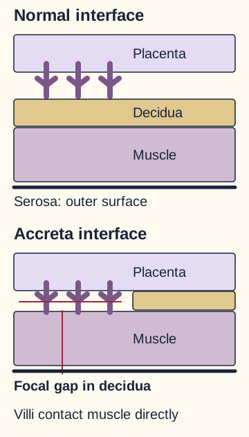
Does oxytocin create a normal separation plane? No. Decidua basalis is the pregnancy-modified maternal endometrium beneath the placenta. Myometrium is the underlying uterine smooth muscle. Chorionic villi are fetal placental structures. Normally, placental separation occurs through the decidual interface after delivery, and uterine contraction compresses maternal vessels at the placental bed.
In placenta accreta spectrum, or PAS, deficient decidualization allows villi to attach directly to, or extend into, myometrium. A scar can disrupt the endometrial-myometrial interface and favor this abnormal attachment. This is the favored explanation, not a claim that every affected patient has a surgical scar or that placental biology is completely understood. [1][3]
Trace from placenta to muscle. The missing intervening layer explains why separation can injure the maternal attachment bed. [1]
Contraction and separation are different processes. Atony is inadequate uterine contraction. Uterotonics can improve tone, including atony coexisting with PAS, but they cannot rebuild absent decidua. Persistent focal adherence despite a firmer uterus should change the working explanation. Increasing traction can disrupt maternal vessels and precipitate major hemorrhage. [1][7]
Trace the force through the interface
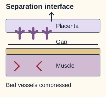
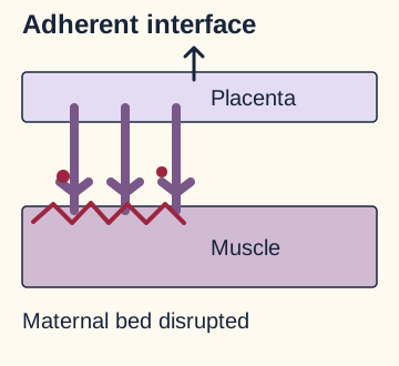
Use the drawing to predict where separation would occur. Open either comparison after making your prediction; the two results can remain side by side in your reading. These are tissue models, not instructions to test placental attachment in a patient.
If decidua is intact, where does separation occur?With a normal decidual interface, separation need not tear away attached muscle. Contraction then helps compress the maternal placental-bed vessels.If villi attach to muscle, what receives the separating force?Force reaches the adherent maternal bed. A stronger contraction may treat concurrent atony, but greater traction does not restore the missing interface.
The complete comparison: intact decidua permits separation at the interface; direct villous attachment transfers force into the maternal bed. Close either comparison to restore its starting view. The model does not estimate force, blood loss, or the safety of extraction.
Apply it: if massage makes the uterus firm but a placental segment remains fixed and bleeding increases during attempted separation, consider PAS while evaluating other hemorrhage causes. Do not interpret normal early coagulation results as protection from a mechanical source of bleeding.
Try it here · Checkpoint 1 of 3
Make your prediction before reading the choices. A first attempt is just a starting point.
Case 1
Show answer and explanations for case 1
A. Deficient decidualization at the implantation interface (Best answer)
A scar-associated defect can leave villi directly attached to muscle. Better contraction cannot recreate a separation interface, so disrupting the fixed segment injures the maternal vascular bed.
Reasoning steps for option A
Why does firm tone not release the anterior placental segment?
Massage and oxytocin correct the initial bogginess, but absent decidual separation at the scar can leave villi fixed to muscle.
What does bleeding with attempted separation indicate here?
Traction disrupts the maternal vascular bed at that fixed attachment, rather than correcting the interface defect.
Which defect accounts for both the scar history and persistent focal fixation?
Deficient decidualization permits direct villous attachment to myometrium despite adequate contraction.
B. Disruption of a maternal genital-tract vessel (Why this does not fit)
Genital trauma can cause postpartum hemorrhage despite firm uterine tone, but no laceration is seen and the bleeding increases specifically during attempted separation of fixed placental tissue.
Reasoning steps for option B
Why could a genital-tract vessel cause bleeding with a firm uterus?
Vaginal birth can injure a maternal vessel independently of uterine tone.
What observation weighs against genital-tract disruption in this patient?
No laceration is seen, while bleeding increases specifically as the fixed anterior placental segment is separated.
Where does the focal trigger localize the bleeding instead?
The placental attachment bed, not a demonstrated genital-tract vessel injury.
C. Inadequate contraction throughout the myometrium (Why this does not fit)
Atony can coexist and explains the initial soft uterus, but it does not explain persistent focal fixation after the uterus becomes firm.
Reasoning steps for option C
Which early finding supports inadequate myometrial contraction?
The uterus is initially boggy during hemorrhage, so atony may contribute at first.
What changes after massage and oxytocin?
The uterus becomes firm, yet the anterior placental segment remains fixed and separation provokes more bleeding.
Why is generalized atony insufficient as the continuing mechanism?
Improved tone can compress vessels but cannot recreate the missing decidual cleavage plane.
D. Premature cleavage through an intact decidual plane (Why this does not fit)
Premature separation causes abruption before delivery. Here the relevant segment has not separated and bleeds when separation is attempted.
Reasoning steps for option D
Why might separation-related bleeding suggest a cleavage-plane problem?
Bleeding rises when separation is attempted, implicating the placental bed.
Does this segment show premature cleavage through intact decidua?
No. After birth it remains attached to the anterior lower wall rather than separating prematurely as in abruption.
What does traction act on if decidua is deficient?
The adherent maternal bed, causing hemorrhage without an intact plane of separation.
E. Primary systemic depletion of coagulation factors (Why this does not fit)
Coagulopathy can complicate major hemorrhage later, but the initial normal tests and focal traction-associated bleeding favor a local attachment problem.
Reasoning steps for option E
Can major postpartum hemorrhage eventually affect coagulation factors?
Yes, depletion can follow substantial blood loss and aggravate bleeding.
What argues against primary factor depletion as this bleeding source?
Initial coagulation studies are normal and bleeding rises with attempted separation of one fixed placental segment.
Which local mechanism should be considered before blaming systemic depletion?
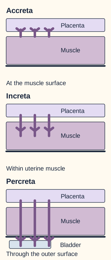
Does the word accreta always mean invasion deep into muscle? PAS is the umbrella term; accreta in the narrow anatomic sense means direct attachment to the myometrial surface without villous invasion into muscle. Increta extends into myometrium. Percreta extends through the uterine wall and serosal surface and may involve adjacent organs, particularly the bladder. These are anatomic descriptions, not three required stages through which every pregnancy progresses. [3]
Follow the villous endpoint rather than the amount of bleeding. At the surface, within muscle, and through the outer boundary distinguish accreta, increta, and percreta. Original simplified anatomy; depth is not drawn to scale.
Before reading the next sentence, trace a path from the uterine cavity to the bladder. Which outer boundary must be crossed? The uterine serosal boundary. Villi within muscle but not through that boundary support increta; full-wall extension supports percreta. The bladder is anterior to the uterus and the rectum is posterior, so the direction of suspected extension changes the adjacent organ being assessed. [13] A bladder-adjacent placenta alone does not prove invasion. Bleeding severity and hematuria warrant attention but cannot independently assign a histologic depth.
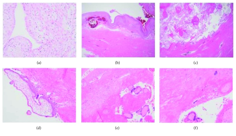
Open the full-size histology imageBartels, Postle, Downey and Brennan, 2018, Figure 1. Panels a and b show decidualized endometrium; c and f show villi touching muscle without intervening decidua; e shows a nonadherent region of the same placenta. Original image, unchanged. Copyright 2018 Helena C. Bartels et al.; Creative Commons Attribution License, version not specified in the accessible publication. Image: Source and caption. [3].
Compare panels c and e: identify the tissue between villi and muscle before deciding whether the interface is abnormal. The visible difference is direct contact in c versus an intervening decidua in e. Now consider a small delivered placental fragment containing muscle fibers. That finding can suggest abnormal separation, but a fragment does not map the full uterine wall and cannot establish percreta. Correlate pathology, specimen sampling, imaging and operative findings. [3]
Accreta is commonly the largest subgroup in older series, often quoted as about three quarters of PAS. That proportion varies with definitions and case selection; it is not a patient-specific probability. Adenomyosis is a different tissue relationship: endometrial glands and stroma within myometrium, rather than placental chorionic villi at an abnormal attachment site. [9] An incidental pelvic somatic finding does not explain villous adherence or substitute for obstetric hemorrhage evaluation.
Risk changes how a scan should be interpreted
Can risk factors be added into a percentage? Not without a validated model. Prior cesarean delivery is the most common risk factor, and concern rises markedly when a current placenta previa overlies a scar. Previa means placental tissue covers the internal cervical os. A low-lying placenta is near but does not cover the os; it is not interchangeable with previa in a risk table. [1][4]
Historical PAS proportions among women with placenta previa, grouped by cesarean number
Prior cesareans
Current cesarean ordinal
Observed proportion
Prior cesareans0
Current cesarean ordinalFirst
Observed proportion3%
Prior cesareans1
Current cesarean ordinalSecond
Observed proportion11%
Prior cesareans2
Current cesarean ordinalThird
Observed proportion40%
Prior cesareans3
Current cesarean ordinalFourth
Observed proportion61%
Prior cesareans4 or more
Current cesarean ordinalFifth or later
Observed proportion67%
These are rounded group observations from the previa subgroup of a cesarean cohort, not predictions for every pregnant patient. The cohort involved cesarean deliveries without labor at academic centers. Its 0.24% figure describes women undergoing a first cesarean in the overall cohort, not all unscarred pregnancies. [2] ACOG explicitly identifies the 3% previa estimate with no prior cesareans. [1]
Try the denominator: a patient with two prior cesareans is approaching her third, not her second. The table therefore points to 40%, not 11%, if she belongs to the previa group. If she has no previa, do not carry that 40% estimate into counseling. The relationship remains clinically important even when an exact individual probability is unavailable.
Other associated histories include curettage, myomectomy or other uterine surgery, intrauterine adhesions such as Asherman syndrome, older maternal age and multiparity. Pregnancy after endometrial ablation is also associated with PAS. [11] In vitro fertilization has an independent association in observational data, but this does not prove a single causal mechanism or justify adding arbitrary percentage points. Absence of previa or prior cesarean does not exclude PAS. [1][8]
Ultrasound first; MRI for selected questions
Experienced obstetric ultrasound assessment, often including transvaginal views and color Doppler, is the initial imaging approach. Multiple irregular placental lacunae, turbulent lacunar flow, loss of the retroplacental clear zone, very thin underlying myometrium and bridging vessels increase suspicion. Abnormality at the bladder interface can affect surgical planning. Transvaginal ultrasound is not the same as a digital cervical examination and is not contraindicated simply because previa is present. [1]
Consider a patient with previa and three prior cesareans whose report says no definite abnormal vessels. Predict whether that report cancels the history. It does not: absence of ultrasound findings does not exclude PAS, and interpretation depends on expertise and the clinical setting. Conversely, one nonspecific sign does not prove PAS or reliably establish its depth.
MRI may help answer a remaining question in difficult posterior placentation or suspected extrauterine extension. It is not an automatic confirmatory test for every suspicious scan. Arrange specialist review and a delivery plan based on the combined evidence, rather than waiting for a threshold score or an MRI label. [1]
Try it here · Checkpoint 2 of 3
Make your prediction before reading the choices. A first attempt is just a starting point.
Case 6
Show answer and explanations for case 6
A. Both patients have an estimated 11% risk because scar counts match (Why this does not fit)
This transfers a conditional estimate to a different placental-location group. Matching prior cesareans is insufficient when previa defined the denominator.
Both X and Y have one previous cesarean, so each would be undergoing a second cesarean.
Which additional condition defines the reported 11% group?
The cohort percentage is specifically among women with placenta previa; X has anterior previa and Y has a fundal placenta.
Can the same 11% be assigned to Y?
No. Her lack of previa places her outside the quoted denominator despite the matching scar count.
B. The 11% estimate applies to Y because her placenta avoids the scar (Why this does not fit)
The quoted denominator requires previa, which Y does not have. Avoiding the lower scar does not turn her into the group being measured.
Reasoning steps for option B
Does avoiding the scar make Y the quoted cohort subject?
No. The 11% observation is not a low-risk category defined by fundal placentation.
Which patient meets both the previa and procedure-ordinal conditions?
X has previa over the anterior scar and one prior cesarean, making the upcoming procedure her second.
What can be said about Y from this figure?
The previa-specific 11% does not quantify her PAS risk.
C. The 11% estimate is relevant to X, but does not quantify Y's risk (Best answer)
One prior cesarean means the current delivery would be the second. X matches both the scar count and previa condition; the supplied cohort figure cannot assign Y a personal risk.
Reasoning steps for option C
How is one previous cesarean translated into the cohort categories?
The upcoming repeat delivery is the second cesarean, matching the 11% ordinal.
Which patient also meets the placental-location denominator?
X has placenta previa over the anterior scar; Y has a fundal placenta clear of the cervix.
What counseling distinction follows?
The group estimate is relevant to X, but cannot provide Y an individual percentage.
D. Neither patient fits because the current cesarean is excluded from the count (Why this does not fit)
The report uses the ordinal number of the current cesarean. A second cesarean corresponds to one previous cesarean.
Reasoning steps for option D
Does a reported second cesarean require two previous operations?
No. One prior operation plus the current operation equals the second.
Which patient therefore fits the reported previa subgroup?
X, whose placenta covers the cervix and overlies the anterior scar.
Why is the claim that neither fits incorrect?
It omits the current cesarean from the ordinal and overlooks X meeting the previa condition.
Takeaway: Before applying a percentage, match the patient to the study denominator.
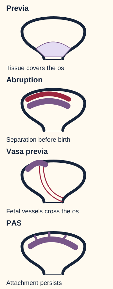
Is painless bleeding always previa, or a retained placenta always PAS? Both shortcuts fail. First assess maternal circulation and fetal status. Then distinguish placental location, premature separation, fetal vessel rupture, failure of uterine contraction and abnormal attachment. PAS can be recognized antenatally and can coexist with previa, so antepartum bleeding does not exclude it.
Locate what is abnormal before naming the condition. Location, separation, vessel course and attachment are different relationships, and more than one can be present.
Sort two examples before checking the explanation: bleeding begins immediately after membrane rupture with sudden fetal bradycardia and a mother whose circulation remains stable; a second patient has abdominal pain after trauma, a persistently tender uterus and fetal deterioration. The first raises concern for fetal blood loss from vasa previa. The second supports placental abruption, with maternal blood collecting behind a prematurely separating placenta. [5][6]
Previa: classically painless bleeding after midpregnancy from a low placental attachment. Locate the placenta by ultrasound before digital cervical examination. Severe bleeding can compromise mother and fetus; initially reassuring fetal status is not a diagnostic requirement. [4]
Abruption: often pain, tenderness or increased uterine tone, with possible fetal compromise. Blood can be concealed, so visible loss can underestimate severity. Hypertension, cocaine, smoking and trauma are associated risks. A normal ultrasound does not rule it out; substantial abruption can cause consumption coagulopathy. [5]
Vasa previa: unprotected fetal vessels traverse the membranes over or near the cervix. Rupture can cause rapid fetal hemorrhage, particularly with membrane rupture. Maternal stability does not reassure about fetal blood volume. [6]
After birth, assess tone, retained tissue, genital tract trauma and coagulation together. A boggy uterus after a prolonged labor supports atony; a firm uterus with a visible bleeding laceration supports trauma. A placenta that has separated but is trapped behind a closing cervix differs from tissue fixed to the uterine wall. Retention can also reflect incomplete separation without PAS. No single elapsed-time cutoff establishes abnormal invasion. [7][10]
Apply the distinction to persistent hemorrhage: normal initial platelets and clotting times do not exclude PAS, and later coagulopathy may develop during massive blood loss. Do not stop evaluating other causes just because one diagnosis fits. Uterotonics remain useful for a coexisting tone problem, but repeated forceful separation is hazardous when abnormal adherence is suspected. [1]
Planning protects the attachment bed
If PAS is suspected before delivery, why not deliver the fetus and then test whether the placenta peels away? Disrupting a vascular attachment site can convert a planned operation into uncontrolled hemorrhage. The generally accepted approach is cesarean hysterectomy with the placenta left in situ after fetal delivery, performed by an experienced multidisciplinary team. The uterine incision is planned to avoid the placenta where feasible. This means the placenta stays with the uterus during hysterectomy, not that every patient is sent home with retained tissue. [1]
Before birth: specialist assessment, shared counseling, anemia evaluation and treatment, surgical and anesthesia planning, blood-bank preparation.
At delivery: avoid placental disruption, deliver the fetus through a planned incision, then complete the agreed surgical strategy with appropriate expertise.
After surgery: monitor for ongoing bleeding, organ injury, infection and complications of major resuscitation.
Choose between these plans for a stable patient at 32 weeks with strongly suspected PAS: wait for labor at a unit without a major transfusion service, or arrange delivery at an experienced level III or IV maternal care facility. The second plan addresses the foreseeable hemorrhage risk before an emergency occurs. Resources include obstetric and pelvic surgical expertise, anesthesia, neonatal care, critical care and a blood bank capable of a massive transfusion response. Suspected urinary tract involvement warrants relevant surgical expertise; a particular specialty title is not a substitute for experience.
Under ACOG/SMFM guidance, planned delivery in an otherwise stable patient is generally scheduled at 34 weeks 0 days through 35 weeks 6 days. This balances prematurity against increasing risk of bleeding or unplanned labor. Persistent bleeding, labor, ruptured membranes, preeclampsia, maternal deterioration or fetal compromise can require earlier delivery. Antenatal corticosteroids are considered according to gestational age and anticipated delivery; an emergency should not wait to complete a course. [1]
Now change the example: the patient develops heavy bleeding and shock at 33 weeks. The consequence is urgent resuscitation and definitive obstetric care, not waiting for the scheduled window or MRI. Obtain help, vascular access and blood products while controlling the source; do not delay treatment for a reassuring early hemoglobin or normal clotting studies. If PAS is discovered unexpectedly in a stable patient before disruption, pause when safe to assemble expertise and consider transfer according to stability and local capability. Instability requires immediate local stabilization and hemorrhage control. [1]
Future fertility matters in counseling. The cited consensus regards expectant management with placenta left in a preserved uterus as investigational. Uterus-preserving approaches can be considered only in carefully selected patients with specialist oversight and explicit discussion of uncertain benefit, hemorrhage, infection, prolonged follow-up and possible delayed hysterectomy. Leaving placenta in situ for fertility preservation is a different strategy from leaving it undisturbed during planned hysterectomy. Methotrexate to hasten placental resorption is not recommended. Routine prophylactic pelvic arterial balloons are not established standard care and can cause vascular complications. [1]
Before independent practice, explain aloud: which tissue interface is deficient, how deeply villi extend, why history still matters after a negative scan, and why avoiding disruption changes delivery planning. You may pause here and return to any section without completing the case bank.
Try it here · Checkpoint 3 of 3
Make your prediction before reading the choices. A first attempt is just a starting point.
Case 16
Show answer and explanations for case 16
A. Emergency cesarean through the lower segment; leave the placenta during hysterectomy (Why this does not fit)
Leaving the placenta undisturbed is appropriate, but the supplied map permits avoiding the placental site rather than incising through it.
Reasoning steps for option A
Which part of this option addresses suspected PAS correctly?
Leaving the placenta attached during hysterectomy avoids deliberate separation of its vascular bed.
What does the scan say about a routine lower-segment incision?
Placenta crosses that incision site, while an accessible fundal region offers a route away from placental tissue.
How should urgency modify incision planning?
Proceed emergently with resuscitation, but use the mapped placenta-avoiding incision when feasible.
B. Emergency cesarean away from the placenta; leave it undisturbed during hysterectomy (Best answer)
Instability requires immediate delivery rather than waiting for the elective window. The map separately supports an incision away from placenta, followed by the definitive approach without placental separation.
Reasoning steps for option B
Why is waiting for the elective delivery window unsafe?
Persistent heavy bleeding, blood pressure 78/45 mmHg and recurrent fetal decelerations demand immediate action.
Where should the uterine incision go according to the supplied map?
An accessible fundal region is away from the placenta spanning the usual lower segment.
What placental handling follows fetal delivery in this definitive approach?
Avoid separation and leave the placenta undisturbed during hysterectomy to limit additional hemorrhage.
C. Emergency cesarean away from the placenta; separate it before hysterectomy (Why this does not fit)
The incision respects the map, but attempted separation would still disrupt the abnormal vascular attachment.
Reasoning steps for option C
Which part of the proposed operation uses the map appropriately?
An incision away from the anterior placenta uses the accessible fundal region for delivery.
Why does that incision not make extraction safe afterward?
Suspected extensive PAS still lacks a normal separation plane, and she is already hypotensive from heavy bleeding.
What should replace placental separation before hysterectomy?
Leave the adherent placenta undisturbed during definitive surgery, even after a placenta-avoiding delivery.
D. Emergency cesarean through the lower segment; separate the placenta before hysterectomy (Why this does not fit)
Urgent delivery is necessary, but this combines an incision through the mapped placental site with separation of the adherent bed, increasing opportunities for severe bleeding.
Reasoning steps for option D
What makes the familiar lower-segment incision particularly hazardous?
Ultrasound maps placental tissue across the usual incision and identifies an accessible fundal alternative.
Why is routine placental removal also hazardous in this patient?
Suspected extensive anterior PAS and ongoing hemorrhagic shock make disruption of the adherent bed especially dangerous.
What two operative choices address both hazards?
Deliver emergently through a placenta-avoiding incision and leave the placenta attached during hysterectomy.
Takeaway: An emergency changes the timing, not the need to use the placental map and avoid disrupting its attachment.
A. Percreta with possible intestinal repair (Why this does not fit)
Percreta is the deepest category, but the catheter-containing anterior organ is the bladder, not intestine. Intestinal expertise would depend on different anatomic involvement.
Reasoning steps for option A
Which depth category does the second sampled site establish?
Villi cross the uterine outer surface, so the deepest demonstrated involvement is percreta.
Which clue rules out intestinal rather than urinary repair?
The invaded hollow organ is anterior to the lower uterus and contains a urinary catheter balloon, identifying bladder.
What operative concern follows from that organ identity?
Possible bladder or urinary tract repair, not bowel repair, accompanies percreta.
B. Percreta with possible urinary tract repair (Best answer)
The second site crosses the serosal boundary, supporting percreta rather than the shallower involvement at the first site. Its extension into the catheter-containing organ identifies bladder involvement and possible urinary tract repair.
Reasoning steps for option B
Why not classify by the first site alone?
Its villi between muscle bundles show increta locally, but the second site crosses the outer uterine surface.
How does the catheter balloon identify the additional involved organ?
It lies in the bladder lumen, identifying the anterior organ whose wall contains the extending villi.
What pair of findings supports the selected combination?
Full-thickness uterine extension supports percreta and bladder-wall extension raises possible urinary tract repair.
C. Increta with possible intestinal repair (Why this does not fit)
This both underestimates the deepest sampled extension and misidentifies the anterior catheter-containing organ as bowel.
Reasoning steps for option C
Which finding might make increta attractive?
The first site contains villi between muscle bundles.
Why is that not the deepest involvement in this specimen?
A second site crosses the uterine outer surface into another organ wall, establishing percreta.
Why is intestinal repair also mismatched?
The anterior hollow organ contains a urinary catheter balloon, localizing involvement to bladder.
D. Increta with possible urinary tract repair (Why this does not fit)
The urinary catheter localizes the adjacent organ correctly, but crossing the uterine outer boundary is more extensive than increta.
Reasoning steps for option D
Which half of this combination fits the specimen?
Urinary tract repair is a concern because villi reach the wall of the catheter-containing bladder.
Why does the increta label understate the deepest site?
Increta covers intramuscular villi at the first site, but villi cross the serosal boundary at the second.
Which depth should govern the combined description?
Percreta follows the deepest demonstrated villous endpoint, even with shallower involvement elsewhere.
Takeaway: Use the deepest demonstrated villous endpoint and the identity of the adjacent organ to anticipate operative needs.
A. Suspected extrauterine involvement; plan obstetric surgery without urinary tract support (Why this does not fit)
The diagnostic uncertainty is appropriately retained, but that concern still affects staffing and preparation for adjacent-organ injury.
Reasoning steps for option A
Why is extrauterine involvement only suspected?
Expert ultrasound and MRI cannot resolve the uterine-bladder outer boundary, so extension is not proved.
Why does omitting urinary support still fail?
Anterior previa over three scars, bridging vessels, marked thinning and unexplained hematuria raise a plausible bladder dissection or repair need.
What decision can precede histologic certainty?
Prepare urinary tract surgical expertise despite an indeterminate invasion depth.
B. Invasion confined to muscle; include urinary tract surgical expertise (Why this does not fit)
Urinary tract expertise is prudent, but an unresolved outer interface does not establish confinement to muscle.
Reasoning steps for option B
What justifies the urinary tract support half of this option?
Hematuria without infection and an unresolved uterine-bladder interface create a possible urinary operative need.
Can the studies establish invasion confined to muscle?
No. Both expert ultrasound and MRI leave the outer boundary indeterminate.
How should uncertainty be described instead?
Suspect possible extrauterine involvement without claiming it is proven or excluded.
C. Suspected extrauterine involvement; include urinary tract surgical expertise (Best answer)
The combined findings raise concern for urinary tract involvement without proving it. Planning for possible bladder dissection or repair is appropriate despite uncertain final depth.
Reasoning steps for option C
What do bridging vessels and a very thin wall suggest?
They heighten PAS concern in this patient with anterior previa and three prior cesareans.
What does unresolved bladder-boundary imaging plus hematuria add?
Possible extrauterine or urinary tract involvement, not histologic proof of bladder invasion.
Why arrange urinary tract expertise now?
The team may need bladder dissection or repair even though final villous depth remains uncertain.
D. Invasion confined to muscle; plan obstetric surgery without urinary tract support (Why this does not fit)
Neither modality establishes an intact outer interface. Hematuria in this high-risk anatomic setting adds concern rather than justification to omit relevant surgical preparation.
Reasoning steps for option D
Why is muscle confinement not established?
Neither ultrasound nor MRI shows a definitively intact uterine-bladder outer boundary.
What prevents omission of urinary tract support?
Unexplained intermittent hematuria and the abnormal anterior interface make bladder involvement a relevant operative concern.
Which two assumptions in this choice overstate reassurance?
It treats indeterminate imaging as proof of muscle-only invasion and then excludes preparation for possible urinary injury.
Takeaway: Uncertain extrauterine extension can change operative preparation without being a proven histologic diagnosis.
A. Arrange local MRI in six weeks before referral (Why this does not fit)
Selective MRI can answer unresolved questions, but a delayed local scan should not be a prerequisite for access to experienced assessment and delivery resources.
Reasoning steps for option A
Could MRI clarify the incompletely seen retroplacental interface?
It may answer selected unresolved anatomy questions, but it is not a mandatory gate before specialist referral.
Why is a six-week local delay problematic at 30 weeks?
Previa over three cesarean scars remains high risk despite no definite lacunae and no current symptoms.
What should occur before waiting on this scan?
Coordinate expert assessment and delivery planning promptly; MRI can be considered selectively within that process.
B. Schedule local cesarean for previa without further interface assessment (Why this does not fit)
The scan has not excluded PAS. A plan for previa alone may lack the surgical and transfusion preparation required for abnormal adherence.
Reasoning steps for option B
Why does a local cesarean for previa alone sound plausible?
Previa itself calls for cesarean delivery and the report notes no definite lacunae.
What prevents that report from excluding PAS?
The retroplacental interface was incompletely visualized in a patient with three prior cesareans.
Which capability might a previa-only plan miss?
Preparation for adherent placenta and major hemorrhage with appropriate surgical and transfusion resources.
C. Refer for coordinated expert ultrasound and delivery planning (Best answer)
Previa over multiple cesarean scars remains high risk after an incomplete study. Referral allows expert reassessment and planning without relying on one absent sign.
Reasoning steps for option C
How should no definite lacunae be interpreted here?
One absent sonographic sign on a community scan with an incompletely seen interface does not rule out PAS.
Which history sustains concern at 30 weeks?
Current previa and three previous cesareans create a high-risk scar-overlying implantation setting.
What follow-up addresses both the scan limitation and delivery risk?
Refer now for experienced ultrasound reassessment and coordinated high-risk delivery planning.
D. Repeat local routine ultrasound in six weeks before referral (Why this does not fit)
A repeat examination may be useful, but waiting six weeks before referral delays planning in a patient already at 30 weeks with substantial risk and an incompletely seen interface.
Reasoning steps for option D
What makes repeat ultrasound potentially useful?
Further views could better visualize the retroplacental interface that the community scan missed.
Why not wait six weeks before referral?
At 30 weeks, waiting would postpone planning to 36 weeks despite previa and three prior scars.
What does current absence of bleeding actually establish?
She is stable now, not that PAS is excluded or specialist planning can safely be deferred.
Takeaway: An incomplete or negative scan does not erase a high-risk implantation history.
A. X to Z: 50 percentage points (Why this does not fit)
The subtraction would match the second and fourth previa groups, but Z has no previa. The table cannot assign Z the fourth-cesarean previa estimate.
Reasoning steps for option A
What calculation makes 50 points seem plausible for X and Z?
One and three prior cesareans map to current second and fourth procedures, giving 61% minus 11%.
Why cannot the fourth-cesarean estimate be assigned to Z?
Z has a fundal placenta clear of the cervix, while every percentage in the table is conditional on previa.
Which similarly scarred comparison does the denominator permit?
Compare X with Y, who both have previa, instead of transferring the value to Z.
B. X to Z: 56 percentage points (Why this does not fit)
This both applies a previa-specific estimate to a fundal placenta and substitutes the fifth-or-later group for a patient with only three prior cesareans.
Reasoning steps for option B
Where does the proposed 56-point difference come from?
It subtracts 11% from the fifth-or-later 67% estimate.
Why does Z fail the table eligibility test?
Her fundal placenta is not previa even though she has three prior cesareans.
What additional ordinal error occurs?
Three previous cesareans imply a current fourth, associated with 61% among previa patients, not fifth-or-later 67%.
C. X to Y: 50 percentage points (Best answer)
X and Y both match the previa denominator. One prior cesarean means a current second procedure, giving 11%; three prior means a current fourth, giving 61%. Their group-estimate difference is 50 percentage points.
Reasoning steps for option C
Which patients can legitimately be compared using the previa table?
X and Y both have placenta previa; Z does not.
Which current cesarean ordinals match their histories?
X has one prior and is undergoing her second; Y has three prior and is undergoing her fourth.
What absolute group-estimate difference follows?
Subtract 11% for X from 61% for Y to obtain 50 percentage points.
D. X to Y: 56 percentage points (Why this does not fit)
X and Y are the appropriate comparison, but 56 points uses the fifth-or-later 67% figure rather than the fourth-cesarean 61% figure applicable to Y.
Reasoning steps for option D
Why is X versus Y the correct comparison pair?
Both have previa, satisfying the table denominator.
Does Y belong to the 67% fifth-or-later group?
No. Three previous cesareans make the current delivery the fourth, corresponding to 61%.
What subtraction corrects the proposed 56 points?
Use 61% minus X's 11%, which is 50 percentage points rather than 67% minus 11%.
Takeaway: Confirm denominator eligibility and translate prior counts before calculating an absolute difference.
A. Repeat uterine artery Doppler; possible bowel surgical support (Why this does not fit)
The posterior localization makes bowel-related support relevant, but another arterial flow measurement will not directly clarify the unresolved tissue boundary after expert anatomic assessment.
Reasoning steps for option A
Which part of this option localizes the surgical concern correctly?
The hollow organ between cervix and sacrum is rectum, so possible bowel support fits the posterior bulge.
Why is another uterine artery Doppler not the best added assessment?
Two expert ultrasounds have already left a tissue boundary unresolved; arterial flow measurement does not map that posterior interface.
What adjunct could address the remaining anatomy question?
Selective expert pelvic MRI may clarify the rectal-side boundary while organ invasion remains unproved.
B. Expert pelvic MRI; possible bowel surgical support (Best answer)
The posterior compartment between cervix and sacrum contains rectum, so possible extrauterine extension there raises a bowel-related surgical question. Selective specialist MRI may help clarify that unresolved boundary after expert ultrasound. Neither involvement nor the need for bowel surgery is yet proven.
Reasoning steps for option B
Where is the unresolved organ boundary?
Posterior to the lower uterus toward the rectum between cervix and sacrum; the anterior bladder boundary is preserved.
Why choose MRI after two expert ultrasound examinations?
Specialist pelvic MRI may help map a difficult posterior tissue interface, unlike repeat uterine artery velocity measurement.
What does possible bowel support mean here?
Prepare for potential rectal involvement without asserting that extension or bowel repair is already established.
C. Repeat uterine artery Doppler; possible bladder surgical support (Why this does not fit)
This selects a vascular measurement rather than additional tissue mapping and directs preparation toward the anterior organ despite the specified posterior uncertainty.
Reasoning steps for option C
Why might lacunae prompt more Doppler testing?
They are associated with abnormal placental vascularity, but the outstanding question is tissue anatomy rather than arterial flow.
Why does possible bladder support miss the indicated compartment?
The anterior uterine-bladder boundary is clearly preserved; the bulge and unresolved boundary lie posteriorly toward rectum.
What would align testing and surgical preparation with the stem?
Consider expert MRI for posterior mapping and provisional bowel-related expertise.
D. Expert pelvic MRI; possible bladder surgical support (Why this does not fit)
MRI is a reasonable adjunct, but the unresolved boundary is posterior toward rectum, not the clearly visualized anterior bladder interface.
Reasoning steps for option D
Which component of this alternative is appropriate?
MRI may provide adjunct anatomic detail after two expert scans fail to resolve the posterior boundary.
Which organ does the specified location implicate instead of bladder?
The hollow organ between cervix and sacrum is rectum, whereas the anterior uterine-bladder interface is preserved.
What preparation should follow the uncertain posterior extension?
Possible bowel surgical support, without treating rectal invasion as proven.
Takeaway: Localize the unresolved compartment first, then choose an adjunct that may clarify its tissue boundary.
A. Treat the findings as previa alone; use a standard previa operation (Why this does not fit)
Multiple PAS-associated findings over repeated scars require more than a location-only plan. Previa and PAS can coexist.
Reasoning steps for option A
Why might confirmed anterior previa invite a location-only operation?
Previa is certain and the bladder interface is poorly seen, but location alone does not characterize adherence.
What findings argue against treating the two-scar placenta as previa alone?
Irregular lacunae, turbulent Doppler flow and markedly thin retroplacental muscle collectively raise PAS suspicion.
Does uncertainty about bladder extension justify omitting PAS precautions?
No. Uncertain depth does not erase evidence of abnormal attachment; plan with PAS expertise without asserting invasion.
B. Treat full-wall extension as most likely; plan routine bladder resection (Why this does not fit)
These signs do not prove percreta or bladder invasion. Possible urinary tract involvement requires relevant expertise, not an automatic resection based on lacunae and flow.
Reasoning steps for option B
Why could thin myometrium and lacunar flow raise concern about the bladder?
They suggest abnormal adherence beneath this anterior previa over two cesarean scars, potentially near the outer wall.
What prevents labeling this percreta with bladder invasion?
The serosal-bladder interface was not adequately assessed, and lacunae or thinning cannot establish full-wall penetration.
Should this scan trigger routine bladder resection?
No. Prepare relevant expertise for possible involvement rather than resecting an organ on an unproven depth assignment.
C. Treat superficial attachment as most likely; plan extraction before hysterectomy (Why this does not fit)
The findings do not establish superficial attachment. Attempted extraction can cause severe hemorrhage even when invasion is not deep.
Reasoning steps for option C
Does a thin residual muscle layer demonstrate superficial accreta?
No. Marked thinning on ultrasound cannot establish the villous endpoint or a safe separation plane.
Why is extraction especially hazardous in this scar-and-previa pattern?
Multiple lacunae and turbulent flow support PAS, in which traction may tear the vascular maternal attachment bed.
What distinction replaces the proposed superficial diagnosis and extraction trial?
Suspect PAS and avoid testing adherence by extraction, even if deep invasion has not been demonstrated.
D. Treat PAS as suspected; retain uncertainty about extrauterine extension (Best answer)
The history and associated ultrasound pattern justify PAS precautions, while an inadequately seen outer interface cannot assign exact depth or establish adjacent-organ involvement.
Reasoning steps for option D
Which combined history and scan signs support suspecting PAS?
Anterior previa over two previous cesareans with irregular lacunae, turbulent flow and markedly thin myometrium support abnormal attachment.
What remains unresolved at the uterine outer boundary?
An inadequately visualized serosal-bladder interface cannot establish extrauterine extension or bladder invasion.
How should the team act despite that unresolved depth?
Use PAS precautions and appropriate expertise while avoiding a claim of specific depth or automatic adjacent-organ resection.
Takeaway: Imaging can justify PAS precautions without proving a histologic depth.
A. Pelvic MRI to map the lower uterine segment (Why this does not fit)
MRI may help selected difficult PAS anatomy, but transvaginal ultrasound is the appropriate initial method for this specific unresolved location.
Reasoning steps for option A
Why might MRI appeal after fetal-head shadowing on abdominal imaging?
A prior cesarean and low anterior placenta can raise complex placentation concerns, while the head obscures the abdominal view.
What specific measurement is needed before cervical palpation?
The placental edge must be located relative to the internal os; targeted transvaginal ultrasound addresses this directly.
When would MRI be more relevant than this proposed first examination?
Reserve MRI for selected complex PAS anatomy; do not substitute it for first-line edge-to-os localization.
B. Transvaginal ultrasound to define the placental edge (Best answer)
Targeted transvaginal views resolve the obscured placental edge-to-os relationship and are safe in suspected previa.
Reasoning steps for option B
What does the abdominal scan fail to show in this bleeding patient?
The fetal head obscures whether the low anterior placental edge reaches or covers the internal os.
How do transvaginal views resolve the decision about digital examination?
They define the edge-to-os relationship without placing a finger through a potentially os-covering placenta.
Does suspected previa prohibit the selected ultrasound approach?
No. Transvaginal imaging is distinct from digital cervical palpation and is appropriate to clarify location.
C. Digital cervical examination to assess the presenting part (Why this does not fit)
A digital examination could provoke substantial bleeding if the placenta covers the os. Placental location should be clarified first.
Reasoning steps for option C
Why might stable vital signs tempt an immediate digital cervical check?
The mother and fetal tracing are reassuring, making bedside assessment of dilation or presenting part seem low risk.
What unknown makes digital palpation unsafe at 31 weeks?
The low placental edge may cover the internal os, which transabdominal imaging cannot resolve behind the fetal head.
Which examination should precede a digital check here?
Map the edge with transvaginal ultrasound first; present stability cannot rule out provoked previa hemorrhage.
D. Sterile speculum examination to inspect the cervix (Why this does not fit)
A speculum examination can investigate a cervical or vaginal bleeding source, but it will not define the internal placental edge-to-os relationship requested here.
Reasoning steps for option D
What can speculum inspection contribute to evaluation of painless bleeding?
It can reveal a visible cervical or vaginal bleeding source without digital palpation.
Why does that inspection not answer this stem?
It cannot map the internal placental edge against the os obscured on the abdominal scan.
What should be selected for the requested localization instead?
Targeted transvaginal ultrasound directly resolves whether this low anterior placenta reaches the cervical opening.
Takeaway: Use ultrasound, including transvaginal views, to establish location before digital cervical examination.
A. Fetal blood loss from exposed membranous vessels (Why this does not fit)
Fetal vessel bleeding explains fetal compromise but not this combination of maternal hypotension, hypertension-associated risk and uterine pain. No membrane-rupture event is supplied.
Reasoning steps for option A
Why could fetal decelerations with scant blood suggest exposed fetal vessels?
Fetal vessel rupture can compromise the fetus despite little visible bleeding.
Which maternal findings contradict fetal blood loss as the whole explanation?
Her blood pressure falls and she has persistent abdominal pain and uterine tenderness, with chronic hypertension as an abruption risk.
Where is the missing blood more plausibly located?
Maternal blood can collect behind a prematurely separating placenta, causing shock despite limited vaginal loss.
B. Maternal intraperitoneal bleeding from uterine rupture (Why this does not fit)
Uterine rupture is another dangerous source of concealed bleeding, but the supplied hypertension-associated tender uterine pattern and lack of a labor or uterine-scar history favor abruption. A complete assessment would still consider other causes.
Reasoning steps for option B
Why does uterine rupture enter the concealed-bleeding differential?
Rupture can cause abdominal pain, fetal decelerations and maternal intraperitoneal blood loss.
Which history favors abruption over rupture here?
No surgery or contractions are reported, whereas chronic hypertension and a persistently tender uterus support abruption.
Does the negative retroplacental ultrasound settle the comparison?
No. Ultrasound can miss abruption; rupture still merits assessment, but the supplied pattern favors concealed placental separation.
C. Maternal bleeding at a placenta covering the cervical opening (Why this does not fit)
Previa can cause significant maternal bleeding, but this placenta is documented clear of the os. The tender painful uterus and concealed-loss pattern favor abruption.
Reasoning steps for option C
How could previa initially explain late-pregnancy maternal hemorrhage?
An os-covering placenta can bleed substantially and eventually compromise fetal status.
What directly contradicts an os-covering source in this patient?
Ultrasound places the placenta clear of the internal os; pain and tenderness also point away from classic previa.
Which mechanism better accounts for hypotension with little external blood?
Concealed maternal hemorrhage behind a prematurely separating placenta fits the painful tender uterus.
D. Maternal blood loss from a bleeding cervical lesion (Why this does not fit)
A cervical lesion could cause visible maternal bleeding, but it does not account well for a persistently painful tender uterus and deterioration out of proportion to the external loss.
Reasoning steps for option D
Why might a cervical lesion seem to localize the visible bleeding?
A cervical lesion can produce vaginal maternal blood without requiring abnormal placental attachment.
Which systemic and uterine signs exceed that explanation?
Falling pressure, fetal decelerations and sustained uterine pain and tenderness are not explained by a small visible cervical bleed.
What should the scant external blood prompt you to infer instead?
Assess for concealed maternal loss from abruption rather than equating visible vaginal blood with total hemorrhage.
E. Concealed maternal bleeding from premature placental separation (Best answer)
Hypertension, pain, tenderness and fetal compromise support abruption. Maternal blood can remain behind the placenta, so external loss and a negative ultrasound underestimate the problem.
Reasoning steps for option E
Which risk and examination findings point to premature separation?
Chronic hypertension, persistent pain and uterine tenderness accompany fetal decelerations.
How can abruption cause shock with scant vaginal bleeding?
Maternal blood can remain concealed behind the placenta rather than draining externally.
Does absence of a sonographic collection exclude this diagnosis?
No. A negative ultrasound does not exclude abruption or negate clinically important concealed bleeding.
Takeaway: Concealed abruption can cause severe maternal and fetal deterioration with little visible blood.
A. Emergency cesarean; anticipate maternal placental-bed bleeding and maternal transfusion (Why this does not fit)
Urgent cesarean is appropriate, but the immediate membrane-rupture event, velamentous insertion and disproportionate fetal deterioration suggest blood is being lost from fetal vessels rather than primarily from the maternal bed.
Reasoning steps for option A
Why does the low placental edge make maternal-bed bleeding tempting?
A low placenta can bleed, and fetal bradycardia makes urgent cesarean necessary.
Which timing and circulation findings instead implicate fetal vessels?
Bleeding begins at membrane rupture with velamentous insertion and fetal rate 65/min, while maternal pulse and pressure stay normal.
What preparation is missing from this proposed emergency plan?
Anticipate neonatal blood loss and transfusion readiness, not just maternal transfusion for a presumed placental-bed source.
B. Emergency cesarean; anticipate cord compression and neonatal ventilation without targeted transfusion preparation (Why this does not fit)
Cord compression can cause bradycardia after membrane rupture, but vaginal bleeding and the uncharted velamentous vessels require preparation for fetal blood loss, not ventilation alone.
Reasoning steps for option B
How can cord compression mimic part of this presentation?
What findings make ventilation alone an incomplete neonatal plan?
Concurrent vaginal bleeding and velamentous vessels of unmapped course raise concern for ruptured fetal vessels.
Which additional resuscitation resource must be anticipated?
Prepare for neonatal transfusion because fetal hypovolemia may accompany the profound bradycardia.
C. Emergency cesarean; anticipate fetal vessel bleeding and neonatal transfusion (Best answer)
The pattern supports ruptured vasa previa with fetal hemorrhage. Severe persistent fetal bradycardia when vaginal birth is not imminent calls for emergency cesarean with neonatal resuscitation and transfusion readiness.
Reasoning steps for option C
What event identifies a plausible source of fetal hemorrhage?
Bleeding starts immediately after membrane rupture in a pregnancy with velamentous cord insertion and uncertain vessel course.
Why prioritize emergency cesarean over awaiting vaginal delivery?
The fetal heart rate falls to 65/min and the cervix is only 2 cm dilated, so birth is not imminent.
Why does normal maternal circulation not lower neonatal concern?
The bleeding may be fetal rather than maternal; neonatal resuscitation must include transfusion readiness.
D. Emergency cesarean; anticipate maternal concealed bleeding and maternal factor replacement (Why this does not fit)
Abruption can require maternal coagulation support, but the painless event immediately after membrane rupture with stable maternal circulation and velamentous insertion more strongly suggests fetal vessel rupture.
Reasoning steps for option D
Why can abruption suggest maternal factor replacement?
A severe abruption can produce concealed maternal hemorrhage and consumption coagulopathy.
Which features favor fetal vessel rupture over that maternal process?
The uterus is nontender, maternal circulation remains stable, and bleeding with marked fetal bradycardia begins immediately after membrane rupture.
What target should accompany urgent delivery here?
Prepare neonatal transfusion for suspected fetal blood loss from vasa previa rather than primarily maternal factor replacement.
Takeaway: Maternal stability does not reassure when bleeding after membrane rupture is accompanied by sudden fetal compromise.
A. Cervical narrowing retains a separated placental segment (Why this does not fit)
A trapped placental segment could interfere with third-stage completion, but the placenta appears complete and hemorrhage improves with restored uterine tone rather than release of trapped tissue.
Reasoning steps for option A
When can cervical narrowing trap placental tissue?
A detached segment may remain behind a closing cervix and contribute to an incomplete third stage.
What observation argues against trapping in this delivery?
The placenta appears complete and bleeding drops when the soft enlarged uterus firms after massage and oxytocin.
What explains the response better than release of a trapped segment?
Restored contraction compresses exposed maternal placental-bed vessels in uterine atony.
B. Contraction compresses maternal placental-bed vessels (Best answer)
The tone-dependent reduction in bleeding after prolonged overdistended labor supports atony. Contraction compresses the maternal vessels exposed at the placental bed.
Reasoning steps for option B
Why was atony plausible after this labor?
Prolonged twin labor can leave an overdistended uterus soft and unable to compress placental-bed vessels effectively.
What change links treatment to hemostasis?
Massage and oxytocin make the uterus firm while heavy bleeding markedly decreases.
Which vessels are being compressed by contraction?
Maternal vessels at the delivered placental bed, not a fixed abnormal villous attachment.
C. Coagulation within an injured cervical vessel stops bleeding (Why this does not fit)
A cervical source is less consistent with the negative laceration assessment and immediate parallel improvement in uterine firmness and bleeding.
Reasoning steps for option C
Why might cervical clotting seem to stop postpartum bleeding?
A lacerated cervical vessel can bleed and eventually clot.
Which findings make that mechanism unlikely here?
No bleeding genital laceration is found, and reduced loss closely follows restored uterine tone.
What accounts for this treatment-linked improvement?
Uterine contraction compresses maternal placental-bed vessels rather than relying on cervical-vessel clotting.
D. Myometrial retraction shears a fixed placental attachment (Why this does not fit)
Disrupting a fixed attachment can worsen hemorrhage. This presentation lacks fixed tissue and improves as tone returns, supporting compression rather than shearing.
Reasoning steps for option D
Why could myometrial retraction be confused with placental shearing?
Contraction normally assists placental separation, so oxytocin might seem to detach a remnant.
What evidence argues there is no fixed attachment to shear?
The placenta has already delivered and appears complete; the enlarged soft uterus firms as bleeding subsides.
Why would shearing fixed PAS tissue be the wrong mechanism?
Tearing an adherent vascular bed risks more bleeding; this pattern instead shows tone-dependent vessel compression.
Takeaway: Uterotonics treat inadequate placental-bed vessel compression, not the missing interface of PAS.
A. Repeat coagulation studies to reassess systemic hemostasis (Why this does not fit)
Serial laboratory reassessment is appropriate in major hemorrhage, but it should not replace direct inspection of a likely anatomic source after instrumental birth.
Reasoning steps for option A
Why are repeat clotting studies reasonable during major hemorrhage?
Coagulopathy can evolve during ongoing blood loss even when initial laboratory results are normal.
Why should laboratory reassessment not be the first focused source search?
Bright continuous bleeding after forceps delivery persists despite a firm fundus, pointing to an anatomic birth-canal injury.
What assessment identifies the more immediate treatable cause?
Inspect the cervix and vagina for lacerations while resuscitation and serial laboratory monitoring continue.
B. Careful examination of the cervix and vagina for lacerations (Best answer)
The instrumental delivery and ongoing bright bleeding despite firm tone make a genital tract laceration a likely treatable source. Resuscitation and assessment proceed together.
Reasoning steps for option B
Which delivery detail increases the likelihood of genital trauma?
Forceps assistance raises concern for a cervical or vaginal laceration.
How do placental and tone findings sharpen that concern?
The placenta appears complete and the uterus stays firm after oxytocin, yet bright-red bleeding continues.
How should the focused examination proceed alongside hemorrhage care?
Inspect cervix and vagina for an actively bleeding injury while resuscitation continues.
C. Ultrasound assessment for residual intrauterine placental tissue (Why this does not fit)
Residual tissue remains a possible cause, but complete-appearing placental delivery and a persistently firm fundus after instrumental birth prioritize examination for trauma.
Reasoning steps for option C
Why can ultrasound for retained tissue be considered despite an apparently complete placenta?
Small residual fragments may remain even when the delivered placenta appears complete.
Which competing source is more strongly indicated now?
Continuous bright bleeding after forceps birth despite a firm fundus favors cervical or vaginal trauma.
What should take priority before an intrauterine tissue search?
Directly examine the birth canal for a treatable laceration, without excluding later retained-tissue evaluation.
D. Reassessment of uterine tone during additional uterotonic treatment (Why this does not fit)
Continued tone assessment matters, but the uterus remains firm despite bleeding. More uterotonic therapy does not identify an injured cervical or vaginal vessel.
Reasoning steps for option D
Why might another uterotonic seem reasonable in postpartum hemorrhage?
Atony commonly causes postpartum bleeding and warrants repeated tone assessment if the uterus becomes soft.
Which supplied observation limits an atony-first strategy?
The fundus remains firm after oxytocin while bright-red bleeding continues following instrumental delivery.
What source does further uterotonic treatment fail to locate?
An injured cervical or vaginal vessel requires inspection and treatment rather than more tone-directed therapy alone.
Takeaway: Persistent bleeding with firm tone after instrumental birth requires an active search for genital tract trauma.
A. 34 weeks 0 days through 35 weeks 6 days, with placenta left in situ during hysterectomy (Best answer)
The stable-patient planning window is 34 weeks 0 days through 35 weeks 6 days. Leaving the placenta undisturbed during cesarean hysterectomy avoids deliberate disruption.
Reasoning steps for option A
Why does this patient qualify for the stable PAS planning window?
At 32 weeks she has strongly suspicious imaging and previa over two scars but no bleeding, labor, rupture or other earlier-delivery indication.
What dates match the ACOG/SMFM stable-patient plan?
Schedule at 34 weeks 0 days through 35 weeks 6 days rather than waiting for term or delivering early without a trigger.
What must remain undisturbed during her chosen definitive surgery?
Leave the placenta attached during cesarean hysterectomy to avoid tearing its vascular bed.
B. 38 weeks 0 days through 39 weeks 0 days, with placenta left in situ during hysterectomy (Why this does not fit)
Waiting this late increases the chance of labor or emergency bleeding in suspected PAS; this is not the recommended stable-patient scheduling window.
Reasoning steps for option B
Why might an asymptomatic patient seem suitable for 38 to 39 weeks?
Waiting could appear to reduce prematurity while preserving the correct plan not to extract the placenta.
What risk makes that late window inappropriate for suspected PAS?
Anterior previa over two prior cesarean scars with strongly suspicious imaging raises the chance of labor or emergency bleeding before term.
What timing retains the correct placental strategy?
Plan cesarean hysterectomy with placenta in situ at 34 weeks 0 days through 35 weeks 6 days if stability persists.
C. 34 weeks 0 days through 35 weeks 6 days, with placental extraction before hysterectomy (Why this does not fit)
The timing is appropriate, but extraction before hysterectomy can disrupt the vascular attachment bed and provoke major bleeding.
Reasoning steps for option C
What component of the extraction-first option is correct?
Its 34 weeks 0 days through 35 weeks 6 days timing matches guidance for stable suspected PAS.
Why does placental extraction defeat the definitive surgical strategy?
With previa, prior scars and strongly suspicious PAS imaging, attempted separation can disrupt the abnormal vascular attachment.
How should the placenta be handled at that same window?
Deliver the fetus and leave the placenta undisturbed during the planned hysterectomy.
D. 32 weeks 0 days through 33 weeks 6 days, with placenta left in situ during hysterectomy (Why this does not fit)
Earlier delivery may be necessary for complications, but none is supplied. This timing adds prematurity without the stated indication that would supersede the usual stable-patient window.
Reasoning steps for option D
Why could severe PAS suspicion encourage delivery at 32 to 33 weeks?
Previa and two cesareans create concern for an unplanned hemorrhagic emergency.
Which absent triggers argue against this earlier date?
She has no bleeding, contractions, ruptured membranes or other medical indication for early birth.
What schedule avoids unnecessary additional prematurity?
Keep the placenta in situ for definitive surgery in the usual 34 weeks 0 days through 35 weeks 6 days window unless complications intervene.
Takeaway: In stable suspected PAS, coordinate the recommended delivery window and avoid placental disruption.
A. Deliver through the bulge and then transfer with the placenta undisturbed (Why this does not fit)
Avoiding later extraction does not protect against hemorrhage from an incision through a suspected placental site. There is no supplied need to deliver before assessing resources.
Reasoning steps for option A
Why does delivering first while leaving the placenta seem protective?
Delivering first may seem to secure the fetus before transfer, while leaving the placenta in place appears to preserve the main PAS hemorrhage precaution.
What makes incision through the anterior vascular bulge dangerous now?
The mother is stable and the fetal tracing reassuring before uterine incision, while the vascular bulge lies beneath the anterior placenta; cutting through that bulge can provoke hemorrhage before transfer.
Why is transfer after that incision not equivalent to pausing before incision?
An incision through the vascular bulge beneath the anterior placenta can trigger major hemorrhage even if no later extraction is attempted; stable mother and fetus permit assessment first.
B. Complete delivery and test separation before requesting additional resources (Why this does not fit)
Testing separation can precipitate severe bleeding. Available stability should be used to prepare, not to provoke a diagnostic hemorrhage.
Reasoning steps for option B
Why might separating the placenta be proposed as a diagnostic test?
Because the vascular bulge is an operative suspicion rather than a confirmed diagnosis, testing separation after delivery may seem to distinguish PAS from an ordinarily detachable placenta before escalating care.
Which resource and stability facts argue against that test?
The markedly vascular bulge is visible before incision, both mother and fetus are stable, and experienced pelvic surgeons and major transfusion support are not immediately available; stability permits preparation rather than a hemorrhage-provoking separation test.
What should precede any attempt to disturb the suspected site?
A separation test could tear the suspected abnormal placental bed before pelvic expertise or major transfusion capacity is available; mobilize resources while stable.
C. Pause before uterine incision, mobilize expertise and assess safe transfer (Best answer)
Stable mother and fetus before uterine incision allow assessment of resources without disrupting the suspected site. If adequate care is unavailable locally, transfer is considered only with an accepting center, reassessment of stability, and safe closure or other appropriate operative disposition.
Reasoning steps for option C
What does the vascular bulge imply before the uterine incision?
The vascular bulge beneath the anterior placenta suggests an abnormally adherent, highly vascular implantation site that should not be incised or disrupted before planning.
Why do stable maternal and fetal findings permit a pause?
Maternal stability and a reassuring fetal tracing mean there is no supplied emergency requiring immediate incision; the surgeon can assess resources and summon experienced help.
How should limited local expertise affect transfer consideration?
This option preserves the undisturbed site: pause, summon expertise, and assess transfer only with accepting-center coordination and safe operative disposition if local resources are inadequate.
D. Proceed with local hysterectomy before assessing whether additional help is available (Why this does not fit)
Hysterectomy may be necessary, but proceeding without evaluating needed expertise and transfusion capacity sacrifices a safe opportunity for preparation.
Reasoning steps for option D
Why could immediate hysterectomy appear definitive?
The striking vascular bulge suggests PAS, for which hysterectomy may provide definitive treatment and avoid a hazardous attempt at placental extraction.
What makes an unprepared local hysterectomy a poor first response?
The mother is stable and the fetal tracing reassuring before uterine incision, but experienced pelvic surgeons and a major transfusion service are not immediately available; these facts favor assessing help and safe transfer before committing to local hysterectomy.
What preparation should stability buy before definitive surgery?
Hysterectomy may ultimately be necessary, but stable pre-incision conditions allow assessment of surgical and blood-bank support before an unprepared local operation.
Takeaway: When unexpected PAS is suspected before incision and both patients are stable, pause before disrupting the site.
A. Stop tone support; obtain urgent expertise without further forceful separation (Why this does not fit)
Escalation and avoiding disruption are appropriate, but the demonstrated response to tone treatment provides a reason to continue addressing coexisting atony.
Reasoning steps for option A
Why is urgent expertise without more traction partly right?
Firm fixation and brisk localized bleeding after attempted separation suggest an adherent placental source, making urgent expertise and avoidance of further disruption appropriate.
Why should massage-responsive bleeding not lose tone support?
Massage improved uterine tone and decreased diffuse bleeding, directly demonstrating an atonic component that still warrants tone support alongside management of the fixed placental site.
Which two hemorrhage mechanisms does stopping tone support conflate?
Massage reduced diffuse bleeding, so ongoing tone support still treats coexisting atony even while fixed tissue prompts urgent PAS-focused help.
B. Continue tone support; complete manual separation before requesting surgical help (Why this does not fit)
Tone support addresses one component, but repeating separation before mobilizing resources could worsen hemorrhage from the adherent bed.
Reasoning steps for option B
Why does retained tissue tempt completion of manual separation?
Retained placental tissue can perpetuate uterine bleeding, so completing manual removal while maintaining tone support may appear to address both retention and atony promptly.
What did the first attempted separation demonstrate about this site?
Part of the placenta remains firmly fixed, and attempted separation already caused brisk localized hemorrhage; these findings favor urgent surgical expertise without repeating forceful separation.
Why request help before any additional manual removal?
The first attempt at separating fixed tissue produced brisk localized hemorrhage; repeated manual extraction before help risks further vascular injury.
C. Stop tone support; perform curettage before requesting surgical help (Why this does not fit)
This both abandons a helpful response and risks disrupting an abnormal attachment before suitable expertise is available.
Reasoning steps for option C
Why might curettage appear to solve retained tissue?
With part of the placenta still retained despite manual efforts, curettage may seem the next way to remove the mechanical cause of bleeding after massage has improved tone.
What do the response to massage and bleeding during separation indicate?
Massage decreased diffuse bleeding, while the fixed placental portion bled briskly with attempted separation; stopping tone support abandons a demonstrated benefit, and curettage risks further disrupting the abnormal attachment before expertise arrives.
Why is stopping tone support plus curettage a double error?
Stopping effective tone support loses control of the diffuse component, while curettage could disrupt the fixed placental bed before expert help arrives.
D. Continue tone support; obtain urgent expertise without further forceful separation (Best answer)
The response to massage supports an atonic component, so tone support can remain useful. Fixed tissue with separation-provoked bleeding adds PAS concern and requires help without repeated disruptive attempts.
Reasoning steps for option D
Which observation establishes a treatable tone component?
Massage firmed the soft uterus and reduced diffuse bleeding, demonstrating a coexisting atonic component that still benefits from tone support.
Which observation adds suspected abnormal placental attachment?
A placental portion remains firmly fixed and bleeds briskly when separation is attempted; this raises PAS concern distinct from atony.
How do continued tone support and avoiding forceful separation address both?
Continue support for massage-responsive atony while obtaining urgent expertise and avoiding more forceful separation of the bleeding fixed portion.
Takeaway: Continue useful treatment for one hemorrhage mechanism while changing the plan for the remaining source.
A. Abnormal adherence is supported; percreta is established by the prenatal image (Why this does not fit)
The operative attachment supports abnormal adherence, but prenatal suspicion of extension is not equivalent to proof, particularly when surgery does not demonstrate it.
Reasoning steps for option A
Why does previa over a scar make prenatal bladder extension concerning?
Previa over a cesarean scar and ultrasound concern for extension toward the bladder create a strong clinical context for deeply invasive PAS, reinforced by operative fixation.
Why do operative findings and fragment orientation limit the imaging claim?
Ultrasound described only possible extension; at surgery the outer uterine surface and bladder separated without visible involvement, and the fragment lacked oriented full-thickness wall or serosa. These findings support abnormal adherence but do not establish serosal crossing.
What evidence would be needed before calling serosal crossing established?
Possible extension toward the bladder on ultrasound is not proof of serosal crossing; surgery showed no visible outer-surface or bladder involvement, and the fragment lacks full-thickness orientation.
B. Abnormal adherence is supported; percreta is not established by these observations (Best answer)
The operative fixation and fragment support an abnormal attachment or separation relationship. Neither an indeterminate prenatal impression nor an unoriented fragment establishes serosal crossing; the observed lack of bladder involvement also does not map every uterine site.
Reasoning steps for option B
What supports abnormal attachment despite an indeterminate prenatal scan?
Firm focal placental attachment at surgery supports abnormal adherence, while the fragment with villi adjacent to smooth muscle adds suggestive but limited tissue evidence.
Why can neither the fragment nor visible bladder separation prove depth?
The fragment has no oriented full-thickness uterine wall or serosa to show crossing; easy separation of the visible bladder interface does not prove what happens at every uterine site.
What conclusion preserves the distinction between adherence and percreta?
Focal operative fixation supports abnormal adherence, but possible prenatal extension and a fragment without serosa cannot establish percreta; an uninvolved bladder does not survey every uterine site.
C. Normal separation is supported; the muscle fibers are clinically incidental (Why this does not fit)
The fragment alone may be nonspecific, but dismissing it as incidental ignores the supplied operative finding of fixed adherence.
Reasoning steps for option C
Why can smooth muscle in a detached fragment seem nonspecific?
Smooth-muscle fibers in a detached, unoriented placental fragment can be nonspecific, and the visibly uninvolved bladder and outer uterine surface may make the prenatal impression seem overcalled.
What operative observation refutes normal separation?
The surgeon found a focal placental area firmly attached; that direct observation cannot be dismissed merely because the detached fragment cannot establish invasion depth.
Why must fragment uncertainty not erase focal fixation?
A limited fragment cannot classify depth, yet the surgeon directly observed a firmly attached placental area, which contradicts normal separation.
D. Invasion is confined to superficial muscle; an uninvolved bladder excludes percreta (Why this does not fit)
An uninvolved bladder does not exclude percreta at another surface, and the fragment does not establish the maximum depth as superficial muscle.
Reasoning steps for option D
Why might an uninvolved bladder suggest only superficial invasion?
Villi adjacent to smooth muscle suggest a myometrial relationship, while easy separation of the bladder may appear to place an upper bound on invasion depth.
Why can this specimen not set an upper bound on villous depth?
The specimen contains neither oriented full-thickness wall nor serosa, so it cannot establish maximum depth; the observed absence of bladder involvement does not map every uterine site or exclude serosal crossing elsewhere.
Why is bladder involvement not required for percreta elsewhere?
Neither a bladder that separates nor an unoriented fragment without serosa proves invasion stops superficially; percreta need not involve the bladder.
Takeaway: Interpret imaging, operative findings and tissue sampling together without treating any incomplete observation as a full-wall map.
A. Secondary coagulation impairment alone; prioritize factor support without source reassessment (Why this does not fit)
Coagulation support is important, but it does not eliminate the need to assess the original attachment-related source and surgical hemostasis.
Reasoning steps for option A
Why does factor replacement seem urgent after diffuse oozing?
After hysterectomy, diffuse oozing, prolonged prothrombin time and fibrinogen of 140 mg/dL strongly support secondary coagulation impairment, making factor replacement an immediate priority.
What initiating event still warrants reassessment of hemostasis?
The hemorrhage began when a placenta fixed to a cesarean site could not be separated without major bleeding; the later coagulation abnormalities add a mechanism but do not establish that surgical hemostasis is complete.
Why cannot late coagulopathy alone close the surgical-source question?
The newly abnormal coagulation tests require factor support, but they do not prove the attachment-related operative bleeding has been controlled; reassess surgical hemostasis.
B. Primary coagulation impairment preceding delivery; replace it as the original diagnosis (Why this does not fit)
The initial normal tests and bleeding localized to attempted separation favor an anatomic initiating event rather than a demonstrated primary coagulation defect.
Reasoning steps for option B
Why might the prolonged PT suggest a primary disorder?
Diffuse oozing with prolonged prothrombin time and markedly low pregnancy fibrinogen is compelling evidence of a coagulation defect that could otherwise explain severe obstetric bleeding.
What chronology contradicts a preexisting coagulation cause?
Platelets and clotting times were normal at hemorrhage onset, major bleeding accompanied attempted separation of the fixed placenta, and coagulation abnormalities developed only after ongoing large-volume blood loss.
Which mechanism began bleeding before clotting tests changed?
Normal platelets and clotting times at onset, followed by bleeding with attempted separation and only later coagulation changes, contradict a demonstrated primary pre-delivery defect.
C. Persistent surgical bleeding alone; prioritize source control without factor support (Why this does not fit)
A surgical source still requires attention, but new diffuse oozing, prolonged clotting time and low fibrinogen provide evidence that local control alone may be insufficient.
Reasoning steps for option C
What makes surgical hemostasis an essential priority?
Major bleeding from a placenta fixed to a prior cesarean site establishes a surgical initiating source, making reassessment for operative bleeding essential.
Which later findings require factor support as well?
After large-volume blood loss, the patient developed diffuse oozing, prolonged prothrombin time and fibrinogen of 140 mg/dL against a supplied late-pregnancy range of 300-600 mg/dL; these findings require coagulation support as well as surgical reassessment.
Why is surgical control alone inadequate after the laboratory shift?
Later diffuse oozing with prolonged PT and fibrinogen 140 mg/dL below the supplied pregnancy range requires factor support alongside surgical reassessment.
D. Attachment-related hemorrhage with secondary coagulation impairment; support factors and reassess surgical hemostasis (Best answer)
The fixed site and chronology identify the initiating attachment-related source, while later tests and diffuse oozing indicate secondary hemostatic impairment. Factor support and reassessment of surgical hemostasis are both required; the data neither prove a residual focal source nor distinguish consumption from dilution.
Reasoning steps for option D
What caused hemorrhage before clotting tests became abnormal?
Bleeding started when the fixed placenta at a cesarean site was disturbed, while initial platelets and clotting times were normal, supporting attachment-related hemorrhage as the initiating event.
How do later PT and fibrinogen findings alter treatment?
After large-volume blood loss, diffuse oozing, prolonged PT and fibrinogen 140 mg/dL below the 300 to 600 mg/dL late-pregnancy range identify an additional hemostatic problem requiring factor support.
Why coordinate factor support with surgical reassessment without presuming a residual focal bleed?
Fixed-site separation initiated hemorrhage while tests were normal; after substantial loss, diffuse oozing and low fibrinogen call for factor support plus reassessment without claiming a proven residual focus or specific consumption versus dilution.
Takeaway: Normal early coagulation studies do not exclude PAS or prevent later hemorrhage-associated coagulopathy.
A. Probable uterine infection; urgent resuscitation, antibiotics and assessment for surgical source control (Best answer)
Retained placental tissue, uterine symptoms and shock suggest a serious uterine infectious complication with bleeding. Expectant preservation can fail; urgent specialist assessment may require surgical source control, including hysterectomy.
Reasoning steps for option A
Which retained-site findings localize the likely infection?
A placenta retained in the uterus plus fever, tenderness, foul discharge and increasing bleeding on day 18 localizes concern to infection at the retained uterine site.
Why do pulse 125 and pressure 88/54 demand more than scheduled follow-up?
A pulse of 125/min and blood pressure of 88/54 mmHg signal shock; urgent resuscitation, antibiotics and assessment for surgical source control supersede scheduled resorption surveillance.
When must the fertility-preserving plan yield to source-control assessment?
Retained placenta with fever, uterine tenderness, foul discharge and bleeding points to a serious uterine infection; shock requires resuscitation, antibiotics and urgent assessment for source control, potentially hysterectomy.
B. Probable isolated atony; urgent uterotonics and transfusion with routine placental follow-up (Why this does not fit)
Atony can cause bleeding but does not account for fever and foul discharge. Treating contraction alone would miss the likely infectious complication.
Reasoning steps for option B
Why could bleeding and shock prompt an atony diagnosis?
Increasing postpartum bleeding with hypotension and tachycardia could prompt an atony-focused response using uterotonics and transfusion.
Which fever and discharge findings contradict isolated atony?
The day-18 fever of 39.2 C, uterine tenderness and foul discharge, with placenta left in place, implicate uterine infection rather than isolated contraction failure; the shock also makes routine placental follow-up inadequate.
Why cannot uterotonics alone address this postpartum deterioration?
Day-18 fever, foul discharge and uterine tenderness are not explained by isolated atony; shock also rules out simply returning to routine placental follow-up.
C. Probable uterine infection; oral antibiotics with continued scheduled resorption surveillance (Why this does not fit)
The likely source is correctly identified, but hypotension and tachycardia make routine outpatient treatment and waiting for resorption inadequate.
Reasoning steps for option C
Which findings make uterine infection the right source?
Uterine tenderness and foul discharge correctly suggest uterine infection, and antibiotics while awaiting resorption may seem to preserve the specialist-selected expectant plan.
Why are oral therapy and resorption surveillance unsafe in this patient?
Blood pressure of 88/54 mmHg and pulse of 125/min, together with high fever and increasing bleeding, require urgent resuscitation, antibiotics and assessment for source control rather than oral treatment and scheduled surveillance.
What escalation is needed when retained-site infection accompanies shock?
The source is plausibly uterine, but pressure 88/54 and pulse 125 with increasing bleeding make oral antibiotics and waiting for resorption unsafe; assess urgently for surgical source control.
D. Probable urinary infection; urgent resuscitation and antibiotics with routine placental follow-up (Why this does not fit)
Urinary infection is possible postpartum, but the local uterine tenderness, foul discharge and bleeding make the retained uterine site more directly implicated. Routine placental follow-up would not address that source.
Reasoning steps for option D
Why could postpartum urinary infection seem plausible?
A postpartum urinary infection can produce fever and systemic instability, making urgent resuscitation and antibiotics a plausible initial response.
Which local symptoms point instead to the retained uterine site?
The stem supplies uterine tenderness, foul discharge and increasing bleeding with retained placenta, as well as no dysuria; these findings more directly implicate the uterus and require reassessment of the retained source.
Why is routine placental follow-up insufficient even with antibiotics?
No dysuria and prominent uterine tenderness, foul discharge and bleeding implicate retained uterine tissue more than the urinary tract; routine surveillance fails to address that source.
Takeaway: New infection or hemorrhage during expectant PAS care can require abandonment of a fertility-preserving plan.
A. Limited late-placental proliferation and renal toxicity concern (Best answer)
Limited cell division in late placenta weakens the proposed antiproliferative benefit. The marked creatinine rise identifies renal dysfunction and adds a patient-specific toxicity concern. ACOG/SMFM does not recommend methotrexate to hasten placental resorption even without this added risk.
Reasoning steps for option A
Why is methotrexate unlikely to speed resorption of a 35-week placenta?
Methotrexate targets proliferating cells, but placental tissue retained after a 35-week delivery has limited proliferative activity, so the proposed benefit is weak.
What does creatinine rising from 0.7 to 2.2 mg/dL add?
Creatinine has risen from 0.7 to 2.2 mg/dL, above the stated normal range, indicating new renal dysfunction that increases concern about drug toxicity; normal liver tests do not erase it.
Would normal renal function make methotrexate recommended for this purpose?
Late placental tissue has limited proliferation, weakening methotrexate benefit; new creatinine 2.2 mg/dL adds renal toxicity concern. Guidance does not recommend this drug even if renal function were normal.
B. High late-placental proliferation and hepatic toxicity already demonstrated (Why this does not fit)
This assumes both rapid late-placental proliferation and demonstrated hepatic injury, neither of which is supported. The supplied abnormality is a new renal problem.
Reasoning steps for option B
Why might trophoblastic origin and known liver risk attract attention?
Methotrexate targets proliferating trophoblastic tissue and has recognized hepatic toxicity, so a learner might extend its early-pregnancy rationale to retained placenta and focus on liver injury.
Which gestational and laboratory facts disprove both assertions?
The placenta is from a 35-week delivery, when proliferative activity is limited; liver tests are normal, whereas creatinine rose from 0.7 to 2.2 mg/dL after hypotension.
Why distinguish potential hepatotoxicity from demonstrated hepatic injury?
A 35-week placenta is not highly proliferative, and normal liver tests show no existing hepatic injury; the new abnormality is creatinine rising after hypotension.
C. Limited late-placental proliferation and hepatic toxicity already demonstrated (Why this does not fit)
Limited proliferation weakens the resorption rationale, but normal liver tests do not demonstrate current hepatic toxicity. The supplied new organ dysfunction is renal.
Reasoning steps for option C
Which half correctly questions the methotrexate target?
Limited proliferation correctly weakens methotrexate's proposed benefit, and its established hepatotoxic potential makes liver safety a reasonable consideration.
Which normal tests refute the claimed current toxicity?
Normal liver tests do not demonstrate hepatic toxicity; the supplied new organ dysfunction is the creatinine rise to 2.2 mg/dL, above the stated 0.6-1.1 mg/dL range.
What patient-specific toxicity concern replaces the unsupported hepatic claim?
Limited late-placental proliferation is correctly identified, but normal liver tests cannot demonstrate hepatotoxicity; elevated creatinine is the supplied organ-specific concern.
D. High late-placental proliferation and renal toxicity concern (Why this does not fit)
Renal dysfunction is a relevant toxicity concern, but late-gestation placenta does not have the high proliferative activity this option assumes.
Reasoning steps for option D
Why is renal toxicity a real concern here?
The marked creatinine rise correctly raises a methotrexate toxicity concern, while the retained placenta's trophoblastic origin may suggest an actively proliferating drug target.
What is wrong with assuming high proliferation after 35 weeks?
The retained placenta follows a 35-week delivery, so its limited late-gestation proliferation undermines the proposed antiproliferative benefit despite the correctly identified renal concern.
Why does recognizing renal risk not rescue an incorrect biological premise?
Creatinine elevation correctly flags renal risk, but a retained 35-week placenta is not a high-proliferation target; methotrexate is not recommended to accelerate resorption.
Takeaway: Assess the proposed biological target and the patient's organ function separately; neither supports methotrexate for placental resorption here.
A. Established recurrent percreta; definitive hysterectomy planning from this scan (Why this does not fit)
The scan is indeterminate at the interface and cannot establish percreta or justify an irreversible conclusion about this pregnancy.
Reasoning steps for option A
Why does implantation over a repair raise recurrent PAS concern?
Prior PAS, implantation directly over the uterine repair and an indistinct interface make recurrent invasive placentation a serious concern.
What does the 12-week scan fail to establish about depth?
At 12 weeks, the ultrasound explicitly cannot distinguish the placental-myometrial interface; it therefore cannot establish recurrence, much less percreta, or justify definitive hysterectomy planning from this scan.
Why is confirmed percreta an overinterpretation at this stage?
Implantation over the prior repair warrants concern, but an indeterminate interface at 12 weeks cannot establish recurrence or percreta; arrange early expert assessment rather than definitive surgery on this image.
B. Resolved prior interface injury; routine follow-up unless bleeding develops (Why this does not fit)
Normal menstruation and absence of bleeding do not show that the repaired interface is normal at the new implantation site.
Reasoning steps for option B
Why might normal menses and no bleeding reassure?
Normal menses between pregnancies and no current bleeding suggest good functional recovery after focal resection and repair.
What unresolved repair-site finding overrides that reassurance?
The current placental tissue lies over the anterior lower uterine repair, and the scan cannot distinguish that interface; symptom-free recovery does not resolve this pregnancy's implantation concern.
Why cannot interpregnancy recovery certify the new interface?
Normal menses and no bleeding do not establish the integrity of the current placental interface over the lower anterior repair; routine symptom-only follow-up overlooks this high-risk site.
C. Possible recurrent attachment disorder; postpone assessment until third-trimester symptoms (Why this does not fit)
Concern is appropriate, but waiting for third-trimester symptoms discards a current opportunity to evaluate high-risk implantation and plan surveillance.
Reasoning steps for option C
Why might an indeterminate early scan encourage postponement?
Recognizing possible recurrence but waiting for later pregnancy may seem reasonable because the 12-week scan is indeterminate and the patient has no bleeding.
Which current risk findings support expert review now?
Prior PAS and current implantation over the repair already identify a high-risk site whose interface remains unresolved; these supplied findings support early expert reassessment rather than waiting for third-trimester symptoms.
What is lost by waiting for third-trimester symptoms?
Indeterminate imaging does not justify waiting for symptoms when placental tissue already overlies a prior PAS repair; reassess the interface early with appropriate expertise.
D. Possible recurrent attachment disorder; early expert reassessment of the repair-site interface (Best answer)
Prior PAS plus implantation over the repair raises concern, while the limited scan does not establish recurrence or depth. Early expert reassessment addresses the unresolved interface before symptoms or an emergency.
Reasoning steps for option D
How does the prior focal repair alter current implantation risk?
The placenta now overlies the anterior lower uterine site previously resected and repaired for PAS, making abnormal attachment there a relevant possibility despite no bleeding.
Why does this scan raise concern but not establish recurrence?
The 12-week ultrasound cannot distinguish the placental-myometrial interface at the repair, so neither recurrence nor invasion depth is established by this scan.
What should expert reassessment resolve before symptoms develop?
Prior focal PAS repair plus implantation over it raises possible recurrence, not a confirmed depth; early expert reassessment addresses what the current scan cannot distinguish.
Takeaway: A new pregnancy requires assessment of its actual implantation site, not reassurance from symptom-free recovery alone.
A. Confirmed attachment disorder; plan definitive surgery without further characterization (Why this does not fit)
The interface finding is not specific enough to confirm PAS or its extent. Planning should follow expert assessment and counseling rather than treating an associated risk as a diagnosis.
Reasoning steps for option A
Why do curettage, adhesions and IVF raise suspicion?
Repeated curettage, treated intrauterine adhesions and IVF combine with an irregular interface to create a persuasive pattern of PAS risk.
Why does an irregular interface not yet confirm PAS?
The supplied ultrasound finding is an irregular placental-myometrial interface, which does not establish PAS or its extent; the associated history supports expert characterization rather than definitive surgery without further assessment.
What must occur before choosing definitive surgery?
Associated risks and an irregular interface justify suspicion, but this single imaging finding cannot establish PAS or extent; obtain expert characterization before any definitive surgical plan.
B. Elevated attachment concern; apply the two-prior-cesarean previa estimate (Why this does not fit)
The concern is reasonable, but this patient does not match the previa-plus-cesarean population. Procedure counts from a different type of uterine injury cannot be substituted into that table.
Reasoning steps for option B
Why might repeated curettage be mistaken for cesarean scars?
Repeated curettage and adhesion treatment represent uterine injury, which may tempt a learner to use a familiar cesarean-based risk table to quantify the concern.
Which placental location and surgery history invalidate the previa table?
This patient has a fundal placenta and no previous cesarean; she does not match the previa-plus-two-prior-cesareans population underlying the proposed estimate.
Why can no 40% previa-cesarean estimate be assigned here?
A fundal placenta without prior cesarean is outside the previa-over-cesarean cohort; curettage counts cannot stand in for two prior cesareans to assign its estimate.
C. Low attachment concern; defer interface reassessment until routine late-pregnancy imaging (Why this does not fit)
Curettage, intrauterine adhesions and IVF are relevant associated risks. Together with an irregular interface, they warrant more attention than the absence of cesarean and previa alone suggests.
Reasoning steps for option C
Why might no cesarean and a fundal placenta lower suspicion?
A fundal placenta and no previous cesarean remove the classic previa-over-cesarean-scar pattern and may lower a learner's initial suspicion.
What history and ultrasound facts still merit reassessment?
Repeated curettage, treated intrauterine adhesions, IVF and a current irregular placental-myometrial interface are all supplied reasons for elevated concern and expert imaging despite the nonclassic presentation.
Why should routine delayed imaging not replace expert review?
No classic scar-previa pattern does not neutralize repeated curettage, treated adhesions, IVF and the present irregular interface; expert evaluation is warranted.
D. Elevated attachment concern; arrange expert imaging without that cohort estimate (Best answer)
The history and interface finding justify expert assessment. Observational associations support concern but neither diagnose PAS nor provide a validated personal percentage for this combination.
Reasoning steps for option D
Which non-cesarean factors make this interface abnormality important?
Repeated curettage and treated adhesions imply prior endometrial injury, IVF is associated with PAS, and a currently irregular placental-myometrial interface adds imaging concern despite no cesarean.
Why is expert imaging preferable to a borrowed numerical estimate?
Expert imaging can characterize the interface without importing the previa-with-prior-cesarean cohort estimate, which does not apply to a fundal placenta without cesarean history.
How should concern be expressed without diagnosing PAS or inventing a personal risk?
These non-cesarean risk associations and irregular interface justify expert imaging without a definitive diagnosis or an imported previa-cesarean percentage.
Takeaway: PAS risk extends beyond cesarean scars, but different risk factors do not share interchangeable numerical weights.
A. No observed bleeding benefit, 6-percentage-point excess occlusion; do not adopt routine balloons (Best answer)
Bleeding above 2 L occurs in 20% of each group, so the observed absolute benefit is zero. Occlusion differs by 6% minus 0%, or 6 percentage points. These synthetic observations illustrate why potential harm and uncertain benefit do not justify routine use, consistent with the cited guidance.
Reasoning steps for option A
What is the observed severe-bleeding difference between groups?
Severe bleeding occurs in 20 of 100 patients in both groups, so the observed absolute difference in that outcome is zero, not a 20-point reduction.
How many percentage points separate occlusion rates?
Occlusion occurs in 6 of 100 balloon patients and 0 of 100 controls: 6% minus 0% equals a six-percentage-point excess.
Why do these hypothetical findings and guidance oppose routine balloons?
Bleeding above 2 L is 20/100 in both hypothetical groups, an observed difference of zero; occlusion 6/100 versus 0/100 is six percentage points higher with balloons, aligning with advice against routine use.
B. No observed bleeding benefit, 0.06-percentage-point excess occlusion; do not adopt routine balloons (Why this does not fit)
The clinical recommendation is appropriate, but 6 of 100 versus 0 of 100 is a 6-percentage-point difference. The proportion 0.06 is not 0.06 percentage points.
Reasoning steps for option B
Why is the no-benefit recommendation partly sound?
Equal bleeding event counts correctly argue against observed benefit, and subtracting occlusion proportions yields 0.06, which can be mistakenly carried directly into a percentage-point label.
What unit error turns 6 of 100 into 0.06 percentage points?
There are 6 occlusions among 100 balloon patients and none among 100 controls: 0.06 minus 0 equals 6% minus 0%, a 6-percentage-point difference, not 0.06 percentage points.
How should an absolute difference of 0.06 be stated?
Subtracting proportions yields 0.06, which equals 6 percentage points rather than 0.06 percentage points; equal bleeding rates do not create a benefit.
C. A 20-percentage-point bleeding reduction, no excess occlusion; adopt routine balloons (Why this does not fit)
This both misreads equal bleeding rates as a benefit and ignores the six arterial occlusions in the balloon group. Core surgical and transfusion preparation remains necessary.
Reasoning steps for option C
Why might the 20% event rate look like balloon benefit?
The intended blood-flow reduction from balloons may make a 20% figure look like a treatment benefit, while identical surgical and transfusion protocols may seem to imply comparable safety.
What do equal bleeding counts and six occlusions show?
Blood loss above 2 L occurs in 20 of 100 patients in each group, so the observed reduction is zero; 6 balloon patients versus no controls develop occlusion, documenting excess harm despite otherwise identical protocols.
Why do equal background protocols not remove intervention-specific harm?
Twenty percent is the event rate in each arm, not a 20-point reduction; six balloon recipients and no controls had arterial occlusion, so matched background care does not justify adoption.
D. A 20-percentage-point bleeding reduction, 6-percentage-point excess occlusion; adopt routine balloons (Why this does not fit)
Twenty percent is each group's event rate, not a reduction. With no observed bleeding benefit and more occlusion, the supplied comparison does not support routine adoption.
Reasoning steps for option D
Why might a claimed 20-point benefit seem to outweigh harm?
If the 20% bleeding event rate is mistaken for a 20-percentage-point benefit, it may appear large enough to outweigh the correctly calculated 6-percentage-point occlusion excess.
What is the actual between-group bleeding difference?
Both groups have 20 bleeding events per 100 patients, so there is no observed bleeding benefit to offset the 6-versus-0 occlusion difference; routine adoption is not supported and conflicts with the cited recommendation.
Why is routine adoption unsupported despite a correctly counted occlusion excess?
Both groups have 20/100 severe bleeding, hence zero observed benefit, while balloons add a six-percentage-point occlusion excess; these data do not support routine use.
Takeaway: Compare outcomes between groups and distinguish proportions from percentage points before deciding whether a proposed adjunct merits routine use.