Trace CFTR dysfunction through airway disease, salt loss and maldigestion, then interpret diagnostic tests and choose function-directed treatment safely.
A child has recurrent chest infections, oily stools and unusually salty sweat. Why would one inherited disorder make airway secretions too dry while leaving too much salt on the skin? Cystic fibrosis begins with impaired epithelial ion transport, but each organ uses that transport differently. Follow the location first, then decide what is retained, lost or unable to reach its destination.
Two affected alleles, not necessarily the same variant
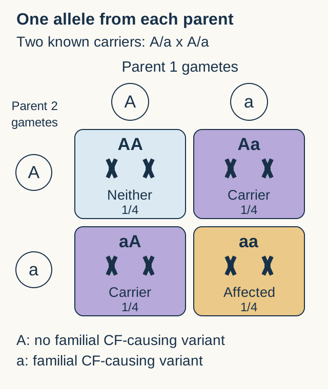
Can healthy parents have an affected child? Yes. CFTR is on chromosome 7, and cystic fibrosis usually follows autosomal recessive inheritance. A child inherits one allele from each parent. Two CF-causing alleles can contain the same variant or different variants. Healthy, unrelated parents can both be carriers; a family history is not required. [1][27]
Choose one allele from the top parent and one from the left parent. A represents an allele without the familial CF-causing variant; a represents an allele with it. This simplified cross assumes two known carriers.
Your prediction: Count the outcomes containing two affected alleles before reading the result. One of four is affected, two of four are carriers, and one of four inherited neither familial variant. The probabilities reset for each pregnancy and do not depend on the child's sex. A previous affected child does not consume the family's risk.
Change the question: Among children known not to have CF in this simplified cross, exclude aa. Two of the three remaining outcomes are carriers. That conditional probability is 2/3, not 1/2. Actual family counseling also considers the detected variants and what testing has excluded. [1]
F508del is a common three-nucleotide deletion that removes phenylalanine at position 508 without shifting the reading frame. Protein is made, but inefficient folding, maturation and delivery cause much of it to be degraded before reaching the surface. Other CFTR variants affect protein production, channel opening or other aspects of function. The categories overlap, so neither every CF allele nor every carrier parent has a deletion. [2][27]
For a different child with deep bacterial and fungal abscesses, ask whether the defect is epithelial transport or phagocyte killing. Chronic granulomatous disease involves the phagocyte oxidase system and can be X-linked or autosomal recessive. A girl's sex cannot exclude it. Functional testing and the complete organ pattern are more useful than a sex-based shortcut. [19]
The airway needs secretion; the sweat duct recovers salt
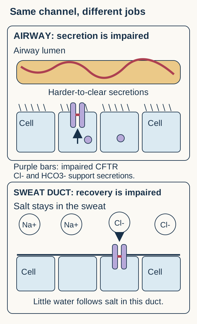
Does CFTR loss have the same effect on every lumen? CFTR is a cAMP-regulated anion channel, including chloride and bicarbonate transport. ATP participates in channel gating; CFTR is not an ATP-driven pump pushing chloride against its gradient. In secretory airway and pancreatic epithelia, anion transport supports the composition and hydration of luminal fluid. [2][3]
Trace the arrows relative to each lumen, not relative to the page. The airway is shown above the sweat duct so the two transport jobs remain distinct. This is a simplified epithelial model, not a measured transport assay.
Predict restoration: In a secreting airway preparation, restoring CFTR can increase anion delivery into the lumen. In a sweat duct, restoration improves salt recovery and lowers final sweat chloride. The opposite luminal chloride effects are not a contradiction; they reflect different epithelial jobs. [3][4]
Airway mucus becomes harder to clear when surface hydration and bicarbonate-dependent secretion properties are disturbed. Sodium absorption through ENaC also participates in surface-fluid regulation, but calling it a universal compensatory overactivity oversimplifies tissue and model differences. Primary newborn-pig experiments demonstrated impaired anion transport without increased sodium absorption. That animal result limits a universal explanation; it does not establish that sodium transport never matters in human airways. [3]
The sweat duct normally recovers chloride and sodium from primary sweat while remaining relatively impermeable to water. Impaired salt recovery therefore produces saltier final sweat, not simply a dehydrated mucus plug in a sweat duct. During heat exposure, excessive salt and water loss can cause extracellular-volume depletion. Secondary renal responses, potassium loss and chloride depletion can maintain hypokalemic metabolic alkalosis. This is not bicarbonate rising merely to balance an electrical charge. [4][20]
Apply it elsewhere: A dehydrated person with high bicarbonate and low urine chloride may be conserving salt appropriately. Ask where salt was lost before diagnosing a primary renal salt-wasting disorder. Symptomatic heat-related depletion needs prompt assessment and monitored fluid and electrolyte replacement.
Try it here · Checkpoint 1 of 3
Make your prediction before reading the choices. A first attempt is just a starting point.
Case 3
Show answer and explanations for case 3
A. Less airway anion secretion and higher final sweat chloride (Why this does not fit)
Both findings would fit further loss of the specified CFTR-dependent functions. The intervention restores channel function rather than inhibiting it. Follow the direction of transport in each tissue before predicting the effect of restoration.
Reasoning steps for option A
What functional change would produce this pair?
Both findings would fit further loss of the specified CFTR-dependent functions.
Is that the intervention being tested?
The intervention restores channel function rather than inhibiting it.
Why does this pair contradict restored CFTR?
Reduced airway secretion and increased sweat chloride both imply diminished CFTR-dependent transport, not restoration.
B. More airway anion secretion and higher final sweat chloride (Why this does not fit)
Restored airway secretion is expected, but a higher sweat chloride implies poorer ductal salt recovery. The treated sweat duct can reabsorb more chloride from the flowing sweat. The airway lumen and the sweat lumen have different normal transport roles.
Reasoning steps for option B
Which half of this prediction fits airway physiology?
Restored airway secretion is expected, but a higher sweat chloride implies poorer ductal salt recovery.
What should improved ductal absorption do to final sweat?
The treated sweat duct can reabsorb more chloride from the flowing sweat.
Why must final sweat chloride fall despite the correct airway prediction?
Restored ductal CFTR reabsorbs more chloride from fixed-flow primary sweat, lowering its final chloride concentration.
C. Less airway anion secretion and lower final sweat chloride (Why this does not fit)
Lower sweat chloride fits better ductal absorption. The stated airway preparation favors secretion, so restoring its anion conductance should not reduce that secretion. A correct prediction in one tissue does not validate the opposite prediction in another.
What is the specified direction of airway transport?
The stated airway preparation favors secretion, so restoring its anion conductance should not reduce that secretion.
Why must airway secretion rise despite the correct sweat prediction?
Restored apical CFTR increases airway anion secretion under the stated secretory conditions.
D. More airway anion secretion and lower final sweat chloride (Best answer)
CFTR supports secretion in the stated airway conditions and chloride absorption in the sweat duct. Restoration therefore increases airway anion delivery while recovering more chloride before sweat reaches the skin. Name the compartment and its usual transport direction before interpreting a chloride result.
Reasoning steps for option D
What are the two normal transport roles?
CFTR supports secretion in the stated airway conditions and chloride absorption in the sweat duct.
How does restoring both affect the two measurements?
Restoration therefore increases airway anion delivery while recovering more chloride before sweat reaches the skin.
How do the two tissues respond differently to restored CFTR?
Airway anion secretion increases, while improved sweat-duct chloride reabsorption lowers final sweat chloride.
Takeaway: The same channel can support different net transport directions in different epithelial environments.
Retained secretions can become permanent airway damage
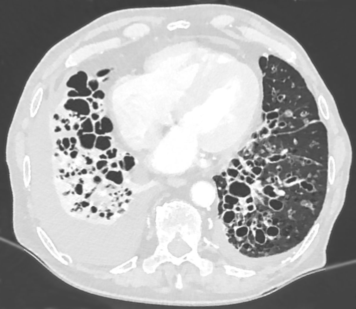
Are all dark spaces on a chest CT the same lesion? In CF, impaired secretion clearance encourages infection and inflammation. Repeated injury can damage bronchial walls and produce bronchiectasis: permanent airway dilation. Staphylococcus aureus is common in younger patients, while Pseudomonas aeruginosa can occur at any age and become persistent, including mucoid forms associated with chronic infection. This is not an obligatory organism timetable. [30][11]
Look for grouped tubular and ring-shaped airways with visible walls, rather than diffuse loss of alveolar tissue. This source image shows bronchiectasis with an accompanying effusion in an older adult; the source does not identify CF. It teaches the structural pattern, not a CF-specific diagnosis. Image: Hellerhoff, original source, CC BY-SA 3.0. image.
Inspect the airway wall: Bronchial enlargement relative to an accompanying artery, lack of normal tapering and visible peripheral bronchi support bronchiectasis. Emphysema primarily destroys alveolar walls. A single image neither establishes the underlying cause nor identifies the organism. In CF, the history connects the structural injury to impaired clearance. [18]
Airway obstruction lowers FEV1/FVC. For example, FEV1 1.8 L with FVC 3.0 L gives a ratio of 0.60; interpret it against the patient's appropriate lower limit. Early disease can have preserved spirometry, and a new decline requires assessment rather than an assumption of inevitable progression. Increased cough, sputum, breathlessness or oxygen need may indicate an exacerbation. [10]
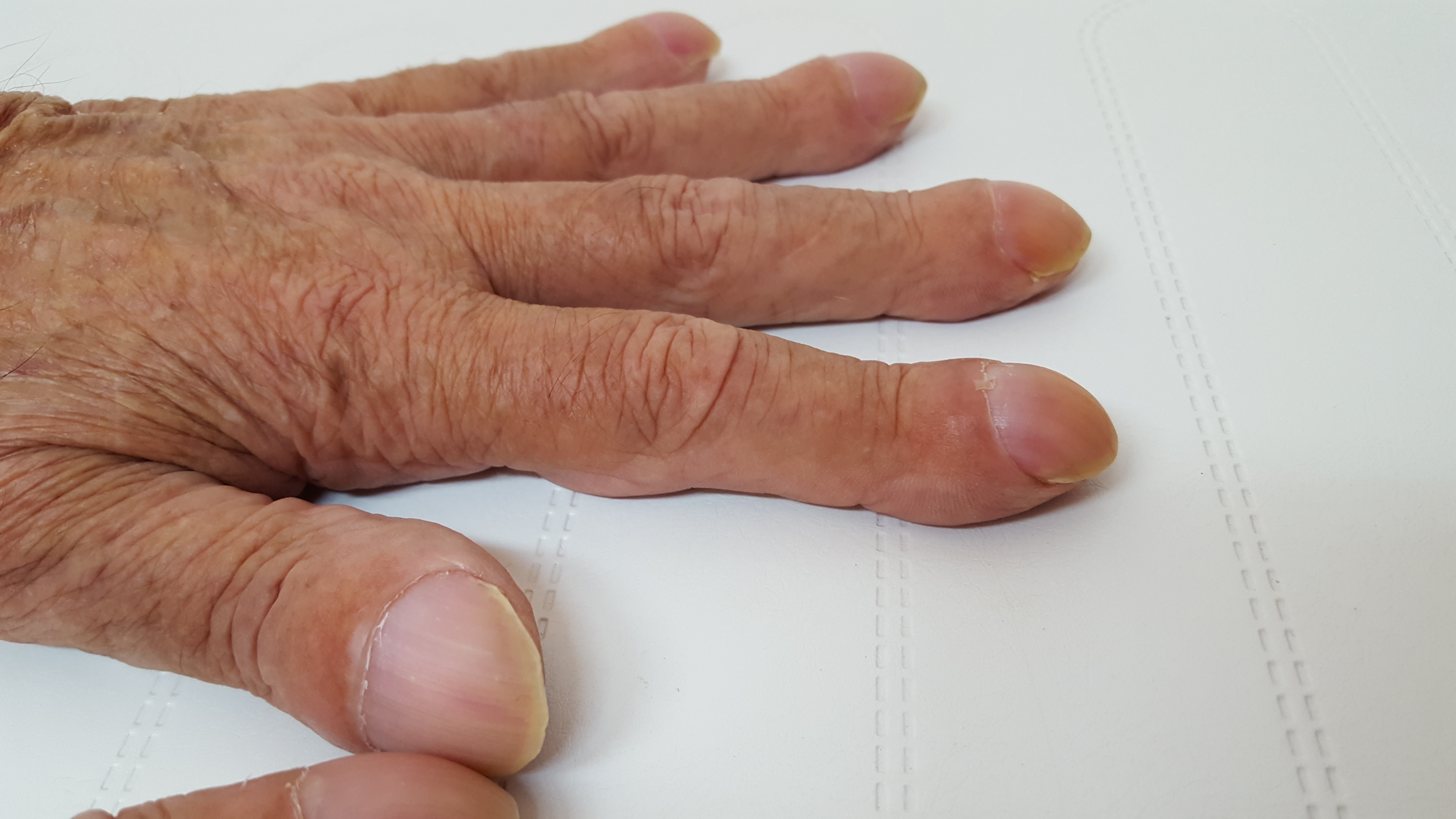
Identify the enlarged fingertips and altered nail contour. Clubbing is a physical finding, not a measurement of oxygen tension or proof of CF. The source does not specify this person's underlying disease. Image: Wesalius, original source, CC BY-SA 4.0. image.
Research on clubbed digits supports involvement of platelet-associated growth factors such as VEGF and PDGF in local vascular and tissue changes. This is a proposed biological explanation, not a rule that clubbing directly measures systemic hypoxemia. [31]
Now consider a different deterioration: Persistent wheeze and opacities after appropriate antibacterial treatment, accompanied by a marked IgE increase and Aspergillus sensitization, raise concern for allergic bronchopulmonary aspergillosis. Fungal presence alone is insufficient, and a lower-lobe distribution does not exclude it in CF. Use the clinical, immunologic and imaging findings together. [12]
A functioning organ still needs a usable outlet
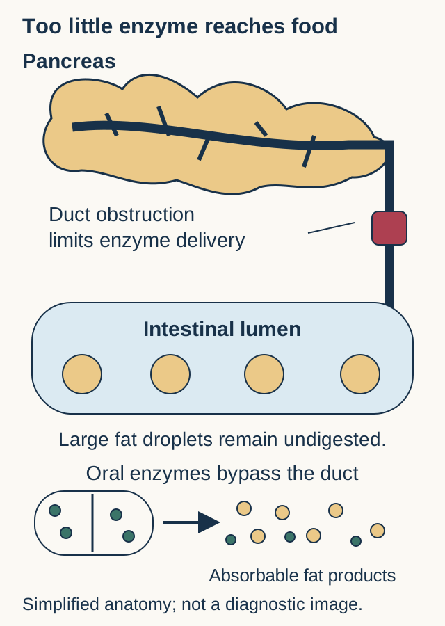
Why can a hungry child lose weight? Pancreatic ducts need fluid and bicarbonate to carry digestive secretions into the intestine. Obstruction and progressive pancreatic injury can reduce delivery of lipase, amylase and proteases. Food arrives, but digestion is incomplete. Greasy, bulky stools and poor growth suggest this exocrine problem; not everyone with CF has pancreatic insufficiency. [7]
Trace two routes into the intestine: the pancreatic duct and an oral dose. Enzyme replacement supplies the destination; it does not repair the blocked duct. Organ shapes and proportions are simplified.
Locate the failure: Increased fecal fat documents impaired fat handling, but not a unique cause. A markedly low fecal elastase in a formed sample supports pancreatic exocrine insufficiency. Predict the treatment consequence: appropriately delivered enzymes can improve digestion even when the original pancreatic abnormality remains. [7]
Predict which function suffers when fat-soluble vitamins are poorly absorbed
Vitamin
Function and possible consequence
VitaminA
Function and possible consequenceRetinal supports visual pigments, including rhodopsin; deficiency can impair dark adaptation and dry the ocular surface.
VitaminD
Function and possible consequenceCalcium handling and bone mineralization suffer; bone pain, low mineral density or defective mineralization can follow.
VitaminE
Function and possible consequenceReduced antioxidant protection can affect nerves and red-cell membranes, producing ataxia, neuropathy or hemolytic vulnerability.
VitaminK
Function and possible consequenceGamma-carboxylation of coagulation proteins is impaired; PT can become prolonged. Factors II, VII, IX and X depend on vitamin K.
Apply the table: Progressive difficulty seeing in dim light points toward a different vitamin-dependent function than bruising with a prolonged PT. Neither finding means that only one vitamin is deficient. Assess enzyme delivery, nutrition and measured deficiencies rather than giving unmonitored large supplement doses. [8][23][24][25][26]
In a newborn, inspissated meconium can obstruct the terminal ileum. Failure to pass meconium, distension and bilious vomiting need urgent assessment. Bubbly intraluminal material and a small unused colon on contrast evaluation support meconium ileus, but do not prove CF. Selected uncomplicated cases can receive specialist-directed contrast-enema treatment after stabilization; perforation or other complications change the surgical plan. CF testing must not delay care of an obstructed infant. [21][22]
CFTR abnormalities also affect the reproductive tract. Most men with CF have an absent or abnormal vas deferens, a mesonephric-duct derivative, and obstructive azoospermia. Sperm production can be preserved. Low-volume semen without sperm, nonpalpable vasa and preserved gonadal findings therefore favor a transport problem rather than primary testicular failure. Sperm retrieval and assisted reproduction may be possible, with partner testing and genetic counseling. The developmental process is more complex than saying every vas deferens never formed. [13][27]
Women can have reduced fertility from cervical secretion changes and overall health, but CF does not establish sterility. Improved health or modulator treatment can alter fertility; contraception and pregnancy planning remain important. [14]
Cholangiocyte dysfunction can contribute to biliary obstruction and fibrosis. CF-associated hepatobiliary disease includes cirrhotic and noncirrhotic portal hypertension. Liver enlargement, a growing spleen, a falling platelet count or collateral vessels deserve evaluation even when albumin and aminotransferases remain normal. Not every organ complication is explained by the same simple mucus plug. [15]
A screen, a functional test and a genotype answer different questions
Does a positive newborn screen establish CF? Immunoreactive trypsinogen is a pancreatic enzyme precursor measured in newborn blood spots. Increased blood levels provide a pancreatic screening signal, not evidence that digestive enzymes are reaching the intestine. Screening programs combine IRT with repeat IRT, variant testing or sequencing in different ways. A positive screen calls for diagnostic evaluation, and a negative screen or limited variant panel does not exclude CF when symptoms are convincing. [28][5]
Quantitative sweat chloride: current interpretation across ages
Chloride, mmol/L
Interpretation
Chloride, mmol/LLess than 30
InterpretationCF is unlikely, but a strongly compatible phenotype or genotype still requires interpretation.
Chloride, mmol/L30 through 59
InterpretationIntermediate. Repeat or additional genetic and functional evaluation may be needed.
Chloride, mmol/L60 or greater
InterpretationSupports CF in an appropriate clinical or screening context. Confirm an abnormal result by repeat testing or an independent diagnostic method.
Place these results: 29 is in the unlikely range, 30 and 59 are intermediate, and 60 reaches the diagnostic threshold. A laboratory report saying quantity not sufficient belongs in none of these ranges: no valid concentration was measured. It is not a negative test. [5][6]
Pilocarpine iontophoresis stimulates sweat for quantitative collection and chloride measurement. For screen-positive newborns, CFF guidance supports testing after 10 days, ideally within the neonatal period, when weight exceeds 2 kg and corrected gestational age is at least 36 weeks. Collection quality matters. Needed care for an infant with presumptive CF should not be postponed until every test is complete. [5][6]
Genotype interpretation requires both variant classification and phase. Two CF-causing variants on opposite alleles are different evidence from two changes on one allele. Some variants have uncertain or varying clinical consequences. Extended sequencing, deletion/duplication analysis, or specialized nasal potential difference or intestinal current testing can help resolve uncertain cases. These functional tests belong in experienced centers. [5]
Transfer the distinction: An asymptomatic screen-positive infant with inconclusive genetic and sweat findings may receive a CRMS/CFSPID designation and follow-up rather than an immediate definitive CF label. An isolated CFTR-related disorder, such as vas deferens abnormalities or pancreatitis without full CF diagnostic criteria, is another distinct category. A normal pancreatic test, a patient's ancestry or one negative panel does not settle these questions. [5]
Try it here · Checkpoint 2 of 3
Make your prediction before reading the choices. A first attempt is just a starting point.
Case 24
Show answer and explanations for case 24
A. CF is excluded because both sweat values are below 40 mmol/L (Why this does not fit)
Older teaching used a higher lower boundary for intermediate sweat results. Current guidance places both values in the intermediate range of 30-59, and the phenotype remains concerning. Apply current thresholds rather than an obsolete adult reference interval.
Reasoning steps for option A
Why might these results once have been called reassuring?
Older teaching used a higher lower boundary for intermediate sweat results.
How are they classified using current criteria?
Current guidance places both values in the intermediate range of 30-59, and the phenotype remains concerning.
How should sweat chloride values of 35 and 39 mmol/L be classified?
Both are intermediate by the current 30-59 mmol/L range, so neither excludes CFTR dysfunction.
B. CFTR dysfunction remains unresolved; pursue specialist genetic and functional evaluation (Best answer)
Intermediate sweat values with a compatible phenotype can require expanded genetic or reference-center functional testing. A limited negative panel and preserved pancreatic function do not exclude CF or another CFTR-related disorder. Match the completeness of the test to the strength of the clinical concern.
Reasoning steps for option B
What does an intermediate sweat result establish?
Intermediate sweat values with a compatible phenotype can require expanded genetic or reference-center functional testing.
Why do the panel and pancreatic findings not close the evaluation?
A limited negative panel and preserved pancreatic function do not exclude CF or another CFTR-related disorder.
What does the limited negative panel leave unanswered in this bronchiectatic patient?
It does not exclude untested CFTR variants or dysfunction; expanded genetics and specialist functional evaluation remain appropriate.
C. CF is established solely by bronchiectasis and two intermediate sweat values (Why this does not fit)
Bronchiectasis and repeated intermediate values raise concern for CFTR dysfunction. They do not independently establish a specific CF diagnosis without further interpretation of function, genotype and phenotype. Concern sufficient for further testing is not the same as diagnostic certainty.
Reasoning steps for option C
Why does this combination deserve further evaluation?
Bronchiectasis and repeated intermediate values raise concern for CFTR dysfunction.
Which certainty would be overstated by this label?
They do not independently establish a specific CF diagnosis without further interpretation of function, genotype and phenotype.
Why do two intermediate sweat tests not by themselves prove CF?
They indicate unresolved CFTR function in a compatible phenotype but do not independently establish the specific diagnosis.
D. A non-CF cause is proven because pancreatic function is normal (Why this does not fit)
Pancreatic insufficiency is common in CF and can aid recognition. Some individuals retain pancreatic function, so its preservation does not prove a different cause of bronchiectasis. Variable organ involvement prevents a single normal organ test from excluding the disorder.
Reasoning steps for option D
Why is pancreatic assessment useful?
Pancreatic insufficiency is common in CF and can aid recognition.
Does a normal result exclude every CF phenotype?
Some individuals retain pancreatic function, so its preservation does not prove a different cause of bronchiectasis.
Can preserved pancreatic function settle the cause of this patient's bronchiectasis?
No; pancreatic-sufficient CF phenotypes exist, and recurrent sinus disease with intermediate sweat chloride still warrants assessment.
Takeaway: Intermediate sweat values need contextual evaluation; limited genotyping is not a universal exclusion test.
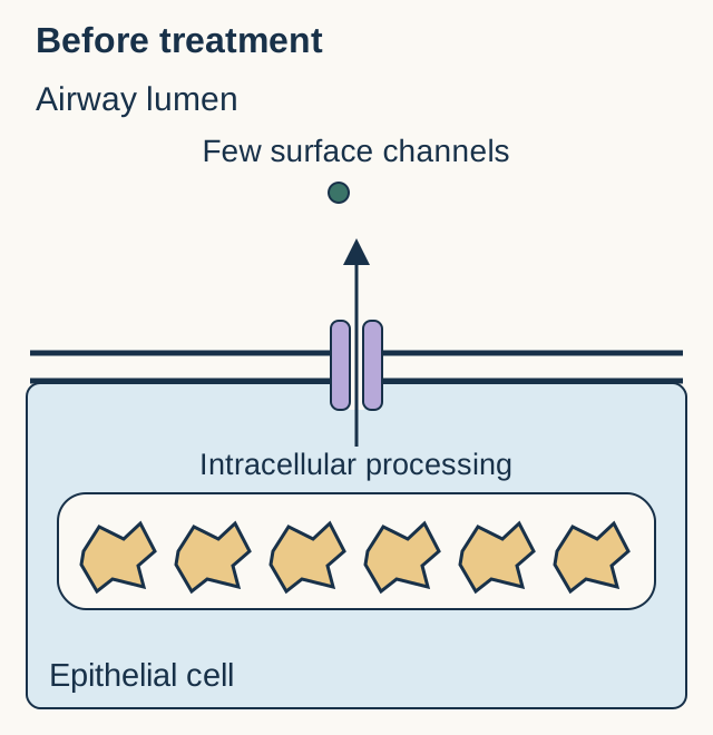
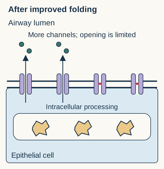
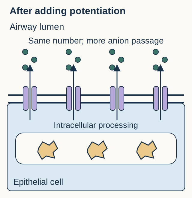
Does putting more CFTR at the surface solve every defect? A corrector improves processing or delivery of suitable protein; examples include lumacaftor, tezacaftor and elexacaftor. A potentiator such as ivacaftor improves activity of suitable channels already at the surface. F508del can affect more than delivery, which helps explain why complementary drugs are combined. [2]
Try the protein experiment. First inspect the untreated preparation. Predict separately what happens to surface-channel number and anion passage after a folding correction, then after adding a potentiator. Open either experiment to compare its altered membrane diagram with your prediction.
Starting preparation: intracellular retention limits surface protein. Icons and ion counts are qualitative, not patient-specific measurements.Test improved folding and surface deliveryMore surface channels do not guarantee that each opens efficiently. Compare both the membrane and the ion paths with the starting preparation.Add improved channel openingSurface number remains similar to the corrected preparation, but more channels conduct. The example separates abundance from function without promising a clinical response for every variant.
Result: Correcting processing can increase surface-channel number. Adding potentiation can increase passage without adding more channels. Protein production, delivery and opening therefore require separate questions. A preparation making no usable protein is not rescued simply by asking nonexistent surface channels to open.
As of the verified March 2026 US indication, elexacaftor/tezacaftor/ivacaftor is indicated for clinical CF from age 2 with at least one qualifying responsive variant or a variant resulting in CFTR protein production. It is not restricted to F508del. Eligibility is not a guarantee of response, and country-specific labeling can differ. Modulators can substantially improve clinical outcomes but do not erase the diagnosis or automatically replace every other treatment. [17]
Apply the safety check: Review drug interactions and liver status before treatment. Current US instructions require liver tests before treatment, monthly for six months, every three months for the next year, then at least annually. Significant liver abnormalities or jaundice require interruption and prompt evaluation. Severe hepatic impairment precludes use; moderate impairment needs particular caution. Strong CYP3A inducers can reduce effectiveness, while inhibitors can require dose adjustment. Pediatric eye examinations and assessment of new visual, severe headache, mood or behavioral symptoms are also important. [17]
Airway clearance addresses retained secretions. Dornase alfa digests extracellular DNA contributing to sputum viscosity; hypertonic saline promotes surface hydration. Neither is an antibiotic. During a pulmonary exacerbation, increase airway-care support as appropriate and select active antibiotics using cultures, prior responses, severity and toxicity. An antipseudomonal beta-lactam such as ceftazidime plus tobramycin is a common combination, not an evidence-proven requirement for every patient. Renal and auditory risks matter. Vancomycin targets relevant gram-positive infection, and maintenance azithromycin is not sufficient acute treatment of a serious Pseudomonas exacerbation. [9][10][29]
For a newly detected Pseudomonas isolate, eradication is different from long-term suppression of persistent infection; a commonly recommended initial regimen is inhaled tobramycin for 28 days. Routine antipseudomonal prophylaxis before acquisition is not the same strategy. [11]
Pancreatic enzymes accompany meals and snacks. Preserve enteric-coated beads and follow product instructions rather than crushing them or mixing them into hot food. Persistent steatorrhea calls for review of timing, administration, dose and other causes. Excessive dosing can cause harm, including fibrosing colonopathy. Individualize calories and fat-soluble vitamin replacement to nutrition and monitoring; severe fat restriction is not the default solution. [7][8]
Finally, pancreatic endocrine dysfunction can cause CF-related diabetes, often from inadequate insulin secretion. Annual oral glucose tolerance testing begins by age 10 under CFF guidance; a normal HbA1c does not exclude it. Confirmed disease generally needs insulin-based management while preserving nutrition. Weight loss despite effective digestive treatment should prompt reassessment rather than another automatic enzyme increase. [16]
Try it here · Checkpoint 3 of 3
Make your prediction before reading the choices. A first attempt is just a starting point.
Case 26
Show answer and explanations for case 26
A. A improves protein processing; B increases channel opening probability (Best answer)
A corrector can increase surface protein, while a potentiator increases activity of channels already there. The abundance increase localizes A's main effect; the further current increase without more channels, gradient or conductance localizes B to opening probability. Separate channel number from the fraction of time those channels conduct.
Reasoning steps for option A
How can processing and gating affect current differently?
A corrector can increase surface protein, while a potentiator increases activity of channels already there.
Which measurements isolate each treatment effect?
The abundance increase localizes A's main effect; the further current increase without more channels, gradient or conductance localizes B to opening probability.
Which measurement assigns A to processing and B to gating?
A raises surface abundance from 10 to 60; B raises current from 7 to 42 without adding channels or changing conductance or gradient.
B. A increases opening of existing channels; B increases delivery of new channels (Why this does not fit)
A gating change can increase current without changing surface abundance. A markedly increases surface abundance, whereas B leaves abundance unchanged, opposite to this proposed pairing. Use the surface-protein measurement to distinguish delivery from activity.
Reasoning steps for option B
What would a pure gating change do to abundance?
A gating change can increase current without changing surface abundance.
Which treatment actually changed abundance?
A markedly increases surface abundance, whereas B leaves abundance unchanged, opposite to this proposed pairing.
Why is the proposed A-gating/B-delivery order reversed?
Only A changes channel abundance, while B changes current at fixed abundance, which favors an opening-probability effect.
C. A increases transcription; B increases the chloride driving gradient (Why this does not fit)
More messenger RNA or a larger driving gradient can alter ion transport. Both messenger RNA and the driving gradient are explicitly unchanged. Do not invoke a variable held constant in the experimental comparison.
Reasoning steps for option C
How could these proposed changes affect transport?
More messenger RNA or a larger driving gradient can alter ion transport.
Were either of those changes observed?
Both messenger RNA and the driving gradient are explicitly unchanged.
Can transcription or driving-gradient changes explain these measured effects?
No; messenger RNA and the ion driving gradient remain unchanged under the recording conditions.
D. A reduces surface delivery; B increases single-channel conductance (Why this does not fit)
Changing delivery and conductance can change aggregate current. Surface delivery increased with A, and conductance was held constant with B. Check the direction of each measured effect before assigning a protein-level explanation.
Reasoning steps for option D
How could delivery and conductance affect current?
Changing delivery and conductance can change aggregate current.
What observations contradict this proposed pair?
Surface delivery increased with A, and conductance was held constant with B.
Why can neither reduced delivery by A nor conductance gain by B explain the data?
A increases surface protein sixfold, and single-channel conductance is explicitly unchanged.
Takeaway: More channels at the surface and more effective opening are complementary, not interchangeable, treatment effects.
Use the supplied findings to localize the problem, decide what the test establishes, and choose the next justified action. Read the explanation after committing to an answer.
Case 1
Show answer and explanations for case 1
A. 1/8 (Why this does not fit)
A one-half carrier probability would give a one-eighth reproductive risk with a carrier partner. Among the three unaffected outcomes of the parental carrier cross, two are carriers rather than one-half. Condition the carrier estimate on what is already known about the relative.
Reasoning steps for option A
Which carrier probability would produce this risk?
A one-half carrier probability would give a one-eighth reproductive risk with a carrier partner.
How does being unaffected change this woman's probability?
Among the three unaffected outcomes of the parental carrier cross, two are carriers rather than one-half.
Why is 1/8 too low for this unaffected sister?
Her carrier chance is 2/3 rather than 1/2; multiplying by the 1/4 affected-child risk with a carrier partner gives 1/6.
B. 1/4 (Why this does not fit)
Two confirmed carriers have a one-fourth affected-child probability per pregnancy. Only the partner is confirmed to be a carrier; her conditional carrier probability remains two-thirds. Multiply the probability of the parental genotype by the risk conditional on that genotype.
Reasoning steps for option B
When does the one-fourth risk apply?
Two confirmed carriers have a one-fourth affected-child probability per pregnancy.
Which parent's genotype remains uncertain?
Only the partner is confirmed to be a carrier; her conditional carrier probability remains two-thirds.
Why does 1/4 overstate the risk before her test?
One-fourth assumes both parents are confirmed carriers, whereas her carrier probability remains 2/3.
C. 1/6 (Best answer)
An unaffected sibling of an affected child of two carriers has a two-thirds carrier probability. Multiplying two-thirds by the one-fourth risk for two carriers gives one-sixth. A conditional carrier probability is not itself the affected-child probability.
Reasoning steps for option C
What fraction of unaffected siblings are carriers in this cross?
An unaffected sibling of an affected child of two carriers has a two-thirds carrier probability.
How does the partner's result affect the calculation?
Multiplying two-thirds by the one-fourth risk for two carriers gives one-sixth.
How do the sister’s status and her partner’s result yield 1/6?
Among unaffected siblings of an affected child, 2/3 carry a variant; two carriers have a 1/4 affected-child risk, giving 1/6.
D. 1/3 (Why this does not fit)
One-third is the chance that an unaffected child of two carriers inherited neither familial variant. The question asks about an affected child in the following generation, not her chance of being a noncarrier. Keep the relative's genotype probability separate from the child's disease probability.
Reasoning steps for option D
What outcome in the original family has probability one-third?
One-third is the chance that an unaffected child of two carriers inherited neither familial variant.
Is that the outcome being requested?
The question asks about an affected child in the following generation, not her chance of being a noncarrier.
Why is 1/3 not the child’s risk?
One-third is her probability of carrying neither familial variant among unaffected siblings, not her child’s disease probability.
Takeaway: First condition on the family information, then calculate the risk in the new parental cross.
A. Protein folding and cell-surface delivery (Best answer)
A processing defect can permit translation but prevent efficient maturation and surface delivery. The protein is synthesized and retained in the endoplasmic reticulum; more mature surface protein appears without more messenger RNA. Measure where the protein fails before choosing the treatment target.
Reasoning steps for option A
Can a processing defect coexist with protein synthesis?
A processing defect can permit translation but prevent efficient maturation and surface delivery.
What experimental finding localizes this rescue?
The protein is synthesized and retained in the endoplasmic reticulum; more mature surface protein appears without more messenger RNA.
What does the apical increase without more CFTR RNA identify?
Rescued folding and delivery of already synthesized deletion protein, previously retained and degraded in the ER.
B. Transcription of CFTR messenger RNA from its gene (Why this does not fit)
Greater transcription can increase the amount of messenger RNA available for translation. Messenger RNA abundance is unchanged, while maturation and surface abundance increase. An increase in surface protein does not necessarily reflect increased transcription.
Reasoning steps for option B
What would a transcriptional effect change first?
Greater transcription can increase the amount of messenger RNA available for translation.
Was that change observed?
Messenger RNA abundance is unchanged, while maturation and surface abundance increase.
Why is transcription not the rescued step?
Transcript abundance is unchanged while mature apical protein increases, indicating post-translational rescue.
C. Initiation of translation at the start codon (Why this does not fit)
Failed initiation prevents production of the encoded protein. Full-length protein is already synthesized before treatment. Existing protein production directs attention beyond translation initiation.
Reasoning steps for option C
What would failed initiation prevent?
Failed initiation prevents production of the encoded protein.
Which baseline result argues against it?
Full-length protein is already synthesized before treatment.
Why is translation initiation not the bottleneck?
Full-length protein lacking one amino acid is already synthesized; its maturation and trafficking are defective.
D. Opening of channels already at the cell surface (Why this does not fit)
A potentiator can increase the opening probability of surface channels. The measured treatment effect is an increase in mature surface protein from an intracellular pool. Surface abundance and channel opening are distinct experimental outcomes.
Reasoning steps for option D
What does a gating intervention change?
A potentiator can increase the opening probability of surface channels.
Which outcome was measured instead?
The measured treatment effect is an increase in mature surface protein from an intracellular pool.
Why does this result not establish improved channel opening?
The observation measures mature surface protein, not opening probability of channels already at that surface.
Takeaway: F508del principally disrupts processing, but the presence, location and function of protein must be considered separately.
A. Persistent renal salt wasting from a loop-segment transport defect (Why this does not fit)
A loop-segment salt-wasting disorder can cause hypokalemic metabolic alkalosis. The low urine chloride during depletion indicates renal conservation rather than persistent renal chloride loss. Interpret urine electrolytes in relation to volume status and treatment exposure.
Reasoning steps for option A
Why could a renal transport defect be considered?
A loop-segment salt-wasting disorder can cause hypokalemic metabolic alkalosis.
How does the measured renal response differ?
The low urine chloride during depletion indicates renal conservation rather than persistent renal chloride loss.
Why does urine chloride of 7 argue against persistent renal wasting?
The volume-depleted kidneys conserve chloride, fitting extrarenal salt loss in sweat rather than ongoing renal chloride loss.
B. Cutaneous salt loss with volume depletion and secondary renal potassium loss (Best answer)
Loss of salt-containing sweat can contract extracellular volume and maintain chloride-responsive alkalosis. Heat exposure, dehydration and low urine chloride support an extrarenal loss with appropriate renal salt conservation. A low urine chloride localizes conservation, not the exact extrarenal source; use the history as well.
Reasoning steps for option B
How can sweating contribute to alkalosis?
Loss of salt-containing sweat can contract extracellular volume and maintain chloride-responsive alkalosis.
Which findings localize the loss in this patient?
Heat exposure, dehydration and low urine chloride support an extrarenal loss with appropriate renal salt conservation.
How does hot-weather sweat loss generate these blood and urine findings?
Sweat salt loss contracts volume; renal chloride conservation and secondary aldosterone-associated potassium loss accompany hypokalemic metabolic alkalosis.
C. Primary mineralocorticoid excess with renal hydrogen loss (Why this does not fit)
Mineralocorticoid excess can produce hypokalemia and metabolic alkalosis. Primary mineralocorticoid excess usually expands effective volume and raises blood pressure, unlike this depleted, orthostatic patient. Distinguish primary hormone excess from the secondary hormonal response to depletion.
Reasoning steps for option C
What laboratory findings could suggest mineralocorticoid excess?
Mineralocorticoid excess can produce hypokalemia and metabolic alkalosis.
What does the circulatory assessment add?
Primary mineralocorticoid excess usually expands effective volume and raises blood pressure, unlike this depleted, orthostatic patient.
Why is primary aldosterone excess less fitting than sweat loss?
Orthostatic volume depletion after heat exposure and low urine chloride conflict with the usual volume-expanded hypertensive state.
D. Chronic hypoventilation with renal compensation (Why this does not fit)
Chronic carbon dioxide retention can increase bicarbonate through renal compensation. The predominant alkalemia and markedly increased bicarbonate after salt loss favor primary metabolic alkalosis, with a compensatory carbon dioxide increase. Identify the primary acid-base process before explaining the compensatory measurement.
Reasoning steps for option D
Why might increased carbon dioxide suggest a respiratory disorder?
Chronic carbon dioxide retention can increase bicarbonate through renal compensation.
Does it explain the direction of the pH change?
The predominant alkalemia and markedly increased bicarbonate after salt loss favor primary metabolic alkalosis, with a compensatory carbon dioxide increase.
Why is PaCO2 48 not the primary disorder?
High bicarbonate with alkalemic pH identifies metabolic alkalosis; elevated PaCO2 is compatible with compensation.
Takeaway: Sweat salt loss can produce a renal response resembling a salt-wasting disorder without a primary renal transport defect.
A. Failure of neutrophil NADPH oxidase activity (Best answer)
Phagocyte oxidase deficiency impairs intracellular microbial killing and can have autosomal recessive inheritance. Deep bacterial and fungal infections with an absent stimulated oxidative burst directly support this defect despite the patient being female. Sex can inform an inheritance pattern but cannot overrule a demonstrated cellular defect.
Reasoning steps for option A
Can this phagocyte disorder affect girls?
Phagocyte oxidase deficiency impairs intracellular microbial killing and can have autosomal recessive inheritance.
Which test localizes the impaired function?
Deep bacterial and fungal infections with an absent stimulated oxidative burst directly support this defect despite the patient being female.
Which defect links Aspergillus abscess to the absent neutrophil burst?
NADPH oxidase failure prevents the normal oxidative burst and impairs microbial killing; autosomal recessive forms can affect girls.
B. Impaired epithelial chloride and bicarbonate transport (Why this does not fit)
Defective CFTR transport can cause recurrent respiratory infection and gastrointestinal disease. The demonstrated abnormality is a neutrophil oxidative response, not epithelial ion transport; sweat and digestive findings do not support that alternative here. Distinguish the infected organ from the cellular function that failed.
Reasoning steps for option B
Why might CFTR dysfunction enter an infection differential?
Defective CFTR transport can cause recurrent respiratory infection and gastrointestinal disease.
What cellular compartment is directly abnormal?
The demonstrated abnormality is a neutrophil oxidative response, not epithelial ion transport; sweat and digestive findings do not support that alternative here.
Why does CFTR dysfunction not explain the decisive assay?
Normal-range sweat chloride and absent stimulated neutrophil oxidative burst point to a phagocyte killing defect rather than an epithelial transport defect.
C. Failure of immunoglobulin class switching (Why this does not fit)
An antibody class-switch defect can cause recurrent infections and susceptibility to selected opportunistic organisms. The supplied functional defect is failure of a stimulated phagocyte oxidative burst rather than an immunoglobulin pattern. Choose the defect measured by the discriminating test rather than by infection history alone.
Reasoning steps for option C
What infection tendency makes an antibody disorder plausible?
An antibody class-switch defect can cause recurrent infections and susceptibility to selected opportunistic organisms.
Which function did this laboratory test actually assess?
The supplied functional defect is failure of a stimulated phagocyte oxidative burst rather than an immunoglobulin pattern.
Why does class-switch failure not explain this assay?
Antibody production may be impaired in class-switch disease, but stimulated neutrophils should still generate an oxidative burst.
D. Deficiency of terminal complement components (Why this does not fit)
Terminal complement deficiency predisposes to particular invasive bacterial infections. It does not explain an absent neutrophil oxidative response to laboratory stimulation. A functional assay can distinguish different immune mechanisms producing recurrent infection.
Reasoning steps for option D
What does terminal complement contribute to host defense?
Terminal complement deficiency predisposes to particular invasive bacterial infections.
Would its absence explain this stimulated-cell result?
It does not explain an absent neutrophil oxidative response to laboratory stimulation.
Why does terminal complement deficiency not explain this pattern?
It predisposes particularly to Neisseria infection, not an intrinsic absent neutrophil oxidative burst with deep fungal abscess.
Takeaway: A demonstrated phagocyte defect is more informative than an assumed sex-linked pattern.
A. Destruction of alveolar septa with loss of elastic recoil (Why this does not fit)
Alveolar destruction can cause airflow obstruction and hyperinflation. The dominant CT abnormality is thick-walled, abnormally wide bronchi rather than diffuse destruction of the intervening alveolar tissue. Localize a structural abnormality before using the obstructive spirometry label to explain it.
Reasoning steps for option A
How can alveolar destruction affect spirometry?
Alveolar destruction can cause airflow obstruction and hyperinflation.
Which structures are abnormal on this CT?
The dominant CT abnormality is thick-walled, abnormally wide bronchi rather than diffuse destruction of the intervening alveolar tissue.
Why is alveolar destruction not the main CT lesion?
The CT shows thick-walled dilated bronchi without diffuse low-attenuation destruction of intervening alveoli.
B. Transient contraction of bronchial smooth muscle (Why this does not fit)
Smooth-muscle contraction can narrow airways and reduce expiratory flow. Transient narrowing does not account for persistent enlargement and lack of tapering of the bronchial lumen. Reversible narrowing and established structural dilation are different airway problems.
Reasoning steps for option B
What physiological effect can smooth-muscle contraction produce?
Smooth-muscle contraction can narrow airways and reduce expiratory flow.
Does it explain the observed bronchial shape?
Transient narrowing does not account for persistent enlargement and lack of tapering of the bronchial lumen.
Smooth-muscle contraction narrows airways; it does not permanently widen thick-walled bronchi or abolish peripheral tapering.
C. Interstitial collagen deposition with traction on otherwise normal airways (Why this does not fit)
Fibrotic interstitial disease can pull bronchi open. The supplied pattern emphasizes thick-walled bronchi in a long suppurative airway course without a described fibrotic interstitial pattern. Look for the surrounding tissue process before attributing bronchial dilation to traction.
Reasoning steps for option C
When can interstitial disease enlarge bronchi?
Fibrotic interstitial disease can pull bronchi open.
What associated evidence would support that explanation?
The supplied pattern emphasizes thick-walled bronchi in a long suppurative airway course without a described fibrotic interstitial pattern.
What evidence is missing for fibrotic traction bronchiectasis?
Interstitial collagen distortion is not described; recurrent infection and thickened bronchial walls favor inflammatory wall injury.
D. Repeated infection and inflammation with permanent bronchial wall injury (Best answer)
Retained secretions can promote infection and inflammatory injury that permanently dilates bronchi. Years of productive exacerbations and airway enlargement with absent tapering fit bronchiectasis in this clinical setting. Bronchiectasis describes structural airway damage; the underlying cause still requires the clinical context.
Reasoning steps for option D
How can impaired clearance cause lasting airway change?
Retained secretions can promote infection and inflammatory injury that permanently dilates bronchi.
Which CT and historical findings fit that process?
Years of productive exacerbations and airway enlargement with absent tapering fit bronchiectasis in this clinical setting.
What links repeated CF infections to these enlarged bronchi?
Chronic infection and inflammation damage airway walls, causing permanent dilation, wall thickening and loss of tapering.
Takeaway: An obstructive pattern describes airflow; CT helps localize the structural disease producing it.
A. A predominantly restrictive change proven by the reduction in FVC (Why this does not fit)
Restriction is established by reduced total lung capacity rather than FVC alone. FVC is nearly unchanged while FEV1 falls substantially, producing a low expiratory ratio. A small FVC change cannot establish restrictive disease.
Reasoning steps for option A
Which volume measurement establishes restriction?
Restriction is established by reduced total lung capacity rather than FVC alone.
How do the supplied expiratory volumes change relative to each other?
FVC is nearly unchanged while FEV1 falls substantially, producing a low expiratory ratio.
Why does near-unchanged FVC not prove restriction?
Restriction requires reduced total lung capacity; FEV1 falls disproportionately, lowering FEV1/FVC from 0.80 to about 0.59.
B. A technical artifact established by comparing visits six months apart (Why this does not fit)
Measurement variability can complicate longitudinal spirometry. The efforts are reproducible and the volume changes accompany a new symptomatic deterioration. Consider quality checks, but do not dismiss a coherent clinical and physiological change as artifact without evidence.
Reasoning steps for option B
Why does test quality matter?
Measurement variability can complicate longitudinal spirometry.
What quality and clinical findings are supplied?
The efforts are reproducible and the volume changes accompany a new symptomatic deterioration.
Why not dismiss the change as a technical artifact?
Reproducible efforts and increased productive cough accompany the fall in FEV1 and ratio below the 0.75 limit.
C. Worsening expiratory airflow obstruction with a ratio near 0.59 (Best answer)
The ratio compares the first-second expired volume with the forced vital capacity. It falls from 0.80 to approximately 0.59, below the supplied lower limit, while FVC changes little. Calculate the ratio and interpret it alongside symptoms and measurement quality.
Reasoning steps for option C
What does FEV1/FVC compare?
The ratio compares the first-second expired volume with the forced vital capacity.
What do the two calculations show?
It falls from 0.80 to approximately 0.59, below the supplied lower limit, while FVC changes little.
What ratios demonstrate worsening expiratory obstruction?
FEV1/FVC falls from 2.4/3.0=0.80 to 1.7/2.9≈0.59, below the laboratory lower limit.
D. Preserved expiratory airflow because FVC remains close to baseline (Why this does not fit)
A preserved FVC indicates that a similar total forced volume can still be exhaled. It does not show that the volume leaves at the same rate; the first-second fraction is substantially lower. Total expired volume and speed of expiration answer different questions.
Reasoning steps for option D
What does a nearly preserved FVC establish?
A preserved FVC indicates that a similar total forced volume can still be exhaled.
What information does the first-second measurement add?
It does not show that the volume leaves at the same rate; the first-second fraction is substantially lower.
Why does stable FVC not mean preserved airflow?
Only 1.7 of 2.9 L is expired in the first second, so the ratio is approximately 0.59 despite near-baseline FVC.
Takeaway: A nearly stable vital capacity can coexist with a substantial decline in expiratory flow.
A. Progressive pulmonary disease from fungal invasion of tissue (Why this does not fit)
Invasive fungal disease can produce respiratory deterioration. The combination of marked IgE increase, sensitization, eosinophilia and persistent mucus-related abnormalities favors an allergic response rather than demonstrating tissue invasion. A positive fungal immune test does not establish invasive infection.
Reasoning steps for option A
What would invasive disease imply about the fungus?
Invasive fungal disease can produce respiratory deterioration.
Which type of host response is actually demonstrated?
The combination of marked IgE increase, sensitization, eosinophilia and persistent mucus-related abnormalities favors an allergic response rather than demonstrating tissue invasion.
Why is fungal invasion not demonstrated?
Marked IgE elevation, Aspergillus sensitization, eosinophilia and mucus plugs indicate allergy rather than evidence of tissue invasion.
B. Allergic bronchopulmonary aspergillosis (Best answer)
Aspergillus hypersensitivity can add airway inflammation and mucus impaction to underlying CF lung disease. Clinical deterioration, marked IgE increase, sensitization and persistent opacities after antibacterial treatment form a coherent allergic pattern. Assess the clinical and immunologic pattern rather than interpreting a fungal finding in isolation.
Reasoning steps for option B
How can Aspergillus worsen airways without tissue invasion?
Aspergillus hypersensitivity can add airway inflammation and mucus impaction to underlying CF lung disease.
Which combined findings support that explanation?
Clinical deterioration, marked IgE increase, sensitization and persistent opacities after antibacterial treatment form a coherent allergic pattern.
What favors ABPA despite susceptibility-directed bacterial treatment?
Persistent wheeze and opacities with IgE rising from 110 to 1650, specific IgE/IgG and eosinophilia indicate allergic airway inflammation.
C. Incidental Aspergillus colonization without an additional disease process (Why this does not fit)
Aspergillus can be present in airways without causing allergic disease. The new immune changes and persistent symptomatic and radiographic deterioration make incidental presence alone insufficient here. Colonization does not explain every deterioration when evidence of a host response is present.
Reasoning steps for option C
Can Aspergillus be present without disease?
Aspergillus can be present in airways without causing allergic disease.
Why is that explanation incomplete in this patient?
The new immune changes and persistent symptomatic and radiographic deterioration make incidental presence alone insufficient here.
Why is incidental colonization insufficient?
It cannot explain the sharp IgE rise, eosinophilia, sensitization and persistent mucus-plugging decline.
D. An isolated bacterial exacerbation with no additional inflammatory process (Why this does not fit)
Bacterial exacerbations commonly worsen symptoms and lung function in CF. Persistent opacities after directed treatment together with the new sensitization-associated immune pattern support an additional process. Response to treatment and newly measured biology can require broadening the assessment.
Reasoning steps for option D
Why is a bacterial exacerbation initially plausible?
Bacterial exacerbations commonly worsen symptoms and lung function in CF.
What remains unexplained after that treatment?
Persistent opacities after directed treatment together with the new sensitization-associated immune pattern support an additional process.
What remains unexplained by the treated bacterial isolate?
The persistent plugs and opacities plus marked IgE elevation, Aspergillus-specific responses and eosinophilia suggest additional allergy.
Takeaway: CF and an allergic response to Aspergillus can coexist; lobar location alone does not separate them.
A. Begin preventive antipseudomonal treatment only after the next culture becomes negative (Why this does not fit)
Prophylaxis would treat a period without documented infection. The organism has already been newly detected, making the current question eradication rather than prevention before acquisition. Distinguish new acquisition from the absence of infection.
Reasoning steps for option A
What clinical state does prophylaxis target?
Prophylaxis would treat a period without documented infection.
What state is established by this sample?
The organism has already been newly detected, making the current question eradication rather than prevention before acquisition.
Why not wait for a negative culture before acting?
Pseudomonas is already newly detected, making eradication rather than pre-acquisition prevention the immediate goal.
B. Observe until persistent symptoms establish a pulmonary exacerbation (Why this does not fit)
Symptoms help assess the need for acute exacerbation treatment. Initial Pseudomonas acquisition is itself a reason to consider eradication; waiting for symptoms can miss that opportunity. A surveillance culture can change management even when lung function is stable.
Reasoning steps for option B
Why are symptoms important in acute illness?
Symptoms help assess the need for acute exacerbation treatment.
Must eradication wait for an exacerbation?
Initial Pseudomonas acquisition is itself a reason to consider eradication; waiting for symptoms can miss that opportunity.
Why not wait for symptoms to become persistent?
First Pseudomonas acquisition merits an eradication attempt before chronic infection, even without an acute exacerbation.
C. Start an inhaled eradication course and arrange follow-up cultures (Best answer)
Guidance favors inhaled treatment for initial or new Pseudomonas growth, commonly tobramycin for 28 days. This is a first isolation in a stable patient, not established persistent infection or an unstable exacerbation. Match the antimicrobial strategy to the stage of airway infection.
Reasoning steps for option C
Which treatment goal applies to initial acquisition?
Guidance favors inhaled treatment for initial or new Pseudomonas growth, commonly tobramycin for 28 days.
What distinguishes this patient from someone needing acute systemic treatment?
This is a first isolation in a stable patient, not established persistent infection or an unstable exacerbation.
Why pair inhaled treatment with repeat cultures?
Treat the new Pseudomonas isolate early and use follow-up cultures to assess clearance or persistence.
D. Commit immediately to indefinite cyclic inhaled suppression (Why this does not fit)
Long-term inhaled suppression may be used for persistent Pseudomonas infection. A first isolation does not yet establish persistent infection after an eradication attempt. Eradication and long-term suppression are different treatment goals.
Reasoning steps for option D
When is chronic suppressive therapy considered?
Long-term inhaled suppression may be used for persistent Pseudomonas infection.
What has not yet been attempted or established?
A first isolation does not yet establish persistent infection after an eradication attempt.
Why is indefinite suppression premature?
This is a first positive culture, not established chronic infection; attempt eradication and assess subsequent cultures first.
Takeaway: Initial acquisition can warrant eradication before a symptomatic exacerbation develops.
A. Select active IV antipseudomonal therapy with the CF team, avoiding reflex tobramycin reuse (Best answer)
Exacerbation treatment considers prior cultures, current susceptibility, severity and drug toxicity. An active beta-lactam is available, while prior ototoxicity and current renal impairment make automatic aminoglycoside reuse inappropriate. Common combination practice is not proof that every patient requires the same two drugs.
Reasoning steps for option A
What information determines antibiotic selection beyond the organism name?
Exacerbation treatment considers prior cultures, current susceptibility, severity and drug toxicity.
Which supplied constraints change the usual combination choice?
An active beta-lactam is available, while prior ototoxicity and current renal impairment make automatic aminoglycoside reuse inappropriate.
How should prior hearing loss alter acute Pseudomonas treatment?
Choose active IV antipseudomonal therapy with the CF team without automatically repeating ototoxic tobramycin amid elevated creatinine.
B. Repeat IV ceftazidime plus tobramycin unchanged, treating two antipseudomonal classes as obligatory (Why this does not fit)
Two antipseudomonal classes are often used during significant exacerbations. Guidance does not establish a universally necessary combination, and unchanged reuse ignores documented auditory and renal risks. Weigh toxicity and available active alternatives rather than treating a common regimen as compulsory.
Reasoning steps for option B
Why might this familiar combination be considered?
Two antipseudomonal classes are often used during significant exacerbations.
Why is unchanged repetition unsafe here?
Guidance does not establish a universally necessary combination, and unchanged reuse ignores documented auditory and renal risks.
Why is repeating tobramycin with ceftazidime not obligatory?
Susceptibility does not erase prior aminoglycoside-associated hearing loss or current renal risk; select an active safer regimen.
C. Replace antipseudomonal therapy with IV vancomycin directed against gram-positive organisms (Why this does not fit)
Vancomycin can be appropriate when clinically relevant resistant gram-positive organisms are present. It does not provide treatment of the documented Pseudomonas, and no MRSA is recovered in this case. Antibiotic substitution must preserve activity against the demonstrated pathogen.
Reasoning steps for option C
Which bacterial group does vancomycin principally target?
Vancomycin can be appropriate when clinically relevant resistant gram-positive organisms are present.
Does it cover the organism requiring treatment?
It does not provide treatment of the documented Pseudomonas, and no MRSA is recovered in this case.
Repeated cultures grow Pseudomonas and no MRSA; vancomycin does not supply needed antipseudomonal activity.
D. Use maintenance azithromycin as the sole therapy and repeat spirometry after clinical recovery (Why this does not fit)
Azithromycin has a role in selected long-term CF regimens. It is not adequate standalone treatment for this hypoxemic exacerbation with documented Pseudomonas. Maintenance anti-inflammatory benefit does not substitute for active treatment of a serious infection.
Reasoning steps for option D
What role can azithromycin have between exacerbations?
Azithromycin has a role in selected long-term CF regimens.
Does that role address this acute illness?
It is not adequate standalone treatment for this hypoxemic exacerbation with documented Pseudomonas.
Why is azithromycin alone inadequate now?
Purulent sputum, hypoxemia and substantial FEV1 decline call for active acute antipseudomonal treatment, not maintenance monotherapy.
Takeaway: Culture activity, clinical severity and individual toxicity all matter when selecting an exacerbation regimen.
A. Hypertonic saline drawing water toward the airway surface (Why this does not fit)
Hypertonic saline increases luminal osmotic concentration and promotes hydration. The first aliquot instead shows shorter DNA fragments without a meaningful osmolality increase. Use the experimentally altered substrate to distinguish two mucus-directed therapies.
Reasoning steps for option A
What does hypertonic saline directly alter?
Hypertonic saline increases luminal osmotic concentration and promotes hydration.
Which different substrate changed in the first aliquot?
The first aliquot instead shows shorter DNA fragments without a meaningful osmolality increase.
Why is hypertonic saline not the first aliquot’s mechanism?
DNA shortens without increased osmolality; hypertonic saline instead draws water toward the airway surface.
B. Ivacaftor increasing opening of epithelial CFTR channels (Why this does not fit)
Ivacaftor can increase the opening probability of responsive channels in epithelial membranes. The described cell-free sputum effect is DNA fragmentation, not a measured epithelial channel response. An effect in collected sputum need not be mediated by a living epithelial channel.
Reasoning steps for option B
Where does a CFTR potentiator act?
Ivacaftor can increase the opening probability of responsive channels in epithelial membranes.
What was present and measured in this experiment?
The described cell-free sputum effect is DNA fragmentation, not a measured epithelial channel response.
Why does ivacaftor not account for the sputum result?
It potentiates epithelial CFTR gating, whereas this aliquot demonstrates extracellular DNA cleavage.
C. Tobramycin inhibiting bacterial protein synthesis (Why this does not fit)
Tobramycin acts on bacterial ribosomes and can reduce susceptible airway bacterial burden. Bacterial protein-synthesis inhibition is not the direct shortening of extracellular DNA demonstrated here. Antimicrobial activity and direct mucus modification are separate mechanisms.
Reasoning steps for option C
What cellular target does tobramycin affect?
Tobramycin acts on bacterial ribosomes and can reduce susceptible airway bacterial burden.
Does that target explain the measured DNA change?
Bacterial protein-synthesis inhibition is not the direct shortening of extracellular DNA demonstrated here.
Why does tobramycin not explain lower viscosity directly?
Bacterial protein-synthesis inhibition is distinct from shortening extracellular DNA fragments.
D. Dornase alfa cleaving extracellular DNA (Best answer)
Dornase alfa is a recombinant DNase that breaks down extracellular DNA contributing to sputum viscosity. DNA fragmentation without an osmotic change distinguishes its direct effect from saline-mediated hydration. Different mucus therapies can complement one another because they act on different contributors to clearance.
Reasoning steps for option D
What substrate does dornase alfa digest?
Dornase alfa is a recombinant DNase that breaks down extracellular DNA contributing to sputum viscosity.
Which result separates that effect from hydration alone?
DNA fragmentation without an osmotic change distinguishes its direct effect from saline-mediated hydration.
What distinguishes dornase alfa from the separate saline effect?
Dornase cleaves extracellular sputum DNA without increasing osmolality; saline increases airway water without fragmenting DNA.
Takeaway: Dornase changes the DNA contribution to viscosity; hypertonic saline changes airway-surface hydration.
A. Loss of small-intestinal absorptive surface from villous injury (Why this does not fit)
Villous injury can cause malabsorption and poor growth. The biopsy preserves villous architecture, while a pancreatic enzyme marker is markedly reduced. Separate failure of digestion before absorption from loss of the absorptive surface.
Reasoning steps for option A
Why might villous disease cause this symptom pattern?
Villous injury can cause malabsorption and poor growth.
Which test localizes the problem elsewhere?
The biopsy preserves villous architecture, while a pancreatic enzyme marker is markedly reduced.
Why is villous loss inconsistent with this child’s tests?
Duodenal villi are preserved and formed-stool elastase is only 55 micrograms/g, pointing to pancreatic enzyme deficiency.
B. Inadequate pancreatic enzyme delivery to the intestinal lumen (Best answer)
Pancreatic exocrine insufficiency impairs digestion, especially of dietary fat. Low fecal elastase in a formed sample, increased fecal fat and preserved villi support impaired enzyme delivery rather than primary mucosal loss. Fecal fat documents malabsorption; pancreatic function testing helps localize its cause.
Reasoning steps for option B
What happens when digestive enzymes do not reach the lumen?
Pancreatic exocrine insufficiency impairs digestion, especially of dietary fat.
How do the stool and biopsy results distinguish that process?
Low fecal elastase in a formed sample, increased fecal fat and preserved villi support impaired enzyme delivery rather than primary mucosal loss.
What localizes the steatorrhea to pancreatic enzyme delivery?
Increased fecal fat with markedly low fecal elastase in formed stool and normal villi favors pancreatic exocrine insufficiency.
C. Selective loss of brush-border lactase activity (Why this does not fit)
Lactase deficiency can produce osmotic symptoms after lactose ingestion. A selective disaccharidase problem does not explain a markedly reduced pancreatic elastase marker with prominent fat malabsorption. Match the malabsorbed nutrient and functional test to the affected digestive compartment.
Reasoning steps for option C
Which nutrient is affected by lactase deficiency?
Lactase deficiency can produce osmotic symptoms after lactose ingestion.
Does that explain the pancreatic marker and oily stools?
A selective disaccharidase problem does not explain a markedly reduced pancreatic elastase marker with prominent fat malabsorption.
Why is isolated lactase deficiency inadequate?
Lactose maldigestion does not explain oily stools, increased fecal fat or elastase of 55 micrograms/g.
D. Failure of terminal ileal bile-acid reabsorption (Why this does not fit)
Bile-acid depletion after substantial ileal dysfunction can impair fat absorption. It does not directly account for the markedly low fecal pancreatic elastase in the supplied formed sample. Several disorders cause steatorrhea; use the localizing measurement rather than stool appearance alone.
Reasoning steps for option D
How can ileal disease affect fat digestion?
Bile-acid depletion after substantial ileal dysfunction can impair fat absorption.
Which measured finding instead supports pancreatic insufficiency?
It does not directly account for the markedly low fecal pancreatic elastase in the supplied formed sample.
Why does ileal bile-acid loss fit less well?
Although it can cause fat malabsorption, markedly low pancreatic elastase directly identifies enzyme insufficiency here.
Takeaway: Oily stools identify a problem with fat handling, not its cause; a pancreatic marker adds localization.
A. An inhibitor neutralizing circulating coagulation factors (Why this does not fit)
A factor inhibitor can prolong a coagulation time. Correction with normal plasma favors deficient factor function rather than an inhibitor persisting in the mixture. Use the mixing result before assigning an immune inhibitor.
Reasoning steps for option A
How can a circulating inhibitor affect clotting time?
A factor inhibitor can prolong a coagulation time.
What does correction in the mixing study suggest?
Correction with normal plasma favors deficient factor function rather than an inhibitor persisting in the mixture.
Why does the corrected mixing study argue against an inhibitor?
Added normal plasma corrects PT, favoring factor deficiency after missed supplements rather than a persistent circulating inhibitor.
B. Consumption of platelets and coagulation factors throughout the circulation (Why this does not fit)
Disseminated coagulation can prolong PT through factor consumption. Preserved platelets and fibrinogen, selective factor findings and the nutritional history do not fit consumption as well as a vitamin-dependent defect. Interpret coagulation times alongside platelet, fibrinogen and clinical findings.
Reasoning steps for option B
Why could consumption prolong PT?
Disseminated coagulation can prolong PT through factor consumption.
Which accompanying abnormalities are not present here?
Preserved platelets and fibrinogen, selective factor findings and the nutritional history do not fit consumption as well as a vitamin-dependent defect.
Why is generalized factor consumption unlikely?
Normal platelets and fibrinogen with low factor VII and preserved factor V suggest selective vitamin K deficiency.
C. Reduced vitamin K-dependent gamma-carboxylation of coagulation proteins (Best answer)
Vitamin K supports gamma-carboxylation needed for normal function of factors II, VII, IX and X. Fat malabsorption with missed supplementation, reduced VII and preserved vitamin K-independent V supports this mechanism. A prolonged PT can reflect impaired factor function despite continued protein synthesis.
Reasoning steps for option C
What biochemical process depends on vitamin K?
Vitamin K supports gamma-carboxylation needed for normal function of factors II, VII, IX and X.
Why does the factor V comparison matter?
Fat malabsorption with missed supplementation, reduced VII and preserved vitamin K-independent V supports this mechanism.
Why does factor VII fall while factor V remains intact?
Vitamin K malabsorption impairs gamma-carboxylation of vitamin K-dependent factor VII; factor V is vitamin K independent.
D. Generalized failure of hepatic coagulation-factor synthesis (Why this does not fit)
Severe hepatic synthetic dysfunction can lower multiple coagulation factors. Preserved factor V with the stated malabsorptive history favors a vitamin K-dependent problem over generalized loss of synthesis. Compare vitamin-dependent and vitamin-independent proteins rather than attributing every prolonged PT to liver failure.
Reasoning steps for option D
How would generalized synthesis failure affect factors?
Severe hepatic synthetic dysfunction can lower multiple coagulation factors.
Which preserved protein favors another explanation?
Preserved factor V with the stated malabsorptive history favors a vitamin K-dependent problem over generalized loss of synthesis.
Why is generalized liver synthesis failure less likely?
Preserved factor V conflicts with broad synthesis failure, whereas missed enzymes and vitamins explain selective factor VII loss.
Takeaway: Preserved factor V can help distinguish a vitamin K-dependent defect from generalized hepatic synthetic failure.
A. Regeneration of the retinal chromophore used in low-light phototransduction (Best answer)
Vitamin A supplies retinal needed for visual pigment function, including rod-mediated vision. Fat malabsorption, dry ocular surfaces and selective dark-adaptation difficulty support inadequate vitamin A availability. Connect the nutrient deficit to the visual system that is selectively impaired.
Reasoning steps for option A
Which vitamin supplies the visual chromophore?
Vitamin A supplies retinal needed for visual pigment function, including rod-mediated vision.
Why does the low-light pattern localize the deficit?
Fat malabsorption, dry ocular surfaces and selective dark-adaptation difficulty support inadequate vitamin A availability.
Why do dim-light difficulty and dry conjunctivae implicate chromophore regeneration?
Fat malabsorption and missed supplements reduce vitamin A availability for rod retinal pigment and dark adaptation.
B. Transmission of light through an opaque lens (Why this does not fit)
Lens opacity can reduce visual acuity and cause glare. The lens is clear and testing localizes dysfunction to rod-mediated responses rather than an optical obstruction. An optical problem and a photoreceptor problem require different evidence.
Reasoning steps for option B
How can lens opacity impair sight?
Lens opacity can reduce visual acuity and cause glare.
What examination result argues against it here?
The lens is clear and testing localizes dysfunction to rod-mediated responses rather than an optical obstruction.
Why is lens opacity not the cause of her night-vision problem?
The lens is clear and testing localizes dysfunction to rod responses rather than blocked light transmission.
C. Maintenance of clear vitreous through normal coagulation (Why this does not fit)
A major coagulation defect can allow ocular bleeding that obstructs vision. No vitreous hemorrhage is present, and the gradual low-light deficit is linked to rod physiology rather than bleeding. Do not attribute every visual problem in fat malabsorption to the same deficient vitamin.
Reasoning steps for option C
How could a coagulation defect impair sight?
A major coagulation defect can allow ocular bleeding that obstructs vision.
Does the observed pattern show that complication?
No vitreous hemorrhage is present, and the gradual low-light deficit is linked to rod physiology rather than bleeding.
Why does vitreous bleeding not explain this pattern?
No vitreous hemorrhage is seen; dry conjunctivae and impaired rod responses point to vitamin A deficiency.
D. Protection of retinal membrane lipids against oxidative injury (Why this does not fit)
Vitamin E supports antioxidant protection, and severe deficiency can affect neural and retinal tissue. The selective dark-adaptation difficulty with dry conjunctivae favors vitamin A-dependent visual pigment and epithelial function more directly. Use the combined ocular pattern rather than treating all fat-soluble deficiencies as interchangeable.
Reasoning steps for option D
Could another fat-soluble deficiency affect retinal tissue?
Yes. Severe vitamin E deficiency can impair antioxidant protection and affect neural or retinal tissue.
Which combined ocular findings favor vitamin A instead?
Selective dark-adaptation difficulty and dry conjunctivae fit vitamin A-dependent visual pigment and epithelial function.
Why is vitamin E antioxidant deficiency less specific?
Selective dark-adaptation difficulty, dry conjunctivae and reduced rod responses directly implicate vitamin A-dependent retinal chromophore.
Takeaway: Dark-adaptation difficulty links a malabsorptive history to vitamin A-dependent photoreceptor function.
A. Failure of vitamin B12-dependent methylmalonyl-CoA metabolism (Why this does not fit)
Vitamin B12 deficiency can cause a posterior-column or peripheral nerve syndrome. The normal B12 result and low alpha-tocopherol with oxidative red-cell fragility favor a different nutrient-dependent process. Similar neurologic patterns require biochemical discrimination.
Reasoning steps for option A
Why might vitamin B12 deficiency be considered?
Vitamin B12 deficiency can cause a posterior-column or peripheral nerve syndrome.
Which measured nutrient and cell response favor another cause?
The normal B12 result and low alpha-tocopherol with oxidative red-cell fragility favor a different nutrient-dependent process.
Does normal B12 with absent macrocytosis support methylmalonyl-CoA failure as the common cause?
No; low alpha-tocopherol and oxidative red-cell fragility instead point to loss of vitamin E membrane protection.
B. Impaired vitamin K-dependent carboxylation of coagulation proteins (Why this does not fit)
Vitamin K deficiency can accompany fat malabsorption. Its principal clinical consequence is impaired coagulation, not this combination of oxidative red-cell fragility and ataxia. The shared absorption route does not mean all fat-soluble vitamins have the same function.
Reasoning steps for option B
Why is vitamin K deficiency possible in this setting?
Vitamin K deficiency can accompany fat malabsorption.
Which manifestations are not explained by that mechanism?
Its principal clinical consequence is impaired coagulation, not this combination of oxidative red-cell fragility and ataxia.
Would impaired vitamin K carboxylation explain oxidative erythrocyte fragility and vibration loss?
No; vitamin K deficiency chiefly impairs coagulation, whereas vitamin E deficiency fits both findings.
C. Reduced activity of enzymes that depend on copper as a cofactor (Why this does not fit)
Copper deficiency can cause neurologic and hematologic abnormalities. Copper is normal, while alpha-tocopherol is low and the measured red-cell defect is susceptibility to oxidation. Use the supplied laboratory discriminator when nutritional syndromes overlap.
Reasoning steps for option C
Why could copper deficiency resemble this illness?
Copper deficiency can cause neurologic and hematologic abnormalities.
What results select a different nutrient?
Copper is normal, while alpha-tocopherol is low and the measured red-cell defect is susceptibility to oxidation.
Can copper-dependent enzyme deficiency be favored when copper is normal?
No; the measured low alpha-tocopherol and oxidative fragility identify an antioxidant defect instead.
D. Reduced protection of cell membranes from oxidative injury (Best answer)
Vitamin E helps protect lipid-containing membranes from oxidation. Low alpha-tocopherol in fat malabsorption with neurologic dysfunction and oxidative red-cell fragility fits impaired antioxidant protection. Vitamin E deficiency can connect neurologic injury with hemolytic vulnerability.
Reasoning steps for option D
What protective role does alpha-tocopherol provide?
Vitamin E helps protect lipid-containing membranes from oxidation.
How do the nerve and red-cell findings fit together?
Low alpha-tocopherol in fat malabsorption with neurologic dysfunction and oxidative red-cell fragility fits impaired antioxidant protection.
Why does the oxidative challenge help distinguish vitamin E deficiency from B12 deficiency?
Red-cell membranes become unusually fragile under oxidation when alpha-tocopherol protection is lost, alongside neurologic injury.
Takeaway: Vitamin E deficiency can affect both nervous tissue and red-cell membrane stability.
A. Higher PTH and greater renal phosphate loss (Why this does not fit)
Higher PTH promotes renal phosphate excretion. Restoring calcium absorption reduces rather than increases the stimulus for secondary PTH secretion. Predict feedback responses from the corrected physiological deficit.
Reasoning steps for option A
How does PTH affect renal phosphate handling?
Higher PTH promotes renal phosphate excretion.
What happens to its stimulus when calcium normalizes?
Restoring calcium absorption reduces rather than increases the stimulus for secondary PTH secretion.
Would normalized calcium drive PTH above its initial 110 pg/mL?
No; removing the hypocalcemic stimulus lowers secondary PTH and therefore lowers PTH-mediated phosphate excretion.
B. Lower PTH and less PTH-driven renal phosphate loss (Best answer)
Low calcium absorption can stimulate secondary hyperparathyroidism. Restoring absorption reduces that stimulus, so PTH and its phosphaturic effect should both fall. Treat the absorption problem and predict the secondary endocrine response.
Reasoning steps for option B
Why was PTH increased initially?
Low calcium absorption can stimulate secondary hyperparathyroidism.
How should its renal effect change after correction?
Restoring absorption reduces that stimulus, so PTH and its phosphaturic effect should both fall.
What happens to PTH-mediated phosphaturia when calcium absorption recovers?
It decreases as secondary PTH secretion falls from its initially elevated value.
C. Lower PTH and greater PTH-driven renal phosphate loss (Why this does not fit)
A fall in PTH is expected when the calcium deficit is corrected. A greater PTH-driven phosphaturic effect is inconsistent with the fall in its hormonal stimulus. Keep the direction of a hormone change aligned with its physiological action.
Reasoning steps for option C
Which part of this pair fits restored calcium absorption?
A fall in PTH is expected when the calcium deficit is corrected.
Does the proposed renal effect follow the same direction?
A greater PTH-driven phosphaturic effect is inconsistent with the fall in its hormonal stimulus.
Can renal phosphate loss increase specifically from PTH while PTH declines?
No; less PTH means less PTH-driven phosphaturia, not more.
D. Higher PTH and less PTH-driven renal phosphate loss (Why this does not fit)
Reduced phosphate loss would fit a reduction in PTH action. The proposed increase in PTH contradicts both the corrected calcium stimulus and the stated reduction in its effect. Separate primary endocrine disease from compensation for a nutritional deficit.
Reasoning steps for option D
What hormonal change could reduce this renal effect?
Reduced phosphate loss would fit a reduction in PTH action.
Does increased PTH fit the corrected calcium state?
The proposed increase in PTH contradicts both the corrected calcium stimulus and the stated reduction in its effect.
Does high PTH paired with low PTH-driven phosphaturia match corrected calcium?
Less phosphate loss fits resolution of the secondary response, but higher PTH does not: corrected calcium lowers PTH, and higher PTH would promote phosphate loss.
Takeaway: Nutritional malabsorption can produce secondary hormonal abnormalities that recede when the initiating deficit is corrected.
A. Loss of ganglion cells in the distal rectum and colon (Why this does not fit)
Distal aganglionosis can cause delayed meconium passage and abdominal distension. The supplied study localizes obstructing material to the ileal lumen rather than identifying a narrowed aganglionic distal segment with proximal colonic dilation. Delayed stool passage alone does not localize a neonatal obstruction.
Reasoning steps for option A
Why can aganglionosis resemble this presentation?
Distal aganglionosis can cause delayed meconium passage and abdominal distension.
Where does contrast localize the obstructing process?
The supplied study localizes obstructing material to the ileal lumen rather than identifying a narrowed aganglionic distal segment with proximal colonic dilation.
What would distal aganglionosis fail to explain on the contrast study?
Multiple intraluminal filling defects in the distal ileum localize the blockage to meconium rather than an aganglionic rectal segment.
B. A congenital membrane obstructing the duodenum (Why this does not fit)
A duodenal obstruction can produce bilious vomiting. It does not account for contrast-demonstrated material obstructing the distal ileum. Use the imaging level rather than the color of emesis alone.
Reasoning steps for option B
Why might a proximal obstruction be considered?
A duodenal obstruction can produce bilious vomiting.
Which finding places the lesion farther distally?
It does not account for contrast-demonstrated material obstructing the distal ileum.
Would a duodenal membrane account for ileal filling defects and an unused colon?
No; the demonstrated obstructing material is distal in the ileum, not a proximal duodenal membrane.
C. Inspissated meconium obstructing the terminal ileum (Best answer)
Thick meconium can obstruct the distal small bowel and leave the downstream colon unused and small. The microcolon and ileal intraluminal filling defects support meconium ileus, which should prompt CF evaluation after urgent obstruction care. Meconium ileus strongly suggests CF but does not establish its diagnosis by itself.
Reasoning steps for option C
How can an ileal obstruction produce a microcolon?
Thick meconium can obstruct the distal small bowel and leave the downstream colon unused and small.
What does the combination of contrast findings identify?
The microcolon and ileal intraluminal filling defects support meconium ileus, which should prompt CF evaluation after urgent obstruction care.
What does reflux into filling defects beyond the microcolon identify?
It identifies inspissated ileal meconium causing meconium ileus; after acute management the newborn warrants CF evaluation.
D. Complete interruption of the ileal lumen by an atretic segment (Why this does not fit)
Ileal atresia can cause neonatal distal obstruction and a microcolon. Contrast reflux into an ileal segment containing multiple obstructing filling defects favors intraluminal meconium rather than a complete interruption at that level. A microcolon describes lack of use; the terminal contrast findings help identify why.
Reasoning steps for option D
Why can atresia also produce a microcolon?
Ileal atresia can cause neonatal distal obstruction and a microcolon.
What additional contrast finding favors a different cause?
Contrast reflux into an ileal segment containing multiple obstructing filling defects favors intraluminal meconium rather than a complete interruption at that level.
Why does reflux into an ileum with multiple filling defects argue against complete ileal atresia?
The contrast reaches the involved ileal segment and outlines intraluminal material rather than ending at an interrupted lumen.
Takeaway: A small unused colon does not identify the cause by itself; follow the contrast to the level and character of obstruction.
A. Double the lipase dose before changing how it is administered (Why this does not fit)
Dose adjustment may help when enzyme replacement is insufficient. The enteric-coated beads are being destroyed and delivery is unreliable; increasing an already substantial dose does not correct this problem. Review administration and safety before escalating enzyme exposure.
Reasoning steps for option A
When might increasing enzyme dose be considered?
Dose adjustment may help when enzyme replacement is insufficient.
What remediable delivery problem is present first?
The enteric-coated beads are being destroyed and delivery is unreliable; increasing an already substantial dose does not correct this problem.
Why not double lipase when the dose is already near the upper meal range?
Grinding enteric beads and mixing them into hot food undermines delivery; more units do not restore their protective coating.
B. Eliminate dietary fat to compensate for persistent steatorrhea (Why this does not fit)
Reducing fat intake may reduce visible oily stools. It can worsen nutrition without correcting the disrupted enzyme delivery identified in the history. A reduction in a symptom-producing nutrient is not a substitute for correcting digestion.
Reasoning steps for option B
Why might fat restriction seem helpful?
Reducing fat intake may reduce visible oily stools.
What underlying problem would remain?
It can worsen nutrition without correcting the disrupted enzyme delivery identified in the history.
What nutritional harm could follow fat elimination in this underabsorbing child?
Fat restriction may worsen nutrition while leaving destroyed enzyme coating and prolonged meal delivery uncorrected.
C. Stop enzymes because persistent symptoms establish treatment failure (Why this does not fit)
Persistent symptoms warrant reassessment of diagnosis and treatment. They do not establish pharmacologic failure when the protective coating and meal delivery have not been preserved. Confirm that a treatment was delivered effectively before declaring it ineffective.
Reasoning steps for option C
What can ongoing symptoms tell the team?
Persistent symptoms warrant reassessment of diagnosis and treatment.
Has an adequate administration trial occurred?
They do not establish pharmacologic failure when the protective coating and meal delivery have not been preserved.
Why do greasy stools not establish that the prescribed enzyme product has failed?
The capsules are being ground and served in hot food, so effective intact-bead administration has not yet been tested.
D. Teach intact-bead use with meals; reassess symptoms and dose (Best answer)
Enteric-coated enzyme beads should not be crushed, and administration should follow the product-specific instructions. Correcting bead handling and timely meal delivery addresses the documented problem before further dose escalation. Enzyme effectiveness depends on delivery as well as the prescribed number of units.
Reasoning steps for option D
Why is preservation of the bead coating important?
Enteric-coated enzyme beads should not be crushed, and administration should follow the product-specific instructions.
Which first action targets the observed failure?
Correcting bead handling and timely meal delivery addresses the documented problem before further dose escalation.
What should be checked after switching to intact beads taken appropriately with meals?
Reassess steatorrhea and actual enzyme dosing after correcting bead handling before considering a dose increase.
Takeaway: Persistent steatorrhea calls for an administration review, not automatic enzyme-dose escalation.
A. Assess for sperm retrieval and assisted reproduction with partner genetic counseling (Best answer)
An absent vas deferens can prevent transport despite preserved sperm production. Low-volume azoospermia with nonpalpable vasa and preserved gonadal findings supports obstruction rather than endocrine failure. Determine whether sperm production or transport is impaired before selecting reproductive treatment.
Reasoning steps for option A
Can sperm production persist when the vasa are absent?
An absent vas deferens can prevent transport despite preserved sperm production.
Which findings support a retrieval-based assessment?
Low-volume azoospermia with nonpalpable vasa and preserved gonadal findings supports obstruction rather than endocrine failure.
Why is sperm retrieval more logical than stimulation of testicular hormones here?
Normal testicular size, FSH and testosterone favor production, while absent vasa explain low-volume azoospermia through transport failure.
B. Begin exogenous testosterone to stimulate spermatogenesis (Why this does not fit)
Testosterone may be considered for selected symptomatic androgen deficiency, with attention to fertility goals. His testosterone is normal, the anatomic obstruction remains, and exogenous testosterone can suppress sperm production. Normal endocrine findings and absent ducts do not support testosterone as a fertility treatment.
Reasoning steps for option B
When might androgen treatment be considered?
Testosterone may be considered for selected symptomatic androgen deficiency, with attention to fertility goals.
Why would it fail to address this patient's problem?
His testosterone is normal, the anatomic obstruction remains, and exogenous testosterone can suppress sperm production.
Could added testosterone bypass absent vasa in this man?
No; it cannot restore ducts and may suppress spermatogenesis despite his normal baseline testosterone.
C. Use gonadotropin treatment for presumed hypothalamic hormone deficiency (Why this does not fit)
Gonadotropin therapy can support sperm production in selected hypogonadotropic disorders. Normal FSH and testosterone with nonpalpable vasa localize the main problem to transport, not deficient hormonal stimulation. Match fertility treatment to the demonstrated level of dysfunction.
Reasoning steps for option C
What disorder can respond to gonadotropins?
Gonadotropin therapy can support sperm production in selected hypogonadotropic disorders.
Do the supplied hormone and anatomical findings support it?
Normal FSH and testosterone with nonpalpable vasa localize the main problem to transport, not deficient hormonal stimulation.
Which finding undercuts a hypothalamic deficiency as the cause of this azoospermia?
Normal FSH and testosterone with bilateral nonpalpable vasa favor obstruction rather than deficient gonadotropin drive.
D. Treat empirically for antibody-mediated sperm dysfunction (Why this does not fit)
Immune interference can impair fertility when sperm are present but their function is affected. Complete azoospermia with nonpalpable ducts and low semen volume supports an anatomical transport problem instead. Distinguish absent sperm in the ejaculate from impaired function of ejaculated sperm.
Reasoning steps for option D
How can immune factors impair conception?
Immune interference can impair fertility when sperm are present but their function is affected.
What more direct explanation is demonstrated here?
Complete azoospermia with nonpalpable ducts and low semen volume supports an anatomical transport problem instead.
What does complete azoospermia with absent vasa imply compared with antibody-mediated dysfunction?
Sperm cannot enter the ejaculate through absent ducts; antibodies would impair sperm already present.
Takeaway: Normal gonadal function can coexist with infertility when the sperm transport pathway is absent or obstructed.
A. Defer contraception until laboratory testing proves that fertility has increased (Why this does not fit)
Fertility testing can help investigate difficulty conceiving. A woman does not need documented restoration of fertility to be at risk of an unintended pregnancy. Contraceptive decisions depend on pregnancy preferences, not proof of prior fertility.
Reasoning steps for option A
When might fertility testing be useful?
Fertility testing can help investigate difficulty conceiving.
Must conception risk be proven before offering contraception?
A woman does not need documented restoration of fertility to be at risk of an unintended pregnancy.
What risk remains while she waits for a fertility laboratory result?
Improved health on modulator therapy can permit pregnancy, and years of prior nonconception do not prevent pregnancy now.
B. Discuss effective contraception and personal pregnancy plans with the CF team (Best answer)
Women with CF can conceive, and improved health or treatment can change fertility. A history of difficulty conceiving is not contraception; her present preference is to avoid pregnancy. Separate subfertility from sterility and review treatment choices during pregnancy planning.
Reasoning steps for option B
Does reduced fertility make pregnancy impossible?
Women with CF can conceive, and improved health or treatment can change fertility.
How does her stated preference guide counseling now?
A history of difficulty conceiving is not contraception; her present preference is to avoid pregnancy.
Why discuss contraception before any proof that the modulator restored fertility?
She wants to avoid pregnancy, and subfertility in CF is not sterility; individualized planning can protect her preference without stopping care.
C. Stop the modulator solely to maintain the prior low chance of conception (Why this does not fit)
Medication changes may be considered for specific adverse effects or pregnancy-related decisions. Using withdrawal of effective CF treatment as contraception risks health without reliably preventing pregnancy. Preserve disease control while using an appropriate reproductive plan.
Reasoning steps for option C
When might a modulator change require discussion?
Medication changes may be considered for specific adverse effects or pregnancy-related decisions.
Is loss of disease control an appropriate contraceptive strategy?
Using withdrawal of effective CF treatment as contraception risks health without reliably preventing pregnancy.
Why is stopping an effective modulator an unreliable means of avoiding pregnancy?
Withdrawal risks loss of respiratory and nutritional benefit yet does not guarantee infertility; contraception addresses the risk directly.
D. Reassure her that pancreatic insufficiency prevents spontaneous conception (Why this does not fit)
Pancreatic insufficiency can contribute to poor nutritional health. It does not establish permanent infertility, and improvement in health can coexist with conception. An organ complication does not function as a contraceptive method.
Reasoning steps for option D
How can nutritional disease affect reproductive health?
Pancreatic insufficiency can contribute to poor nutritional health.
Does it eliminate the possibility of pregnancy?
It does not establish permanent infertility, and improvement in health can coexist with conception.
Does pancreatic insufficiency rule out spontaneous pregnancy after her health improves?
No; pancreatic insufficiency does not confer sterility, and improved weight and health may alter conception prospects.
Takeaway: Reduced fertility is not contraception, including after effective disease-modifying therapy.
A. Normal albumin excludes clinically important hepatobiliary disease (Why this does not fit)
Albumin helps assess hepatic synthetic function. Preserved synthesis does not exclude portal hypertension, especially when splenomegaly and collateral vessels are present. Assess portal vascular consequences separately from synthetic function.
Reasoning steps for option A
What does albumin help measure?
Albumin helps assess hepatic synthetic function.
Which disease consequence is demonstrated despite that normal result?
Preserved synthesis does not exclude portal hypertension, especially when splenomegaly and collateral vessels are present.
Which findings outweigh normal albumin when assessing portal disease?
Splenomegaly, a platelet decline to 92,000/microliter and portosystemic collaterals indicate portal hypertension despite preserved synthesis.
B. Thrombocytopenia alone establishes primary immune platelet destruction (Why this does not fit)
Immune destruction can cause isolated thrombocytopenia. Splenomegaly and portosystemic collaterals provide a coherent portal explanation for the falling platelet count. Integrate platelet counts with examination and vascular imaging.
Reasoning steps for option B
Why could an immune process enter the differential?
Immune destruction can cause isolated thrombocytopenia.
What additional findings favor a portal process?
Splenomegaly and portosystemic collaterals provide a coherent portal explanation for the falling platelet count.
Why is immune platelet destruction not sufficient to explain this trajectory?
It does not account for both new splenomegaly and ultrasound-demonstrated portosystemic collaterals.
C. CF-associated portal hypertension is plausible, including a noncirrhotic form (Best answer)
CF hepatobiliary disease can include portal hypertension with or without established cirrhosis. Collaterals, splenomegaly and falling platelets support portal hypertension even when routine liver chemistry and albumin remain normal. Normal routine liver tests do not replace assessment of the spleen and portal circulation.
Reasoning steps for option C
Must CF-related portal hypertension always reflect established cirrhosis?
CF hepatobiliary disease can include portal hypertension with or without established cirrhosis.
What findings support evaluation now?
Collaterals, splenomegaly and falling platelets support portal hypertension even when routine liver chemistry and albumin remain normal.
Why consider noncirrhotic portal hypertension despite normal bilirubin and aminotransferases?
Portal collaterals and splenic sequestration can appear without abnormal routine chemistry or established cirrhosis.
D. Normal aminotransferases make the collateral vessels clinically insignificant (Why this does not fit)
Aminotransferases reflect aspects of hepatocellular injury. They do not measure portal pressure, and collateral vessels are not dismissed by a normal enzyme result. Use each laboratory test for the physiological question it can answer.
Reasoning steps for option D
What do aminotransferases principally reflect?
Aminotransferases reflect aspects of hepatocellular injury.
Do they exclude the vascular finding?
They do not measure portal pressure, and collateral vessels are not dismissed by a normal enzyme result.
Can normal aminotransferases make ultrasound collaterals incidental?
No; aminotransferases measure injury, not portal pressure, and collateral vessels are direct evidence of portal vascular consequences.
Takeaway: Portal hypertension can be clinically important before routine liver chemistry or albumin becomes abnormal.
A. Diabetes is excluded; no change is needed because HbA1c is below 6.5% (Why this does not fit)
HbA1c is useful in many diabetes settings. It may miss CF-related diabetes; repeated diagnostic postchallenge glucose values are not negated by a lower HbA1c. A reassuring screening marker cannot override a diagnostic test that detects the relevant physiology.
Reasoning steps for option A
Why might HbA1c seem reassuring?
HbA1c is useful in many diabetes settings.
What do the repeated postchallenge results establish?
It may miss CF-related diabetes; repeated diagnostic postchallenge glucose values are not negated by a lower HbA1c.
Does HbA1c of 5.5% reverse two 2-hour glucose values above 200 mg/dL?
No; CF-related diabetes can be missed by HbA1c while repeated stable-state OGTT results meet diagnostic criteria.
B. The results represent transient stress hyperglycemia that should be ignored (Why this does not fit)
Acute illness and glucocorticoids can produce transient glucose abnormalities. Both tests were obtained during stable health without glucocorticoids and meet the diagnostic threshold. Interpret a glucose abnormality in the clinical state in which it was measured.
Reasoning steps for option B
When is stress hyperglycemia plausible?
Acute illness and glucocorticoids can produce transient glucose abnormalities.
Are those conditions present on either test day?
Both tests were obtained during stable health without glucocorticoids and meet the diagnostic threshold.
Why are the 216 and 209 mg/dL values not merely illness-related spikes?
Both were measured on separate days during stable health without glucocorticoids and exceed the 200 mg/dL threshold.
C. Excess dietary carbohydrate is the primary problem; restrict total calorie intake (Why this does not fit)
Diet can influence postprandial glucose. The repeated diagnostic values and weight decline in CF support an insulin-deficient disorder rather than a rationale for nonspecific calorie restriction. Avoid worsening catabolism while treating abnormal glucose regulation.
Reasoning steps for option C
Why might diet be considered in hyperglycemia?
Diet can influence postprandial glucose.
Which features argue against simply restricting calories?
The repeated diagnostic values and weight decline in CF support an insulin-deficient disorder rather than a rationale for nonspecific calorie restriction.
Why is calorie restriction especially inappropriate with these glucose tests?
The patient is losing weight despite adequate calories; repeated diagnostic OGTT values favor insulin deficiency requiring treatment without worsening catabolism.
D. CF-related diabetes is supported; plan insulin therapy with the CF team (Best answer)
CF-related diabetes can first appear as postchallenge hyperglycemia from insufficient insulin secretion. Two diagnostic 2-hour values in stable health support the diagnosis despite normal fasting glucose and a low HbA1c. Use the appropriate glucose test and preserve nutrition while treating insulin deficiency.
Reasoning steps for option D
Can CF-related diabetes occur without fasting hyperglycemia?
CF-related diabetes can first appear as postchallenge hyperglycemia from insufficient insulin secretion.
Why are these repeated results diagnostically important?
Two diagnostic 2-hour values in stable health support the diagnosis despite normal fasting glucose and a low HbA1c.
What is the treatment implication of postchallenge diabetes despite normal fasting glucose?
Repeated 2-hour results above 200 mg/dL support CF-related diabetes and insulin-based management while maintaining nutrition.
Takeaway: In CF, abnormal postchallenge glucose can precede fasting hyperglycemia and may be missed by HbA1c.
A. Arrange prompt repeat quantitative sweat testing at an experienced center (Best answer)
An insufficient sweat collection is uninterpretable, not a negative chloride result. This infant is old and large enough for a repeat attempt, while the screen and one variant leave the diagnosis unresolved. A specimen-quality failure should lead to adequate testing rather than a diagnostic label.
Reasoning steps for option A
What does quantity not sufficient mean?
An insufficient sweat collection is uninterpretable, not a negative chloride result.
Which supplied features support a prompt repeat attempt?
This infant is old and large enough for a repeat attempt, while the screen and one variant leave the diagnosis unresolved.
What does a quantity-not-sufficient sweat report require in this 18-day-old, 3.2-kg infant?
Prompt repeat quantitative collection at an experienced center, because no chloride value exists to resolve the positive screen.
B. Classify the infant as unaffected because no high chloride value was reported (Why this does not fit)
An adequate low chloride result can make CF less likely. No concentration was measured, so the report supplies no evidence of normal CFTR function. Absence of a result is different from a normal result.
Reasoning steps for option B
What type of result could be reassuring?
An adequate low chloride result can make CF less likely.
Was such a result actually obtained?
No concentration was measured, so the report supplies no evidence of normal CFTR function.
What evidence is missing before calling this screen-positive infant unaffected?
An adequate measured low sweat chloride result is missing; quantity not sufficient is not a normal test.
C. Diagnose CF solely from the increased IRT and one variant (Why this does not fit)
A positive newborn screen identifies infants requiring diagnostic evaluation. IRT is not specific, and one detected variant does not by itself establish CF. Distinguish screening sensitivity from diagnostic confirmation.
Reasoning steps for option C
What does a positive screen accomplish?
A positive newborn screen identifies infants requiring diagnostic evaluation.
What evidence remains insufficient for a definitive diagnosis?
IRT is not specific, and one detected variant does not by itself establish CF.
Why does one CF-causing variant with increased IRT still require a diagnostic test?
A positive screen and one variant raise suspicion but do not establish CF without adequate confirmatory evaluation.
D. Defer all diagnostic evaluation until respiratory symptoms appear (Why this does not fit)
Symptoms can provide evidence for CF in patients missed by screening. The purpose of a positive newborn screen is timely evaluation before disease manifestations are required. Do not wait for complications to resolve a positive screening result.
Reasoning steps for option D
Why can symptoms sometimes trigger CF testing?
Symptoms can provide evidence for CF in patients missed by screening.
Why is waiting unnecessary here?
The purpose of a positive newborn screen is timely evaluation before disease manifestations are required.
Why not wait for respiratory symptoms before repeating the sweat test?
Screening is intended to prompt diagnosis before symptoms, and this infant is old and large enough for another collection attempt.
Takeaway: An insufficient sample cannot rule out CF and should not delay timely diagnostic evaluation.
A. The result establishes two affected CFTR alleles because two variants were counted (Why this does not fit)
Two CF-causing variants on opposite alleles can provide important evidence of CF. Phasing places both detected variants on one allele, so the count does not demonstrate biallelic involvement. Count affected alleles rather than sequence changes alone.
Reasoning steps for option A
When can two variants support biallelic disease?
Two CF-causing variants on opposite alleles can provide important evidence of CF.
How does their location change that interpretation?
Phasing places both detected variants on one allele, so the count does not demonstrate biallelic involvement.
How many affected chromosomes do the two phased maternal variants demonstrate?
Only one; both changes are in cis on the maternal chromosome, not one on each parental allele.
B. The normal sweat values prove that both identified variants are biologically harmless (Why this does not fit)
Low sweat chloride makes CF less likely in this context. It does not reclassify established variant pathogenicity; carrying changes on one allele can coexist with low sweat chloride. A patient's genotype configuration and a variant's classification are separate questions.
Reasoning steps for option B
What do these sweat results suggest clinically?
Low sweat chloride makes CF less likely in this context.
Do they establish that the variants are harmless in every genotype?
It does not reclassify established variant pathogenicity; carrying changes on one allele can coexist with low sweat chloride.
Why do 18 and 21 mmol/L sweat chloride values not reclassify the maternal variants?
Low sweat chloride makes current CF less likely but does not render established CF-causing variants harmless when both reside on one allele.
C. No additional CFTR abnormality can exist on the paternal allele because sequencing detected none (Why this does not fit)
A negative result narrows the variants detected by the assay performed. It does not establish that every possible CFTR abnormality was excluded, especially when test scope and technical limits matter. Interpret a negative allele result within the actual testing method rather than as universal biological proof.
Reasoning steps for option C
What does a negative sequencing result establish?
No relevant variant was detected within the tested regions and capabilities of that assay.
Does it exclude every possible CFTR abnormality?
No. Test scope and technical limits remain relevant if later clinical evidence raises concern.
What limitation remains after no paternal variant is detected by this assay?
The test does not prove that every possible paternal CFTR abnormality is absent, although none is demonstrated here.
D. Both detected variants affect one allele, so this result alone does not establish biallelic CF (Best answer)
Variants in cis occupy the same inherited allele. The paternal allele has no detected variant, and the low sweat values do not independently confirm CF; the center must integrate the complete evaluation. Phase matters when using genotype as diagnostic evidence.
Reasoning steps for option D
What does it mean for two variants to be in cis?
Variants in cis occupy the same inherited allele.
What diagnostic evidence has not been established by counting them?
The paternal allele has no detected variant, and the low sweat values do not independently confirm CF; the center must integrate the complete evaluation.
What does parental phasing resolve that counting two variants cannot?
Both CF-causing changes are maternal and in cis, so the observed genotype does not demonstrate biallelic CF.
Takeaway: Two sequence changes on one allele are not the same genetic evidence as disease-causing changes on both alleles.
A. Exclude the combination solely because F508del is absent (Why this does not fit)
F508del is an important genotype for which this combination is used. The current US indication is broader and includes qualifying variants resulting in CFTR protein production. A familiar genotype should not be mistaken for the entire current indication.
Reasoning steps for option A
Why is F508del often associated with this treatment?
F508del is an important genotype for which this combination is used.
Does its absence establish ineligibility under the current indication?
The current US indication is broader and includes qualifying variants resulting in CFTR protein production.
Does absence of F508del alone settle eligibility under the March 2026 US indication?
No; the indication also covers qualifying variants resulting in CFTR protein production in patients meeting its other criteria.
B. Defer eligibility assessment until adulthood (Why this does not fit)
Age restrictions are part of modulator prescribing. The current US indication includes patients aged 2 years and older, so this child is not excluded by age. Check the current product-specific age requirement rather than assuming adult-only use.
Reasoning steps for option B
Why must age be checked?
Age restrictions are part of modulator prescribing.
Does the supplied age fall outside the indicated range?
The current US indication includes patients aged 2 years and older, so this child is not excluded by age.
Does age eight require waiting until adulthood for this combination?
No; the current US indication includes children aged two years and older.
C. Assess candidacy using the current indication, genotype evidence and safety profile (Best answer)
The current US indication includes clinical CF from age 2 with a qualifying responsive or protein-producing variant. This child is not excluded solely by the absence of F508del, but eligibility does not replace liver, interaction and individual benefit-risk assessment. Use current product-specific criteria and distinguish eligibility from guaranteed response.
Reasoning steps for option C
What broader variant category is included in the indication?
The current US indication includes clinical CF from age 2 with a qualifying responsive or protein-producing variant.
What assessment remains necessary after eligibility is considered?
This child is not excluded solely by the absence of F508del, but eligibility does not replace liver, interaction and individual benefit-risk assessment.
Why is expert genotype review still needed even though this child is eight and lacks F508del?
The protein-producing disease-causing variant may qualify under the current indication; safety, interactions and individual benefit-risk still require assessment.
D. Select ivacaftor alone because any produced CFTR protein has a gating defect (Why this does not fit)
Ivacaftor can help suitable responsive variants with impaired channel activity. Protein production alone does not establish an isolated gating defect or ivacaftor-monotherapy responsiveness. Protein presence, functional defect and drug response are related but distinct properties.
Reasoning steps for option D
Which functional defect can make a potentiator useful?
Ivacaftor can help suitable responsive variants with impaired channel activity.
Does evidence of protein production establish that defect?
Protein production alone does not establish an isolated gating defect or ivacaftor-monotherapy responsiveness.
Does a protein-producing variant necessarily justify ivacaftor alone?
No; protein production does not establish a gating defect or responsiveness to ivacaftor monotherapy.
Takeaway: Modulator indications change; absence of one common variant is not a permanent class-wide exclusion rule.
A. Continue unchanged and repeat liver tests at the next annual visit (Why this does not fit)
Long-term monitoring eventually becomes less frequent in stable treatment. New jaundice and marked liver-test abnormalities are an interruption signal, not a reason to wait for a routine annual check. Clinical benefit does not negate a serious adverse-effect signal.
Reasoning steps for option A
When can annual monitoring be part of routine care?
Long-term monitoring eventually becomes less frequent in stable treatment.
Why is routine timing inappropriate now?
New jaundice and marked liver-test abnormalities are an interruption signal, not a reason to wait for a routine annual check.
Why cannot improved breathing justify annual rather than immediate liver assessment?
Jaundice with ALT seven times and bilirubin three times the upper limit requires interruption and prompt evaluation despite respiratory benefit.
B. Interrupt the modulator and promptly evaluate and monitor the suspected liver injury (Best answer)
The product instructions call for interruption with significant liver-test increases or symptoms of liver injury. The ALT increase and jaundice meet that concern despite improved respiratory symptoms; evaluation must also consider other causes. An adverse-effect signal requires action without assuming that the medication is the only possible cause.
Reasoning steps for option B
What safety response is required for significant liver injury findings?
The product instructions call for interruption with significant liver-test increases or symptoms of liver injury.
Why do the current findings override routine continuation?
The ALT increase and jaundice meet that concern despite improved respiratory symptoms; evaluation must also consider other causes.
Which findings trigger interruption rather than routine continuation of the modulator?
New jaundice and marked ALT and bilirubin elevations three weeks after initiation warrant interruption, investigation and monitoring.
C. Reduce the dose by half and wait for spontaneous resolution without evaluation (Why this does not fit)
Dose adjustments are used in certain specified interaction or hepatic-impairment settings. New symptomatic liver injury calls for interruption and assessment, not an improvised half-dose strategy without evaluation. Use the adverse-event instructions rather than substituting a dosing adjustment for investigation.
Reasoning steps for option C
When can a dose adjustment be appropriate?
Dose adjustments are used in certain specified interaction or hepatic-impairment settings.
What makes this situation different?
New symptomatic liver injury calls for interruption and assessment, not an improvised half-dose strategy without evaluation.
Why is an improvised half dose not enough for this liver-test pattern?
Symptomatic marked liver injury requires interruption and assessment rather than dose reduction without investigation.
D. Replace pancreatic enzymes and continue the modulator unchanged (Why this does not fit)
Enzyme delivery problems can cause gastrointestinal symptoms in pancreatic insufficiency. They do not adequately explain this new marked hepatocellular injury pattern with jaundice after treatment initiation. Do not attribute all gastrointestinal symptoms in CF to the pancreas.
Reasoning steps for option D
Why might enzyme replacement be reviewed for gastrointestinal symptoms?
Enzyme delivery problems can cause gastrointestinal symptoms in pancreatic insufficiency.
Which objective findings demand a different safety response?
They do not adequately explain this new marked hepatocellular injury pattern with jaundice after treatment initiation.
Why cannot pancreatic enzyme adjustment explain away the new jaundice?
Enzyme delivery issues may cause gastrointestinal symptoms but do not explain ALT seven times and bilirubin three times the upper limit after modulator initiation.
Takeaway: A meaningful pulmonary response does not justify continuing through a significant liver-injury signal.